No More Sinus Lifts - Implantología Dental | SIESI
8
| 14 | Smile Dental Journal | Volume 5, Issue 1 - 2010 No More Sinus Lifts Application of Concepts Stemming from Orthopedic Surgery for Effective Dental Implant Procedures in the Distal Maxilla Abstract In cases of bone atrophy of the maxilla different bone buildup procedures as prerequisite for the subsequent installation of implants are advocated by the traditional concepts in oral implantology. For the lateral segments of the maxilla, this often involves “sinus lifts”, a procedure which was introduced in 1986 and has been spread and taught widely. Since “sinus lift” is usually carried out in a separate surgery, it increases the number of necessary treatment steps and affects the predictability of the treatment outcome. They also increase both cost and treatment time. Sinus lifts are accepted by patients as long as they are told there is no way to implants without doing so. No patient would ever ask to undergo this procedure. In fact, as our 11 year experience shows, sinus lifts are, as a rule, absolutely avoidable. The technique of basal (or lateral) implantology is based on stabile, cortical engagement of endosseous implant parts. The implants are adapted to reach maximum cortical engagement in resorption free zones: basal screw implants are inserted vertically or in angle up to 30 degrees off the vertical. Angulation adapters provide the possibility to connect cemented bridges to the implants, even if the endosseous implant parts are divergent. Basal screw implants utilize opposing corticals and resorption free bone areas, preferably bone near muscle attachment regions; lateral basal implants utilize the horizontal bone supply, they are inserted from the lateral aspect of the jaw bone. At the same time they may pass through and even utilize spongious bone areas, but their success does not depend at all on bone being available between the corticals or osseous integration in areas other than the cortical bone, even if under functional load these areas are hardened. This leads to completely changed therapeutic option in the lateral maxilla: sinus lifts have become avoidable because all patients have sufficient horizontal bone naturally, even if vertical bone is missing. This article reviews the available literature on basal implants and lines out a treatment concept without bone augmentation for the upper jaw. Keywords: Basal implants, BOI, Sinus lift, Augmentation, Immediate loading. Introduction Partially or completely edentulous patients have a demand for teeth replacement and their first intention is to get fixed teeth. With removable dentures becoming less accepted today, practitioners need little effort to convince patients to receive dental implants and undergo rough course related to their installation. According to traditional concepts, pre-implantological treatment in the upper jaw often requires bone augmentations, bone transplants or a combination of both. Sinus lifts have been introduced at time when the dental implant industry produced types implants which do not suit the anatomical situation right away, but require “augmentations”. In up to 80% of cases with pronounced jaw bone atrophy, these (bullet type) implants donot fit into the bone morphology. A typical example for the verbal confusion created by the industry was introduction of a product sold under the misleading brand name “Bonefit ® ”*, a large, bullet type Stefan Ihde Dr. med.dent. Private practice, Switzerland [email protected] Sigmar Kopp DDS, PhD Private practice, Germany [email protected] *Bonefit ® : A trademark of Institute Straumann, Switzerland
-
Upload
khangminh22 -
Category
Documents
-
view
0 -
download
0
Transcript of No More Sinus Lifts - Implantología Dental | SIESI

| 14 | Smile Dental Journal | Volume 5, Issue 1 - 2010
No More Sinus LiftsApplication of Concepts Stemming from Orthopedic Surgery for Effective Dental Implant Procedures in the Distal Maxilla
AbstractIn cases of bone atrophy of the maxilla different bone buildup procedures as prerequisite for the subsequent installation of implants are advocated by the traditional concepts in oral implantology. For the lateral segments of the maxilla, this often involves “sinus lifts”, a procedure which was introduced in 1986 and has been spread and taught widely. Since “sinus lift” is usually carried out in a separate surgery, it increases the number of necessary treatment steps and affects the predictability of the treatment outcome. They also increase both cost and treatment time. Sinus lifts are accepted by patients as long as they are told there is no way to implants without doing so. No patient would ever ask to undergo this procedure. In fact, as our 11 year experience shows, sinus lifts are, as a rule, absolutely avoidable. The technique of basal (or lateral) implantology is based on stabile, cortical engagement of endosseous implant parts. The implants are adapted to reach maximum cortical engagement in resorption free zones: basal screw implants are inserted vertically or in angle up to 30 degrees off the vertical. Angulation adapters provide the possibility to connect cemented bridges to the implants, even if the endosseous implant parts are divergent. Basal screw implants utilize opposing corticals and resorption free bone areas, preferably bone near muscle attachment regions; lateral basal implants utilize the horizontal bone supply, they are inserted from the lateral aspect of the jaw bone. At the same time they may pass through and even utilize spongious bone areas, but their success does not depend at all on bone being available between the corticals or osseous integration in areas other than the cortical bone, even if under functional load these areas are hardened. This leads to completely changed therapeutic option in the lateral maxilla: sinus lifts have become avoidable because all patients have sufficient horizontal bone naturally, even if vertical bone is missing. This article reviews the available literature on basal implants and lines out a treatment concept without bone augmentation for the upper jaw.
Keywords: Basal implants, BOI, Sinus lift, Augmentation, Immediate loading.
IntroductionPartially or completely edentulous patients have a demand for teeth replacement and their first intention is to get fixed teeth. With removable dentures becoming less accepted today, practitioners need little effort to convince patients to receive dental implants and undergo rough course related to their installation. According to traditional concepts, pre-implantological treatment in the upper jaw often requires bone augmentations, bone transplants or a combination of both. Sinus lifts have been introduced at time when the dental implant industry produced types implants which do not suit the anatomical situation right away, but require “augmentations”. In up to 80% of cases with pronounced jaw bone atrophy, these (bullet type) implants donot fit into the bone morphology.
A typical example for the verbal confusion created by the industry was introduction of a product sold under the misleading brand name “Bonefit®”*, a large, bullet type
Stefan IhdeDr. med.dent.
Private practice,Switzerland
Sigmar KoppDDS, PhD
Private practice, Germany
*Bonefit®: A trademark of Institute Straumann, Switzerland
Smile Dental Journal | Volume 5, Issue 1 - 2010 | 15 |
problems of our profession today:
• Through utilizing horizontal, vertical and oblique bone support, these devices can be implanted under all anatomical conditions, even immediately post-extraction. No bone buildup is required and that is freeing the implantologist from performing all augmentations including “sinus lifts”.
•When planned and carried out properly with enough implants, the devices allow immediate loading even in cases exhibiting severe jaw bone atrophy.
• The polished smooth surface especially in the area of gingival penetration is a built-in prophylaxis that makes peri-implantitis preventable forever.
All these properties meet the requirements of our patients for fast and enduring treatment.Simple treatment plans are set up to avoid sinus lifts and typical plans will be explained here. It is a question of anatomy and surgical experience whether the most distal implant in the maxilla should be a screwed basal implant or a disk-type design. The importance of a really stabile distal support for prosthetics, particularly under
(Fig. 1) A typical basal implant for lateral insertion (BOI® brand) with a stable base plate, reduced vertical implant portions, two integrated bending areas, reduced and polished mucosal penetration diameter.
implant. Only in very rare cases, this implant really fitted the bone, but only few practitioners realized the contradiction buried within this brand name. Instead of using implants which really fitted the bone and the patient’s needs, it became accepted that the patients jaw bone had to be multi-operated to produce a size and shape that actually fitted the implants. This is a unique development in surgery on living humans. Contributing to increasing the confusion, many practitioners failed to make clear distinctions between indications for augmentations: “aesthetical indications” for augmentations in the visible zone were mixed or called wrongly “no-bone-indications” and vice versa. In the turmoil, dental implantology became expensive, lengthy and unpredictable and all this was accepted and appraised as the “Gold standard”.
As so often in life, when the vast majority of thinkers and workers in a profession reach the same opinion, it is time to ask questions. These questions where brought up by a small group of basal implantologists two decades ago. This group simply was empathetic enough with their patients, and wanted to invest their skills and methods to help the patients instead of referring to the maxillofacial surgeons for adapting the patient to the industrial provided implants. Consequently, they openly searched for and found a different solution. This is the way that the basal approach was developed (Julliet1, Scortecci, Donsimoni, Spahn, Ihde, Kopp).
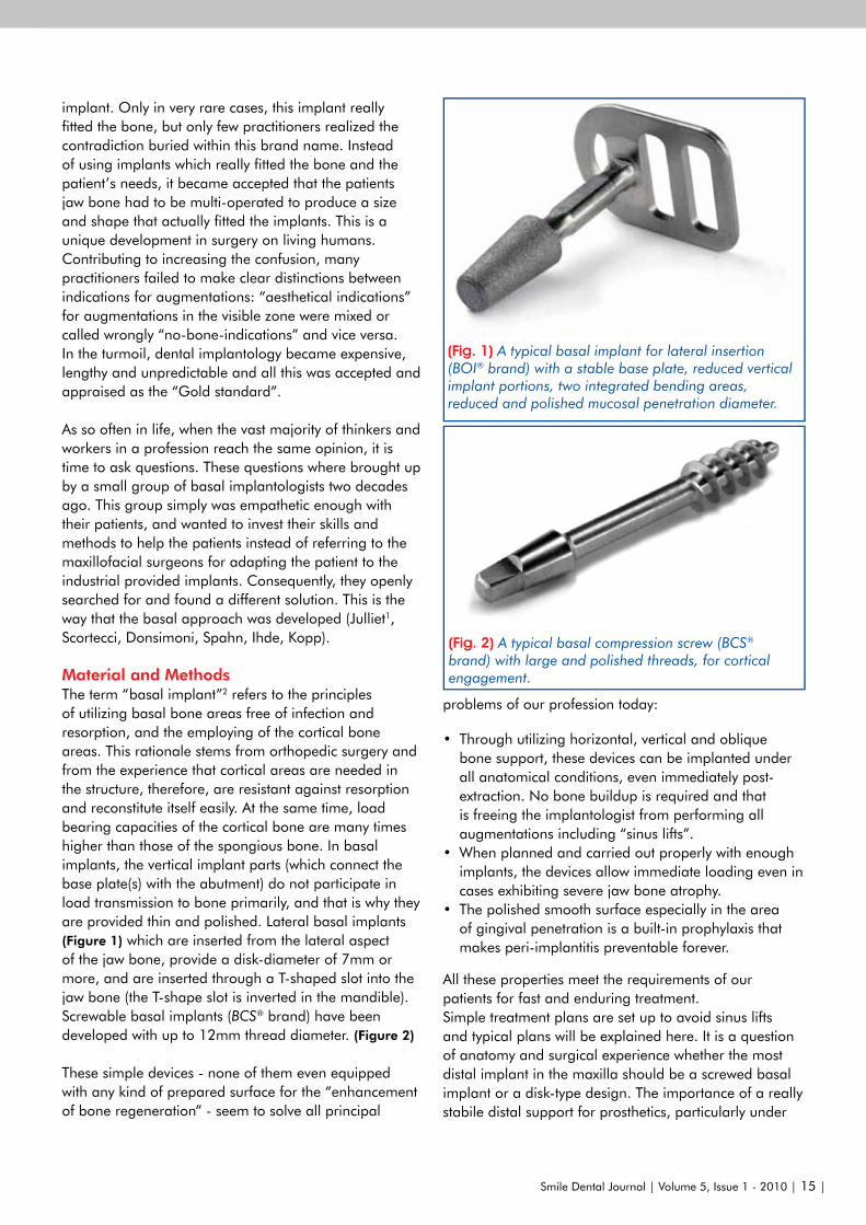
Material and MethodsThe term “basal implant”2 refers to the principles of utilizing basal bone areas free of infection and resorption, and the employing of the cortical bone areas. This rationale stems from orthopedic surgery and from the experience that cortical areas are needed in the structure, therefore, are resistant against resorption and reconstitute itself easily. At the same time, load bearing capacities of the cortical bone are many times higher than those of the spongious bone. In basal implants, the vertical implant parts (which connect the base plate(s) with the abutment) do not participate in load transmission to bone primarily, and that is why they are provided thin and polished. Lateral basal implants (Figure 1) which are inserted from the lateral aspect of the jaw bone, provide a disk-diameter of 7mm or more, and are inserted through a T-shaped slot into the jaw bone (the T-shape slot is inverted in the mandible). Screwable basal implants (BCS® brand) have been developed with up to 12mm thread diameter. (Figure 2)
These simple devices - none of them even equipped with any kind of prepared surface for the “enhancement of bone regeneration” - seem to solve all principal
(Fig. 2) A typical basal compression screw (BCS® brand) with large and polished threads, for cortical engagement.
| 16 | Smile Dental Journal | Volume 5, Issue 1 - 2010
immediate load conditions, should however never be in dispute.
1. Full arch reconstructionsa. Implant installations anterior to the maxillary sinuses:Just as in the “all on four” or “all on six”-concept, an adequate number of implants can be placed anterior to the maxillary sinus thus avoiding sinus lift. Typical examples for this kind of treatment are shown in figures 3 and 4.
(Fig. 3a) Preoperative panoramic view: implants were planned to replace missing and ailing teeth as well as a removable denture.
(Fig. 3b) Postoperative panoramic view of the same patient 6 weeks after implants placement and subsequent loading. Cortical anchoring of the implants is a fundamental principle of the therapy.
(Fig. 4c) 12 month later control x-ray with fixed metal ceramic bridges.
b. Implant installation on both sides of the maxillary sinuses:When enough horizontal bone is left distally to the maxillary sinus, we alternatively use wider implants as tubero-pterygoid-implants. Typical designs are “STC” or TPG” screws, which allow screw connected prosthetics (Figures 5 - 6).
One-piece BCS implants for cementation of prosthetics are in most cases easy to use and suitable, because the problems of a screw connection between the bridge and the implant are avoided. Uniform direction of the insertion can be achieved easily through angulation adapters or bending.
(Fig. 4a) Preoperative panoramic view of a 46 years old male patient.
(Fig. 3c) Intraoral view 6 weeks postoperatively with a metal- plastic bridge installed.
(Fig. 4b) Immediately after extraction of all teeth, the implants were placed and loaded right away.
Smile Dental Journal | Volume 5, Issue 1 - 2010 | 17 |
The number of necessary basal implants for a full maxillary reconstruction is between 4 and 12. Four implants require meticulous masticatory control, sufficient and good quality of bone in the strategic implant positions, as well as perfect patient compliance. The more implants are placed in the maxilla, the safer treatment develops. With implants becoming more and more affordable and suitable (due to changes in design), and since we have learned from a large number of treated patients how to utilize the available bone better, we were able to increase the number of implants that could be placed in the maxilla. Due to the greater softness of the maxillary bone, it is recommended never to under-equipping this jaw with implants, especially when loading it immediately. Immediate loading requires an even distribution of
(Fig. 6) The combination of tubero-pterygoid screws with BOI right in front of the tuberosity gives a great vertical stability. Flapped over struts of BOI® in the area of 2nd molars and SFF-screws for fixation provide additional lateral stabilization.
masticatory forces between all implants involved. This distribution is done through the bridge, which is a perfect external splinting device. Because the metal used to enforce the bridge tends to be elastic, the dimensions of the metal frame must be chosen adequately: it is not enough to design the dimensions of the metal frame in a fracture-proof manner. The thickness of the metal structure has to guarantee stiffness and stability and a force distribution between all implants, which means that it should not allow elastic deformation of the metal frame while being loaded by typical masticatory forces. This applies also to metal cores of segmental bridges described later in the text. Typical dimensions providing enough stiffness are 2.5mm width x 3mm height. One should keep in mind that an adequate height influences the stiffness on vertical forces more than the width does.
2. Segments in the lateral maxillaa. Segments including posterior tooth supportOften, stabile second molars are available and the implantologist may want to include them into the treatment plan, while leaving the anterior teeth disconnected from the bridge. This approach saves us from increasing the bone volume in the area of the first molar or second premolar, because during the healing phase, enough stability for immediate function is provides by the tooth and thereafter the implants are integrated and performing. Single base plate implants may be placed under the sinus in as little as 3mm vertical bone height, utilizing stabile cortical anchoring. In the area of the premolars double or triple base plate implants are applicable. Basal screws may be used as an alternative in the premolar area. While basal (lateral) implants are utilizing the lateral and medial walls of the maxillary bone, basal screw implants utilize the cortical bone in the floor of the nose and the maxillary sinus for stabile anchoring (figure 7).
(Fig. 7) Two basal implants are inserted in the upper right maxillary bone and immediately connected to the second molar. 12 years postoperative panoramic view. The vertical bone loss is about 1.5mm. It is affecting the load transmitting base plates of the implants.
(Fig. 5) Screws with internal threads (as in area 18) or one-piece implants (as in area 28) are equally suitable for equipping the tubero-pterygoid region. Today, we tend to use machined, thin implants without surface enlargement and an aggressive thread-design. Oral hygiene in the distal maxilla is often difficult and therefore we put our emphasis in designing the bridges which allow self-cleaning by the tongue and suppuration away from the mucosal penetration area of the implants.
| 18 | Smile Dental Journal | Volume 5, Issue 1 - 2010
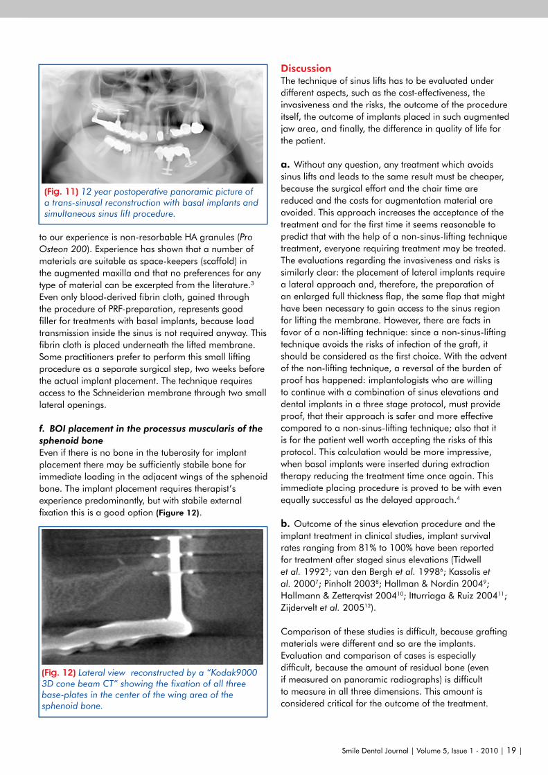
(Fig. 10a) BAC implant
(Fig. 10b) BAC implant for external screw fixation in the palatinal and the vestibular side of the maxilla. The longer (right) strut of the base plate engages nicely in the resorption stabile bone of the zygomatic process of the maxilla. Screws with a thread diameter of 2.4mm are used.
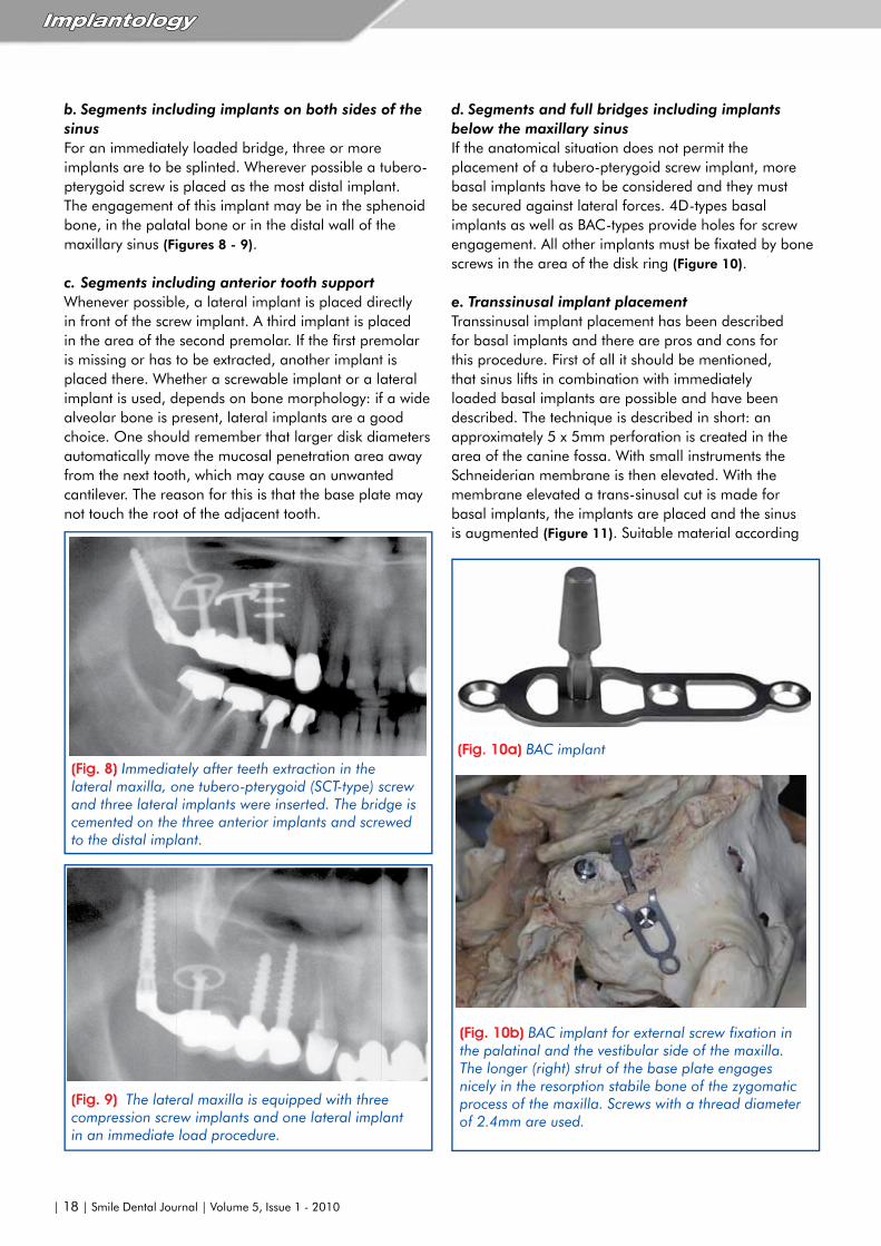
b. Segments including implants on both sides of the sinusFor an immediately loaded bridge, three or more implants are to be splinted. Wherever possible a tubero-pterygoid screw is placed as the most distal implant. The engagement of this implant may be in the sphenoid bone, in the palatal bone or in the distal wall of the maxillary sinus (Figures 8 - 9).
c. Segments including anterior tooth supportWhenever possible, a lateral implant is placed directly in front of the screw implant. A third implant is placed in the area of the second premolar. If the first premolar is missing or has to be extracted, another implant is placed there. Whether a screwable implant or a lateral implant is used, depends on bone morphology: if a wide alveolar bone is present, lateral implants are a good choice. One should remember that larger disk diameters automatically move the mucosal penetration area away from the next tooth, which may cause an unwanted cantilever. The reason for this is that the base plate may not touch the root of the adjacent tooth.
(Fig. 8) Immediately after teeth extraction in the lateral maxilla, one tubero-pterygoid (SCT-type) screw and three lateral implants were inserted. The bridge is cemented on the three anterior implants and screwed to the distal implant.
d. Segments and full bridges including implants below the maxillary sinusIf the anatomical situation does not permit the placement of a tubero-pterygoid screw implant, more basal implants have to be considered and they must be secured against lateral forces. 4D-types basal implants as well as BAC-types provide holes for screw engagement. All other implants must be fixated by bone screws in the area of the disk ring (Figure 10).
e. Transsinusal implant placementTranssinusal implant placement has been described for basal implants and there are pros and cons for this procedure. First of all it should be mentioned, that sinus lifts in combination with immediately loaded basal implants are possible and have been described. The technique is described in short: an approximately 5 x 5mm perforation is created in the area of the canine fossa. With small instruments the Schneiderian membrane is then elevated. With the membrane elevated a trans-sinusal cut is made for basal implants, the implants are placed and the sinus is augmented (Figure 11). Suitable material according
(Fig. 9) The lateral maxilla is equipped with three compression screw implants and one lateral implant in an immediate load procedure.
Smile Dental Journal | Volume 5, Issue 1 - 2010 | 19 |
(Fig. 11) 12 year postoperative panoramic picture of a trans-sinusal reconstruction with basal implants and simultaneous sinus lift procedure.
to our experience is non-resorbable HA granules (Pro Osteon 200). Experience has shown that a number of materials are suitable as space-keepers (scaffold) in the augmented maxilla and that no preferences for any type of material can be excerpted from the literature.3 Even only blood-derived fibrin cloth, gained through the procedure of PRF-preparation, represents good filler for treatments with basal implants, because load transmission inside the sinus is not required anyway. This fibrin cloth is placed underneath the lifted membrane. Some practitioners prefer to perform this small lifting procedure as a separate surgical step, two weeks before the actual implant placement. The technique requires access to the Schneiderian membrane through two small lateral openings.
f. BOI placement in the processus muscularis of the sphenoid boneEven if there is no bone in the tuberosity for implant placement there may be sufficiently stabile bone for immediate loading in the adjacent wings of the sphenoid bone. The implant placement requires therapist’s experience predominantly, but with stabile external fixation this is a good option (Figure 12).
DiscussionThe technique of sinus lifts has to be evaluated under different aspects, such as the cost-effectiveness, the invasiveness and the risks, the outcome of the procedure itself, the outcome of implants placed in such augmented jaw area, and finally, the difference in quality of life for the patient.
a. Without any question, any treatment which avoids sinus lifts and leads to the same result must be cheaper, because the surgical effort and the chair time are reduced and the costs for augmentation material are avoided. This approach increases the acceptance of the treatment and for the first time it seems reasonable to predict that with the help of a non-sinus-lifting technique treatment, everyone requiring treatment may be treated. The evaluations regarding the invasiveness and risks is similarly clear: the placement of lateral implants require a lateral approach and, therefore, the preparation of an enlarged full thickness flap, the same flap that might have been necessary to gain access to the sinus region for lifting the membrane. However, there are facts in favor of a non-lifting technique: since a non-sinus-lifting technique avoids the risks of infection of the graft, it should be considered as the first choice. With the advent of the non-lifting technique, a reversal of the burden of proof has happened: implantologists who are willing to continue with a combination of sinus elevations and dental implants in a three stage protocol, must provide proof, that their approach is safer and more effective compared to a non-sinus-lifting technique; also that it is for the patient well worth accepting the risks of this protocol. This calculation would be more impressive, when basal implants were inserted during extraction therapy reducing the treatment time once again. This immediate placing procedure is proved to be with even equally successful as the delayed approach.4
b. Outcome of the sinus elevation procedure and the implant treatment in clinical studies, implant survival rates ranging from 81% to 100% have been reported for treatment after staged sinus elevations (Tidwell et al. 19925; van den Bergh et al. 19986; Kassolis et al. 20007; Pinholt 20038; Hallman & Nordin 20049; Hallmann & Zetterqvist 200410; Itturriaga & Ruiz 200411; Zijdervelt et al. 200512).
Comparison of these studies is difficult, because grafting materials were different and so are the implants. Evaluation and comparison of cases is especially difficult, because the amount of residual bone (even if measured on panoramic radiographs) is difficult to measure in all three dimensions. This amount is considered critical for the outcome of the treatment.
(Fig. 12) Lateral view reconstructed by a “Kodak9000 3D cone beam CT” showing the fixation of all three base-plates in the center of the wing area of the sphenoid bone.

| 20 | Smile Dental Journal | Volume 5, Issue 1 - 2010
with poor bone conditions to be told that implants are not an option, or if there are, the options are expensive, lengthy, and invasive bone augmentation procedures are required. These patients are often left without a possible choice for implants. Is that a failure? In an era when nearly all edentulous patients would prefer fixed teeth rather than removable dentures, perhaps we need to start counting failures as soon as the patient is rejected for an implantological treatment.
In addition to survival rates, clinical studies in dental implantology should also measure patient-centered outcomes. How does the patient feel in respect to their oral health? Oral health related to the quality of life (OHQoL) has been summarized by the following:
• chew and eat full range of foods native to diet• speak clearly• socially acceptable smile • socially acceptable dentofacial profile• comfortable and free from pain • have fresh breath
(Fig. 13) While on the left side (L) the placement of a tuber-pterygoid screw was possible, no stability could be reached for a screw implant on the right side (R). Therefore a basal implant was used to support the bridge. Both lateral implants were secured by SSF-bone screws.
c. Outcome of implant treatment in cases without augmentation:A number of references are available on basal implant treatment. Donsimoni et al. reported a 97% survival rate and a 100% clinical success rate13. Similar results have been reported by Scortecci14, Kopp, Ihde & Mutter15, and Ihde16. If the body of literature in crestal implantology is compared to basal implantology, it becomes clear that only few specialists are eager to undertake the burden of scientific work and publishing. Since only a few universities are involved in this research, and because industry-derived money supply for increasing the literature on this technique is missing, an impacting number of publications can not be expected. Nevertheless, enough evidence for the basal approach can be found easily and the quality of the articles and the research are almost the same when compared to the publications on crestal implants. Literature search is not easy, because most of it is published in French and German language.
d. Differences in QOL (Quality Of Life)Implant survival or success is the “gold standard” for measuring the efficacy of dental implants, yet these definitions vary widely from a study to another. Several different definitions have been proposed,2,17,18 but no consensus has been reached. In some studies, success is defined as the survival of the prosthesis, in others; it is the survival of the implant. When the prosthesis is considered, implants not subjected to loading due to improper angulation may be scored as successful provided the prosthesis doesn’t fail because it is supported by other implants.19 Some studies account for all implants placed and report all removals as failures, while others report failures that occur following loading. Early trials of Brånemark implants reported by Adell et al.20,21 excluded all implants loaded less than one year. Walton22 has demonstrated a wide variation in success rates when replacement, repair, and modification of prostheses are taken into account. These studies suggest that we do not have a clear definition of failure and when to start counting failures. With the emergence and popularity of immediate load protocols, it is imperative that failures are counted as soon as implants are placed. It is reasonable to differentiate between “early” and “late” failure in delayed loading protocols; however, to be able to compare delayed loading implant systems to immediate load systems, failures must be counted immediately.
How early is too early? What about those who are turned away in the dental office because they are not “good candidates” for implants. This is not discussed or quantified in the literature. It is common for patients
(Fig. 14) The combination of transgingival inserted compression screws (KOS®) with basal implants (BOI® and BCS®) proved as an enduring option for fixation of the bridges.

Smile Dental Journal | Volume 5, Issue 1 - 2010 | 21 |
In immediate loading protocols, failure rates and OHQoL should be measured early. We are beginning to measure this in all our patients as an important way to differentiate between patient-centered “success” and clinical “success”. We challenge other researchers in our field to do the same.
ConclusionThere are many options for immediate implant placement and implant loading in the distal maxilla available and some were shown here as an example. All these technologies using appropriate implants are developed and implemented to avoid time-and money-consuming procedures like sinus lift. Today, the application of traditional implants connected to sinus lifting procedures needs a good reason because minimal invasive alternatives are available and should be a part of complete information to a well informed patient.
References 1. Julliet JM. United States Patent 3,925,892. 1975.2. Albrektsson T, Zarb G, Worthington P, Eriksson AR. The long-term
efficacy of currently used dental implants: a review and proposed criteria of success. Int J Oral Maxillofac Implants. 1986;1(1):11-25.
3. Bornstein MM, Chappuis V, von Arx T, Buser D. Performance of dental implants after staged sinus floor elevation procedures: 5-year results of a prospective study in partially edentulous patients. Clin Oral Implants Res. 2008;19(10):1034-43.
4. Kopp S, Kopp W. Comparison of immediate vs. delayed basal implants. J Maxillofac Oral Surg. 2008;7(1):116-22
5. Tidwell JK, Blijdorp PA, Stoelinga PJ, Brouns JB, Hinderks F. Composite grafting of the maxillary sinus for placement of endosteal implants. A preliminary report of 48 patients. Int J Oral Maxillofac Surg. 1992;21(4):204-9.
6. van den Bergh JP, ten Bruggenkate CM, Krekeler G, Tuinzing DB. Sinusfloor elevation and grafting with autogenous iliac crest bone. Clin Oral Implants Res. 1998;9(6):429-35.
7. Kassolis JD, Rosen PS, Reynolds MA. Alveolar ridge and sinus augmentation utilizing platelet-rich plasma in combination with freeze-dried bone allograft: case series. J Periodontol. 2000;71(10):1654-61.
8. Pinholt EM. Branemark and ITI dental implants in the human bone-grafted maxilla: a comparative evaluation. Clin Oral Implants Res. 2003;14(5):584-92.
9. Hallman M, Nordin T. Sinus floor augmentation with bovine hydroxylapatite mixed with fibrin glue and later placement of nonsubmerged implants: a retrospective study in 50 patients. Int J Oral Maxillofac Implants. 2004;19(2):222-7.
10. Hallman M, Zetterqvist L. A 5-year prospective follow-up study of implant-supported fixed prostheses in patients subjected to maxillary sinus floor augmentation with an 80:20 mixture of bovine hydroxyapatite and autogenous bone. Clin Implant Dent Relat Res. 2004;6(2):82-9.
11. Iturriaga MT, Ruiz CC. Maxillary sinus reconstruction with calvarium bone grafts and endosseous implants. J Oral Maxillofac Surg. 2004;62(3):344-7.
12. Zijderveld SA, Zerbo IR, van den Bergh JP, Schulten EA, ten Bruggenkate CM. Maxillary sinus floor augmentation using a beta-tricalcium phosphate (Cerasorb) alone compared to autogenous bone grafts. Int J Oral Maxillofac Implants. 2005;20(3):432-40.
13. Donsimoni J-M, Gabrieleff D, Bermot P, Dohan D. Les implants maxillofaciaux à plateaux d`assise; Concepts et technologies orthopediques, rehabilitations maxillomandibulaires, reconstructions maxillo-faciales, rehabilitations dentaires partielles, techniques de reintervention, meta-analyse. 6e partie: une meta-analyse. Implantodontie. 2004;13:217-28.
14. Scortecci G. Mise en fonction immédiate dans le traitement imptantaire de l’édenté total maxillaire et/ou mandibulaire. Implantodontie. 1999;34:29-38.
15. Ihde S, Mutter L. Versorgung von Freieindsituationen mit basal osseointegrierten Implantaten (BOI) bei reduziertem vertikalen Knochenangebot. Deutsch Zahnärztl Zeitschr. 2003;58:94-102.
16. Ihde S. Principles of BOI. Heidelberg: Springer Verlag; 2005.17. Smith DE, Zarb GA. Criteria for success of osseointegrated
endosseous implants. J Prosthet Dent. 1989;62:567-72.18. Schnitman PA, Shulman LB. Recommendations of the consensus
development conference on dental implants. J Am Dent Assoc. 1979;98:373-7.
19. Morris HF, Ochi S. Influence of two different approaches to reporting implant survival outcomes for five different prosthodontic applications. Ann Periodontol. 2000;5:90-100.
20. Adell R, Lekholm U, Rockler B, Branemark PI. A 15-year study of osseointegrated implants in the treatment of the edentulous jaw. Int J Oral Surg. 1981;10:387-416.
21. Adell R. Clinical results of osseointegrated implants supporting fixed prostheses in edentulous jaws. J Prosthet Dent. 1983;50: 251-4.
22. Walton TR. The outcome of implant-supported fixed prostheses from the prosthodontic perspective: proposal for a classification protocol. Int J Prosthodont. 1998;11:595-601.