Simultaneous Determination of Dopamine and Uric Acid by Electrocatalytic Oxidation on a Carbon Paste...
7
Electrochimica Acta 91 (2013) 36–42 Contents lists available at SciVerse ScienceDirect Electrochimica Acta jou rn al h om epa ge: www.elsevier.com/locate/electacta Simultaneous determination of dopamine and uric acid in biological samples on the pretreated pencil graphite electrode Esmaeel Alipour a,∗ , Mir Reza Majidi a , Afsaneh Saadatirad a , Sayed mahdi Golabi a , Ali Mohammad Alizadeh b a Electroanlytical Chemistry Lab. Department of Analytical Chemistry, Faculty of Chemistry, University of Tabriz, Tabriz, Iran b Cancer Research Center, Tehran University of Medical Sciences, Tehran, Iran a r t i c l e i n f o Article history: Received 17 October 2012 Received in revised form 11 December 2012 Accepted 19 December 2012 Available online 27 December 2012 Keywords: Pretreated pencil graphite electrode Uric acid Dopamine Differential pulse voltammetry Human serum Urine a b s t r a c t An electrochemically pretreated pencil graphite electrode (PGE) has been proposed for simultaneous determination of dopamine (DA) and uric acid (UA) in the presence of high levels of ascorbic acid (AA). The electrochemical pretreatment of PGE was achieved in phosphate buffer (pH 7) by the potential recycling between 1.5–2.0 V/SCE for 100 cycles, and at the scan rate of 100 mV s −1 . The influence of electrochemical pretreatment of PGE on the separation of overlapped peaks of AA, DA and UA were studied and optimum conditions were suggested. Differential pulse voltammetry (DPV) was used for simultaneous determina- tion of DA and UA in the presence of high concentrations of AA. The peak separation between DA and AA, and also between DA and UA was up to 260 and 160 mV, respectively. The calibration curves were in the range of 0.15–15 M and 0.3–150 M for DA and UA in the presence of AA, respectively. The detection limits (S/N = 3) were 0.033 and 0.12 M for DA and UA, respectively. The results reveal that pretreat- ment of the pencil graphite electrode surface not only improves its electrochemical catalytic activities towards the oxidation of DA and UA, but also resolves the overlapped oxidation peaks of AA, DA and UA into three well-defined peaks. The present method can apply for UA and DA determination in human serum and urine, and also for some commercial pharmaceutical samples without additional sample pre-treatment. © 2012 Elsevier Ltd. All rights reserved. 1. Introduction Dopamine, ascorbic acid, and uric acid are important human body compounds that play determining role in metabolism [1], hence, monitoring the their concentration in biologic fluids such as blood and urine may prevent and control many diseases. These are electrochemically active compounds that can be deter- mined by different techniques. In recent years, the development of voltammetric methods for their determination in human body fluids such as urine and serum has received consider- able interest. However, in most cases, overlapped voltammetric responses, poor selectivity and low reproducibility make their simultaneous tracking highly difficult [2]. To overcome these prob- lems, various modified electrodes have been constructed and used such as organic redox mediators modified electrode [3], polymers modified electrodes [4–10], nanoparticles modified elec- trodes [11–14], boron-doped diamond modified electrode [15], ∗ Corresponding author. Tel.: +98 411 3393126; fax: +98 411 3340191. E-mail address: [email protected] (E. Alipour). carbon–ceramic modified electrode [16], pyrolytic graphite mod- ified electrodes [17,18], screen-printed carbon modified electrode [19], carbon ionic liquid modified electrode [20], carbon nan- otube modified electrodes [13,21–23], RNA modified electrode [24], and finally glassy carbon electrode modified via electrochemi- cal oxidation [25]. In spite of selectivity of these voltammetric techniques, cheaper or more sensitive and selective methods are still needed to develop detection routes for uric acid and dopamine in the presence of AA due to their clinical signifi- cance. Electrochemical pretreatment of pencil graphite electrode seems to be a simple, less time consuming and more applica- ble strategy in comparison to other procedures. This strategy eliminates the use of some toxic compounds required in mod- ification of electrode surface. Recently, we have successfully used electrochemically pretreated pencil graphite electrode for development of various DNA hybridization biosensors [26–31]. In the present study, a simple sensor based on PPGE described for simultaneous determination of DA and UA in the pres- ence of high levels of AA. The analytical performance of this sensor was evaluated by differential pulse voltammetry technique. 0013-4686/$ – see front matter © 2012 Elsevier Ltd. All rights reserved. http://dx.doi.org/10.1016/j.electacta.2012.12.079
-
Upload
independent -
Category
Documents
-
view
0 -
download
0
Transcript of Simultaneous Determination of Dopamine and Uric Acid by Electrocatalytic Oxidation on a Carbon Paste...
St
EAa
b
a
ARR1AA
KPUDDHU
1
bhaTmobarslupt
0h
Electrochimica Acta 91 (2013) 36– 42
Contents lists available at SciVerse ScienceDirect
Electrochimica Acta
jou rn al h om epa ge: www.elsev ier .com/ locate /e lec tac ta
imultaneous determination of dopamine and uric acid in biological samples onhe pretreated pencil graphite electrode
smaeel Alipoura,∗, Mir Reza Majidia, Afsaneh Saadatirada, Sayed mahdi Golabia,li Mohammad Alizadehb
Electroanlytical Chemistry Lab. Department of Analytical Chemistry, Faculty of Chemistry, University of Tabriz, Tabriz, IranCancer Research Center, Tehran University of Medical Sciences, Tehran, Iran
r t i c l e i n f o
rticle history:eceived 17 October 2012eceived in revised form1 December 2012ccepted 19 December 2012vailable online 27 December 2012
eywords:retreated pencil graphite electroderic acid
a b s t r a c t
An electrochemically pretreated pencil graphite electrode (PGE) has been proposed for simultaneousdetermination of dopamine (DA) and uric acid (UA) in the presence of high levels of ascorbic acid (AA). Theelectrochemical pretreatment of PGE was achieved in phosphate buffer (pH 7) by the potential recyclingbetween 1.5–2.0 V/SCE for 100 cycles, and at the scan rate of 100 mV s−1. The influence of electrochemicalpretreatment of PGE on the separation of overlapped peaks of AA, DA and UA were studied and optimumconditions were suggested. Differential pulse voltammetry (DPV) was used for simultaneous determina-tion of DA and UA in the presence of high concentrations of AA. The peak separation between DA and AA,and also between DA and UA was up to 260 and 160 mV, respectively. The calibration curves were in therange of 0.15–15 �M and 0.3–150 �M for DA and UA in the presence of AA, respectively. The detection
opamineifferential pulse voltammetryuman serumrine
limits (S/N = 3) were 0.033 and 0.12 �M for DA and UA, respectively. The results reveal that pretreat-ment of the pencil graphite electrode surface not only improves its electrochemical catalytic activitiestowards the oxidation of DA and UA, but also resolves the overlapped oxidation peaks of AA, DA and UAinto three well-defined peaks. The present method can apply for UA and DA determination in humanserum and urine, and also for some commercial pharmaceutical samples without additional samplepre-treatment.
. Introduction
Dopamine, ascorbic acid, and uric acid are important humanody compounds that play determining role in metabolism [1],ence, monitoring the their concentration in biologic fluids suchs blood and urine may prevent and control many diseases.hese are electrochemically active compounds that can be deter-ined by different techniques. In recent years, the development
f voltammetric methods for their determination in humanody fluids such as urine and serum has received consider-ble interest. However, in most cases, overlapped voltammetricesponses, poor selectivity and low reproducibility make theirimultaneous tracking highly difficult [2]. To overcome these prob-
ems, various modified electrodes have been constructed andsed such as organic redox mediators modified electrode [3],olymers modified electrodes [4–10], nanoparticles modified elec-rodes [11–14], boron-doped diamond modified electrode [15],∗ Corresponding author. Tel.: +98 411 3393126; fax: +98 411 3340191.E-mail address: [email protected] (E. Alipour).
013-4686/$ – see front matter © 2012 Elsevier Ltd. All rights reserved.ttp://dx.doi.org/10.1016/j.electacta.2012.12.079
© 2012 Elsevier Ltd. All rights reserved.
carbon–ceramic modified electrode [16], pyrolytic graphite mod-ified electrodes [17,18], screen-printed carbon modified electrode[19], carbon ionic liquid modified electrode [20], carbon nan-otube modified electrodes [13,21–23], RNA modified electrode [24],and finally glassy carbon electrode modified via electrochemi-cal oxidation [25]. In spite of selectivity of these voltammetrictechniques, cheaper or more sensitive and selective methodsare still needed to develop detection routes for uric acid anddopamine in the presence of AA due to their clinical signifi-cance.
Electrochemical pretreatment of pencil graphite electrodeseems to be a simple, less time consuming and more applica-ble strategy in comparison to other procedures. This strategyeliminates the use of some toxic compounds required in mod-ification of electrode surface. Recently, we have successfullyused electrochemically pretreated pencil graphite electrode fordevelopment of various DNA hybridization biosensors [26–31].In the present study, a simple sensor based on PPGE described
for simultaneous determination of DA and UA in the pres-ence of high levels of AA. The analytical performance ofthis sensor was evaluated by differential pulse voltammetrytechnique.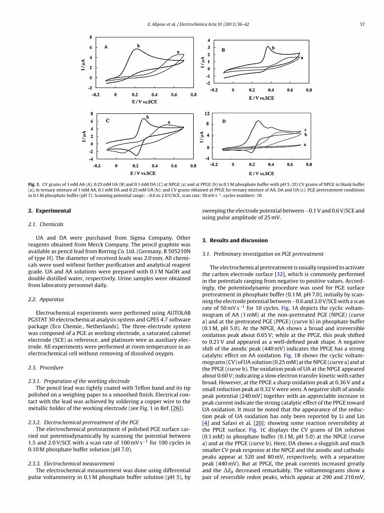
E. Alipour et al. / Electrochimica Acta 91 (2013) 36– 42 37
F d at P( obtaini rate: 5
2
2
raocgdf
2
Ppwete
2
2
ptm
2
r10
2
p
ig. 1. CV grams of 1 mM AA (A), 0.25 mM UA (B) and 0.1 mM DA (C) at NPGE (a) ana), in ternary mixture of 1 mM AA, 0.1 mM DA and 0.25 mM UA (b); and CV grams
n 0.1 M phosphate buffer (pH 7): Scanning potential range: −0.6 to 2.0 V/SCE, scan
. Experimental
.1. Chemicals
UA and DA were purchased from Sigma Company. Othereagents obtained from Merck Company. The pencil graphite wasvailable as pencil lead from Rotring Co. Ltd. (Germany, R 505210Nf type H). The diameter of received leads was 2.0 mm. All chemi-als were used without further purification and analytical reagentrade. UA and AA solutions were prepared with 0.1 M NaOH andouble distilled water, respectively. Urine samples were obtainedrom laboratory personnel daily.
.2. Apparatus
Electrochemical experiments were performed using AUTOLABGSTAT 30 electrochemical analysis system and GPES 4.7 softwareackage (Eco Chemie., Netherlands). The three-electrode systemas composed of a PGE as working electrode, a saturated calomel
lectrode (SCE) as reference, and platinum wire as auxiliary elec-rode. All experiments were performed at room temperature in anlectrochemical cell without removing of dissolved oxygen.
.3. Procedure
.3.1. Preparation of the working electrodeThe pencil lead was tightly coated with Teflon band and its tip
olished on a weighing paper to a smoothed finish. Electrical con-act with the lead was achieved by soldering a copper wire to the
etallic holder of the working electrode (see Fig. 1 in Ref. [26]).
.3.2. Electrochemical pretreatment of the PGEThe electrochemical pretreatment of polished PGE surface car-
ied out potentiodynamically by scanning the potential between.5 and 2.0 V/SCE with a scan rate of 100 mV s−1 for 100 cycles in.10 M phosphate buffer solution (pH 7.0).
.3.3. Electrochemical measurementThe electrochemical measurement was done using differential
ulse voltammetry in 0.1 M phosphate buffer solution (pH 5), by
PGE (b) in 0.1 M phosphate buffer with pH 5. (D) CV grams of NPGE in blank buffered at PPGE for ternary mixture of AA, DA and UA (c). PGE pretreatment conditions0 mV s−1, cycles numbers: 10.
sweeping the electrode potential between −0.1 V and 0.6 V/SCE andusing pulse amplitude of 25 mV.
3. Results and discussion
3.1. Preliminary investigation on PGE pretreatment
The electrochemical pretreatment is usually required to activatethe carbon electrode surface [32], which is commonly performedin the potentials ranging from negative to positive values. Accord-ingly, the potentiodynamic procedure was used for PGE surfacepretreatment in phosphate buffer (0.1 M, pH 7.0), initially by scan-ning the electrode potential between −0.6 and 2.0 V/SCE with a scanrate of 50 mV s−1 for 10 cycles. Fig. 1A depicts the cyclic voltam-mogram of AA (1 mM) at the non-pretreated PGE (NPGE) (curvea) and at the pretreated PGE (PPGE) (curve b) in phosphate buffer(0.1 M, pH 5.0). At the NPGE, AA shows a broad and irreversibleoxidation peak about 0.65 V; while at the PPGE, this peak shiftedto 0.21 V and appeared as a well-defined peak shape. A negativeshift of the anodic peak (440 mV) indicates the PPGE has a strongcatalytic effect on AA oxidation. Fig. 1B shows the cyclic voltam-mograms (CV) of UA solution (0.25 mM) at the NPGE (curve a) and atthe PPGE (curve b). The oxidation peak of UA at the NPGE appearedabout 0.60 V; indicating a slow electron transfer kinetic with ratherbroad. However, at the PPGE a sharp oxidation peak at 0.36 V and asmall reduction peak at 0.32 V were seen. A negative shift of anodicpeak potential (240 mV) together with an appreciable increase inpeak current indicate the strong catalytic effect of the PPGE towardUA oxidation. It must be noted that the appearance of the reduc-tion peak of UA oxidation has only been reported by Li and Lin[4] and Safavi et al. [20]; showing some reaction reversibility atthe PPGE surface. Fig. 1C displays the CV grams of DA solution(0.1 mM) in phosphate buffer (0.1 M, pH 5.0) at the NPGE (curvea) and at the PPGE (curve b). Here, DA shows a sluggish and muchsmaller CV peak response at the NPGE and the anodic and cathodic
peaks appear at 520 and 80 mV, respectively, with a separationpeak (440 mV). But at PPGE, the peak currents increased greatlyand the �Ep decreased remarkably. The voltammograms show apair of reversible redox peaks, which appear at 290 and 210 mV,
38 E. Alipour et al. / Electrochimica Acta 91 (2013) 36– 42
Fig. 2. (A) DP voltammograms of 1.1 mM AA, 0.1 mM UA and 0.02 mM DA obtained in 0.1 M phosphate buffer (pH 5), at various PPGE. PGE pretreatment conditions: Scanningp 0, (c)i in 0.11
rr
3r
tcN∼wmueesdtfctfPmattawBecttc
ndstmt(
not changed. The increase of �Ep between DA and AA peaks wasobserved up to 80 cycles and then remains constant. Therefore, apotential recycling of 100 cycles was considered as optimum.
Fig. 3. DP voltammograms obtained at PPGE for 0.6 mM AA in 0.1 M phosphate
otential range: −0.6 to 2.0 V/SCE, scan rate: 50 mV s−1, cycles numbers: (a) 0, (b) 1n 0.1 M phosphate buffer (pH 5.0), at various PPGE. PGE pretreatment conditions00 mV s−1, cycle numbers: (a) 10, (b) 20, (c) 30, (d) 50, and (e) 100.
espectively. The value of �Ep = Ep.a − Ep.c = 80 mV indicates that theeversibility of DA at the PPGE considerably improved.
.2. Effect of electrochemical pretreatment on pencil leadeactivity
What happens at PGE surface following the electrochemical pre-reatment? The core in pencil leads is mainly graphite, a form ofarbon in which each atom is connected together by week bonds.early, 65 wt% of H pencil lead is constituted from graphite and30 wt% is clay, and the remaining 5 wt% is an electro-inactiveax or polymer used as binder. Clay is a naturally-occurring alu-inosilicate showing the ion exchange property [33] and can be
sed as modifier in preparing modified electrode. In clay modifiedlectrode, the film of clay plays the role of a cation exchange andxhibits the voltammetric responses of surface confined cationicpecies. However, the present clay as constituent in pencil leadoes not exhibit any voltammetric signal in DA, AA and UA solu-ions in phosphate buffer, where they exist in neutral or anionicorms [34]. However, the graphitic parts of pencil lead surface, inontact with supporting electrolyte is cleaned during the oxida-ive electrochemical treatment and find various oxygen containingunctionalities. The enhancement in electrochemical activity ofPGE toward the studied compounds and in comparison with NPGEay be attributed to an increase of oxygen containing groups, such
s phenolic, carbonyl and carboxyl groups on electrode surface or tohe formation of graphite oxide films during electrochemical pre-reatment of PGE [35–38]. The formation of graphite oxide filmsfter electrochemical oxidation of graphite electrodes has beenell established and this topic has been thoroughly reviewed byesenhard and Fritz [37]. On the basis of literature investigation,vidence for increased surface oxygen concentration upon electro-hemical pretreatment of carbon based electrodes has promptedhe investigators to propose that activation results from electronransfer mediation by oxygen-containing groups such as phenolic,arboxylic and carbonyl functionalities [36].
In the present study, according to Fig. 1, we conclude that PPGEot only can accelerate the oxidation of AA, DA and UA, but alsoramatically enhance their peaks separation, rendering feasible theimultaneous determination of AA, DA and UA. Fig. 1D represents
he cyclic voltammograms of the NPGE in blank (curve a), in ternaryixture of AA, DA and UA (curve b), and of the PPGE in the sameernary mixture (curve c). From CV gram obtained at the NPGEcurve b), it is impossible to deduce any information about the
20, (d) 40 and (e) 80. (B) DP voltammograms of 0.6 mM AA and 9 �M DA obtained M phosphate buffer (pH 7.0): scanning potential range: 1.5–2.0 V/SCE, scan rate:
electroactive species present in solution. While, using PPGE asworking electrode, two well-defined anodic peaks are observed forDA and UA oxidation, and also anodic peak of AA appears as shoul-der of DA peak (Fig. 1D, curve c). These observations reveal thatsuch pretreated electrode can be used only for determination ofUA in the presence of AA and DA, and in order to resolve the over-lapped peaks of DA and AA, and so determination of DA, the PGEpretreatment must be optimized.
3.3. Effect of potential sweep parameters – optimization of PGEpretreatment.
In order to determine the effect of scan number on the reactivityof the pretreated electrode, several PGEs were pretreated in phos-phate buffer (0.1 M, pH 7.0), in potential range of −0.6–2 V/SCE. Thescan rate was fixed at 50 mV s−1. Then, the pretreated electrodeswere used for simultaneous detection of AA, UA and DA. Fig. 2Ashows DPVs obtained using these electrodes in phosphate solu-tion (0.1 M, pH 5), containing AA, UA and DA. With increasing thescan number during PGE pretreatment, negative shift was observedfor AA oxidation peak, while oxidation peaks of UA and DA were
buffer (pH 5.0) in the absence of DA (a), and in the presence of various concen-trations of DA, (b) 0.15 �M, (c) 4.24 �M, (d) 7.48 �M, (e) 11.10 �M, (f) 13.32 �Mand (g) 14.87 �M. Inset: Related calibration graph. PGE pretreatment conditions in0.1 M phosphate buffer (pH 7.0): scanning potential range: 1.5–2.0 V/SCE, scan rate:100 mV s−1, cycle numbers: 100.
E. Alipour et al. / Electrochimica Acta 91 (2013) 36– 42 39
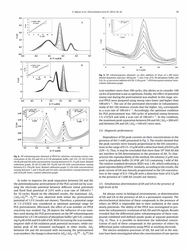
Fig. 4. DP voltammograms obtained at PPGE for solutions containing various con-centrations of AA, DA and UA in 0.1 M phosphate buffer (pH 5.0). (A) AA 0.5 mM,UA 40 �M and DA with concentrations varying between 0.15–15 �M. Inset: Relatedcalibration graph. (B) AA 0.5 mM, DA 10 �M and UA with concentrations varyingbva
tnaf(poPrboindbs
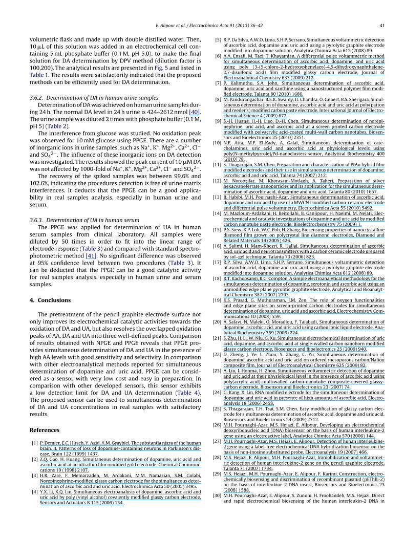
Fig. 5. DP voltammograms obtained: (a) after addition of 10 �L of a 200 times
etween 0.5–150 �M. Inset: Related calibration graph. (C) DA with concentrationsarying between 1 and 5.36 �M and UA with concentrations varying between 4.9nd 26.8 �M. Insets: related calibration graph.
In order to improve the peak separation between DA and AA,he potentiodynamic pretreatment of the PGE carried out by scan-ing the electrode potential between different initial potentialsnd fixed final potential of 2.0 V with a scan rate of 100 mV s−1
or 10 cycles. Based on the obtained results, the maximum �Ep
�Ep = EpDA − Ep
AA) was observed with initial the pretreatmentotential of 1.5 V (results not shown). Therefore, a potential rangef 1.5–2 V/SCE was considered as optimum potential range forGE pretreatment. Afterward, the effect of scan number on PPGEeactivity was studied. Fig. 2B depicts the influence of scan num-ers used during the PGE pretreatment on the DP voltammogramsbtained for a 0.1 M solution of phosphate buffer (pH 5.0), contain-ng 9 �M of DA and 0.6 mM of AA. With increasing the scan number,
egative shift of AA oxidation peak potential increases; while oxi-ation peak of DA remained unchanged. In other words, �Epetween DA and AA increased with increasing the pretreatmentcan numbers. No change is observed in �Ep (�Ep = Ep
DA − EpAA) for
diluted dopamine injection (40 mg mL−1) into 5 mL of 0.1 M phosphate buffer (pH5.0), (b–g) successive addition of 0.38–2.28 �g mL−1 of DA into parent solution. Inset:related calibration graph.
scan numbers more than 100 cycles, this allows us to consider 100cycles of potential scans as optimum. Finally, the effect of potentialsweep rate during the pretreatment was studied. In this stage, sev-eral PPGE were prepared using sweep rates lower and higher than100 mV s−1. The use of the pretreated electrodes in voltammetricstudy of AA + DA mixture reveals that the higher �Ep correspondsto a scan rate of 100 mV s−1. Accordingly, the optimum conditionfor PGE pretreatment was 100 cycles of potential sweep between1.5–2 V/SCE and with a scan rate of 100 mV s−1. In this condition,the maximum peak separation between DA and AA (�Ep = 260 mV)and between DA and UA (�Ep = 160 mV) were seen.
3.4. Diagnostic performances
Dependence of DA peak currents on their concentrations in thepresence of AA (1 mM) presented in Fig. 3. The results showed thatthe peak currents were linearly proportional to the DA concentra-tions in the range of 0.15–15 �M with a detection limit of 0.033 �M(S/N = 3). Thus, it may be concluded that more than 103 fold AA didnot interfere in DA determination in the presence of AA. To char-acterize the reproducibility of the method, DA solution (2 �M) wasused in phosphate buffer (0.10 M, pH 5.0) containing 1 mM of AA.The relative standard deviation (RSD) was 1.4%, which indicating agood reproducibility of PPGE. Similarly, according to the results, thepeak current of UA was linearly proportional to the UA concentra-tion in the range of 0.3–150 �M with a detection limit of 0.12 �Min the presence of 1 mM AA (results not shown).
3.5. Simultaneous determination of DA and UA in the presence ofhigh levels of AA
AA always exists in biological environments, so determinationof DA and UA in the presence of AA is a permanent requisite. Theelectrochemical detection of these compounds in the presence ofothers on NPGE is impossible due to their oxidation at the samenearly potentials. The ability of the PPGE was investigated to pro-mote the voltammetric resolution of AA, DA and UA signals. Resultsrevealed that the differential pulse voltammograms of these com-pounds exhibited well defined anodic peaks at separate potentials(Fig. 4). Thus, it is possible to determine the individual concen-
tration of each of these compounds in the presence of others bydifferential pulse voltammetry using PPGE as working electrode.The electro-oxidation processes of DA, AA and UA in the mix-ture have been investigated when the concentration of one species
40 E. Alipour et al. / Electrochimica Acta 91 (2013) 36– 42
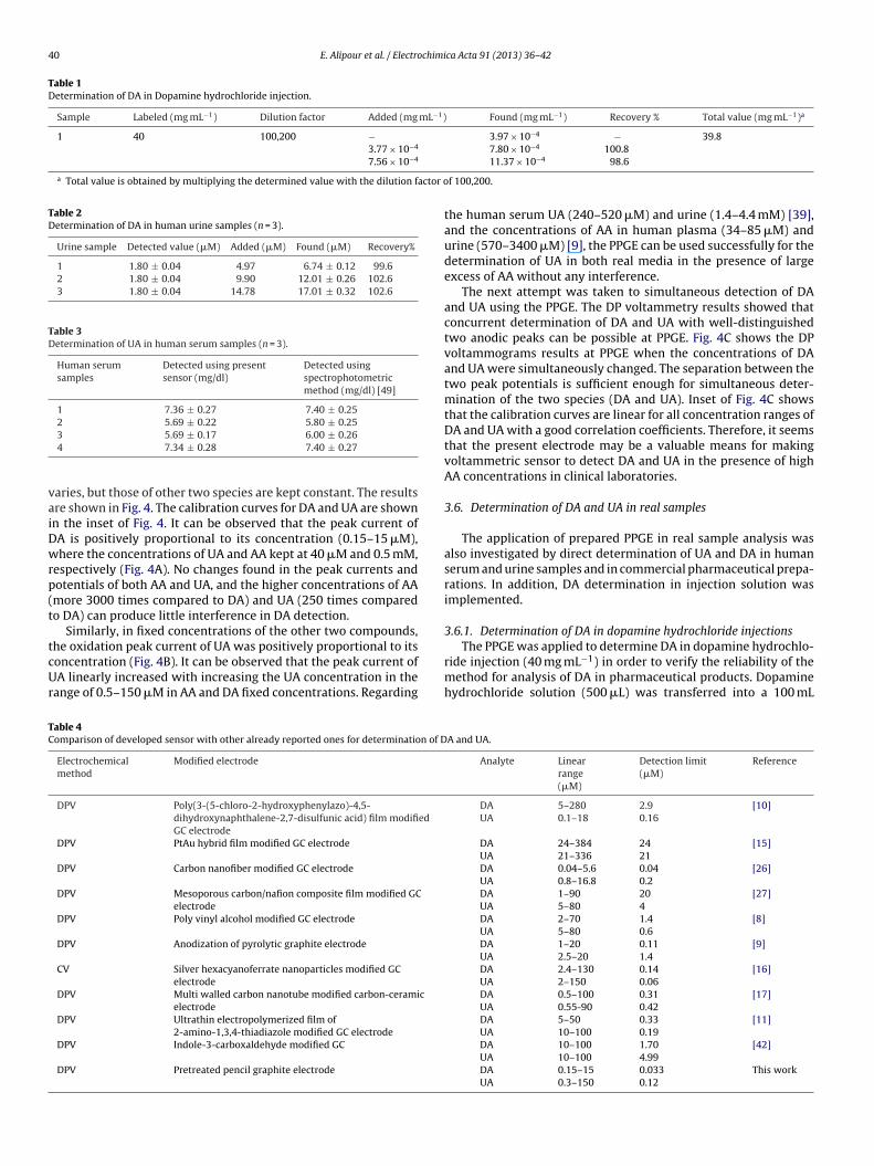
Table 1Determination of DA in Dopamine hydrochloride injection.
Sample Labeled (mg mL−1) Dilution factor Added (mg mL−1) Found (mg mL−1) Recovery % Total value (mg mL−1)a
1 40 100,200 − 3.97 × 10⁻⁴ − 39.83.77 × 10−4 7.80 × 10⁻⁴ 100.87.56 × 10−4
a Total value is obtained by multiplying the determined value with the dilution factor o
Table 2Determination of DA in human urine samples (n = 3).
Urine sample Detected value (�M) Added (�M) Found (�M) Recovery%
1 1.80 ± 0.04 4.97 6.74 ± 0.12 99.62 1.80 ± 0.04 9.90 12.01 ± 0.26 102.63 1.80 ± 0.04 14.78 17.01 ± 0.32 102.6
Table 3Determination of UA in human serum samples (n = 3).
Human serumsamples
Detected using presentsensor (mg/dl)
Detected usingspectrophotometricmethod (mg/dl) [49]
1 7.36 ± 0.27 7.40 ± 0.252 5.69 ± 0.22 5.80 ± 0.25
vaiDwrp(t
tcUr
The PPGE was applied to determine DA in dopamine hydrochlo-
TC
3 5.69 ± 0.17 6.00 ± 0.264 7.34 ± 0.28 7.40 ± 0.27
aries, but those of other two species are kept constant. The resultsre shown in Fig. 4. The calibration curves for DA and UA are shownn the inset of Fig. 4. It can be observed that the peak current ofA is positively proportional to its concentration (0.15–15 �M),here the concentrations of UA and AA kept at 40 �M and 0.5 mM,
espectively (Fig. 4A). No changes found in the peak currents andotentials of both AA and UA, and the higher concentrations of AAmore 3000 times compared to DA) and UA (250 times comparedo DA) can produce little interference in DA detection.
Similarly, in fixed concentrations of the other two compounds,he oxidation peak current of UA was positively proportional to its
oncentration (Fig. 4B). It can be observed that the peak current ofA linearly increased with increasing the UA concentration in theange of 0.5–150 �M in AA and DA fixed concentrations. Regarding
able 4omparison of developed sensor with other already reported ones for determination of D
Electrochemicalmethod
Modified electrode
DPV Poly(3-(5-chloro-2-hydroxyphenylazo)-4,5-dihydroxynaphthalene-2,7-disulfunic acid) film modifiedGC electrode
DPV PtAu hybrid film modified GC electrode
DPV Carbon nanofiber modified GC electrode
DPV Mesoporous carbon/nafion composite film modified GCelectrode
DPV Poly vinyl alcohol modified GC electrode
DPV Anodization of pyrolytic graphite electrode
CV Silver hexacyanoferrate nanoparticles modified GCelectrode
DPV Multi walled carbon nanotube modified carbon-ceramicelectrode
DPV Ultrathin electropolymerized film of2-amino-1,3,4-thiadiazole modified GC electrode
DPV Indole-3-carboxaldehyde modified GC
DPV Pretreated pencil graphite electrode
11.37 × 10⁻⁴ 98.6
f 100,200.
the human serum UA (240–520 �M) and urine (1.4–4.4 mM) [39],and the concentrations of AA in human plasma (34–85 �M) andurine (570–3400 �M) [9], the PPGE can be used successfully for thedetermination of UA in both real media in the presence of largeexcess of AA without any interference.
The next attempt was taken to simultaneous detection of DAand UA using the PPGE. The DP voltammetry results showed thatconcurrent determination of DA and UA with well-distinguishedtwo anodic peaks can be possible at PPGE. Fig. 4C shows the DPvoltammograms results at PPGE when the concentrations of DAand UA were simultaneously changed. The separation between thetwo peak potentials is sufficient enough for simultaneous deter-mination of the two species (DA and UA). Inset of Fig. 4C showsthat the calibration curves are linear for all concentration ranges ofDA and UA with a good correlation coefficients. Therefore, it seemsthat the present electrode may be a valuable means for makingvoltammetric sensor to detect DA and UA in the presence of highAA concentrations in clinical laboratories.
3.6. Determination of DA and UA in real samples
The application of prepared PPGE in real sample analysis wasalso investigated by direct determination of UA and DA in humanserum and urine samples and in commercial pharmaceutical prepa-rations. In addition, DA determination in injection solution wasimplemented.
3.6.1. Determination of DA in dopamine hydrochloride injections
ride injection (40 mg mL−1) in order to verify the reliability of themethod for analysis of DA in pharmaceutical products. Dopaminehydrochloride solution (500 �L) was transferred into a 100 mL
A and UA.
Analyte Linearrange(�M)
Detection limit(�M)
Reference
DAUA
5–2800.1–18
2.90.16
[10]
DAUA
24–38421–336
2421
[15]
DAUA
0.04–5.60.8–16.8
0.040.2
[26]
DAUA
1–905–80
204
[27]
DAUA
2–705–80
1.40.6
[8]
DAUA
1–202.5–20
0.111.4
[9]
DAUA
2.4–1302–150
0.140.06
[16]
DAUA
0.5–1000.55-90
0.310.42
[17]
DAUA
5–5010–100
0.330.19
[11]
DAUA
10–10010–100
1.704.99
[42]
DAUA
0.15–150.3–150
0.0330.12
This work
chimi
v1ts1Tm
3
iTp
woaww
1ibs
3
sdepacfs
4
oopovhwdecaTor
R
[
[
[
[
[
[
[
[
[
[
[
[
[
[
[
[
[
[
[
[
E. Alipour et al. / Electro
olumetric flask and made up with double distilled water. Then,0 �L of this solution was added in an electrochemical cell con-aining 5 mL phosphate buffer (0.1 M, pH 5.0), to make the finalolution for DA determination by DPV method (dilution factor is00,200). The analytical results are presented in Fig. 5 and listed inable 1. The results were satisfactorily indicated that the proposedethods can be efficiently used for DA determination.
.6.2. Determination of DA in human urine samplesDetermination of DA was achieved on human urine samples dur-
ng 24 h. The normal DA level in 24 h urine is 424–2612 nmol [40].he urine sample was diluted 2 times with phosphate buffer (0.1 M,H 5) (Table 2).
The interference from glucose was studied. No oxidation peakas observed for 10 mM glucose using PPGE. There are a number
f inorganic ions in urine samples, such as Na+, K+, Mg2+, Ca2+, Cl−
nd SO42−. The influence of these inorganic ions on DA detection
as investigated. The results showed the peak current of 10 �M DAas not affected by 1000-fold of Na+, K+, Mg2+, Ca2+, Cl− and SO4
2−.The recovery of the spiked samples was between 99.6% and
02.6%, indicating the procedures detection is free of urine matrixnterferences. It deducts that the PPGE can be a good applica-ility in real samples analysis, especially in human urine anderum.
.6.3. Determination of UA in human serumThe PPGE was applied for determination of UA in human
erum samples from clinical laboratory. All samples wereiluted by 50 times in order to fit into the linear range oflectrode response (Table 3) and compared with standard spectro-hotometric method [41]. No significant difference was observedt 95% confidence level between two procedures (Table 3). Itan be deducted that the PPGE can be a good catalytic activityor real samples analysis, especially in human urine and serumamples.
. Conclusions
The pretreatment of the pencil graphite electrode surface notnly improves its electrochemical catalytic activities towards thexidation of DA and UA, but also resolves the overlapped oxidationeaks of AA, DA and UA into three well-defined peaks. Comparisonf results obtained with NPGE and PPGE reveals that PPGE pro-ides simultaneous determination of DA and UA in the presence ofigh AA levels with good sensitivity and selectivity. In comparisonith other electroanalytical methods reported for simultaneousetermination of dopamine and uric acid, PPGE can be consid-red as a sensor with very low cost and easy in preparation. Inomparison with other developed sensors, this sensor exhibits
low detection limit for DA and UA determination (Table 4).he proposed sensor can be used to simultaneous determinationf DA and UA concentrations in real samples with satisfactoryesults.
eferences
[1] P. Demier, E.C. Hirsch, Y. Agid, A.M. Graybiel, The substantia nigra of the humanbrain. II. Patterns of loss of dopamine-containing neurons in Parkinson’s dis-ease, Brain 122 (1999) 1437.
[2] Z.Q. Gao, H. Huang, Simultaneous determination of dopamine, uric acid andascorbic acid at an ultrathin film modified gold electrode, Chemical Communi-cations 19 (1998) 2107.
[3] H.R. Zare, F. Memarzadeh, M. Ardakani, M.M. Namazian, S.M. Golabi,
Norepinephrine-modified glassy carbon electrode for the simultaneous deter-mination of ascorbic acid and uric acid, Electrochimica Acta 50 (2005) 3495.[4] Y.X. Li, X.Q. Lin, Simultaneous electroanalysis of dopamine, ascorbic acid anduric acid by poly (vinyl alcohol) covalently modified glassy carbon electrode,Sensors and Actuators B 115 (2006) 134.
[
ca Acta 91 (2013) 36– 42 41
[5] R.P. Da Silva, A.W.O. Lima, S.H.P. Serrano, Simultaneous voltammetric detectionof ascorbic acid, dopamine and uric acid using a pyrolytic graphite electrodemodified into dopamine solution, Analytica Chimica Acta 612 (2008) 89.
[6] A.A. Ensafi, M. Taei, T. Khayamian, A differential pulse voltammetric methodfor simultaneous determination of ascorbic acid, dopamine, and uric acidusing poly (3-(5-chloro-2-hydroxyphenylazo)-4,5-dihydroxynaphthalene-2,7-disulfonic acid) film modified glassy carbon electrode, Journal ofElectroanalytical Chemistry 633 (2009) 212.
[7] P. Kalimuthu, S.A. John, Simultaneous determination of ascorbic acid,dopamine, uric acid and xanthine using a nanostructured polymer film modi-fied electrode, Talanta 80 (2010) 1686.
[8] M. Pandurangachar, B.E.K. Swamy, U. Chandra, O. Gilbert, B.S. Sherigara, Simul-taneous determination of dopamine, ascorbic acid and uric acid at poly(pattonand reeder’s) modified carbon paste electrode, International Journal of Electro-chemical Science 4 (2009) 672.
[9] S.-H. Huang, H.-H. Liao, D.-H. Chen, Simultaneous determination of norepi-nephrine, uric acid, and ascorbic acid at a screen printed carbon electrodemodified with polyacrylic acid-coated multi-wall carbon nanotubes, Biosen-sors and Bioelectronics 25 (2010) 2351.
10] N.F. Atta, M.F. El-Kady, A. Galal, Simultaneous determination of cate-cholamines, uric acid and ascorbic acid at physiological levels usingpoly(N-methylpyrrole)/Pd-nanoclusters sensor, Analytical Biochemistry 400(2010) 78.
11] S. Thiagarajan, S.M. Chen, Preparation and characterization of PtAu hybrid filmmodified electrodes and their use in simultaneous determination of dopamine,ascorbic acid and uric acid, Talanta 74 (2007) 212.
12] M. Noroozifar, M. Khorasani-Motlagh, A. Taheri, Preparation of silverhexacyanoferrate nanoparticles and its application for the simultaneous deter-mination of ascorbic acid, dopamine and uric acid, Talanta 80 (2010) 1657.
13] B. Habibi, M.H. Pournaghi-Azar, Simultaneous determination of ascorbic acid,dopamine and uric acid by use of a MWCNT modified carbon-ceramic electrodeand differential pulse voltammetry, Electrochimica Acta 55 (2010) 5492.
14] M. Mazloum-Ardakani, H. Beitollahi, B. Ganjipour, H. Naeimi, M. Nejati, Elec-trochemical and catalytic investigations of dopamine and uric acid by modifiedcarbon nanotube paste electrode, Bioelectrochemistry 75 (2009) 1.
15] P.S. Siew, K.P. Loh, W.C. Poh, H. Zhang, Biosensing properties of nanocrystallinediamond film grown on polycrystal line diamond electrodes, Diamond andRelated Materials 14 (2005) 426.
16] A. Salimi, H. Mam-Khezri, R. Hallaj, Simultaneous determination of ascorbicacid, uric acid and neurotransmitters with a carbon ceramic electrode preparedby sol–gel technique, Talanta 70 (2006) 823.
17] R.P. Silva, A.W.O. Lima, S.H.P. Serrano, Simultaneous voltammetric detectionof ascorbic acid, dopamine and uric acid using a pyrolytic graphite electrodemodified into dopamine solution, Analytica Chimica Acta 612 (2008) 89.
18] R.T. Kachoosangi, R.G. Compton, A simple electroanalytical methodology for thesimultaneous determination of dopamine, serotonin and ascorbic acid using anunmodified edge plane pyrolitic graphite electrode, Analytical and Bioanalyt-ical Chemistry 387 (2007) 2793.
19] K.S. Prasad, G. Muthuraman, J.M. Zen, The role of oxygen functionalitiesand edge plane sites on screen-printed carbon electrodes for simultaneousdetermination of dopamine, uric acid and ascorbic acid, Electrochemistry Com-munications 10 (2008) 559.
20] A. Safavi, N. Maleki, O. Moradlou, F. Tajabadi, Simultaneous determination ofdopamine, ascorbic acid, and uric acid using carbon ionic liquid electrode, Ana-lytical Biochemistry 359 (2006) 224.
21] S. Zhu, H. Li, W. Niu, G. Xu, Simultaneous electrochemical determination of uricacid, dopamine, and ascorbic acid at single-walled carbon nanohorn modifiedglassy carbon electrode, Biosensors and Bioelectronics 25 (2009) 940.
22] D. Zheng, J. Ye, L. Zhou, Y. Zhang, C. Yu, Simultaneous determination ofdopamine, ascorbic acid and uric acid on ordered mesoporous carbon/Nafioncomposite film, Journal of Electroanalytical Chemistry 625 (2009) 82.
23] A. Liu, I. Honma, H. Zhou, Simultaneous voltammetric detection of dopamineand uric acid at their physiological level in the presence of ascorbic acid usingpoly(acrylic acid)-multiwalled carbon-nanotube composite-covered glassy-carbon electrode, Biosensors and Bioelectronics 23 (2007) 74.
24] G. Kang, X. Lin, RNA modified electrode for the simultaneous determination ofdopamine and uric acid in presence of high amounts of ascorbic acid, Electro-analysis 18 (2006) 2458.
25] S. Thiagarajan, T.H. Tsai, S.M. Chen, Easy modification of glassy carbon elec-trode for simultaneous determination of ascorbic acid, dopamine and uric acid,Biosensors and Bioelectronics 24 (2009) 2712.
26] M.H. Pournaghi-Azar, M.S. Hejazi, E. Alipour, Developing an electrochemicaldeoxyribonucleic acid (DNA) biosensor on the basis of human interleukine-2gene using an electroactive label, Analytica Chimica Acta 570 (2006) 144.
27] M.H. Pournaghi-Azar, M.S. Hejazi, E. Alipour, Detection of hunan interleukine-2 gene using a label-free electrochemical DNA hybridization biosensor on thebasis of non-inosine substituted probe, Electroanalysis 19 (2007) 466.
28] M.S. Hejazi, E. Alipour, M.H. Pournaghi-Azar, Immobilization and voltammet-ric detection of human interleukine-2 gene on the pencil graphite electrode,Talanta 71 (2007) 1734.
29] M.S. Hejazi, M.H. Pournaghi-Azar, E. Alipour, F. Karimi, Construction, electro-
chemically biosensing and discrimination of recombinant plasmid (pEThIL-2)on the basis of interleukine-2 DNA insert, Biosensors and Bioelectronics 23(2008) 1588.30] M.H. Pournaghi-Azar, E. Alipour, S. Zununi, H. Froohandeh, M.S. Hejazi, Directand rapid electrochemical biosensing of the human interleukin-2 DNA in

4 chimi
[
[
[
[
[
[
[
[[
[
[
Acta 37 (1972) 141.
2 E. Alipour et al. / Electro
unpurified polymerase chain reaction (PCR)-amplified real samples, Biosensorsand Bioelectronics 24 (2008) 524.
31] E. Alipour, M.H. Pournaghi-Azar, M. Parvizi, S.M. Golabi, M.S. Hejazi, Elec-trochemical detection and discrimination of single copy gene target DNA innon-amplified genomic DNA, Electrochimica Acta 56 (2011) 1925.
32] R.L. Mc Creery, K.K. Cline, in: P. Kissinger, W.R. Heineman (Eds.), LaboratoryTechniques in Electroanalytical Chemistry, second ed., Marcel Dekker Inc., NewYork, 1996.
33] A.J. Bard, W.E. Rudzinski, Preparative Chemistry Using Supported Reagents,Academic, San Diego, 1987.
34] A.J. Bard, L.A. Faulkner, Electrochemical Methods, Fundamentals and Applica-tions, second ed., John Wiley Inc., New York, 2001.
35] A. Dekanski, J. Stevanovic, R. Stevanovic, B.Z. Nikolic, V.M. Jovanovic, Glassy
carbon electrodes - I. Characterization and electrochemical activation, Carbon39 (2001) 1195.36] L.J. Kepley, A.J. Bard, Ellipsometric, electrochemical, and elemental charac-terization of the surface phase produced on glassy carbon electrodes byelectrochemical activation, Analytical Chemistry 60 (1988) 1459.
[
ca Acta 91 (2013) 36– 42
37] J.O. Besenhard, H.P. Fritz, The electrochemistry of black carbons, AngewandteChemie International Edition 22 (1983) 950.
38] A.J. Bard, Integrated Chemical Systems, John Wiley Inc., New York, 1994.39] P.T. Kissinger, L.A. Pachla, L.D. Reynolds, S. Wright, Review of uric acid method-
ology: Analytical methods for measuring uric acid in biological samples andfood products, Journal of the Association of Official Analytical Chemists 70(1987) 1.
40] K.-C. Loh, A.H. Shlossberg, E.C. Abbott, S.R. Salisbury, M.-H. Tan, Phaeochro-mocytoma: a ten-year survey, QJM-Monthly Journal of the Association ofPhysicians 90 (1997) 51.
41] J.V. Pillegi, J. DiGiorgio, R.D. Wybenga, A one-tube serum uric acid method usingphosphotungstic acid as protein precipitant and color reagent, Clinica Chimica
42] D. Deletioglu, E. Hasdemir, A.O. Solak, Simultaneous determination ofdopamine and uric acid in the presence of ascorbic acid at the indole-3-carboxaldehyde modified glassy carbon electrode, Current AnalyticalChemistry 6 (2010) 203.