The Power of Play: Design Lessons for Extending the Lifespan of Obsolete Computers

Epilepsy & Behavior 21 (2011) 52–59
Contents lists available at ScienceDirect
Epilepsy & Behavior
j ourna l homepage: www.e lsev ie r.com/ locate /yebeh
Should “migralepsy” be considered an obsolete concept? A multicenter retrospectiveclinical/EEG study and review of the literature
Alberto Verrotti a, Giangennaro Coppola b, Alessia Di Fonzo a, Elisabetta Tozzi c, Alberto Spalice d,Paolo Aloisi c, Raffaella Bruschi e, Paola Iannetti d, Maria Pia Villa e, Pasquale Parisi e,⁎a Child Neurology and Department of Pediatrics, University of Chieti, Chieti, Italyb Child Neuropsychiatry, University of Naples, Naples, Italyc Child Neuropsychiatry, University of L'Aquila, L'Aquila, Italyd Child Neurology, Chair of Pediatrics, First Faculty of Medicine, Sapienza University, Rome, Italye Child Neurology, Pediatric Headache Centre, Sleep Disorders Centre, Second Faculty of Medicine, Sapienza University, Rome, Italy
⁎ Corresponding author at: Child Neurology, HeadachSleep Disorders, II Faculty of Medicine, Sapienza UniverVia di Grottarossa, 1035–1039, Rome, Italy.
E-mail addresses: [email protected], parp
1525-5050/$ – see front matter © 2011 Elsevier Inc. Aldoi:10.1016/j.yebeh.2011.03.004
a b s t r a c t
a r t i c l e i n f oArticle history:Received 12 January 2011Revised 28 February 2011Accepted 4 March 2011Available online 15 April 2011
Keywords:Peri-ictal headachePostictal headacheIctal epileptic headacheMigraineEpilepsyMigralepsyHemicrania epilepticaStatus migrainosusStatus epilepticusEpilepsy
The few reports that have been published on the current International Classification of Headache Disorders,Second Edition (ICHD-II), criteria for migralepsy and hemicrania epileptica have highlighted the considerableconfusion regarding this “hot topic” within both headache and epilepsy classifications (ICHD-II andInternational League Against Epilepsy [ILAE]). Indeed, the ICHD-II describes a migraine-triggered seizure asa rare event in which a seizure occurs during migraine aura; on the other hand, hemicrania epileptica isdescribed as an “ictal headache” that occurs “synchronously” with a partial seizure. To confuse matters evenfurther, neither the term migralepsy nor the term hemicrania epileptica is included in the currently usedILAE classification. On the basis of both a review of “migralepsy” cases in the literature and 16 additionalretrospective multicenter cases, we suggest that the term migraine-triggered seizure or migralepsy be deletedfrom the ICHD-II classification until unequivocal evidence is provided of its existence, and that the term ictalepileptic headache be introduced into the ILAE classification.
e Paediatric Center, Paediatricsity, c/o Sant'Andrea Hospital,
[email protected] (P. Parisi).
l rights reserved.
© 2011 Elsevier Inc. All rights reserved.
1. Introduction
Although not fully elucidated, a relationship between migraineand epilepsy has long been postulated, with clinical and epidemio-logical studies demonstrating that both entities are highly comorbid[1–12]. As both these disorders are characterized by transientparoxysmal episodes of altered brain function, one condition maybe mistaken for the other [1–12]. Epilepsy and migraine may eithercoexist independently in the same individual or be associated bychance; the outcome of this comorbidity is that one of these disordersmay lead to, or mimic, the other.
Although the nature of this association is unclear, several plausibleexplanations do exist, including: the two disorders coexist by chance;headache is part (or even the sole ictal phenomenon) of seizuresor the postictal state; both disorders share a common underlying
etiology; epilepsy mimics the symptoms of migraine (as in benignchildhood epilepsy); lastly, migraine with aura triggers seizures,a phenomenon referred to as migralepsy [1]. Recently, Parisi et al.[13–22] suggested that the term ictal epileptic headache be used forpatients whose headache rarely represents the sole ictal epilepticmanifestation; on the basis of articles and cases previously publishedby both their group [13–22] and other groups [2,23–34], theysuggested that the “migralepsy concept” might not exist at all, andthat headache is simply the first ictal epileptic symptom in most“migralepsy” cases [13–22]. In other words, “migralepsy” probablyrepresents an epileptic event that starts with an “ictal epilepticheadache” followed by other sensory/motor/autonomic ictal epilepticsigns/symptoms.
Nonetheless, the term migralepsy (“migraine-triggered seizure”) iscurrently included in the International Classification of HeadacheDisorders, Second Edition (ICHD-II); its use is based on the fulfillmentof two criteria: (1) migraine fulfilling criteria for 1.2 Migraine with aura(MA); (2) a seizure fulfilling the International League Against Epilepsy(ILAE) classification diagnostic criteria for one type of epileptic attackoccurring within 1 hour of a migraine aura.
53A. Verrotti et al. / Epilepsy & Behavior 21 (2011) 52–59
In this article, we review and discuss all the “migralepsy” casespreviously described in the literature (Table 1), adding a multicenterretrospective series of sixteen new “potential” cases (Table 2).
2. “Migralepsy” and “ictal epileptic headache” cases inthe literature
The term migralepsy was first used in 1960 by Lennox and Lennox[1] to describe a condition wherein “ophthalmic migraine withperhaps nausea and vomiting was followed by symptoms character-istic of epilepsy” [1].
There are at least 50 potential cases of migralepsy reported in theliterature [1–3,5,8,23–31] (Table 1). However, the diagnosis in themajority of these cases is uncertain because the information availableis not clear (38%), the cases do not fulfill the current ICHD-II criteria(30%), or the diagnosis is questionable (28%) (Table 1) [31]. Indeed,most of the previous reports of “migralepsy” are complicated casesthat do not provide a meaningful and unequivocal migraine–epilepsysequence or are occipital seizures that imitate migraine with aura[4,8,10–12]. The term migralepsy does not appear either in previousclassifications of the ILAE or in the recent report by the ILAECommission on the Classification and Terminology of seizures andepilepsies [36], whereas the current ICHD-II [35] definesmigralepsy asone of the complications of migraine, in the paragraph coded 1.5.5, inwhich migralepsy is referred to as a “migraine-triggered seizure.”
Another interesting aspect is that, according to the definitionprovided in the ICHD-II, migralepsy is considered to be associatedwith MA attacks alone; by contrast, some authors have described amigraine without aura (MO) attack as representing a trigger for anepileptic seizure [14,30,31], and (unlike the current ICHD-II criteria)the development of an epileptic seizure more than 1 hour after an MAattack is not excluded [30].
As regards the ictal EEG abnormalities in these types of patients, itshould be borne in mind that although unequivocal epileptiformabnormalities usually point to a diagnosis of epilepsy, the lack of clearepileptic spike–wave activity is frequent in other ictal autonomicmanifestations, such as Panayiotopoulos syndrome [19,20], as wellas in patients with a deep epileptic focus arising, for example, from
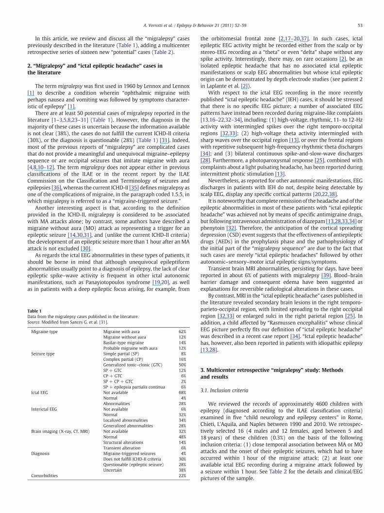
Table 1Data from the migralepsy cases published in the literature.Source. Modified from Sances G. et al. [31].
Migraine type Migraine with aura 62%Migraine without aura 12%Basilar-type migraine 14%Probable migraine with aura 12%
Seizure type Simple partial (SP) 8%Complex partial (CP) 16%Generalized tonic–clonic (GTC) 50%SP + GTC 12%CP + GTC 6%SP + CP + GTC 2%SP + epilepsia partialis continua 6%
Ictal EEG Not available 68%Normal 4%Abnormalities 28%
Interictal EEG Not available 6%Normal 32%Localized abnormalities 34%Generalized abnormalities 28%
Brain imaging (X-ray, CT, MRI) Not available 32%Normal 48%Structural alterations 14%Transient alteration 6%
Diagnosis Migraine-triggered seizures 4%Does not fulfill ICHD-II criteria 30%Questionable (epileptic seizure) 28%Uncertain 38%
Comorbidities 22%
the orbitomesial frontal zone [2,17–20,37]. In such cases, ictalepileptic EEG activity might be recorded either from the scalp or bystereo-EEG recording as a “theta” or even “delta” shape without anyspike activity. Interestingly, there may, on rare occasions [2], be anisolated epileptic headache that has no associated ictal epilepticmanifestations or scalp EEG abnormalities but whose ictal epilepticorigin can be demonstrated by depth electrode studies (see patient 2in Laplante et al. [2]).
With respect to the ictal EEG recording in the more recentlypublished “ictal epileptic headache” (IEH) cases, it should be stressedthat there is no specific EEG picture; a number of associated EEGpatterns have instead been recorded during migraine-like complaints[13,16–22,32–34], including: (1) high-voltage, rhythmic, 11- to 12-Hzactivity with intermingled spikes over the right temporo-occipitalregions [32,33]; (2) high-voltage theta activity intermingled withsharp waves over the occipital region [13], or over the frontal regionswith repetitive subsequent high-frequency rhythmic theta discharges[34]; and (3) bilateral continuous spike-and-slow-wave discharges[28]. Furthermore, a photoparoxysmal response [25], combined withcomplaints about a light pulsating headache, has been reported duringintermittent photic stimulation [13].
Nevertheless, as reported for other autonomic manifestations, EEGdischarges in patients with IEH do not, despite being detectable byscalp EEG, display any specific cortical patterns [20,22,38].
It is noteworthy that complete remission of the headache andof theepileptic abnormalities in most of these patients with “ictal epilepticheadache” was achieved not by means of specific antimigraine drugs,but following intravenous administration of diazepam [13,28,33,34] orphenytoin [32]. Therefore, the anticipation of the cortical spreadingdepression (CSD) event suggests that the effectiveness of antiepilepticdrugs (AEDs) in the prophylaxis phase and the pathophysiology ofthe initial part of the “migralepsy sequence” are due to the fact thatsuch cases are merely “ictal epileptic headaches” followed by otherautonomic–sensory–motor ictal epileptic signs/symptoms.
Transient brain MRI abnormalities, persisting for days, have beenreported in about 6% of patients with migralepsy [39]. Blood–brainbarrier damage and consequent edema have been suggested asexplanations for reversible radiological alterations in these cases.
By contrast, MRI in the “ictal epileptic headache” cases published inthe literature revealed secondary brain lesions in the right temporo-parieto-occipital region, with limited spreading to the right occipitalregion [32,33] or enlarged sulci in the right parietal region [25]. Inaddition, a child affected by “Rasmussen encephalitis” whose clinicalEEG picture perfectly fits our definition of “ictal epileptic headache”was described in a recent case report [34]. “Ictal epileptic headache”has, however, also been reported in patients with idiopathic epilepsy[13,28].
3. Multicenter retrospective “migralepsy” study: Methodsand results
3.1. Inclusion criteria
We reviewed the records of approximately 4600 children withepilepsy (diagnosed according to the ILAE classification criteria)examined in five “child neurology and epilepsy centers” in Rome,Chieti, L'Aquila, and Naples between 1990 and 2010. We retrospec-tively selected 16 (4 males and 12 females, aged between 5 and18 years) of these children (0.3%) on the basis of the followinginclusion criteria: (1) close temporal association between MA or MOattacks and the onset of their epileptic seizures, which had to haveoccurred within 1 hour of the migraine attack; (2) at least oneavailable ictal EEG recording during a migraine attack followed bya seizure within 1 hour. See Table 2 for the details and clinical/EEGpictures of the sample.
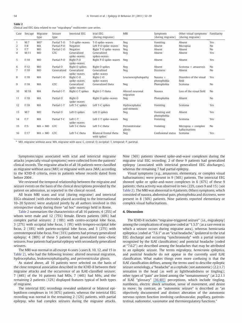
Table 2Clinical and EEG data related to our “migralepsy” multicenter case series.
Case Sex/age Migrainetype
Seizuretype
Interictal EEG Ictal EEG(during migraine)
MRI Symptoms(during migraine)
Other visual symptoms(during migraine)
Familiarity
1 M/7 MOa Partial T-O T-O spike–waves T-O spike–waves Neg Vomiting Absent Yes2 F/8 MA Partial P-O Negative Left P-O spike–waves Neg Absent Micropsia No3 F/7 MO Partial C-O Negative Right T-O spike–waves Neg Absent Absent No4 M/11 MO GTC Generalized
spike–wavesGeneralizedspikes-waves
Neg Absent Absent Yes
5 F/10 MO Partial P-O Right P-Ospike–waves
Right P-O spike–waves Neg Absent Absent Yes
6 F/12 MO Partial O Right O spikes Right O spikes Neg Absent Scotoma + amaurosis No7 F/10 MO Generalized Generalized
spike–wavesGeneralizedspike–waves
Neg Dizziness Absent No
8 F/18 MA Partial C-O Right C-Ospike–waves
Right C-Ospike–waves
Leucoencephalopathy Nausea +photophobia
Disorders of the visualfield
Yes
9 F/18 MA GTC Generalizedspike–waves
Generalized theta Neg Photophobia Scotoma No
10 M/18 MA Partial C-T Right C-T spikes Right C-T theta Altered neuronalmigration
Nausea Loss of the visual field No
11 F/18 MA Partial O Right Ospike–waves
Right O spike–waves Neg Photophobia Absent No
12 F/18 MA Partial C-T Left T-C spikes Left T-C spikes Hydrocephalusand stenosis
Vomiting Scotoma Yes
13 M/7 MO Partial O Left O spikes Left O spikes Neg Vomiting andphotophobia
Absent Yes
14 F/7 MA Partial T-C Left C-Tspike–waves
Left C-T spike–waves Neg Vomiting Scotoma Yes
15 F/5 MA + MO GTC Left T-C theta Left T-C theta Periventriculargliosis
Vomiting Micropsia + complexhallucinations
No
16 F/17 MA + MO GTC Left T-C theta Bilateral frontal thetawith spikes
Neg Confusional status Scotoma Yes
a MO, migraine without aura; MA, migraine with aura; C, central; O, occipital; T, temporal; P, parietal.
54 A. Verrotti et al. / Epilepsy & Behavior 21 (2011) 52–59
Symptoms/signs associated with ictal and interictal migraineattacks (especially visual symptoms)were collected from the patients’clinical records. The migraine attacks of all 16 patients were classifiedas migraine without aura (MO) or migraine with aura (MA) accordingto the ICHD-II criteria, even in patients whose records dated frombefore 2004 .
We reviewed the temporal relationship between the migraine andseizure events on the basis of the clinical descriptions provided by thepatient on admission, as reported in the clinical record.
All brain MRI scans and ictal (during migraine) and interictalEEGs obtained (with electrodes placed according to the International10–20 System) were analyzed jointly by all authors involved in thisretrospective study during three “ad hoc” meetings held in Rome.
Table 2 summarizes the characteristics of all 16 patients, 4 (25%) ofwhom were male and 12 (75%) female. Eleven patients (69%) hadcomplex partial seizures: 2 (18%) with centro-occipital lobe focus,3 (27%) with occipital lobe focus, 1 (9%) with temporo-occipital lobefocus, 2 (18%) with parieto-occipital lobe focus, and 3 (27%) withcentrotemporal lobe focus. Five (31%) patients had primary generalizedepilepsy; 4 (80%) of these 5 patients had generalized tonic–clonicseizures. Four patients had partial epilepsywith secondarily generalizedseizures.
The MRI was normal in all except 4 cases (cases 8, 10, 12, and 15 inTable 2), who had the following lesions: altered neuronal migration,hydrocephalus, leukoencephalopathy, and periventricular gliosis.
As stated above, all 16 patients were selected on the basis ofthe close temporal association described in the ICHD-II between theirmigraine attacks and the occurrence of an ILAE-classified seizure;7 (44%) of the 16 patients had MOs, 7 (44%) had MAs, and theremaining 2 patients (12%) displayed features typical of both typesof migraine.
The interictal EEG recordings revealed unilateral or bilateral epi-leptiform complexes in 14 (87%) patients, whereas the interictal EEGrecording was normal in the remaining 2 (12%) patients, with partialepilepsy, who had complex seizures during the migraine attacks.
Nine (56%) patients showed spike-and-wave complexes during themigraine ictal EEG recording; 2 of these 9 patients had generalizedepilepsy (associated with interictal generalized EEG discharges),whereas the remaining 7 had partial epilepsy.
Visual symptoms (e.g., amaurosis, elementary, or complex visualhallucinations) were present in 9 (56%) patients. The interictal EEGshowed spike or spike-and-wave complexes in 6 (67%) of these 9patients; theta activity was observed in two (22%, cases 9 and 15) (seeTable 2). TheMRIwas abnormal in 4 patients. Others symptoms,whichconsisted of nausea, abdominal pain, photophobia and dizziness, werepresent in 8 (50%) patients. Nine patients reported elementary orcomplex visual hallucinations.
4. Discussion
The ICHD-II includes “migraine-triggered seizure” (i.e., migralepsy)among the complications ofmigraine coded at “1.5.5” (as a rare event inwhich a seizure occurs during migraine aura), whereas hemicraniaepileptica (coded at “7.6.1” as an “ictal headache,” ipsilateral to the ictalEEG discharge and occurring “synchronously” with a partial seizurerecognized by the ILAE classification) and postictal headache (codedat “7.6.2”) are described among the headaches that may be attributedto an epileptic seizure. The terms migralepsy, hemicrania epileptica,and postictal headache do not appear in the currently used ILAEclassification. What makes things even more confusing is that theILAE classification defines, among the terms used to describe epilepticseizure semeiology, a “headache” asa cephalic (not autonomic) (2.2.1.7)sensation in the head (as well as lightheadedness or tingling);other types of “pain” are listed among the “somatosensory” (at 2.2.1.1of ILAE “glossary” [36,40]) perceptions, which include tingling,numbness, electric shock sensation, sense of movement, and desireto move; by contrast, an “autonomic seizure” is described as “anobjectively documented and distinct alteration of the autonomicnervous system function involving cardiovascular, pupillary, gastroin-testinal, sudomotor, vasomotor and thermoregulatory functions.”

55A. Verrotti et al. / Epilepsy & Behavior 21 (2011) 52–59
The 16 new retrospective cases described here (Table 2) and thepreviouslypublishedmigralepsy cases (Table1) illustrate thedifficultiesthat may be encountered when attempting to make a diagnosis, aswell as the inadequacy of the recent ICHD-II “migralepsy” diagnosticcriteria [31]. Moreover, a recent review of the literature (Table 1)identified a remarkably large number of purely epileptic disordersamong cases reported asmigralepsy [31]. Indeed, one patient describedin a case report we published previously [13] responded (immediatelyfrom both the clinical and EEG points of view) to intravenousanticonvulsant (diazepam) administration, as did other cases (threeresponded to diazepam and one to phenytoin) [28,32–34].
Moreover, Maggioni et al. [30], when discussing two of their“migralepsy” cases, stressed the role of a preexisting low epilepticthreshold, suggesting that further studies be designed to assess theneed to include MO-triggered epileptic seizures in the diagnosticcriteria for migralepsy. They also suggested that migralepsy mightoccur more frequently than is reported because the epileptic eventovershadows the MO [30].
With respect to our “migralepsy” cases (Table 2), we found, on thebasis of the afore-described temporal “inclusion criteria,” only 16 ofapproximately 4600 epileptic children whose seizure “occurredwithin 1 hour of a MA or MO attack”; we should also point outthat we even included (in accordance with Maggioni et al. [30],though in contrast to the current ICHD-II “migralepsy” criteria, point1), patients manifesting MO as the sole migraine type associated with“epileptic seizures” (patients 1, 3–7, and 13), thus not adopting MAas a mandatory condition (the “sine qua non” condition requestedby the current ICHD-II classification to make a diagnosis of “migraine-triggered seizure” or “migralepsy”).
With respect to the EEG findings, we decided to select (seeInclusion Criteria) only subjects with an ictal EEG recorded duringthe “migraine phase.” Interestingly, all 16 patients displayed focal orgeneralized “ictal EEG abnormalities” during the migraine attacks.Moreover, it is worth bearing in mind, as mentioned above, that only4% of previous reports of “migralepsy” cases fulfill the current ICHD-IIdiagnostic criteria, and that a migraine-phase EEG was availablein only 32% of those cases [31]. There seems to be no correlationbetween specific EEG abnormalities and MA or MO in the children weidentified (see Table 2); indeed, the spike or spike-and-wave pattern,which is the most commonly observed EEG pattern, was associatedwith both MA and MO (Table 2), whereas EEG theta activity wassurprisingly associated exclusively with MA or a “double migrainepattern,” in which MA and MO coexisted (patients 9, 10, 15, and 16).Fourteen of 16 children (the exceptions were cases 2 and 3) displayedinterictal EEG abnormalities. Our sample (Table 2) exhibited amarked prevalence of “migralepsy” in females (8 F vs 4 M), whereasthe “migralepsy association” with MO (as the sole migraine type)prevailed in males (75%).
Another intriguing finding in our patients with “migralepsy”(Table 2) is the lack of a correlation between specific cortical locali-zation of the EEG abnormalities and a synchronous headache onset.Indeed, whereas the “occipital localization” has previously beenreported to prevail, or even be exclusive, in migralepsy, we did notobserve any specific cortical localization in our 16 patients; for example,we recorded occipital (Fig. 1), centrotemporal (Fig. 2), and evenfrontal (Fig. 3) ictal EEG abnormalities, respectively, in patients 6, 14,and 16, all of whom presented with an ictal headache (see Table 2).
Moreover, no specific EEG pattern emerges from either the“migralepsy” cases listed in Table 2 or the ictal epileptic headachecases that have recently been published [13,16–22,32–34], with awide range of patterns and localizations (occipital, temporoparietal,and frontal) being recorded during migraine-like complaints. Thesefindings suggest, as we have previously hypothesized [15], that theepileptic focus that activates the trigeminovascular system mightremain purely autonomic (it being associated exclusively withmigraine complaints), without ictal neuronal activation of nonauto-
nomic cortical areas (different neuronal network thresholds appear tobe involved); in this case, the focus fails to reach the symptomato-genic threshold required to induce sensory–motor manifestations,as has been described for other ictal autonomic manifestations inPanayiotopoulos syndrome [41].
The diagnosis in these rare cases is complicated even further by thefact that the epileptic headache may be isolated; that is, it is notassociated with any other ictal epileptic manifestations or EEGabnormalities that can be detected by scalp EEG recording [2]. Inthis regard, it is often impossible (in 20 to 40% of patients) to detectictal epileptic activity by means of scalp EEG recording in other typesof epileptic manifestations, such as in frontal lobe epilepsy [37].
Another noteworthy issue that causes some confusion is thecommon “epilepsy–migraine” sequence, which has widely beenignored despite its high prevalence in occipital epilepsy, in which“postictal” headache occurs in 50% of patients. Indeed, althougha migraine-type headache is a common postictal phenomenonthat occurs in approximately 50% of patients with epilepsy, it isoften neglected because of the dramatic manifestation of the seizure.This condition has been included in the recent ICHD-II [35] and isdiagnosed on the basis of the following criteria: (1) headache hastension-type headache features or, if in a patient with migraine,migraine headache features and fulfills criteria 2–4; (2) the patienthas had a partial or generalized seizure; (3) headache develops within3 hours of the seizure; (4) headache resolves within 72 hours of theseizure. In this regard, Schön and Blau [42] described a series of 100patients with epilepsy, 51 of whom had postictal headache, which hasbeen reported particularly in idiopathic occipital seizures [43]. It islikely that the seizure discharges in the occipital lobes trigger agenuine migraine headache through cortical spreading depression(CSD) and trigeminovascular or brainstemmechanisms, as previouslysuggested by our group [15–20]. Indeed, the onset of epilepticseizuresmay facilitate that of CSD to a greater degree than the onset ofCSD facilitates that of epileptic seizures. This may explain why, in theclinical context, patients with epilepsy with postictal headache (51%according to Schön and Blau) [42] are more likely to be observed thansubjects with migraine with epileptic seizures [15–20].
Yet another source of confusion is the lack of an accuratedescription of symptoms during “ictal epileptic headache”; indeed,an “ictal epileptic headache” is usually orbital or manifests itself aspainful discomfort that does not have a clear localization within thehead and, thus, displays a “pattern” different from that of “migraine-associated” pain. By contrast, “postictal headache” is more character-istic of migraine symptoms; a slight impairment in consciousnessmay be associated with “ictal epileptic headache” more often thanit is with postictal headache conditions, during which patients aremore likely to be able to clearly describe their “postictal” symptoms(severity, type, and localization of pain).
A possible partial overlap between headache and epilepsy, whichon rare occasions may even be a complete overlap (e.g., in patientswith “ictal epileptic headache”), is supported by clinical/EEG andgenetic studies on familial hemiplegic migraine (FHM) [44–46]. InFHM, errors in the same gene may be associated with migraine insome cases and with epilepsy in others [46].
To sum up, the authors of most of the “migralepsy” casespreviously published in the literature have highlighted the fact thatthe EEG and clinical features of such subjects may be suggestive ofoccipital lobe seizures, although the information currently available isinsufficient to confirm this hypothesis. In this regard, it should beborne in mind that ictal EEG recordings during the migraine phaseare available in only 32% of previously published “migralepsy” cases,which prevents any firm conclusions from being drawn regarding“migralepsy” EEG-associated patterns. Indeed, Sances et al. suggestedthat ICHD-II 1.5.5 (the paragraph defining migralepsy) be relocated tothe ICHD-II Appendix until a larger number of migralepsy cases arereported or the condition is better characterized [31].

Fig. 1. Case 6 in Table 2. (A) Right occipital theta activity with sporadic spikes during migraine. (B) Normal EEG some months later.
56 A. Verrotti et al. / Epilepsy & Behavior 21 (2011) 52–59
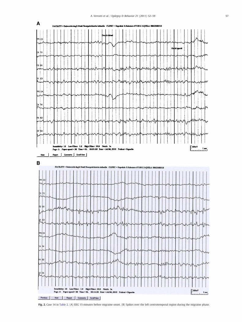
Fig. 2. Case 14 in Table 2. (A) EEG 15 minutes before migraine onset. (B) Spikes over the left centrotemporal region during the migraine phase.
57A. Verrotti et al. / Epilepsy & Behavior 21 (2011) 52–59
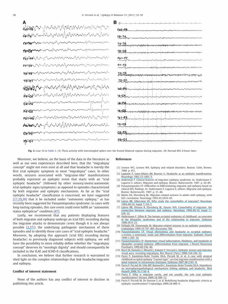
Fig. 3. Case 16 in Table 2. (A) Theta activity with intermingled spikes over the frontal bilateral regions during migraine. (B) Normal EEG 6 hours later.
58 A. Verrotti et al. / Epilepsy & Behavior 21 (2011) 52–59
Moreover, we believe, on the basis of the data in the literature aswell as our own experiences described here, that the “migralepsyconcept” might not even exist at all and that headache is merely thefirst ictal epileptic symptom in most “migralepsy” cases. In otherwords, seizures associated with "migraine-like" manifestationsprobably represent an epileptic event that starts with an “ictalepileptic headache” (followed by other sensory/motor/autonomicictal epileptic signs/symptoms) as opposed to episodes characterizedby both migraine and epileptic mechanisms. As far as the “ictalepileptic headache” classification is concerned, we have suggested[17,19,20] that it be included under “autonomic epilepsy,” as hasrecently been suggested for Panayiotopoulos syndrome; in cases withlong-lasting episodes, this rare event could even fulfill an “autonomicstatus epilepticus” condition [47].
Lastly, we recommend that any patients displaying featuresof both migraine and epilepsy undergo an ictal EEG recording duringthe migraine attacks to demonstrate (even though it is not alwayspossible [2,37]) the underlying pathogenic mechanism of theseepisodes and to identify those rare cases of “ictal epileptic headache.”Moreover, by adopting this approach (ictal EEG recording duringheadache) in previously diagnosed subjects with epilepsy, we willhave the possibility to more reliably define whether the “migralepsyconcept” deserves its “nosologic dignity” and should consequently beincluded in the ILAE and ICHD-II classifications.
In conclusion, we believe that further research is warranted toshed light on the complex relationships that link headache/migraineand epilepsy.
Conflict of interest statement
None of the authors has any conflict of interest to disclose inpublishing this article.
References
[1] Lennox WG, Lennox MA. Epilepsy and related disorders. Boston: Little, Brown;1960. p. 451.
[2] Laplante P, Saint-Hilaire JM, Bouvier G. Headache as an epileptic manifestation.Neurology 1983;33:1493–5.
[3] Anderman F. Clinical features of migraine–epilepsy syndrome. In: Andermann F,Lugaresi E, editors. Migraine and epilepsy. Boston: Butterworth; 1987. p. 20–89.
[4] Panayiotopoulos CP. Difficulties in differentiating migraine and epilepsy based onclinical EEG findings. In: Andermann F, Lugaresi E, editors. Migraine and epilepsy.Boston: Butterworth; 1987. p. 31–46.
[5] Marks DA, Ehrenberg BL. Migraine-related seizures in adults with epilepsy, withEEG correlation. Neurology 1993;43:2476–83.
[6] Lipton RB, Silberstein SD. Why study the comorbidity of migraine? Neurology1994;44(10, Suppl 7):S4–5.
[7] Lipton RB, Ottman R, Ehrenberg BL, Hauser WA. Comorbidity of migraine: theconnection between migraine and epilepsy. Neurology 1994;44(10, Suppl 7):S28–32.
[8] Andermann F, Zifkin B. The benign occipital epilepsies of childhood: an overviewof the idiopathic syndromes and of the relationship to migraine. Epilepsia1998;39:9–23.
[9] Velioglu SK, Ozmenoglu M. Migraine-related seizures in an epileptic population.Cephalalgia 1999;19:797–801 discussion 766.
[10] Panayiotopoulos CP. Visual phenomena and headache in occipital epilepsy:a review, a systematic study and differentiation from migraine. Epileptic Disord1999;1:205–16.
[11] Panayiotopoulos CP. Elementary visual hallucination, blindness, and headache inidiopathic occipital epilepsy: differentiation from migraine. J Neurol NeurosurgPsychiatry 1999;66:536–40.
[12] Barré M, Hamelin S, Minotti L, Kahane P, Vercueil L. Epileptic seizure and migrainevisual aura: revisiting migralepsy. Rev Neurol (Paris) 2008;164:246–52.
[13] Parisi P, Kasteleijn-Nolst Trenite DGA, Piccioli M, et al. A case with atypicalchildhood occipital epilepsy “Gastaut type”: an ictal migraine manifestation with agood response to intravenous diazepam. Epilepsia 2007;48:2181–6.
[14] Parisi P, Piccioli M, Villa MP, Buttinelli C, Kasteleijn-Nolst Trenéte DGA. Hypothesison neurophysiopathological mechanisms linking epilepsy and headache. MedHypoth 2008;70:1150–4.
[15] Parisi P. Why is migraine rarely, and not usually, the sole ictal epilepticmanifestation? Seizure 2009;18:309–12.
[16] Parisi P, Piccioli M, De Sneeuw S, et al. Redefining headache diagnostic criteria asepileptic manifestation? Cephalalgia 2009;28:408–9.

59A. Verrotti et al. / Epilepsy & Behavior 21 (2011) 52–59
[17] Parisi P. Who's still afraid of the link between headache and epilepsy? Somereactions to and reflections on the article by Marte Helene Bjørk and co-workers.J Headache Pain 2009;10:327–9.
[18] Piccioli M, Parisi P, Tisei P, Villa MP, Buttinelli C, Kasteleijn-Nolst Trenité DGA. Ictalheadache and visual sensitivity. Cephalalgia 2009;29:194–203.
[19] Parisi P, Kasteleijn-Nolst Trenitè DGA. “Migralepsy”: a call for revision of thedefinition. Epilepsia 2010;51:932–3.
[20] Kasteleijn-Nolst Trenité DGA, Verrotti A, Di Fonzo A, et al. Headache, epilepsy andphotosensitivity: how are they connected? J Headache Pain 2010;11:469–76.
[21] Striano P, Belcastro V, Parisi P. Status epilepticus migrainosus: clinical,electrophysiologic, and imaging characteristics. Neurology 2011;76:761.
[22] Parisi P. Comments on the article by Fusco L. et al. entitled "Migraine triggered byepileptic discharges in a Rasmussen's encephalitis patient after surgery" BrainDev. doi:10.1016/j.braindev.2011.03.008. [Electronic publication ahead of print]on line in PubMed, April 4, 2011.
[23] Isler H, Wieser HG, Egli M. Hemicrania epileptica: synchronous ipsilateral ictalheadache with migraine features. In: Andermann F, Lugaresi E, editors. Migraineand epilepsy. Boston: Butterworth; 1987. p. 249–63.
[24] Niedermeyer E. Migraine-triggered epilepsy. Clin EEG 1993;24:37–43.[25] Walker MC, Smith SJ, Sisodya SM, Shorvon SD. Case of simple partial status
epilepticus in occipital lobe epilepsy misdiagnosed as migraine: clinical,electrophysiological, and magnetic resonance imaging characteristics. Epilepsia1995;36:1233–6.
[26] Friedenberg S, Dodick DW. Migraine-associated seizure: a case of reversible MRIabnormalities and persistent non dominant hemisphere syndrome. Headache2000;40:487–90.
[27] Milligan TA, Bromfield E. A case of migralepsy. Epilepsia 2005;46:2–6.[28] Ghofrani M, Mahvelati F, Tonekaboni H. Headache as a sole manifestation in
nonconvulsive status epilepticus. J Child Neurol 2006;21:981–3.[29] Merlino G, Valente MR, D'Anna S, Gigli GL. Seizures with prolonged EEG
abnormalities duringanattackofmigrainewithout aura.Headache2007;47:919–22.[30] Maggioni F, Mampreso E, Ruffatti S, Viaro F, Lunardelli V, Zanchin G. Migralepsy: is
the current definion too narrow? Headache 2008;48:1129–32.[31] Sances G, Guaschino E, Perucca P, Allena M, Ghiotto N, Manni R. Migralepsy: a call
for revision of the definition. Epilepsia 2009;50:2487–96.[32] Perucca P, Terzaghi M, Manni R. Status epilepticus migrainosus: clinical,
electrophysiologic, and imaging characteristics. Neurology 2010;75:373–4.
[33] Belcastro V, Striano P, Pierguidi L, Calabresi P, Tambasco N. Ictal epileptic headachemimicking statusmigrainosus: EEG andDWI-MRI findings. Headache2011;51:160–2.
[34] Fusco L, Specchio N, Ciofetta G, Longo D, Trivisano M, Vigevano F. Migrainetriggered by epileptic discharges in a Rasmussen's encephalitis patient aftersurgery. Brain Dev October 23 2010, doi:10.1016/j.braindev.2010.09.014. EPub.
[35] International Headache Society. The International Classification of HeadacheDisorders: 2nd edition. Cephalalgia 2004;24(Suppl 1):9–160.
[36] Berg AT, Berkovic SF, Brodie MJ, et al. Revised terminology and concepts fororganization of seizures and epilepsies: report of the ILAE Commission onClassification and Terminology, 2005–2009. Epilepsia 2010;51:676–85.
[37] Derry CP, Harvey AS, Walker MC, Duncan JS, Berkovic SF. NREM arousalparasomnias and their distinction from nocturnal frontal lobe epilepsy: a videoEEG analysis. Sleep 2009;32:1637–44.
[38] Kokkinos V, KoutroumanidisM, Tsatsou K, Koupparis A, Tsiptsios D, PanayiotopoulosCP. Multifocal spatiotemporal distribution of interictal spikes in Panayiotopoulossyndrome. Clin Neurophysiol 2010;121:859–69.
[39] Mateo I, Foncea N, Vicente I, Gomez Beldarrain M, Garcia-Monco JC. Migraine-associated seizures with recurrent and reversible magnetic resonance imagingabnormalities. Headache 2004;44:265–70.
[40] BlumeWT, Lüders HO, Mizrahi E, Tassinari C, van Emde BoasW, Engel Jr J. Glossaryof descriptive terminology for ictal semiology: report of the ILAE task force onclassification and terminology. Epilepsia 2001;42:1212–8.
[41] Koutroumanidis M. Panayiotopoulos syndrome: an important electroclinicalexample of benign childhood system epilepsy. Epilepsia 2007;48:1044–53.
[42] Schön F, Blau JN. Post-epileptic headache and migraine. J Neurol NeurosurgPsychiatry 1987;50:1148–52.
[43] Ekstein D, Schachter SC. Postictal headache. Epilepsy Behav 2010;19:151–5.[44] Riant F, Ducros A, Ploton C, Banbance C, Depienne C, Tournie-Lasserve E. De novo
mutations in ATP1A2 and CACNA1A are frequent in early-onset sporadichemiplegic migraine. Neurology 2010;75:967–72.
[45] Escayg A, Goldin AL. Critical review and invited commentary: sodium channelSCN1A and epilepsy: mutations and mechanisms. Epilepsia 2010;51:1650–8.
[46] LudvigssonP,HesdorfferD,OlafssonE, KjartanssonO,HauserWA.Migrainewith aurais a risk factor for unprovoked seizures in children. Ann Neurol 2006;59:210–3.
[47] Ferrie CD, Caraballo R, Covanis A, et al. Autonomic status epilepticus inPanayiotopoulos syndrome and other childhood and adult epilepsies: a consensusview. Epilepsia 2007;48:1165–72.
Copyright © 2022 FDOKUMEN