ELA Curriculum Grade 7 PACC Standard Term 1 Term 2 Term 3 ...

Short-Term and Long-Term Outcomes After SimultaneousResection of Colorectal Malignancies and Synchronous LiverMetastases
Eduardo de Santibanes • Diego Fernandez • Carlos Vaccaro •
Guillermo Ojea Quintana • Fernando Bonadeo • Juan Pekolj •
Carlos Bonofiglio • Ernesto Molmenti
Published online: 8 June 2010
� Societe Internationale de Chirurgie 2010
Abstract
Background We evaluated the simultaneous resection of
colorectal malignancies and synchronous liver metastases.
Methods Between June 1982 and June 2006, a total of
752 patients underwent resection of colorectal hepatic
metastases. In all, 185 (25%) of them underwent simulta-
neous resection of the hepatic lesions and the correspond-
ing primary tumors.
Results The median hospital stay was 8 days (range 4–
24 days), with a median operating time of 4 h (range 2–8 h).
Altogether, 62 (33.5%) patients required intraoperative
transfusion of packed red blood cells (median 2.1 IU, range
1–5 IU), and 25 (13.5%) were given frozen fresh plasma
(median 2.1 IU, range 1–4 IU). The morbidity rate was
20.5%. There were two postoperative deaths (mortality rate
1.08%) within 30 days of the surgical intervention. Major
hepatectomy was associated with greater morbidity (37.2%
vs. 16.2%, P \ 0.01) and mortality (4.7% vs. 0%, P \ 0.05)
rates. For the overall survivals (OS) at 3 and 5 years were
60.1% (52.3–67.85%) and 36.1% (27.4-44.8%), respec-
tively. Disease-free survivals (DFS) at 3 and 5 years were
37.7% (30.2–45.3%) and 26.5% (18.7–34.3%), respectively.
Transfusion of blood products, CEA level C 200 ng/dl, and
N2 node status were found to be prognostic factors by uni-
variate analysis. CEA level C 200 ng/dl and N2 node status
achieved prognostic significance by multivariate analysis.
Conclusions The simultaneous resection of colorectal
malignancies and synchronous liver metastases is safe, avoids
an additional intervention, can be performed with low mor-
bidity and mortality, and is associated with good oncologic
outcomes. Node stage N2 and CEA level C 200 ng/dl should
be given special consideration when selecting patients.
Keywords Liver � Colorectal � Gastrointestinal �Oncology � Biliary � Critical care
Introduction
Colorectal cancer is one of the most frequent malignancies
throughout the world [1–6]. Resection of liver metastases
presenting after removal of the primary tumor can be
associated with 5-year survivals of up to 38% [7–17].
When they present synchronously (23–51% of resectable
cases [18]), however, the survival decreases to B20% [10,
19]. Except in selected cases (i.e., right colectomy, wedge
liver resection), synchronous metastases were systemati-
cally treated with two laparotomies [10, 11]. Current
studies, however, suggest that simultaneous resections can
be performed safely with comparable or even better out-
comes than staged procedures [20–31]. Furthermore,
simultaneous resections are associated with improved
patient comfort [23], massive reduction in tumor mass,
better response to chemotherapy [30], potential decreased
E. de Santibanes (&) � D. Fernandez � J. Pekolj
Hepato-pancreato-biliary and Liver Transplant Sections,
Department of General Surgery, Hospital Italiano, Buenos Aires,
Argentina
e-mail: [email protected]
C. Vaccaro � G. O. Quintana � F. Bonadeo
Colorectal Section, Department of General Surgery, Hospital
Italiano, Buenos Aires, Argentina
C. Bonofiglio
Department of Anesthesia, Hospital Italiano, Buenos Aires,
Argentina
E. Molmenti
Department of General Surgery, North Shore – LIJ Health
System, Great Neck, NY, USA
123
World J Surg (2010) 34:2133–2140
DOI 10.1007/s00268-010-0654-6

risk of cancer cell dissemination [31, 32], and avoidance of
an additional surgical procedure [33].
The purpose of this study was to evaluate our 24-year
experience with simultaneous resection of synchronous
colorectal cancer and liver metastases.
Patients and Methods
From June 1982 to June 2006, a total of 2,700 patients with
colorectal malignancy and 1,375 with malignant hepatic
tumors underwent resection at the Colorectal and Hepa-
tobiliary Section of the Hospital Italiano of Buenos Aires.
Of these, 185 (13.45%) underwent simultaneous colorectal
and hepatic resection for colorectal malignancy with syn-
chronous hepatic metastases. There were 114 males and 71
females, with a median age of 63 years (range 16–
86 years). Complications were classified according to
Dindo’s scheme [33], except for transfusion of blood
products, which was not considered a complication.
Preoperative Staging
Routine preoperative evaluation included a complete
physical examination, chest radiography, carcinoembry-
onic antigen (CEA) (obtained at our laboratory in 79 of the
185 patients) and cancer antibody (CA) 19-9 levels,
colonoscopy, abdominal ultrasonography (US), and com-
puted tomography (CT) scans of the chest, abdomen, and
pelvis. Positron emission tomography was not part of our
protocol due to lack of medical insurance coverage.
Patient Selection
A total of 492 patients with colorectal cancer and synchro-
nous liver metastases were evaluated. Simultaneous liver
and colorectal resection was undertaken in 185 cases
(37.6%). Simultaneous liver and colorectal resection was not
undertaken in patients with performance status American
Society of Anesthesiologists (ASA) IV; emergency resection
in the setting of obstruction, perforation, or hemorrhage [28];
tumors whose colorectal origin could not be clearly estab-
lished; extrahepatic metastases; failure to achieve negative
intraoperative colorectal (R0) and hepatic (R0) resection
margins; or inability to retain a postresection liver volume of
at least 35% of the total liver volume [34]. Size and number
of liver metastases were not considered exclusionary factors.
All of the resections were performed by surgical teams that
had experience with both procedures.
Preoperative Care
Mechanical bowel preparation was with 90 ml of an oral
phosphate solution. Antibiotic prophylaxis consisted of
750 mg oral ciprofloxacin and 1 g intravenous ornidazole
administered less than 1 h prior to surgery.
Surgical Technique
Prior to 1999, we used inhalational anesthetic agents. In
1999, we adopted total intravenous anesthesia (TIVA).
Central venous pressure (CVP) was kept at \5 cm H2O,
and vasopressors were added whenever necessary to avoid
hypotension.
The abdomen was entered through a midline incision
and its contents explored thoroughly. Colorectal resection
was undertaken prior to liver resection. An R0 colorectal
resection was achieved in those cases where all criteria
were verified. Prior to continuing with the liver resection,
all surgical instruments and drapes were removed and
replaced with clean sterile ones.
The falciform ligament was divided to allow for US
evaluation of the liver. The midline incision was extended
transversely for metastases located in Couinaud’s segments
VI or VII. Major hepatectomy was defined as resection of
at least three segments [35]. Liver transection was carried
out with a cavitation ultrasonic aspirator (CUSA; Valley,
Boulder, CO, USA) and/or by means of Kelly clamp tissue
crushing. Argon beam coagulation was used for hemosta-
sis. We applied the Pringle maneuver only in cases where
bleeding was encountered during the parenchymal tran-
section despite CVP \ 5 cm H2O. We did not use pre-
conditioning. An initial occlusion period of 20 min was
followed (if necessary) by a rest period of 5 min and a
second occlusion period of 15 min. In cases of multiple
metastases with no involvement of large vessels, we per-
formed wedge resection with margins of at least 1 cm. At
the end of the procedure, we placed one to three suction
drains that were removed 3–5 days later in the absence of
bile leaks. Postoperative recovery was in the intensive care
unit (ICU).
Follow-up
Routine postoperative follow-up was every 4 months and
included a complete physical examination, US or CT, liver
function tests, complete blood count, and assays for elec-
trolytes, blood urea nitrogen (BUN), creatinine, and CEA
as well as the prothrombin time (PT) and partial throm-
boplastin time (PTT). Median follow-up was 35 months
(range 12–128 months). All patients received chemother-
apy according to the following guidelines for stage IV
tumors: prior to 1996, 5-fluorouracil (5FU) and leucovorin
[36]; from 1996 to 2003, FOLFIRI [folinic acid (leucovo-
rin)/5FU/irinotecan] [37, 38]; and since 2003, FOLFOX
[folinic acid (leucovorin)/5FU/oxaliplatin] and FOLFIRI
[39–41].
2134 World J Surg (2010) 34:2133–2140
123
Statistical Analysis
Continuous data were expressed as the median and range.
Overall and disease-free survival rates were calculated with
the Kaplan–Meier method and expressed as median values
with 95% confidence intervals (95% CI). Survival curves
were compared using the log-rank test. Results were con-
sidered to be statistically significant at P B 0.05.
Results
Technical Aspects
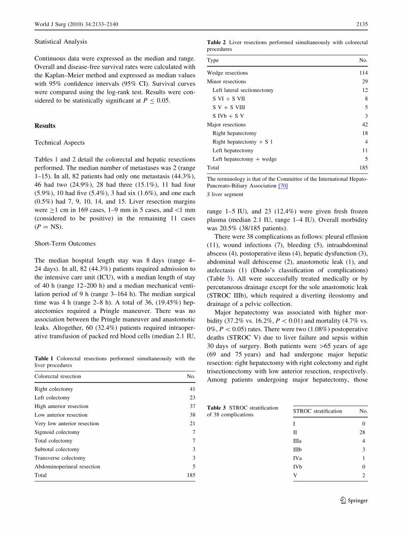
Tables 1 and 2 detail the colorectal and hepatic resections
performed. The median number of metastases was 2 (range
1–15). In all, 82 patients had only one metastasis (44.3%),
46 had two (24.9%), 28 had three (15.1%), 11 had four
(5.9%), 10 had five (5.4%), 3 had six (1.6%), and one each
(0.5%) had 7, 9, 10, 14, and 15. Liver resection margins
were C1 cm in 169 cases, 1–9 mm in 5 cases, and\1 mm
(considered to be positive) in the remaining 11 cases
(P = NS).
Short-Term Outcomes
The median hospital length stay was 8 days (range 4–
24 days). In all, 82 (44.3%) patients required admission to
the intensive care unit (ICU), with a median length of stay
of 40 h (range 12–200 h) and a median mechanical venti-
lation period of 9 h (range 3–164 h). The median surgical
time was 4 h (range 2–8 h). A total of 36, (19.45%) hep-
atectomies required a Pringle maneuver. There was no
association between the Pringle maneuver and anastomotic
leaks. Altogether, 60 (32.4%) patients required intraoper-
ative transfusion of packed red blood cells (median 2.1 IU,
range 1–5 IU), and 23 (12.4%) were given fresh frozen
plasma (median 2.1 IU, range 1–4 IU). Overall morbidity
was 20.5% (38/185 patients).
There were 38 complications as follows: pleural effusion
(11), wound infections (7), bleeding (5), intraabdominal
abscess (4), postoperative ileus (4), hepatic dysfunction (3),
abdominal wall dehiscense (2), anastomotic leak (1), and
atelectasis (1) (Dindo’s classification of complications)
(Table 3). All were successfully treated medically or by
percutaneous drainage except for the sole anastomotic leak
(STROC IIIb), which required a diverting ileostomy and
drainage of a pelvic collection.
Major hepatectomy was associated with higher mor-
bidity (37.2% vs. 16.2%, P \ 0.01) and mortality (4.7% vs.
0%, P \ 0.05) rates. There were two (1.08%) postoperative
deaths (STROC V) due to liver failure and sepsis within
30 days of surgery. Both patients were [65 years of age
(69 and 75 years) and had undergone major hepatic
resection: right hepatectomy with right colectomy and right
trisectionectomy with low anterior resection, respectively.
Among patients undergoing major hepatectomy, those
Table 1 Colorectal resections performed simultaneously with the
liver procedures
Colorectal resection No.
Right colectomy 41
Left colectomy 23
High anterior resection 37
Low anterior resection 38
Very low anterior resection 21
Sigmoid colectomy 7
Total colectomy 7
Subtotal colectomy 3
Transverse colectomy 3
Abdominoperineal resection 5
Total 185
Table 2 Liver resections performed simultaneously with colorectal
procedures
Type No.
Wedge resections 114
Minor resections 29
Left lateral sectionectomy 12
S VI ? S VII 8
S V ? S VIII 5
S IVb ? S V 3
Major resections 42
Right hepatectomy 18
Right hepatectomy ? S 1 4
Left hepatectomy 11
Left hepatectomy ? wedge 5
Total 185
The terminology is that of the Committee of the International Hepato-
Pancreato-Biliary Association [70]
S liver segment
Table 3 STROC stratification
of 38 complicationsSTROC stratification No.
I 0
II 28
IIIa 4
IIIb 3
IVa 1
IVb 0
V 2
World J Surg (2010) 34:2133–2140 2135
123

aged C 65 years had a significant higher mortality rate
(16.7% vs. 0%, P = 0.02).
Long-Term Outcomes
Tumor recurrence was observed in 64% of cases (119/185
patients). Recurrences were detected (on occasions at more
than one site) in: liver (n = 98), lung (n = 43), peritoneum
(n = 16), abdominal wall (n = 3), bone (n = 4), and
adrenal gland (n = 1). There was no association between
site of recurrence and location of the primary tumor within
the colon and rectum. The 3- and 5-year overall survival
rates were 60.1% and 36.1%, respectively. The corre-
sponding disease-free survivals were 37.8% and 27.9%,
respectively.
Colorectal tumors were staged according to the TNM
classification. There were 4 T1, 15 T2, 148 T3, and 18 T4
cases. N staging showed 62 N0, 80 N1, and 43 N2 cases. T
stage was not prognostic. Overall survival (OS) and dis-
ease-free survival (DFS) were inversely correlated with the
N stage (Table 4).
The median weight of the hepatic resection specimens
was 356 g (range 3–1800 g), with a median metastasis
diameter of 4.5 cm (range 1–28 cm). Size did not correlate
with prognosis.
The median number of liver metastases was 2.29 (range
1–15). Statistical analysis (not shown) revealed that one or
two versus three or more liver metastases showed a sig-
nificant inverse correlation with DFS but had no effect on
OS (Table 5).
Resection margins and CA 19-9 levels had no effect on
either OS or DFS. The median preoperative CEA value was
23 ng/ml (range 0.2–550.0 ng/ml). The CEA level was
correlated with the outcome (Table 6).
Transfusion of packed red blood cells and fresh frozen
plasma showed an inverse correlation with both OS and
DFS (Table 7). Transfusion of blood products, CEA level,
and N2 node status were found to be prognostic by uni-
variate analysis (Table 8). On multivariate analysis, only
the CEA level and N2 node status reached significance
(Table 9).
Discussion
More than a decade ago, Hughes et al. [13] recommended
simultaneous resections in patients with single metastases
who could physiologically tolerate simple removal of the
liver lesion in the setting of an uncomplicated colorectal
resection with minimal blood loss and contamination.
Since then, more extensive hepatic resections have been
attempted [20, 27]. However, despite thorough preopera-
tive workups with US, CT, magnetic resonance imaging
(MRI), and 18F-fluorodeoxyglucose positron emission
tomography (FDG-PET) [42–47], widespread metastases
are encountered in approximately 30% of patients at the
time of surgery. The 64% recurrence rate in our series, with
most lesions located in the liver, suggests the presence of
microscopic residual disease undetectable at the time of
surgery. This finding also supports the use of adjuvant
therapies during both the pre- and postoperative periods
and could serve as a criticism for the absence of PET
scanning in our protocol.
Although no randomized trials have been published so
far, recent series on simultaneous resections reported
similar or improved short-term and equivalent long-term
outcomes [8, 20, 23, 26–28, 42–51]. Accordingly, the
Standards Practice Task Force, the American Society of
Colon and Rectal Surgeons [52], and a recent systematic
review [49] listed simultaneous colorectal and liver resec-
tions among recommended guidelines with levels of evi-
dence III, grade C II or III, and grade C, respectively.
Martin et al. [20] reported that simultaneous resections
had fewer complications (49% vs. 67%, P \ 0.01), shorter
median hospital stays (10 vs. 18 days, P \ 0.01), and
similar mortality rates (2.2% vs. 2.8%, P = NS) when
compared to staged procedures. Moreover, when compar-
ing patients who underwent major liver resections (lobec-
tomy or more), those with simultaneous surgeries had a
lower morbidity rate (60% vs. 70%, P = 0.03), shorter
operating times, decreased hospital stays, and a twofold
decrease in laparotomy complications. Capussotti et al. [53,
54] and Martin et al. [20] reported that the combined
morbidity was greater after staged procedures than after
Table 4 N stage (TNM classification) and long-term outcome
N stage 3-Year DFS 5-Year DFS 3-Year OS 5-Year OS
N0 (n = 64) 38.7% (25.3–52.1) 30.3% (16.8–43.8) 56.8% (43.1–70.5) 41.6% (26.4–56.8)
N1 (n = 79) 44.9% (33.1–56.6) 33.8% (21.4–46.1) 65.9% (54.4–77.4) 41.5% (27.8–55.2)
N2 (n = 42) 17.8% (5.2–30.3) 9.2% (0–23.9)* 50.4% (34.4–66.4) 11.9% (12.0–24.5)*
Results are the mean and 95% CI
DFS disease-free survival, OS overall survival
* P \ 0.05
2136 World J Surg (2010) 34:2133–2140
123

simultaneous resections, with no difference in mortality.
Martin et al. [20] reported no difference in severe
morbidities.
Weber et al. [27], despite performing major resections in
more than half of their patients (56% vs. 31%, P = 0.01),
found no difference in median hospital stay (17 vs.
16 days), morbidity (23% vs. 32%), or mortality (0% vs.
0%) rates. In their series, there was no difference in long-
term OS or DSF among both groups (94%, 45%, and 21%
at 1, 3, and 5 years after simultaneous resections, and 92%,
45%, and 22% after delayed resections, respectively).
Chua et al. [28] compared 64 patients with concurrent
resections to 32 patients with staged resections. Both
groups had comparable primary tumors, liver metastases,
and treatment types (colectomies and hepatectomies).
There were no perioperative deaths or differences in mor-
bidity rates (53% and 41%, respectively; P = 0.8); and the
median hospital stay was significantly shorter after con-
current resections (10 and 17 days, respectively;
P \ 0.001).
Our data compare favorably with the above series. We
encountered low morbidity and mortality rates (21.08%
and 1.08%, respectively) and a shorter hospital stay
(median 8 days), even with 24% major liver procedures
and 31% low anterior resections. Our OS (60.1% and
36.1% at 3 and 5 years, respectively) and DFS (37.8% and
27.9% at 3 and 5 years, respectively), even in the absence
of PET-CT and neoadjuvant therapy, were similar to those
of recent series. Although there are differing views with
respect to the effect of increased venous pressure on
intestinal anastomoses, we did not encounter any deleteri-
ous effect from the Pringle maneuver in the setting of
Table 5 Number of metastases and survival
No. of metastases 3-Year DFS 5-Year DFS 3-Year OS 5-Year OS
1–2 (n = 155) 41.3% (31.9–50.7) 31.5% (21.8–41.1) 63.9% (454.7–73.1) 36.2% (25.9–46.4)
C3 (n = 30) 26.7% (14.4–39.1) 14.4% (1.7–27.1)* 50.5% (36.5–64.5) 37.1% (20.3–53.9)
Results are the mean and 95% CI
* P \ 0.05
Table 6 CEA level and survival
CEA level 3-Year DFS 5-Year DFS 3-Year OS 5-Year OS
(\200 ng/ml) (n = 14) 68.8% (43.2–94.3) 68.8% (43.2–94.3) 92.3% (77.8–100) 82.1% (59.1–100)
(C200 ng/ml) (n = 65) 21.1% (10.7–31.5)* 16.5% (6.5–26.5)* 53.3% (40.6–66.1)* 19.4 (7.7–31.1)*
Results are the mean and 95% CI
CEA carcinoembryonic antigen
* P \ 0.05
Table 7 Transfusion of blood products and survival
Transfusion 3-Year DFS 5-Year DFS 3-Year OS 5-Year OS
No (n = 123) 40.2% (30.7–49.6) 30.1% (20.4–39.8) 65.7% (56.4–74.9) 39.4% (28.2–50.7)
Yes (n = 62) 29.9% (17.5–42) 17.7% (3.9–31.4)* 50.1% (36.7–63.3) 25.6% (11.5–39.6)*
Results are the mean and 95% CI
* P \ 0.05
Table 8 Prognostic factors for disease-free survival
Factor Univariate analysis Multivariate analysis
Score P Score P
CEA C 200 4.9 (1.7–13.7)* 0.002 4.7 (1.7–13.2)* 0.003
N2 node status 1.8 (1.2–2.7)* 0.004 1.7 (0.9–3.1)* 0.05
Transfusion 1.4 (0.9–2.1)* 0.05 1.3 (0.7–2.2) 0.38
Results are the mean and 95% CI
* P \ 0.05
Table 9 Prognostic factors for overall survival
Factor Univariate analysis Multivariate analysis
Score P Score P
CEA C 200 10.1 (2.4–42.3)* 0.001 9.5 (2.3–40.0)* 0.002
N2 node status 1.9 (1.2–2.9)* 0.006 1.9 (1.0–3.6)* 0.04
Transfusion 1.5 (1.0–2.3)* 0.04 0.9 (0.5–1.6) 0.7
Results are the mean and 95% CI
* P \ 0.05
World J Surg (2010) 34:2133–2140 2137
123

CVP \ 5 cm H2O [18, 20, 53, 55, 56]. Our length of
mechanical ventilation and ICU stay have diminished since
the establishment of a postanesthesia recovery room that
allows stabilization of patients prior to their transfer to the
surgical ward.
Several variables have been proposed as predictors of
outcome after metachronous liver resection [11, 14, 16,
56–61]. Advanced tumor stage [62] and CEA serum levels
[200 ng/ml are invariably associated with extensive pri-
mary colorectal disease and represent poor prognostic
factors [7]. Patients with one or two liver metastases had a
similar prognosis, and those with three or more lesions had
decreased 5-year survival (13% vs. 28%, respectively,
P \ 0.0001) [60]. The extent of liver involvement was also
related to outcome by Doci et al. [63], who observed that
patients with \25% liver involvement had a better 5-year
survival than those with 25–50% involvement (40% and
19%, respectively; P = 0.005). Similar results were
reported by Ekberg et al. [61], who reported no 5-year
survivors among those with [50% liver involvement.
Several studies have demonstrated the importance of the
surgical resection margin as an independent predictor of
outcome. Patients with positive margins were reported to
have a median survival time (14.2 months) similar to that
of selected untreated patients [62, 63]. However, extended
resections showed no increased survival when compared to
resections with 1- to 2-cm free margins. In our series, we
observed that even margins \1 cm have no deleterious
effect on survival. This observation could be explained (at
least in part) by the small number of patients with margins
\1 mm or by the fact that the cavitation and aspiration
effect of the CUSA could potentially decrease the margin
available for pathology examination.
Based on 1,001 consecutive cases, Fong et al. [7] pro-
posed a risk score based on five clinical criteria: nodal
status of the primary tumor; disease-free interval
\12 months; more than one lesion; preoperative CEA level
[200 ng/ml; and size of the largest lesion [5 cm. Each
criterion was assigned one point. The 5-year actuarial
survival for patients with 0 points was 60%. No patients
with 5 points survived 5 years. Nordlinger et al. [14], after
evaluating 1,568 cases, proposed a classification similar to
Fong’s but with age ([60 years) and margin positivity as
additional variables. In our series, N2 node stage and CEA
level C200 ng/dl had a negative impact on long-term
survival in cases of simultaneous resection.
The time frame of our series straddles the introduction
of multislice helical CT scans as well as improvements in
staging and the development of effective systemic che-
motherapy to down-stage hepatic metastases [48, 64–70].
This could explain the widespread disease found in 30% of
patients at laparotomy. We currently recommend neoad-
juvant chemotherapy in patients with unfavorable
prognostic factors such as advanced colorectal tumors,
more that three metastases, tumors in intimate proximity to
major hepatic vessels (in order to preserve liver paren-
chyma), CEA C 200 ng/dl, or radiologic evidence of
enlarged lymph nodes in the hepatic pedicle or the mes-
entery of the colon/rectum.
In patients C65 years of age who require resections of
[65% of the total liver volume, we perform the liver
resection first in an attempt to decrease morbidity and
mortality. In those [65 years, and in the absence of the
unfavorable prognostic factors mentioned above, we pro-
ceed with simultaneous resection.
Conclusions
The simultaneous resection of a colorectal malignancy and
synchronous liver metastases is a safe, efficient approach
that avoids additional major surgical intervention. It can be
performed with low morbidity and mortality and is asso-
ciated with good oncologic outcomes. Node stage N2 and
CEA level C 200 ng/dl should be given special consider-
ation when selecting patients.
References
1. Geoghegan JG, Scheele J (1999) Treatment of colorectal liver
metastases. Br J Surg 86:158–169
2. NHS Center for Reviews and Dissemination (1997) Effective
health care: the management of colorectal cancer, vol 3, no. 6.
University of York, York
3. NH Executive (1997) Guidance on commissioning cancer ser-
vices: improving outcomes in colorectal cancer: the research
evidence. Department of Health, London
4. Jemal A, Tiwari RC, Murray T (2004) Cancer statistics, 2004.
CA Cancer J Clin 54:8–29
5. Liu LX, Zhang WH, Jiang HC (2003) Current treatment for liver
metastases from colorectal cancer. World J Gastroenterol 9:193–
200
6. Saito H (2000) Screening for colorectal cancer. Dis Colon Rec-
tum 43(Suppl):S78–S84
7. Fong Y, Cohen AM, Fortner JG et al (1997) Liver resection for
colorectal metastases. J Clin Oncol 15:938–946
8. Nadig DE, Wade TP, Fairchild RB et al (1997) Major hepatic
resection: indication and results in a national hospital system
from 1998 to 1992. Arch Surg 132:115–119
9. Nordlinger B, Jaeck D, Guiget (1992) Multicentric retrospective
study by the French Surgical Association. In: Nordlinger B, Jaeck
D et al (eds) Treatment of hepatic metastases of colorectal cancer.
Springer-Verlag, Paris, pp 129–146
10. Scheele J, Stangl R, Altendorf-Hofmann A et al (1991) Indicators
of prognosis after hepatic resection for colorectal secondaries.
Surgery 110:13–29
11. Doci R, Gennari L, Bignami P et al (1991) One hundred patients
with hepatic metastases from colorectal cancer treated by resec-
tion: analysis of prognosis determinants. Br J Surg 78:797–801
12. Schlag P, Hohenberger P, Herfath C (1990) Resection of liver
metastases in colorectal cancer: competitive analysis of treatment
2138 World J Surg (2010) 34:2133–2140
123

results in synchronous versus metachronous metastases. Eur
J Surg Oncol 16:360–365
13. Hughes KS, Simon R, Songhorabodi S et al (1988) Resection of
the liver for colorectal metastases: a multi-institutional study of
indication for resection. Surgery 103:278–288
14. Nordlinger B, Guiguet M, Vaillant JC et al (1996) Surgical
resection of colorectal carcinoma metastases to the liver: a
prognostic scoring system to improve case selection, based on
1568 patients—Association Francaise de Chirurgie. Cancer
77:1254–1262
15. Elias D, Cavalcanti A, Sabourn JC et al (1998) Results of 136
curative hepatectomies with a safety margin of less than 10 mm
for colorectal metastases. J Surg Oncol 69:88–93
16. Fong Y, Fortner J, Sun RL et al (1999) Clinical score for pre-
dicting recurrence after hepatic resection for metastatic colorectal
cancer: analysis of 1001 consecutive cases. Ann Surg 230:309–
321
17. Minagawa M, Makuuki M, Torsilli G et al (2000) Extension of
the frontiers of surgical indications in the treatment of liver
metastases from colorectal cancer: long term results. Ann Surg
231:487–499
18. Minagawa M, Yamamoto, Miwa S et al (2006) Selection criteria
for simultaneous resection in patients with synchronous liver
metastasis. Arch Surg 141:1006–1012
19. Lambert LA, Colacchio TA, Barth RJ (2000) Interval hepatic
resection of colorectal metastases improves patient selection.
Arch Surg 135:473–480
20. Martin R, Paty P, Fong Y et al (2003) Simultaneous liver and
colorectal resections are safe for synchronous colorectal liver
metastases. J Am Coll Surg 197:233–242
21. Lyass S, Zamir G, Matot I et al (2001) Combined colon and
hepatic resection for synchronous colorectal liver metastases.
J Surg Oncol 78:17–21
22. Vogt P, Raab R, Ringe B et al (1991) Resection of synchronous
liver metastases from colorectal cancer. World J Surg 15:62–67
23. Weber JC, Bachellier P, Oussoultzoglou E et al (2003) Simul-
taneous resection of colorectal primary tumour and synchronous
liver metastases. Br J Surg 90:956–962
24. Doko M, Zovak M, Ledinsky M et al (2000) Safety of simulta-
neous resections of colorectal cancer and liver metastases. Coll
Antropol 24:381–390
25. Tanaka K, Shimada H, Matsuo K et al (2004) Outcome after
simultaneous colorectal and hepatic resection for colorectal
cancer with synchronous metastases. Surgery 136:650–659
26. Fujita S, Akasu T, Moriya Y (2000) Resection of synchronous
liver metastases from colorectal cancer. Jpn J Clin Oncol 30:7–11
27. De Santibanes E, Bonadeo F, Pekolj J et al (2002) Simultaneous
colorectal and hepatic resection for colorectal cancer: post-
operative and long-term outcomes. J Am Coll Surg 195:196–202
28. Chua HK, Sondenaa K, Tsiotos GG et al (2004) Concurrent vs.
staged colectomy and hepatectomy for primary colorectal cancer
with synchronous hepatic metastases. Dis Colon Rectum
47:1310–1316
29. Cady B, Stone MD (1991) The role of surgical resection of liver
metastases in colorectal carcinoma. Semin Oncol 18:399–406
30. Espi A, Arenas J, Garcia-Granero E et al (1996) Relationship of
curative surgery on natural killer cell activity in colorectal cancer.
Dis Colon Rectum 39:429–434
31. Topal B, Aerts JL, Roskams T et al (2005) Cancer cell dissem-
ination during curative surgery for colorectal liver metastases.
Eur J Surg Oncol 31:506–511
32. Raa ST, Oosterling SJ, van der Kaaij NP et al (2005) Surgery
promotes implantation of disseminated tumor cells, but does not
increase growth of tumor cell clusters. J Surg Oncol 92:124–129
33. Dindo D, Demartines N, Clavien PA (2004) Classification of
surgical complications: a new proposal with evaluation in a
cohort of 6336 patients and results of a survey. Ann Surg
240:205–213
34. Adam R (2007) Colorectal cancer with synchronous liver
metastases. Br J Surg 94:129–131
35. Strasberg SM (1997) Terminology of liver anatomy and liver
resections: coming to grips with hepatic Babel. J Am Coll Surg
184:413–434
36. Moertel CG (1994) Chemotherapy for colorectal cancer. N Engl
J Med 330:1136–1142
37. Douillard JY, Cunningham D, Roth AD et al (2000) Irinotecan
combined with fluorouracil compared with fluorouracil alone as
first-line treatment for metastatic colorectal cancer: a multicentre
randomised trial. Lancet 355:1041–1047
38. Rougier P, Van Cutsem E, Bajetta E et al (1998) Randomised
trial of irinotecan versus fluorouracil by continuous infusion after
fluorouracil failure in patients with metastatic colorectal cancer.
Lancet 352:1407–1412
39. Rothenberg ML, Oza AM, Bigelow RH et al (2003) Superiority
of oxaliplatin and fluorouracil-leucovorin compared with either
therapy alone in patients with progressive colorectal cancer after
irinotecan and fluorouracil-leucovorin: interim results of a phase
III trial. J Clin Oncol 21:2059–2069
40. De Gramont A, Figer A, Seymour M et al (2000) Leucovorin and
fluorouracil with or without oxaliplatin as first-line treatment in
advanced colorectal cancer. J Clin Oncol 18:2938–2947
41. Giacchetti S, Perpoint B, Zidani R et al (2000) Phase III multi-
center randomized trial of oxaliplatin added to chronomodulated
fluorouracil-leucovorin as first-line treatment of metastatic colo-
rectal cancer. J Clin Oncol 18:136–147
42. Glove C, Douse P, Kane F et al (2002) Accuracy of investigations
for asymptomatic colorectal liver metastases. Dis Colon Rectum
45:476–484
43. Hueber RH, Park KC, Sheph JF et al (2000) A meta-analysis of the
literature for whole-body FDG PET. J Nucl Med 41:1177–1189
44. Vall C, Andia E, Sanchez A et al (2001) Hepatic metastases from
colorectal cancer: preoperative detection and assessment of
respectability with helical CT. Radiology 218:55–60
45. Kinkel K, Lu Y, Both M et al (2002) Detection of hepatic
metastasis from cancer of the gastrointestinal tract by using
noninvasive imaging methods (US, CT, MR imaging, PET): a
meta-analysis. Radiology 224:748–756
46. Fong Y, Saldinger P, Akhurst T et al (1999) Utility of 18F-FDG
positron emission tomography scanning on selection of patients
for resection of hepatic colorectal metastases. Am J Surg
178:282–287
47. John TG, Garden OJ (1993) Needle tract seeding of primary and
secondary liver carcinoma after percutaneous liver biopsy. HPB
Surg 6:199–203
48. Fernandez FG, Drebin JA, Linehan DC et al (2004) Five-year
survival after resection of hepatic metastases from colorectal
cancer in patients screened by positron emission tomography with
F-18 fluorodeoxyglucose (FDG-PET). Ann Surg 240:438–450
49. Hillingso JG, Wille-Jorgensen P (2009) Staged or simultaneous
resection of synchronous liver metastases from colorectal cancer:
a systematic review. Colorectal Dis 11:3–10
50. Jaeck D, Bachellier P, Weber JC et al (1996) Surgical treatment
of synchronuos hepatic metastases of colorectal cancer: simul-
taneous or delayed resection. Ann Chir 50:507–512
51. Foster JH, Berman MM (1997) Long-term survival; colon and
rectum metastases. In: Solid liver tumor. Saunders, Philadelphia,
pp 221–225
52. Otchy D, Hyman NH, Simmang C et al (2004) Practice param-
eters for colon cancer. Dis Colon Rectum 47:1269–1284
53. Capussotti L, Ferrero A, Vigano L et al (2007) Major liver
resections synchronous with colorectal surgery. Ann Surg Oncol
14:195–201
World J Surg (2010) 34:2133–2140 2139
123

54. Capussotti L, Vigano L, Ferrero A et al (2007) Timing of
resection of liver metastases synchronous to colorectal tumor:
proposal for prognosis-based decisional model. Ann Surg Oncol
14:1143–1150
55. Elias D, Detroz B, Lasser P et al (1995) Is simultaneous
hepatectomy and intestinal anastomosis safe? Am J Surg
169:254–260
56. Jenkins LT, Millikan KW, Bines SD et al (1997) Hepatic resec-
tion for metastatic colorectal cancer. Am Surg 63:605–610
57. Iwatsuki S, Dvorchik I, Madariaga JR et al (1999) Hepatic
resection for metastatic colorectal adenocarcinoma: a proposal of
a prognostic scoring system. J Am Coll Surg 189:291–299
58. Yamamoto J, Shimada K, Kosuge T et al (1999) Factors influ-
encing survival of patients undergoing hepatectomy for colorectal
metastases. Br J Surg 86:332–337
59. Yasui K, Hirai T, Kato T et al (1997) A new macroscopic clas-
sification predicts prognosis for patient with liver metastases from
colorectal cancer. Ann Surg 226:582–586
60. Taylor M, Forster J, Langer B et al (1997) A study of prognostic
factor for hepatic resection for colorectal metastases. Am J Surg
173:467–471
61. Ekberg H, Tranberg KG, Andersson R et al (1987) Pattern of
recurrence in liver resection for colorectal secondaries. World
J Surg 11:541
62. Scheele J, Stangl R, Altendorf-Hofmann A (1990) Hepatic
metastases from colorectal carcinoma: impact of surgical resec-
tion on the natural history. Br J Surg 77:1241–1246
63. Doci R, Bignami P, Gennari L (1996) Liver metastasis: clinico-
pathological prognostic factors in metastasis from colorectal
cancer. Ann Ital Chir 67:761–765
64. Thelen A, Jonas S, Benckert C et al (2007) Simultaneous versus
staged liver resection of synchronous liver metastases from
colorectal cancer. Int J Colorectal Dis 22:1269–1276
65. Mentha G, Arnaud AD, Sylvain T et al (2008) ‘Liver first’
approach in the treatment of colorectal cancer with synchronous
liver metastases. Dig Surg 25:430–435
66. Allen P, Kemeny N, Jarnagi W et al (2003) Importance of
response to neoadjuvant chemotherapy in patients undergoing
resection of synchronous colorectal liver metastases. J Gastroin-
test Surg 7:109–117
67. Mentha G, Majno PE, Andres A et al (2006) Neoadjuvant che-
motherapy and resection of advanced synchronous liver metas-
tases before treatment of the colorectal primary. Br J Surg
93:872–878
68. Benoist S, Nordlinger B (2007) Neoadjuvant treatment before
resection of liver metastases. Eur J Surg Oncol 33(Suppl 2):535–541
69. Nordlinger B, Sorbye H, Glimelius B et al (2008) Perioperative
chemotherapy with FOLFOX4 and surgery versus surgery alone
for resectable liver metastases from colorectal cancer (EORTC
Intergroup trial 40983): a randomised controlled trial. Lancet
371:1007–1016
70. Strasberg SM, Belghiti J, Clavien PA et al (2000) The Brisbane
2000 terminology of liver anatomy and resections. HPB (Oxford)
2:333–339
2140 World J Surg (2010) 34:2133–2140
123

Reproduced with permission of the copyright owner. Further reproduction prohibited without permission.
Copyright © 2022 FDOKUMEN