Improved Palliation of Cerebral Metastases in Epithelial ...
Upload
independentCategory
view
1download
0
Hindawi Publishing CorporationDermatology Research and PracticeVolume 2012, Article ID 156068, 14 pagesdoi:10.1155/2012/156068
Review Article
Biomarkers as Key Contributors in Treating MalignantMelanoma Metastases
Camila Ferreira de Souza, Alice Santana Morais, and Miriam Galvonas Jasiulionis
Pharmacology Department, Federal University of Sao Paulo, 04039-032 Sao Paulo, SP, Brazil
Correspondence should be addressed to Miriam Galvonas Jasiulionis, [email protected]
Received 12 May 2011; Accepted 17 August 2011
Academic Editor: Gerald E. Pierard
Copyright © 2012 Camila Ferreira de Souza et al. This is an open access article distributed under the Creative CommonsAttribution License, which permits unrestricted use, distribution, and reproduction in any medium, provided the original work isproperly cited.
Melanoma is a human neurocristopathy associated with developmental defects in the neural crest-derived epidermal melanocytes.At the present time, at least three hypotheses were identified that may explain melanoma aetiology, as follows: (1) a model of linearprogression from differentiated melanocytes to metastatic cancer cells (2) a model involving the appearance of melanoma stem-like cells, and (3) an epigenetic progenitor model of cancer. Treating metastatic melanoma is one of the most serious challengesin the 21st century. This is justified because of a subpopulation of cells presenting a remarkable molecular heterogeneity, which isable to explain the drug resistance and the growing mortality rates worldwide. Fortunately, there are now evidences sustaining theimportance of genetic, epigenetic, and metabolomic alterations as biomarkers for classification, staging, and better managementof melanoma patients. To illustrate some fascinating insights in this field, the genes BRAFV600E and CTLA4 have been recognizedas bona fide targets to benefit melanoma patients. Our research attempts to carefully evaluate data from the literature in order tohighlight the link between a molecular disease model and the key contribution of biomarkers in treating malignant melanomametastases.
1. Introduction
Historically, the first report of malignant tumor forma-tions in the skin from anatomic sites where nevi hadpreviously existed was proposed by Virchow [1]. Melan-otic nevi and cutaneous melanoma are defined as humanneurocristopathies associated with developmental defectsin the neural crest-derived epidermal melanocytes. Oneconsequence of changes in skin melanocyte developmentis its malignant transformation to cutaneous melanoma[2, 3]. In the sixties, Clark and coauthors described threedifferent clinical types of primary human skin melanomasbased on histological growth patterns: superficial spreadingmelanoma (SSM), nodular melanoma (NM), and lentigomaligna melanoma (LMM). Any melanoma, except NM,was showed to present a biphasic growth pattern: an initialand long period of time characterized by superficial growthfollowed by rapid deeper invasion. Curiously, NM subtypedoes not appear to have a superficial growth component,and it was characterized by uniform invasiveness. Moreover,
they observed five anatomic levels of invasion (Clark’s levelsI–V), based on extracellular matrix architecture, as follows:level I (tumor cells were above the basement membrane—insitu melanoma), level II (tumor cells invading the papillarydermis), level III (tumor cells invading both papillary and theupper part of reticular dermis) level IV (tumor cells showingany significant invasion of the reticular dermis, wherecollagen begins to be organized into bundles), and levelV (tumor cells invading subcutaneous tissues—metastaticmelanoma). Afterwards, Reed (1976) identified acral lentigi-nous melanoma (ALM) as another histopathological subtypeof this disease, which occurs in glabrous (palmar, plantar,and subungual) skin [4, 5]. As a whole, higher levelsof invasion were correlated with poor prognosis [4]. Themain purpose of this article is to discuss relevant pointsof view on current advances and limitations in studyingand treating malignant melanoma, emphasizing how earlyevents driving tumorigenesis might help us to understandmetastatic disease, and the key role of discovering newbiomarkers in this scenario.
2 Dermatology Research and Practice
2. Malignant Melanoma Progression: FromNormal Melanocytes to Metastatic Stage
Nowadays, there are three main hypotheses proposed toexplain malignant melanoma origin and progression. Firstly,there is a linear invasion model proposed by Clark et al.[6], in which melanoma begins to develop gradually fromdifferentiated precursor melanocytes. The earliest step ofthis neoplastic process is viewed as a focal proliferationof mature melanocytes, and the resultant lesion is knownas common acquired melanocytic nevus. Afterwards, thereis a stage in which a melanocytic nevus is comprisedby melanocytes showing nuclear atypia and an aberrantdifferentiation program, and the resultant premalignantlesion is known as melanocytic dysplasia. Some dysplasticor atypic melanocytes have spreading capability withinthe epidermis and a distinctive profile of invasive growthconfined to the papillary dermis. This primary malignantlesion is termed radial growth melanoma (RGP) and doesnot form metastasis. The appearance of a new populationof cells with aggressive biological potential, growing in anexpansive way and perpendicular direction, and invadingthe reticular dermis is correlated to a more dangerous stepknown as primary vertical growth melanoma (VGP). VGPmelanoma is clinically dangerous because of a cell subpopu-lation presenting competence for metastasis. However, thesecells and surrounding environment do not have sufficientangiogenesis or lymphangiogenesis capabilities to supportmetastases. VGP melanoma cells can progress to a metastaticstage, in which deeper invasion of the subcutaneous tissuesand extensive neoangiogenesis are important properties. Asa hallmark of metastatic developmental stage, a cluster ofprogeny cells are able to colonize distant tissues from theorigin of the primary melanoma [6, 7]. The molecularmechanisms of invasion and metastasis require changes incell motility gene expression, and these processes occur bycoordination of cell extension, adhesion, deadhesion, andcontraction steps [7]. Importantly, not all melanomas arisefrom melanocytes that pass through the atypical melanocyticnevus stage and not all atypical melanocytes progress tothe malignant disease. The evidence in which malignantmelanoma does not universally arise from nevus and thatthis may be the exception rather than the rule was proposedby Clark et al. in 1969 [4]. Hence, two other models wereproposed. One of them is a melanoma stem cell and tumormicroenvironment model, in which melanoma stem-likecells (tumor-initiating cells) lead directly to the observedclinical outcomes without progressing through intermediatestages. Additionally, fibroblasts, endothelial cells, and inflam-matory cells from the tumor microenvironment would beable to contribute to and support metastases [8]. Finally,there is an epigenetic progenitor model to explain thecommon basis of cancer proposed by Feinberg et al. [9], inwhich early epigenetic changes occur in tissue-specific (non-cancerous) stem-cells. According to this polyclonal model,this primary alteration can be due to events within the stemcells themselves, the influence of the stromal compartment,or microenvironmental injury. Later, epigenetic plasticityand genetic alterations would drive tumor progression [9].
Thus, the epigenetic progenitor model of cancer providesnew insights to understand the malignant transformation.Since global epigenetic abnormalities accumulate duringcancer development, it is reasonable to hypothesize thatderegulation in epigenetic mechanisms may be involved inmelanoma aetiology.
3. Staging of Metastatic Melanoma at Diagnosisand Prognosis
Malignant melanoma is a complex disease and patientsdiagnosed at metastatic stage have a poor prognosis becauseof the remarkable molecular heterogeneity and resistanceof melanoma cells to apoptotic processes and classicalchemotherapy interventions [10, 11]. Metastatic melanomacells tend to disseminate to multiple organs, including butnot limited to brain, lungs, liver, and bone. Currently,there is no effective cure for advanced disease. Completeremissions after chemotherapeutic regimens rarely benefitmore than 20% of patients [11]. According to the NationalCancer Institute 68,130 new cases and 8,700 deaths frommalignant melanoma were estimated in the United States in2010 (available at http://www.cancer.gov/). Taken together,these data suggest that malignant melanoma represents animportant public health problem in the 21st century.
As a result of multivariate analysis of 7,972 patientswith metastatic melanoma performed by American JointCommittee on Cancer (AJCC), the clinicopathologic factorsthat define the M-category strata to the conventional TNM(tumor, node, metastasis) system should be evaluated bytwo dominant criteria. The first one is the site(s) of distantmetastases, as follows: M1a—nonvisceral (skin, subcuta-neous (soft) tissue, or distant lymph nodes), M1b—lung or acombination of lung and skin or subcutaneous metastases,and M1c—the other visceral metastatic sites. The secondcriterion is the increased lactic dehydrogenase (LDH) serumlevel that is a powerful and an independent predictor ofsurvival among patients with stage IV melanoma. In thiscase, patients are all categorized as M1c, regardless of thesite(s) of their distant disease. These staging for metastaticmelanoma have been approved by the International UnionAgainst Cancer (UICC) TNM Committee. One-year survivalrates among 7,972 stage IV patients were 62% for M1a (betterprognosis), 53% for M1b (intermediate prognosis), and 33%for M1c (worst prognosis) melanomas (P < 0.0001). Theoverall prognosis of all patients with stage IV melanomaremains poor, even among those who are classified asM1a. For this reason, the Melanoma Staging Committeerecommended no stage grouping for stage IV melanomas toclinical trial studies [12].
4. The Key Role of Biomarkers in MelanomaClassification Refinement
It is well known that malignant melanoma developmentcorrelates with genetic alterations and sun exposure sinceultraviolet radiation is the major risk factor [13]. Currently,aberrant epigenetic patterns, including DNA methylation,
Dermatology Research and Practice 3
canonical and variant histone marks, nucleosome position-ing, and noncoding RNAs (specifically microRNAs), areemerging as dynamic hallmarks of earliest and later eventsdriving melanoma genesis and progression [14–16]. Toillustrate this, we recently demonstrated that nonmetastaticand metastatic murine melanoma cells treated in vitro,respectively, with Trichostatin A and 5-aza-2′-deoxycytidine(pharmacological inhibitors of histone deacetylases enzymesand DNA methyltransferases, resp.) showed reduction oftumor growth in vivo when inoculated in syngeneic mice.Moreover, we observed changes in epigenetic machinerycomponents even in premalignant melanocytes, suggestingthat epigenetic marks act as key contributors to melanocytefull malignant transformation and melanoma aggressiveness[16]. A recent study has pointed out the role of chemicals asmelanoma risk factors, such as the polychlorinated biphenylspesticides [17]. This study uncovers the existence of otherrisk factors associated with melanoma development, whichare not yet well characterized.
The classification scheme recommended by The WorldHealth Organization (WHO) to identify primary melanomasdistinguishes four main melanoma subtypes based onhistological growth patterns: SSM, LMM, NM, and ALM[4, 5, 18]. However, there are reasons to be concernedabout classical classification schemes. There is a groupof pathologists who recognize a subgroup designated asmalignant melanoma of unclassifiable histogenetic type[18]. The meta-analysis study conducted worldwide showedthat histological type of primary melanomas did not havepredictive clinical outcome once systemic metastasis haddeveloped [19]. Moreover, distinct types of melanomas haddifferent susceptibility to ultraviolet light [20], suggestingthat not all melanomas arise from areas exposed to the sun.Because of this, another classification system has been used toclassify primary melanomas arising from nonglabrous skin,as follows: melanomas occurring in nonchronically or inter-mittently sun-damaged skin and in chronically sun-damagedskin. This classification system comprises acral melanomasand mucosal melanomas, which can be separated based onpatterns of chromosomal aberrations [20]. Furthermore, it iswell known that melanoma cells express multiple molecularphenotypes. This feature has been associated with the pres-ence of a subpopulation of cells showing stem-cell properties,such as the ability to differentiate into several mesenchymallineages, including melanocytic cells [8]. Importantly, arecent study reported the presence of a temporarily distinctsubpopulation of slow-cycling melanoma cells, which seemto be an essential feature for continuous tumor growth[21]. Apart from genetic alterations, another possible expla-nation to melanoma cells heterogeneity is the epigeneticreprogramming as an adaptation of cells to a sustainedmicroenvironmental stress condition [9, 16]. Taken together,these findings highlight the urgency in understanding some-how molecular variables can predict subtypes biologicallymore homogeneous to refine the WHO melanoma clas-sification scheme and improve melanoma treatment. Inthis way, current studies have been performed to identifyreliable biomarkers to predict melanoma clinical outcome.Unfortunately, most of the studies characterizing “omics”
expression do not emphasize or describe with sufficientdetails any melanoma classification system. This scenario wasable to explain, at least in part, the poor correlation betweendiscovered biomarkers from basic research with results ofclinical trials and their potential clinical exploitation. Here,we present a compilation of studies conducted according tothe new hypothesis, in which identifying patient’s subgroupsmore biologically homogeneous needs an integrated analysisinvolving histomorphological measurements, transcriptome,proteomic and metabolomic studies, and epidemiologic andclinical variables. Such data suggest that this evaluationscheme is more accurate than the currently classificationbased only on histological features.
Study 1. Alonso et al. [22] performed an integrated studybased on the combination of tissue microarray approachwith clinical and histopathological data. The aim of thestudy was to identify new proteins differentially involvedat specific stages of melanoma as candidate biomarkersfor target therapy. A total of 175 human specimens wereretrospectively evaluated, including 10 nevi, 28 RGP primarymelanomas, 66 VGP primary melanomas, and 71 metastases(34 skin metastases and 37 nodal metastases). As a result, thisstudy demonstrated that each step in malignant melanomadevelopment was characterized by the expression of aspecific signature. Among the most detected alterationsobserved were upregulation of cyclin A, cyclin D1, CDK1 andCDK2 (cell cycle regulatory proteins), survivin (apoptosis),and active form of STAT1 (transcription factor) in RGPmelanomas relative to nevi; simultaneous upregulation ofKi-67 and downregulation of p27KIP1 (cell cycle regulatoryproteins) in VGP melanomas relative to RGP melanomasor benign lesions; upregulation of cyclin D1 and cyclinD3, and a loss of p16INK4a (cell cycle regulatory proteins),and downregulation of BCL2 (apoptosis) and MUM1 (tran-scription factor) in metastatic melanomas relative to VGPmelanomas. Although many of identified changes are stagespecific, it seems that an increasing degree of expressionof cyclins and CDK(s), in conjunction with a loss of CDKinhibitors, facilitate the progression to advanced clinical andhistological stages. Curiously, cyclin D1 protein expressionwas negative in all nevi and markedly expressed in RGPmelanomas and metastases, and significantly downregu-lated in VGP melanomas. As discussed by authors, thesefindings suggest a critical role for cyclin D1 in melanomapathology, and illustrate how melanoma progression ismeticulously and dynamic coordinated. Thus, to progressfrom nevus to metastasis, each step is distinguished by theexpression of a characteristic set of molecular marks or,perhaps, by their differential levels of expression as thoseobserved for cyclin D1. In fact, our experience with globalgene expression analysis using high-density oligonucleotidemicroarrays in a murine melanoma progression modelsupports this idea, in which several kinetic profiles of geneexpression from nontumorigenic melanocytes to metastaticmelanoma cell lineage were observed (see [16], and data notpublished). Moreover, Alonso’s group [22] observed a defec-tive deregulation of apoptosis by upregulation of survivinprotein expression from nevi to RGP, VGP, and metastatic
4 Dermatology Research and Practice
melanoma, suggesting a parallel between apoptosis resistanceand melanoma progression. Since 30 patients had VGPand metastatic melanomas, a simultaneous analysis in theexpression of a combination of relevant molecular markersin the same patients from both stages was performed.In this way, this study also developed a predictor modelfor survival identifying a group of patients with shorteroverall survival associated with VGP melanoma, which wasrelated to the absence of p16INK4a or the presence of BCL6protein expression in conjunction with either high Ki-67expression or positive p21CIP1 [22]. Although apparentlyparadoxical because of its cyclin-dependent inhibitory andtumor suppressor activities, a recent study reported thatp21CIP1 may also act as a tumor promoting factor byinhibiting apoptosis [23].
Study 2. Curtin et al. [20] performed an elegant studyin which they compared genome-wide alterations in thenumber of DNA copies, and mutational status of BRAFand NRAS, in attempt to clarify the relationship amongclinical heterogeneity, histological characteristics, degree ofsun exposure and susceptibility to ultraviolet light, andgenomic instability. A total of 126 tumor specimens wereevaluated and classified in four groups based on theirlocation and differences in the degree of sun exposure, asfollows: 36 specimens of acral melanoma, 20 specimens ofmucosal melanoma, 30 specimens of melanoma arising fromskin with chronic sun-induced damage, and 40 specimensof melanoma arising from skin without chronic sun-induceddamage. All primary melanomas had an invasive componentin which tumor cells predominated over stromal cells.As a result, they observed and discussed that there weredistinct sets of genetic alterations, suggesting that melanomadevelops by different mechanistic pathways in responseto different selective influences. As a whole, there weremarked differences in aberrant genomic regions among thegroups. These differences were more pronounced betweenmelanomas on skin that were relatively or absolutely pro-tected from the sun (glabrous skin in acral melanomas andmucosal epithelia membranes in mucosal melanomas) andmelanomas on skin exposed to the sun. Acral and mucosalmelanomas had a significantly higher degree of chromoso-mal aberrations, but they involved different genomic regions.The commonest alterations distinguishing these two groupswere gains involving the CCND1 locus, gains and lossesinvolving chromosomes 22 and 4q, respectively, in the groupexposed chronically to the sun, while the losses involvingchromosome 10q were more frequently observed in thegroup with no skin-induced damages. Specifically, eighty-one percent of tumors on skin without chronic sun-induceddamage frequently had mutation in BRAF together withfewer copies of PTEN, or mutations in NRAS alone. Inaccordance to this finding and as reviewed by authors,BRAF and NRAS genes do not show typical ultraviolet“fingerprint” mutations. On the other hand, the majority ofmelanomas in the other three groups did not have mutationsin BRAF or NRAS but instead had increased copy numberof the downstream genes CCND1 or CDK4. These findingsimplicate CDK4 and CCND1 as independent oncogenes in
melanomas presenting this genetic background (absence ofmutations in BRAF and NRAS). As discussed by authors,their findings are of great clinical importance because ofpossible prevention and potential therapeutic strategies.Hence, in melanomas in which BRAF or NRAS mutationsare present, they would be expected to be responsive totherapeutic interventions targeting the RAS-RAF-ERK andPI3K pathways, such as sorafenib [20]. In parallel, a recentmulticentric study reported that the treatment of metastaticmelanomas carrying BRAFV600E mutations with a selectivesmall molecule inhibitor PLX4032 resulted in complete orpartial regression of disease in most of the patients [24].
Studies 3 and 4. Viros et al. [25] performed a studybased on genotype-phenotype point of view, in which theyrefined a current classification scheme of distinct melanomasubtypes proposed by WHO, integrating to this existingclassification model continuous variables, such as a panelof histomorphological measures (scatter of intraepidermalmelanocytes, nesting of intraepidermal melanocytes, cyto-plasmic pigmentation of neoplastic melanocytes, cell size,cell shape, nuclear size, and nuclear shape), and unorderedcategorical variables, such as anatomic site, WHO subtype,melanomas arising from skin with and without evidence ofchronic sun-induced damage, acral grouping and gender,and correlated them with the mutation status of BRAF(exon 15, which includes codon 600) and NRAS (exons 1and 2, only if no BRAF mutation was detected, since thereis a very low cooccurrence of these aberrations). A totalof 302 primary radial growth melanomas were evaluated,and areas with vertical growth were excluded, except inthe case of nodular melanoma, in which the assessmentwas made in any small intraepidermal component adjacentto the nodular portion wherever possible. The aim of thisstudy was improving melanoma classification by integratinggenetic and morphologic features, and clinical informationto provide a better understanding regarding malignantmelanoma “histogenetic” origin and group melanoma ina more homogeneous biological category. As a result, theauthors observed that melanomas with BRAF mutationsshowed distinct morphological features, such as increasedupward migration and nest formation of intraepidermalmelanocytes, thickening of the involved epidermis, andsharper demarcation to the surrounding skin, and hadlarger, rounder, and more pigmented tumor cells comparedto melanomas without BRAF mutation. Moreover, amongthe clinical variables, they identified age <55 years as thesingle most predictive factor for BRAF mutation. When thisage cutoff was applied to an independent cohort of 4,785patients, a better prognosis associated to the development ofmetastases to regional nodes were observed whereas patientsthat, on the basis of their age, were less likely to have aBRAF mutant melanoma more frequently displayed satellite,in-transit metastasis, and visceral metastasis. On the otherhand, NRAS mutation failed in distinguishing melanomason the basis of these morphological features. Finally, authorsdiscussing the importance of improving the classificationscheme in two ways: to facilitate stratification for therapy, aswell as retrospective analysis of existing clinical trials [25].
Dermatology Research and Practice 5
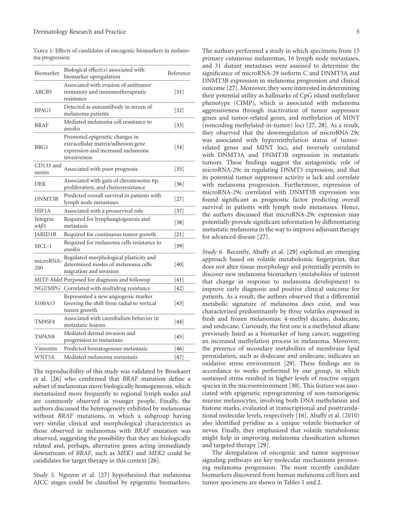
Table 1: Effects of candidates of oncogenic biomarkers in melano-ma progression.
BiomarkerBiological effect(s) associated withbiomarker upregulation
Reference
ABCB5Associated with evasion of antitumorimmunity and immunotherapeuticresistance
[31]
BPAG1Detected as autoantibody in serum ofmelanoma patients
[32]
BRAFMediated melanoma cell resistance toanoikis
[33]
BRG1
Promoted epigenetic changes inextracellular matrix/adhesion geneexpression and increased melanomainvasiveness
[34]
CD133 andnestin
Associated with poor prognosis [35]
DEKAssociated with gain of chromosome 6p,proliferation, and chemoresistance
[36]
DNMT3BPredicted overall survival in patients withlymph node metastases
[27]
HIF1A Associated with a prosurvival role [37]
Integrinα4β1
Required for lymphangiogenesis andmetastasis
[38]
JARID1B Required for continuous tumor growth [21]
MCL-1Required for melanoma cells resistance toanoikis
[39]
microRNA-200
Regulated morphological plasticity anddetermined modes of melanoma cellsmigration and invasion
[40]
MITF-Mdel Purposed for diagnosis and followup [41]
NG2/MPG Correlated with multidrug resistance [42]
S100A13Represented a new angiogenic markerfavoring the shift from radial to verticaltumor growth
[43]
TM9SF4Associated with cannibalism behavior inmetastatic lesions
[44]
TSPAN8Mediated dermal invasion andprogression to metastasis
[45]
Vimentin Predicted hematogenous metastasis [46]
WNT5A Mediated melanoma metastasis [47]
The reproducibility of this study was validated by Broekaertet al. [26] who confirmed that BRAF mutation define asubset of melanomas more biologically homogeneous, whichmetastasized more frequently to regional lymph nodes andare commonly observed in younger people. Finally, theauthors discussed the heterogeneity exhibited by melanomaswithout BRAF mutations, in which a subgroup havingvery similar clinical and morphological characteristics asthose observed in melanomas with BRAF mutation wasobserved, suggesting the possibility that they are biologicallyrelated and, perhaps, alternative genes acting immediatelydownstream of BRAF, such as MEK1 and MEK2 could becandidates for target therapy in this context [26].
Study 5. Nguyen et al. [27] hypothesized that melanomaAJCC stages could be classified by epigenetic biomarkers.
The authors performed a study in which specimens from 15primary cutaneous melanomas, 16 lymph node metastases,and 31 distant metastases were assessed to determine thesignificance of microRNA-29 isoform C and DNMT3A andDNMT3B expression in melanoma progression and clinicaloutcome [27]. Moreover, they were interested in determiningtheir potential utility as hallmarks of CpG island methylatorphenotype (CIMP), which is associated with melanomaaggressiveness through inactivation of tumor suppressorgenes and tumor-related genes, and methylation of MINT(noncoding methylated-in-tumor) loci [27, 28]. As a result,they observed that the downregulation of microRNA-29cwas associated with hypermethylation status of tumor-related genes and MINT loci, and inversely correlatedwith DNMT3A and DNMT3B expression in metastatictumors. These findings suggest the antagonistic role ofmicroRNA-29c in regulating DNMT3 expression, and thatits potential tumor suppressor activity is lack and correlatewith melanoma progression. Furthermore, expression ofmicroRNA-29c correlated with DNMT3B expression wasfound significant as prognostic factor predicting overallsurvival in patients with lymph node metastases. Hence,the authors discussed that microRNA-29c expression maypotentially provide significant information by differentiatingmetastatic melanoma in the way to improve adjuvant therapyfor advanced disease [27].
Study 6. Recently, Abaffy et al. [29] exploited an emergingapproach based on volatile metabolomic fingerprint, thatdoes not alter tissue morphology and potentially permits todiscover new melanoma biomarkers (metabolites of interestthat change in response to melanoma development) toimprove early diagnosis and positive clinical outcome forpatients. As a result, the authors observed that a differentialmetabolic signature of melanoma does exist, and wascharacterized predominantly by three volatiles expressed infresh and frozen melanomas: 4-methyl decane, dodecane,and undecane. Curiously, the first one is a methylated alkanepreviously listed as a biomarker of lung cancer, suggestingan increased methylation process in melanoma. Moreover,the presence of secondary metabolites of membrane lipidperoxidation, such as dodecane and undecane, indicates anoxidative stress environment [29]. These findings are inaccordance to works performed by our group, in whichsustained stress resulted in higher levels of reactive oxygenspecies in the microenvironment [30]. This feature was asso-ciated with epigenetic reprogramming of non-tumorigenicmurine melanocytes, involving both DNA methylation andhistone marks, evaluated at transcriptional and posttransla-tional molecular levels, respectively [16]. Abaffy et al. (2010)also identified pyridine as a unique volatile biomarker ofnevus. Finally, they emphasized that volatile metabolomicmight help in improving melanoma classification schemesand targeted therapy [29].
The deregulation of oncogenic and tumor suppressorsignaling pathways are key molecular mechanisms promot-ing melanoma progression. The most recently candidatebiomarkers discovered from human melanoma cell lines andtumor specimens are shown in Tables 1 and 2.
6 Dermatology Research and Practice
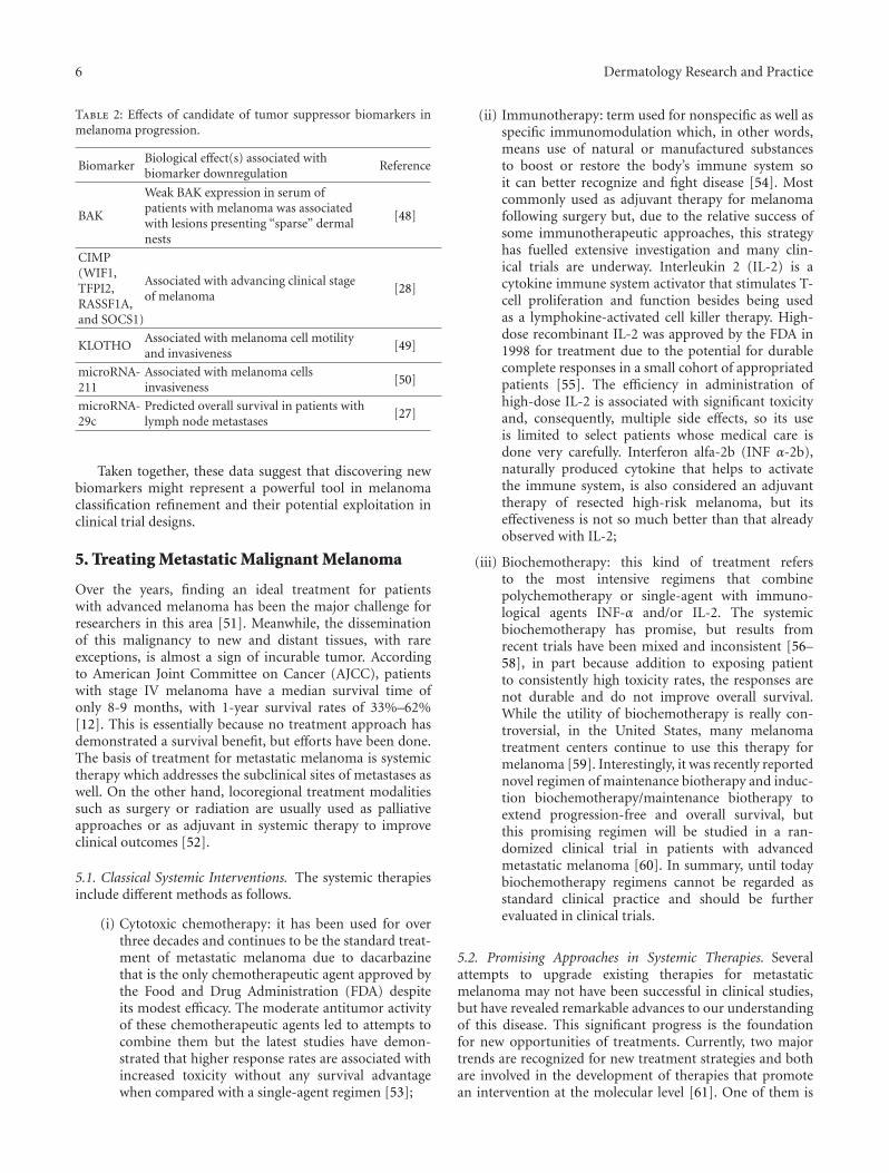
Table 2: Effects of candidate of tumor suppressor biomarkers inmelanoma progression.
BiomarkerBiological effect(s) associated withbiomarker downregulation
Reference
BAK
Weak BAK expression in serum ofpatients with melanoma was associatedwith lesions presenting “sparse” dermalnests
[48]
CIMP(WIF1,TFPI2,RASSF1A,and SOCS1)
Associated with advancing clinical stageof melanoma
[28]
KLOTHOAssociated with melanoma cell motilityand invasiveness
[49]
microRNA-211
Associated with melanoma cellsinvasiveness
[50]
microRNA-29c
Predicted overall survival in patients withlymph node metastases
[27]
Taken together, these data suggest that discovering newbiomarkers might represent a powerful tool in melanomaclassification refinement and their potential exploitation inclinical trial designs.
5. Treating Metastatic Malignant Melanoma
Over the years, finding an ideal treatment for patientswith advanced melanoma has been the major challenge forresearchers in this area [51]. Meanwhile, the disseminationof this malignancy to new and distant tissues, with rareexceptions, is almost a sign of incurable tumor. Accordingto American Joint Committee on Cancer (AJCC), patientswith stage IV melanoma have a median survival time ofonly 8-9 months, with 1-year survival rates of 33%–62%[12]. This is essentially because no treatment approach hasdemonstrated a survival benefit, but efforts have been done.The basis of treatment for metastatic melanoma is systemictherapy which addresses the subclinical sites of metastases aswell. On the other hand, locoregional treatment modalitiessuch as surgery or radiation are usually used as palliativeapproaches or as adjuvant in systemic therapy to improveclinical outcomes [52].
5.1. Classical Systemic Interventions. The systemic therapiesinclude different methods as follows.
(i) Cytotoxic chemotherapy: it has been used for overthree decades and continues to be the standard treat-ment of metastatic melanoma due to dacarbazinethat is the only chemotherapeutic agent approved bythe Food and Drug Administration (FDA) despiteits modest efficacy. The moderate antitumor activityof these chemotherapeutic agents led to attempts tocombine them but the latest studies have demon-strated that higher response rates are associated withincreased toxicity without any survival advantagewhen compared with a single-agent regimen [53];
(ii) Immunotherapy: term used for nonspecific as well asspecific immunomodulation which, in other words,means use of natural or manufactured substancesto boost or restore the body’s immune system soit can better recognize and fight disease [54]. Mostcommonly used as adjuvant therapy for melanomafollowing surgery but, due to the relative success ofsome immunotherapeutic approaches, this strategyhas fuelled extensive investigation and many clin-ical trials are underway. Interleukin 2 (IL-2) is acytokine immune system activator that stimulates T-cell proliferation and function besides being usedas a lymphokine-activated cell killer therapy. High-dose recombinant IL-2 was approved by the FDA in1998 for treatment due to the potential for durablecomplete responses in a small cohort of appropriatedpatients [55]. The efficiency in administration ofhigh-dose IL-2 is associated with significant toxicityand, consequently, multiple side effects, so its useis limited to select patients whose medical care isdone very carefully. Interferon alfa-2b (INF α-2b),naturally produced cytokine that helps to activatethe immune system, is also considered an adjuvanttherapy of resected high-risk melanoma, but itseffectiveness is not so much better than that alreadyobserved with IL-2;
(iii) Biochemotherapy: this kind of treatment refersto the most intensive regimens that combinepolychemotherapy or single-agent with immuno-logical agents INF-α and/or IL-2. The systemicbiochemotherapy has promise, but results fromrecent trials have been mixed and inconsistent [56–58], in part because addition to exposing patientto consistently high toxicity rates, the responses arenot durable and do not improve overall survival.While the utility of biochemotherapy is really con-troversial, in the United States, many melanomatreatment centers continue to use this therapy formelanoma [59]. Interestingly, it was recently reportednovel regimen of maintenance biotherapy and induc-tion biochemotherapy/maintenance biotherapy toextend progression-free and overall survival, butthis promising regimen will be studied in a ran-domized clinical trial in patients with advancedmetastatic melanoma [60]. In summary, until todaybiochemotherapy regimens cannot be regarded asstandard clinical practice and should be furtherevaluated in clinical trials.
5.2. Promising Approaches in Systemic Therapies. Severalattempts to upgrade existing therapies for metastaticmelanoma may not have been successful in clinical studies,but have revealed remarkable advances to our understandingof this disease. This significant progress is the foundationfor new opportunities of treatments. Currently, two majortrends are recognized for new treatment strategies and bothare involved in the development of therapies that promotean intervention at the molecular level [61]. One of them is
Dermatology Research and Practice 7
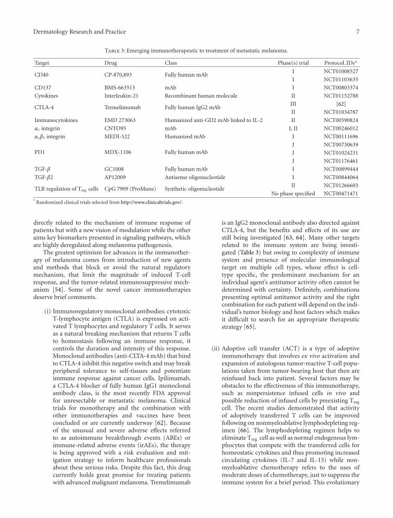
Table 3: Emerging immunotherapeutic to treatment of metastatic melanoma.
Target Drug Class Phase(s) trial Protocol IDs∗
CD40 CP-870,893 Fully human mAbI NCT01008527
I NCT01103635
CD137 BMS-663513 mAb I NCT00803374
Cytokines Interleukin-21 Recombinant human molecule II NCT01152788
CTLA-4 Tremelimumab Fully human IgG2 mAbIII [62]
II NCT01034787
Immunocytokines EMD 273063 Humanized anti-GD2 mAb linked to IL-2 II NCT00590824
αv integrin CNTO95 mAb I, II NCT00246012
αvβ3 integrin MEDI-522 Humanized mAb I NCT00111696
PD1 MDX-1106 Fully human mAbI NCT00730639
I NCT01024231
I NCT01176461
TGF-β GC1008 Fully human mAb I NCT00899444
TGF-β2 AP12009 Antisense oligonucleotide I NCT00844064
TLR regulation of Treg cells CpG 7909 (ProMune) Synthetic oligonucleotideII NCT01266603
No phase specified NCT00471471∗
Randomized clinical trials selected from http://www.clinicaltrials.gov/.
directly related to the mechanism of immune response ofpatients but with a new vision of modulation while the otheraims key biomarkers presented in signaling pathways, whichare highly deregulated along melanoma pathogenesis.
The greatest optimism for advances in the immunother-apy of melanoma comes from introduction of new agentsand methods that block or avoid the natural regulatorymechanism, that limit the magnitude of induced T-cellresponse, and the tumor-related immunosuppressive mech-anism [54]. Some of the novel cancer immunotherapiesdeserve brief comments.
(i) Immunoregulatory monoclonal antibodies: cytotoxicT-lymphocyte antigen (CTLA) is expressed on acti-vated T lymphocytes and regulatory T cells. It servesas a natural breaking mechanism that returns T cellsto homeostasis following an immune response, itcontrols the duration and intensity of this response.Monoclonal antibodies (anti-CLTA-4 mAb) that bindto CTLA-4 inhibit this negative switch and may breakperipheral tolerance to self-tissues and potentiateimmune response against cancer cells. Ipilimumab,a CTLA-4 blocker of fully human IgG1 monoclonalantibody class, is the most recently FDA approvalfor unresectable or metastatic melanoma. Clinicaltrials for monotherapy and the combination withother immunotherapies and vaccines have beenconcluded or are currently underway [62]. Becauseof the unusual and severe adverse effects referredto as autoimmune breakthrough events (ABEs) orimmune-related adverse events (irAEs), the therapyis being approved with a risk evaluation and mit-igation strategy to inform healthcare professionalsabout these serious risks. Despite this fact, this drugcurrently holds great promise for treating patientswith advanced malignant melanoma. Tremelimumab
is an IgG2 monoclonal antibody also directed againstCTLA-4, but the benefits and effects of its use arestill being investigated [63, 64]. Many other targetsrelated to the immune system are being investi-gated (Table 3) but owing to complexity of immunesystem and presence of molecular immunologicaltarget on multiple cell types, whose effect is cell-type specific, the predominant mechanism for anindividual agent’s antitumor activity often cannot bedetermined with certainty. Definitely, combinationspresenting optimal antitumor activity and the rightcombination for each patient will depend on the indi-vidual’s tumor biology and host factors which makesit difficult to search for an appropriate therapeuticstrategy [65].
(ii) Adoptive cell transfer (ACT) is a type of adoptiveimmunotherapy that involves ex vivo activation andexpansion of autologous tumor-reactive T-cell popu-lations taken from tumor-bearing host that then arereinfused back into patient. Several factors may beobstacles to the effectiveness of this immunotherapy,such as nonpersistence infused cells in vivo andpossible reduction of infused cells by preexisting Treg
cell. The recent studies demonstrated that activityof adoptively transferred T cells can be improvedfollowing on nonmyeloablative lymphodepleting reg-imen [66]. The lymphodepleting regimen helps toeliminate Treg cell as well as normal endogenous lym-phocytes that compete with the transferred cells forhomeostatic cytokines and thus promoting increasedcirculating cytokines (IL-7 and IL-15) while non-myeloablative chemotherapy refers to the uses ofmoderate doses of chemotherapy, just to suppress theimmune system for a brief period. This evolutionary
8 Dermatology Research and Practice
Table 4: Recent report vaccine approaches to advanced malignant melanoma.
Phase trial Purpose and brief comments Reference
II
Treatment of 54 patients with a patient-specific tumor cell vaccines consisting ofautologous dendritic cells, incubated with IL-4 and suspended in GM-CSF, whichhad phagocytized irradiated tumor cells from an autologous tumor cell line.Treatment was well-tolerated and the projected 5-year survival rate is an impressive54% at a median followup of 4.5 years for the 30 surviving patients.
[74]
II
Immunization of unresectable stage III or stage IV M1a melanoma patients withrecombinant MAGE-A3 protein combined with adjuvant systems AS15 or AS02B.The combination of MAGE-A3 and AS15 yielded higher specific Ab titers, morerobust T-cell induction and long-lasting clinical responses or SD in metastaticmelanoma.
[75]
II
To identify markers predictive of the clinical activity of the MAGE-A3antigen-specific cancer immunotherapeutic (ASCI) from gene expression profilingby microarrays. A gene-signature derived from pretreatment tumor biopsies hasbeen developed and shown to predict clinical benefit.
[76]
II
Routinely intratumoral injections of OncoVEXGM−CSF, an oncolytic herpes simplexvirus vector encoding granulocyte monocyte colony-stimulating factor (GM-CSF).It was observed an improvement rate and durability of response when compared toother treatment options available to patients with advanced melanoma.
[77]
IIILearn more about the safety and risks of using OncoVEXGM−CSF. Results of a phaseII trial of OncoVEXGM−CSF were encouraging and led to the design of this Phase IIItrial.
NCT00769704∗
III
Comparative study between metastatic melanoma patients treated with gp100:209–217(210M) peptide followed by high-dose IL-2 and high-dose IL-2 alone. Thepeptide vaccine plus HD IL-2 promoted significant improvement inprogression-free survival (PFS) without a clear impact on survival.
[78]
∗Clinical trials selected from http://www.clinicaltrials.gov/.
technique to treat cancer has demonstrated signifi-cant progress, mediating objective tumor regressionsin significant percentage of patients who stayed onthis treatment [67]; however, it is important tomention it is not available as standard treatmentfor advanced melanoma. Many studies are underway,and the new investigations are focusing the transferof T cell genetically modified to express melanoma-specific T-cell receptors [68].
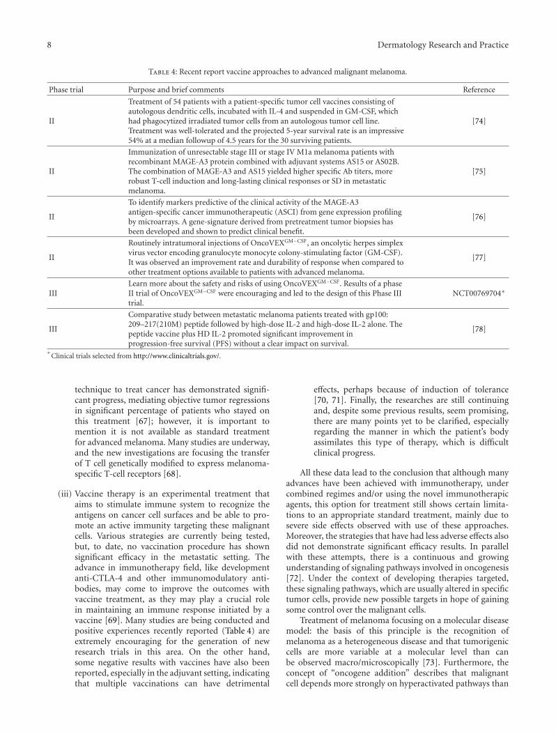
(iii) Vaccine therapy is an experimental treatment thataims to stimulate immune system to recognize theantigens on cancer cell surfaces and be able to pro-mote an active immunity targeting these malignantcells. Various strategies are currently being tested,but, to date, no vaccination procedure has shownsignificant efficacy in the metastatic setting. Theadvance in immunotherapy field, like developmentanti-CTLA-4 and other immunomodulatory anti-bodies, may come to improve the outcomes withvaccine treatment, as they may play a crucial rolein maintaining an immune response initiated by avaccine [69]. Many studies are being conducted andpositive experiences recently reported (Table 4) areextremely encouraging for the generation of newresearch trials in this area. On the other hand,some negative results with vaccines have also beenreported, especially in the adjuvant setting, indicatingthat multiple vaccinations can have detrimental
effects, perhaps because of induction of tolerance[70, 71]. Finally, the researches are still continuingand, despite some previous results, seem promising,there are many points yet to be clarified, especiallyregarding the manner in which the patient’s bodyassimilates this type of therapy, which is difficultclinical progress.
All these data lead to the conclusion that although manyadvances have been achieved with immunotherapy, undercombined regimes and/or using the novel immunotherapicagents, this option for treatment still shows certain limita-tions to an appropriate standard treatment, mainly due tosevere side effects observed with use of these approaches.Moreover, the strategies that have had less adverse effects alsodid not demonstrate significant efficacy results. In parallelwith these attempts, there is a continuous and growingunderstanding of signaling pathways involved in oncogenesis[72]. Under the context of developing therapies targeted,these signaling pathways, which are usually altered in specifictumor cells, provide new possible targets in hope of gainingsome control over the malignant cells.
Treatment of melanoma focusing on a molecular diseasemodel: the basis of this principle is the recognition ofmelanoma as a heterogeneous disease and that tumorigeniccells are more variable at a molecular level than canbe observed macro/microscopically [73]. Furthermore, theconcept of “oncogene addition” describes that malignantcell depends more strongly on hyperactivated pathways than
Dermatology Research and Practice 9
Table 5: Principal and secundary melanoma molecular subtypes [73].
Detailed subtypes Pathway(s) Key gene/biomarker(s) Potentially relevant therapeutics
1.1 MAPK BRAFBRAF inhibitors, MEK inhibitors, Hsp90inhibitors
1.2 MAPK BRAF/PTEN(BRAF inhibitors) AND (PI3K inhibitors, AKTinhibitors or mTOR inhibitors)
1.3 MAPK BRAF/AKT(BRAF inhibitors) AND (AKT inhibitors ormTOR inhibitors)
1.4 BRAF/CDK4 BRAF inhibitors AND CDK inhibitors
2.1 c-KIT c-KIT Gleevec & other c-KIT inhibitors
3.1 GNAQ GNA11 GNAQ MEK inhibitors
3.2 GNAQ GNA11 GNA11 MEK inhibitors
4.1 NRAS NRASMAPK & PI3K inhibitors, Farnesyl transferaseinhibitors
5.1 MITF MITF HDAC inhibitors
6.1 AKT/PI3K PTENPI3K inhibitors, AKT inhibitors or mTORinhibitors
6.2 AKT/PI3K AKT AKT inhibitors or mTOR inhibitors
6.3 AKT/PI3K PI3KPI3K inhibitors, AKT inhibitors or mTORinhibitors
7.1 CDK ARF/INK4 CDK inhibitors
7.2 CDK CDK4 CDK inhibitors
7.3 CDK CCND1/Cyclin D1 CDK inhibitors
8.1 PR3/BCL BCL-2 TBD
8.2 PR3/BCL P53 TBD
9 Placeholder for any new subtype of patients that is not currently defined
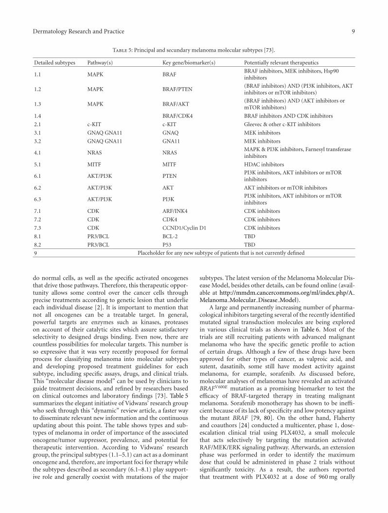
do normal cells, as well as the specific activated oncogenesthat drive those pathways. Therefore, this therapeutic oppor-tunity allows some control over the cancer cells throughprecise treatments according to genetic lesion that underlieeach individual disease [2]. It is important to mention thatnot all oncogenes can be a treatable target. In general,powerful targets are enzymes such as kinases, proteaseson account of their catalytic sites which assure satisfactoryselectivity to designed drugs binding. Even now, there arecountless possibilities for molecular targets. This number isso expressive that it was very recently proposed for formalprocess for classifying melanoma into molecular subtypesand developing proposed treatment guidelines for eachsubtype, including specific assays, drugs, and clinical trials.This “molecular disease model” can be used by clinicians toguide treatment decisions, and refined by researchers basedon clinical outcomes and laboratory findings [73]. Table 5summarizes the elegant initiative of Vidwans’ research groupwho seek through this “dynamic” review article, a faster wayto disseminate relevant new information and the continuousupdating about this point. The table shows types and sub-types of melanoma in order of importance of the associatedoncogene/tumor suppressor, prevalence, and potential fortherapeutic intervention. According to Vidwans’ researchgroup, the principal subtypes (1.1–5.1) can act as a dominantoncogene and, therefore, are important foci for therapy whilethe subtypes described as secondary (6.1–8.1) play support-ive role and generally coexist with mutations of the major
subtypes. The latest version of the Melanoma Molecular Dis-ease Model, besides other details, can be found online (avail-able at http://mmdm.cancercommons.org/ml/index.php/AMelanoma Molecular Disease Model).
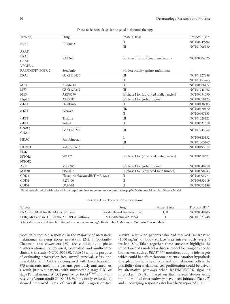
A large and permanently increasing number of pharma-cological inhibitors targeting several of the recently identifiedmutated signal transduction molecules are being exploredin various clinical trials as shown in Table 6. Most of thetrials are still recruiting patients with advanced malignantmelanoma who have the specific genetic profile to actionof certain drugs. Although a few of these drugs have beenapproved for other types of cancer, as valproic acid, andsutent, dasatinib, some still have modest activity againstmelanoma, for example, sorafenib. As discussed before,molecular analyses of melanomas have revealed an activatedBRAFV600E mutation as a promising biomarker to test theefficacy of BRAF-targeted therapy in treating malignantmelanoma. Sorafenib monotherapy has shown to be ineffi-cient because of its lack of specificity and low potency againstthe mutant BRAF [79, 80]. On the other hand, Flahertyand coauthors [24] conducted a multicenter, phase 1, dose-escalation clinical trial using PLX4032, a small moleculethat acts selectively by targeting the mutation activatedRAF/MEK/ERK signaling pathway. Afterwards, an extensionphase was performed in order to identify the maximumdose that could be administered in phase 2 trials withoutsignificantly toxicity. As a result, the authors reportedthat treatment with PLX4032 at a dose of 960 mg orally
10 Dermatology Research and Practice
Table 6: Selected drugs for targeted melanoma therapy.
Target(s) Drug Phase(s) trial Protocol IDs∗
BRAF PLX4032II NCT00949702
III NCT01006980
ARAF
RAF265 In Phase 1 for malignant melanoma NCT00304525BRAF
CRAF
VEGFR-2
RAFPDGFRVEGFR-2 Sorafenib Modest activity against melanoma —
BRAF GSK2118436 III NCT01227889
II NCT01153763
MEK AZD6244 II NCT00866177
MEK GSK1120212 III NCT01245062
MEK AZD8330 In phase I for (advanced malignancies) NCT00454090
Hsp90 AT13387 In phase I for (solid tumors) NCT00878423
c-KIT Dasatinib II NCT00436605
c-KIT GleevecIII, NCT00470470
II NCT00667953
c-KIT Tasigna III NCT01028222
c-KIT Sutent II NCT00631618
GNAQGSK1120212 III NCT01245062
GNA11
HDAC PanobinostatI, NCT00925132
III NCT01065467
HDAC1 Valproic acid I NCT00495872
PI3KSF1126 In phase I for (advanced malignancies) NCT00658671MTOR1
MTOR2
AKT MK2206 In phase I for (solid tumors) NCT00848718
MTOR OSI-027 In phase I for (advanced solid tumors) NCT00698243
CDK4 Flavopiridol/alvocidib/HMR 1275 II NCT00005971
CDK4 P276-00 II NCT00835419
CDK4 UCN-01 II NCT00072189∗
Randomized clinical trials selected from http://mmdm.cancercommons.org/ml/index.php/A Melanoma Molecular Disease Model.
Table 7: Dual Therapeutic intervention.
Targets Drug Phase(s) trial Protocol IDs∗
BRAF and MEK for the MAPK pathway Sorafenib and Temsirolimus I, II NCT00349206
PI3K, AKT and mTOR for the AKT/PI3K pathway MK2206 plus AZD6244 I NCT01021748∗
Clinical trials selected from http://mmdm.cancercommons.org/ml/index.php/A Melanoma Molecular Disease Model.
twice daily induced responses in the majority of metastaticmelanomas carrying BRAF mutations [24]. Importantly,Chapman and coworkers [80] are conducting a phase3 interventional, randomized, controlled and multicenterclinical trial study (NCT01006980, Table 6) with the purposeof evaluating progression-free, overall survival, safety andtolerability of PLX4032 as compared with Dacarbazine in675 metastatic melanoma patients previously untreated. Asa result just yet, patients with unresectable stage IIIC orstage IV melanoma (AJCC) positive for BRAFV600E mutationreceiving Vemurafenib (PLX4032, 960 mg orally twice daily)showed improved rates of overall and progression-free
survival relative to patients who had received Dacarbazine(1000 mg/m2 of body surface area intravenously every 3weeks) [80]. Taken together, these successes highlight theimportance of a molecular disease model focusing on specificbiomarkers, such as BRAFV600E mutation, as bona fide targetswhich could benefit melanoma patients. Another hypothesisto explain low activity of Sorafenib in melanoma cells is thepossibility that melanoma cell proliferation could be drivenby alternative pathways when RAF/MEK/ERK signalingis blocked [79, 81]. Based on this, several studies usinginhibitors of distinct pathways have been initiated (Table 7)and encouraging response rates have been reported [82].
Dermatology Research and Practice 11
The amount of studies presented here and the emergenceof new therapeutic opportunities show that the scientificcommunity is really committed to finding the best way toovercome this challenge, the treatment for metastatic malig-nant melanoma. Since melanoma is much more than a singledisease, the real benefit most likely will be require the useof multiple strategies together (chemotherapy, immunother-apy, targeted therapy). But if on one hand the positivesynergistic effect of these agents is achieved by the other, anextra burden of toxicity and unknown events arising fromthe interaction of these agents cannot be ignored. It is knownthat, besides the intrinsic characteristics of each person, thebiology of melanoma, as well as other types of cancer, has alsoa few distinct characteristics even among patients in samedisease stage, which leads us to another challenge, beforetreatment choice: identifying the tumors and patients thatbest suited to respond to certain therapies.
6. Conclusion
Recent discoveries have provided fascinating insights intomalignant melanoma development. It is clear that dereg-ulation in distinct oncogenes and tumor suppressors areinvolved in malignant melanoma progression, as evaluatedby results of compiled studies presented here. Thus, there areevidences in which more than one route leads to melanocytefull malignant transformation and melanoma progression.The major question to be considered by researchers,clinicians, and dermatopathologists is the limited successof discovered biomarkers to predict or identify groupsof patients with substantial risk for metastasis. Unfortu-nately, no reliable biomarker that significantly translatesinto effectiveness therapeutic responses and overall mediansurvival rate benefits to patients with advanced disease hasbeen identified. Perhaps, this characteristic can be due tomelanoma heterogeneity, substantial differences in experi-mental and clinical trials design, inadequate classificationschemes, environmental-associated risk factors, and person’scondition such as age, gender, and immune competence.Although a true biomarker is a molecule capable of dis-tinguishing a specific stage of disease, the data reportedhere strongly suggest that understanding primary clonalevents that occur in primary malignant melanomas throughmelanoma classification refinement may help us to identifymolecular marks acting immediately downstream. Thesesecondary alterations provide new insights for predicting themetastatic behavior in a more stable genomic, epigenomic,and metabolomic context. Hence, additional studies arerequired to identify distinct subpopulations of melanomacells, including circulating tumor-initiating cells in attemptto identify these distinct molecular pathways and classify,in a more reliable way, patient candidates for further per-sonalized medicine. Recently, Vidwans’ group (2011) devel-oped a molecular disease model that classifies melanomasinto molecular subtypes based on genetic status of keybiomarker(s)/pathway(s) and their combination, which arepotential targets for existing therapeutic interventions [73].This molecular classification of melanomas might be apowerful strategy used to refine melanoma classification and
to guide appropriate target therapy. Thus, because melanomais a heterogeneous disease, biomarkers can allow us toidentify patients who best respond to certain therapeuticapproach. And therefore standard treatments are currentlyavailable to be based primarily on usage, together ornot, chemotherapies (dacarbazine) or/and imunotherapies(high-dose recommbinant IL-2, Ipilimumab), but require arigorous preclinical evaluation as well as a constant medicalsupervision. On the other hand, another barrier for studyingand treating melanoma metastasis is the limited availabilityof fresh melanoma tissues representing early stages of diseaseand adjacent normal skin. Because of this, different sourcescan be exploited to initial screening, such as an animal modelthat mimicries the “natural” evolution of disease. In this way,a murine melanoma progression model developed by ourgroup [83] has shown good correlation with data reportedin humans, especially with respect to epigenetic therapy,highlighting the potential translational application from thebasic research to clinical research, which might be especiallyimportant in metastatic disease [16]. In conclusion, thecharacterization of distinct subpopulations of melanomacells and signaling pathways by integrating classificationscheme opens the avenue to the development of moreresponsive antimelanoma therapy.
Acknowledgments
The authors would like to thank Universidade Federal de SaoPaulo and Fundacao de Amparo a Pesquisa do Estado deSao Paulo (09/51462-9, to C. F. Souza; 09/03335-8, to A. S.Morais; 06/61293-1, to M.G.Jasiulionis). C. F. Souza and A.S. Morais contributed equally to this review.
References
[1] G. E. De Schweinitz and E. A. Shumway, “Concerningmelanoma of the choroid with report of one case of thischaracter and of another exhibiting a pigmented sarcomaof the choroid early in its development,” Transactions of theAmerican Ophthalmological Society, vol. 10, part 3, pp. 439–451, 1905.
[2] V. Gray-Schopfer, C. Wellbrock, and R. Marais, “Melanomabiology and new targeted therapy,” Nature, vol. 445, no. 7130,pp. 851–857, 2007.
[3] L. Hou and W. J. Pavan, “Transcriptional and signalingregulation in neural crest stem cell-derived melanocyte devel-opment: do all roads lead to Mitf?” Cell Research, vol. 18, no.12, pp. 1163–1176, 2008.
[4] W. H. Clark Jr., L. From, E. A. Bernardino, and M. C. Mihm,“The histogenesis and biologic behavior of primary humanmalignant melanomas of the skin,” Cancer Research, vol. 29,no. 3, pp. 705–727, 1969.
[5] R. J. Reed, “Acral lentiginous melanoma,” in New Concepts inSurgical Pathology of the Skin, pp. 89–90, Wiley, New York, NY,USA, 1979.
[6] W. H. Clark Jr., D. E. Elder, and D. Guerry, “A study of tumorprogression: the precursor lesions of superficial spreading andnodular melanoma,” Human Pathology, vol. 15, no. 12, pp.1147–1165, 1984.
12 Dermatology Research and Practice
[7] C. Gaggioli and E. Sahai, “Melanoma invasion—currentknowledge and future directions,” Pigment Cell Research, vol.20, no. 3, pp. 161–172, 2007.
[8] S. E. Zabierowski and M. Herlyn, “Melanoma stem cells: thedark seed of melanoma,” Journal of Clinical Oncology, vol. 26,no. 17, pp. 2890–2894, 2008.
[9] A. P. Feinberg, R. Ohlsson, and S. Henikoff, “The epigeneticprogenitor origin of human cancer,” Nature Reviews Genetics,vol. 7, no. 1, pp. 21–33, 2006.
[10] K. A. Parker, S. Glaysher, M. Polak et al., “The molecular basisof the chemosensitivity of metastatic cutaneous melanoma tochemotherapy,” Journal of Clinical Pathology, vol. 63, no. 11,pp. 1012–1020, 2010.
[11] M. S. Soengas and S. W. Lowe, “Apoptosis and melanomachemoresistance,” Oncogene, vol. 22, no. 20, pp. 3138–3151,2003.
[12] C. M. Balch, J. E. Gershenwald, S. J. Soong et al., “Final versionof 2009 AJCC melanoma staging and classification,” Journal ofClinical Oncology, vol. 27, no. 36, pp. 6199–6206, 2009.
[13] E. D. Pleasance, R. K. Cheetham, P. J. Stephens et al., “Acomprehensive catalogue of somatic mutations from a humancancer genome,” Nature, vol. 463, no. 7278, pp. 191–196, 2010.
[14] S. Sharma, T. K. Kelly, and P. A. Jones, “Epigenetics in cancer,”Carcinogenesis, vol. 31, no. 1, pp. 27–36, 2009.
[15] L. Sigalotti, A. Covre, E. Fratta et al., “Epigenetics of humancutaneous melanoma: setting the stage for new therapeuticstrategies,” Journal of Translational Medicine, vol. 8, article 56,2010.
[16] F. Molognoni, A. T. Cruz, F. M. Meliso et al., “Epigeneticreprogramming as a key contributor to melanocyte malignanttransformation,” Epigenetics, vol. 6, no. 4, pp. 451–465, 2011.
[17] R. P. Gallagher, A. C. MacArthur, T. K. Lee et al., “Plasma levelsof polychlorinated biphenyls and risk of cutaneous malignantmelanoma: a preliminary study,” International Journal ofCancer, vol. 128, no. 8, pp. 1872–1880, 2011.
[18] V. J. McGovern, A. J. Cochran, and E. P. Van der Esch,“The classification of malignant melanoma, its histologicalreporting and registration: a revision of the 1972 Sydneyclassification,” Pathology, vol. 18, no. 1, pp. 12–21, 1986.
[19] C. M. Balch, “Cutaneous melanoma: prognosis and treatmentresults worldwide,” Seminars in Surgical Oncology, vol. 8, no.6, pp. 400–414, 1992.
[20] J. A. Curtin, J. Fridlyand, T. Kageshita et al., “Distinct setsof genetic alterations in melanoma,” New England Journal ofMedicine, vol. 353, no. 20, pp. 2135–2147, 2005.
[21] A. Roesch, M. Fukunaga-Kalabis, E. C. Schmidt et al., “Atemporarily distinct subpopulation of slow-cycling melanomacells is required for continuous tumor growth,” Cell, vol. 141,no. 4, pp. 583–594, 2010.
[22] S. R. Alonso, P. Ortiz, M. Pollan et al., “Progression incutaneous malignant melanoma is associated with distinctexpression profiles: a tissue microarray-based study,” Ameri-can Journal of Pathology, vol. 164, no. 1, pp. 193–203, 2004.
[23] B. Sestakova, L. Ondrusova, and J. Vachtenheim, “Cell cycleinhibitor p21/WAF1/CIP1 as a cofactor of MITF expression inmelanoma cells,” Pigment Cell and Melanoma Research, vol. 23,no. 2, pp. 238–251, 2010.
[24] K. T. Flaherty, I. Puzanov, K. B. Kim et al., “Inhibitionof mutated, activated BRAF in metastatic melanoma,” NewEngland Journal of Medicine, vol. 363, no. 9, pp. 809–819, 2010.
[25] A. Viros, J. Fridlyand, J. Bauer et al., “Improving melanomaclassification by integrating genetic and morphologic fea-tures,” PLoS Medicine, vol. 5, no. 6, article e120, 2008.
[26] S. M. C. Broekaert, R. Roy, I. Okamoto et al., “Genetic andmorphologic features for melanoma classification,” PigmentCell and Melanoma Research, vol. 23, no. 6, pp. 763–770, 2010.
[27] T. Nguyen, C. Kuo, M. B. Nicholl et al., “Downregulation ofmicroRNA-29c is associated with hypermethylation of tumor-related genes and disease outcome in cutaneous melanoma,”Epigenetics, vol. 6, no. 3, pp. 388–394, 2011.
[28] A. Tanemura, A. M. Terando, M. S. Sim et al., “CpG Islandmethylator phenotype predicts progression of malignantmelanoma,” Clinical Cancer Research, vol. 15, no. 5, pp. 1801–1807, 2009.
[29] T. Abaffy, R. Duncan, D. D. Riemer et al., “Differential volatilesignatures from skin, naevi and melanoma: a novel approachto detect a pathological process,” PLoS ONE, vol. 5, no. 11,Article ID e13813, 2010.
[30] A. C. E. Campos, F. Molognoni, F. H. M. Melo et al., “Oxida-tive stress modulates DNA methylation during melanocyteanchorage blockade associated with malignant transforma-tion,” Neoplasia, vol. 9, no. 12, pp. 1111–1121, 2007.
[31] T. Schatton, U. Schutte, N. Y. Frank et al., “Modulation of T-cell activation by malignant melanoma initiating cells,” CancerResearch, vol. 70, no. 2, pp. 697–708, 2010.
[32] T. Shimbo, A. Tanemura, T. Yamazaki, K. Tamai, I. Katayama,and Y. Kaneda, “Serum anti-BPAG1 auto-antibody is a novelmarker for human melanoma,” PloS one, vol. 5, no. 5, ArticleID e10566, 2010.
[33] K. Boisvert-Adamo and A. E. Aplin, “Mutant B-RAF mediatesresistance to anoikis via Bad and Bim,” Oncogene, vol. 27, no.23, pp. 3301–3312, 2008.
[34] S. V. Saladi, B. Keenen, H. G. Marathe, H. Qi, K. V. Chin, andI. L. de la Serna, “Modulation of extracellular matrix/adhesionmolecule expression by BRG1 is associated with increasedmelanoma invasiveness,” Molecular Cancer, vol. 9, article 280,2010.
[35] A. Fusi, U. Reichelt, A. Busse et al., “Expression of the stemcell markers nestin and CD133 on circulating melanoma cells,”Journal of Investigative Dermatology, vol. 131, no. 2, pp. 487–494, 2011.
[36] M. S. Khodadoust, M. Verhaegen, F. Kappes et al., “Melanomaproliferation and chemoresistance controlled by the DEKoncogene,” Cancer Research, vol. 69, no. 16, pp. 6405–6413,2009.
[37] R. Busca, E. Berra, C. Gaggioli et al., “Hypoxia-induciblefactor 1α is a new target of microphthalmia- associatedtranscription factor (MITF) in melanoma cells,” Journal of CellBiology, vol. 170, no. 1, pp. 49–59, 2005.
[38] B. Garmy-Susini, C. J. Avraamides, M. C. Schmid et al.,“Integrin α4β1 signaling is required for lymphangiogenesisand tumor metastasis,” Cancer Research, vol. 70, no. 8, pp.3042–3051, 2010.
[39] K. Boisvert-Adamo, W. Longmate, E. V. Abel, and A. E. Aplin,“Mcl-1 is required for melanoma cell resistance to anoikis,”Molecular Cancer Research, vol. 7, no. 4, pp. 549–556, 2009.
[40] I. Elson-Schwab, A. Lorentzen, and C. J. Marshall, “Mic-roR-NA-200 family members differentially regulate morphologicalplasticity and mode of melanoma cell invasion,” PLoS ONE,vol. 5, no. 10, Article ID e13176, 2010.
[41] Y. Wang, S. Radfar, S. Liu, A. I. Riker, and H. T. Khong,“Mitf-Mdel, a novel melanocyte/melanoma-specific isoformof microphthalmia-associated transcription factor-M, as acandidate biomarker for melanoma,” BMC Medicine, vol. 8,article 14, 2010.
Dermatology Research and Practice 13
[42] M. Chekenya, C. Krakstad, A. Svendsen et al., “The progenitorcell marker NG2/MPG promotes chemoresistance by acti-vation of integrin-dependent PI3K/Akt signaling,” Oncogene,vol. 27, no. 39, pp. 5182–5194, 2008.
[43] D. Massi, M. Landriscina, A. Piscazzi et al., “S100A13 is a newangiogenic marker in human melanoma,” Modern Pathology,vol. 23, no. 6, pp. 804–813, 2010.
[44] F. Lozupone, M. Perdicchio, D. Brambilla et al., “The humanhomologue of Dictyostelium discoideum phg1A is expressedby human metastatic melanoma cells,” EMBO Reports, vol. 10,no. 12, pp. 1348–1354, 2009.
[45] O. Berthier-Vergnes, M. E. Kharbili, A. de La Fouchardiereet al., “Gene expression profiles of human melanoma cellswith different invasive potential reveal TSPAN8 as a novelmediator of invasion,” British Journal of Cancer, vol. 104, no.1, pp. 155–165, 2011.
[46] M. Li, B. Zhang, B. Sun et al., “A novel function for vimentin:the potential biomarker for predicting melanoma hematoge-nous metastasis,” Journal of Experimental and Clinical CancerResearch, vol. 29, no. 1, article 109, 2010.
[47] S. K. Dissanayake, M. Wade, C. E. Johnson et al., “The Wnt5A/protein kinase C pathway mediates motility in melanoma cellsvia the inhibition of metastasis suppressors and initiation ofan epithelial to mesenchymal transition,” Journal of BiologicalChemistry, vol. 282, no. 23, pp. 17259–17271, 2007.
[48] C. Longo, G. Gambara, V. Espina et al., “A novel biomarkerharvesting nanotechnology identifies Bak as a candidatemelanoma biomarker in serum,” Experimental Dermatology,vol. 20, no. 1, pp. 29–34, 2011.
[49] T. C. Camilli, M. Xu, M. P. O’Connell et al., “Loss ofKlotho during melanoma progression leads to increasedfilamin cleavage, increased Wnt5A expression, and enhancedmelanoma cell motility,” Pigment Cell and Melanoma Research,vol. 24, no. 1, pp. 175–186, 2011.
[50] J. Mazar, K. De Young, D. Khaitan et al., “The regulation ofmiRNA-211 expression and its role in melanoma cell invasive-ness,” PLoS ONE, vol. 5, no. 11, Article ID e13779, 2010.
[51] C. Garbe, T. K. Eigentler, U. Keilholz, A. Hauschild, and J.M. Kirkwood, “Systematic review of medical treatment inmelanoma: current status and future prospects,” Oncologist,vol. 16, no. 1, pp. 5–24, 2011.
[52] S. Bhatia, S. S. Tykodi, and J. A. Thompson, “Treatment ofmetastatic melanoma: an overview,” Oncology, vol. 23, no. 6,pp. 488–496, 2009.
[53] M. B. Lens and T. G. Eisen, “Systemic chemotherapy inthe treatment of malignant melanoma,” Expert Opinion onPharmacotherapy, vol. 4, no. 12, pp. 2205–2211, 2003.
[54] D. Schadendorf, S. M. Algarra, L. Bastholt et al., “Immuno-therapy of distant metastatic disease,” Annals of Oncology, vol.20, supplement 6, pp. 41–50, 2009.
[55] M. B. Atkins, M. T. Lotze, J. P. Dutcher et al., “High-doserecombinant interleukin 2 therapy for patients with metastaticmelanoma: analysis of 270 patients treated between 1985 and1993,” Journal of Clinical Oncology, vol. 17, no. 7, pp.2105–2116, 1999.
[56] O. Eton, S. S. Legha, A. Y. Bedikian et al., “Sequential bio-chemotherapy versus chemotherapy for metastatic melanoma:results from a phase III randomized trial,” Journal of ClinicalOncology, vol. 20, no. 8, pp. 2045–2052, 2002.
[57] N. J. Ives, R. L. Stowe, P. Lorigan, and K. Wheatley, “Chemo-therapy compared with biochemotherapy for the treatmentof metastatic melanoma: a meta-analysis of 18 trials involving2,621 patients,” Journal of Clinical Oncology, vol. 25, no. 34,pp. 5426–5434, 2007.
[58] M. B. Atkins, J. Hsu, S. Lee et al., “Phase III trial comparingconcurrent biochemotherapy with cisplatin, vinblastine,dacarbazine, interleukin-2, and interferon α-2b with cisplatin,vinblastine, and dacarbazine alone in patients with metastaticmalignant melanoma (E3695): a trial coordinated by theEastern Cooperative Oncology Group,” Journal of ClinicalOncology, vol. 26, no. 35, pp. 5748–5754, 2008.
[59] D. R. Minor, D. Moore, C. Kim et al., “Prognostic factors inmetastatic melanoma patients treated with biochemotherapyand maintenance immunotherapy,” Oncologist, vol. 14, no. 10,pp. 995–1002, 2009.
[60] S. J. O’Day, M. B. Atkins, P. Boasberg et al., “Phase II multi-center trial of maintenance biotherapy after induction con-current biochemotherapy for patients with metastatic mela-noma,” Journal of Clinical Oncology, vol. 27, no. 36, pp.6207–6212, 2009.
[61] K. T. Flaherty, “Chemotherapy and targeted therapy combi-nations in advanced melanoma,” Clinical Cancer Research,vol. 12, no. 7, part 2, pp. 2366s–2370s, 2006.
[62] F. S. Hodi, S. J. O’Day, D. F. McDermott et al., “Improvedsurvival with ipilimumab in patients with metastaticmelanoma,” New England Journal of Medicine, vol. 363, no. 8,pp. 711–723, 2010.
[63] A. Ribas, A. Hauschild, R. Kefford et al., “Phase III, open-label,randomized, comparative study of tremelimumab (CP-675,206) and chemotherapy (temozolomide [TMZ] or dacar-bazine [DTIC]) in patients with advanced melanoma,” Journalof Clinical Oncology, vol. 26, 2008, ASCO Meeting Abstracts:Abstr LBA9011.
[64] J. M. Kirkwood, P. Lorigan, P. Hersey et al., “Phase II trialof tremelimumab (CP-675,206) in patients with advancedrefractory or relapsed melanoma,” Clinical Cancer Research,vol. 16, no. 3, pp. 1042–1048, 2010.
[65] M. Sznol, “Promising immunotherapeutic approaches to thetreatment of metastatic melanoma: modulation of the im-mune response,” Community Oncology, vol. 5, no. 3 SUPPL.,pp. 14–22, 2008.
[66] S. A. Rosenberg, N. P. Restifo, J. C. Yang, R. A. Morgan,and M. E. Dudley, “Adoptive cell transfer: a clinical path toeffective cancer immunotherapy,” Nature Reviews Cancer, vol.8, no. 4, pp. 299–308, 2008.
[67] S. A. Rosenberg and M. E. Dudley, “Adoptive cell therapy forthe treatment of patients with metastatic melanoma,” CurrentOpinion in Immunology, vol. 21, no. 2, pp. 233–240, 2009.
[68] N. N. Hunder, H. Wallen, J. Cao et al., “Treatment of meta-static melanoma with autologous CD4+ T cells againstNY-ESO-1,” New England Journal of Medicine, vol. 358, no.25, pp. 2698–2703, 2008.
[69] A. M. M. Eggermont, “Advances in systemic treatment ofmelanoma,” Annals of Oncology, vol. 21, no. 7, pp. 339–344,2010.
[70] D. L. Morton, N. Mozzillo, J. F. Thompson et al., “An inter-national, randomized, phase III trial of bacillus Calmette-Guerin (BCG) plus allogeneic melanoma vaccine (MCV) orplacebo after complete resection of melanoma metastatic toregional or distant sites,” Journal of Clinical Oncology, vol. 25,2007, ASCO Meeting Abstracts: Abstr 8508.
[71] M. B. Faries, E. C. Hsueh, X. Ye, M. Hoban, and D. L. Morton,“Effect of granulocyte/macrophage colony-stimulating factoron vaccination with an allogeneic whole-cell melanomavaccine,” Clinical Cancer Research, vol. 15, no. 22, pp.7029–7035, 2009.
14 Dermatology Research and Practice
[72] K. T. Flaherty, “Combination therapy: key data and ongoingstudies combining targeted and cytotoxic agents,” CommunityOncology, vol. 5, no. 3 SUPPL., pp. 23–30, 2008.
[73] S. J. Vidwans, K. T. Flaherty, D. E. Fisher, J. M. Tenenbaum,M. D. Travers, and J. Shrager, “A melanoma molecular diseasemodel,” PLoS ONE, vol. 6, no. 3, Article ID e18257, 2011.
[74] R. O. Dillman, S. R. Selvan, P. M. Schiltz et al., “Phase II trialof dendritic cells loaded with antigens from self-renewing,proliferating autologous tumor cells as patient-specificantitumor vaccines in patients with metastatic melanoma:final report,” Cancer Biotherapy and Radiopharmaceuticals,vol. 24, no. 3, pp. 311–319, 2009.
[75] W. H. Kruit, S. Suciu, B. Dreno et al., “Immunization withrecombinant MAGE-A3 protein combined with adjuvantsystems AS15 or AS02B in patients with unresectable andprogressive metastatic cutaneous melanoma: a randomizedopen-label phase II study of the EORTC Melanoma Group(16032–18031),” Journal of Clinical Oncology, vol. 26, 2008,ASCO Meeting Abstracts: Abstr 9065.
[76] J. Louahed, O. Gruselle, S. Gaulis et al., “Expression ofdefined genes identified by pretreatment tumor profiling:association with clinical responses to the GSK MAGE-A3 im-munotherapeutic in metastatic melanoma patients (EORTC16032–18031),” Journal of Clinical Oncology, vol. 26, 2008,ASCO Meeting Abstracts: Abstr 9045.
[77] N. N. Senzer, H. L. Kaufman, T. Amatruda et al., “Phase IIclinical trial with a second generation, GM-CSF encoding,oncolytic herpesvirus in unresectable metastatic melanoma,”Journal of Clinical Oncology, vol. 26, 2008, ASCO MeetingAbstracts: Abstr 9008.
[78] D. J. Schwartzentruber, D. Lawson, J. Richards et al., “A phaseIII multi-institutional randomized study of immunizationwith the gp100: 209–217(210M) peptide followed by high-dose IL-2 compared with high-dose IL-2 alone in patientswith metastatic melanoma,” Journal of Clinical Oncology, vol.27, 2009, ASCO Meeting Abstracts: Abstr 9011.
[79] P. A. Ott, A. Hamilton, C. Min et al., “A phase II trial ofsorafenib in metastatic melanoma with tissue correlates,”PLoS ONE, vol. 5, no. 12, Article ID e15588, 2010.
[80] P. B. Chapman, A. Hauschild, C. Robert et al., “Improvedsurvival with vemurafenib in melanoma with BRAF V600Emutation,” New England Journal of Medicine, vol. 364, no. 26,pp. 2507–2516, 2011.
[81] D. A. Murphy, S. Makonnen, W. Lassoued, M. D. Feldman,C. Carter, and W. M. F. Lee, “Inhibition of tumor endothelialERK activation, angiogenesis, and tumor growth by sorafenib(BAY43-9006),” American Journal of Pathology, vol. 169, no. 5,pp. 1875–1885, 2006.
[82] R. K. Amaravadi, L. M. Schuchter, D. F. McDermott et al.,“Phase II trial of temozolomide and sorafenib in advancedmelanoma patients with or without brain metastases,” ClinicalCancer Research, vol. 15, no. 24, pp. 7711–7718, 2009.
[83] S. M. Oba-Shinjo, M. Correa, T. I. Ricca et al., “Melanocytetransformation associated with substrate adhesion impedi-ment,” Neoplasia, vol. 8, no. 3, pp. 231–241, 2006.
Copyright © 2022 FDOKUMEN