A novel Drosophila antisense scaRNA with a predicted guide function
Upload
independentCategory
view
3download
0
SLTIMMJA
E(vrpmppEsmmp((t(sf
A
a
Journal of the American College of Cardiology Vol. 43, No. 4, 2004© 2004 by the American College of Cardiology Foundation ISSN 0735-1097/04/$30.00Published by Elsevier Inc. doi:10.1016/j.jacc.2003.08.055
hort- and Long-Term Recovery ofeft Ventricular Function Predicted at theime of Primary Percutaneous Coronary
ntervention in Anterior Myocardial Infarctionatthijs Bax, MD, Robbert J. de Winter, MD, PHD, Carl E. Schotborgh, MD, Karel T. Koch, MD, PHD,artijn Meuwissen, MD, PHD, Michiel Voskuil, MD, Rob Adams, RN, Karla J. J. Mulder,
an G. P. Tijssen, PHD, Jan J. Piek, MD, PHD, FACCmsterdam, the Netherlands
OBJECTIVES The aim of this study was to determine predictors of left ventricular (LV) function recoveryat the time of primary percutaneous coronary intervention (PCI).
BACKGROUND Angiographic, intracoronary Doppler flow, and electrocardiographic variables have beenreported to be predictors of recovery of LV function after acute myocardial infarction (MI).We directly compared the predictive value of Thrombolysis In Myocardial Infarction (TIMI)flow grade, corrected TIMI frame count (cTfc), myocardial blush grade, coronary Dopplerflow velocity analysis, and resolution of ST-segment elevation for recovery of LV function inpatients undergoing primary PCI for acute MI.
METHODS We prospectively studied 73 patients who underwent PCI for an acute anterior MI. Recoveryof global and regional LV function was measured using an echocardiographic 16-segmentwall motion index (WMI) before PCI, at 24 h, at one week, and at six months. Directly aftersuccessful PCI, coronary flow velocity reserve (CFR), cTfc, TIMI flow grade, and myocardialblush grade were assessed.
RESULTS Mean global and regional WMI improved gradually over time from 1.86 � 0.23 before PCIto 1.54 � 0.34 at six-month follow-up (p � 0.0001) and from 2.39 � 0.30 before PCI to 1.87� 0.48 at six-month follow-up (p � 0.0001), respectively. Multivariate analysis revealed CFRas the only independent predictor for global and regional recovery of LV function at sixmonths.
CONCLUSIONS Doppler-derived CFR is a better prognostic marker for LV function recovery after anteriorMI than other currently used parameters of myocardial reperfusion. (J Am Coll Cardiol2004;43:534–41) © 2004 by the American College of Cardiology Foundation
dL
nbpc
M
Pppmswbvpvtnafl
arly restoration of perfusion after myocardial infarctionMI) reduces mortality, limits infarct size, and preserves leftentricular (LV) function (1–3). The primary objective ofeperfusion therapy is not only to restore epicardial vesselatency but also to reperfuse tissue in order to maintainyocyte integrity and function and, thus, LV function. At
resent, it is unclear which diagnostic method in the acutehase of MI accurately predicts the recovery of LV function.lectrocardiographic (ECG) determinants such as ST-
egment deviation resolution (4) and (in)direct measure-ents of microvascular function after reperfusion therapyay indicate recovery of LV function (5,6). Angiographic
redictors include Thrombolysis In Myocardial InfarctionTIMI) flow grade (2,7,8), corrected TIMI frame countcTfc) (9), and myocardial blush grade as surrogates forissue reperfusion (10). Coronary flow velocity reserveCFR) obtained by digital subtraction cine-angiographyignificantly correlated with regional myocardial function atollow-up in the setting of acute MI (5). Both Doppler-
From the Department of Cardiology, Academic Medical Center, University ofmsterdam, Amsterdam, the Netherlands.Manuscript received June 16, 2003; revised manuscript received July 23, 2003,
accepted August 5, 2003.
erived CFR and blood flow velocity pattern may indicateV function recovery (11–13).The purpose of this study was to identify early determi-
ants (at the time of reperfusion) of recovery of LV functiony a direct comparison of the aforementioned parameters inatients with acute MI treated with primary percutaneousoronary intervention (PCI).
ETHODS
atient selection. We studied 100 consecutive patientsresenting with a first, acute, anterior MI treated withrimary PCI. Acute MI was defined as chest pain lastingore than 30 min in conjunction with persistent ST-
egment elevation in the precordial leads. Exclusion criteriaere cardiogenic shock defined as systolic blood pressureelow 90 mm Hg despite conservative measurements, pre-ious anterior MI, previous coronary artery bypass grafting,rior LV ejection fraction �40%, LV hypertrophy (inter-entricular septum or posterior wall �12 mm), absence ofhoracic windows for echocardiography, three-vessel coro-ary artery disease, TIMI grade 2 or 3 flow at time of initialngiography, or unsuccessful PCI defined as no antegradeow and/or �50% residual stenosis in the infarct-related
rtery (IRA). All patients gave informed consent to the
shPPsstPfDlwnaoi(peCwvhlp4cCumSiLem(sLmvca
frDmcRptc
aamfiawtvwmr�(
fe�(busai[
draSwmwVCmtoicpAcom
535JACC Vol. 43, No. 4, 2004 Bax et al.February 18, 2004:534–41 LV Recovery Predicted During Primary PCI
tudy before the procedure. The institutional review boardad approved the study protocol.rimary angioplasty and Doppler flow measurements.rimary PCI was performed within 6 h after the onset ofymptoms via 6F sheath in the femoral artery, according totandard clinical practice with provisional stent implanta-ion. Coronary angiography was performed at the end ofCI for off-line flow analyses. Five to 10 min after success-
ul PCI, blood flow velocity was measured with a 0.014 inchoppler wire (FloWire, Jomed, Ulestraten, The Nether-
ands) distal to the lesion. Coronary flow velocity reserveas determined as the ratio of adenosine (20 �g intracoro-ary), induced hyperemic average peak flow velocity (APV),nd baseline APV. Flow velocities were recorded continu-usly on videotape (FloMap, Jomed). Coronary flow veloc-ty reserve was also measured in an angiographically normaldiameter stenosis �30%) reference artery at the end of therocedure. A 12-lead ECG was performed before and at thend of PCI to evaluate ST-segment deviation.oncomitant medical therapy. All patients were treatedith aspirin 300 mg orally and heparin 5,000 IU intra-enously before the procedure. An additional 2,500 IUeparin intravenously was administered if the procedure
asted more than 90 min. According to the protocol,atients subsequently received unfractionated heparin for8 h, aspirin 100 mg daily, and ticlopidine 250 mg orlopidogrel 75 mg once daily after stent placement.aptopril was administered within 24 h after PCI andptitrated if possible to 25 mg three times a day,etoprolol 50 mg twice a day, uptitrated if possible.
tatin treatment was started the day after admissionrrespective of serum cholesterol values.V function evaluation and follow-up. Two-dimensionalchocardiography was performed immediately before pri-ary PCI with a commercially available imaging system
Philips SONOS 2500, 2.0/2.5 MHz transducer). Data wastored on videotape. Echocardiographic evaluation of theV function was repeated at day one, at one week, and at sixonths follow-up. After five weeks, a gated radionuclide
entriculography was performed. At six months follow-up,oronary angiography was repeated to assess vessel patency
Abbreviations and AcronymsAPV � average peak flow velocityCFR � coronary flow velocity reservecTfc � corrected TIMI frame countECG � electrocardiogram/electrocardiographic/
electrocardiographyIRA � infarct-related arteryLAD � left anterior descendingLV � left ventricle/left ventricularMI � myocardial infarctionPCI � percutaneous coronary interventionTIMI � Thrombolysis In Myocardial InfarctionWMI � wall motion score index
nd/or restenosis. At six months, all patients were evaluated e
or major events, defined as death from all causes, non-fataleinfarction, repeat PCI, or coronary artery bypass grafting.
ata extraction. The sum of ST-segment elevations waseasured manually 80 ms after the end of the QRS
omplex (J-point) in leads I, aVL, and V1 through V6.esolution of ST-segment elevation was expressed as aercentage of the initial ST-segment elevation. Resolu-ion of �70% was defined as indicative for good myo-ardial reperfusion (14).
Collateral flow to the IRA was graded before PCI,ccording to Rentrop’s classification (15). The TIMI flownd myocardial blush were graded (7,10), and cTfc waseasured (9) off-line. The rate–pressure product was de-
ned as the product of heart rate and systolic blood pressuret the end of the procedure. Doppler flow velocity spectraere analyzed off-line to determine the following parame-
ers: diastolic APV, diastolic deceleration time with a cutoffalue of 600 ms (11), average antegrade systolic flow velocityith a cutoff value of 6.5 cm/s (11), the calculated ratio ofean diastolic-to-systolic flow velocity and early systolic
etrograde flow velocity defined as retrograde peak velocity10 cm/s, and duration �60 ms as previously described
16).A 16-segment model was used to determine systolic LV
unction (17). All segments with a good delineation of thendocardium were scored: 1 � normal, 2 � hypokinesis, 3
akinesis, 4 � dyskinesis. Global wall motion score indexWMI) was calculated by summation of the scores dividedy the number of analyzed segments. Nine segments weresed to calculate regional WMI: basal and mid-antero-eptal; mid-septal; apico-septal; apico-lateral; basal-, mid-,nd apico-anterior; and apico-inferior—usually represent-ng the perfusion territory of the left anterior descendingLAD] artery.
Recovery of global and regional LV function wasefined as the difference in global and, respectively,egional WMI before PCI and that at specific time pointst follow-up.tatistical analysis. The study cohort consisted of patientsho had an uneventful follow-up with an analyzable six-onth follow-up echocardiography. The primary end pointas recovery of LV function at six months, as defined above.ariables are presented as percentage of number of patients.ontinuous variables are expressed as mean � SD. Nor-ally distributed variables were tested by two-tailed Studenttest for paired or unpaired data, as appropriate, or by
ne-way analysis of variance (ANOVA) for more than twondependent groups of data. The categorical variables wereompared by chi-square or Fisher exact test where appro-riate. Changes in global and regional WMI were tested byNOVA for repeated measures. A p value �0.05 was
onsidered statistically significant. Regression lines werebtained by least squares regression method. After deter-ining univariate predictors of recovery of LV function and
jection fraction, multivariate stepwise linear regression

acaS(
R
Ct
T
A
M
H
S
H
D
P
B
C
A
A
S
P
T
T
G
R
*
536 Bax et al. JACC Vol. 43, No. 4, 2004LV Recovery Predicted During Primary PCI February 18, 2004:534–41
nalysis was applied to univariate variables with a signifi-ance level lower than 0.15. Qualitative variables were codeds 1 when the property was present and as 0 when absent.tatistical analysis was performed with SPSS version 10.0.7
able 1. Baseline and Clinical Characteristics and Univariate Pre
n � 73%
1 Day Mean(�SD) p
ge � 55 yrsYes 45 0.11 (0.28)No 55 0.06 (0.23)ale genderYes 84 0.06 (0.24)No 16 0.19 (0.30)ypertensionYes 26 0.02 (0.29)No 74 0.09 (0.24)
mokingYes 60 0.09 (0.24)No 40 0.04 (0.27)ypercholesterolemiaYes 29 0.05 (0.22)No 71 0.09 (0.26)iabetes mellitusYes 8 �0.07 (0.19)No 92 0.09 (0.26)
ositive family historyYes 49 0.09 (0.28)No 51 0.06 (0.24)
eta blockerYes 14 0.09 (0.26)No 86 0.08 (0.25)
alcium antagonistYes 9 0.09 (0.18)No 91 0.08 (0.26)
spirinYes 10 0.19 (0.15)No 90 0.06 (0.26)
CE inhibitorYes 6 �0.11 (0.34)No 94 0.09 (0.24)
tatinYes 10 0.03 (0.31)No 90 0.08 (0.25)
reinfarct anginaYes 63 0.11 (0.26)No 37 0.03 (0.23)
ime to arrival �2.0 hYes 54 0.07 (0.27)No 46 0.10 (0.24)
ime to reperfusion �3.0 hYes 55 0.10 (0.24)No 45 0.06 (0.27)lobal WMI before PCI �1.9Yes 56 0.07 (0.30)No 44 0.11 (0.18)
egional WMI before PCI �2.4Yes 45 0.000 (0.29)No 55 0.16 (0.20)
Variables with a p value �0.15 were submitted to multivariate analysis for LV funcACE � angiotensin-converting enzyme; LV � left ventricular; PCI � percutane
SPSS Inc., Chicago, Illinois). k
ESULTS
linical events. One patient died of a hemorrhagic strokehree days after primary PCI, and one patient of end-stage
rs for Regional LV Function Recovery
Regional LV Function Recovery
1 Week Mean(�SD) p Value
6 Months Mean(�SD) p Value
NS NS*0.32 (0.35) 0.54 (0.55)0.34 (0.38) 0.49 (0.42)
0.06 NS*0.29 (0.35) 0.49 (0.46)0.51 (0.39) 0.63 (0.54)
NS NS*0.28 (0.33) 0.36 (0.41)0.36 (0.37) 0.53 (0.47)
NS NS0.33 (0.37) 0.42 (0.45)0.33 (0.34) 0.55 (0.48)
0.15 NS0.44 (0.41) 0.52 (0.52)0.30 (0.31) 0.48 (0.43)
NS NS0.11 (0.24) 0.40 (0.73)0.35 (0.36) 0.49 (0.44)
NS NS0.39 (0.43) 0.51 (0.46)0.28 (0.27) 0.43 (0.45)
NS NS0.30 (0.38) 0.37 (0.36)0.34 (0.37) 0.52 (0.49)
NS NS0.30 (0.35) 0.65 (0.56)0.33 (0.38) 0.48 (0.46)
0.02 NS0.71 (0.17) 0.70 (0.45)0.30 (0.37) 0.48 (0.47)
NS NS0.21 (0.58) 0.41 (0.58)0.34 (0.36) 0.50 (0.47)
0.08 NS0.59 (0.35) 0.68 (0.61)0.31 (0.37) 0.49 (0.45)
0.14 NS0.39 (0.40) 0.54 (0.47)0.25 (0.23) 0.44 (0.48)
NS NS0.28 (0.42) 0.47 (0.43)0.39 (0.30) 0.57 (0.53)
0.1 0.080.39 (0.33) 0.60 (0.53)0.25 (0.40) 0.41 (0.38)
NS NS0.30 (0.39) 0.51 (0.50)0.37 (0.34) 0.51 (0.45)
0.02 NS0.21 (0.34) 0.46 (0.45)0.42 (0.36) 0.56 (0.50)
covery evaluation.ronary intervention; WMI � wall motion index.
dicto
Value
NS
0.13
NS
NS
NS
NS
NS
NS
NS
NS
0.12
NS
NS
NS
NS
NS
0.11
tion reous co
idney failure with progressive heart failure at 147 days’

T
S
L
C
S
A
T
c
M
R
C
B
H
D
A
D
S
C
S
*
v
537JACC Vol. 43, No. 4, 2004 Bax et al.February 18, 2004:534–41 LV Recovery Predicted During Primary PCI
able 2. Angiographic, Doppler, and Procedural Characteristics and Univariate Predictors for Regional LV Function Recovery
Regional LV Function Recovery
n � 73%
1 Day Mean(�SD) p Value
1 Week Mean(�SD) p Value
6 Months Mean(�SD) p Value
ingle-vessel disease 0.07 0.06 0.12*Yes 78 0.11 (0.26) 0.37 (0.39) 0.56 (0.46)No 22 �0.02 (0.19) 0.16 (0.23) 0.35 (0.51)
ocation of occlusion NS NS NS*Before septal 15 0.11 (0.20) 0.28 (0.20) 0.57 (0.43)Between septal and diagonal 43 0.03 (0.21) 0.30 (0.27) 0.44 (0.49)Distal to first diagonal 42 0.11 (0.30) 0.38 (0.50) 0.58 (0.51)
ollaterals NS NS NS*No collaterals 40 0.07 (0.24) 0.26 (0.33) 0.51 (0.48)Rentrop grade 1 47 0.09 (0.25) 0.39 (0.42) 0.54 (0.51)Rentrop grade 2 13 0.08 (0.30) 0.26 (0.21) 0.44 (0.41)Rentrop grade 3 0
tent implantation NS NS NSYes 62 0.09 (0.26) 0.31 (0.39) 0.45 (0.52)No 38 0.09 (0.25) 0.35 (0.35) 0.56 (0.45)
bciximab NS NS NSYes 18 0.09 (0.23) 0.20 (0.21) 0.52 (0.46)No 82 0.09 (0.26) 0.35 (0.38) 0.52 (0.48)
IMI flow after PCI 0.06 0.07 NSGrade 1 1 �0.5 �0.5 0.06Grade 2 20 0.07 (0.20) 0.30 (0.29) 0.45 (0.44)Grade 3 79 0.09 (0.25) 0.34 (0.37) 0.54 (0.49)
Tfc after PCI NS NS NS�40 43 0.05 (0.25) 0.30 (0.35) 0.52 (0.47)30–40 31 0.10 (0.25) 0.39 (0.42) 0.58 (0.42)�30 26 0.11 (0.27) 0.29 (0.33) 0.42 (0.56)yocardial blush grade after PCI 0.08 NS NSGrade 1 4 �0.13 (0.50) �0.08 (0.59) 0.24 (0.20)Grade 2 39 0.02 (0.23) 0.30 (0.33) 0.43 (0.47)Grade 3 57 0.13 (0.23) 0.36 (0.37) 0.59 (0.49)
ate-pressure product �10,000 0.11 NS 0.1Yes 61 0.12 (0.28) 0.59 (0.49)No 39 0.02 (0.20) 0.40 (0.46)
FR LAD NS 0.06 �0.0001�1.50 33 0.04 (0.28) 0.24 (0.27) 0.26 (0.41)1.50–1.75 41 0.11 (0.25) 0.28 (0.41) 0.53 (0.48)�1.75 26 0.11 (0.22) 0.50 (0.37) 0.81 (0.39)
aseline APV LAD (cm/s) NS NS NS�15 27 0.06 (0.27) 0.25 (0.41) 0.55 (0.51)15–20 33 0.08 (0.26) 0.31 (0.25) 0.45 (0.40)�20 40 0.11 (0.24) 0.40 (0.41) 0.55 (0.52)yperemic APV LAD (cm/s) NS 0.13 NS�25 34 0.04 (0.28) 0.22 (0.36) 0.46 (0.52)25–35 32 0.07 (0.25) 0.31 (0.29) 0.50 (0.40)�35 34 0.14 (0.23) 0.44 (0.42) 0.59 (0.51)iastolic deceleration time �600 ms NS NS NSYes 55 0.08 (0.28) 0.31 (0.39) 0.51 (0.44)No 45 0.09 (0.25) 0.37 (0.37) 0.55 (0.56)
verage systolic flow velocity �6.5 cm/s NS NS NSYes 31 0.02 (0.23) 0.26 (0.35) 0.57 (0.47)No 69 0.11 (0.22) 0.39 (0.36) 0.59 (0.44)iastolic-systolic velocity ratio �3 0.12 NS 0.03Yes 62 0.12 (0.24) 0.40 (0.41) 0.67 (0.47)No 38 0.03 (0.18) 0.25 (0.23) 0.42 (0.36)
ystolic retrograde flow velocity �10 cm/s 0.008 0.02 0.03Yes 30 �0.03 (0.30) 0.17 (0.34) 0.33 (0.43)No 70 0.14 (0.21) 0.40 (0.36) 0.59 (0.49)
FR reference vessel NS NS NS�2.0 16 0.11 (0.22) 0.40 (0.46) 0.34 (0.44)2.0–2.8 64 0.07 (0.28) 0.31 (0.37) 0.54 (0.48)�2.8 19 0.11 (0.21) 0.35 (0.30) 0.57 (0.50)
T-resolution �70% NS NS NSYes 55 0.12 (0.31) 0.30 (0.36) 0.55 (0.40)No 45 0.05 (0.21) 0.38 (0.44) 0.48 (0.54)
Variables with a p value �0.15 were submitted to multivariate analysis for LV function recovery evaluation.
APV � average peak flow velocity; CFR � coronary flow velocity reserve; cTfc � corrected TIMI frame count; LAD � left anterior descending coronary artery; LV � leftentricular; PCI � percutaneous coronary intervention; TIMI � Thrombolysis In Myocardial Infarction.

fgpccrarataBadaSamws
�a�gb
cmhvmadsAw�be(c0brc
pMRsrbfr
pdfg1
aprGpDpn(Lfrrtc2�
D
IaalTdapkC
l moti
538 Bax et al. JACC Vol. 43, No. 4, 2004LV Recovery Predicted During Primary PCI February 18, 2004:534–41
ollow-up. One patient underwent coronary artery bypassrafting at day 89. Eight patients underwent repeat PCI (4atients showed target lesion restenosis, 1 acute stentlosure at day 14, and 3 patients underwent non-LADoronary artery repeat PCI). Six patients with significantestenosis at six months’ follow-up were excluded from thenalysis because of possible influences on LV functionecovery. Ten patients with an uncomplicated clinical courset follow-up refused a reangiography. The remaining 73 ofhe initial 100 patients constitute the study cohort and werenalyzed in the present study.aseline characteristics. Baseline clinical characteristics
re shown in Table 1. Angiographic, Doppler, and proce-ural characteristics are shown in Table 2. Mean age of thenalyzed patients was 54 � 12 years. Mean summatedT-segment elevation before PCI was 26.5 � 14.8 mV andfter the procedure 7.9 � 6.1 mV resulting in 18.6 � 12.2V absolute ST-segment resolution. The relative resolutionas 70.2 � 23.0%. Mean time between reperfusion and
econd ECG was 106 � 30 min.Mean cTfc in the LAD coronary artery after PCI was 4421 and in the reference vessel 29 � 12. A significant
ssociation existed between TIMI flow grade and cTfc (r �0.72, p � 0.0001), TIMI flow grade and myocardial blush
rade (r � 0.45, p � 0.0001), and cTfc and myocardiallush grade (r � �0.53, p � 0.0001).At the end of the PCI procedure, mean CFR in the LAD
oronary artery was 1.62 � 0.37 (range, 1.0 to 2.6), with aean baseline APV of 20.1 � 8.5 cm/s and a mean
yperemic APV of 32.0 � 14.1 cm/s. The mean referenceessel CFR was 2.43 � 0.53. In the LAD coronary artery,ean diastolic decleration time was 635 � 382 ms, mean
verage systolic flow velocity 9.9 � 8.4 cm/s, meaniastolic-to-systolic flow velocity 4.2 � 6.2, and mean earlyystolic retrograde flow velocity 6.3 � 11 cm/s. BaselinePV in the LAD coronary artery showed fair correlationsith TIMI flow grade (r � 0.31, p � 0.007) and cTfc (r �0.54, p � 0.0001). No association was found between
aseline APV and myocardial blush grade. A fair correlationxisted between baseline APV and ST-segment resolutionr � �0.40, p � 0.003). Hyperemic APV in the LADoronary artery was associated with TIMI flow grade (r �.35, p � 0.003), cTfc (r � �0.50, p � 0.0001), myocardiallush grade (r � 0.25, p � 0.04), and with ST-segmentesolution (r � 0.29, p � 0.04). There was no significant
Table 3. Recovery of Global and Regional Lef
Global WMI
Before PCI 1.86 (0.23)Recovery† at one day 0.03 (0.22)Recovery† at one week 0.20 (0.28)Recovery† at six months 0.32 (0.34)
*p value obtained in analysis of variance for repeated measuspecific time points. Values are given as means � SD.
PCI � percutaneous coronary intervention; WMI � wal
orrelation between CFR and any of the angiographic r
arameters or between CFR and ST-segment resolution.ean peak CK-MB was 486 � 255 �g/l.
ecovery of global and regional LV function. A progres-ive improvement of short-term and long-term global andegional LV function was documented (Table 3). Allaseline variables were used to identify univariate predictorsor short-term and long-term global (data not shown) andegional (Tables 1 and 2) LV function recovery.
The relation between CFR and LV function recovery islotted in Figure 1. All patients with a CFR �2.0 imme-iately after primary PCI showed improvement of LVunction (Fig. 1A). No relation existed between angio-raphic parameters and recovery of LV function (Fig. 1B,C, and 1D).Coronary flow velocity reserve of the LAD coronary
rtery directly after primary PCI was the only independentredictor in multivariate analysis of global (Table 4) andegional (Table 5) LV function recovery at six months.lobal and regional LV function recovery at one week were
redicted by clinical, echocardiographic, angiographic, andoppler-derived variables (Tables 4 and 5). Independent
redictors of short-term LV function improvement wereot similar to those predicting long-term improvementTables 4 and 5).V ejection fraction. Mean ejection fraction at five weeks’
ollow-up was 47 � 13%. Multivariate regression analysisevealed CFR in the LAD coronary artery and early systolicetrograde flow velocity as independent predictors of ejec-ion fraction at five weeks (coefficient of constant, 14.6;oefficient of CFR, 15.7; 95% confidence interval, 5.8 to5.5; p � 0.003; coefficient of systolic retrograde flow,10.0; 95% confidence interval, �18.0 to �2.1; p � 0.01).
ISCUSSION
n our homogenously selected group of patients with a firstnterior acute MI, Doppler-derived CFR obtained directlyfter primary PCI was the only independent predictor ofong-term global and regional recovery of LV function.hrombolysis In Myocardial Infarction flow grading pre-icted global recovery of LV function at one week, but nott other time points. No other angiographic parameter afterrimary PCI predicted LV function recovery. To ournowledge, this is the first study that directly comparedFR with other prognostic variables for LV function
tricular Function
Value* Regional WMI p Value*
0.0001 2.39 (0.30) 0.00010.09 (0.25)0.33 (0.37)0.52 (0.48)
ts; †recovery defined as WMI before PCI minus WMI at
on index.
t Ven
p
remen
ecovery at six months after primary PCI.

ScotpwtoSveippm
tMArLgtuarbrce
Fi(f
539JACC Vol. 43, No. 4, 2004 Bax et al.February 18, 2004:534–41 LV Recovery Predicted During Primary PCI
T-segment resolution and LV function recovery. Inontrast with earlier studies (14,18,19), we could not dem-nstrate a relation between ST-segment recovery and ejec-ion fraction nor between ST-segment recovery and im-rovement of LV function. Our results are in accordanceith the results of Poli et al. (19) as they found, with respect
o six-month functional recovery, no additional predictionf ST-segment resolution next to myocardial blush grade.T-segment resolution is proposed as a marker of micro-ascular reperfusion (20). However, in our study, no relationxisted between ST-segment resolution and CFR, althought was associated with baseline and hyperemic APV. Inrevious studies, ST-segment resolution was slower inatients with anterior MI than with non-anterior MI. This
igure 1. Relation between six-month change in global wall motion inntervention (A) and change in WMI as a function of corrected ThromboC), and TIMI flow grade (D). The regression lines and 95% confidence inunction after six months.
ay explain the absence of association between ST resolu- d
ion and CFR in our study in patients with only anteriorI.
ngiographic parameters in relation to LV functionecovery. Our current knowledge on factors influencingV function recovery after acute MI is based on angio-raphic studies. In large, multicenter studies evaluatinghrombolysis, TIMI flow grading appeared to be of clinicalse for risk stratification (2,8,21). In our study, TIMI flowfter PCI showed a weak correlation with LV functionecovery at one week (r � 0.30, p � 0.015), and myocardiallush grade was weakly correlated with regional functionecovery at one day (r � 0.27, p � 0.02). Our studyonsisted of non-high-risk patients (excluding shock, lowjection fraction, previous anterior MI, and excluding car-
(WMI) and coronary flow velocity reserve after percutaneous coronaryn Myocardial Infarction (TIMI) frame count, (B) myocardial blush gradels are shown. Change in WMI �0 reflects improvement of left ventricular
dexlysis Iterva
iac events on follow-up). This may be the reason for a
dgipDrprntaroor
vflapp(TsaapflfnctaapSp
faaD
mCmitCirtifCtLappdtp
AWca
Rm2a
TV
R
R
R
*
pb
TV
R
R
R
*
p
540 Bax et al. JACC Vol. 43, No. 4, 2004LV Recovery Predicted During Primary PCI February 18, 2004:534–41
iminished ability to detect a relationship between angio-raphic parameters and LV function recovery, whereas CFRs a potent predictor of LV function recovery in theseatients.oppler-flow parameters in relation to LV function
ecovery. In our study, CFR was the only independentredictor of long-term global and regional LV functionecovery. Coronary flow velocity reserve after PCI predictedot only the change in LV function over six months but alsohe ventriculographic ejection fraction at five weeks that isssociated with long-term mortality. Coronary flow velocityeserve as a predictor being superior to the other parametersf myocardial perfusion may be explained by the direct wayf interrogating the microvascular bed, thereby more accu-ately reflecting microvascular integrity and function.
Iwakura et al. (16) demonstrated altered coronary flowelocity patterns as the appearance of systolic retrogradeow, diminished systolic antegrade flow, and rapid deceler-tion of diastolic flow in patients with the no-reflowhenomenon after reperfusion therapy. These flow velocityatterns appeared to be inversely related with in-hospital22) and with one-month recovery of LV function (11).his is in accordance with our findings that absence of early
ystolic retrograde flow immediately after primary PCI wasssociated with recovery of global and regional LV functiont one-day follow-up and with regional LV function im-rovement at one week. At five weeks, systolic retrogradeow, next to CFR, independently correlated with ejectionraction. However, long-term LV function changes wereot predicted by altered coronary flow velocity patterns inontrast with CFR. Surprisingly, CFR was not assessed inhe aforementioned studies. Although altered flow patternsfter primary PCI could predict in-hospital complicationsnd mortality (23), it is unclear if these flow patterns canredict also long-term mortality.tudy limitations. This study was designed to evaluate
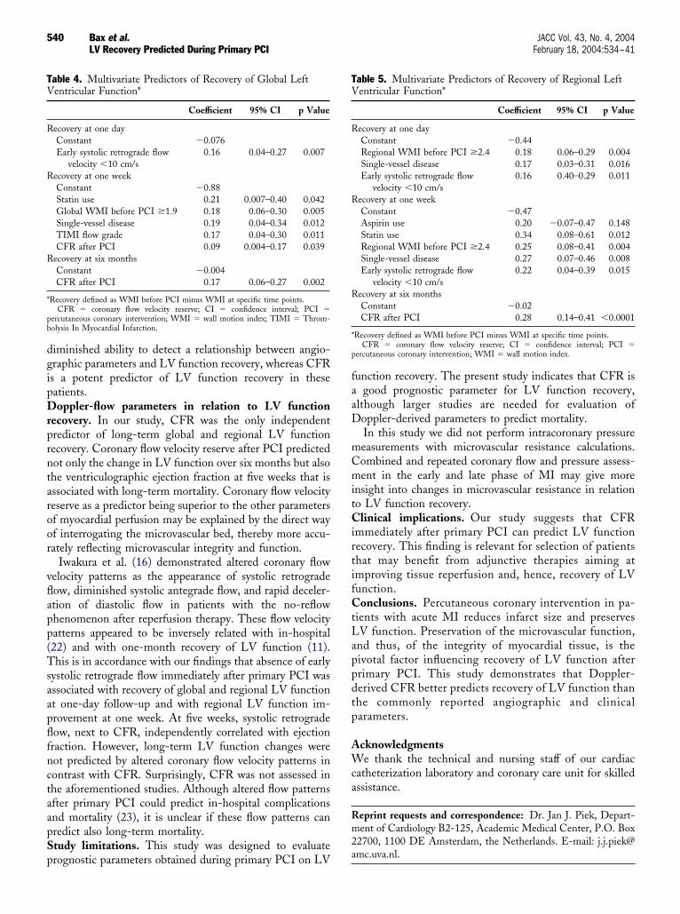
able 4. Multivariate Predictors of Recovery of Global Leftentricular Function*
Coefficient 95% CI p Value
ecovery at one dayConstant �0.076Early systolic retrograde flow
velocity �10 cm/s0.16 0.04–0.27 0.007
ecovery at one weekConstant �0.88Statin use 0.21 0.007–0.40 0.042Global WMI before PCI �1.9 0.18 0.06–0.30 0.005Single-vessel disease 0.19 0.04–0.34 0.012TIMI flow grade 0.17 0.04–0.30 0.011CFR after PCI 0.09 0.004–0.17 0.039
ecovery at six monthsConstant �0.004CFR after PCI 0.17 0.06–0.27 0.002
Recovery defined as WMI before PCI minus WMI at specific time points.CFR � coronary flow velocity reserve; CI � confidence interval; PCI �
ercutaneous coronary intervention; WMI � wall motion index; TIMI � Throm-olysis In Myocardial Infarction.
rognostic parameters obtained during primary PCI on LV
unction recovery. The present study indicates that CFR isgood prognostic parameter for LV function recovery,
lthough larger studies are needed for evaluation ofoppler-derived parameters to predict mortality.In this study we did not perform intracoronary pressureeasurements with microvascular resistance calculations.ombined and repeated coronary flow and pressure assess-ent in the early and late phase of MI may give more
nsight into changes in microvascular resistance in relationo LV function recovery.
linical implications. Our study suggests that CFRmmediately after primary PCI can predict LV functionecovery. This finding is relevant for selection of patientshat may benefit from adjunctive therapies aiming atmproving tissue reperfusion and, hence, recovery of LVunction.
onclusions. Percutaneous coronary intervention in pa-ients with acute MI reduces infarct size and preservesV function. Preservation of the microvascular function,nd thus, of the integrity of myocardial tissue, is theivotal factor influencing recovery of LV function afterrimary PCI. This study demonstrates that Doppler-erived CFR better predicts recovery of LV function thanhe commonly reported angiographic and clinicalarameters.
cknowledgmentse thank the technical and nursing staff of our cardiac
atheterization laboratory and coronary care unit for skilledssistance.
eprint requests and correspondence: Dr. Jan J. Piek, Depart-ent of Cardiology B2-125, Academic Medical Center, P.O. Box
2700, 1100 DE Amsterdam, the Netherlands. E-mail: [email protected].
able 5. Multivariate Predictors of Recovery of Regional Leftentricular Function*
Coefficient 95% CI p Value
ecovery at one dayConstant �0.44Regional WMI before PCI �2.4 0.18 0.06–0.29 0.004Single-vessel disease 0.17 0.03–0.31 0.016Early systolic retrograde flow
velocity �10 cm/s0.16 0.40–0.29 0.011
ecovery at one weekConstant �0.47Aspirin use 0.20 �0.07–0.47 0.148Statin use 0.34 0.08–0.61 0.012Regional WMI before PCI �2.4 0.25 0.08–0.41 0.004Single-vessel disease 0.27 0.07–0.46 0.008Early systolic retrograde flow
velocity �10 cm/s0.22 0.04–0.39 0.015
ecovery at six monthsConstant �0.02CFR after PCI 0.28 0.14–0.41 �0.0001
Recovery defined as WMI before PCI minus WMI at specific time points.CFR � coronary flow velocity reserve; CI � confidence interval; PCI �
ercutaneous coronary intervention; WMI � wall motion index.

R
1
1
1
1
1
1
1
1
1
1
2
2
2
2
541JACC Vol. 43, No. 4, 2004 Bax et al.February 18, 2004:534–41 LV Recovery Predicted During Primary PCI
EFERENCES
1. Gruppo Italiano per lo Studio della Streptochinasi nell’InfartoMiocardico (GISSI-I). Effectiveness of intravenous thrombolytictreatment in acute myocardial infarction. Lancet 1986;1:397–401.
2. The GUSTO Angiographic Investigators. The effects of tissue plas-minogen activator, streptokinase, or both on coronary-artery patency,ventricular function, and survival after acute myocardial infarction.N Engl J Med 1993;329:1615–22.
3. de Boer MJ, Suryapranata H, Hoorntje JC, et al.. Limitation of infarctsize and preservation of left ventricular function after primary coronaryangioplasty compared with intravenous streptokinase in acute myocar-dial infarction. Circulation 1994;90:753–61.
4. Andrews J, Straznicky IT, French JK, et al.. ST-segment recovery addsto the assessment of TIMI 2 and 3 flow in predicting infarct wallmotion after thrombolytic therapy. Circulation 2000;101:2138–43.
5. Suryapranata H, Zijlstra F, MacLeod DC, et al.. Predictive value ofreactive hyperemic response on reperfusion on recovery of regionalmyocardial function after coronary angioplasty in acute myocardialinfarction. Circulation 1994;89:1109–17.
6. Greaves K, Dixon SR, Fejka M, et al.. Myocardial contrast echocar-diography is superior to other known modalities for assessing myocar-dial reperfusion after acute myocardial infarction. Heart 2003;89:139–44.
7. The TIMI Study Group. The Thrombolysis In Myocardial Infarction(TIMI) trial. N Engl J Med 1985;312:932–6.
8. Lincoff AM, Topol EJ, Califf RM, et al.. Significance of a coronaryartery with thrombolysis in myocardial infarction grade 2 flow “pa-tency” (outcome in the thrombolysis and angioplasty in myocardialinfarction trials). Thrombolysis and Angioplasty in Myocardial Infarc-tion Study Group. Am J Cardiol 1995;75:871–6.
9. Gibson CM, Cannon CP, Daley WL, et al.. TIMI frame count: aquantitative method of assessing coronary artery flow. Circulation1996;93:879–88.
0. van’t Hof AWJ, Liem A, Suryapranata H, et al.. Angiographicassessment of myocardial reperfusion in patients treated with primaryangioplasty for acute myocardial infarction. Circulation 1998;97:2302–6.
1. Kawamoto T, Yoshida K, Akasaka T, et al.. Can coronary blood flowvelocity pattern after primary percutaneous transluminal coronaryangioplasty predict recovery of regional left ventricular function inpatients with acute myocardial infarction? Circulation 1999;100:339–45.
2. Mazur W, Bitar JN, Lechin M, et al.. Coronary flow reserve maypredict myocardial recovery after myocardial infarction in patients withTIMI grade 3 flow. Am Heart J 1998;136:335–44.
3. Tsunoda TF, Nakamura MF, Wakatsuki TF, et al.. The pattern ofalteration in flow velocity in the recanalized artery is related to leftventricular recovery in patients with acute infarction and successfuldirect balloon angioplasty. J Am Coll Cardiol 1998;32:338 –44.
4. van’t Hof AW, Liem A, de Boer MJ, Zijlstra F. Clinical value of12-lead electrocardiogram after successful reperfusion therapy for acutemyocardial infarction. Zwolle Myocardial Infarction study group.Lancet 1997;350:615–9.
5. Blanke H, Cohen M, Karsch KR, et al.. Prevalence and significance ofresidual flow to the infarct zone during the acute phase of myocardialinfarction. J Am Coll Cardiol 1985;5:827–31.
6. Iwakura K, Ito H, Takiuchi S, et al.. Alternation in the coronary bloodflow velocity pattern in patients with no reflow and reperfused acutemyocardial infarction. Circulation 1996;94:1269–75.
7. Schiller NB, Shah PM, Crawford M, et al.. Recommendations forquantitation of the left ventricle by two-dimensional echocardiogra-phy: American Society of Echocardiography Committee on Standards,Subcommittee on Quantitation of Two-Dimensional Echocardio-grams. J Am Soc Echocardiogr 1989;2:358–67.
8. Matetzky S, Novikov M, Gruberg L, et al.. The significance ofpersistent ST elevation versus early resolution of ST segment elevationafter primary PTCA. J Am Coll Cardiol 1999;34:1932–8.
9. Poli A, Fetiveau R, Vandoni P, et al.. Integrated analysis of myocardialblush and ST-segment elevation recovery after successful primaryangioplasty: real-time grading of microvascular reperfusion and pre-diction of early and late recovery of left ventricular function. Circula-tion 2002;106:313–8.
0. de Lemos JA, Braunwald E. ST segment resolution as a tool forassessing the efficacy of reperfusion therapy. J Am Coll Cardiol2001;38:1283–94.
1. The GUSTO IIb Investigators. A clinical trial comparing primarycoronary angioplasty with tissue plasminogen activator for acutemyocardial infarction. N Engl J Med 1997;336:1621–8.
2. Wakatsuki T, Nakamura M, Tsunoda T, et al.. Coronary flow velocityimmediately after primary coronary stenting as a predictor of ventric-ular wall motion recovery in acute myocardial infarction. J Am CollCardiol 2000;35:1835–41.
3. Yamamuro A, Akasaka T, Tamita K, et al.. Coronary flow velocitypattern immediately after percutaneous coronary intervention as apredictor of complications and in-hospital survival after acute myocar-dial infarction. Circulation 2002;106:3051–6.
Copyright © 2022 FDOKUMEN