Serum levels of β-catenin as a potential marker for genotype 4/hepatitis C-associated...
7
ONCOLOGY REPORTS 00: 0-00, 0000 Abstract. The global rising incidence of hepatocellular carcinoma (HCC), which parallels the increase of hepatitis C virus (HCV) prevalence, has sparked a renewed interest in discovering additional HCC serum markers. In this study, we investigated the clinical use of serum E-cadherin, ICAM, MMP-2, VEGF, OPN and β-catenin as potential diagnostic makers for HCV/genotype 4-associated HCC. Twenty cases of healthy subjects, 11 cases with asymptomatic HCV/genotype 4 carriers (ASC), 28 cases of chronic hepatitis (CH) cases and 32 patients with HCC were enrolled in this study. Serum levels of proteins were measured by a sandwich-enzyme- linked (ELISA) assay. The diagnostic accuracy of each candidate marker was evaluated using receiver-operating characteristic (ROC) curve analysis, reporting the area under the curve (AUC) and its 95% confidence interval (CI). We demonstrated that serum β-catenin levels were significantly elevated in patients with HCC compared to those with CH, ASC and healthy controls. Among the six studied markers, β-catenin was also found to be the only marker that can significantly discriminate between patients with HCC and those with CH; therefore, β-catenin could be considered as a potential marker for early diagnosis of HCV-associated HCC in patients infected with HCV genotype 4. Introduction Hepatocellular carcinoma (HCC) is the fifth most common cancer and one of the leading causes of cancer death in the world (1). It has heterogeneous geographical distribution, with its greatest incidence in Asia and sub-Saharan Africa, where hepatitis B infection is endemic (2). Its incidence has also been increasing steadily in the United States and Western Europe due to the high prevalence of hepatitis C (3,4). Egypt has the highest prevalence of hepatitis C virus (HCV) infection, with 14% of the population infected and seven million with chronic liver hepatitis (5,6). Little is known about the molecular mechanisms by which HCV initiates HCC. Molecular studies directed at mapping the etiology of HCV disease progression to HCC are expected to provide new insights on the management of this increasing problem and hence are of great global health interest. However, the lack of molecular makers that can characterize the stage of HCC, regardless of the etiologic factor, and tumor progression precludes the effective diagnosis and prognosis of HCC. Currently, HCC diagnosis relies on the radiology imaging systems, and elevated serum α-fetoprotein (AFP). Serum AFP is not always elevated to a diagnostic level in all patients in small HCC, thus, considerable numbers of patients with more advanced stages would be missed unless other diagnostic tools are used (7,8). Moreover, the level of AFP may be elevated in non-malignant chronic liver diseases, including chronic hepatitis and cirrhosis (9,10). Several biomarkers, such as des-γ-carboxyprothrombin/prothrombin induced by vitamin K absence-II, lens cularis agglutinin- reactive (AFP-L3), and glypican-3, have been examined for their ability to diagnose early HCC (11). However, there is a need for additional serum biomarkers to improve the detection of HCC at its early stage. Protein expression profiling studies have detected many proteins that are associated with hepato- carcinogenesis. For example, serum level of soluble E-cadherin was shown be elevated in patients with HCC, and is associated with early recurrence or extrahepatic metastasis (12). The level of other serum proteins, such as serum intercellular adhesion molecule-1 (sICAM-1) (13), serum matrix metallo- proteinase-2 (MMP-2) (14), vascular endothelial growth factor (VEGF) (15), plasma osteopontin (OPN) (16), and β-catenin (17) has been shown to be elevated in patients with HCC and Serum levels of β-catenin as a potential marker for genotype 4/ hepatitis C-associated hepatocellular carcinoma ABDEL-RAHMAN N. ZEKRI 1 , ABEER A. BAHNASSY 1 , HANAA M. ALAM EL-DIN 1 , HEBA M. MORSY 1 , SAbry SHAArAWy 1 , NAGIA Z. MOHARRAM 2 and SAYED S. DAOUD 3 1 Virology and Immunology Unit, Cancer Biology Department, National Cancer Institute; 2 Faculty of Science, Zoology Department, Cairo University, Cairo, Egypt; 3 Department of Pharmaceutical Sciences, Center for Integrated biotechnology, Washington State University, Pullman, WA, USΑ Received XXXXX; Accepted XXXXX DOI: 10.3892/or_xxxxxxxx 22 23 24 25 26 27 28 29 30 31 32 33 34 35 36 37 38 39 40 41 42 43 44 45 46 47 48 49 50 51 52 53 54 55 56 57 58 59 60 61 62 63 1 2 3 4 5 6 7 8 9 10 11 12 13 14 15 16 17 18 19 20 21 Correspondence to: Dr Sayed Daoud, Department of Pharmaceutical Sciences, Center for Integrated biotechnology, 340D Wegner Hall, Pullman, WA 99164-6534, USA E-mail: [email protected] Abbreviations: HCC, hepatocellular carcinoma; sE-cadherin, soluble human epithelial cadherin; ICAM-1, intercellular adhesion molecule; total MMP-2, active and pro-matrix metalloproteinase 2; VEGF, vascular endothelial growth factor; OPN, human osteopontin; CH, chronic hepatitis; ASC, asymptomatic HCV carriers; HCV, hepatitis C virus; AFP, α-fetoprotein; NO; nitric oxide; MMPs, matrix metalloproteinases; TIMPs, tissue inhibitor of MMPs; RGD, arginine- glycine-aspartic acid; ROC, receiver-operating characteristic curve Key words: hepatocellular carcinoma, chronic hepatitis C, hepatitis C virus genotype 4, serum markers, β-catenin, E-cadherin
-
Upload
independent -
Category
Documents
-
view
2 -
download
0
Transcript of Serum levels of β-catenin as a potential marker for genotype 4/hepatitis C-associated...

ONCOLOGY repOrts 00: 0-00, 0000
Abstract. the global rising incidence of hepatocellular carcinoma (HCC), which parallels the increase of hepatitis C virus (HCV) prevalence, has sparked a renewed interest in discovering additional HCC serum markers. In this study, we investigated the clinical use of serum e-cadherin, ICAM, MMp-2, VeGF, OpN and β-catenin as potential diagnostic makers for HCV/genotype 4-associated HCC. twenty cases of healthy subjects, 11 cases with asymptomatic HCV/genotype 4 carriers (AsC), 28 cases of chronic hepatitis (CH) cases and 32 patients with HCC were enrolled in this study. serum levels of proteins were measured by a sandwich-enzyme-linked (eLIsA) assay. the diagnostic accuracy of each candidate marker was evaluated using receiver-operating characteristic (rOC) curve analysis, reporting the area under the curve (AUC) and its 95% confidence interval (CI). We demonstrated that serum β-catenin levels were significantly elevated in patients with HCC compared to those with CH, AsC and healthy controls. Among the six studied markers, β-catenin was also found to be the only marker that can significantly discriminate between patients with HCC and those with CH; therefore, β-catenin could be considered as a potential marker for early diagnosis of HCV-associated HCC in patients infected with HCV genotype 4.
Introduction
Hepatocellular carcinoma (HCC) is the fifth most common cancer and one of the leading causes of cancer death in the world (1). It has heterogeneous geographical distribution, with its greatest incidence in Asia and sub-saharan Africa, where hepatitis B infection is endemic (2). Its incidence has also been increasing steadily in the United States and Western europe due to the high prevalence of hepatitis C (3,4). egypt has the highest prevalence of hepatitis C virus (HCV) infection, with 14% of the population infected and seven million with chronic liver hepatitis (5,6). Little is known about the molecular mechanisms by which HCV initiates HCC. Molecular studies directed at mapping the etiology of HCV disease progression to HCC are expected to provide new insights on the management of this increasing problem and hence are of great global health interest. However, the lack of molecular makers that can characterize the stage of HCC, regardless of the etiologic factor, and tumor progression precludes the effective diagnosis and prognosis of HCC. Currently, HCC diagnosis relies on the radiology imaging systems, and elevated serum α-fetoprotein (AFp). serum AFp is not always elevated to a diagnostic level in all patients in small HCC, thus, considerable numbers of patients with more advanced stages would be missed unless other diagnostic tools are used (7,8). Moreover, the level of AFp may be elevated in non-malignant chronic liver diseases, including chronic hepatitis and cirrhosis (9,10). several biomarkers, such as des-γ-carboxyprothrombin/prothrombin induced by vitamin K absence-II, lens cularis agglutinin-reactive (AFp-L3), and glypican-3, have been examined for their ability to diagnose early HCC (11). However, there is a need for additional serum biomarkers to improve the detection of HCC at its early stage. Protein expression profiling studies have detected many proteins that are associated with hepato-carcinogenesis. For example, serum level of soluble e-cadherin was shown be elevated in patients with HCC, and is associated with early recurrence or extrahepatic metastasis (12). the level of other serum proteins, such as serum intercellular adhesion molecule-1 (sICAM-1) (13), serum matrix metallo-proteinase-2 (MMp-2) (14), vascular endothelial growth factor (VeGF) (15), plasma osteopontin (OpN) (16), and β-catenin (17) has been shown to be elevated in patients with HCC and
Serum levels of β-catenin as a potential marker for genotype 4/ hepatitis C-associated hepatocellular carcinomaABdeL-rAHMAN N. ZeKrI1, ABeer A. BAHNAssY1, HANAA M. ALAM eL-dIN1,
HeBA M. MOrsY1, SAbry SHAArAWy1, NAGIA Z. MOHArrAM2 and sAYed s. dAOud3
1Virology and Immunology unit, Cancer Biology department, National Cancer Institute; 2Faculty of science, Zoology department, Cairo university, Cairo, egypt; 3department of pharmaceutical sciences,
Center for Integrated biotechnology, Washington State University, Pullman, WA, USΑ
received XXXXX; Accepted XXXXX
dOI: 10.3892/or_xxxxxxxx
222324252627282930313233343536373839404142434445464748495051525354555657585960616263
123456789
101112131415161718192021
Correspondence to: dr sayed daoud, department of pharmaceutical Sciences, Center for Integrated biotechnology, 340D Wegner Hall, Pullman, WA 99164-6534, USAe-mail: [email protected]
Abbreviations: HCC, hepatocellular carcinoma; se-cadherin, soluble human epithelial cadherin; ICAM-1, intercellular adhesion molecule; total MMp-2, active and pro-matrix metalloproteinase 2; VeGF, vascular endothelial growth factor; OpN, human osteopontin; CH, chronic hepatitis; AsC, asymptomatic HCV carriers; HCV, hepatitis C virus; AFp, α-fetoprotein; NO; nitric oxide; MMps, matrix metalloproteinases; tIMps, tissue inhibitor of MMps; rGd, arginine-glycine-aspartic acid; rOC, receiver-operating characteristic curve
Key words: hepatocellular carcinoma, chronic hepatitis C, hepatitis C virus genotype 4, serum markers, β-catenin, e-cadherin

ZeKrI et al: MOLeCuLAr MArKers FOr HCV-AssOCIAted HCC2
123456789
101112131415161718192021222324252627282930313233343536373839404142434445464748495051525354555657585960
616263646566676869707172737475767778798081828384858687888990919293949596979899
100101102103104105106107108109110111112113114115116117118119120
hence could be used as prognostic markers for early detection of HCC. However, limited information is available about the extent to which these proteins could be used as prognostic markers for viral-associated HCC.
In view of the possible diagnostic role of these serum proteins in the development of HCC initiated by the HCV/genotype 4, the serum levels of the above mentioned proteins were measured in 91 egyptian subjects using a sandwich ELISA methods in four groups. We have analyzed the correlation and predictive value of these markers in screening patients at risk of HCC.
Materials and methods
Patients and sample collection. serum samples for the assessment of biomarkers were obtained from 91 consecutive individuals. samples were obtained from patients presented before treatment to the specialized liver clinic of the National Cancer Institute, Cairo university during the period of January to december 2009. Informed consent was obtained from each patient and the study protocols conformed to the ethics guidelines of the Institutional review Board. subjects were divided into four groups. Group 1 (HCC; n=32) included patients with histological proven HCC. Group 2 (CH; n=28) included patients with chronic hepatitis, with or without cirrhosis. Group 3 (AsC; n=11) included patients infected with HCV with histological confirmed non-cirrhotic chronic hepatitis (asymptomatic carriers). All patients were HCV positive as detected by hepatitis C antibody and HCV rt-pCr (18), and HBV negative as detected by hepatitis B antigen and HBV dNA/pCr (19). Group 4 (Control; n=20) included
normal healthy subjects with no history of liver disease that were negative for HCV and HBV. the clinicopathological data of the subjects in this study at initial diagnosis were collected, which included gender, age, liver function tests, kidney function tests, and AFp levels are presented in table I.
sera collected from 10 ml of coagulated blood by centri-fugation were immediately separated and frozen at -80˚C until assayed.
Measurement of serum biomarkers. serum levels of soluble human epithelial cadherin (se-cadherin), sICAM-1, active and proMMp-2 (total MMp-2), VeGF, human OpN and β-catenin were measured by a commercially available enzyme-linked immunosorbent assay (eLIsA) kit (r&d systems, Inc., Minneapolis, MN) according to the manufacturer's instructions. each sample was examined in duplicate and the average value (mean) was used for data analysis. the cut-off was considered as mean ± 2sd of the negative controls.
Statistical analysis. data are expressed as the mean ± sd. Comparisons between groups were analyzed by the χ2 test or Fisher's exact test for categorical variables, and by the Mann-Whitney test or Student's t-test when appropriate for quantitative variables. receiver-operating characteristics curves (rOC) were constructed to evaluate the diagnostic performance of the serum makers in discriminating HCC from other groups. Sensitivity, specificity, positive and negative predictor values and diagnostic accuracy were calculated in accordance with standard methods. p<0.05 for a two-tailed test was considered statistically significant. All statistical analyses were performed using the spss software version 15.0 (spss, Chicago, IL).
table I. Clinical characteristics of study subjects.
Variables HCC (n=32) CH (n=28) AsC (n=11) Control (n=20) p-value
Age mean, years (range) 54a (40-76) 50.5ab (31-63) 44bc (22-56) 32c (27-53) <0.001Gender (male/female) 29/3 21/7 9/2 16/4AFp 855a (175-1390) 13.5b (0.4-975.8) 1.4c (0.53-7.35) 0.69c (0.43-1.8) <0.001d-Bilirubin 0.92a (0.14-20.4) 0.64a (0.2-9.24) 0.78a (0.05-0.93) 0.05b (0.02-0.09) <0.001I-Bilirubin 1.05a (0.55-9.4) 0.86a (0.5-6.38) 0.14b (0.04-0.59) 0.11 (0.03-0.45) <0.001Ast 58a (23-138) 68.5a (25-138) 29b (15-39) 9b (5-13) <0.001ALt 46a (26-100) 55a (12-89) 35b (14-51) 6c (3-11) <0.001Alk-phosphatase 111a (63-389) 117a (37-304) 122a (60-194) 36b (23-56) <0.001Albumin 2.65b (1.3-3.2) 4a (1.9-5.4) 2.8b (2-3.2) 2.7b (2.3-3.7) <0.001urea 37 (12-129) 28 (18-150) 30 (16-58) 44 (9.6-112) 0.285Creatinine 1 (0.5-6.39) 0.95 (0.5-2.7) 0.9 (0.6-1.4) 1.11 (0.52-1.9) 0.348HB 10.95b (6.2-12.8) 11.5b (7.9-13.3) 14.1a (12.3-17) 13.3a (11.9-14.5) <0.001tLC 4b (0.53-11.4) 6.45ab (2.7-12.5) 5.3ab (3.6-9) 7.5a (4.45-10) <0.001pLt 105.5c (62-650) 105.5bc (7.6-293) 190ab (20-400) 195a (159-315) <0.001pt 16.05 (14-24) 15.8 (13-20.7) 14.4 (13.0-15.1) 13.0 (13- 14.5 0.303pC 50.5b (33-84) 52b (20-90) 85a (68-100) 100a (100-100) <0.001FBs 97.5 (60-254) 95 (70-199) 98 (67-162) 112.5 (67-145) 0.285
Groups indicated by different superscripted initials are significantly different from each other. HCC, hepatocellular carcinoma; CH, chronic hepatitis; AsC, asymptomatic HCV carriers; AFp, α-fetoprotein; ALt, alanine aminotransferase; Ast, aspartate aminotransferase.

ONCOLOGY repOrts 00: 0-00, 0000 3
123456789
101112131415161718192021222324252627282930313233343536373839404142434445464748495051525354555657585960
616263646566676869707172737475767778798081828384858687888990919293949596979899
100101102103104105106107108109110111112113114115116117118119120
Results and Discussion
Patient profiles. A total of 91 subjects, 75 men and 16 women were included in the study. detailed demographic data are shown in table I. the age of patients with HCC, CH and AsC were significantly older than healthy controls (p<0.001). Likewise, the mean age of patients with CH was significantly higher than that of AsC patients or healthy controls (p<0.001). The level of serum AFP in patients with HCC was significantly higher than those of healthy controls, CH and AsC (p<0.001). the serum AFp levels in the CH patients were also signif-icantly higher than those in AsC patients and healthy controls (p<0.001).
Serum levels of E-cadherin, ICAM-1, MMP-2, VEGF, OPN, and β-catenin as diagnostic markers. the results of the present
study showed that the serum levels of four out of the six studied markers (β-catenin, e-cadherin, sICAM and OpN) were significantly higher in patients with HCC and CH compared to AsC patients and healthy controls (Fig. 1). the high serum levels of those cell adhesion markers could be related to the process of fibrosis and cirrhosis in HCV-infected livers, which usually precedes the development of carcinoma, especially that no significant difference was detected between HCC and CH patients. Among those four markers, the serum levels of β-catenin were significantly higher in patients with HCC compared to CH patients (cut-off value of 1379.5 pg/ml for HCC patients vs. 737 pg/ml for CH patients) (Fig. 1). similarly, the serum levels of β-catenin were also higher in patients with CH than in AsC patients and healthy controls. these data are in agreement with previous reports by sun et al (20), where higher serum level of β-catenin were measured in patients with
Figure 1. scatter plots for (A) e-cadherin, (B) sICAM, (C) MMp-2, (d) VeGF, (e) OpN, (F) β-catenin. Levels are presented as ng/ml in hepatocellular carcinoma.HCC; chronic hepatitis, CH; asymptomatic carriers (AsC) and healthy controls. the cut-off value is indicated.

ZeKrI et al: MOLeCuLAr MArKers FOr HCV-AssOCIAted HCC4
123456789
101112131415161718192021222324252627282930313233343536373839404142434445464748495051525354555657585960
616263646566676869707172737475767778798081828384858687888990919293949596979899
100101102103104105106107108109110111112113114115116117118119120
HCC compared with those in CH patients. the authors of this study suggested that serum level of β-catenin could be used clinically to complement the current AFp diagnostic test for more accurate detection of early HCC. therefore, the ability to measure β-catenin in the peripheral blood of HCC patients would offer a valuable, non-invasive diagnostic and prognostic marker for HCC. the expression of β-catenin in HCC has also been detected immunohistochemically by several groups. In these studies, mutated nuclear β-catenin overexpression has been associated with increased cell proliferation, poorer cellular differentiation and reduced survival rate (21,22). these mutations of β-catenin lead to a nuclear accumulation of aberrant β-catenin proteins that stimulate the activity of other
transcription factors, such as cyclin d1 and c-myc (23,24). In dysplastic nodules, a cytoplasmic expression of β-catenin has been observed (25). Axin, an important regulator of β-catenin, is mutated in about 10% of HCC cases, leading to an activation of the Wnt pathway (26). However, mutations in the axin gene have been identified only in HCC that lack mutations in the β-catenin gene (27,28).
soluble e-cadherin was found to be circulating in the biological fluids of healthy individuals, but elevated in cancer patients (29) as well as in individuals with systemic inflam-matory response syndrome (30). the median concentration of serum e-cadherin reported in our study was significantly higher in patients with CH and HCC compared to those with
Figure 2. serum levels of (A) e-cadherin, (B) sICAM, (C) MMp-2, (d) VeGF, (e) OpN, (F) β-catenin in hepatocellular carcinoma (HCC); chronic hepatitis (CH); asymptomatic HCV carriers (AsC); and healthy controls. the box plots indicate the median values (triangles), the minimum and maximum values (squares and crosses, respectively). The box defines the boundaries of the first and third quartiles of data.
ONCOLOGY repOrts 00: 0-00, 0000 5
123456789
101112131415161718192021222324252627282930313233343536373839404142434445464748495051525354555657585960
616263646566676869707172737475767778798081828384858687888990919293949596979899
100101102103104105106107108109110111112113114115116117118119120
AsC and healthy controls (Fig. 1). In contrast to β-catenin, no significant difference was observed in the median concen-tration of serum e-cadherin between CH and HCC patients (Fig. 2A). earlier studies showed that the median serum E-cadherin levels were significantly elevated in HCC patients before surgery compared to healthy subjects (10.759 ng/ml vs. 5.798 ng/ml, p<0.05) (15). Moreover, high serum e-cadherin (≥8.000 ng/ml) was significantly associated with early recurrence and extra-hepatic metastasis. therefore, serum e-cadherin could be considered as a potential prognostic marker for patients with HCC (31), albeit with limited prognostic value in the early detection of HCC.
there have been few reports investigating the relationship between serum ICAM-1 concentrations, hepatitis and hepato-carcinogenesis. these reports showed that ICAM-1 is produced and secreted by tumor cells and that serum ICAM-1 could be used as a marker for disease progression and prognosis in patients with HCC (32). In the present study, the median level of sICAM was significantly higher in patients with HCC and CH compared to those of AsC and healthy controls (Fig. 1). However, there was no significant difference in the level of sICAM between HCC and CH (Fig. 2b). Our findings thus confirmed previous reports by Shimizu et al (32) who reported that high serum concentrations of ICAM-1 or an increasing level of sICAM-1 over time are significant risk factors for the occurrence of HCC in patients with HCV-associated CH or liver cirrhosis (LC). thus, regular measurements of sICAM-1 concentrations would be of clinical significance and could be used not only as a prognostic marker for early detection of HCC in patients with CH or LC, but also as a marker of disease progression after HCC treatment.
the median concentrations of serum MMp-2 reported in the present study were significantly higher in patients with CH compared to those with HCC (Fig. 2C). these results demon-strate the important role of MMp-2 in the development of HCV-induced fibrosis as well as in disease progression to cirrhosis. Our data are in agreement with those of Kuyvenhoven et al (33) who demonstrated that serum levels of MMp-2 as detected by ELISA, were significantly higher in HCC patients than those of healthy controls but comparable to patients with
chronic liver disease. Additional studies showed that circulating serum MMp-2 levels were increased in patients with liver cirrhosis and reported a wide overlap in patients with CH and healthy controls (34). serum levels of proMMp-2 were determined to be highly elevated in chronic liver diseases than in normal controls and were strongly correlated to type IV collagen in sera of CH patients. therefore, it is possible to consider the serum level of proMMp-2 as a follow-up marker in patients with CH (35).
The present study showed no significant increase in the serum level of OpN in patients with CH compared to those with HCC (Fig. 2C). However, this level was significantly higher in patients with CH and HCC than in AsCs and healthy controls (Fig. 1). Huang et al (36) have demonstrated that serum OPN level correlates well with liver fibrosis and inflam-mation. they reported that a significant difference in the mean plasma OpN levels between HCV patients with severe fibrosis and those with mild fibrosis (4.29±1.01 vs. 2.15±0.63 ng/ml, respectively p<0.001) and suggested that plasma OpN could be used as a marker to evaluate the severity of liver damage in HCV-infected persons. In contrast to our data, Kim et al (37) reported that plasma OpN levels in HCC patients (median 954 ng/ml, range 168-5,742) were significantly higher than in those with chronic liver diseases (381 ng/ml, 29-1,688) or healthy controls (155 ng/ml, 10-766) (p<0.001) with a diagnostic sensitivity and specificity of 87 and 82%; respectively, suggesting a superior diagnostic accuracy for serum OpN. this discrepancy in the results between both studies could be attributed to the fact that all patients included in our study were positive for HCV with a preponderance of genotype 4.
previous reports by Jinno et al (38) indicated that serum VeGF levels increased gradually along with disease progression in patients with hepatitis, cirrhosis and HCC. there was a significant difference in the level of serum VEGF between HCC and other patient groups, but not among hepatitis patients, cirrhotic patients and normal controls. Zhao et al (39) reported that serum VeGF detected by eLIsA was significantly higher in HCC patients than in those with benign liver lesions and healthy controls; however, no significant difference was found between patients with benign hepatic diseases and healthy controls or between patients with benign hepatic diseases and those with cirrhosis. In the present study, the median VEGF level showed no statistically significant difference between patients with CH, HCC and healthy controls (Fig. 2d). the data are consistent with that reported by Niu et al (40) who reported that the mean serum levels of VeGF in normal healthy controls, cirrhotic patients, patients with benign liver tumor, and those with HCC were 158.46±41.84, 90±22.42, 156.34±41.32 and 164.42±76.07 ng/ml; respectively. As a result, serum levels of VEGF were significantly higher in HCC than in cirrhotic patients, whereas no significant differences between patients with HCC and healthy controls were noted. these data highlight the important role of VeGF in disease progression from liver cirrhosis to HCC, and its limited prognostic value in differentiating between benign liver tumors and HCC (15).
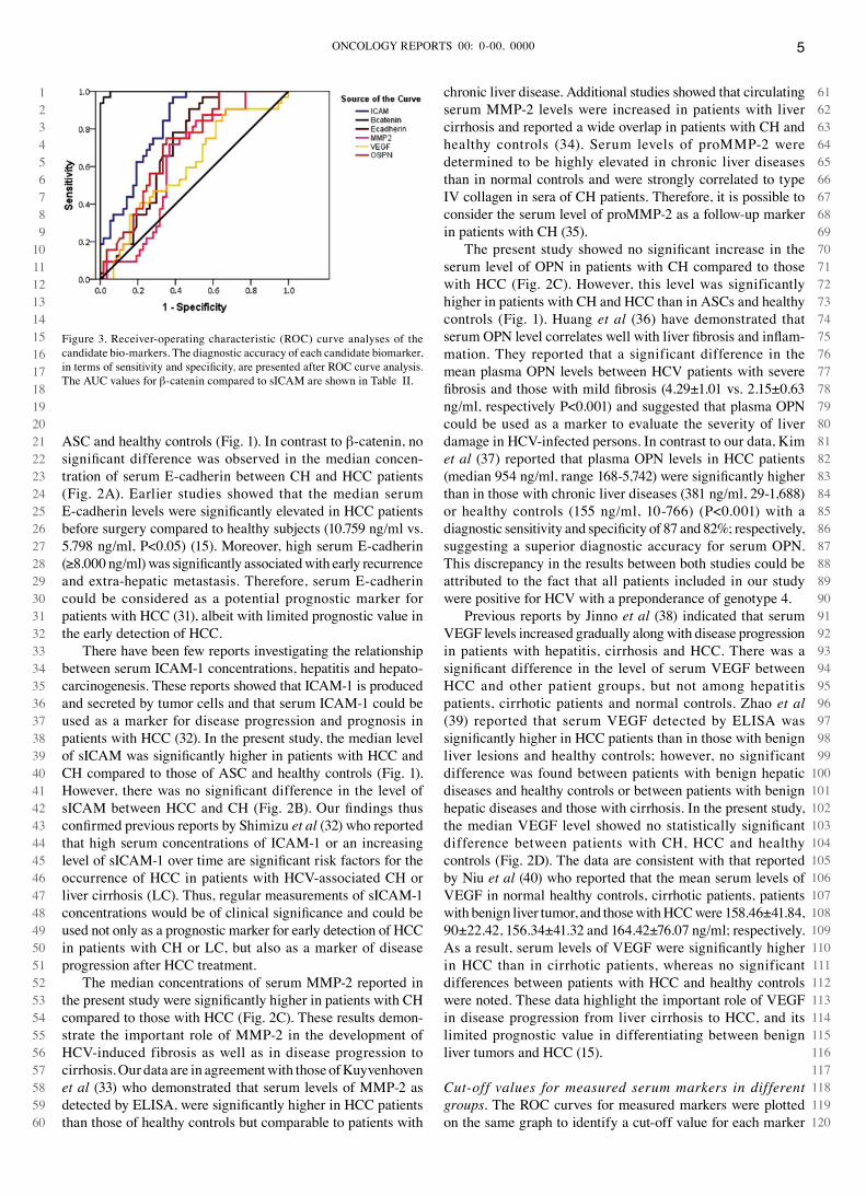
Cut-off values for measured serum markers in different groups. the rOC curves for measured markers were plotted on the same graph to identify a cut-off value for each marker
Figure 3. receiver-operating characteristic (rOC) curve analyses of the candidate bio-markers. the diagnostic accuracy of each candidate biomarker, in terms of sensitivity and specificity, are presented after rOC curve analysis. the AuC values for β-catenin compared to sICAM are shown in table II.

ZeKrI et al: MOLeCuLAr MArKers FOr HCV-AssOCIAted HCC6
123456789
101112131415161718192021222324252627282930313233343536373839404142434445464748495051
that would best distinguish HCC from the other groups investigated (Fig. 3). Based on the rOC analysis, the optimal cut-off values for sICAM and β-catenin for HCC and CH patients were ≥55.9 ng/ml, and ≥997.3 pg/ml, respectively. there were no satisfactory cut-off values for the other studied markers when comparing these two groups. For sICAM, we found that 23/32 (71.8%) HCC patients had an optimal cut-off level of ≥55.9 ng/ml, compared to 15/28 (53.6%) cases with CH. In contrast, 31/32 (96.9%) of HCC patients had β-catenin levels ≥997.3 pg/ml compared to 2/28 (7.1%) in patients with CH. the area under the curves (AuC) for e-cadherin, sICAM, MMp-2, VeGF, OpN and β-catenin were 0.708, 0.821, 0.630, 0.603, 0.712 and 0.998, respectively. therefore, the serum level of β-catenin could possibly be used as a valuable marker to discriminate patients with hepatocellular carcinoma from those with chronic active hepatitis. As shown in table II, the sensitivity and specificity values for β-catenin, at a cut-off value of ≥997.3 pg/ml, were 96.9 and 92.6%, respectively. For sICAM, these values at a cut-off value ≥55.9 ng/ml were 71.9 and 47%, respectively. this suggests that the optimal value for β-catenin showed a higher sensitivity and specificity in discriminating between HCC and CH than sICAM.
In conclusion, our study showed that serum β-catenin levels were significantly elevated in patients with HCC as compared to those with CH, AsC and healthy controls. Among the six studied markers, β-catenin was found to be the only one that can significantly discriminate between patients with HCC and those with CH; therefore, β-catenin could be considered as a potential marker for early diagnosis in selecting high-risk patients for the HCC surveillance program. Further large scale studies are worthwhile to confirm these observations and to elucidate the clinical significance of serum β-catenin in patients with HCC.
References
1. Nordenstedt H, White DL and El-Serag Hb: Changing pattern of epidemiology in hepatocellular carcinoma. dig Liver dis 42 (suppl 3): s206-s214, 2010.
2. Bosch FX, ribes J and Borras J: epidemiology of primary liver cancer. semin Liver dis 19: 271-285, 1999.
3. Cabibbo G and Craxi A: epidemiology, risk factors and surveillance of hepatocellular carcinoma. eur rev Med pharmacol sci 14: 352-355, 2010.
4. el-serag HB and Mason AC: rising incidence of hepatocellular carcinoma in the united states. N engl J Med 340: 745-750, 1999.
5. Lehman EM and Wilson ML: Epidemic hepatitis C virus infection in egypt: estimates of past incidence and future morbidity and mortality. J Viral Hepatol 16: 650-658, 2009.
6. Lehman EM and Wilson ML: Epidemiology of hepatitis viruses among hepatocellular carcinoma cases and healthy people in egypt: a systematic review and meta-analysis. Int J Cancer 124: 690-697, 2009.
7. spangenberg HC, thimme r and Blum He: serum markers of hepatocellular carcinoma. semin Liver dis 26: 385-390, 2006.
8. tateishi r, Yoshida H, Matsuyama Y, Mine N, Kondo Y and Omata M: diagnostic accuracy of tumor markers for hepato-cellular carcinoma: a systematic review. Hepatol Int 2: 17-30, 2008.
9. sherman M, peltekian KM and Lee C: screening for hepato-cellular carcinoma in chronic carriers of hepatitis B virus: incidence and prevalence of hepatocellular carcinoma in a North American urban population. Hepatology 22: 432-438, 1995.
10. di Bisceglie AM and Hoofnagle JH: elevations in serum α-fetoprotein levels in patients with chronic hepatitis B. Cancer 64: 2117-2120, 1989.
11. Wright LM, Kreikemeier JT and Fimmel CJ: A concise review of serum markers for hepatocellular cancer. Cancer detect prev 31: 35-44, 2007.
12. soyama A, eguchi s, takatsuki M, Kawashita Y, Hidaka M, tokai H, Nagayoshi s, Mochizuki s, Matsumoto s, Mochizuki s, Matsumoto s, Hamasaki K, tajima Y and Kanematsu t: Significance of the serum level of soluble E-cadherin in patients with HCC. Hepatogastroenterology 55: 1390-1393, 2008.
13. Sun y, Mi W, Cai J, ying W, Liu F, Lu H, Qiano y, Jia W, bi X, Lu N, Liu S, Qian X and Zhao X: Quantitative proteomic signature of liver cancer cells: tissue transglutaminase-2 could be a novel protein candidate of human hepatocellular carcinoma. J proteome res 7: 3847-3859, 2008.
14. yeh HC, Lin SM, Chen MF, Pan TL, Wang PW and yeh CT: evaluation of serum matrix metalloproteinase (MMp)-9 to MMp-2 ratio as a biomarker in hepatocellular carcinoma. Hepatogastroenterology 57: 98-102, 2010.
15. schoenleber sJ, Kurtz dM, talwalkar JA, roberts Lr and Gores GJ: prognostic role of vascular endothelial growth factor in hepatocellular carcinoma: systematic review and meta-analysis. Br J Cancer 100: 1385-1392, 2009.
16. Zhao L, Li T, Wang y, Pan y, Ning H, Hui X, Xie H, Wang J, Han Y, Liu Z and Fan d: elevated plasma osteopontin level is predictive of cirrhosis in patients with hepatitis B infection. Int J Clin pract 62: 1056-1062, 2008.
17. Zulehner G, Mikula M, schneller d, van Zijl F, Huber H, Sieghart W, Grasl-Kraupp b, Waldhör, Peck-radosavijevic M, beug H and Mikulits W: Nuclear beta-catenin induces an early liver progenitor phenotype in hepatocellular carcinoma and promotes tumor recurrence. Am J pathol 176: 472-481, 2010.
18. Zekri A, Alam el-din HM, Bahnassy AA, el-shehabi AM, El-Leethy H, Omar A and Khaled HM: TrUGENE sequencing versus INNO-LipA for sub-genotyping of HCV genotype-4. J Med Virol 75: 412-420, 2005.
19. Zekri A, Hafez MM, Mohamed NI, Hassan ZK, el-sayed MH, Khaled MM and Mansour t: Hepatitis B virus (HBV) genotypes in egyptian pediatric cancer patients with acute and chronic active HBV infection. Virol J 4: 474-481, 2007.
20. Jemal A, Siegel r, Xu J and Ward E: Cancer Statistics, 2010. CA Cancer J Clin 60: 277-300, 2010.
21. Wong CM, Fan ST and Ng IO: beta-catenin mutation and overexpression in hepatocellular carcinoma: clinicopathologic and prognostic significance. Cancer 92: 136-145, 2001.
22. Inagawa s, Itabashi M, Adachi s, Kawamoto t, Hori M, shimazaki J, Yoshimi F and Fukao K: expression and prognostic roles of beta-catenin in hepatocellular carcinoma: correlation with tumor progression and postoperative survival. Clin Cancer res 8: 450-456, 2002.
23. Kim Y, sills rC and Houle Cd: Overview of the molecular biology of hepatocellular neoplasms and hepatoblastomas of the mouse liver. toxicol pathol 33: 175-180, 2005.
24. reed Kr, Athineos D, Meniel VS, Wilkins JA, ridgway rA, Burke Zd, Muncan V, Clarke Ar and sansom OJ: B-catenin deficiency, but not Myc deletion, suppresses the immediate phenotypes of ApC loss in the liver. proc Natl Acad sci usA 105: 18919-18923, 2008.
table II. rOC curve values of studied markers between HCC and CH patients.
rOC ICAM ≥55.9 ng/ml β-catenin ≥997.3 pg/mlvalues (%) (%)
sensitivity 71.9 96.9Specificity 47 92.6AuC 0.642 0.995ppV 60.53 93.93NpV 59 96.15
rOC, receiver-operating characteristic; AuC, area under the curve; NpV, negative predictive value; ppV, positive predictive value; CH, chronic active hepatitis; HCC, hepatocellular carcinoma.

ONCOLOGY repOrts 00: 0-00, 0000 7
25. Nhieu JT, renard CA, Wei y, Cherqui D, Zafrani ES and Buendia MA: Nuclear accumulation of mutated beta-catenin in hepatocellular carcinoma is associated with increased cell proliferation. Am J pathol 155: 703-710, 1999.
26. Nakamura t, Hamada F, Ishidate t, Anai K, Kawahara K, Toyoshima K and Akiyama T: Axin, an inhibitor of the Wnt signaling pathway, interacts with beta-catenin, GsK-3beta and ApC and reduces the beta-catenin level. Genes Cells 3: 395-403, 1998.
27. Park Jy, Park WS, Nam SW, Kim Sy, Lee SH, yoo NJ, Lee Jy and park CK: Mutations of beta-catenin and AXIN 1 genes are a late event in human hepatocellular carcinogenesis. Liver Int 25: 70-76, 2005.
28. Zucman-rossi J, Benhamouche s, Godard C, Boyault s, Grimber G, Balabaud C, Cunha As, Bioulac-sage p and perret C: differential effects of inactivated Axin1 and activated beta-catenin mutations in human hepatocellular carcinomas. Oncogene 26: 774-780, 2007.
29. Kuefer r, Hofer Md, Zorn Cs, engel O, Volkmer BG, Juarez-Brito MA, eggel M, Gschwend Je, rubin MA and day ML: Assessment of a fragment of e-cadherin as a serum biomarker with predictive value for prostate cancer. Br J Cancer 92: 2018-2023, 2005.
30. Pittard AJ, Galley HF and Webster Nr: Soluble E-cadherin concentrations in patients with systemic inflammatory response syndrome and multiorgan dysfunction syndrome. Br J Anaesth 76: 629-631, 1996.
31. Moriyama M, Matsumura H, shioda J, Aoki H, Nakamura H, Arakawa Y, Nirei K, Yamagami H, Kaneko M, tanaka N and Arakawa Y: Measurement of human intercellular adhesion molecule 1 in the blood is useful for predicting the occurrence of hepatocellular carcinoma from chronic hepatitis C and liver cirrhosis. Intervirology 49: 327-338, 2006.
32. shimizu Y, Minemura M, tsukishiro t, Kashii Y, Miyamoto M, Nishimori H, Higuchi K and Watanabe A: Serum concentration of intercellular adhesion molecule-1 in patients with hepato-cellular carcinoma is a marker of the disease progression and prognosis. Hepatology 22: 525-531, 1995.
33. Kuyvenhoven JP, van Hoek b, blom E, van Duijn W, Hanemaaijer r, Verheijen JH, Lamers Cb and Verspaget HW: Assessment of the clinical significance of serum matrix metalloproteinases MMp-2 and MMp-9 in patients with various chronic liver diseases and hepatocellular carcinoma. thromb Haemost 89: 718-725, 2003.
34. Lichtinghagen r, Huegel O, seifert t, Haberkorn CI, Michels d, Flemming p, Bahr M and Boeker KH: expression of matrix metalloproteinase-2 and -9 and their inhibitors in peripheral blood cells of patients with chronic hepatitis C. Clin Chem 46: 183-192, 2000.
35. ebata M, Fukuda Y, Nakano I, Katano Y, Fujimoto N and Hayakawa t: serum levels of tissue inhibitor of metallo-proteinases-2 and of precursor from a matrix metalloproteinase-2 in patients with liver disease. Liver 17: 293-299, 1997.
36. Huang W, Zhu G, Huang M, Lou G, Liu y and Wang S: Plasma osteopontin concentration correlates with the severity of hepatic fibrosis and inflammation in HCV-infected subjects. Clin Chim Acta 411: 675-678, 2010.
37. Kim J, Ki ss, Lee sd, Han CJ, Kim YC, park sH, Cho sY, Hong YJ, park HY, Lee M, Jung HH, Lee KH and Jeong sH: elevated plasma osteopontin levels in patients with hepatocellular carcinoma. Am J Gastroenterol 101: 2051-2059, 2006.
38. Jinno K, tanimizu M and Hyodo I: Circulating vascular endothelial growth factor (VeGF) is a possible tumor marker for metastasis in human hepatocellular carcinoma. J Gastroenterol 33: 376-382, 1998.
39. Zhao J, Hu J, Cai J, Yang X and Yang Z: Vascular endothelial growth factor expression in serum of patients with hepatocellular carcinoma. Chin Med J 116: 772-776, 2003.
40. Niu Q, Tang Zy, Ma ZC, Qin LX and Zhang LH: Serum vascular endothelial growth factor is a potential biomarker of metastatic recurrence after curative resection of hepatocellular carcinoma. World J Gastroenterol 6: 565-568, 2000.