Determination of thorium and uranium in solution by laser-induced breakdown spectrometry
Upload
independentCategory
view
2download
0
SEQUENTIAL EXTRACTION PROCEDURE
FOR DETERMINATION OF URANIUM, THORIUM, RADIUM,
LEAD AND POLONIUM RADIONUCLIDES BY ALPHA
SPECTROMETRY IN ENVIRONMENTAL SAMPLES
J. M. OLIVEIRA AND F. P. CARVALHO*)
Instituto Tecnológico e NuclearDepartamento de Protecção Radiológica e Segurança Nuclear
E.N. 10, 2686-953 Sacavém, Portugal
A sequential extraction technique was developed and tested for common naturally-occurringradionuclides. This technique allows the extraction and purification of uranium, thorium, radium,lead, and polonium radionuclides from the same sample. Environmental materials such as water,soil, and biological samples can be analyzed for those radionuclides without matrix interferencesin the quality of radioelement purification and in the radiochemical yield. The use of isotopic trac-ers (232U, 229Th, 224Ra, 209Po, and stable lead carrier) added to the sample in the beginning of thechemical procedure, enables an accurate control of the radiochemical yield for each radioelement.The ion extraction procedure, applied after either complete dissolution of the solid sample withmineral acids or co-precipitation of dissolved radionuclide with MnO2 for aqueous samples,includes the use of commercially available pre-packed columns from Eichrom® and ion exchangecolumns packed with Bio-Rad resins, in altogether three chromatography columns. All radioactiveelements but one are purified and electroplated on stainless steel discs. Polonium is spontaneouslyplated on a silver disc. The discs are measured using high resolution silicon surface barrier detec-tors. 210Pb, a beta emitter, can be measured either through the beta emission of 210Bi, or stored fora few months and determined by alpha spectrometry through the in-growth of 210Po. This sequen-tial extraction chromatography technique was tested and validated with the analysis of certified ref-erence materials from the IAEA. Reproducibility was tested through repeated analysis of the samehomogeneous material (water sample).
1 Introduction
The analysis of alpha-emitting radionuclides in environmental materials is of theutmost interest in order to assess radiation exposure to the natural radioactive backgroundand to technologically enhanced radioactive environments [1]. Several radiochemicalanalytical procedures have been developed based on extraction with liquid organic sol-vents, ion exchange column chromatography, and ready-to-use packed columns [2-4].Most of these procedures are time consuming, generate high amounts of toxic waste, suchas organic solvents, and do not always allow for high chemical recovery yields.
For many years we have been using column chromatography to extract and purify theradioactive elements of uranium and thorium decay series with good results [5-7]. As themain requirements of radiochemical procedures are reproducibility and reliability, highrecovery yields, and low time demands, we tested several modifications to optimise theprocedures and to incorporate ready-to-use packed columns.
Czechoslovak Journal of Physics, Vol. 56 (2006), Suppl. D D545
*) E-mail: [email protected]
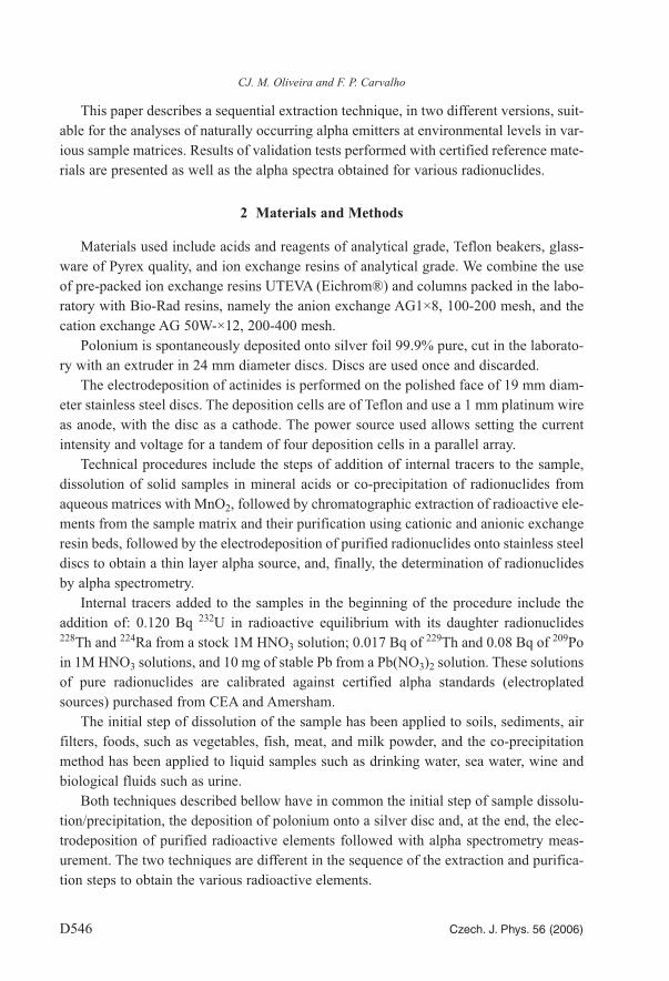
This paper describes a sequential extraction technique, in two different versions, suit-able for the analyses of naturally occurring alpha emitters at environmental levels in var-ious sample matrices. Results of validation tests performed with certified reference mate-rials are presented as well as the alpha spectra obtained for various radionuclides.
2 Materials and Methods
Materials used include acids and reagents of analytical grade, Teflon beakers, glass-ware of Pyrex quality, and ion exchange resins of analytical grade. We combine the useof pre-packed ion exchange resins UTEVA (Eichrom®) and columns packed in the labo-ratory with Bio-Rad resins, namely the anion exchange AG1×8, 100-200 mesh, and thecation exchange AG 50W-×12, 200-400 mesh.
Polonium is spontaneously deposited onto silver foil 99.9% pure, cut in the laborato-ry with an extruder in 24 mm diameter discs. Discs are used once and discarded.
The electrodeposition of actinides is performed on the polished face of 19 mm diam-eter stainless steel discs. The deposition cells are of Teflon and use a 1 mm platinum wireas anode, with the disc as a cathode. The power source used allows setting the currentintensity and voltage for a tandem of four deposition cells in a parallel array.
Technical procedures include the steps of addition of internal tracers to the sample,dissolution of solid samples in mineral acids or co-precipitation of radionuclides fromaqueous matrices with MnO2, followed by chromatographic extraction of radioactive ele-ments from the sample matrix and their purification using cationic and anionic exchangeresin beds, followed by the electrodeposition of purified radionuclides onto stainless steeldiscs to obtain a thin layer alpha source, and, finally, the determination of radionuclidesby alpha spectrometry.
Internal tracers added to the samples in the beginning of the procedure include theaddition of: 0.120 Bq 232U in radioactive equilibrium with its daughter radionuclides228Th and 224Ra from a stock 1M HNO3 solution; 0.017 Bq of 229Th and 0.08 Bq of 209Poin 1M HNO3 solutions, and 10 mg of stable Pb from a Pb(NO3)2 solution. These solutionsof pure radionuclides are calibrated against certified alpha standards (electroplatedsources) purchased from CEA and Amersham.
The initial step of dissolution of the sample has been applied to soils, sediments, airfilters, foods, such as vegetables, fish, meat, and milk powder, and the co-precipitationmethod has been applied to liquid samples such as drinking water, sea water, wine andbiological fluids such as urine.
Both techniques described bellow have in common the initial step of sample dissolu-tion/precipitation, the deposition of polonium onto a silver disc and, at the end, the elec-trodeposition of purified radioactive elements followed with alpha spectrometry meas-urement. The two techniques are different in the sequence of the extraction and purifica-tion steps to obtain the various radioactive elements.
CJ. M. Oliveira and F. P. Carvalho
D546 Czech. J. Phys. 56 (2006)

3 Experimental
Sample preparation. Solid samples such as soils (about 0.5 g) or freeze-dried foods(about 5 g) are weighted with an analytical balance. Isotopic tracers are pipetted onto thesample in a Teflon beaker and the sample covered with concentrated HNO3 and HCl. Wetashing is performed with gentle heating on a hotplate. Small amounts of HF are added todissolve the silica fraction. Occasionally, drops of H2O2 are added to oxidize organic mat-ter and to ensure conversion of oxidation state of tracers and radionuclides of the sample.After complete dissolution of the sample material, the solution is evaporated to drynessand the residue further treated with concentrated HCl to remove all traces of nitric acid.
Liquid samples such as water (3 – 5 L) are acidified at the collection point or imme-diately after filtration in order to avoid loss of radionuclides from the soluble phase ontothe container walls. The isotopic tracers are added and the sample homogenized by vig-orous stirring. At the moment of analyses, MnCl2 is added to the sample with KMnO4,and air bubbled overnight to ensure thorough mixing. Ammonium hydroxide is used toraise the pH to 8-9 under continuous stirring and pH control with a pH electrode.Manganese precipitates at this pH as a brown flock. The precipitate is allowed to settlefor several hours on the bottom of the beaker and the supernatant siphonated with apump. The final volume is centrifuged in order to discard as much water as possible. Theprecipitate is dissolved directly in the centrifuge tubes with HCl-H2O2 mixture, and trans-ferred with washes into a Pyrex beaker. The sample solution is gently evaporated to dry-ness.
Polonium deposition. The beaker containing the dry residue obtained either from thesolid sample wet ash or from the precipitate of aqueous sample will be used for poloni-um deposition. The sample residue is dissolved with 0.5M HCl and 200 mg of ascorbicacid is added. Polonium is spontaneously plated overnight onto a silver disc with mag-netic stirring at room temperature.
In both cases after polonium deposition, the solution is recuperated, evaporated to dryresidue and the residue treated with HNO3 to destroy the ascorbic acid. From this pointonwards, the sample is dissolved in a different solution according to the chromatograph-ic separation schema to be adopted.
If 226Ra determination is wanted, the formation of radioactive equilibrium betweenthe 228Th of the sample and its 224Ra daughter should be ensured. For this purpose thesample should be stored for at least 15 days from the collection date to the chromato-graphic separation. However, sample preparation and polonium deposition can be per-formed during these 15 days.
Chromatographic separation – Technique 1. The sample residue is dissolved in asmall volume of 3M HNO3 – 1M Al(NO3)3, 10 mL or 15 mL as needed. Add 2 mL or 3mL of ferrous sulphamate and swirl. Add 200 mg of ascorbic acid and mix [8]. This solu-tion is transferred to a UTEVA column (column I) preconditioned with 3M HNO3 (Fig.1). The sample volume plus washes until a total of 20 ml are collected in a first eluatefraction. This fraction contains Ra and Pb still mixed with other elements such as Fe, Mn,Pu and Am and it is saved to be purified in column II. Take a note of date and hour of thisseparation of 224Ra from 228Th for correction of 224Ra decay. Further work on column I
Sequential extraction procedure for alpha emitting natural radionuclides
Czech. J. Phys. 56 (2006) D547

includes the elution with 5 mL of 9M HCl to convert column to chloride system (discard)
and elution with 20 mL of the mixture 5M HCl-0.05M oxalic acid. The eluate of this frac-
tion contains thorium sufficiently purified to be electroplated. In samples from some
locations neptunium may occur and will be co-eluted with thorium. However, this is not
the case in most of the current environmental materials analyzed. Further elution of col-
umn I with 15 mL of 0.01M HCl, removes uranium from the resin bed, ready for the elec-
trodeposition step.
CJ. M. Oliveira and F. P. Carvalho
D548 Czech. J. Phys. 56 (2006)
UTEVA
AG 50Wx12
Col. II
Col. I
Col. IIIAG1x8
3M HNO3(Ra,Pb,Mn,Fe,...)
5M HCl
5M HCl
0- 40 ml 5M HCL (Pb)
2M HCl, 60 ml
H2O60ml
Pb Th U
9M HCldiscard
5M HCl -0.05M Oxalic acid
0.01M HCl
40-90 ml 5M HCl
Ra
Sample in
3M HNO3 + 1M Al(NO3)3 +0.6M ferrous sulphamate + ascorbic acid
3M HNO3
9M HCl5M HCl -0.05M Oxalic acid
2M HCldiscard
Fig1
0.01M HCl
Fig. 1. Flow diagram of the sequential extraction and purification of radionuclides afterpolonium deposition (Technique 1).

Sequential extraction procedure for alpha emitting natural radionuclides
Czech. J. Phys. 56 (2006) D549
The first fraction obtained, containing Ra and Pb, is slowly evaporated to dryness andthe residue treated with small volumes of concentrate HCl to convert salts into chlorides.The residue is dissolved with 6mL of 5M HCL and fed onto a cation exchange columnAG 50W×12, with 4mL resin bed previously equilibrated with this acid. The first frac-tion of eluate, 40 mL corresponding to the sample volume plus washes with 5M HCl,contain lead and it is saved for further purification. Continuing the column elution with5M HCl, the eluate from 40 mL to 90 mL will contain radium. The radium solution issaved for the electrodeposition step.
The fraction containing lead is taken to dryness and the residue dissolved in a smallvolume of 2M HCl. This lead solution is fed onto column III, an anion exchange resinAG1×8, with 10 mL resin bed previously equilibrated with 2M HCl. The first 60 mL ofeluate corresponding to the sample volume and washes are discarded. Elution with 60 mLof distilled water will strip lead from the column. Lead solution is saved for co-precipi-tation of 210Pb as lead chromate.
Technique 2. After polonium plating, the sample solution is evaporated to residue anddissolved with 6 mL of 5M HCl (Fig. 2). This solution is fed into a column with 4 mLAG 50W×12 resin bed pre-equilibrated with 5M HCl (column I). The sample solutionplus washes with 5M HCl until 40 mL of eluate volume, will drain the U and Pb throughthe column. This fraction is saved for further purification.
Continuing the elution of the column I with 5M HCl, radium is eluted in the fractionfrom 40 to 90 mL, sufficiently purified to be electroplated. Take a note of date and hourof radium elution for correction of 224Ra radioactive decay. Further elution of the columnwith 40 mL of 0.5M oxalic acid will remove thorium, purified for electrodeposition.
The first eluate fraction of column I, containing U and Pb, is taken to dryness and theresidue dissolved in a small volume, 10 mL (or 15 mL if residue is abundant) of 3MHNO3 – 1M Al(NO3)3. Add 2 mL (or 3 mL) of ferrous sulphamate and swirl. Add 200mg of ascorbic acid and mix. This solution is fed in to a UTEVA column (column II) preconditioned with 3M HNO3.
The sample volume plus two washes of the beaker and column, 5 mL of 3M HNO3
each, are recuperated as a first eluate fraction. This fraction contains Pb but still requiresfurther purification. Continuing elution of column II with 5 mL of 9M HCl will convertcolumn to chloride system. Elution with 20 mL of 5M HCl-0.05M oxalic acid will cleanup the column and will remove traces of thorium and iron that later could interfere withthe electrodeposition. Elution with 15 mL of HCl 0.01M strips uranium from the column,ready to go for electrodeposition.
The first eluate fraction, containing lead, is slowly evaporated and the residue recu-perated in concentrated HCl, dried again to remove nitric acid and redissolved in a smallvolume of 2M HCl. This solution is passed through an AG1×8 column, pre-equilibratedwith 2M HCl, followed by 60 mL 2M HCl washes. Lead is then removed from the col-umn with 60 mL of distilled H2O and saved for precipitation as lead chromate.
Electrodeposition. Electrodeposition of uranium and thorium is performed on the pol-ished surface of stainless steel discs, in Teflon cells with a platinum wire as anode,inspired on the design proposed by Talvatie [9]. The eluate fraction containing uraniumor thorium for electrodeposition, are evaporated to dryness and the residue treated with a

CJ. M. Oliveira and F. P. Carvalho
D550 Czech. J. Phys. 56 (2006)
Fig 2
U Ra ThPb
Col. I
Col. II
Col. III
AG 50Wx12
UTEVA
AG 1x 8
5M HCl0-40 ml (U,Pb)
5M HCl40-90ml 0.5M Oxalic acid
40 ml
3M HNO3
3M HNO3
2M HCl
9M HCl discard5M HCl-0.05M Oxalic acdiscard
0.01M HCl
Sample in 5M HCl
5M HCl
2M HCldiscard
H2O
3M HNO3 + 1M Al(NO3)3 +0.6M ferrous sulphamate + ascorbic acid
2M HCl
0.5M Oxalic acid
9M HCl5M HCl-0.05M Oxalic ac
0.01M HCl
Fig. 2. Flow diagram of the sequential extraction and purification of radionuclides after poloni-um deposition (Technique 2)
few drops of HNO3 to convert salts. The dry residue is dissolved with 10 mL of ammo-
nium solution (100g L–1 of (NH4)2SO4) at pH 2 adjusted with H2SO4 and transferred to
the electrode position cell with rinses with ammonium solution [2]. The electrodeposition
is made during 1 hour with the electric current of 1A. The disc surface is than rinsed with
distilled water and ethanol and strongly heated on the hotplate. Usually, a blue-like colour
taints the stainless steel surface and no deposit is visible.

Sequential extraction procedure for alpha emitting natural radionuclides
Czech. J. Phys. 56 (2006) D551
Radium is electrodeposited following a different procedure. The solution purifiedabove (radium fraction for electrodeposition) is gently evaporated to dryness. The residueis dissolved with 0.35M ammonium acetate – 0.1M HNO3 and the solution transferredwith washes to the deposition cell [10]. Electrodeposition on a stainless steel disc is per-formed during 3 hours with a electric current of 600 mA. At the end, the disc surface isclear and no deposit should be visible.
Alternatively, radium can be electroplated from a solution of 1 mL of 0.1M HNO3 +9 mL of ethanol, during 30 min with 70 mA electric current [2,11]. However, this methodhas no advantage over the one described above.
Lead is recuperated from the purified solution obtained as described in section above,according to the following procedure. The solution is slowly evaporated to dryness andthe residue dissolved with a few drops of HNO3. Evaporate again and dissolve the residuewith 30 mL of distilled water and 1mL of acetate buffer solution. Bring solution to boil-ing and, always on the hotplate, add 1 mL of 10% Na2CrO4 solution [2]. A yellow pre-cipitate of PbCrO4 is formed. Maintain the heating for 30 min. Allow to cool and recu-perate the precipitate by filtration. Determine the radiochemical yield by the gravimetricmethod and store the filter with the precipitate for several months (4-6). Then dissolvethe filter and precipitate in the presence of a new amount of the tracer 209Po and proceedwith the deposition of polonium to determine 210Pb through the in-growth of 210Po sincethe date of precipitation. Alternatively, the filter with lead chromate may be mounted ona sample holder and counted with a low background beta counter using the beta radiationemitted by 210Bi and after formation of the radioactive equilibrium of 210Bi with 210Pb.However, the determination of 210Pb through the 210Po in growth is much more sensitiveand accurate.
Alpha measurements. The measurement of radionuclides is performed using siliconsurface barrier detectors R-Type, 100 µm depletion depth, and ion implanted detectorsULTRA-AS (Ortec). The ion implanted detectors, 450 mm2 active surface, are assembledin groups of 8 in OCTETE Plus (EG&G). Counting time is adjusted to the activity in thesample discs in order to obtain a counting statistics better than 5% relative uncertainty.
The performance of purification and electrodeposition steps is crucial to obtain alphasources of good quality. Typically in our sources the energy resolution measured on the210Po peak is 45-50 keV with surface barrier detectors and 20 keV with ion implanteddetectors. Fig. 3 shows typical spectra of environmental samples.
The average recovery yields obtained are reported in Table 1 based on analyticalresults of a large number of environmental samples with different matrices. In general theyields are consistently high and similar for both techniques.
Validation and Testing. Validation of the procedures was made for reproducibility andaccuracy using certified reference materials from the IAEA in the available matrices.
The IAEA-385 reference material, sediment from the Irish Sea, was analysed by bothtechniques described above. Table 2 shows the results, as well as the IAEA recommend-ed concentration values for this material. There is, in general, a good agreement betweenthe results obtained by each technique used by us, and also between the results of ourdeterminations and the IAEA values.

CJ. M. Oliveira and F. P. Carvalho
D552 Czech. J. Phys. 56 (2006)
Fig. 3. Alpha spectrogram of sources prepared using the techniques described herein. From topto bottom: uranium, thorium, radium and polonium spectra. The main energies are indicated for
each radionuclide.

Sequential extraction procedure for alpha emitting natural radionuclides
Czech. J. Phys. 56 (2006) D553
Sample matrix
Separation technique
n U Th Ra Po Pb
2 16 0.71 ± 0.10 0.37 ± 0.10 0.16 ± 0.09 0.86 ± 0.09 0.47 ± 0.20Water 1 23 0.58 ± 0.17 0.51 ± 0.18 0.27 ± 0.20 0.86 ± 0.09 0.47 ± 0.16
Suspended matter
1 21 0.83 ± 0.09 0.59 ± 0.17 0.31 ± 0.11 0.84 ± 0.11 0.48 ± 0.10
Biota 1 19 0.88 ± 0.12 0.47 ± 0.18 0.22 ± 0.12 0.80 ± 0.08 0.28 ±0.24
Sediments 1 17 0.83 ± 0.06 0.73 ± 0.14 0.09 ± 0.02 0.88 ± 0.09 --
238U 235U 234U 232Th 230Th 226Ra210Po
(=210Pb)IAEA-385Recommended values Mean and range
29.428.0-30.5
1.361.24-1.51
27.225.8-28.6
33.832.6-34.5
31.830.0-34.9
22.721.8-24.0
35.531.2-38.9
Technique 1 Mean ± 1 σ of the mean (n=3)
31.4±0.8 1.44±0.01 31.3±1.2 33.3±0.8 31.6±1.3 25.0±4.2 36.9±0.8
Technique 2 Mean ± 1 σ of the mean (n=4)
31.7±1.0 1.36±0.08 30.3±0.7 29.3±1.1 32.0±2.7 22.0±1.6 36.9±0.8
Table 1. Recovery yields (mean ± 1σ) of the radiochemical separation procedures applied in theanalyses of n independent environmental samples, determined with the internal isotopic tracers
added to the sample
Table 2. Analytical results for reference material IAEA-385, marine sediment from the Irish Sea(Bq kg–1 dry weight)
The IAEA-414 reference material, mixed fish sample from the Irish Sea, was ana-lyzed as an example of organic matrix with high fat content. Our results are comparableto the IAEA values and show that a consistent analytical performance is obtained in thismatrix also (Table 3).
No liquid sample is available as a reference material. We tested reproducibility of ourprocedure in a water sample from the tap. A 30 L sample was split into 3 portions of equalvolume, acidified and than the subsamples were analyzed in parallel following the pro-cedure outlined above as technique 1. Results are in the confidence interval of the mean± 1 standard deviation, indicating high reproducibility (Table 4).
Robustness of the performance when the technique is applied to samples with differ-ent matrix was tested analyzing cabbage, potatoes, fish filet, meat, water, urine, soils andsediments with high yield of recovery and complete separation of radioactive elements.Precision was tested also through participation in analytical inter-laboratory worldwidecomparisons analysing blind samples (round robin tests) organized by IAEA and WHO,with good results.[12-14] Table 5 shows the results of the intercomparison exercise foruranium isotopes in a urine sample.

CJ. M. Oliveira and F. P. Carvalho
D554 Czech. J. Phys. 56 (2006)
238U 235U 234U 226Ra 210Po (=210Pb) IAEA-414 Recommended values Mean and range
1.11 (1.06-1.117)
0.050 (0.045-0.058)
1.22 (1.14-1.27)
1.40 (0.59-1.76)
2.22 (1.55-2.60)
Mean ± 1 σ of the mean (n=3) Technique 1
1.08±0.08 0.064±0.012 1.38 ±0.09 1.10±0.02 1.96±0.03
Table 3. Analytical results for the certified reference material IAEA-414, mixed with fish muscle from the Irish Sea
Table 4. Analytical results (Bq m–3) of three replicate water sub samples
Table 5. IAEA interlaboratory analytical comparison results (mBq in sample ± 2σ) in a urinesample organized by the IAEA [12]
Subsample
238U 235U 234U 232Th 230Th 226Ra210Po
a 24.9±0.9 1.2±0.1 27.3±0.9 0.16±0.07 0.96±0.13 17.0±1.3 4.3±0.2 b 24.2±0.8 1.1±0.1 27.6±0.9 0.09±0.03 0.79±0.09 17.4±1.3 4.5±0.2 c 24.2±0.8 1.2±0.1 25.9±0.9 0.15±0.05 0.93±0.12 18.5±1.7 4.4±0.2 Mean ± 1 σ of themean
24.4±0.2 1.2±0.03 26.9±0.5 0.13±0.02 0.93±0.08 17.6±0.4 4.4±0.06
238U 235U 234UIAEA Reference value
21.5±0.25 0.99±0.012 20.7±0.406
Our determination 20.4±1.01 1.20±0.205 19.6±0.977
The comparison with other sequential extraction techniques used before, show thatthe current technique has the advantages of separating a large set of radioactive elementsin less time (about half of the time required before), with similar high performance, lesschromatography columns, less manipulation, and generating a lower volume of acidwaste. The disadvantage is the relatively higher cost of each analysis with the purchaseof ready-to-use chromatography columns although this becomes compensated by thelower technical time needed in the analytical work.
4 Final considerations
This radiochemical procedure, in comparison with procedures used before, allows forobtaining results faster. In three days, either by technique 1 or technique 2, samples arecompletely analyzed from the polonium deposition to the completion of electrodeposi-tion of all other radionuclides on discs.
Sequential extraction procedure for alpha emitting natural radionuclides
Czech. J. Phys. 56 (2006) D555
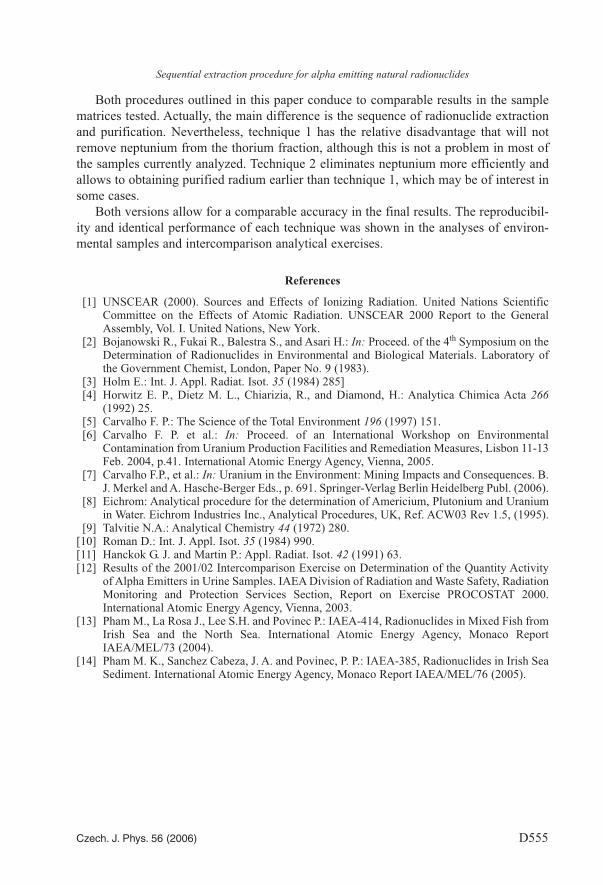
Both procedures outlined in this paper conduce to comparable results in the samplematrices tested. Actually, the main difference is the sequence of radionuclide extractionand purification. Nevertheless, technique 1 has the relative disadvantage that will notremove neptunium from the thorium fraction, although this is not a problem in most ofthe samples currently analyzed. Technique 2 eliminates neptunium more efficiently andallows to obtaining purified radium earlier than technique 1, which may be of interest insome cases.
Both versions allow for a comparable accuracy in the final results. The reproducibil-ity and identical performance of each technique was shown in the analyses of environ-mental samples and intercomparison analytical exercises.
References
[1] UNSCEAR (2000). Sources and Effects of Ionizing Radiation. United Nations ScientificCommittee on the Effects of Atomic Radiation. UNSCEAR 2000 Report to the GeneralAssembly, Vol. I. United Nations, New York.
[2] Bojanowski R., Fukai R., Balestra S., and Asari H.: In: Proceed. of the 4th Symposium on theDetermination of Radionuclides in Environmental and Biological Materials. Laboratory ofthe Government Chemist, London, Paper No. 9 (1983).
[3] Holm E.: Int. J. Appl. Radiat. Isot. 35 (1984) 285][4] Horwitz E. P., Dietz M. L., Chiarizia, R., and Diamond, H.: Analytica Chimica Acta 266
(1992) 25.[5] Carvalho F. P.: The Science of the Total Environment 196 (1997) 151.[6] Carvalho F. P. et al.: In: Proceed. of an International Workshop on Environmental
Contamination from Uranium Production Facilities and Remediation Measures, Lisbon 11-13Feb. 2004, p.41. International Atomic Energy Agency, Vienna, 2005.
[7] Carvalho F.P., et al.: In: Uranium in the Environment: Mining Impacts and Consequences. B.J. Merkel and A. Hasche-Berger Eds., p. 691. Springer-Verlag Berlin Heidelberg Publ. (2006).
[8] Eichrom: Analytical procedure for the determination of Americium, Plutonium and Uraniumin Water. Eichrom Industries Inc., Analytical Procedures, UK, Ref. ACW03 Rev 1.5, (1995).
[9] Talvitie N.A.: Analytical Chemistry 44 (1972) 280.[10] Roman D.: Int. J. Appl. Isot. 35 (1984) 990.[11] Hanckok G. J. and Martin P.: Appl. Radiat. Isot. 42 (1991) 63.[12] Results of the 2001/02 Intercomparison Exercise on Determination of the Quantity Activity
of Alpha Emitters in Urine Samples. IAEA Division of Radiation and Waste Safety, RadiationMonitoring and Protection Services Section, Report on Exercise PROCOSTAT 2000.International Atomic Energy Agency, Vienna, 2003.
[13] Pham M., La Rosa J., Lee S.H. and Povinec P.: IAEA-414, Radionuclides in Mixed Fish fromIrish Sea and the North Sea. International Atomic Energy Agency, Monaco ReportIAEA/MEL/73 (2004).
[14] Pham M. K., Sanchez Cabeza, J. A. and Povinec, P. P.: IAEA-385, Radionuclides in Irish SeaSediment. International Atomic Energy Agency, Monaco Report IAEA/MEL/76 (2005).
Copyright © 2022 FDOKUMEN