Sensitivity to implant materials in patients with total knee arthroplasties
7
Sensitivity to implant materials in patients with total knee arthroplasties Donatella Granchi a, * , Elisabetta Cenni a , Domenico Tigani b , Giovanni Trisolino b , Nicola Baldini a,b , Armando Giunti a,b a Laboratory for Pathophysiology of Orthopedic Implants, Istituti Ortopedici Rizzoli, via di Barbiano 1/10, 40136 Bologna, Italy b Department of Orthopedics and Traumatology, Istituti Ortopedici Rizzoli, Bologna, Italy Received 26 September 2007; accepted 27 November 2007 Available online 21 December 2007 Abstract Materials used for total knee arthroplasty (TKA), may elicit an immune response whose role in the outcome of the arthroplasty is still unclear. The aim of this study was to evaluate the frequency of sensitization in patients who had undergone TKA, and the clinical impact of this event on the outcome of the implant. Ninety-four subjects were recruited, including 20 patients who had not yet undergone arthroplasty, 27 individuals who had a well-functioning TKA, and 47 patients with loosening of TKA components. Sensitization was detected by using patch testing includ- ing haptens representative of cobalt-based alloys (CoCrMo), titanium-based alloys (TiAlV), and bone cements. The frequency of positive skin reactions to metals increased significantly after TKA, either stable or loosened (No Implant 20%; Stable TKA 48.1%, p ¼ 0.05; Loosened TKA 59.6%, p ¼ 0.001, respectively). We found a higher frequency of positive patch testing to vanadium in patients who had a Stable TKA with at least one TiAlV component (39.1%, p ¼ 0.01). The medical history for metal allergy seems to be a risk factor, because the TKA failure was fourfold more likely in patients who had symptoms of metal hypersensitivity before TKA. The prognostic value was supported by survival anal- ysis, because in these individuals the outcome of the implant was negatively influenced (the logrank test Chi square 5.1, p ¼ 0.02). This study confirms that in patients with a TKA the frequency of positive patch testing is higher than in the normal population, although no predictive value is attributable to the sensitization because patch testing was not able to discriminate between stable and loose implants. On the contrary, the presence of symptoms of metal allergy before implantation should be taken into account as a potential risk factor for TKA failure. Ó 2007 Elsevier Ltd. All rights reserved. Keywords: Knee replacement; Hypersensitivity; Cobalt alloy; Titanium alloy 1. Introduction Total knee arthroplasty (TKA) has been a major advance in the treatment of knee disabilities, predictably achieving excel- lent results at a long-term follow-up with relatively low perio- perative morbidity [1,2]. In over 95% of patients TKA relieves pain, improves functional status, and restores most of their normal activities of daily living. In spite of the excellent out- come, TKA can fail over time, and the revision rate over 5 or more years is 2% of knees and 2.1% of patients [1]. TKA fail- ure is generally multifactorial even though the main reasons may be ascribed to mechanical and biological causes [3,4]. Chronic inflammation following the generation of wear parti- cles has been recognized as the main biological mechanism leading to implant failure [5]. Moreover, if degradation prod- ucts are able to interact with the immune system, undesirable immunotoxic effects may be induced, including a delayed-type hypersensitivity reaction (DTH) [6e9]. Metals and acrylic ce- ments represent the main components of TKA: in contact with biological fluids they undergo corrosion and wear, and metal ions or other molecules may induce a DTH [10]. Nickel, cobalt, and chromium are known to be the most common sen- sitizers, [9,11] but also hypersensitivity to titanium and vana- dium has been described [12,13]. Polymeric biomaterials, namely acrylic bone cements, are not easily chemically de- graded and immunogenic reactions to poly-methyl-methacry- late or other constituents have been occasionally reported [14]. The possible correlation between hypersensitivity and * Corresponding author. Tel./fax: þ39 51 6366748. E-mail address: [email protected] (D. Granchi). 0142-9612/$ - see front matter Ó 2007 Elsevier Ltd. All rights reserved. doi:10.1016/j.biomaterials.2007.11.038 Available online at www.sciencedirect.com Biomaterials 29 (2008) 1494e1500 www.elsevier.com/locate/biomaterials
-
Upload
independent -
Category
Documents
-
view
3 -
download
0
Transcript of Sensitivity to implant materials in patients with total knee arthroplasties
Available online at www.sciencedirect.com
Biomaterials 29 (2008) 1494e1500www.elsevier.com/locate/biomaterials
Sensitivity to implant materials in patients with total knee arthroplasties
Donatella Granchi a,*, Elisabetta Cenni a, Domenico Tigani b, Giovanni Trisolino b,Nicola Baldini a,b, Armando Giunti a,b
a Laboratory for Pathophysiology of Orthopedic Implants, Istituti Ortopedici Rizzoli, via di Barbiano 1/10, 40136 Bologna, Italyb Department of Orthopedics and Traumatology, Istituti Ortopedici Rizzoli, Bologna, Italy
Received 26 September 2007; accepted 27 November 2007
Available online 21 December 2007
Abstract
Materials used for total knee arthroplasty (TKA), may elicit an immune response whose role in the outcome of the arthroplasty is still unclear.The aim of this study was to evaluate the frequency of sensitization in patients who had undergone TKA, and the clinical impact of this event onthe outcome of the implant. Ninety-four subjects were recruited, including 20 patients who had not yet undergone arthroplasty, 27 individualswho had a well-functioning TKA, and 47 patients with loosening of TKA components. Sensitization was detected by using patch testing includ-ing haptens representative of cobalt-based alloys (CoCrMo), titanium-based alloys (TiAlV), and bone cements. The frequency of positive skinreactions to metals increased significantly after TKA, either stable or loosened (No Implant 20%; Stable TKA 48.1%, p¼ 0.05; Loosened TKA59.6%, p¼ 0.001, respectively). We found a higher frequency of positive patch testing to vanadium in patients who had a Stable TKA with atleast one TiAlV component (39.1%, p¼ 0.01). The medical history for metal allergy seems to be a risk factor, because the TKA failure wasfourfold more likely in patients who had symptoms of metal hypersensitivity before TKA. The prognostic value was supported by survival anal-ysis, because in these individuals the outcome of the implant was negatively influenced (the logrank test Chi square 5.1, p¼ 0.02). This studyconfirms that in patients with a TKA the frequency of positive patch testing is higher than in the normal population, although no predictive valueis attributable to the sensitization because patch testing was not able to discriminate between stable and loose implants. On the contrary, thepresence of symptoms of metal allergy before implantation should be taken into account as a potential risk factor for TKA failure.� 2007 Elsevier Ltd. All rights reserved.
Keywords: Knee replacement; Hypersensitivity; Cobalt alloy; Titanium alloy
1. Introduction
Total knee arthroplasty (TKA) has been a major advance inthe treatment of knee disabilities, predictably achieving excel-lent results at a long-term follow-up with relatively low perio-perative morbidity [1,2]. In over 95% of patients TKA relievespain, improves functional status, and restores most of theirnormal activities of daily living. In spite of the excellent out-come, TKA can fail over time, and the revision rate over 5 ormore years is 2% of knees and 2.1% of patients [1]. TKA fail-ure is generally multifactorial even though the main reasonsmay be ascribed to mechanical and biological causes [3,4].
* Corresponding author. Tel./fax: þ39 51 6366748.
E-mail address: [email protected] (D. Granchi).
0142-9612/$ - see front matter � 2007 Elsevier Ltd. All rights reserved.
doi:10.1016/j.biomaterials.2007.11.038
Chronic inflammation following the generation of wear parti-cles has been recognized as the main biological mechanismleading to implant failure [5]. Moreover, if degradation prod-ucts are able to interact with the immune system, undesirableimmunotoxic effects may be induced, including a delayed-typehypersensitivity reaction (DTH) [6e9]. Metals and acrylic ce-ments represent the main components of TKA: in contact withbiological fluids they undergo corrosion and wear, and metalions or other molecules may induce a DTH [10]. Nickel,cobalt, and chromium are known to be the most common sen-sitizers, [9,11] but also hypersensitivity to titanium and vana-dium has been described [12,13]. Polymeric biomaterials,namely acrylic bone cements, are not easily chemically de-graded and immunogenic reactions to poly-methyl-methacry-late or other constituents have been occasionally reported[14]. The possible correlation between hypersensitivity and
Table 1
Data on the patients
No Implant
(n¼ 20)
Stable TKA
(n¼ 27)
Loosened TKA
(n¼ 47)
Gender
Males 6 (28.7%) 5 (18.5%) 16 (34.0%)
Females 14 (71.3%) 22 (81.5%) 31 (66.0%)
Age (years)
Mean� standard deviation 65.2� 8 66.1� 10 70.4� 6
Median 69 68 70
Range 42e84 42e84 57e79
Indication for arthroplasty
Idiopathic OAa 18 (90.0%) 23 (85.1%) 42 (89.4%)
Post-traumatic OA 2 (10%) 1 (3.7%) 3 (6.4%)
Other causesb e 3 (11.1%) 2 (4.2%)
Positive medical history
for metal allergy
2 (10%) 1 (3.7%) 7 (14.9%)
Follow-up (months)
Median e 18 24
Range e 9.6e120 4.8e132
Other implants e 8 (29.6%) 11 (23.4%)
Metal composition of the implant(s)
CoCrMo alloy e 3 (11.1%) 16 (34.0%)
1495D. Granchi et al. / Biomaterials 29 (2008) 1494e1500
implant failure has been previously investigated, but the mainquestion about the causeeeffect relationship between theseevents has not yet been solved.
One of the major difficulties in understanding the clinicalimplications of hypersensitivity to the implant components isthe lack of universally accepted testing methods [15]. Severalin vitro tests based on the reactivity of immune cells tometaleprotein complexes have been proposed in diagnosinga systemic hypersensitivity to the implant components[11,16,17], but they have some faults that limit large-scale ap-plication, including the high costs and the need to be performedin qualified laboratories. In addition, the number of immunecells recruited from biological samples does not allow an ex-haustive assay of all the immunogenic substances containedin metal alloys and bone cements, and the elevated toxicityof some chemicals often hampers their in vitro testing[11,18]. Alternatively, the in vivo approach, i.e. the epicutane-ous skin-testing (patch testing), is cheap and allows to estimatesimultaneously several haptens [19]. Although patch testing isthe most common method used to diagnose contact allergy tometals, its validity in determining a deep-tissue hypersensitiv-ity lets some doubts [10,11]. Nevertheless, its usefulness in de-tecting the sensitization to implant materials may be improvedif patients are tested with an appropriate series of haptens ac-cording to the prosthesis components [19,20].
An additional limit in establishing the role of the sensitivityto the implant components is the paucity of clinical studiesproviding clear data of a connection between metal sensitivityand outcome of the implant [15]. The majority of investigatorssuggest that hypersensitivity can be a contributing factor to im-plant failure, because of the high proportion of metal DTH inpatients with prosthesis loosening [10], the shorter lifespan ofthe implant in patients having positive patch testing [19], andthe histological findings of hypersensitivity-like reaction in tis-sue around the artificial joint, especially in metal-on-metalbearings [21e25]. Most studies have been performed in co-horts of patients undergoing a total hip arthroplasty (THA)[10], while few data are available for patients with TKA [9].From a theoretical point of view, the proportion of positiveskin reactions in patients with TKA could differ from those ob-served in THA for several reasons, including the biomechanicsof the joint influencing the metal ion release.
The main goals of our pilot study were to evaluate (i) thefrequency of skin sensitization in patients who had undergoneTKA, and (ii) the clinical impact of this event on the outcomeof the implant. For this purpose we chose to use patch testingby applying a panel of haptens representative of cobalt-basedalloys (CoCrMo), titanium-based alloys (TiAlV), and bonecements.
2. Patients and methods
TiAlV alloy e 1 (3.7%) 2 (4.3%)
CoCrMo/TiAlV alloys e 23 (85.2%) 27 (57.4%)
2.1. Study designUnknown e e 2 (4.3%)
Cemented TKA 26 (96.3%) 43 (95.6%)
a Osteoarthritis.b Other causes were fractures (2), septic OA (2), rheumatoid arthritis (1).
Ninety-four Caucasian individuals were enrolled in this retrospective
caseecontrol study. The study design was approved by the institutional ethical
committee on human research and signed informed consent was obtained from
the patients. The sample size was calculated on the basis of the literature data
considering the prevalence of sensitization in general population and the mean
value of the sensitization frequency recorded in patients undergoing total joint
replacement [10,11,19]. A personal history was collected from each individ-
ual, including details concerning diseases that had led to TKA, other diseases,
presence of other bone or joint implants, previous orthopaedic surgery, and
drug intake. A medical history of hypersensitivity to metal before TKA was
documented in 10 patients (10.6%): it was verified by reports of previous
skin-testing or investigated through a questionnaire aiming to verify the pres-
ence of contact dermatitis symptoms. Nobody of our patients presented cuta-
neous symptoms of metal allergy at time of the study inclusion. Patients who
were using corticosteroid or other immunosuppressive drugs were excluded
from the study.
Consecutive patients were recruited among individuals who were admitted
to the orthopaedic department for pre- and post-operative medical examina-
tion planned for the TKA surgery. Three groups of patients were evaluated:
the first group (No Implant) included 20 patients who were candidates for
TKA; the second group consisted of 27 individuals who had already under-
gone TKA that was clinically and radiographically stable (Stable TKA); the
third group consisted of 47 patients who had already undergone total knee
arthroplasty but showed clinical and radiographic evidence of failure. More
details on the groups are shown in Table 1. The implantation of knee prosthe-
sis in both pre- and stable TKA groups, as well as the replacement of failed
TKA, was performed by one orthopaedic surgeon who is specialized in knee
surgery. The knee performance was valued using the ‘Knee Society Clinical
Rating System’ [26] that establishes a ‘total knee score’ depending on pain,
range of motion, and stability, and a ‘function score’ based on walking and
stair climbing. Radiographic diagnosis of failure was made on the results of
anteroposterior, lateral and skyline views according to the roentgenographic
evaluation scoring system of the ‘Knee Society’ [27]. A shift or subsidence
of implant position on sequential radiographs also indicates loosening [28],
as well as progressive widening of the cement-bone or bone prosthesis inter-
face and cement fragmentation under a component [29]. The total-body bone
scan with technetium-99m-labeled hydroxymethylene diphosphonate was

1496 D. Granchi et al. / Biomaterials 29 (2008) 1494e1500
performed to confirm the radiographic suspicion of loosening. In patients who
had an increase in inflammation markers, either hexamethylpropyleneamine
oxide-labeled granulocyte scintigraphy or aspiration were performed to prove
the presence of infection [30].
Nineteen TKA patients (25.7%) had an additional bone or joint implant at
another site, including hip prosthesis and fixation devices. The type of implant
was known in 97% of cases, since two patients arrived to our observation with
an antibiotic-loaded bone cement spacer, and neither information nor radio-
graphic documentation of the previous implant were available.
2.2. Patch testing
Hypersensitivity to metals was tested before the surgery by using the
following haptens: 5% nickel sulphate, 1% cobalt chloride, 2% chromium
trichloride, 0.5% potassium dichromate, 2% ferric chloride, 2% molybdenum
chloride, 2% manganese chloride, 2% vanadium trichloride, 1% aluminium
chloride, 1% niobium chloride, and 2% titanium dioxide. The last hapten
was chosen as an oxide, rather than salt, to mimic the host reactivity of tita-
nium-based devices, which form on their surface a titanium dioxide film.
Haptens for bone cements were the following: 5% methyl-methacrylate,
2% butyl-methacrylate, 2% triethylene glycol dimethacrylate, 2% ethylene
glycol dimethacrylate, 2% N,N-dimethyl-paratoluidine, 5% hydroxy-ethyl-
methacrylate, 2% benzoyl-peroxide, and 1% hydroquinone monobenzyl ether
(F.I.R.M.A SpA, Florence, Italy). Vaseline, which was the carrier in the patch
testing, was assayed as a negative control. A drop of each hapten was
smeared on Haye’s chamber test, which was applied to the back of the
patient. After 48e72 h, patch testing was evaluated by observers who were
unaware of clinical information or the composition of the implant. Skin reac-
tions were graded according to their intensity as 0 (no reaction), plus or
minus reaction (week erythema only, doubtful), 1þ (erythema with edema
covering at least 50% of the patch test site), 2þ (erythema and papules cov-
ering at least 50% the patch test site), and 3þ (vesicles or bullae covering
least 50% of the patch test site) [31].
2.3. Calculations and statistical analysis
Table 2
Frequency (%) of positive skin reactions to metal haptens in patient groups
No Implant TKA
(n¼ 20)
Stable TKA
(n¼ 27)
Loosened TKA
(n¼ 47)
At least one hapten 15.0b,c 44.4 57.4
Nickel sulphate 10.0 7.4 23.4
Chromiuma 5.0 7.4 19.1
Cobalt chloride 5.0 11.1 12.8
Ferric chloride 0 0 2.2
Molybdenum chloride 0 0 4.3
Manganese chloride 5.0 3.7 23.4
Titanium dioxide 5.0 0 2.1
Quantitative results were expressed as mean plus and minus standard de-
viation. The nonparametric analysis of variance (KruskaleWallis) was applied
to detect the effects of the various clinical variables on the quantitative results,
and the ManneWhitney test was applied to detect specific differences between
groups. In each group, the frequency of positive patch testing was calculated,
and the Chi square test with Fisher’s exact test were used to highlight differ-
ences attributable to clinical variables. Probabilistic measures, such as sensi-
tivity, specificity, and likelihood ratios, as well as their 95% confidence
limits (95% CL), were used to describe the ‘diagnostic accuracy’ of our series
of haptens in diagnosing DTH patients who had a previously documented
metal hypersensitivity [32]. The same method was used to assess the relation-
ship between patch testing result and TKA status. In this context, specificity
and sensitivity signified the effectiveness of the patch testing at excluding
or at detecting TKA failure, and positive and negative likelihood ratios de-
scribed the discriminatory properties of positive and negative test results.
Positive likelihood ratios above 10 and negative likelihood ratios below 0.1
provide convincing diagnostic evidence, whereas those above 5 and below
0.2 give good diagnostic evidence [33]. The KaplaneMeier product-limit
method was used to estimate the 5-year survival frequency of the TKA, in
our case series since revision was considered as the end point of interest;
the logrank ‘‘ManteleCox’’ test was applied to highlight significant differ-
ences among the survival curves of groups [34]. Differences were considered
significant if the p value was less or equal to 0.05.
3. Results
Aluminium chloride 0 0 0Niobium chloride 0 0 0
Vanadium trichloride 5.0b 33.3 19.1
3.1. Sensitization and status of the implant a Positive patch testing for at least one of the two tested haptens.b Chi square p value< 0.05 in comparison to the ‘Stable TKA’ group.c Chi square p value< 0.005 in comparison to the ‘Loosened TKA’ group.The frequency of skin positive reaction was not influencedby age or presence of other implants, but in all groups females
showed a higher percentage of positive patch testing (data notshown). The frequency of positive skin reactions to at least onehapten (metal or cement) was significantly higher in group ofpatients tested after TKA (No Implant 20%, TKA 55.4%; Chisquare 7.9, p¼ 0.005), and no differences were observedbetween stable and loosened implants (Stable TKA 48.1%,Loosened TKA 59.6%; Chi square 0.91, p¼ 0.46). No differ-ences were found with regards to the intensity of skin reaction:the proportion of 1þ, 2þ, and 3þ patch testing was similar inall groups and no doubtful reactions were observed.
In ‘No Implant’ group, the frequency of positive patch test-ing for at least one metal hapten was 15%, and differed signif-icantly from that observed in patients with Stable TKA (44.4%,Chi square 4.6, p¼ 0.05) and Loosened TKA (57.4%, Chisquare 10.2, p¼ 0.001). When the reactivity against each hap-ten was considered, we found a higher frequency of positivepatch testing to vanadium which resulted significant in patientswith Stable TKA ( p¼ 0.02) (Table 2). Niobium chloride wastested only in half of the patients and it was never foundpositive.
The frequency of positive reaction to at least one of thebone cement components (Table 3) was similar in all patientgroups. No significant difference was found when positiveskin reaction for each hapten was considered.
The group of patients with Stable TKA was split accordingto the presence of clinical symptoms (Table 4), and the 14patients who complained of a moderate pain related to theimplant showed a high frequency of sensitization to metalhaptens, mostly to vanadium. The proportion of positive patchtesting was higher than that found in patients without clinicalsymptoms, and significantly higher than that observed in pa-tients who were candidated for TKA. These findings did notdepend on the different length of follow-up in the two groups,because it was identical in patients with and without clinicalsymptoms (median time 18 months).
Patients who had a Loosened TKA were divided accordingto the cause of the failure, i.e. septic (n¼ 17) and aseptic loos-ening (n¼ 21), and mechanical failure including malrotation(n¼ 5) associated with patellar maltracking (n¼ 3) and stiff-ness (n¼ 1) (Table 5). The 21 patients who had an aseptic
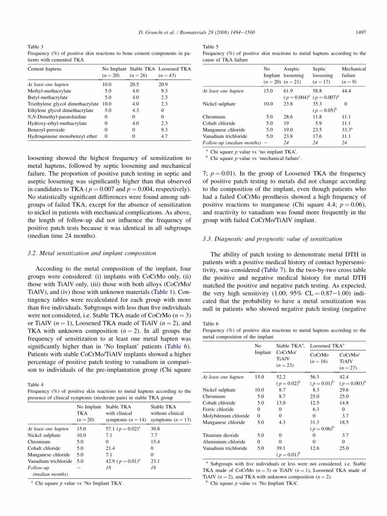
Table 3
Frequency (%) of positive skin reactions to bone cement components in pa-
tients with cemented TKA
Cement haptens No Implant
(n¼ 20)
Stable TKA
(n¼ 26)
Loosened TKA
(n¼ 43)
At least one hapten 10.0 20.5 20.9
Methyl-methacrylate 5.0 4.0 9.3
Butyl-methacrylate 5.0 4.0 2.3
Triethylene glycol dimethacrylate 10.0 4.0 2.3
Ethylene glycol dimethacrylate 5.0 4.3 0
N,N-Dimethyl-paratoluidine 0 0 0
Hydroxy-ethyl-methacrylate 0 4.0 2.3
Benzoyl-peroxide 0 0 9.3
Hydroquinone monobenzyl ether 0 0 4.7
Table 5
Frequency (%) of positive skin reactions to metal haptens according to the
cause of TKA failure
No
Implant
(n¼ 20)
Aseptic
loosening
(n¼ 21)
Septic
loosening
(n¼ 17)
Mechanical
failure
(n¼ 9)
At least one hapten 15.0 61.9
( p¼ 0.004)a58.8
( p¼ 0.007)a44.4
Nickel sulphate 10.0 23.8 35.3
( p¼ 0.05)b0
Chromium 5.0 28.6 11.8 11.1
Cobalt chloride 5.0 19 5.9 11.1
Manganese chloride 5.0 19.0 23.5 33.3a
Vanadium trichloride 5.0 23.8 17.6 11.1
Follow-up (median months) e 24 24 24
a Chi square p value vs ‘no implant TKA’.b Chi square p value vs ‘mechanical failure’.
1497D. Granchi et al. / Biomaterials 29 (2008) 1494e1500
loosening showed the highest frequency of sensitization tometal haptens, followed by septic loosening and mechanicalfailure. The proportion of positive patch testing in septic andaseptic loosening was significantly higher than that observedin candidates to TKA ( p¼ 0.007 and p¼ 0.004, respectively).No statistically significant differences were found among sub-groups of failed TKA, except for the absence of sensitizationto nickel in patients with mechanical complications. As above,the length of follow-up did not influence the frequency ofpositive patch tests because it was identical in all subgroups(median time 24 months).
3.2. Metal sensitization and implant composition
Table 6
Frequency (%) of positive skin reactions to metal haptens according to the
metal composition of the implant
No
Implant
Stable TKAa,
CoCrMo/
TiAlV
(n¼ 23)
Loosened TKAa
CoCrMo
(n¼ 16)
CoCrMo/
TiAlV
(n¼ 27)
According to the metal composition of the implant, fourgroups were considered: (i) implants with CoCrMo only, (ii)those with TiAlV only, (iii) those with both alloys (CoCrMo/TiAlV), and (iv) those with unknown materials (Table 1). Con-tingency tables were recalculated for each group with morethan five individuals. Subgroups with less than five individualswere not considered, i.e. Stable TKA made of CoCrMo (n¼ 3)or TiAlV (n¼ 1), Loosened TKA made of TiAlV (n¼ 2), andTKA with unknown composition (n¼ 2). In all groups thefrequency of sensitization to at least one metal hapten wassignificantly higher than in ‘No Implant’ patients (Table 6).Patients with stable CoCrMo/TiAlV implants showed a higherpercentage of positive patch testing to vanadium in compari-son to individuals of the pre-implantation group (Chi square
Table 4
Frequency (%) of positive skin reactions to metal haptens according to the
presence of clinical symptoms (moderate pain) in stable TKA group
No Implant
TKA
(n¼ 20)
Stable TKA
with clinical
symptoms (n¼ 14)
Stable TKA
without clinical
symptoms (n¼ 13)
At least one hapten 15.0 57.1 ( p¼ 0.02)a 30.8
Nickel sulphate 10.0 7.1 7.7
Chromium 5.0 0 15.4
Cobalt chloride 5.0 21.4 0
Manganese chloride 5.0 7.1 0
Vanadium trichloride 5.0 42.9 ( p¼ 0.01)a 23.1
Follow-up
(median months)
e 18 18
a Chi square p value vs ‘No Implant TKA’.
7; p¼ 0.01). In the group of Loosened TKA the frequencyof positive patch testing to metals did not change accordingto the composition of the implant, even though patients whohad a failed CoCrMo prosthesis showed a high frequency ofpositive reactions to manganese (Chi square 4.4; p¼ 0.06),and reactivity to vanadium was found more frequently in thegroup with failed CoCrMo/TiAlV implant.
3.3. Diagnostic and prognostic value of sensitization
The ability of patch testing to demonstrate metal DTH inpatients with a positive medical history of contact hypersensi-tivity, was considered (Table 7). In the two-by-two cross tablethe positive and negative medical history for metal DTHmatched the positive and negative patch testing. As expected,the very high sensitivity (1.00; 95% CL¼ 0.87e1.00) indi-cated that the probability to have a metal sensitization wasnull in patients who showed negative patch testing (negative
At least one hapten 15.0 52.2
( p¼ 0.02)b56.3
( p¼ 0.01)b42.4
( p¼ 0.003)b
Nickel sulphate 10.0 8.7 8.3 29.6
Chromium 5.0 8.7 25.0 25.0
Cobalt chloride 5.0 13.0 12.5 14.8
Ferric chloride 0 0 6.3 0
Molybdenum chloride 0 0 0 3.7
Manganese chloride 5.0 4.3 31.3
( p¼ 0.06)b18.5
Titanium dioxide 5.0 0 0 3.7
Aluminium chloride 0 0 0 0
Vanadium trichloride 5.0 39.1
( p¼ 0.01)b12.6 25.0
a Subgroups with five individuals or less were not considered, i.e. Stable
TKA made of CoCrMo (n¼ 3) or TiAlV (n¼ 1), Loosened TKA made of
TiAlV (n¼ 2), and TKA with unknown composition (n¼ 2).b Chi square p value vs ‘No Implant TKA’.
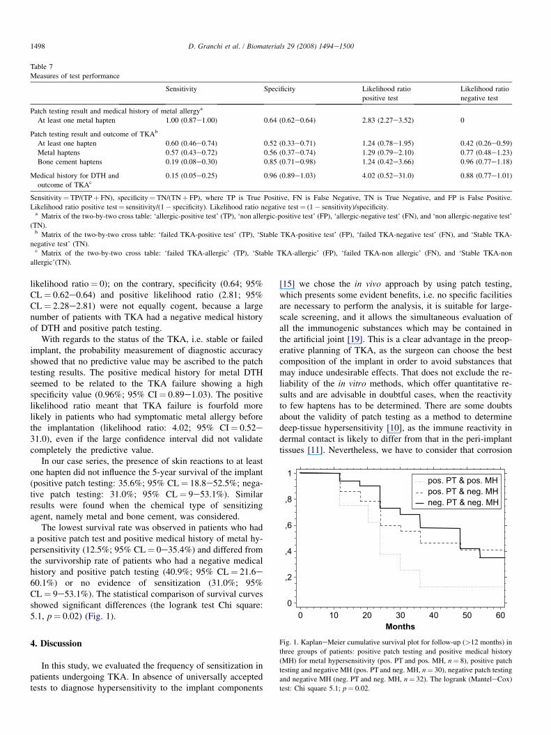
Table 7
Measures of test performance
Sensitivity Specificity Likelihood ratio
positive test
Likelihood ratio
negative test
Patch testing result and medical history of metal allergya
At least one metal hapten 1.00 (0.87e1.00) 0.64 (0.62e0.64) 2.83 (2.27e3.52) 0
Patch testing result and outcome of TKAb
At least one hapten 0.60 (0.46e0.74) 0.52 (0.33e0.71) 1.24 (0.78e1.95) 0.42 (0.26e0.59)
Metal haptens 0.57 (0.43e0.72) 0.56 (0.37e0.74) 1.29 (0.79e2.10) 0.77 (0.48e1.23)
Bone cement haptens 0.19 (0.08e0.30) 0.85 (0.71e0.98) 1.24 (0.42e3.66) 0.96 (0.77e1.18)
Medical history for DTH and
outcome of TKAc0.15 (0.05e0.25) 0.96 (0.89e1.03) 4.02 (0.52e31.0) 0.88 (0.77e1.01)
Sensitivity¼ TP/(TPþ FN), specificity¼ TN/(TNþ FP), where TP is True Positive, FN is False Negative, TN is True Negative, and FP is False Positive.
Likelihood ratio positive test¼ sensitivity/(1� specificity). Likelihood ratio negative test¼ (1� sensitivity)/specificity.a Matrix of the two-by-two cross table: ‘allergic-positive test’ (TP), ‘non allergic-positive test’ (FP), ‘allergic-negative test’ (FN), and ‘non allergic-negative test’
(TN).b Matrix of the two-by-two cross table: ‘failed TKA-positive test’ (TP), ‘Stable TKA-positive test’ (FP), ‘failed TKA-negative test’ (FN), and ‘Stable TKA-
negative test’ (TN).c Matrix of the two-by-two cross table: ‘failed TKA-allergic’ (TP), ‘Stable TKA-allergic’ (FP), ‘failed TKA-non allergic’ (FN), and ‘Stable TKA-non
allergic’(TN).
0
,2
,4
,6
,8
1
0 10 20 30 40 50 60
neg. PT & neg. MHpos. PT & neg. MHpos. PT & pos. MH
1498 D. Granchi et al. / Biomaterials 29 (2008) 1494e1500
likelihood ratio¼ 0); on the contrary, specificity (0.64; 95%CL¼ 0.62e0.64) and positive likelihood ratio (2.81; 95%CL¼ 2.28e2.81) were not equally cogent, because a largenumber of patients with TKA had a negative medical historyof DTH and positive patch testing.
With regards to the status of the TKA, i.e. stable or failedimplant, the probability measurement of diagnostic accuracyshowed that no predictive value may be ascribed to the patchtesting results. The positive medical history for metal DTHseemed to be related to the TKA failure showing a highspecificity value (0.96%; 95% CI¼ 0.89e1.03). The positivelikelihood ratio meant that TKA failure is fourfold morelikely in patients who had symptomatic metal allergy beforethe implantation (likelihood ratio: 4.02; 95% CI¼ 0.52e31.0), even if the large confidence interval did not validatecompletely the predictive value.
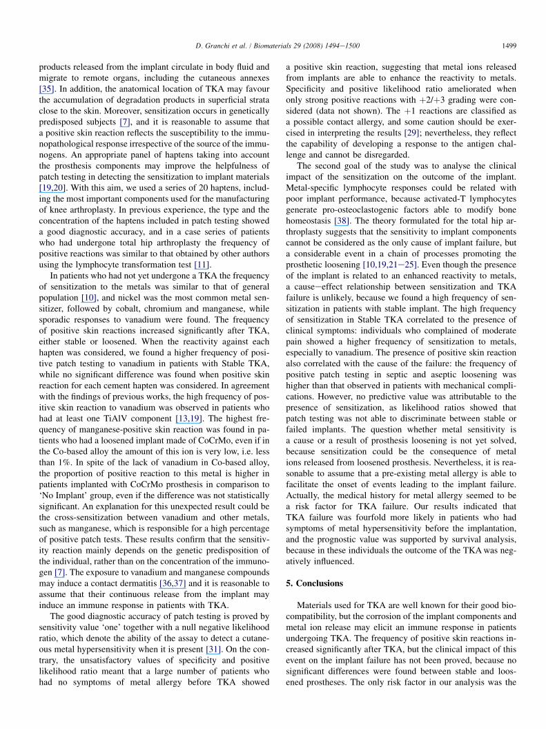
In our case series, the presence of skin reactions to at leastone hapten did not influence the 5-year survival of the implant(positive patch testing: 35.6%; 95% CL¼ 18.8e52.5%; nega-tive patch testing: 31.0%; 95% CL¼ 9e53.1%). Similarresults were found when the chemical type of sensitizingagent, namely metal and bone cement, was considered.
The lowest survival rate was observed in patients who hada positive patch test and positive medical history of metal hy-persensitivity (12.5%; 95% CL¼ 0e35.4%) and differed fromthe survivorship rate of patients who had a negative medicalhistory and positive patch testing (40.9%; 95% CL¼ 21.6e60.1%) or no evidence of sensitization (31.0%; 95%CL¼ 9e53.1%). The statistical comparison of survival curvesshowed significant differences (the logrank test Chi square:5.1, p¼ 0.02) (Fig. 1).
Months
Fig. 1. KaplaneMeier cumulative survival plot for follow-up (>12 months) in
three groups of patients: positive patch testing and positive medical history
(MH) for metal hypersensitivity (pos. PT and pos. MH, n¼ 8), positive patch
testing and negative MH (pos. PT and neg. MH, n¼ 30), negative patch testing
and negative MH (neg. PT and neg. MH, n¼ 32). The logrank (ManteleCox)
test: Chi square 5.1; p¼ 0.02.
4. Discussion
In this study, we evaluated the frequency of sensitization inpatients undergoing TKA. In absence of universally acceptedtests to diagnose hypersensitivity to the implant components
[15] we chose the in vivo approach by using patch testing,which presents some evident benefits, i.e. no specific facilitiesare necessary to perform the analysis, it is suitable for large-scale screening, and it allows the simultaneous evaluation ofall the immunogenic substances which may be contained inthe artificial joint [19]. This is a clear advantage in the preop-erative planning of TKA, as the surgeon can choose the bestcomposition of the implant in order to avoid substances thatmay induce undesirable effects. That does not exclude the re-liability of the in vitro methods, which offer quantitative re-sults and are advisable in doubtful cases, when the reactivityto few haptens has to be determined. There are some doubtsabout the validity of patch testing as a method to determinedeep-tissue hypersensitivity [10], as the immune reactivity indermal contact is likely to differ from that in the peri-implanttissues [11]. Nevertheless, we have to consider that corrosion
1499D. Granchi et al. / Biomaterials 29 (2008) 1494e1500
products released from the implant circulate in body fluid andmigrate to remote organs, including the cutaneous annexes[35]. In addition, the anatomical location of TKA may favourthe accumulation of degradation products in superficial strataclose to the skin. Moreover, sensitization occurs in geneticallypredisposed subjects [7], and it is reasonable to assume thata positive skin reaction reflects the susceptibility to the immu-nopathological response irrespective of the source of the immu-nogens. An appropriate panel of haptens taking into accountthe prosthesis components may improve the helpfulness ofpatch testing in detecting the sensitization to implant materials[19,20]. With this aim, we used a series of 20 haptens, includ-ing the most important components used for the manufacturingof knee arthroplasty. In previous experience, the type and theconcentration of the haptens included in patch testing showeda good diagnostic accuracy, and in a case series of patientswho had undergone total hip arthroplasty the frequency ofpositive reactions was similar to that obtained by other authorsusing the lymphocyte transformation test [11].
In patients who had not yet undergone a TKA the frequencyof sensitization to the metals was similar to that of generalpopulation [10], and nickel was the most common metal sen-sitizer, followed by cobalt, chromium and manganese, whilesporadic responses to vanadium were found. The frequencyof positive skin reactions increased significantly after TKA,either stable or loosened. When the reactivity against eachhapten was considered, we found a higher frequency of posi-tive patch testing to vanadium in patients with Stable TKA,while no significant difference was found when positive skinreaction for each cement hapten was considered. In agreementwith the findings of previous works, the high frequency of pos-itive skin reaction to vanadium was observed in patients whohad at least one TiAlV component [13,19]. The highest fre-quency of manganese-positive skin reaction was found in pa-tients who had a loosened implant made of CoCrMo, even if inthe Co-based alloy the amount of this ion is very low, i.e. lessthan 1%. In spite of the lack of vanadium in Co-based alloy,the proportion of positive reaction to this metal is higher inpatients implanted with CoCrMo prosthesis in comparison to‘No Implant’ group, even if the difference was not statisticallysignificant. An explanation for this unexpected result could bethe cross-sensitization between vanadium and other metals,such as manganese, which is responsible for a high percentageof positive patch tests. These results confirm that the sensitiv-ity reaction mainly depends on the genetic predisposition ofthe individual, rather than on the concentration of the immuno-gen [7]. The exposure to vanadium and manganese compoundsmay induce a contact dermatitis [36,37] and it is reasonable toassume that their continuous release from the implant mayinduce an immune response in patients with TKA.
The good diagnostic accuracy of patch testing is proved bysensitivity value ‘one’ together with a null negative likelihoodratio, which denote the ability of the assay to detect a cutane-ous metal hypersensitivity when it is present [31]. On the con-trary, the unsatisfactory values of specificity and positivelikelihood ratio meant that a large number of patients whohad no symptoms of metal allergy before TKA showed
a positive skin reaction, suggesting that metal ions releasedfrom implants are able to enhance the reactivity to metals.Specificity and positive likelihood ratio ameliorated whenonly strong positive reactions with þ2/þ3 grading were con-sidered (data not shown). The þ1 reactions are classified asa possible contact allergy, and some caution should be exer-cised in interpreting the results [29]; nevertheless, they reflectthe capability of developing a response to the antigen chal-lenge and cannot be disregarded.
The second goal of the study was to analyse the clinicalimpact of the sensitization on the outcome of the implant.Metal-specific lymphocyte responses could be related withpoor implant performance, because activated-T lymphocytesgenerate pro-osteoclastogenic factors able to modify bonehomeostasis [38]. The theory formulated for the total hip ar-throplasty suggests that the sensitivity to implant componentscannot be considered as the only cause of implant failure, buta considerable event in a chain of processes promoting theprosthetic loosening [10,19,21e25]. Even though the presenceof the implant is related to an enhanced reactivity to metals,a causeeeffect relationship between sensitization and TKAfailure is unlikely, because we found a high frequency of sen-sitization in patients with stable implant. The high frequencyof sensitization in Stable TKA correlated to the presence ofclinical symptoms: individuals who complained of moderatepain showed a higher frequency of sensitization to metals,especially to vanadium. The presence of positive skin reactionalso correlated with the cause of the failure: the frequency ofpositive patch testing in septic and aseptic loosening washigher than that observed in patients with mechanical compli-cations. However, no predictive value was attributable to thepresence of sensitization, as likelihood ratios showed thatpatch testing was not able to discriminate between stable orfailed implants. The question whether metal sensitivity isa cause or a result of prosthesis loosening is not yet solved,because sensitization could be the consequence of metalions released from loosened prosthesis. Nevertheless, it is rea-sonable to assume that a pre-existing metal allergy is able tofacilitate the onset of events leading to the implant failure.Actually, the medical history for metal allergy seemed to bea risk factor for TKA failure. Our results indicated thatTKA failure was fourfold more likely in patients who hadsymptoms of metal hypersensitivity before the implantation,and the prognostic value was supported by survival analysis,because in these individuals the outcome of the TKA was neg-atively influenced.
5. Conclusions
Materials used for TKA are well known for their good bio-compatibility, but the corrosion of the implant components andmetal ion release may elicit an immune response in patientsundergoing TKA. The frequency of positive skin reactions in-creased significantly after TKA, but the clinical impact of thisevent on the implant failure has not been proved, because nosignificant differences were found between stable and loos-ened prostheses. The only risk factor in our analysis was the
1500 D. Granchi et al. / Biomaterials 29 (2008) 1494e1500
presence of a positive medical history for DTH, which influ-ences negatively the implant lifespan, and increases fourfoldthe likelihood to have a TKA failure. Finally, our resultsshow that patch testing may be a suitable method to evaluatesimultaneously and rapidly the immunogenic substances con-tained in the artificial joint. The usefulness of patch testingdoes not exclude the reliability of the in vitro methods whichare advisable in doubtful cases and/or when the reactivity tofew haptens has to be confirmed.
Acknowledgments
Funded by grants from Italian Ministry of the Health for theNational Hospitals and Research Institutes (Ricerca Corrente).The study is part of the activities of the European Cooperationin the field of Scientific and Technical Research (COST), Ac-tion 537 ‘‘Core laboratories for the improvement of MedicalDevices in Clinical Practice from the Failure of the ExplantedProstheses Analyses (FEPA)’’.
References
[1] Kane RL, Saleh KJ, Wilt TJ, Bershadsky B, Cross III WW, MacDonald RM,
et al. Total knee replacement. Evid Rep Technol Assess 2003;86:1e8.
[2] NIH Consensus Panel. NIH consensus statement on total knee replace-
ment December 8e10, 2003. J Bone Joint Surg Am 2004;86:1328e35.
[3] Sundfeldt M, Carlsson LV, Johansson CB, Thomsen P, Gretzer C. Aseptic
loosening, not only a question of wear: a review of different theories.
Acta Orthopaedica 2006;77:177e97.
[4] Callaghan JJ, O’rourke MR, Saleh KJ. Why knees fail: lessons learned.
J Arthroplasty 2004;19:31e4.
[5] Pizzoferrato A, Cenni E, Ciapetti G, Granchi D, Savarino L, Stea S.
Inflammatory response to metals and ceramics. In: Barbucci R, editor.
Integrated biomaterials science. New York: Kluwer Academic/Plenum
Publishers; 2002. p. 735e91.
[6] FDA e Center for devices and radiological health. Guidance for industry
and FDA reviewers e immunotoxicity testing guidance. FDA; May 6,
1999. p. 1e16.
[7] Descotes J. Importance of immunotoxicity in safety assessment: a medi-
cal toxicologist’s perspective. Toxicol Lett 2004;149:103e8.
[8] Granchi D, Savarino L, Ciapetti G, Cenni E, Rotini R, Mieti M, et al.
Immunological changes in patients with primary osteoarthritis of the
hip after total joint replacement. J Bone Joint Surg Br 2003;85:758e64.
[9] Niki Y, Matsumoto H, Otani T, Yatabe T, Kondo M, Yoshimine F, et al.
Screening for sintomatic metal sensitivity: a prospective study of 92 pa-
tients undergoing total knee arthroplasty. Biomaterials 2005;26:1019e26.
[10] Hallab N, Merritt K, Jacobs JJ. Metal sensitivity in patients with ortho-
paedic implants. J Bone Joint Surg Am 2001;83:428e36.
[11] Hallab NJ, Anderson S, Stafford T, Glant T, Jacobs JJ. Lymphocyte
responses in patients with total hip arthroplasty. J Orthop Res 2005;
232:384e91.
[12] Lalor PA, Revell PA, Gray AB, Wright S, Railton GT, Freeman MA.
Sensitivity to titanium. A cause of implant failure? J Bone Joint Surg
Br 1991;73:25e8.
[13] Cancilleri F, De Giorgis P, Verdoia C, Parrini L, Lodi A, Crosti C. Al-
lergy to components of total hip arthroplasty before and after surgery.
Ital J Orthop Traumatol 1992;18:407e10.
[14] Haddad FS, Cobb AG, Bentley G, Levell NJ, Dowd PM. Hypersensitivity
in aseptic loosening of total hip replacements. The role of constituents of
bone cement. J Bone Joint Surg Br 1996;78:546e9.
[15] Jacobs JJ, Hallab NJ. Loosening and osteolysis associated with metal-on-
metal bearings: a local effect of metal hypersensitivity? J Bone Joint Surg
Am 2006;88:1171e2.
[16] Granchi D, Ciapetti G, Savarino L, Stea S, Filippini F, Sudanese A, et al.
Expression of the CD69 activation antigen on lymphocytes of patients
with hip prosthesis. Biomaterials 2000;21:2059e65.
[17] Valentine-Thon E, Schiwara HW. Validity of MELISA for metal sensitiv-
ity testing. Neuroendocrinol Lett 2003;24:57e64.
[18] Granchi D, Ciapetti G, Savarino L, Cavedagna D, Donati ME,
Pizzoferrato A. Assessment of metal extract toxicity on human lympho-
cytes cultured in vitro. J Biomed Mater Res 1996;31:183e91.
[19] Granchi D, Cenni E, Trisolino G, Giunti A, Baldini N. Sensitivity to
implant materials in patients undergoing total hip replacement. J Biomed
Mater Res B Appl Biomater 2006;77:257e64.
[20] Van der Valk PGM, Devos SA, Coenraads PJ. Evidence-based diagnosis
in patch testing. Contact Derm. 2003;48:121e5.
[21] Davies AP, Willert HG, Campbell PA, Learmonth ID, Case CP. An
unusual lymphocytic perivascular infiltration in tissues around contempo-
rary metal-on-metal joint replacements. J Bone Joint Surg Am 2005;87:
18e27.
[22] Willert HG, Buchhorn GH, Fayyazi A, Flury R, Windler M, Koster G,
et al. Metal-on-metal bearings and hypersensitivity in patients with arti-
ficial hip joints. A clinical and histomorphological study. J Bone Joint
Surg Am 2005;87:28e36.
[23] Park YS, Moon YW, Lim SJ, Yang JM, Ahn G, Choi YL. Early osteolysis
following second-generation metal-on-metal hip replacement. J Bone
Joint Surg Am 2005;87:1515e21.
[24] Milosev I, Trebse R, Kovac S, Cor A, Pisot V. Survivorship and retrieval
analysis of sikomet metal-on-metal total hip replacements at a mean of
seven years. J Bone Joint Surg Am 2006;88:1173e82.
[25] Korovessis P, Petsinis G, Repanti M, Repantis T. Metallosis after contem-
porary metal-on-metal total hip arthroplasty: five- to nine-year follow-up.
J Bone Joint Surg Am 2006;88:1183e91.
[26] Insall J, Dorr L, Scott R, Scott N. Rationale of the knee society clinical
rating system. Clin Orthop Relat Res 1989;248:13e4.
[27] Ewald FC. The knee society total arthroplasty roentgenographic evalua-
tion and scoring system. Clin Orthop Relat Res 1989;248:9e12.
[28] Bertin KC. Evaluation of the unsuccessful total knee arthroplasty. In:
Engh GA, Rorabeck CH, editors. Revision total knee arthroplasty. Balti-
more, MD, USA: Williams and Wilkins; 1997. p. 28e45.
[29] Schenk MM, Murray KD. Radiographic evaluation of the painful total
knee replacement. In: Saleh KJ, Wood KC, Gafni A, Gross AE, editors.
Revision total knee arthroplasty. Philadelphia, PA, USA: Lippincott-
Raven; 1999. p. 355e70.
[30] Sudanese A, Toni A, Busanelli L, Furno A, Montina P, Marraro M, et al.
Diagnostic protocol in prosthetic loosening. Chir Organi Mov 1994;79:
257e67.
[31] Drake LA, Dorner W, Goltz RW, Graham GF, Lewis CW, Pariser DM,
et al. Guidelines of care for contact dermatitis. Committee on guidelines
of care. J Am Acad Dermatol 1995;32:109e13.
[32] Bossuyt PM, Reitsma JB, Bruns DE, Gatsonis CA, Glasziou PP,
Irwig LM, et al. Standards for reporting of diagnostic accuracy steering
group. Towards complete and accurate reporting of studies of diagnostic
accuracy: the STARD initiative. BMJ 2003;326:41e4.
[33] Deeks JJ. Systematic reviews in health care: systematic reviews of
evaluations of diagnostic and screening tests. BMJ 2001;323:
157e62.
[34] Kocher MS, Zurakowski D. Clinical epidemiology and biostatistics:
a primer for orthopaedic surgeons. J Bone Joint Surg Am 2004;86:
607e20.
[35] Coleman RF, Herrington J, Scales JT. Concentration of wear products in
hair, blood, and urine after total hip replacement. BMJ 1973;1:527e9.
[36] High WA, Ayers RA, Adams JR, Chang A, Fitzpatrick JE. Granuloma-
tous reaction to titanium alloy: an unusual reaction to ear piercing.
J Am Acad Dermatol 2006;55:716e20.
[37] Motolese A, Truzzi M, Giannini A, Seidenari S. Contact dermatitis and
contact sensitization among enamellers and decorators in the ceramics
industry. Contact Dermatitis 1993;28:59e62.
[38] Purdue PE, Koulouvaris P, Potter HG, Bryan NJ, Sculco TP. The cellular
and molecular biology of periprosthetic osteolysis. Clin Orthop Relat Res
2006;454:251e61.