Sensitive bi-enzymatic biosensor based on polyphenoloxidases–gold nanoparticles–chitosan hybrid...
10
Sensitive bi-enzymatic biosensor based on polyphenoloxidases–gold nanoparticles–chitosan hybrid film–graphene doped carbon paste electrode for carbamates detection Thiago M.B.F. Oliveira a,b , M. Fátima Barroso a,c , Simone Morais a, ⁎, Mariana Araújo d , Cristina Freire d , Pedro de Lima-Neto b , Adriana N. Correia b , Maria B.P.P. Oliveira c , Cristina Delerue-Matos a a REQUIMTE, Instituto Superior de Engenharia do Porto, Instituto Politécnico do Porto, Rua Dr. Bernardino de Almeida 431, 4200-072 Porto, Portugal b GELCORR, Dep. de Química Analítica e Físico-Química, Centro de Ciências, Universidade Federal do Ceará, Bloco 940, Campus do Pici, 60455-970 Fortaleza, CE, Brazil c REQUIMTE, Dep. de Ciências Químicas, Faculdade de Farmácia, Universidade do Porto, Rua de Jorge Viterbo Ferreira 228, 4050-313 Porto, Portugal d REQUIMTE, Dep. de Química e Bioquímica, Faculdade de Ciências, Universidade do Porto, Rua do Campo Alegre s/n, 4169-007 Porto, Portugal abstract article info Article history: Received 21 November 2013 Received in revised form 3 February 2014 Accepted 22 February 2014 Available online 5 March 2014 Keywords: Bi-enzymatic biosensor Graphene modified electrode Gold nanoparticles Chitosan hybrid film Carbamates A bi-enzymatic biosensor (LACC–TYR–AuNPs–CS/GPE) for carbamates was prepared in a single step by electro- deposition of a hybrid film onto a graphene doped carbon paste electrode (GPE). Graphene and the gold nanopar- ticles (AuNPs) were morphologically characterized by transmission electron microscopy, X-ray photoelectron spectroscopy, dynamic light scattering and laser Doppler velocimetry. The electrodeposited hybrid film was composed of laccase (LACC), tyrosinase (TYR) and AuNPs entrapped in a chitosan (CS) polymeric matrix. Exper- imental parameters, namely graphene redox state, AuNPs:CS ratio, enzymes concentration, pH and inhibition time were evaluated. LACC–TYR–AuNPs–CS/GPE exhibited an improved Michaelis–Menten kinetic constant (26.9 ± 0.5 M) when compared with LACC–AuNPs–CS/GPE (37.8 ± 0.2 M) and TYR–AuNPs–CS/GPE (52.3 ± 0.4 M). Using 4-aminophenol as substrate at pH 5.5, the device presented wide linear ranges, low detection limits (1.68 × 10 -9 ± 1.18 × 10 -10 –2.15 × 10 -7 ± 3.41 × 10 -9 M), high accuracy, sensitivity (1.13 × 10 6 ± 8.11 × 10 4 –2.19 × 10 8 ± 2.51 × 10 7 %inhibition M -1 ), repeatability (1.2–5.8% RSD), reproducibility (3.2–6.5% RSD) and stability (ca. twenty days) to determine carbaryl, formetanate hydrochloride, propoxur and ziram in citrus fruits based on their inhibitory capacity on the polyphenoloxidases activity. Recoveries at two fortified levels ranged from 93.8 ± 0.3% (lemon) to 97.8 ± 0.3% (orange). Glucose, citric acid and ascorbic acid do not interfere signifi- cantly in the electroanalysis. The proposed electroanalytical procedure can be a promising tool for food safety control. © 2014 Elsevier B.V. All rights reserved. 1. Introduction Carbamates are one of the principal classes of pesticides that are being largely used to increase crop yield. However, their residues may pose serious environmental and health problems [1,2]. The adverse effects of several carbamates were reported, and they include renal, hepatic, neurological, reproductive, immune, and metabolic functions in both humans and animals [3,4]. Some of them are classed as endo- crine disrupting chemicals [5] and regarded as priority pollutants by the United States Environmental Protection Agency [6]. Biosensor technology has been considered as a key tool for the implementation of the new European Union directives because of the negligible waste generation, minimization of use of hazardous substances, high sensitivity and selectivity, as well as, the in situ real- time monitoring capacity [7,8]. In this perspective, the biosensing of environmental pollutants, particularly agrochemicals, using enzymes as biorecognition element has increased pronouncedly in the last years [1,9–14]. Still, many of these devices need to improve their perfor- mance because of the low maximum residue limits (MRLs) established worldwide for pesticides [15,16]. Considerable positive synergistic effects on the current signal can be attained by combining several enzymes [17–21]. Enzyme selection and their sources have a major in- fluence on the biosensor sensitivity [19,22]. The few studies dedicated to bi-enzymatic biosensors [17–21,23–25] reported in the last ten years are summarized in Table 1S (Supplementary material). As far as the authors know, there is no publication related to the application of bi-enzymatic biosensors for the quantification of pesticides in food com- modities or in other real samples. Moreover, there is a general lack of validated biosensor-based procedures for analysis of food samples [1,12,13,26]. The main drawback of the application of enzymatic biosensors to complex matrices is the susceptibility of the transducer to surface passivation. Furthermore, enzymatic products may undergo partial Bioelectrochemistry 98 (2014) 20–29 ⁎ Corresponding author. Tel.: +351 228340500; fax: +351 228321159. E-mail address: [email protected] (S. Morais). http://dx.doi.org/10.1016/j.bioelechem.2014.02.003 1567-5394/© 2014 Elsevier B.V. All rights reserved. Contents lists available at ScienceDirect Bioelectrochemistry journal homepage: www.elsevier.com/locate/bioelechem
-
Upload
independent -
Category
Documents
-
view
1 -
download
0
Transcript of Sensitive bi-enzymatic biosensor based on polyphenoloxidases–gold nanoparticles–chitosan hybrid...

Bioelectrochemistry 98 (2014) 20–29
Contents lists available at ScienceDirect
Bioelectrochemistry
j ourna l homepage: www.e lsev ie r .com/ locate /b ioe lechem
Sensitive bi-enzymatic biosensor based on polyphenoloxidases–goldnanoparticles–chitosan hybrid film–graphene doped carbon pasteelectrode for carbamates detection
Thiago M.B.F. Oliveira a,b, M. Fátima Barroso a,c, Simone Morais a,⁎, Mariana Araújo d, Cristina Freire d,Pedro de Lima-Neto b, Adriana N. Correia b, Maria B.P.P. Oliveira c, Cristina Delerue-Matos a
a REQUIMTE, Instituto Superior de Engenharia do Porto, Instituto Politécnico do Porto, Rua Dr. Bernardino de Almeida 431, 4200-072 Porto, Portugalb GELCORR, Dep. de Química Analítica e Físico-Química, Centro de Ciências, Universidade Federal do Ceará, Bloco 940, Campus do Pici, 60455-970 Fortaleza, CE, Brazilc REQUIMTE, Dep. de Ciências Químicas, Faculdade de Farmácia, Universidade do Porto, Rua de Jorge Viterbo Ferreira 228, 4050-313 Porto, Portugald REQUIMTE, Dep. de Química e Bioquímica, Faculdade de Ciências, Universidade do Porto, Rua do Campo Alegre s/n, 4169-007 Porto, Portugal
⁎ Corresponding author. Tel.: +351 228340500; fax: +E-mail address: [email protected] (S. Morais).
http://dx.doi.org/10.1016/j.bioelechem.2014.02.0031567-5394/© 2014 Elsevier B.V. All rights reserved.
a b s t r a c t
a r t i c l e i n f oArticle history:Received 21 November 2013Received in revised form 3 February 2014Accepted 22 February 2014Available online 5 March 2014
Keywords:Bi-enzymatic biosensorGraphene modified electrodeGold nanoparticlesChitosan hybrid filmCarbamates
A bi-enzymatic biosensor (LACC–TYR–AuNPs–CS/GPE) for carbamates was prepared in a single step by electro-deposition of a hybridfilmonto a graphene doped carbon paste electrode (GPE). Graphene and the gold nanopar-ticles (AuNPs) were morphologically characterized by transmission electron microscopy, X-ray photoelectronspectroscopy, dynamic light scattering and laser Doppler velocimetry. The electrodeposited hybrid film wascomposed of laccase (LACC), tyrosinase (TYR) and AuNPs entrapped in a chitosan (CS) polymeric matrix. Exper-imental parameters, namely graphene redox state, AuNPs:CS ratio, enzymes concentration, pH and inhibitiontime were evaluated. LACC–TYR–AuNPs–CS/GPE exhibited an improved Michaelis–Menten kinetic constant(26.9 ± 0.5 M) when compared with LACC–AuNPs–CS/GPE (37.8 ± 0.2 M) and TYR–AuNPs–CS/GPE (52.3 ±0.4 M). Using 4-aminophenol as substrate at pH 5.5, the device presented wide linear ranges, low detectionlimits (1.68 × 10⁻⁹ ± 1.18 × 10⁻¹⁰–2.15 × 10⁻⁷ ± 3.41 × 10⁻⁹ M), high accuracy, sensitivity (1.13 × 10⁶ ±8.11 × 10⁴–2.19 × 10⁸± 2.51 × 10⁷ %inhibitionM−1), repeatability (1.2–5.8% RSD), reproducibility (3.2–6.5% RSD)and stability (ca. twenty days) to determine carbaryl, formetanate hydrochloride, propoxur and ziram in citrusfruits based on their inhibitory capacity on the polyphenoloxidases activity. Recoveries at two fortified levels rangedfrom 93.8 ± 0.3% (lemon) to 97.8 ± 0.3% (orange). Glucose, citric acid and ascorbic acid do not interfere signifi-cantly in the electroanalysis. The proposed electroanalytical procedure can be a promising tool for food safetycontrol.
© 2014 Elsevier B.V. All rights reserved.
1. Introduction
Carbamates are one of the principal classes of pesticides that arebeing largely used to increase crop yield. However, their residues maypose serious environmental and health problems [1,2]. The adverseeffects of several carbamates were reported, and they include renal,hepatic, neurological, reproductive, immune, and metabolic functionsin both humans and animals [3,4]. Some of them are classed as endo-crine disrupting chemicals [5] and regarded as priority pollutants bythe United States Environmental Protection Agency [6].
Biosensor technology has been considered as a key tool for theimplementation of the new European Union directives because ofthe negligible waste generation, minimization of use of hazardoussubstances, high sensitivity and selectivity, as well as, the in situ real-time monitoring capacity [7,8]. In this perspective, the biosensing of
351 228321159.
environmental pollutants, particularly agrochemicals, using enzymesas biorecognition element has increased pronouncedly in the lastyears [1,9–14]. Still, many of these devices need to improve their perfor-mance because of the low maximum residue limits (MRLs) establishedworldwide for pesticides [15,16]. Considerable positive synergisticeffects on the current signal can be attained by combining severalenzymes [17–21]. Enzyme selection and their sources have a major in-fluence on the biosensor sensitivity [19,22]. The few studies dedicatedto bi-enzymatic biosensors [17–21,23–25] reported in the last tenyears are summarized in Table 1S (Supplementary material). As far asthe authors know, there is no publication related to the application ofbi-enzymatic biosensors for thequantification of pesticides in food com-modities or in other real samples. Moreover, there is a general lack ofvalidated biosensor-based procedures for analysis of food samples[1,12,13,26].
The main drawback of the application of enzymatic biosensorsto complex matrices is the susceptibility of the transducer to surfacepassivation. Furthermore, enzymatic products may undergo partial

(A)
(B)
(C)
21T.M.B.F. Oliveira et al. / Bioelectrochemistry 98 (2014) 20–29

-2.0
-1.5
-1.0
-0.5
0.0
0.5
1.0
1.5
2.0
IIcIc
IIa
Second step(chemical-electrochemical)
First step(electrochemical)
I /
μA
E / V (vs. Ag/AgCl/Cl-sat)
Ia
-0.4 -0.2 0.0 0.2 0.4 0.6 0.8
(A)
Fig. 2. A) Electrochemical behavior of 4-aminophenol (4.75 × 10⁻⁵ M in 0.04 MBritton–Robinson buffer, pH 5.5) obtained with the bare GPE (dashed line) andwith the LACC–TYR/GPE (solid line) after an incubation time of 20 min. Scan rate50 mV/s. B) Mechanistic proposal.
Fig. 2 (continued).
22 T.M.B.F. Oliveira et al. / Bioelectrochemistry 98 (2014) 20–29
electropolymerization onto the bare electrodes, yielding polyaromaticcompounds which increase the capacitance and negatively influencethe analytical response [19,27]. Protective polymeric films are interest-ing strategies to overcome these limitations. Chitosan (CS) is a naturalpolysaccharidewhichhas been extensively studied over the last two de-cades as a nontoxic, renewable and biodegradable polymer [20,28]. Dueto the presence of amino and hydroxyl groups, CS film exhibits multiplefunctionalities. CS can be cross-linked with nanomaterials [9,20,24,29],inorganic complexes [30], and biological elements [20,31], and used assupport for blends with other polymers [32]. CS has excellentmembrane-forming ability, high permeability towards water, good ad-hesion and biocompatibility providing a suitable microenvironmentfor electroimmobilization of biomolecules on different working surfaces[14,29,33]. However, CS has asmain disadvantage to act as an insulator,which hinders the charge-transfer process. In order to improve thecurrent signal, the enrichment of CS matrix with metallic nanoparticleshas shown interesting results [21,24,29,33].
Graphene shows great promise for the development of electrochem-ical biosensors due to its excellent mechanical flexibility, fast electrontransfer, and good biocompatibility [13,34–36]. In addition, its electro-catalytic action diminishes the overpotential associated to electroactivecompounds, minimizing the interferences that occur in real samples[37]. Graphene can enhance direct electron transfer between enzymesand electrodes. It has been also reported that the use of graphene asso-ciated with metal nanoparticles can form exceptionally stable and cost-effective biosensors [35,38]. No graphene-based bi-enzymatic biosensorwas found in the literature so far.
Thus, the goal of this study was to explore the synergistic advan-tages of combining CS (good adhesion and biocompatibility), AuNPs(high superficial area, conductivity and electron transfer rate), twopolyphenoloxidases (Trametes versicolor laccase, LACC, and Agaricusbisporus tyrosinase, TYR, which present high and selective catalyticactivity towards phenolic compounds), and a graphene doped carbonpaste electrode (20% (w/w); GPE) (high electrocatalytic activity, con-ductivity and adsorptive character) to prepare a novel bi-enzymatic bio-sensor. The construction of the device is based on a fast single stepelectrodeposition of an improved hybrid thin film (LACC–TYR–AuNPs–CS) onto the GPE surface. The developed bi-enzymatic biosensor exhib-
Fig. 1. TEM micrographs of graphene flakes obtained by sonication-assisted exfoliation ofby Hummers and Offeman method. (C) Representative XPS spectrum of as-prepared grapyrrolidone.
ited high accuracy, precision, sensitivity and stability for quantificationof worldwide used carbamates, i.e. formetanate hydrochloride (FMT),carbaryl (CBR), propoxur (PPX) and ziram (ZRM) in citrus (orange,tangerine and lemon) samples.
2. Materials and methods
2.1. Reagents
The polyphenoloxidases LACC (0.5 U mg−1) and TYR (1.0 U mg−1),and the substrate 4-aminophenol (4-AMP) were purchased from
graphite in N-methyl-2-pyrrolidone before (A) and after (B) the oxidation procedurephene flakes obtained by sonication-assisted exfoliation of graphite in N-methyl-2-
0
5
10
15
20
25
30
-Ip
/ μA
LACC : TYR (w/w)4:1 3:1 2:1 1:1 1:0 0:1 1:2 1:3 1:4
Fig. 3. Intensity of the 4-AMP (4.75 × 10⁻⁵M in Britton–Robinson buffer, pH 5.5) cathodicpeak current at−0.07 V obtained with the LACC–TYR–AuNPs–CS/GPE for different ratios(w/w) of LACC and TYR dispersed in the AuNPs–CS composite matrix. Error bars are thestandard deviation of three replicates.
23T.M.B.F. Oliveira et al. / Bioelectrochemistry 98 (2014) 20–29
Sigma-Aldrich (Germany). The carbamates CBR (CAS: 63-25-2), FMT(CAS: 23422-53-9), PPX (CAS: 114-26-1) and ZRM (CAS: 137-30-4)were supplied from Fluka (Pestanal®, Germany). Citric and ascorbicacids, and paraffin oil binder were obtained from Merck (Germany).D(+)-glucose anhydrous was from Scharlau (Spain). Spectroscopicgrade graphite powder was purchased from Ultracarbon (Spain). Medi-ummolecular weight chitosan (250–300 kDa, DD 93%, apparent viscos-ity 150 cps) was purchased from Altakitin (Portugal). Other chemicalswere of reagent grade and used without further purification. All solu-tions were prepared with ultrapure water (ρ=18MΩ cm−1) obtainedby a Simplicity 185 apparatus (Millipore, Molsheim, France).
Graphene was prepared by sonication-assisted exfoliation of graph-ite [59]. Briefly, 10 g of graphite powder was sonicated in 100 mL ofN-methyl-2-pyrrolidone (Sigma-Aldrich) using a probe sonicatorwith a titanium tip (Bandelin Sonoplus) for 6 h. The dispersionwas cen-trifuged at 500 rpm for 45 min; the supernatant containing the dis-persed graphene flakes was removed and then filtered through anylon 0.2 μm pore size membrane (Whatman). The resulting powderwas dried by vacuum, at room temperature, for several days. The Hum-mers and Offeman method was employed to obtain oxidized graphene.The as-prepared graphenewas submitted to a reactionwith concentratedsulfuric acid, sodium nitrate, and potassium permanganate in absence ofwater [39]. Both as-prepared graphene and graphene oxide were charac-terized by transmission electron microscopy (TEM, Hitachi H-9000NA)with the microscope operating at an accelerating voltage of 200–300 kV. The as-prepared graphene was also characterized by X-ray pho-toelectron spectroscopy (XPS, VG Scientific ESCALAB 200A spectrometer)using non-monochromatized Al Ka radiation (1486.6 eV).
AuNPs were synthesized by the Turkevich method, through thereduction of 0.01% gold (III) chloride solution by citrate and ascorbicacids [40]. The hydrodynamic size and potential zeta of the nanoparti-cles were characterized by dynamic light scattering and laser Dopplervelocimetry, respectively, using a Zetasizer Nano ZS (Malvern Instru-ments Ltd., Malvern, UK) at 25 °C.
2.2. Bi-enzymatic biosensor construction
Initially, a carbon paste was prepared bymixing spectroscopic gradegraphite powder with a paraffin oil binder (70:30%, w/w) and carefullyhand-mixing it in a mortar and pestle. Subsequently, this paste wasdoped with 20% (w/w) of graphene. This proportion was selectedbased on prior studies developed by our team [13]. The resultant com-posite material showed excellent characteristics (high conductivityand electron transfer rate) as transducer. The graphene doped carbonpaste was packed into a handmade cavity of a Teflon® tube (1.0mm in-ternal diameter) and then provided by a stainless steel piston. The GPEsurfacewas smoothed against a plainwhite paper and rinsedwith ultra-pure water before each measurement.
The composite material was produced by mixing different amountsof AuNPs, CS solution (1%, w/v), LACC and TYR. CS solution (1%, w/v)was prepared by dissolving 0.1 g chitosan powder in 10 mL of 0.05 Macetic acid solution. Several proportions of AuNPs (10, 20, 30, 40, 50,60 and 70%, v/v) and CS (1%, w/v) solutions were evaluated. This mix-ture was then enriched with LACC and TYR in several ratios, namely,4.0:1.0; 3.0:1.0; 2.0:1.0; 1.0:1.0; 1.0:0.0; 0.0:1.0; 1.0:2.0; 1.0:3.0, and1.0:4.0% (w/w) to produce the uniform composite material LACC–TYR–AuNPs–CS. The LACC–TYR–AuNPs–CS/GPE biosensor was obtainedby immersion of the GPE in the above solution and applying a constantpotential of−1.5 V for 200 s [41]. These parameters allow electrodepo-sition of the hybrid film in a single step. Then, the device was washedwith ultrapure water. When not in use, it was stored at 4 °C.
2.3. Electrochemical experiments
Cyclic voltammetry (CV), square-wave voltammetry (SWV) andelectrochemical impedance spectroscopy (EIS) assays were performed
at room temperature (20–22 °C) using an electrochemical systemAutolab PGSTAT-30 (Eco Chemie, The Netherlands) and GPES/FRA soft-ware. The electrochemical cell was assembled with the developed bi-enzymatic biosensor as the working electrode, a Ag/AgCl/KCl (3.0 M)reference electrode, and a platinum counter electrode.
Optimization of the electroanalytical procedure was performedusing 4.75 × 10⁻⁵ M 4-AMP as substrate in 0.04 M Britton–Robinsonbuffer (BR; pH 5.5). For the pesticide quantification by the proposedbi-enzymatic biosensor, the SWV parameters, i.e., the frequency, pulseamplitude and height of the potential step were optimized based onthe maximum value of peak current (Ip), displacement of the potentialpeak (Ep), and alterations on half-peak width (ΔEp/2), since their valuesexert considerable influence on the sensitivity of the electrochemicalprocedure. The optimal SWV parameters were a frequency of 100 Hz,pulse amplitude of 40 mV and step of 3 mV. The apparent Michaelis–Menten constant Km (M) was determined using substrate concentra-tions ranging from 9.90 × 10⁻⁶ to 1.23 × 10⁻⁴ mol L−1 at the optimalexperimental parameters.
EIS experiments were performed in the same supporting electrolyteusing 4-AMP as redox mediator, for a frequency range from 10−1 to105 Hz, amplitude perturbation of 5 mV and applying the half-wavepotentials of the 4-AMP reduction peaks in the absence (0.15 V) andin the presence (0.008 V) of the enzymes.
2.4. Electroanalytical characteristics
The selected carbamates were quantified based on their capacity toinhibit the catalytic reaction of the substrate 4.75 × 10⁻⁵ M 4-AMP per-formed by the bi-enzymatic system. The inhibition percentages (IR, %)of the 4-AMP analytical peak at −0.07 V and the concentrations of thedifferent carbamates were employed to obtain the analytical data,according to Eq. (1):
%IR ¼ 1− Ip=Ip0
� �h i� 100 ð1Þ
where Ip0 and Ip are the peak currents before and after the standard
addition of the carbamate pesticides, respectively.Standard deviations of the intercepts and the average of slopes of the
straight lines from the analytical curves were used to determine thedetection (LOD) and quantification (LOQ) limits [42].

0
200
400
600
800
f
e
d
c
b
-Z"
/Ω
Z ' /Ω
a
(A)
Rs
Rp
Cp
Zw
(B)
-16
-12
-8
-4
0
4
8
LACC-TYR/GPE LACC-TYR-CS/GPE LACC-TYR-AuNPs-CS/GPE
I / μ
A
E / V (vs. Ag/AgCl/Cl-sat)
(C)
0 200 400 600 800 1000 1200
-0.3 0.0 0.3 0.6 0.9 1.2
Fig. 4. (A) Nyquist plots of (a) bare GPE, (b) CS/GPE, (c) AuNPs–CS/GPE, (d) LACC–TYR–AuNPs–CS/GPE, (e) LACC–TYR/GPE, and (f) LACC–TYR–CS/GPE, for a frequencyrange of 10−1 to 105 Hz and amplitude perturbation of 5 mV. Experimental conditions:4.75 × 10⁻⁵ M 4-AMP as redox mediator in 0.04 M Britton–Robinson buffer (pH 5.5),conditioning potential of 0.15 and 0.008 V in the absence and in the presence of theenzymes, respectively. (B) Equivalent electrical circuit comprising the resistance of theelectrolyte (Rs/Ω), the polarization resistance (Rp/Ω), the Warburg impedance (Zw/Ω),and the capacitance of the system (Cp/F). (C) Cyclic voltammograms of 4.75 × 10⁻⁵ M4-AMP (Britton–Robinson buffer, pH 5.5) at 50 mV/s on LACC–TYR/GPE, LACC–TYR–CS/GPE, and LACC–TYR–AuNPs–CS/GPE.
0
10
20
30
40
50
60
- I p
/ μ
A
pH
LACC-AuNPs-CS/GPE TYR-AuNPs-CS/GPE LACC-TYR-AuNPs-CS/GPE
4.0 4.5 5.0 5.5 6.0 6.5 7.0
Fig. 5. Relation between Ip and pH obtained with the LACC–AuNPs–CS/GPE (2%), TYR–AuNPs–CS/GPE (1%) and LACC–TYR–AuNPs–CS/GPE (LACC:TYR ratio of 2.0:1.0%, w/w).Experimental conditions: 4.75 × 10⁻⁵ M 4-AMP (0.04 M Britton–Robinson buffer), scanrate of 50 mV/s and incubation time of 20 min. Error bars are the standard deviation ofthree replicates.
24 T.M.B.F. Oliveira et al. / Bioelectrochemistry 98 (2014) 20–29
2.5. Application to citrus fruits
Samples of orange, tangerine and lemon were obtained from localmarkets (Oporto region, Portugal), and taken, chopped andhomogenizedin accordancewith guidelines of the European Council Directive [43]. TheQuick, Easy, Cheap, Effective, Rugged and Safe— QuEChERS method was
employed for the extraction step [44,45]. Briefly, an aliquot of 10±0.05 gof homogenized sample was transferred to a tube containing the buffer–salt mixture 6 g MgSO4/1.5 g NaCl/1.5 g C6H5Na3O7·2H2O (UCT, Bristol,USA). Next, 10 mL of acetonitrile was added and the QuEChERS tubewas shaken vigorously for 1 min. After centrifugation in a 2.16 Sartoriuscentrifuge (Sigma, Goettingen, Germany), for 3min at 4000 rpm, the sol-vent layer was evaporated under vacuum in a Büchi B-940 rotary evapo-rator (Büchi, Switzerland), and then with a gentle stream of nitrogen tocomplete dryness. The residue was re-dissolved with 10 mL of 0.04 MBR buffer solution at pH 5.5 containing 4.75 × 10⁻⁵ M 4-AMP, immedi-ately before the electroanalysis. Validation of the pesticide residuemeth-odology was performed by recovery assays of fortified citrus samples attwo spiking levels (0.01–3.14 mg kg−1). All measurements were carriedout in triplicate by the standard addition method.
3. Results and discussion
3.1. Graphene characterization
TEM micrographs of the reduced graphene (Fig. 1A) and oxidized(Fig. 1B) graphene confirm the success of the graphite exfoliation inN-methyl-2-pyrrolidone which produced graphene flakes with fewlayers, irregular shape and size, different thicknesses, and length varyingfrom ~500 nm to ~1.5 μm. In addition, it can be observed that the mor-phology and the size of this nanomaterial were not significantly affectedduring the oxidation procedure by the Hummers and Offeman method.The influence of the redox state of graphene flakes in the electrochemicalsignal of the substrate 4-AMP was assessed. The as-prepared reducedgraphene showed an analytical signal 2.1 times higher than the oxidizedform. This result is due to the higher electric conductivity of the reducedgraphene when compared to the oxidized sample, since upongraphene oxidation there is some disruption of the extended π delo-calization due to the formation of oxygen group functionalities with-in graphene layers. Consequently, the reduced form is moreadequate for electroanalysis with 4-AMP substrate. XPS surfaceatomic characterization (Fig. 1C) indicated that the as-prepared re-duced graphene is composed by 87.0% carbon, 12.4% oxygen and0.6% nitrogen. The presence of nitrogen is due to a small contamina-tion of the solvent used (N-methyl-2-pyrrolidone), which doesn'tcompromise the quality of the material as transducer.

Table 1Carbamate pesticide calibration and recovery data obtained with the optimized LACC–TYR–AuNPs–CS/GPE biosensor in QuEChERS extracts of citrus fruits. Voltammetric conditions:4.75 × 10⁻⁵ M 4-AMP (0.04 M Britton–Robinson buffer, pH 5.5), frequency 100 Hz, pulse amplitude 40 mV and scan increment 3 mV.
Parameter aCBR bFMT cPPX dZRM
Linear range/M 9.90 × 10⁻⁸ to 2.91 × 10⁻⁶ 9.99 × 10⁻⁷ to 3.21 × 10⁻⁵ 4.99 × 10⁻⁷ to 1.92 × 10⁻⁵ 9.99 × 10⁻⁸ to 3.38 × 10⁻⁷
Intercept/% inhibition −2.22 6.40 2.99 −2.32eSDa/% inhibition ±0.21 ±0.13 ±0.08 ±0.12Slope/% inhibition M−1 2.92 × 10⁷ 1.82 × 10⁶ 1.13 × 10⁶ 2.19 × 10⁸fSDb/% inhibition M−1 ±1.82 × 10⁶ ±1.08 × 10⁵ ±8.11 × 10⁴ ±2.51 × 10⁷
r 0.9994 0.9992 0.9988 0.9989gLOD/M 1.98 × 10⁻⁸ 2.15 × 10⁻⁷ 1.87 × 10⁻⁷ 1.68 × 10⁻⁹
SDLOD/M ±1.22 × 10⁻¹⁰ ±3.41 × 10⁻⁹ ±6.03 × 10⁻⁹ ±1.18 × 10⁻¹⁰
hLOQ/M 6.60 × 10⁻⁸ 7.17 × 10⁻⁷ 6.25 × 10⁻⁷ 5.62 × 10⁻⁹
SDLOQ/M ±4.06 × 10⁻¹⁰ ±8.37 × 10⁻⁹ ±9.44 × 10⁻⁹ ±3.94 × 10⁻¹⁰
Spiking assays in citrus fruitsaCBR bFMT cPPX dZRM
Spiking level I/(mg/kg) 0.01 0.63 0.52 0.01Recovery/% in orange 96.3 ± 0.4 95.1 ± 0.1 93.9 ± 0.2 95.6 ± 0.4Recovery/% in tangerine 95.7 ± 0.2 96.3 ± 0.4 94.2 ± 0.1 96.5 ± 0.1Recovery/% in lemon 94.9 ± 0.1 94.8 ± 0.6 93.8 ± 0.3 95.2 ± 0.2Spiking level II/(mg/kg) 0.03 3.14 2.55 0.04Recovery/% in orange 96.8 ± 0.1 96.1 ± 0.3 95.6 ± 0.2 97.8 ± 0.3Recovery/% in tangerine 95.7 ± 0.2 96.6 ± 0.3 96.9 ± 0.2 97.3 ± 0.3Recovery/% in lemon 94.8 ± 0.1 95.4 ± 0.5 96.2 ± 0.1 97.1 ± 0.1
a CBR: carbaryl.b FMT: formetanate hydrochloride.c PPX: propoxur.d ZRM: ziram.e SDa: standard deviation of intercept.f SDb: standard deviation of the slope.g LOD: detection limit.h LOQ: quantification limit.
25T.M.B.F. Oliveira et al. / Bioelectrochemistry 98 (2014) 20–29
3.2. Bi-enzymatic biosensor construction
3.2.1. Concentration ratio of AuNPs and chitosan in the composite materialCS has a gelification process at pH values below its pKa (~6.5) due
to the protonation of the hydroxyl and amine groups. In these condi-tions, the produced material can be used as microenvironment toelectroimmobilize at−1.5 V several materials such as metallic nano-particles and enzymes onto different surfaces due to its suitablebiocompatibility and cross-linking ability [14,25,28,20,41].
In this work, negatively charged AuNPs were used in the CS matrix(pH= 5.5) to enhance the conductivity. The synthesized AuNPs exhib-ited a mean hydrodynamic diameter of 37 nm and an average zeta po-tential of −38 mV which indicate a physically stable nanosuspensionand low tendency to form aggregates [40]. The effects of the AuNPs inthe CS matrix were investigated, after electroimmobilization onto theGPE, through the intensity of the peak current of the substrate 4-AMP(4.75 × 10⁻⁵ M in BR buffer at pH 5.5). On a bare GPE, the electrochem-ical behavior of 4-AMP (evaluated from−0.3 to 0.6 V) is represented bya quasi-reversible process (Fig. 2A) with well-defined anodic (peak Ia at0.31 V) and cathodic (peak Ic at 0.15 V) peaks, which are related toformation of a quinone-imine derivative (Fig. 2B) [46]. When pure CSwas electroimmobilized onto the GPE, a decrease of the peak currentswas observed due to the insulating properties of this biopolymer.The proportion of AuNPs in the CS matrix (10, 20, 30, 40, 50, 60and 70%, v/v) was optimized. A linear increase of the anodic and ca-thodic (peak Ic at 0.15 V; −Ip/μA = 3.57 × 10⁻² ± 2.31 × 10⁻⁴ +4.99 × 10⁻² ± 1.46 × 10⁻⁴ [AuNPs:CS]/(% v/v); r = 0.9984;n = 7) peak currents was observed for all ratios tested, suggestinga better conductivity and sensitivity for analytical applications. How-ever, the composite material became less consistent and more sus-ceptible to lixiviation from the GPE surface above 40% (v/v). For thisratio, the values of Ip increased ca. 2.3 times. Based on these results,a proportion of AuNPs 40% (v/v) in the CS solution was selected forthe subsequent experiments.
3.2.2. Concentration ratio of LACC and TYR in the hybrid filmIn acid conditions, polyphenoloxidases catalyze the oxidation process
of 4-AMP (Fig. 2B) [12]. Therefore, the quinone-imine derivative pro-duced in the first step is converted to p-benzoquinone at the secondstep (Fig. 2B), which may be further reduced to p-hydroquinone at−0.07 V (peak IIc, Fig. 2A) on the bi-enzymatic biosensor through an irre-versible process. This step has a slowkinetic and appears to determine therate of the redox reaction. In this work, this cathodic peak (peak IIc at−0.07 V) was selected as analytical signal due to its higher intensityand stability.
The optimumproportion of LACC and TYR in the AuNPs–CS compos-itematrixwas determined. According to the optimization results exhib-ited in Fig. 3, when LACC and TYR are used together, and particularlywhen LACC exists in larger amounts, there is a clear synergistic effectthat promotes the amplification of the 4-AMP electrochemical signal.LACC contributes more significantly than TYR to the catalytic oxidationof the substrate having a higher impact on the increase of the Ip values.The best resultswere observed for the LACC:TYR ratio of 2.0:1.0% (w/w)(corresponding to 1:1 U/U) which was considered the optimal propor-tion. These results are similar with those attained by Kochana et al. [19]that co-immobilized the enzymes in a titania gel matrix.
3.3. Electrochemical characterization of the biosensor
Studies were conducted by EIS in order to evaluate the influence ofeach modification in the interface properties of the biosensor. The ob-tained Nyquist plots are presented in Fig. 4A, and the equivalent electri-cal circuit used to fit the electrochemical impedance data is presented inFig. 4B. According to the standard complex function representation, theimpedance data can be described as a real Z′(ω) and an imaginary part Z″(ω) (Eqs. (2)–(3)) [47]:
Z′ ωð Þ ¼ Rs þ Rp= 1þω2 � Rp2 � Cp
2� �
ð2Þ
-10
-8
-6
-4
-2
0
Blank
Inhi
bitio
n
(A)
0
25
50
75
100
% I
R
[CBR] / μM -1.5
-1.2
-0.9
-0.6
-0.3
0.0
Blank
Inhi
bitio
n
(B)
0
15
30
45
60
75
% I
R
[FMT] / μM
-7
-6
-5
-4
-3
-2
-1
0
(C)
Blank
5
10
15
20
25
Inhi
bitio
n
% I
R
[PPX] / μM
-10
-8
-6
-4
-2
0
Inhi
bitio
n
Blank
(D)
0.0 0.8 1.6 2.4 3.2 0 5 10 15 20 25 30 35
0 5 10 15 20 0.0 0.1 0.2 0.3 0.4
0
20
40
60
80
% I
R
[ZRM] / μM
I /
μA
I /
μA
I /
μA
I /
μA
E / V (vs. Ag/AgCl/Cl-sat) E / V (vs. Ag/AgCl/Cl-sat)
-0.4 -0.2 0.0 0.2 0.4 0.6 -0.4 -0.2 0.0 0.2 0.4 0.6
E / V (vs. Ag/AgCl/Cl-sat) E / V (vs. Ag/AgCl/Cl-sat)
-0.4 -0.2 0.0 0.2 0.4 0.6 -0.4 -0.2 0.0 0.2 0.4 0.6
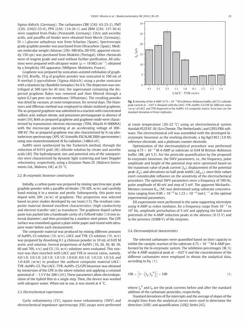
Fig. 6. Square-wave voltammograms of 4.75 × 10⁻⁵ M 4-AMP (0.04 M Britton–Robinson buffer, pH 5.5) obtained with the developed LACC–TYR–AuNPs–CS/GPE bi-enzymaticbiosensor for quantification of (A) carbaril (CBR; 9.90 × 10⁻⁸–2.91 × 10⁻⁶ M), (B) formetanate (FMT; 9.99 × 10⁻⁷–3.21 × 10⁻⁵ M), (C) propoxur (PPX; 4.99 × 10⁻⁷–1.92 × 10⁻⁵ M)and (D) ziram (ZRM; 9.99 × 10⁻⁸–3.38 × 10⁻⁷ M) in QuEChERS extracts of orange samples by the standard addition method. Square-wave voltammetric conditions: frequency100 Hz, pulse amplitude 40 mV and scan increment 3 mV. The inserts refer to the calibration curves obtained for each carbamate pesticide by inhibition of the enzymatic catalysis (inhi-bition percentage, %IR=−2.22± 0.21+ 2.92 × 10⁷± 1.82 × 10⁶ [CBR]/M; r= 0.9994; n= 8; %IR= 6.40± 0.13+ 1.82 × 10⁶± 1.08 × 10⁵ [FMT]/M; r= 0.9992; n= 8; %IR= 2.99±0.08 + 1.13 × 10⁶ ± 8.11 × 10⁴ [PPX]/M; r = 0.9988; n = 9; %IR = −2.32 ± 0.12 + 2.19 × 10⁸ ± 2.51 × 10⁷ [ZRM]/M; r = 0.9989; n = 8).
26 T.M.B.F. Oliveira et al. / Bioelectrochemistry 98 (2014) 20–29
−Z″ ωð Þ ¼ ω � Rp2 � Cp= 1þω2 � Rp
2 � Cp2
� �ð3Þ
where Rs/Ω is the resistance of the electrolyte; Rp/Ω the polarizationresistance; Cp/F is the capacitance of the system andω is the angular fre-quency (ω=2× π× f; f is the ac-frequency) [47]. GPE showed excellentconductivity and a Z′/−Z″ linear relation (line a), suggesting a mass-transfer process controlled by diffusion. When CS was electrodepositedonto the GPE, a large capacitive arc was observed (line b), indicating anincrease of the charge-transfer resistance (Rct = 767 Ω). However,when CS was enriched with AuNPs (line c), the charge-transfer valuesdecreased sharply (Rct = 407 Ω), showing that the gold nanoparticlesclearly improved the conductivity of the hybrid film.
Regarding the best pathway to immobilize LACC and TYR onto theelectrode, cyclic voltammograms of the three alternatives tested, namely,dispersion in the AuNPs–CS composite material, dispersion in pure CSmatrix, and directly onto the GPE by drip-coating (solution containing20 U mL−1 LACC and 10 U mL−1 TYR) are presented in Fig. 4C whilethe corresponding EIS results may be observed in Fig. 4A (lines d–f).The lower charge-transfer resistance (Rct = 527 Ω; Fig. 4A, line d) andconsequent higher peak intensity (Fig. 4C) were reached when theenzymes were dispersed in the AuNPs–CS composite. For the other twocases (lines e and f for drip-coating and dispersion in pure CS matrix,respectively), the Rct was higher and quite similar, indicating that the
immobilization processes were also efficient, but in the presence ofAuNPs the systemexhibited a better performance as electrochemical bio-sensor. AuNPs are widely used nanomaterials because of their large spe-cific surface area, strong adsorption ability, and high conductivity [33].Their conductivity characteristics improve the electron transfer betweenthe enzyme redox center and the electrode surface [37]. They can strong-ly interact with biomaterials and they have been used as a mediator toimmobilize biomolecules and to efficiently retain their activity [33].
3.4. Effect of the pH and incubation time on the biosensor response
Although the two selected enzymes are polyphenoloxidases, eachone has a different working pH range where maximum activity occurs(2.0–6.0 for LACC and 5.0–8.0 for TYR). Moreover, the optimum pH ishighly dependent on the enzyme source and on the substrate used[12,19,22,48]. This experimental parameter is crucial for the develop-ment of bi-enzymatic systems. The pH of the electrolyte was rangedfrom 4 to 7 to characterize the individual activity of LACC and TYR,and the activity of the bi-enzymatic system proposed (Fig. 5). Thecurrent response resulting from the enzyme-catalyzed reaction (peakIIc observed at −0.07 V, Fig. 2A) achieved a maximum value at pH 5.0and 6.0, respectively, for LACC–AuNPs–CS/GPE and TYR–AuNPs–CS/GPE.These results are in accordance to those reported for other individualLACC- and TYR-based biosensors [8,49–51]. By using both enzymes

27T.M.B.F. Oliveira et al. / Bioelectrochemistry 98 (2014) 20–29
at the optimum ratio (2.0:1.0, w/w), the peak current was enhancedca. 1.6 to 2.1 times at pH 5.5 indicating a significant synergistic effectof the LACC–TYR conjugate system. Also, the increase of the pH causeda linear displacement of the peak potential to more negative values(−Ep/V = −0.64 ± 0.009 + 0.16 ± 0.023 pH; r = 0.9987; n = 7)confirming a proton-dependence of the substrate. A pH of 5.5 wasselected as the optimum.
The apparent Michaelis–Menten constant Km (M) [29,52], whichreflects both the enzymatic affinity and the kinetic constants was deter-mined based on the Lineweaver–Burk Eq. (4):
1=Isð Þ ¼ Km=Imaxð Þ � 1=Cð Þ þ 1=Imaxð Þ ð4Þ
where Is is the steady state current (A) after the addition of substrate,Imax corresponds to maximum current (A) obtained in the linear rangeand C is the concentration (M) of the substrate in the solution. Thus,for a linear regression equation obtained from the relationship between1/Is and 1/C data, the slope and intercept of the linear fit correspond toKm/Imax and 1/Imax, respectively. LACC–TYR–AuNPs–CS/GPE (Km of26.9 ± 0.5 M) showed the most interesting properties when comparedwith the LACC–AuNPs–CS/GPE (Km of 37.8 ± 0.2 M) and TYR–AuNPs–CS/GPE (Km of 52.3 ± 0.4 M) since lower Km values indicate highergrade of affinity of the immobilized enzymes to the substrate [53].
Considering that the analytical peak of the substrate (−0.07 V,peak IIc at Fig. 2A) results from the reduction of the p-benzoquinoneto p-hydroquinone at the second chemical–electrochemical step(Fig. 2B) and that the formation of p-benzoquinone is slow and deter-minant for the reaction rate, the incubation time for the preparationof the biosensor surface was also optimized. The peak current in-creased until 20 min and remained approximately constant thereaf-ter (data not shown). Thus, this durationwas selected as theworkingincubation/stabilization time before the analytical application of theconstructed LACC–TYR–AuNPs–CS/GPE biosensor. Thereafter, andconcerning the quantification of the selected carbamates, no incuba-tion time between each SWV measurement was applied.
3.5. Electroanalytical characteristics
Using the optimized square-wave voltammetric parameters (fre-quency 100 Hz, pulse amplitude 40 mV and scan increment 3 mV),calibration data were obtained for CBR, FMT, PPX and ZRM (Table 1).The lower inhibition percentages were observed for PPX, while thehigher were detected for ZRM. These patterns of variation are relatedwith the inherent toxicity of each pesticide for the employed bi-enzymatic system. The analytical curves (Fig. 6) presented wide linearranges, suitable linearity and low dispersion of the data, even at lowconcentrations, with correlation coefficients (r) ranging from 0.9988(PPX) to 0.9994 (CBR) (Table 1). The lower LOD and LOQwere reached,by order, for ZRM b CBR b PPX b FMT. The selected carbamates are ex-tensively applied for the protection of fruit and vegetable crops withMRLs ranging from 0.05 to 0.5 mg kg−1 (w/w) [16,17]. The attainedLODs calculated in a fresh weight basis (0.001 mg kg−1 for ZRM,0.004 mg kg−1 for CBR, 0.039 mg kg−1 for PPX and 0.048 mg kg−1 forFMT) allow application of the electroanalytical procedure for fruit safetyquality control.
Repeatability and reproducibility were also assessed by the relativestandard deviations (RSD) of different measurements. For intra-dayrepeatability (n = 10), RSD values ranged from 1.2 to 2.8%, and forinter-day repeatability (n= 5) from 3.2 to 5.8%. Reproducibility studieswere made using four different biosensors and the attained RSD variedbetween 3.2 and 6.5%. The stability of the biosensor was also tested. Thecatalytic properties of the bi-enzymatic system retained 93.6% of itsinitial current response over a period of twenty days.
Overall, the results show that the developed LACC–TYR–AuNPs–CS/GPE biosensor presents suitable analytical characteristics forquantification of the selected carbamates. The results achieved with
the proposed biosensor are in agreement or compare favorably withthose reported previously (scarce and mainly based on acetylcholines-terase) for CBR [54–57], PPX [56], FMT [10,13] and ZRM [13,58].
3.6. Application to citrus fruits
The validation of the biosensor-based procedure was performed byrecovery assays performed at two spiking levels (0.01–3.14 mg kg−1
w/w) in citrus fruits (orange, tangerine and lemon) (Table 1). Fig. 6 dis-plays representative recovery assays using the standard additionsmethod. Acceptable recoveries for trace pesticides determination werefound ranging from 93.9 ± 0.2 to 97.8 ± 0.3% for orange, from 94.2 ±0.1 to 97.3 ± 0.3% for tangerine, and from 93.8 ± 0.3% to 97.1 ± 0.1%for lemon samples. The lower recoveries were attained for PPX,while the superior were obtained for ZRM probably due to the differentsensitivity of the biosensor to these compounds (the lower for PPX,1.13 × 10−6 M, and the higher for ZRM, 2.19 × 10⁻⁸ M; Table 1).Globally and as expected, a slightly better performancewas observedfor the higher fortified level tested.
Orange, tangerine and lemon are important sources of glucose, citricacid and ascorbic acid. Consequently, theywere tested as possible inter-ferences in the analytical signal of the LACC–TYR–AuNPs–CS/GPEbiosensor (results not shown). Experiments were carried out in thepresence of several interference:substrate ratios (1:10, 1:1 and 10:1(v/v)). The higher biases (%) were detected at the maximum ratiowhich is seldom to occur in 10 g of the analyzed citrus fruits. The resultscorresponded to a response reduction of 4.7 ± 0.3% for glucose, b7.5 ±0.1% for citric acid and b11.5 ± 0.4% for ascorbic acid. Therefore, theseresults corroborate the reliability of the pesticide residue methodologyproposed.
4. Conclusions
In this first study that explores the electrochemical and catalyticproperties of a bi-enzymatic device based on a GPE, an accurate andsensitive LACC–TYR–AuNPs–CS/GPE biosensor was developed for car-bamate pesticide determination in citrus fruits. The chitosan polymericmatrix produced at pH 5.5 proved to be a suitable microenvironment tocarry AuNPs and the selected polyphenoloxidases by a single and fastelectrodeposition step onto the GPE. Although pure chitosan increasedthe charge-transfer resistance of the device, the use of AuNPs allowedovercoming this problem by reducing the Rct of the device, even in thepresence of the enzymes. Synergistic positive effects were detected be-tween LACC and TYR at the optimum pH promoting high sensitivity ofthe proposed bi-enzymatic system. Also, the amine and hydroxylgroups of the chitosan in acid conditions attributed high stability (ca.twenty days) to the biosensor. Thus, the proposed and validated elec-troanalytical procedures can be an interesting strategy for food safetycontrol of carbamate pesticide residues in fruits. Furthermore, itpresents several advantages when compared to the traditional chro-matographic techniques employed, namely, it is clearly faster, easier,more environmental friendly while being amenable to integration forin situ determination.
Acknowledgments
The authors thank CAPES and FCT for the financial support throughthe project (Nº. 313/11). This work received also financial supportfrom theEuropeanUnion (FEDER funds through COMPETE) andNation-al Funds (FCT, Fundação para a Ciência e Tecnologia) through projectPest-C/EQB/LA0006/2013. The work also received financial supportfrom the European Union (FEDER funds) under the framework ofQREN through Project NORTE-07-0124-FEDER-000069. Maria doCarmo Pereira and Pedro Carneiro are acknowledged for their help onpreparation of the AuNPs. M.F. Barroso is grateful to FCT for her post-
28 T.M.B.F. Oliveira et al. / Bioelectrochemistry 98 (2014) 20–29
doc fellowship SFRH/BPD/78845/2011. T.M.B.F. Oliveira is grateful toUERN for his PhD fellowship.
Appendix A. Supplementary data
Supplementary data to this article can be found online at http://dx.doi.org/10.1016/j.bioelechem.2014.02.003.
References
[1] J.S.V. Dyk, B. Pletschke, Review on the use of enzymes for the detection of organo-chlorine, organophosphate and carbamate pesticides in the environment,Chemosphere 82 (2011) 291–307.
[2] S. Morais, E. Dias, M.L. Pereira, Carbamates: human exposure and health effects, in:M. Jokanovic (Ed.), The Impact of Pesticides, WY Academy Press, Cheyenne, 2012,pp. 21–38.
[3] E. Dias, M. Gomes, C. Domingues, E. Ramalheira, S. Morais, M.L. Pereira, Subacute ef-fects of the thiodicarb pesticide on target organs of male Wistar rats: biochemical,histological, and flow cytometry studies, J. Toxicol. Environ. Health A 76 (2013)533–539.
[4] U. Schulte-Oehlmann, J. Oehlmann, F. Keil, Before the curtain falls: endocrine-activepesticides — a German contamination legacy, Rev. Environ. Contam. Toxicol. 213(2011) 137–159.
[5] WHO (World Health Organization), State of the Science of Endocrine DisruptingChemicals 2012, United Nations Environment Programme and the World HealthOrganization, Geneva, 2013.
[6] US EPA (United States Environmental Protection Agency), National Survey of Pesti-cides in DrinkingWaterWells, Phase II Report, EPA 570/8-91-020, National TechnicalInformation Service, Springfield, 2012.
[7] M. Farré, L. Kantiani, S. Pérez, D. Barceló, Sensors and biosensors in support of EUdirectives, TrAC, Trends Anal. Chem. 28 (2009) 170–185.
[8] M.D. Fusco, C. Tortolini, D. Deriu, F. Mazzei, Laccase-based biosensor for the determi-nation of polyphenol index in wine, Talanta 81 (2010) 235–240.
[9] C.I.L. Justino, T.A.P. Rocha-Santos, A.C. Duarte, Advances in point-of-care technolo-gies with biosensors based on carbon nanotubes, TrAC, Trends Anal. Chem. 45(2013) 24–36.
[10] F.W.P. Ribeiro, M.F. Barroso, S. Morais, S. Viswanathan, P. De Lima-Neto, A.N. Correia,M.B.P.P. Oliveira, C. Delerue-Matos, Simple laccase-based biosensor for formetanatehydrochloride quantification in fruits, Bioelectrochemistry 95 (2014) 7–14.
[11] R.K. Shervedani, A. Amini, Direct electrochemistry of dopamine on gold–Agaricusbisporus laccase enzyme electrode: characterization and quantitative detection,Bioelectrochemistry 84 (2012) 25–31.
[12] T.M.B.F. Oliveira, M.F. Barroso, S. Morais, P. De Lima-Neto, A.N. Correia, M.B.P.P.Oliveira, C. Delerue-Matos, Biosensor based on multi-walled carbon nanotubespaste electrode modified with laccase for pirimicarb pesticide quantification,Talanta 106 (2013) 137–143.
[13] T.M.B.F. Oliveira, M.F. Barroso, S. Morais, M. Araújo, C. Freire, P. De Lima-Neto, A.N.Correia, M.B.P.P. Oliveira, C. Delerue-Matos, Laccase–Prussian blue film–graphenedoped carbon paste modified electrode for carbamate pesticides quantification,Biosens. Bioelectron. 47 (2013) 292–299.
[14] S. Prakash, T. Chakrabarty, A.K. Singh, V.K. Shahi, Polymer thin films embedded withmetal nanoparticles for electrochemical biosensors applications, Biosens. Bioelectron.41 (2013) 43–53.
[15] EU pesticide residues database, EU pesticide residues database for all the EU-MRLs,(Available at) http://ec.europa.eu/food/plant/protection/pesticides/database(August 2013).
[16] ANVISA (Brazilian Health Surveillance Agency), Monographs of authorized pesti-cides, (Accessed at) http://portal.anvisa.gov.br/wps/content/Anvisa+Portal/Anvisa/Inicio/Agrotoxicos+e+Toxicologia/Assuntos+de+Interesse/Monografias+de+Agrotoxicos/Monografias (August 2013).
[17] M. ElKaoutit, I. Naranjo-Rodriguez, K.R. Temsamani, M. Domínguez, J.L.H.-H. DeCisneros, Investigation of biosensor signal bioamplification: comparison of directelectrochemistry phenomena of individual laccase, and dual laccase–tyrosinasecopper enzymes, at a sonogel–carbon electrode, Talanta 75 (2008) 1348–1355.
[18] O.D. Leite, K.O. Lupetti, O. Fatibello-Filho, I.C. Vieira, A.M. Barbosa, Synergic effectstudies of the bi-enzymatic system laccase–peroxidase in a voltammetric biosensorfor catecholamines, Talanta 59 (2003) 889–896.
[19] J. Kochana, P. Nowak, A. Jarosz-Wilkołaska, M. Bieroń, Tyrosinase/laccase bienzymebiosensor for amperometric determination of phenolic compounds, Microchem. J.89 (2008) 171–174.
[20] D. Odaci, A. Telefoncu, S. Timur, Maltose biosensing based on co-immobilization ofα-glucosidase and pyranose oxidase, Bioelectrochemistry 79 (2010) 108–113.
[21] E. Zapp, D. Brondani, I.C. Vieira, J. Dupont, C.W. Scheeren, Bioelectroanalytical deter-mination of rutin based on bi-enzymatic sensor containing iridium nanoparticles inionic liquid phase supported in clay, Electroanalysis 23 (2011) 764–776.
[22] Z. Yang, F. Wu, Catalytic properties of tyrosinase from potato and edible fungi,Biotechnol. 5 (2006) 344–348.
[23] C. Chouteau, S. Dzyadevych, C. Durrieu, J.-M. Chovelon, A bi-enzymatic whole cellconductometric biosensor for heavy metal ions and pesticides detection in watersamples, Biosens. Bioelectron. 21 (2005) 273–281.
[24] H. Wang, R. Yuan, Y. Chai, H. Niu, Y. Cao, H. Liu, Bi-enzyme synergetic catalysisto in situ generate coreactant of peroxydisulfate solution for ultrasensitiveelectrochemiluminescence immunoassay, Biosens. Bioelectron. 37 (2012) 6–10.
[25] M. Yang, Y. Yang, Y. Yang, G. Shen, R. Yu, Bienzymatic amperometric biosensor forcholine based on mediator thionine in situ electropolymerized within a carbonpaste electrode, Anal. Biochem. 334 (2004) 127–134.
[26] J. Cancino, C.A. Razzino, V. Zucolotto, S.A.S. Machado, The use of mixed self-assembledmonolayers as a strategy to improve the efficiency of carbamate detectionin environmental monitoring, Electrochim. Acta 87 (2013) 717–723.
[27] M. Diaconu, S.C. Litescu, G.L. Radu, Bienzymatic sensor based on the use of redoxenzymes and chitosan–MWCNT nanocomposite. Evaluation of total phenoliccontent in plant extracts, Microchim. Acta 172 (2011) 177–184.
[28] J. Li, S. Zivanovic, P.M. Davidson, K. Kit, Production and characterization of thick, thinand ultra-thin chitosan/PEO films, Carbohydr. Polym. 83 (2011) 375–382.
[29] C. Zhai, S. Xia, W. Zhao, Z. Gong, X. Wang, Acetylcholinesterase biosensor based onchitosan/Prussian blue/multiwall carbon nanotubes/hollow gold nanospheres nano-composite film by one-step electrodeposition, Biosens. Bioelectron. 42 (2013)124–130.
[30] C.L. De Vasconcelos, D.E.S. Dos Santos, T.N.C. Dantas, M.R. Pereira, J.L.C. Fonseca,Effect of molecular weight and ionic strength on the formation of polyelectrolytecomplexes based on poly(methacrylic acid) and chitosan, Biomacromolecules 7(2006) 1245–1252.
[31] S.P. Strand, S. Danielsen, B.E. Christensen, K.M. Vårum, Influence of chitosan structureon the formation and stability of DNA–chitosan polyelectrolyte complexes,Biomacromolecules 6 (2005) 3357–3366.
[32] Y. Martı́nez, J. Retuert, M. Yazdani-Pedram, H. Cölfen, Hybrid ternary organic–inor-ganic films based on interpolymer complexes and silica, Polymer 45 (2004)3257–3265.
[33] K.-J. Huang, J. Li, Y.-Y.Wu, Y.-M. Liu, Amperometric immunobiosensor forα-fetoproteinusing Au nanoparticles/chitosan/TiO2–graphene composite based platform,Bioelectrochemistry 90 (2013) 18–23.
[34] M.J. Allen, V.C. Tung, R.B. Kaner, Honeycomb carbon: a review of graphene, Chem.Rev. 110 (2010) 132–145.
[35] T. Kuila, S. Bose, P. Khanra, A.K. Mishra, N.H. Kim, J.H. Lee, Recent advances ingraphene-based biosensors, Biosens. Bioelectron. 26 (2011) 4637–4648.
[36] M. Pumera, A. Ambrosi, A. Bonanni, E.L.K. Chng, H.L. Poh, Graphene for electrochemicalsensing and biosensing, Trac, Trends Anal. Chem. 29 (2010) 954–965.
[37] A. Sassolas, B. Prieto-Simón, J.-L. Marty, Biosensors for pesticide detection: newtrends, Am. J. Anal. Chem. 3 (2012) 210–232.
[38] S. Andreescu, L.A. Luck, Studies of the binding and signaling of surface-immobilizedperiplasmic glucose receptors on gold nanoparticles: a glucose biosensor applica-tion, Anal. Biochem. 375 (2008) 282–290.
[39] C.A. Amarnath, C.E. Hong, N.H. Kim, B.-C. Ku, T. Kuila, J.H. Lee, Efficient synthesisof graphene sheets using pyrrole as a reducing agent, Carbon 49 (2011)3497–3502.
[40] J. Kimling, M. Maier, B. Okenve, V. Kotaidis, H. Ballot, A. Plech, Turkevich method forgold nanoparticle synthesis revisited, J. Phys. Chem. B 110 (2006) 15700–15707.
[41] M.-H. Xue, Q. Xu, M. Zhou, J.-J. Zhu, In situ immobilization of glucose oxidase inchitosan–gold nanoparticle hybrid film on Prussian blue modified electrode forhigh-sensitivity glucose detection, Electrochem. Commun. 8 (2006) 1468–1474.
[42] J.N. Miller, J.C. Miller, Statistics and Chemometrics for Analytical Chemistry, PearsonPrentice Hall, United Kingdom, 2005.
[43] European Council Directive 2002/63/CE, Establishing community methods ofsampling for the official control of pesticide residues in and on products of plantand animal origin and repealing Directive 79/700/EEC, Off. J. Eur. CommunitiesL187 (2002) 30-31.
[44] M. Anastassiades, S.J. Lehotay, D. Stajnbaher, F.J. Schenck, Fast and easymultiresiduemethod employing acetonitrile extraction/partitioning and “dispersive solid-phaseextraction” for the determination of pesticide residues in produce, J. AOAC Int. 86(2003) 412–431.
[45] P. Paíga, S. Morais, T. Oliva-Teles, M. Correia, C. Delerue-Matos, S.C. Duarte, A. Pena,C.M. Lino, Extraction of ochratoxin A in bread samples by the QuEChERS methodol-ogy, Food Chem. 135 (2012) 2522–2528.
[46] H.J. Salavagione, J. Arias, P. Garcés, E. Morallón, C. Barbero, J.L. Vázquez,Spectroelectrochemical study of the oxidation of aminophenols on platinumelectrode in acid medium, J. Electroanal. Chem. 565 (2004) 375–383.
[47] M.E. Orazem, B. Tribollet, Electrochemical Impedance Spectroscopy, John Wiley &Sons, Inc., Hoboken, 2008.
[48] G.-Q. Zhang, Q.-J. Chen, H.-X.Wang, T.B. Ng, A laccasewith inhibitory activity againstHIV-1 reverse transcriptase from the mycorrhizal fungus Lepiota ventriosospora,J. Mol. Catal. B Enzym. 85–86 (2013) 31–36.
[49] E. Casero, M.D. Petit-Domínguez, L. Vázquez, I. Ramírez-Asperilla, A.M. Parra-Alfambra,F. Pariente, E. Lorenzo, Laccase biosensors based on different enzyme immobilizationstrategies for phenolic compounds determination, Talanta 115 (2013) 401–408.
[50] L. Tang, Y. Zhou, G. Zeng, Z. Li, Y. Liu, Y. Zhang, G. Chen, G. Yang, X. Lei, M. Wu, Atyrosinase biosensor based on ordered mesoporous carbon–Au/L-lysine/Au nano-particles for simultaneous determination of hydroquinone and catechol, Analyst138 (2013) 3552–3560.
[51] L. Yang, H. Xiong, X. Zhang, S. Wang, A novel tyrosinase biosensor based onchitosan–carbon-coated nickel nanocomposite film, Bioelectrochemistry 84(2012) 44–48.
[52] Y. Song, K. Qu, C. Zhao, J. Ren, X. Qu, Graphene oxide: intrinsic peroxidase catalyticactivity and its application to glucose detection, Adv. Mater. 22 (2010) 2206–2210.
[53] M. Gamero, F. Pariente, E. Lorenzo, C. Alonso, Nanostructured rough gold electrodesfor the development of lactate oxidase-based biosensors, Biosens. Bioelectron. 25(2010) 2038–2044.
[54] B. Bucur, D. Fournier, A. Danet, J.-L. Marty, Biosensors based on highly sensitiveacetylcholinesterases for enhanced carbamate insecticides detection, Anal. Chim.Acta. 562 (2006) 115–121.

29T.M.B.F. Oliveira et al. / Bioelectrochemistry 98 (2014) 20–29
[55] J. Caetano, S.A.S. Machado, Determination of carbaryl in tomato “in natura” using anamperometric biosensor based on the inhibition of acetylcholinesterase activity,Sensors Actuators B 129 (2008) 40–46.
[56] G.S. Nunes, D. Barceló, B.S. Grabaric, J.M. Díaz-Cruz, M.L. Ribeiro, Evaluation of ahighly sensitive amperometric biosensor with low cholinesterase charge immobilizedon a chemicallymodified carbon paste electrode for trace determination of carbamatesin fruit, vegetable and water samples, Anal. Chim. Acta. 399 (1999) 37–49.
[57] X.Wang, L. Chen, S. Xia, Z. Zhu, J. Zhao, J. Chovelon, N.J. Renaul, Tyrosinase biosensorbased on interdigitated electrodes for herbicides determination, Int. J. Electrochem.Sci. 1 (2006) 55–61.
[58] M.T.P. Pita, A.J. Reviejo, F.J.M. Villena, J.M. Pingarrón, Amperometric selectivebiosensing of dimethyl- and diethyldithiocarbamates based on inhibition processesin a medium of reversed micelles, Anal. Chim. Acta. 340 (1997) 89–97.
[59] U. Khan, I. O’Connor, Y.K. Gun’ko, J.N. Coleman, The Preparation of Hybrid Films ofCarbon Nanotubes and Nano-graphite/graphene with Excellent Mechanical andElectrical Properties, Carbon 48 (2010) 2825–2830