Self-Assembled pH-Sensitive Nanoparticles: A Platform for Oral Delivery of Protein Drugs
6
www.afm-journal.de FEATURE ARTICLE © 2010 WILEY-VCH Verlag GmbH & Co. KGaA, Weinheim 3695 www.MaterialsViews.com wileyonlinelibrary.com Adv. Funct. Mater. 2010, 20, 3695–3700 By Kiran Sonaje, Kun-Ju Lin, Jiun-Jie Wang, Fwu-Long Mi, Chiung-Tong Chen, Jyuhn-Huarng Juang, and Hsing-Wen Sung* 1. Introduction Production of pharmaceutically active peptide/protein drugs in large quantities has become feasible. The oral route is consid- ered to be the most convenient and comfortable means of drug administration, leading to a higher patient compliance. Drug molecules can traverse the intestinal epithelium by a transcel- lular means involving endocytotic process or by passing between the cells through a process known as the paracellular pathway. [1] Nevertheless, oral administra- tion of hydrophilic macromolecules such as peptide/protein drugs is encountered with many difficulties, as the drugs have to confront various major barriers in the gas- trointestinal (GI) tract. First of all, peptide/ protein drugs get denatured readily by the low pH of gastric medium in the stomach. Secondly, different digestive enzymes in the stomach and small intestine may lead to degradation of peptide/protein drugs. [2] A carrier system is therefore required to protect peptide/protein drugs from the harsh environment in the GI tract. Finally, the intestinal epithelium is a major barrier to the absorption of hydrophilic macro- molecules as they cannot diffuse across cells through the lipid- bilayer cell membranes, due to their high molecular weights and hydrophilicity. [3] Attentions have been drawn to improving the paracellular transport of hydrophilic macromolecules. [4] The transport of hydrophilic macromolecules via the paracellular pathway is, how- ever, severely restricted by the presence of tight junctions which are located at the luminal aspect of adjacent epithelial cells. [5] Tight junctions consist of a complex of trans-membrane (e.g., occuludin, claudin, JAM) and cytoplasmic (e.g., ZO-1, ZO-2, ZO-3, cingulin) proteins and are dynamically regulated by com- plex physiological mechanisms. [6] ZO-1 proteins are thought to be a linkage molecule between occludin and actin cytoskeleton and play important roles in the rearrangement of cell–cell contacts. A number of intestinal permeation enhancers have been explored to enhance the oral bioavailability of hydrophilic macro- molecules. It has been reported that chitosan (CS), a cationic polysaccharide, can adhere to epithelial surfaces and open the tight junctions between contiguous cells. [7] Recent hypothesis suggested that the interactions between the positively-charged CS and the negatively-charged portion of the tight junction pro- tein ZO-1, leads to translocation of ZO-1 to the cytoskeleton and subsequently increases the paracellular permeability. [8] Via this paracellular pathway, we developed a pH-sensitive nanoparticle (NP) system covered with CS for oral delivery of insulin for the treatment of insulin-dependent diabetic patients. Self-Assembled pH-Sensitive Nanoparticles: A Platform for Oral Delivery of Protein Drugs The oral route is considered to be the most convenient and comfortable means of drug administration for patients. Nevertheless, oral administration of hydrophilic macromolecules such as peptide/protein drugs is encountered with many difficulties. To overcome these difficulties, a pH-sensitive nanoparticle (NP) carrier system, self-assembled by chitosan (CS) and poly- γ-glutamic acid ( γ-PGA), is developed for paracellular transports of insulin. Oral administration of insulin-loaded NPs shows a significant hypoglycemic action in diabetic rats and the corresponding relative bioavailability of insulin is approximately 15%. These findings suggest that the developed NP system is a promising carrier for improved transmucosal delivery of insulin in the small intestine. Besides insulin, this NP carrier system is expected to serve as a platform for oral delivery of hydrophilic macromolecules such as pharmaceutically active peptides/ proteins, glycosaminoglycans, and oligonucleotides. Studies on the detailed mechanism of tight-junction opening by CS or its derivatives are in progress, which is of paramount importance and needs to be established to aid further development in the use of NPs for oral delivery of hydrophilic macromolecules. DOI: 10.1002/adfm.201001014 [∗] K. Sonaje, Prof. H.-W. Sung Department of Chemical Engineering National Tsing Hua University Hsinchu 30013, Taiwan, R.O.C. E-mail: [email protected] Dr. K.-J. Lin, Dr. J.-J. Wang Department of Medical Imaging and Radiological Sciences Chang Gung University Taoyuan, Taiwan, R.O.C. Dr. F.-L. Mi Department of Biotechnology Vanung University Chungli, Taoyuan, Taiwan, R.O.C. Dr. C.-T. Chen Division of Medical Engineering Research National Health Research Institutes Zhunan, Miaoli, Taiwan, R.O.C. Dr. J.-H. Juang Division of Endocrinology and Metabolism Chang Gung University and Memorial Hospital Taoyuan, Taiwan, R.O.C.
Transcript of Self-Assembled pH-Sensitive Nanoparticles: A Platform for Oral Delivery of Protein Drugs

www.afm-journal.de
FEATU
R
www.MaterialsViews.com
Self-Assembled pH-Sensitive Nanoparticles: A Platform for Oral Delivery of Protein Drugs
E ARTIC
By Kiran Sonaje , Kun-Ju Lin , Jiun-Jie Wang , Fwu-Long Mi , Chiung-Tong Chen , Jyuhn-Huarng Juang , and Hsing-Wen Sung *
LE
The oral route is considered to be the most convenient and comfortable means of drug administration for patients. Nevertheless, oral administration of hydrophilic macromolecules such as peptide/protein drugs is encountered with many diffi culties. To overcome these diffi culties, a pH-sensitive nanoparticle (NP) carrier system, self-assembled by chitosan (CS) and poly- γ -glutamic acid ( γ -PGA), is developed for paracellular transports of insulin. Oral administration of insulin-loaded NPs shows a signifi cant hypoglycemic action in diabetic rats and the corresponding relative bioavailability of insulin is approximately 15%. These fi ndings suggest that the developed NP system is a promising carrier for improved transmucosal delivery of insulin in the small intestine. Besides insulin, this NP carrier system is expected to serve as a platform for oral delivery of hydrophilic macromolecules such as pharmaceutically active peptides/proteins, glycosaminoglycans, and oligonucleotides. Studies on the detailed mechanism of tight-junction opening by CS or its derivatives are in progress, which is of paramount importance and needs to be established to aid further development in the use of NPs for oral delivery of hydrophilic macromolecules.
1. Introduction
Production of pharmaceutically active peptide/protein drugs in large quantities has become feasible. The oral route is consid-ered to be the most convenient and comfortable means of drug
© 2010 WILEY-VCH Verlag GmbH & Co. KGaA, WeinheiAdv. Funct. Mater. 2010, 20, 3695–3700
DOI: 10.1002/adfm.201001014
[∗] K. Sonaje , Prof. H.-W. Sung Department of Chemical EngineeringNational Tsing Hua UniversityHsinchu 30013, Taiwan, R.O.C. E-mail: [email protected] Dr. K.-J. Lin , Dr. J.-J. Wang Department of Medical Imaging and Radiological SciencesChang Gung UniversityTaoyuan, Taiwan, R.O.C. Dr. F.-L. Mi Department of BiotechnologyVanung UniversityChungli, Taoyuan, Taiwan, R.O.C. Dr. C.-T. Chen Division of Medical Engineering ResearchNational Health Research InstitutesZhunan, Miaoli, Taiwan, R.O.C. Dr. J.-H. Juang Division of Endocrinology and MetabolismChang Gung University and Memorial HospitalTaoyuan, Taiwan, R.O.C.
administration, leading to a higher patient compliance. Drug molecules can traverse the intestinal epithelium by a transcel-lular means involving endocytotic process or by passing between the cells through a process known as the paracellular pathway. [ 1 ] Nevertheless, oral administra-tion of hydrophilic macromolecules such as peptide/protein drugs is encountered with many diffi culties, as the drugs have to confront various major barriers in the gas-trointestinal (GI) tract. First of all, peptide/protein drugs get denatured readily by the low pH of gastric medium in the stomach. Secondly, different digestive enzymes in the stomach and small intestine may lead to degradation of peptide/protein drugs. [ 2 ] A carrier system is therefore required to protect peptide/protein drugs from the harsh environment in the GI tract. Finally, the intestinal epithelium is a major barrier to the absorption of hydrophilic macro-
molecules as they cannot diffuse across cells through the lipid-bilayer cell membranes, due to their high molecular weights and hydrophilicity. [ 3 ]
Attentions have been drawn to improving the paracellular transport of hydrophilic macromolecules. [ 4 ] The transport of hydrophilic macromolecules via the paracellular pathway is, how-ever, severely restricted by the presence of tight junctions which are located at the luminal aspect of adjacent epithelial cells. [ 5 ] Tight junctions consist of a complex of trans-membrane (e.g., occuludin, claudin, JAM) and cytoplasmic (e.g., ZO-1, ZO-2, ZO-3, cingulin) proteins and are dynamically regulated by com-plex physiological mechanisms. [ 6 ] ZO-1 proteins are thought to be a linkage molecule between occludin and actin cytoskeleton and play important roles in the rearrangement of cell–cell contacts.
A number of intestinal permeation enhancers have been explored to enhance the oral bioavailability of hydrophilic macro-molecules. It has been reported that chitosan (CS), a cationic polysaccharide, can adhere to epithelial surfaces and open the tight junctions between contiguous cells. [ 7 ] Recent hypothesis suggested that the interactions between the positively-charged CS and the negatively-charged portion of the tight junction pro-tein ZO-1, leads to translocation of ZO-1 to the cytoskeleton and subsequently increases the paracellular permeability. [ 8 ] Via this paracellular pathway, we developed a pH-sensitive nanoparticle (NP) system covered with CS for oral delivery of insulin for the treatment of insulin-dependent diabetic patients.
m 3695wileyonlinelibrary.com

FEATU
RE
ARTI
CLE
3696
www.afm-journal.dewww.MaterialsViews.com
Hsing-Wen Sung is Tsing Hua Chair Professor at the Department of Chemical Engineering, National Tsing Hua University, Taiwan. He received his PhD degree from Georgia Institute of Technology in 1988, after which, he worked as a R&D engineer at the Baxter Healthcare Corporation, USA. He started an academic career in 1993 as an Associate
Professor at the Department of Chemical Engineering, National Central University, Taiwan. His research interests are tissue engineering, drug/gene delivery and biomaterials.
Diabetes is caused by the inability of the pancreas to control the blood glucose concentration. It is a pervasive, chronic and often insidious disease that affects 18–20 million people in the U.S. (nearly 30% of whom are undiagnosed) and 200 million worldwide. This will increase to at least 300 million by 2025, according to recent estimates. [ 9 ] Multiple daily injections of insulin are currently the standard treatment for insulin-dependent diabetic patients. However, poor control of blood glucose level and poor patient compliance are associated with this method. [ 3 ] Therefore, better ways of insulin administra-tion are being sought. These include alternative administration routes such as oral, nasal, or inhaled routes that can improve patient compliance. A variety of approaches have been studied in the past to overcome the problems encountered with the oral delivery of insulin, but with little success. The development of an effective oral insulin formulation has been hampered by problems of delivering a suffi cient supply of insulin to the body at the time when it is needed. This is because insulin is largely broken down in the digestive system, much of the insulin deliv-ered orally does not enter the bloodstream, and there is an undesirable variability in the rate of insulin absorption. [ 10 ]
The basic concept of our study is that the orally administered NPs with excessive mucoadhesive CS on their surfaces may pro-long their residence in the small intestine and infi ltrate into the mucus layer. Subsequently, the infi ltrated NPs transiently open the tight junctions between epithelial cells while becoming unstable and disintegrated, due to their pH-sensitivity, to release the preloaded insulin. The released insulin then permeates through the opened paracellular pathway to the bloodstream, which is the ultimate destination for the drug ( Figure 1 ). [ 11 , 12 ]
2. Preparation of the NPs
In a typical study, the NPs were prepared by a simple ionic-gela-tion method using the positively charged CS with an average
© 2010 WILEY-VCH Verlag Gmwileyonlinelibrary.com
Figure 1 . Schematic illustrations of the presumed mechanism of the paraceinsulin released from test NPs [using the proximal small intestine (duodenas an example]: NPs adhere and infi ltrate into the mucus layer of intestininfi ltrated NPs disintegrate due to their pH sensitivity and release the loadapproach the tight junctions; simultaneously, the excessive CS on their sutight junctions between the adjacent epithelial cells; the released insulin pthe opened paracellular pathway. Insets showing TEM micrographs of the Nvalues. Reproduced with permission. [ 12 ] Copyright, 2009 Elsevier.
Fasted State pH 2.0 – 3.7
StomachFed State pH 1.2 pH 1.0 – 3.7
pH 6.0 – 7.0
100 nm
100 nm
EC
ML
TJs
NPs
Systemic Circulation
NPs: Nanoparticles, ML: Mucus Layer, TJs: Tight Junctions, EC: Epithelial Cells
Insulin
Duodenum and
Jejunum
Fasted State pH 2.0 – 3.7
StomachFed State pH 1.2 pH 1.0 – 3.7
pH 6.0 – 7.0
100 nm
100 nm
EC
ML
TJs
NPs
Systemic Circulation
NPs: Nanoparticles, ML: Mucus Layer, TJs: Tight Junctions, EC: Epithelial Cells
Insulin
Duodenum and
Jejunum
Fasted State pH 2.0 – 3.7
StomachFed State pH 1.2 pH 1.0 – 3.7
pH 6.0 – 7.0
100 nm
100 nm
EC
ML
TJs
NPs
Systemic Circulation
NPs: Nanoparticles, ML: Mucus Layer, TJs: Tight Junctions, EC: Epithelial Cells
Insulin
Duodenum and
Jejunum
molecular weight of 80 kDa and negatively charged poly- γ -glutamic acid ( γ -PGA) with an average molecular weight of 60 kDa. [ 13 ] CS is non-toxic, biodegradable, and soft-tissue com-patible [ 14 ] ; it has been widely used as a food additive and as a weight-loss product and been developed as a safe excipient in drug formulations. [ 15 ] γ -PGA, an anionic peptide, is a water-sol-uble, biodegradable, and non-toxic polymer. [ 16 ] The NPs were self-assembled instantaneously upon addition of an aqueous γ -PGA (together with peptide/protein drugs such as insulin) into an aqueous CS under magnetic stirring at room temperature. [ 13 , 17 ] This technique is promising, as the NPs can be prepared under aqueous conditions at room temperature, thus preventing the bioactivity of peptide/protein drugs from decreasing.
A transmission electron microscopy (TEM) image of the NPs prepared in deionized water (pH ≈ 6.0) is shown in Figure 1 . [ 12 ] The as-prepared NPs were of spherical shape with a smooth surface. The mean particle size and the zeta-potential value of
bH & Co. KGaA, Wein
llular transport of um and jejunum) al epithelium; the ed insulin as they rfaces opens the
ermeates through Ps at distinct pH
pH 7.4
100 nm
pH 6.0 – 7.2
100 nm
Chitosan
pH 7.4
100 nm
pH 6.0 – 7.2
100 nm
Chitosan
pH 7.4
100 nm
pH 6.0 – 7.2
100 nm
Chitosan
the prepared NPs can be controlled by the relative concentrations of CS to γ -PGA used. The size of the NPs used in the subsequent in vitro and in vivo studies was approxi-mately 200 nm with a positive surface charge ( ≈ 25 mV) or CS dominated (or shelled) on the surface. [ 11 , 12 ]
3. In Vitro Characteristics
3.1. pH-Sensitivity of the NPs
It is known that the natural pH environment in the GI tract varies from acidic in the stomach to slightly alkaline in the small intestine. [ 18 ] The p K a values of the amine groups ( − NH 2 ) on CS and the carboxyl groups ( − COOH) on γ -PGA are approximately 6.5 and 2.9, respec-tively. [ 19 ] In the range of pH 2.5 − 6.6, CS and γ -PGA are ionized. The ionized CS and γ -PGA can form polyelectrolyte complexes, which results in a matrix structure with a spherical
heim Adv. Funct. Mater. 2010, 20, 3695–3700

FEATU
RE A
RTIC
LE
www.afm-journal.dewww.MaterialsViews.com
0 200 400 600 800 1000 1200 14000
10
20
30
40
50
60
70
80
90
100
200 220 240 260
-30000
-20000
-10000
0
10000
20000Released InsulinStandard Insulin
(n = 5)
pH 2.5
Cu
mu
lativ
e Am
oun
t of
In
sulin
R
elea
sed
(%)
Time (min)
pH 6.6 pH 7.0
a
0 4 8 12 16 20 2430
40
50
60
70
80
90
100
110
Control Group at pH 6.6
Multi-ion-crosslinked NPs at pH 6.6
Control Group at pH 7.0
Multi-ion-crosslinked NPs at pH 7.0
NPs at pH 6.6NPs at pH 7.0T
EE
R o
f in
itia
l Val
ue
(%)
Time (h)
NPsMulti-ion-crosslinked NPs
pH 7.0
b
Elli
ptic
ity
(deg
cm
2dm
ol-1
)
Wavelength (nm)
Figure 2 . a) Release profi les of insulin from test nanoparticles at pH 2.5, 6.6, 7.0, and 7.4, simulating the pH environments in the fasting stomach, duodenum, jejunum and the body fl uid at intercellular spaces between epithelial cells, respectively. Inset: Circular dichroism spectra of the insulin released from the test nanoparticles at pH 7.4 and the standard insulin. b) Effects of test nanoparticles on TEER values of Caco-2 cell monolayers at pH 6.6 or pH 7.0. Control Group: the group incubated with free-form insulin. Reproduced with permission. [ 11 ] Copyright, 2008 Elsevier.
shape (CS/ γ -PGA NPs); however, the CS/ γ -PGA NPs at pH 2.5 or 6.6 were swollen slightly. Outside of this pH range (e.g., pH 1.2 and 7.4), the CS/ γ -PGANPs became unstable and sub-sequently disintegrated. This is because the − COO − groups on γ -PGA became protonated at lower pH values while the amine groups on CS tended to be deprotonated at pH values above 6.5, leading to the disintegration of CS/ γ -PGA NPs and limiting the effi cacy of the insulin delivery at the jejunum and the ileum, in which their pH values are neutral or slightly basic. [ 17 , 20 ]
To increase the pH stability of NPs and thus enhance their in vivo bioavailability, magnesium sulfate (MgSO 4 ) and tripolyphos-phate sodium (TPP) were introduced in the preparation of NPs. [ 11 , 12 ] Our Fourier-transform infrared(FT-IR) spectroscopy and X-ray diffraction (XRD) results indicated that the sponta-neous interaction between CS, insulin, γ -PGA, MgSO 4 , and TPP can form an ionically crosslinked network-structure, leading to the formation of NPs (multi-ion-crosslinked NPs). The multi-ion-crosslinked NPs were more compact than their counterparts without incorporation of MgSO 4 and TPP (CS/ γ -PGA NPs), while their zeta-potential values were comparable. Additionally, the multi-ion-crosslinked NPs had a superior stability over a broader pH range (pH 2.0–7.2) than CS/ γ -PGA NPs (Figure 1 ).
3.2. Drug-Release Profi les
Zinc (Zn 2 + ) is usually added to the biosynthesis and storage of insulin to enhance its stability. [ 17 , 20 ] In the preparation of test NPs, it was noted that insulin may conjugate with γ -PGA via Zn 2 + and thus increase its loading effi ciency (LE) and loading content (LC) in NPs. Here, the LE represents the percentage of insulin used that is encapsulated in the NPs, while the LC is the percentage of insulin per NP (w/w). The insulin LE and LC of CS/ γ -PGA NPs were found to be 55 ± 3.3% and 14.1 ± 0.8% ( n = 5 batches), respectively. [ 11 ] For multi-ion-crosslinked NPs, the insulin LE and LC can be signifi cantly increased to 71.8 ± 1.1% and 19.1 ± 0.6%, respectively. [ 12 ]
Figure 2 a shows the release profi les of insulin from CS/ γ -PGA NPs and multi-ion-crosslinked NPs at pH 2.5, 6.6, 7.0, and 7.4, simulating the pH environments in the fasting stomach, duo-denum, jejunum and the body fl uid at the intercellular spaces between epithelial cells, respectively. [ 11 ] The condition at pH 1.2 (simulating the pH environment in the stomach after meals) was not considered in the study, due to disintegration of studied NPs as described above. As a result, the test NPs prepared in the present study may be orally administered only before meals (pH 2.5–3.7), when conventional injectable insulin therapy can also be administered.
At pH 2.5, the cumulative amount of insulin released from CS/ γ -PGA NPs was about 25%, while it was approximately 70% at pH 6.6 and 7.0 (simulating the pH environments in the duo-denum, jejunum, and proximal ileum), which failed to provide an adequate retention of the preloaded insulin. In contrast, with the addition of MgSO 4 and TPP to the NPs (multi-ion-crosslinked NPs), the cumulative amounts of insulin released were signifi -cantly reduced to 15% at pH 2.5 and 35% at pH 6.6 or 7.0. At pH 7.4 (simulating the pH environment in the body fl uid at intercellular spaces), the multi-ion-crosslinked NPs became unstable and disintegrated, resulting in a fairly fast release of
© 2010 WILEY-VCH Verlag GAdv. Funct. Mater. 2010, 20, 3695–3700
insulin. Thus, the multi-ion-crosslinked NPs are more likely to release the preloaded insulin only when they infi ltrate into the mucus layer and approach epithelial cells. As indicated by the circular dichroism spectra (the inset in Figure 2 a), no signifi cant change in conformation was observed for the insulin released from test NPs at pH 7.4, compared with the standard insulin. [ 11 ]
3.3. Transepithelial Electrical Resistance and Insulin Transport
Evaluation of test NPs in opening intercellular tight junctions was conducted in vitro in Caco-2 cell monolayers cultured in a Transwell insert. After incubation of test NPs with Caco-2 cell monolayers, the change of transepthelial electrical resistance (TEER) for the tightness of cell monolayers was measured using an electrical resistance system. Measurements of the TEER of Caco-2 cell monolyers can be used to predict their paracellular permeability; [ 21 ] a reduction in TEER between their donor and receiver compartments indicates an increased paracellular per-meability, or the opening of intercellular tight junctions. [ 22 ]
mbH & Co. KGaA, Weinheim 3697wileyonlinelibrary.com

FEATU
RE
ARTI
CLE
3698
www.afm-journal.dewww.MaterialsViews.com
In the experiment, different pH values (6.6, 7.0, and 7.4) were employed in the donor compartment to simulate the luminal pH conditions in the small intestinal segments (duodenum, jejunum, and ileum, respectively), [ 18 , 23 ] whereas the receiver compartment was maintained at pH 7.4, simulating the pH environment underneath the epithelial cells. Hence, there was a pH gradient within the paracellular gap. As shown in Figure 2 b, test NPs with a positive surface charge (or CS shelled on the surface) were able to effectively reduce the TEER of Caco-2 cell monolayers. [ 13 , 24 ] After removal of the incubated NPs, a gradual increase in TEER was observed. These results indicated that the prepared NPs with CS shelled on the surface could transiently and reversibly open the tight junctions between contiguous cells and allowed transport of insulin via the paracellular pathway.
However, this observation was pH-dependent and the amount of insulin transported was signifi cantly decreased with an increase in pH. [ 11 ] The apparent permeability coeffi cients ( P app ) values of insulin for test NPs at pH 6.6 and 7.0 (4.73 ± 0.35 × 10 − 6 cm s − 1 and 3.98 ± 0.38 × 10 − 6 cm s − 1 , respectively) were signifi cantly greater than that at pH 7.4 (1.41 ± 0.23 × 10 − 6 cm s − 1 ). [ 12 ] It has been suggested that the interaction of the positively-charged CS with the negatively-charged sites on cell surfaces and tight junc-tions induces a redistribution of the tight junction protein ZO-1 and F-actins, thus accompanying the increased paracellular perme-ability. [ 20 ] With increasing pH, CS became less positively charged and therefore had a less effect on the intercellular permeability.
In an attempt to overcome this problem, CS was conju-gated with trimethyl groups for the synthesis of N -trimethyl CS (TMC). [ 25 ] The quaternized TMC, which shows a higher aqueous solubility than CS in a broader pH range, was used to prepare NPs (TMC/ γ -PGA NPs). It was found that TMC/ γ -PGA NPs still retained a positive surface charge with a zeta-potential value of 17.3 mV at pH 7.4. TEER measurements and transport studies implied that CS/ γ -PGA NPs can be effective as an insulin car-rier only in a limited area of the intestinal lumen where the pH
© 2010 WILEY-VCH Verlag Gwileyonlinelibrary.com
Figure 3 . a) Fluorescence images of Caco-2 cell monolayers that had been incdonor compartment. The pH in the receiver compartment was maintained at 7images of Caco-2 cell monolayers immunofl uorescently stained for ZO-1 protlabeled nanoparticles (NPs) for 20 or 120 min (the opening of tight junctionsControl group: the group without incubation with NPs. CS: chitosan. Reprodu
15 μμm
Cy5-CS SuperimposedFA-γ-PGA Cy3-Insulin
Depth
0 μm
5 μm
10 μm
15 μm
a
ain
ain
values are close to the p K a of CS. In contrast, TMC/ γ -PGA NPs may be a suitable carrier for transmucosal delivery of insulin within the entire intestinal tract. [ 25 ]
3.4. Confocal Laser Scanning Microscopy Visualization
The distribution of the constituent components of test NPs [Cy5-labeled CS (in blue), fl uoresceinamine-isomer-I-labeled γ -PGA (FA-labeled γ -PGA, in green) and Cy3-labeled insulin (in red)] at the intercellular spaces of cell monolayers is shown in Figure 3 a. [ 12 ] As shown, most incubated NPs were intact at the apical side of cell monolayers (0 μ m in depth), as indicated by the blue/green/red superimposed spots (i.e., the white spots). With increasing the depth of cell monolayers (5–15 μ m), the white spots in the superimposed images started to disappear, indicating that the NPs at the intercellular spaces became unstable and dis-integrated, supporting our basic concept as mentioned earlier.
The opening of intercellular tight junctions by test NPs is shown in Figure 3 b. [ 20 ] Before incubation of test NPs (the control group), the staining for ZO-1 proteins showed a continuous ring appear-ance between contiguous cells. After incubation with test NPs, the staining for ZO-1 proteins appeared discontinuous, indicating the opening of intercellular tight junctions. With time increasing, this phenomenon was more remarkable. After removal of test NPs, the discontinuous staining for ZO-1 proteins appeared again, indicating the restoration of intercellular tight junctions. [ 11 , 20 ]
4. Animal Studies
4.1. Mucoadhesion of NPs
The in vivo functionality of test NPs was investigated in a rat model (Wistar, 180–200 g). In the study, the fl uorescence-labeled
mbH & Co. KGaA, Weinheim Adv. Funct. Mater. 2010, 20, 3695–3700
ubated with fl uorescence-labeled nanoparticles for 120 min at pH 6.6 in the .4. Reproduced with permission. [ 11 ] Copyright 2008, Elsevier. b) Fluorescence eins and nuclei after incubation with the Fluorescein isothiocyanate (FITC)-
was indicated by white arrows) and their counterparts after removal of NPs. ced with permission. [ 20 ] Copyright 2006, IOP publishing.
15 μm
desopmirepuS1-OZ Nuclei FITC-CS
Control Group
20 min fter NPs cubation
120 min fter NPs cubation
After removal of NPs
b
FEATU
RE A
RTIC
LE
www.afm-journal.dewww.MaterialsViews.com
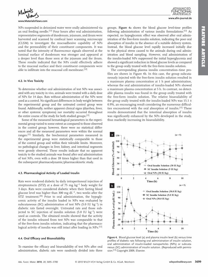
Figure 4 . Blood glucose level (a) and plasma insulin level (b) versus time profi les of diabetic rats following oral administration of insulin solution, oral administration of insulin-loaded nanoparticles (NPs) or subcuta-neous (SC) administration of insulin solution. (Reproduced with permis-sion. [ 12 ] Copyright 2009, Elsevier.
-10
10
30
50
70
90
110
130
0 2 4 6 8 10
Blo
od G
luco
se (%
of i
nit
ial)
Time (h)
Oral Insulin Solution (30 IU/kg)
SC Insulin Solution (5 IU/kg)
Oral NPs (30 IU/kg)
a (n = 5)
-10
10
30
50
70
90
110
130
0 2 4 6 8 10Pla
sma
Insu
lin L
evel
(µIU
/mL
)
Time (h)
Oral Insulin Solution (30.0 IU/kg)
SC Insulin Solution (5.0 IU/kg)
Oral NPs (30.0 IU/kg)
b (n = 5)
NPs suspended in deionized water were orally administered via an oral feeding needle. [ 12 ] Four hours after oral administration, representative segments of duodenum, jejunum, and ileum were harvested and scanned by confocal laser scanning microscopy (CLSM) to investigate the mucoadhesive capability of NPs and the permeability of their constituent components. It was noted that the intensity of fl uorescence signals observed at the luminal surface of duodenum was stronger and appeared at a deeper level than those seen at the jejunum and the ileum. These results indicated that the NPs could effectively adhere to the mucosal surface and their constituent components were able to infi ltrate into the mucosal cell membrane. [ 12 ]
4.2. In Vivo Toxicity
To determine whether oral administration of test NPs was associ-ated with any toxicity in vivo, animals were treated with a daily dose of NPs for 14 days. Rats without administration of the NPs were used as a control. No signifi cant differences in body weight between the experimental group and the untreated control group were found. Additionally, neither apparent clinical signs (diarrhea, fever, or other systemic symptoms) nor mortality occurred throughout the entire course of the study for both studied groups. [ 12 ]
Some of the measured hematological parameters in the experi-mental group varied to some extent as compared to those observed in the control group; however, there were no statistical differ-ences and all the measured parameters were within the normal ranges. [ 12 ] Similarly, the biochemical parameters measured in the experimental group were statistically comparable to those of the control group and within their tolerable limits. Moreover, no pathological changes in liver, kidney, and intestinal segments were grossly observed. These results indicate that no apparent toxicity to the studied animals was found after oral administration of test NPs, even with a dose 18 times higher than that used in the subsequent pharmacodynamic/pharmacokinetic study.
4.3. Pharmacological Activity of Loaded Insulin
Rats were rendered diabetic by daily intraperitoneal injection of streptozotocin (STZ) at a dose of 75 mg kg − 1 body weight for 3 days. Rats were considered diabetic when their fasting blood glucose level was higher than 300 mg dL − 1 , two weeks after the STZ treatment. [ 26 ] Prior to oral administration, the hypogly-cemic activity of the insulin loaded in NPs was evaluated by subcutaneous (SC) administration of test NPs (5.0 IU kg − 1 ) in diabetic rats fasted overnight. Untreated rats and those sub-jected to SC injection of insulin solution (5.0 IU kg − 1 ) were used as controls. The obtained results showed that the activity of the insulin released from test NPs was comparable to that of the free-form insulin solution, indicating that the pharmaco-logical activity of insulin was still intact after loading in NPs. [ 12 ]
4.4. Oral Effi cacy and Bioavailability
To examine the effi cacy and bioavailability of test NPs after oral administration, diabetic rats were randomly divided into three
© 2010 WILEY-VCH Verlag GmAdv. Funct. Mater. 2010, 20, 3695–3700
groups. Figure 4 a shows the blood glucose level-time profi les following administration of various insulin formulations. [ 12 ] As expected, no hypoglycemic effect was observed after oral admin-istration of the free-form insulin solution, indicating the poor oral absorption of insulin in the absence of a suitable delivery system. Instead, the blood glucose level rapidly increased initially due to the physical stress caused to the animals during oral admin-istration and blood sampling. However, oral administration of the insulin-loaded NPs suppressed the initial hyperglycemia and showed a signifi cant reduction in blood glucose levels as compared to the group orally treated with the free-form insulin solution.
The corresponding plasma insulin concentration–time pro-fi les are shown in Figure 4 b. In this case, the group subcuta-neously injected with the free-form insulin solution resulted in a maximum plasma concentration at 1 h post administration, whereas the oral administration of insulin-loaded NPs showed a maximum plasma concentration at 5 h. In contrast, no detect-able plasma insulin was found in the group orally treated with the free-form insulin solution. The relative bioavailability of the group orally treated with the insulin-loaded NPs was 15.1 ± 0.9%, an encouraging result considering the numerous diffi cul-ties encountered with the oral absorption of insulin. [ 12 ] These results demonstrated that the intestinal absorption of insulin was signifi cantly enhanced by the NPs developed in the study, thus markedly increasing its bioavailability.
bH & Co. KGaA, Weinheim 3699wileyonlinelibrary.com

FEATU
RE
ARTI
CLE
3700
www.afm-journal.dewww.MaterialsViews.com
5. Oral Administration of Other Hydrophilic Macromolecules
Besides insulin, our recent results also demonstrated that the same NP carrier system can be used for oral administration of other hydrophilic macromolecules such as calcitonin and heparin. The NP system has also been evaluated for the pos-sible oral delivery of aptamers. Calcitonin, a peptide hormone of 32 amino acid residues, has been used for the treatment of osteoporosis. Heparin, a naturally occurring glycosaminoglycan, is a potent inhibitor of blood coagulation. Aptamers are short single-stranded oligonucleotides, either RNA or DNA, that fold into well-defi ned three-dimensional structures and bind to their ligand by complementary shape interactions. These hydrophilic macromolecules cannot be absorbed in the GI tract, presumably because of their hydrophilicity and molecular size. As such, they have poor oral bioavailability and are administered parenterally.
We have recently developed a NP system covered with CS for oral delivery of heparin. The internal structure of the prepared NPs was examined by small-angle X-ray scattering (SAXS). The SAXS profi les suggest that the NPs are associated with a two-phase system and consist of the CS/heparin complex microdo-mains surrounded by the CS matrix. No signifi cant anticoagu-lant activity was detected after oral administration of the free-form heparin solution in a rat model, while administration of NPs orally was effective in the delivery of heparin into the blood stream; the absolute bioavailability was found to be approximately 21%. The biodistribution of the drug carrier, 99 mTc-labeled CS, in rats was studied by the single-photon emission computed tomography after oral administration of the radio-labeled NPs. No signifi cant radioactivity was found in the internal organs, indicating a minimal absorption of CS into the systemic circula-tion. These results suggest that the NPs described here can be employed as a potential carrier for oral delivery of heparin.
6. Summary and Perspectives
In summary, a NP carrier system shelled with CS for oral delivery of peptide/protein drugs was successfully developed, using a simple and mild ionic-gelation method. The as-prepared NPs with CS shells on the surface could transiently open the tight junctions between contiguous cells and increase their paracellular permeability. The results obtained in the animal study indicated that these NPs were well tolerated without showing any detectable toxicity. Additionally, the in vivo results clearly indicated that the effect of our insulin-loaded NPs on reducing the blood glucose level in a diabetic rat model.
We are now studying the detailed mechanism of tight-junc-tion opening by CS or its derivatives in vitro and in vivo. Under-standing the mechanism of interaction between NPs covered with CS (or its derivatives) and epithelial cells is of paramount impor-tance and needs to be established to aid further development in the use of NPs for oral delivery of hydrophilic macromolecules. Specifi c alteration of the epithelial junctional complex exposed to these NPs has not been elucidated. We hypothesize that the NPs covered by CS (or its derivatives) interact with specifi c mem-brane protein moieties (e.g., JAM1, CAR, CD55, CD44, CD47, or CD18) on or near the epithelial tight junctions to elicit the desired
© 2010 WILEY-VCH Verlag Gwileyonlinelibrary.com
reversible opening of tight junctions. What remains to be under-stood and verifi ed, however, is the ultrastructural changes in the epithelial junctional complex after treatment with NPs. Such detailed morphological information is obtained by high-resolution TEM and combined with the known biological assays and molec-ular dynamic simulations to elucidate the precise mechanism regarding how NPs interact with epithelial tight junctions.
Acknowledgements This work was supported by a grant from the National Science Council (NSC 97–2120-M-007–001), Taiwan, Republic of China. This article is part of a Special Issue on Nanomaterials Research by Chinese Scientists.
Received: May 20, 2010 Published online: October 14, 2010
[ 1 ] M. Hayashi , M. Tomita , S. Awazu , Adv. Drug Del. Rev. 1997 , 28 , 191 . [ 2 ] M. Goldberg , I. Gomez-Orellana , Nat. Rev. Drug Discov. 2003 , 2 , 289 . [ 3 ] E. S. Khafagy , M. Morishita , Y. Onuki , K. Takayama , Adv. Drug Del.
Rev. 2007 , 59 , 1521 . [ 4 ] A. F. Kotze , H. L. Lueen , B. J. de Leeuw , B. G. de Boer , J. Coos Verhoef ,
H. E. Junginger , J. Controlled Release 1998 , 51 , 35 . [ 5 ] P. D. Ward , T. K. Tippin , D. R. Thakker , Pharm. Sci. Tech. Today 2000 ,
3 , 346 . [ 6 ] N. N. Salama , N. D. Eddington , A. Fasano , Adv. Drug Delivery Rev.
2006 , 58 , 15 . [ 7 ] G. Ranaldi , I. Marigliano , I. Vespignani , G. Perozzi , Y. Sambuy ,
J. Nutr. Biochem. 2002 , 13 , 157 . [ 8 ] J. M. Smith , M. Dornish , E. J. Wood , Biomaterials 2005 , 26 , 3269 . [ 9 ] Medtech Insight , 2005 , 7 , 319 ; http://www.medtechinsight.com
(accessed May, 2009) . [ 10 ] G. P. Carino , E. Mathiowitz , Adv. Drug Del. Rev. 1999 , 35 , 249 . [ 11 ] Y. H. Lin , K. Sonaje , K. M. Lin , J. H. Juang , F. L. Mi , H. W. Yang ,
H.-W. Sung , J. Controlled Release 2008 , 132 , 141 . [ 12 ] K. Sonaje , Y.-H. Lin , J.-H. Juang , S.-P. Wey , C.-T. Chen , H.-W. Sung ,
Biomaterials 2009 , 30 , 2329 . [ 13 ] Y. H. Lin , C. K. Chung , C. T. Chen , H. F. Liang , S. C. Chen ,
H. W. Sung , Biomacromolecules 2005 , 6 , 1104 . [ 14 ] W. Wei , Y. Lan , H. Gang , W. Lian-Yan , W. Jie , H. Xue , S. Zhi-Guo ,
M. Guang-Hui , Adv. Mater. 2008 , 20 , 2292 . [ 15 ] N. N. David , J. G. Jordan , M. C. Juliana , L. Robert , G. A. Daniel , Adv.
Mater. 2009 , 21 , 847 . [ 16 ] T. Akagi , M. Higashi , T. Kaneko , T. Kida , M. Akashi , Macromol.
Biosci. 2005 , 5 , 598 . [ 17 ] Y. H. Lin , F. L. Mi , C. T. Chen , W. C. Chang , S. F. Peng , H. F. Liang ,
H. W. Sung , Biomacromolecules 2007 , 8 , 146 . [ 18 ] D. F. Evans , G. Pye , R. Bramley , A. G. Clark , T. J. Dyson ,
J. D. Hardcastle , Gut 1988 , 29 , 1035 . [ 19 ] W. C. Lin , D. G. Yu , M. C. Yang , Colloids Surf., B 2005 , 44 , 143 . [ 20 ] Y. H. Lin , C. T. Chen , H. F. Liang , A. R. Kulkarni , P. W. Lee ,
C. H. Chen , H. W. Sung , Nanotechnology 2007 , 18 , 105102 . [ 21 ] Z. Ma , L. Y. Lim , Pharm. Res. 2003 , 20 , 1812 . [ 22 ] X. He , M. Sugawara , Y. Takekuma , K. Miyazaki , Antimicrob. Agents
Chemother. 2004 , 48 , 2604 . [ 23 ] L. Shargel , A. B. C. Yu , Applied Biopharmaceutic & Pharmacokinetics ,
4th ed. McGraw-Hill New York 1999 . [ 24 ] F. L. Mi , Y. Y. Wu , Y. H. Lin , K. Sonaje , Y. C. Ho , C. T. Chen ,
J. H. Juang , H. W. Sung , Bioconjug. Chem. 2008 , 19 , 1248 . [ 25 ] J. Old , L. Connelly , J. Francis , K. Branch , G. Fry , E. Deane , Comp.
Clin. Pathol. 2005 , 14 , 130 . [ 26 ] C. Damge , P. Maincent , N. Ubrich , J. Controlled Release 2007 , 117 , 163 .
mbH & Co. KGaA, Weinheim Adv. Funct. Mater. 2010, 20, 3695–3700