TEMPLATE-ASSEMBLED SYNTHETIC G-QUARTETS ...
207
TEMPLATE-ASSEMBLED SYNTHETIC G-QUARTETS (TASQS) by MEHRAN NIKAN A THESIS SUBMITTED IN PARTIAL FULFILLMENT OF THE REQUIREMENTS FOR THE DEGREE OF DOCTOR OF PHILOSOPHY in THE FACULTY OF GRADUATE STUDIES (Chemistry) THE UNIVERSITY OF BRITISH COLUMBIA November 2008 © Mehran Nikan, 2008
-
Upload
khangminh22 -
Category
Documents
-
view
2 -
download
0
Transcript of TEMPLATE-ASSEMBLED SYNTHETIC G-QUARTETS ...

TEMPLATE-ASSEMBLED SYNTHETIC G-QUARTETS (TASQS)
by
MEHRAN NIKAN
A THESIS SUBMITTED IN PARTIAL FULFILLMENT OFTHE REQUIREMENTS FOR THE DEGREE OF
DOCTOR OF PHILOSOPHY
in
THE FACULTY OF GRADUATE STUDIES
(Chemistry)
THE UNIVERSITY OF BRITISH COLUMBIA
November 2008
© Mehran Nikan, 2008

Abstract
Fabrication of functional supramolecular structures requires a certain degree of control
which may not be achieved by relying solely on noncovalent interactions. The current study
aims to investigate the effect of a rigid cavitand template on morphology, function and stability
of lipophilic G-quadruplexes. The first Chapter of this thesis introduces different aspects of G
quadruplex chemistry and explains how these structures are particularly suited for the creation of
supramolecular architectures.
The second Chapter of this thesis presents the synthesis and self-assembly of a new class
of supramolecular architectures composed of four guanosines attached to a rigid cavitand
template. These structures, named template-assembled synthetic G-quartets (TASQs), were
synthesized via the “click” reaction and manifest an ordered topology dictated by the template.
The lipophilic TASQs were found to self-associate spontaneously to form a singular basket-like
structure in chloroform. Moreover, it was found that TASQs form cation-free G-quartets which
exhibit remarkable stability under this condition.
TASQs self-associate in chloroform
11

The third Chapter of this thesis describes the preparation, characterization and solution
study of the cation-bound complexes TASQNa, TASQK, TASQCs, and TASQSr2.
Cations play a major role in controlling the morphology and stability of G-quadruplexes. The
analysis of the cation-specific structures of TASQs reveals the formation of a monomeric G
quartet for Na and Sr2, a dimeric system for Cs and a mixture of monomers and dimers for K.
The factors governing the formation of these structures were evaluated, the selectivities of
TASQs for cations were determined, and the cation-dependent structural transformations were
studied.
____ ____
r
(b) (c) (d)
Polymorphism of TASQcation assemblies: Only structures consistent with (a) and(c) were observed
The fourth Chapter describes the efforts towards synthesizing a hydrophilic TASQ via
the “click” reaction. The following steps have been taken: 1) a water-soluble cavitand has been
successfully synthesized and characterized, which can potentially serve as a hydrophilic
template, and 2) two oligonucleotides have been appropriately functionalized and preliminary
coupling reactions were attempted. The next phases of this research along with potential future
directions are discussed in Chapter five.
(a)
111

Table of Contents
Abstract.ii
Table of Contents iv
List of Tables ix
List of Figures x
List of Schemes xv
List of Abbreviations xvi
Acknowledgements xviii
Co-Authorship xix
CHAPTER 1: INTRODUCTION 1
1.1 Background 1
1.2 Thesis Goals 2
1.3 Thesis Overview 2
1.4 G-Quartet in Supramolecular Chemistry and Biology 31.4.1 Properties of Nucleotides 31.4.2 Guanine Self-Assembly: G-Quartet vs. G-Ribbon 51.4.3 Nucleic Acid G-Quadruplexes 61.4.4 Lipophilic G-Quadruplexes 8
1.4.4.1 Structure of Lipophilic G-Quadruplexes 91.4.5 The Role of Cations 111.4.6 Cation-Free G-Quartets 121.4.7 A G-Quartet with Extra H-Bonds 141.4.8 G-Quartet in Material Science and Nanotechnology 15
1.4.8.1 Liquid Crystalline Phases 161.4.8.2 Supramolecular Polymers 171.4.8.3 Nanoparticle Assembly 181.4.8.4 Molecular Electronics 191.4.8.5 Nanowires and G-Wires 201.4.8.6 Nanomachines 21
1.4.9 G-Quartet and Chiral Resolution 231.4.10 G-Quartet and Equilibriums 25
1.4.10.1 Dynamic Covalent Chemistry (DCC) 25
iv

1.4.10.2 Dynamic Cation Binding and Release 291.4.11 Nucleic Acid G-Quadruplexes as Probes 30
1.4.11.1 G-Quadruplexes as Cation-Sensors 301.4.11.2 G-Quadruplexes as Protein-Sensors 331.4.11.3 G-Quadruplexes as Oligonucleotide-Sensors 35
1.4.12 G-Quadruplexes as Catalysts 361.4.13 Miscellaneous G-Quadruplex-Based Structures 39
1.4.13.1 Peptide Nucleic Acid (PNA) G-Quadruplex 391.4.13.2 Bunch-Oligonucleotides 40
1.5 Summary and Conclusions 42
1.6 References 43
CHAPTER 2: Synthesis, Characterization, and Solution Studies of LipophilicTemplate-Assembled Synthetic G-Quartets (TASQs) 51
2.1 Introduction 51
2.2 Rationale Behind Thesis 51
2.3 Results and Discussion 522.3.1 TASQ via “Click” Reaction 522.3.2 Synthesis of Cavitands 6a-c 542.3.3 Synthesis of 5’-Azido-2’,3’-O-Isopropylidene Guanosine 9 552.3.4 Synthesis of TASQs lOa-c 552.3.5 The Signal Assignment Strategy 56
2.3.5.1 Signal Assignment 582.3.6 Regiochemistry 642.3.7 Solution Structure Determination 652.3.8 Variable-Temperature 1H NMR Spectroscopic Studies 682.3.9 Infrared Spectroscopic Studies 722.3.10 CD Spectroscopic Studies 742.3.11 Diffusion NMR Studies 75
2.3.11.1 Translational Self-Diffusion 762.3.11.2 The Theory of Pulse Field-Gradient (PFG) NMR Spectroscopy 772.3.11.3 The Size Determination of TASQ lOc by PFG NMR Spectroscopy 80
2.3.12 The Role of Water 83
2.4 Summary and Conclusions 85
2.5 Experimental 86
V

2.5.1 General .862.5.2 Synthesis of Cavitands 6a-c 872.5.3 Synthesis of 5’-Azido-2’,3’-O-Isopropylidene Guanosine 9 882.5.4 Synthesis of TASQs lOa-c 882.5.5 Synthesis of 5’-Azido-2’,3’-O-Isopropylidene Adenosine 15 902.5.6 Synthesis of Compound 16c 922.5.7 PFG NMR Spectroscopic Experiments 93
2.5.7.1 General 932.5.7.2 Experimental (Bruker) Parameters for PFG NMR of lOc-16c in CDC13 932.5.7.3 Experimental (Bruker) Parameters for PFG NMR of lOc-16c in DMSO-d6...94
2.6 References 95
CHAPTER 3: Cation-Complexation Behavior of TASQs: The Preparation andStructural Characterization of TASQCation Assemblies 98
3.1 Introduction 98
3.2 Rationale for the Study of TASQCation Assemblies 98
3.3 Results and Discussion 1003.3.1 Synthesis of TASQs 1003.3.2 ‘H NMR Spectroscopic Studies 1003.3.3 TASQ 10cNa 102
3.3.3.1 The Size Determination of TASQ 10cNa4 by PFG NMR Spectroscopy 1053.3.4 TASQ lOcSr24 107
3.3.4.1 The Size Determination of TASQ lOc Sr24 by PFG NMR Spectroscopy 1103.3.5 TASQ 10cK 1113.3.6 TASQ 10cCs 114
3.3.6.1 The Size Determination of TASQ 10cCs by PFG NMR Spectroscopy 1153.3.7 The Anion Effect 1193.3.8 Gas-Phase Structures 1203.3.9 Selectivities of TASQs 1223.3.10 Interconversion of TASQ lOcNa4to TASQ lOc 1253.3.11 CD Spectroscopy 127
3.4 Summary and Conclusions 128
3.5 Experimental 1303.5.1 General 1303.5.2 Picrate Extraction Experiments 1313.5.3 PFG NMR Spectroscopic Experiments 132
vi

3.5.3.1 Experimental (Bruker) Parameters for PFG NMR Experiments 1323.5.4 Atomic Absorption Experiments 133
3.6 References 134
CHAPTER 4: Towards Hydrophilic TASQs 138
4.1 Introduction 138
4.2 Rationale for the Study of Hydrophilic TASQs 138
4.3 Results and Discussion 1394.3.1 The Oligonucleotide Approach 139
4.3.1.1 Solid-Phase Functionalization of the Oligonucleotides 1414.3.1.2 The Coupling Reaction of 5 ‘-Azido Oligunucleotides 20a-b 142
4.3.2 The Water-Soluble Cavitand Approach 1474.3.2.1 Synthesis of Phosphate-Footed Tetrakis (O-Propargyl) Cavitand 30 148
4.3.3 The Phosphoramidite Approach 151
4.4 Conclusions 155
4.5. Experimental 1574.5.1 Phosphate-Footed Tetrakis (O-Propargyl) Cavitand Synthesis 157
4.5.1.1 General 1574.5.1.2 Synthesis of Dodecol 22 1584.5.1.3 Synthesis of Tetrabromo Dodecol 23 1584.5.1.4 Synthesis of Hydroxyl-Footed Tetrabromo Cavitand 24 1594.5.1.5 Synthesis of TBDMS-Protected Tetrabromo Cavitand 25 1604.5.1.6 Synthesis of TBDMS-Protected Tetrol 26 1614.5.1.7 Synthesis of TBDMS-Protected Tetrakis (O-Propargyl) Cavitand 27 1614.5.1.8 Synthesis of Hydroxyl-Footed Tetrakis (O-Propargyl) Cavitand 28 1624.5.1.9 Synthesis of t-Butyl Phosphorylated Tetrakis (O-Propargyl) Cavitand 29.... 1634.5.1.10 Synthesis of Phosphate-Footed Tetrakis (O-Propargyl) Cavitand 30 164
4.5.2 Oligonucleotide Synthesis 1654.5.2.1 General 1654.5.2.2 Synthesis of 5 ‘-lodo Oligunucleotides 18a-b 1664.5.2.3 Synthesis of 5 ‘-Azido Oligunucleotides 20a-b 166
4.5.3 Phosphoramidite Synthesis 1704.5.3.1 General 1704.5.3.2 Synthesis of Tetrakisphosphoramidite Cavitand 34 1704.5.3.3 Synthesis ofN2-Isobutyryl-2’,3’-O-Isopropylidene Guanosine 33 172
vii

4.5.3.4 Synthesis ofN2-Isobutyryl-2’,3 ‘-O-Isopropylidene GuanosinePhosphoramidite 36 173
4.6 References 174
CHAPTER 5: Summary, and Future Work 177
5.1 Thesis Summary 177
5.2 Future Work 1795.2.1 Lipophilic TASQ Project 180
5.2.1.1 Complementary Work 1805.2.1.1 Future Applications 180
5.2.2 Hyclrophilic TASQ Project 1835.2.2.1 Complementary Work 1835.2.2.2 Future Applications 186
5.3 References 187
viii

List of Tables
Table 2.1. Spectral assignments of lOc in DMSO-d6and CDC13 at ambient temperature ... 59Table 2.2. Table of chemical shifts for a solution of lOc in CDC13at four different
temperatures 72Table 2.3. Diffusion coefficients (D) of lOc and 16c in CDC13and DMSO-d6at 295 K 81Table 3.1. Diffusion coefficients (D) of lOcNa4and 16c in CDC13at 295 K 105Table 3.2. Diffusion coefficients (D) of 1OcSr2and 16c in CDC13at 295 K 110Table 3.3. Diffusion coefficients (D) of 1OcCs and lOc in CDC13 at 295 K 117Table 3.4. The selectivities of TASQs. The magnitude of these selectivities is estimated as
more than a factor of 20 between each pair of cations 124Table 3.5. Na content (tg/g) of TASQ samples 133Table 4.1. Reaction conditions examined for the couplinga of 20a to 6a 143
ix

List of Figures
Figure 1.1. A tetrol cavitand.1Figure 1.2. Two sample nucleotides of guanine and cytosine. The numbering schemes for the
base and sugar subunits are also presented 3Figure 1.3. The purine (adenine, guanine) and pyrimidine (cytosine, thymine, uracil) bases of
nucleic acids 4Figure 1.4. The syn and anti conformations of guanosine 4Figure 1.5. Watson-Crick and Hoogsteen base-pairing of nucleobases. Cytosine has to be
protonated to be able to participate in Hoogsteen pairing with guanine or anothermolecule of cytosine. This mode of H-bonding is important in the formation oftriplexes and i-motifs 5
Figure 1.6. A schematic representation of a) G-quartet, b) G-quadruplex (the G-quartet unitsare rotated about 300 in respect to each other). Images (c) and (d) depictG-ribbons (arrows indicate dipole moments) 6
Figure 1.7. a) Tetramolecular, b,c) bimolecular and d) unimolecular G-quadruplexes (arrowsindicate 3’—+ 5’ polarity) 8
Figure 1.8. A schematic representation of an octamer formed from 3 ‘,5 ‘-didecanoyl-2’-deoxyguanosine 9
Figure 1.9. A schematic representation of the crystal structure of a lipophilic guanosinehexadecamer formed in the presence of a) K and Cs picrates, b) M2 picrates.The H-bonding of the picrate anion to the quartets is also presented 10
Figure 1.10. The syn conformation adopted by 8-(4-PN-dimethylamino) guanosine directs theformation of a cation-free G-quartet 12
Figure 1.11. a) Calix[4jarene-guanosine conjugate, and b) calix[4jarene-guanosine dimers... 13Figure 1.12. The network of cation-free G-quartet made from guanine on gold surfaces. The
H-bonds that connect the adjacent G-quartets are marked with circles 14Figure 1.13. The G-quartet obtained from 8-aryl-2’-deoxyguanosine. The extra H-bonds are
circled 15Figure 1.14. A schematic representation of the liquid crystalline phases obtained from
lipophilic or hydrophilic guanosine compounds. Depending on concentration,ions, and temperature, either the cholesteric or the hexagonal mesophases isobtained 16
Figure 1.15. The self-assembly of bisiminoboronate-guanosine to a G-quartet-based polymericfilm in the presence of K. The B-N dative bond of the monomer helps rigidifythe polymer and also prevents the hydrolysis of the reversible boronate-guanosineester bond 17
Figure 1.16. a) 3 ‘-thiolpropyl deoxyguanosine phosphate and b) gold nanoparticle assemblyusing G-quartet formation (cations are omitted for clarity) 18
Figure 1.17. A schematic representation of the simple transistor made from G-ribbons 19Figure 1.18. A schematic representation of the competing structures which can be adopted by
telomeric DNA d(G4T2G4). The experimental conditions can be adjusted to favorthe formation of G-wires. As shown below the formation of a G-wire is the resultof the interaction between the 3’ end of one helix and the 5’ end of another helix(cations are omitted for clarity) 20
x

Figure 1.19. A schematic representation of a G-quadruplex-based nanomachine. Thequadruplex-forming strand extends into a duplex upon addition of acomplementary strand (C-fuel) and shrinks back to G-quadruplex via addition ofG-fuel (cations are omitted for clarity) 21
Figure 1.20. Copper-driven transition of a G-quadruplex to a random coil and vice versa 22Figure 1.21. Homochiral and heterochiral G-quadruplexes obtained from extraction of
(D, L)-5’-silyl-2’,3’-isopropylidene guanosine with Ba2 and Kpicrates 23Figure 1.22. a) The lipophilic guanosine compound capable of chiral discrimination between
(D) and (L) N-(2,4-dinitrophenyl)-tryptophan, and b) potassium salt ofN-(2,4-dinitrophenyl)-(L,D)-tryptophan 24
Figure 1.23. Preparation of a library of acyihydrazones (E, F) and G-quartet hydrazones (G, H)from the reaction of guanosine hydrazide A and serine hydrazide B withaldehydesCandD 25
Figure 1.24. A schematic representation of the reaction of acridone (A), peptide (P), andglutathione (G, both oxidized and reduced forms) in the presence and absence ofthe G-quadruplex template 27
Figure 1.25. Synthesis of a unimolecular G-quadruplex using reversible olefin metathesis. ... 28Figure 1.26. Interconversion of a bisguanine compound between so! and gel states 29Figure 1.27. A) po!ydiacety!ene (PDA) liposome, B) a polydiacetylene (PDA) liposome
functionalized with G-rich oligonucleotides (non fluorescent) and thecorresponding microarray, and C) activated liposome (red fluorescent) due to theformation of G-quadruplex 31
Figure 1.28. A schematic representation of the on/off nanoswitch used for the electrochemicaldetection of potassium ion 31
Figure 1.29. The structure of the thrombin-binding aptamer containing two pyrene moietiesand the resulting chair-type conformation. The interaction of the fluorophores isonly possible in the presence of potassium ions 32
Figure 1.30. Schematic illustration of the three-component duplex. Addition of thrombin leadsto the formation of the G-quadruplex, which coincides with the release of theQ-strand and an enhancement in the fluorescence 33
Figure 1.31. A) Formation of monolayer, B) addition and capture of thrombin, C) addition andbinding of the secondary aptamer, and D) oxidation and detection of Cd2with anion-selective electrode 34
Figure 1.32. Proposed mechanisms for the recognition of the G-quadruplex-based molecularbeacon 36
Figure 1.33. A schematic representation of the deoxyribozyme catalyzing the photocleavage ofa thymine cyclobutane dimer. The G-quadruplex absorbs and transmits the low-energy radiation (—3O5 nm) which is outside the normal absorption of DNA(>250nm) 37
Figure 1.34. A proline modified G-quadruplex catalyzes the aldol reaction between acetoneand an aldehyde-appended porphyrin 38
Figure 1.35. Hybrid tetramolecular G-quadruplexes formed from DNA (blue) and PNA (Red).Two structures of diagonally opposite (A) or adjacent (B) are possible 39
Figure 1.36. a) Schematic representation of the bunch-oligonucleotides (Arrows indicate5’—. 3’ polarity), b) G-quadruplexes obtained from bunch-oligonucleotides indifferent orientations. Structure 3b was not formed and 4b and 4c were obtainedin minor quantities 41
xi

Figure 2.1. The important 2D NMR correlations observed for compound lOc (shown brokenapart for the convenience of illustrating the correlations) 57
Figure 2.2. 400 MHz ‘H-’H COSY of lOc at 25 °C in DMSO-d6 60Figure 2.3. 400 MHz ‘H-’3C HMQC of lOc at 25 °C in DMSO-d6.The signals corresponding
to diastereoto?ic pairs of H5’a/ H5’b, Ha/Hb, andH1/H0are circled 61Figure 2.4. 400 MHz ‘H- H COSY of lOc at 25 °C in CDC13 62Figure 2.5. 400 MHz ‘H-’3CHMQC of 10c at 25 °C in CDC13. The signals corresponding to
diastereotopic pairs of H5 ‘a! H5 ‘b, Ha/Hb, and H/H0are circled 63Figure 2.6. a) NOEs expected for 1,4 and 1,5 triazoles. b) NOEs observed for a solution of
lOa in DMSO at 400 MHz at 25 °C 64Figure 2.7. 400 MHz ‘H NMR of lOc at 25 °C in a) CDCI3,and b) DMSO-d6 65Figure 2.8. 400 MHz ‘H NMR of lOc at —40 °C in CDC13 indicating the H-bonded (NH2b),
and the non-H-bonded (NH2a) amino signals 66Figure 2.9. a) Inter and intra-base NOE correlations in a G-quartet, b) Intra-base NOE
correlations in syn (strong Hi ‘/H8 and weak H2’/H8) and anti (medium HI ‘/H8and strong H2 ‘/H8) conformers,24c) NOEs indicative of the formation of Gquartet, and d) NOEs indicative of the syn conformation at 400 MHz in CDC13 at—40 °C 67
Figure 2.10. Variable-temperature experiments on a solution of TASQ lOc in CDC13 at 400MHz. Arrows mark signals of: red: water, black: H-bonded NH2,and yellow: theaverage NH2 69
Figure 2.11. Changes in the chemical shift of coalesced NH2 signal of lOc in CDC12-CDC12from 50-100°C 70
Figure 2.12. The donor and acceptor faces of guanine in a G-quartet unit 71Figure 2.13. The amino (NH2)and imino (NH) stretching region (3200-3600 cm’) of the
infrared spectrum of a 3 xl 02 M solution of TASQ lOc in CDC13 at RT. Theassociation bands at 3310 and 3470 cm’ are due to H-bonding 73
Figure 2.14. CD spectra of a 0.2 mM solution of TASQ lOc in chloroform and DMSO 75Figure 2.15. The translational diffusion of a molecule from point 1 to point 2, which is
independent of its path 76Figure 2.16. a) A simple gradient pulse sequence, b) The BPLED sequence. In this sequence,
short gradients of opposite polarity are used, which are separated by a 180° pulse.This combination reduces the eddy current effects (induced by gradient pulses),and improves the line shape 78
Figure 2.17. Normalized signal decay as a function of the b value at 295 K for a) lOc-16c inDMSO-d6,b) lOc-16c in CDC13.The diffusion coefficients (D) were calculatedfor the representative signal of Hi’. [b value = (2 yg6)2(A-6/3) s/m2] 82
Figure 2.18. Normalized signal decay as a function of the g (gradient strength) at 295 K for a)the Hi’ signal of lOc in DMSO-d6,and b) the Hi’ signal of lOc in CDCI3 83
Figure 3.1. ‘H NMR spectra of TASQcation assemblies in CDC13at 400 MHz at 25 °C. Theregion between 6 and 12 ppm’s has been assigned to make comparison easier.The signals labeled “p” and “s” indicate picrate and solvent respectively. TASQ10cCs gave two sets of signals, one of which has been marked with asterisks.iOi
Figure 3.2. ‘H-’H COSY of TASQ 10c.Naat 400 MHz in CDCI3 at 25 °C 103Figure 3.3. TASQ 10cNa shows NOEs similar to those shown by the cation-free system.
These effects are indicative of a) the formation of G-quartet and, b) the synconformation at 400 MHz in CDCI3 at —40 °C. The NH2 signals which appearbroad at RT, sharpen up and become clearly visible at low temperature 104
xli

Figure 3.4. Picrate signal integration for TASQ 10cNa at 400 MHz in CDC13 at 25 °C.... 105Figure 3.5. Normalized signal decay as a function of the b value at 295 K for 10cNa-16c in
CDC13. The diffusion coefficients (D) were calculated for the representativesignal of Hi’. [b value = (2’ygö)2(A-6/3) s/rn2] 106
Figure 3.6. Normalized signal decay as a function of the gradient strength (g) at 295 K for a)the Hi’ signal of 10cNa, and b) the Hi’ signal of!6c in CDCI3 106
Figure 3.7. ‘H-’H COSY of TASQ !0c.Sr2at400 MHz in CDCI3 at 25 °C 108Figure 3.8. Portions of a 100 ms NOESY spectrum of TASQ 10cSr2acquired at 400 MHz at
—40 °C in CDC13 indicative of a) the formation of G-quartet, and b) the synconformation. The NH2 signals which are broadened up at RT are visible at lowternperature 109
Figure 3.9. Picrate signal integration for TASQ !0cSr2at 400 MHz in CDC13 at 25 °C.... 109Figure 3.10. Normalized signal decay as a function of the b value at 295 K for !Oc.Sr2tl6c in
CDC13. The diffusion coefficients (D) were calculated for the representativesignal of Hi’. [b value = (2yg6)2(z-/3)s/rn2] 110
Figure 3.11. Normalized signal decay as a function of the gradient strength (g) at 295 K for a)the Hi’ signal of 10cSr2,and b) the Hi’ signal of 16c in CDC13 111
Figure 3.12. Partial ‘H NMR spectrum of TASQ !0cK at 400 MHz at —40 °C displayingthree sets of signals. The two sets of small signals related to a second “new”species has been marked with asterisks. The signals labeled “p” and “s”corresponds to the picrate and the solvent, respectively 112
Figure 3.13. Portions of a 100 ms NOESY spectrum of TASQ 10cK acquired at 400 MHz inCDC13 112
Figure 3.14. Picrate signal integration for TASQ 10cK at 400 MHz in CDC13 at 25 C 113Figure 3.15. Partial ‘H-’H NOESY of TASQ lOa.Cs4 acquired at 400 MHz at —40 °C in
CDC13. Signals shown are indicative of the formation of G-quartet 114Figure 3.16. Picrate signal integration for TASQ 10cCs at 400 MHz in CDC13 at 25 °C. ... 115Figure 3.17. Normalized signal decay as a function of the b value at 295 K for !0cCs-10c in
CDC13. The diffusion coefficients (D) were calculated for the representativesignal of NH. [b value = (2ityg.ö)2(A-ö/3) s/rn2] 117
Figure 3.18. Normalized signal decay as a function of the gradient strength (g) at 295 K for a)the NH signal of 10cCs, and b) the NH signal of !Oc in CDC13 118
Figure 3.19. Proposed G-quartet-basecl structures of TASQcation assemblies a) isolatedG-quartet, b,d) homodimers, and c) heterodimer. Only structures consistent with(a) and (c) were observed 119
Figure 3.20. ESI-MS spectrum of TASQ 10aSr2in positive mode. Two species of [TASQ10aSr]2(mlz = 1145) and [(TASQ !0a)2Sr]2(m/z = 2246) were observed inthe gas-phase 121
Figure 3.21. MALDI-MS spectrum of TASQ !OaSr2in positive mode. Two species of[TASQ !0aSr] (rn/z = 2290) and [(TASQ 10a)2Sr] (m/z 4492) wereobserved in the gas-phase (note that MALDI typically generates only singlycharged species) 121
Figure 3.22. a) ‘H NMR spectrum of pure TASQ 10aCs, b) ‘H NMR spectrum of pure+ 1TASQ lOaNa , c) H NMR spectrum recorded after extraction of TASQ lOa
with an equimolar mixture of sodium and cesium picrates 124Figure 3.23. Schematic representation of the conversion of TASQ !0cNa to TASQ lOc upon
addition of [2.2.2] cryptand 125
xiii

Figure 3.24. a) ‘H NMR spectrum of a 2.4 mlvi solution of TASQ 1OcNa at 400 MHz inCDC13 at 25°C, b) The spectrum recorded after addition of the 8-fold excess of2.2.2j cryptand to the same solution (note the free picrate signal at 8.8 ppm), c)H NMR spectrum of a 2.4 mM of TASQ lOc after addition of the 8-fold excess
of [2.2.2] cryptand, and d) ‘H NMR spectrum of a solution of TASQ lOc at 400MHz in CDC13 at 25°C 126
Figure 3.25. CD spectra of a 0.2 mM solution of TASQ lOc and TASQ lOccation assembliesin chloroform 128
Figure 4.1. Tris-(benzyltriazolylmethyl)amine (TBTA) 145Figure 4.2. The upper and lower rims of the cavitand 149Figure 4.3. HPLC chromatograms of the crude (top), and the pure 20a (bottom) 168Figure 4.4. ESI-MS spectrum of 20a in negative mode. The signals at (mlz = 1278.2,
1300.1, and 1316.1) correspond to [M-H], [M+Na-2H] -, and [M+K-2Hfrespectively 168
Figure 4.5. HPLC chromatograms of the crude (top), and pure 20b (bottom) 169Figure 4.6. MALDI-MS spectrum of 20b in positive mode. The signal at (m/z = 1888.8)
corresponds to [M+H] 169Figure 4.7. ESI-MS spectrum of 34 in positive mode. Only one species was observed in the
solution. The signals at (mlz = 1457.7) and (m/z = 1558.9) correspond to [M+H]and [M+NEt3+H]respectively 171
Figure 4.8. The 200 MFIz 31P NMR spectrum of a solution of 5a reacted with 2-cyanoethyl-]\cN-diisopropylchloro phosphoramidite in CD2CI2at 25 °C. The signal at 149.9ppm corresponds to the product. (Note that the P-diastereomers are not resolvedat200MHz) 171
Figure 5.1. a) A ditopic TASQ monomer, b) Three possible arrays derived from selfassembly of compound 39: a linear polymer 39a, a sandwiched dimer 39b, and astacked polymer 39c (only one stack is shown) 181
xiv

List of Schemes
Scheme 2.1. Thermal vs. catalytic 1 ,3-dipolar cycloaddition reaction 53Scheme 2.2. The synthesis of cavitands 6a-c 54Scheme 2.3. The synthesis of 5’-azido guanosine 9 55Scheme 2.4. The synthesis of TASQs lOa-c 56Scheme 2.5. The Synthesis of Compound 16c 80Scheme 2.6. The Synthesis of Nucleoside 15 91Scheme 4.1. Proposed synthesis of an all parallel hydrophilic TASQ with the sequences of
5’-TG4T-3’ and 5’-G4-3’ using the oligunucleotide approach 140Scheme 4.2. The solid-phase functionalization of 17a-b 142Scheme 4.3. Proposed synthesis of a hydrophilic TASQ using the water-soluble cavitand
approach 148Scheme 4.4. The synthesis of phosphate-footed tetrakis (O-propargyl) cavitand 30 150Scheme 4.5. Proposed synthesis of a hydrophilic TASQ using the phosphoramidite
approach 152Scheme 4.6. The transient protection of 7 153Scheme 4.7. The synthesis of tetrakisphosphoramidite cavitand 34 153Scheme 4.8. The synthesis ofN2-isobutyryl-2’,3 ‘-O-isopropylidene guanosine
phosphoramidite 154Scheme 4.9. Proposed synthesis of a hydrophilic TASQ using the phosphoramidite
approach 155Scheme5.1. a) Proposed synthesis of a pentameric TASQ analog using the “click” reaction,
b) Isoguanosine pentamer 182Scheme 5.2. Proposed reactants for the synthesis of a hydrophilic TASQ using the “click”
reaction and their preparation methods 185
xv

List of Abbreviations
AFM atomic force microscopyaq. aqueousAr aromaticBBI broadband inverse (probe)BPLED bipolar pulse longitudinal eddy current delayBn benzylBu butylBz benzylCD circular dichroismCOSY correlation (NMR) spectroscopyCPG controlled pore glassCPK Corey-Pauling-KoltunCTAB cetyltrimethylammonium bromide
change in chemical shiftd doubletDCC dynamic covalent chemistryDCL dynamic covalent libraryDCM dichloromethaneDCTB 2- [(2E)-3 -(4-tert-butylphenyl)-2-methylprop-2-enylidene]malononitrileDHB 2,5-dihydroxybenzoic acidDMA N-N-dimethylacetamideDMF N N-dimethylformamideDMSO dimethylsulfoxideDMT dimethoxytritylDNA deoxyribonucleic acidEDTA ethylenediamineESI electrospray ionizationEt ethyl (-CH2CH3)EtOAc ethyl acetateEtOH ethanoleq. equivalentsEXAFS extended X-ray absorption fine structureFRET Förster resonance energy transferGMP guanosine monophosphateHIV human immunodeficiency virusHMQC heteronuclear multiple quantum coherenceHPA hydroxylpicolinic acidHPLC high performance (pressure) liquid chromatographyi-Pr isopropylJR infraredISE ion-selective electrodeL litrem multipletM parent mass (mass spectra) or moles per litre
xvi

MALDI matrix assisted laser desorption ionizationMB molecular beaconMe methyl (-CH3)MeCN acetonitrileMeOH methanolMHz megahertzMs methanesulfonylMS mass spectrometry or spectrumMW molecular weightn-BuLi n-butyllithiumNBS N-bromosuccinimidenm nanometer(s)NMR nuclear magnetic resonanceNOE nuclear Overhauser effectNOESY nuclear Overhauser enhancement spectroscopyPDA polydiacetylenePFG pulse field-gradientPh phenyl groupPNA peptide-nucleic acidq quartet (‘H NMR)RF radio frequencyRNA ribonucleic acidRP-HPLC reverse-phase high performance (pressure) liquid chromatographyRT room temperatures singlet (‘H NMR)SANS small-angle neutron scatteringt tripletTASP template assembled synthetic proteinTASQ template assembled synthetic G-quartetTBA thrombin-binding aptamerTBAF tetrabutylammonium fluorideTBDMS tert-butyldimethylsilylTBTA tris-(benzyltriazolylmethyl)aminet-Bu tert-butylTCEP tris(2-carboxyethyl)phosphine hydrochlorideTEA triethylamineTFA 2,2,2-trifluoroacetic acidTHF tetrahydrofuranTLC thin layer chromatographyTMPyP4 tetramethylpyridinium porphyrinTOF time of flightTs para-toluenesulfonylUV ultravioletvs. versusv/v volume per volume
xvii

Acknowledgements
First, I would like to thank my supervisor, Professor John Sherman, who shaped my
mind as a researcher and taught me to think critically and not to be satisfied with the minimum.
I really appreciate his patience, constructive discussions and the time he spent reading and
improving the manuscripts. I am also grateful to Dr. David Perrin for inspiring this project, and
his helpful suggestions and constant encouragement.
Also, I wish to thank both present and past members of the Sherman group for creating a
friendly atmosphere in the lab. Special thanks to Aifredo Franco Cea, Jon Freeman and Grant
Bare for proofreading sections of this thesis.
I would like as well to acknowledge the staff of the NMR laboratory, Dr. Nick Burlinson,
Dr. Maria Ezhova and Zorana Danilovic for providing assistance and sharing their valuable
experience, Bert Meuller for conducting atomic absorption experiments, Fred Rosell for helping
with his CD apparatus and the staff of the mass spectrometry laboratory and biological services.
I want to thank Dr Marco Ciufolini as well for his helpful suggestions regarding the
synthesis of the water-soluble cavitand.
Finally, I would also like to express my sincere thanks, appreciation and respect to my
family and friends.
xviii

Co-Authorship
This thesis work has been conducted and written solely by the author and not in
collaboration with other parties (including experimental design and data analyses). The
published and submitted manuscripts have been prepared by the author and have been edited by
Professor John Sherman.
xix

CHAPTER 1: Introduction
1.1 Background
Supramolecular chemistry deals with non-covalent interactions between molecules. One
of its challenges is the organization of small molecules into well-defined assemblies.’ One way
to approach this problem is to use a molecular template to which structural units can be
attached.2 The molecules which are generally used as templates are rigid molecules that direct
the building blocks in well-defined spatial arrangements. Numerous supramolecular systems
containing both rigid and flexible subunits have been designed and synthesized following this
approach.3 This strategy has also been used to address the protein folding problem.4 The
cavitands, rigid macrocycles with an enforced cavity, are ideal scaffolds which have been used
for synthesis of de novo proteins (Figure 1.1). The resulting four-helix bundles called caviteins,
have been studied in our group and have shown a high degree of stability compared to other
template-assembled synthetic proteins (TASPs).5’6
Figure 1.1. A tetrol cavitand
1

1.2 Thesis Goals
In the present study we are utilizing the concept of template-assembly along with H-
bonding interactions towards the directed organization of G-quartet assemblies. G-quartets are
cyclic tetramers of guanines which are important in biology and supramolecular chemistry.7 The
major goals of this project are the followings:
1) To design and synthesize a family of cavitand-linked guanines such that the cavitand
helps direct the guanines into a G-quartet. We have named these compounds template-
assembled synthetic G-quartets (TASQ5).
2) To demonstrate the utility of the cavitand template in controlling the morphology of
G-quadruplexes (stacks of G-quartets).
1.3 Thesis Overview
This thesis is organized in five chapters. Chapter 1 will describe the supramolecular
and biological significance of the G-quartets and G-quadruplexes, and will present selected
examples of the relevant works. Chapter 2 will detail the design, synthesis, and solution
properties of the first lipophilic template-assembled synthetic G-quartets (TASQs) which are
capable of forming a cation-free G-quartet in solution. Chapter 3 will discuss the cation
complexation behavior of the TASQs, and investigate the preparation and characterization of
the TASQcation assemblies. Chapter 4 will present the attempts at synthesizing a
hydrophilic TASQ including the synthesis of a water-soluble phosphate-footed
tetrapropargyl ether cavitand. Chapter 5 will present the thesis summary, and will suggest
2

directions for future research. An introduction on nucleotides will be presented below,
followed by a discussion on the structure of G-quartets.
1.4 G-Quartet in Supramolecular Chemistry and Biology
1.4.1 Properties of Nucleotides
Nature has always been a source of inspiration for supramolecular chemists. Vital
biopolymers such as DNA and RNA are both constructed from smaller units selected by the
powerful mechanism of evolution. These smaller units, called nucleotides, (Figure 1.2) are
composed of a heterocyclic base (purine or pyrimidine), a sugar (D-ribose or D-2 ‘-deoxyribose),
and a phosphate group (at 3’ or 5’ positions). Figure 1.3 shows the nucleobases of DNA and
RNA which are all capable of forming highly ordered H-bonds.8’9
Figure 1.2. Two sample nucleotides of guanine and cytosine. The numbering schemes for thebase and sugar subunits are also presented.
NH2
5N CytosinePhosphate 8JjGuanIne
GLN.LQ2
HO—_
-
, Ribose2 -Deoxyribose O-F’—O
Phosphate 0
3

Figure 1.3. The purine (adenine, guanine) and pyrimidine (cytosine, thymine, uracil) bases ofnucleic acids.
<4JN$NH2 00
Adenine Guanine Cytosine Thymine Uracil
The nucleobases in DNA and RNA are all connected to the ribose moieties through an N
glycosidic bond and can exist in two different conformations known as syn and anti (Figure 1.4).
The anti conformation is the dominant conformation in the naturally occurring DNA. 8,9
Figure 1.4. The syn and anti conformations of guanosine.
0 0
HO HO
ZNH2
1;JClI JClI
Syn Anti
Because of their geometry, all bases in DNA can form Watson-Crick H-bonds in which a
purine pairs with a pyrimidine base (Figure 1.5). There are also other modes of H-bonding
including the Hoogsteen pairing, which is probably more significant than the other non-Watson-
Crick H-bonds (Figure 1 5)•10 This mode of H-bonding plays a key role in the formation of the
G-quartet)’
4

Figure 1.5. Watson-Crick and Hoogsteen base-pairing of nucleobases. Cytosine has to beprotonated to be able to participate in Hoogsteen pairing with guanine or another molecule ofcytosine. This mode of H-bonding is important in the formation of triplexes and i-motifs.’2
H H-- -o /
H HNT— N// R0
Hoogsteen
1.4.2 Guanine Self-Assembly: G-Quartet vs. G-Ribbon
Guanine bases, present in guanosine compounds, guanine-rich strands of DNA and RNA,
and DNA-like polymers, are able to self-associate and form a tetrameric structure called a G
quartet (Figure 1.6a).7”1”3 Stacks of G-quartets form a three dimensional structure known as a
G-quadruplex (Figure 1 .6b).’4”5 As shown in Figure 1.6a, each G-quartet is stabilized by eight
hydrogen bonds. Some positively charged metal cations such as Na, K, and Sr2 can also be
sandwiched between the planes of the G-quartets and their inclusion increases the stability of the
system.’6”7 These cations are coordinated by the carbonyl oxygen atoms of the G-quartets.
Guanine aggregation in the absence of eations (in lipophilic solvents or on the solid phases)
generally results in the formation of ribbon-like structures.18 Depending on the solvent and the
sugar substituents, one of the structures of (c) or (d) might be formed (Figures 1 .6c and 1 .6d).
5
Watson-Crick
Hoogsteen

Overall Dipole
G-quartet formation has been known for many years, but little attention was paid to this
phenomenon.’9 The early studies on G-quartets date back to 1962 when guanine tetramer was
Figure 1.6. A schematic representation of a) G-quartet, b) G-quadruplex (the G-quartet units arerotated about 300 in respect to each other). Images (c) and (d) depict G-ribbons (arrows indicatedipole moments).
R
H
a)
c)
d)
1.4.3 Nucleic Acid G-Quadruplexes
0:No Dipole
6
discovered as the main component of the gels formed from the 3’ and 5’-
guanosinemonophsphate (GMP) in water.11 There is currently considerable interest in this area
of research because of the potential role of these structures in many biological events.7 G-rich
sequences are abundant in some biologically important regions, such as the telomeric ends of
eukaryotic chromosomes, the promoter regions of DNA,20’2’and the immunoglobulin switch
regions22,and are capable of forming G-quadruplexes in vitro. There is also evidence for their
existence and function in vivo.23
Telomeric DNA shrinks with each successive division of cells, thus limiting the number
of divisions a cell can undergo. In cancerous cells, the telomerase enzyme elongates the
telomeric DNA, enabling more divisions of cells. This enzyme is only active in cancerous cells
but inactive in most somatic cells. It has been suggested that formation of a G-quadruplex could
inhibit cancer because the telomerase enzyme only binds to the unfolded telomere. When a
single stranded telomere folds over on itself and forms a G-quadruplex, the action of the
telomerase is terminated. Therefore, ligands that can selectively stabilize or bind to G
quadruplexes are of utmost importance. Some of these ligands have been shown to accelerate
the folding of a single strand of DNA to aG-quadruplex.24’25
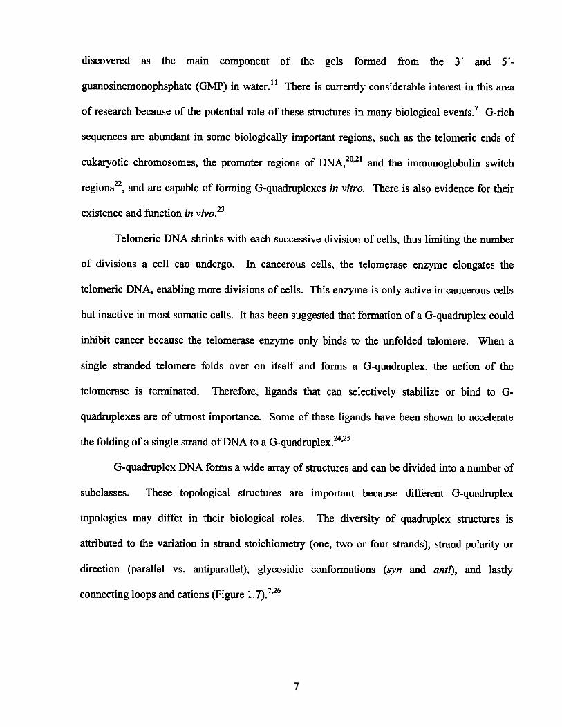
G-quadruplex DNA forms a wide array of structures and can be divided into a number of
subclasses. These topological structures are important because different G-quadruplex
topologies may differ in their biological roles. The diversity of quadruplex structures is
attributed to the variation in strand stoichiometry (one, two or four strands), strand polarity or
direction (parallel vs. antiparallel), glycosidic conformations (syn and anti), and lastly
connecting loops and cations (Figure 1 7)726
7

Figure 1.7. a) Tetramolecular, b,c) bimolecular and d) unimolecular G-quadruplexes (arrowsindicate 3’—+ 5’ polarity).
1.4.4 Lipophilic G-Quadruplexes
Compared to the studies conducted on nucleic acid G-quadruplexes, the research on
lipophilic systems has been slow for many years, mainly due to solubility problems. With the
introduction of the solubilizing protecting groups such as silyl ethers, the research in this area
has been given a new boost.’3 Gottarelli’s, Davis’s and Spada’s labs have been particularly
active in this area of research and contributed extensively to the development of this field.
Lipophilic G-quadruplexes are useful materials for constructing supramolecular systems and are
also ideal models for the nucleic acid G-quadruplexes.’3’27
In 1995, Gottarelli and colleagues demonstrated that the lipophilic guanosine compounds
are capable of extracting alkali metal picrates from aqueous phase to chloroform.28 Later on they
established the structure of the G-ribbons (shown in Figures 1 .6c and 1 .6d)’8 and in collaboration
with Davis’s lab solved the structure of the first lipophilic G-quadruplex in solution.29 We will
start this section with a review of the important findings from the above mentioned research
groups followed by a brief discussion on the role of cations.
8

1.4.4.1 Structure of Lipophilic G-Quadrupiexes
One of the early studies conducted on 3 ‘,5 ‘-didecanoyl-2’-deoxyguanosine in 1999
showed that this lipophilic guanosine compound forms an octamer in the presence of potassium
ions (Figure 1 .8).29 The elimination of the phosphate groups ensures that the cations are only
extracted by the guanine bases and coordinated to the central core of the G-quartet.
Figure 1.8. A schematic representation of an octamer formed from 3 ‘,5 ‘-didecanoyl-2deoxyguanosine.
NH2KI
C9H9CHCI3
S
Gottarelli, Davis and colleagues demonstrated based on an extensive NMR analysis that
the observed octamer is made of an all-anti and an aIl-syn tetramer sandwiching a central cation
(Figure 1.8). The researchers also concluded that the formation of the octamer is the first stage
in the formation of the higher order guanine aggregates. This conclusion was completely in line
with the previous observations made in hydrophilic systems.’7 Another important achievement
was accomplished in 2000 when the first crystal structure of lipophilic G-quadruplexes was
obtained.30 This crystal was a hexadecamer formed from a solution of 5’-silyl-2’,3’-
isopropylidene guanosine and K and Cs picrates in acetonitrile (Figure 1 .9a). It was composed
of four stacked G-quartets (3 .3 A apart) resembling the crystals obtained from the parallel
S
9

nucleic acid G-quadruplexes.3’Three potassium ions were coordinated along the central axis
and a cesium ion was positioned on the upper quartet providing a cap for the system. Recently,
it was shown that the hexadecamer is also the dominant species in the solution.32
Figure 1.9. A schematic representation of the crystal structure of a lipophilic guanosinehexadecamer formed in the presence of a) K and Cs picrates, b) M2 picrates. The H-bondingof the picrate anion to the quartets is also presented.
NNHE :: NHpNo
Me<‘N I
N’NH
a)
2
Si
____________
I
____________
t-Bu P
M2+ :M2 = Ba2,Sr2 b) —
The lipophilic G-quadruplexes are capable of binding both monovalent and divalent
cations.7’13 Besides the complexes of the monovalent cations, Davis’s group was also successful
in obtaining the crystal structures of the hexadecamers formed from some divalent cations such
as Sr2,33 and Ba2.34 In contrast to the first crystal, the divalent cations were only found
between the upper and lower octamers, but no cation was coordinated to the inner G-quartets
(Figure 1 .9b). This observation has also been made in the nucleic acid G-quadruplexes and is
due to the greater repulsive forces between the divalent cations.35 The crystal structures also
revealed that the anions contribute to the stability of the lipophilic 0-quadruplexes by interacting
10

with the non-H-bonded amino proton of the guanines. Therefore, the anion acts as a clip for the
quartets and holds the system together like an anionic belt (Figure 1.9). Anions such as 2,6-
dinitrophenol, which makes stronger H-bonds with the quartets, provide better kinetic and
thermodynamic stability for the system.36 In addition to anions, cations also influence the kinetic
and thermodynamic stability of the lipophilic G-quadruplexes.37 Compared to the complexes
formed by the monovalent cations such as Na and K, some divalent cations such as Sr2 and
Ba2provide a stronger ion-dipole interaction and as a result form more stable complexes.34
1.4.5 The Role of Cations
G-quadruplexes are templated and stabilized by cations. The coordination of cations to
G-quartet layers was predicted and discovered about 30 years ago even before obtaining the
crystal structures of the G-quadruplexes.’7 The early studies on 5 ‘-GMP showed that in the
presence of certain cations such as K and Sr2, a more stable gel with considerably higher
melting temperature (Tm) is obtained.38 The following order of cation selectivity was proposed
+ + + •+ +17for 5 -GMP based on the optimum fit model: K >Na , Rb >>Li ,Cs . Later studies on nucleic
acid G-quadruplexes showed that, in addition to the size of the cations, the free energy of the
coordination and, more importantly, the heat of dehydration of cations, contribute to the cation
selectivities.39’4°The selectivities observed for nucleic acid G-quadruplexes are very similar to
the one observed for 5 ‘-GMP. A recent study on the telomeric G-quadruplex, for example,
2+ + + + .+ +41 . .determined the following order: Sr > K >Na Rb >Li >Cs . Cation-induced polymorphism
of G-quadruplexes has also been under extensive investigation for years. Sen and Gilbert
showed that a sodium-potassium switch can cause a structural transition from a parallel to an
11

antiparallel system.42 Recent advances in the NMR techniques along with the crystallographic
analyses and X-ray studies in solution-phase (EXAFS) have provided a wealth of information
about the role of cations.31’35’43’44 These studies are not limited only to hydrophilic G
quadruplexes. Wu and co-workers have successfully localized the 23Na,45 39K,46 and 87Rb47
cations residing inside the lipophilic G-quadruplexes using solid-state NMR techniques. Aside
from the direct detection of cations, the structural impact of cations on lipophilic G-quadruplexes
has also been studied and will be discussed in the upcoming sections.
1.4.6 Cation-Free G-Quartets
Although cations appear to be needed to stabilize G-quartet structures, this is not always
the case. Sessler’s group reported the formation of a cation-free G-quartet in the solution phase
and solid-state from a guanosine compound substituted with a sterically demanding group on the
C8 position (Figure 1.1 O).48 The bulky substituent makes the formation of the anti conformation
unfavorable and directs the formation of a cation-free G-quartet by blocking one of the faces of
the guanine. The G-ribbons which have been reported to date have had anti conformations.
Figure 1.10. The syn conformation adopted by 8-(4-AN-dimethylamino) guanosine directs theformation of a cation-free G-quartet.
H3C’0N1NH H N)xN>-c-bH:
RO—J2
G-Quartet
OR OR OR ORG-Ribbon
•— ( 3)2 Anti Syn
12

Recently, Davis et al. reported the synthesis and characterization of a calix[4jarene-
guanosine conjugate.49 The product is barely soluble in dry CDC13,but is more soluble in water-
saturated CDC13. NMR analysis revealed that the conjugate forms a H-bonded dimer in this
media which is held together by a water-bound G-quartet (Figure 1.11). The dimer was also
found to be a ditopic receptor having two discrete binding sites for cations and anions.
Figure 1.11. a) Calix[4]arene-guanosine conjugate, and b) calix[4]arene-guanosine dimers.
G—O / ‘....—Q \ //N -.. /N
MX11 H20
Bu = butyl, TBS = tert-butyldimethylsilyl
Similar to guanosine, the guanine base itself has also been shown to form a cation-free G
quartet. More recently, Besenbacher and co-workers reported the formation of a network of
cation-free G-quartets on a gold surface (Figure 1.1 2).° STM studies showed that the 0-quartet
was only the kinetic product and that the cyclic structure converted to 0-ribbons upon annealing.
h2N
13

Figure 1.12. The network of cation-free G-quartet made from guanine on gold surfaces. The H-bonds that connect the adjacent G-quartets are marked with circles.
1.4.7 A G-Quartet with Extra H-Bonds
In G-quartets, only one of the amino protons of the guanine participates in H-bonding.
The other proton is either exposed to the solvent or bound to the anions.36 Rivera and colleagues
reported the synthesis of an 8-aryl-2 ‘-deoxyguanosine compound (Figure 1.13) which was
capable of forming extra H-bonds in the presence of K.5’ Variable-temperature ‘H NMR and
dilution experiments showed that the resulting G-quadruplex had considerable stability. Rivera
et al. attributed the enhanced stability to a combination of factors including the formation of
extra H-bonds and increased 7t-stacking interactions. The formation of a G-quartet from
14

modified guanine bases at N2 and C8 positions have also been reported by Wu’s and Davis’s
groups.52’53
Figure 1.13. The G-quartet obtained from 8-aryl-2’-deoxyguanosine. The extra H-bonds arecircled.
R: Isobutyryl
+K
-K
1.4.8 G-Quartet in Material Science and Nanotechnology
G-quartets and G-quadruplexes owe their formation and properties to the unique structure
of the guanine. The presence of the H-bond donor (NH and NH2) and acceptor (06 and N7)
faces along with its high self-association constants (KGG 1 O- 1 O M’ vs. < 5 M in CDCI3)
and cation-coordination ability has made guanine an ideal candidate for the construction of
supramolecular structures.54 We give here a brief survey of the recent developments in this field.
Although the main focus of this section will be on lipophilic systems, some examples from
hydrophilic systems will also be presented in the appropriate place.
R.
R
15

1.4.8.1 Liquid Crystalline Phases
As mentioned earlier, an octamer made from two stacked G-quartets is the basic
observable unit of lipophilic G-quadruplexes in solution.29 Under suitable conditions, not an
octamer but a long columnar aggregate is the final product of the self-assembly.55 Gottarelli and
colleagues reported that the polymeric structures formed from lipophilic G-quartets exhibit
lyotropic liquid crystalline properties (Figure 1.1 4),56 Liquid crystal is a phase of matter which
is somewhere between a liquid and a solid crystal.57 The authors used small-angle neutron
scattering (SANS) and NMR analysis to show that the aggregates are composed of G-quartets,
regularly stacked on top of each other. Such behavior of lipophilic G-quadruplexes is very
similar to the gel/liquid-crystal formation of 5 ‘-GMP and some G-rich oligonucleotides in
water.58’59 Compounds such as folic acid which form tetrameric structures in water,60 and G
ribbons’8are also known to form liquid crystalline phases under suitable condition.
Figure 1.14. A schematic representation of the liquid crystalline phases obtained from lipophilicor hydrophilic guanosine compounds. Depending on concentration, ions, and temperature, eitherthe cholesteric or the hexagonal mesophases is obtained.
Cholesteric Hexagonal
16

1.4.8.2 Supramolecular Polymers
There have been a number of investigations aimed at the formation of polymeric films by
making use of the structural capabilities of the nucleotides.6’ One commonly used strategy to
achieve this is to functionalize the chain end of an appropriately chosen spacer with
complementary bases. The predictable interaction of these recognition sites affords the
supramolecular product.54’6’ This strategy has been recently employed by Barboiu et al. to
prepare a G-quartet based membrane film.62 Bisiminoboronate-guanosine (Figure 1.15), the
symmetric monomer used in this study, forms a mixture of G-ribbons and other oligomers in the
absence of cations, but solidifies upon addition of K. The resulting structure was found to be an
ordered membrane film which allows rapid transport of electronlproton and Na/K.
Figure 1.15. The self-assembly of bisiminoboronate-guanosine to a G-quartet-based polymericfilm in the presence of K. The B-N dative bond of the monomer helps rigidify the polymer andalso prevents the hydrolysis of the reversible boronate-guanosine ester bond.
H O
HHH’
)—i--ç
0’ ‘H,H
H’0’, HM
-
—“N’ 0’ ‘Ho
H
17

1.4.8.3 Nanoparticle Assembly
Gaining control over the self-assembly of nanoparticles is essential for the formation of
functional nanomaterials.63’64 Mirkin and Li have reported the self-assembly of gold
nanoparticles attached to the thiolated G-rich oligonucleotides.65 In their experiment, 13 nm Au
nanoparticles were first functionalized with thiolated guanosine derivative such as 3 ‘-thiolpropyl
deoxyguanosine phosphate (Figure 1.16) and were then exposed to cations. This led to the
aggregation and consequent precipitation of the gold nanoparticles. Mirkin et al. have observed
a cation-dependency for the self-assembly process (K>>Cs>Na) which suggests the
involvement of G-quartet motifs. More recently, Shen and co-workers reported that the effect of
cations on G-quartet induced aggregation of nanoparticle can be controlled by altering the length
of the G-rich sequences.66 It should also be noted that the non-specific interaction of DNA bases
with gold nanoparticles is one of the challenges of this kind of studies.
Figure 1.16. a) 3’-thiolpropyl deoxyguanosine phosphate and b) gold nanoparticle assemblyusing G-quartet formation (cations are omitted for clarity).
HO_-JN NH2
:aa) b)
18

1.4.8.4 Molecular Electronics
Biological molecules such as DNA have long been considered for the formation of
bioelectronics.67’68 Recently, it was established that double-stranded DNA is capable of charge
transfer but is very sensitive to structural changes.69 Guanine base itself has a low ionization
potential and can assemble into an ordered structure in solution or on solid surfaces. Studies
conducted by Calzolari et al.,70’7’ Barton72 and Chatgilialoglu et al.73 have suggested that the
structure of G-quartets could be used for the development of molecular electronics. However,
there is still a need for more experimental work to support that. In comparison, more studies
have been done on G-ribbons.74’75 A study performed by Rinaldi and colleagues showed that the
G-ribbons deposited between two gold electrodes on a mica surface (‘—1 00 nm away) are capable
of conducting current (Figure 1.17, also see Figure l.6c).74 The current-potential curves (I-V)
recorded by the authors showed a strong distance dependency. At short distances (60nm) the
device exhibited diode-like properties while at longer distances (.--800 nm) it showed typical
metal-semiconductor-metal properties. The random orientation of G-ribbons between the
electrodes with respect to their overall dipoles was found to be a major problem for this study.
Figure 1.17. A schematic representation of the simple transistor made from G-ribbons.
G-Ribbon
Gold ElectrodeGold Electrode
Si Si02A’,
Ag
19

1.4.8.5 Nanowires and G-Wires
DNA-based nanotubes and nanowires have been in the center of attention for their
potential sensing or transport capabilities.76 In the early 1990’s Sen and Gilbert reported that
DNA sequences with a 3’- or 5 ‘-terminal guanine form high molecular weight structures in the
presence of K which are distinct from smaller G-quadruplexes.77 A few years later, Marsh and
Henderson reported the AFM images of the polymers obtained from telomeric DNA d(G4T2G4)
in the presence of Mg2 and termed them G-wires (Figure 1.1 8).78 G-wires are linear and
generally have very few bends, range from 10 nm to ljim, and have considerable
thermodynamic stabilities. In contrast to double helices, the G-wires hold their shapes and
structures when placed on a mica surface.79 G-wires and their close relatives, the frayed wires80
d (A15G15)11 are currently under investigation as novel nanomaterials.
Figure 1.18. A schematic representation of the competing structures which can be adopted bytelomeric DNA d(G4T2G4). The experimental conditions can be adjusted to favor the formationof G-wires. As shown below the formation of a G-wire is the result of the interaction betweenthe 3’ end of one helix and the 5’ end of another helix (cations are omitted for clarity).
5’ 3’
1k1ilil i1-
U U U U U U U
1kG-wires
-
1kA -1 A A 1 A A
G-Quadruplexes
L’ L’ L’ L’ L’ ‘ L’ -
G-Quadruplexes
20

1.4.8.6 Nanomachines
DNA-nanomachines have been inspired by biological molecular machines such as
myosin and DNA-polymerase and are still in an early experimental stage. These systems are
made by self-assembly and rely on sequence-specific interactions of DNA to perform a special
task (e.g sensing, delivering or moving).81’82 In 2002, Li and Tan reported that G-quadruplex-to
duplex transition, and vice versa, generates an extending-shrinking movement which can be the
basis of a nanomotor.83 Shortly thereafter, Alberti and Mergny employed a similar paradigm to
construct a nanomachine based on the same G-quadruplex-to-duplex cycle.84 As shown in
Figure 1.19, the machine oscillates between an open and a closed state by the addition of two
complementary strands (C-fuel and G-fuel) and generates a double-stranded waste. A 5’-
fluorescein donor and a 3 ‘-rhodamine acceptor were connected to the G-quadruplex to monitor
the structural transition by FRET.
Figure 1.19. A schematic representation of a G-quadruplex-based nanomachine. Thequadruplex-forming strand extends into a duplex upon addition of a complementary strand (C-fuel) and shrinks back to G-quadruplex via addition of G-fuel (cations are omitted for clarity).
C-fuel
open state
closed state G-iuel
waste duplex
21

In addition to duplex fueling, external parameters such as light or pH can also be used to
trigger a conformational transition. Recently, Mergny et a!. reported the copper-mediated
transition of a G-quadruplex to a random coil using a bisquinolinium ligand.85 The ligand can
adopt two different conformations depending on whether it is free (V-shaped) or Cu-complexed
(linear). Only the V-shaped conformation has affinity for the G-quadruplex. Mergny et a!.
designed a reversible cycle based on the copper-mediated transition of these two conformations.
In the absence of copper the ligand is active and stabilizes the G-quadruplex structure. The
addition of copper deactivates the ligand and at the same time denatures the DNA. Subsequent
addition of EDTA traps the copper ion and regenerates the G-quadruplex. The whole process
can be monitored by CD spectroscopy (Figure 1.20).
Figure 1.20. Copper-driven transition of a G-quadruplex to a random coil and vice versa.
Stacked Ligand
Cu2
[Cu][H2edta]
H2edta
22

1.4.9 G-Quartet and Chiral Resolution
The importance of chirality at molecular level and in natural supramolecular systems
(e.g. DNA and proteins) is clear.86 Chiral supramolecular systems can be assembled from chiral
or achiral building blocks. Guanosine compounds are chiral and the chirality information is
contained in the ribose unit.87 Davis’s group showed that when a mixture of (D, L)-5’-silyl-
2’,3 ‘-isopropylidene guanosine in CH2C12was mixed with an aqueous solution of barium picrate,
only homochiral G-quadruplexes were formed (Figure 1.21 )•34 In contrast, when potassium
picrate was used for extraction, a mixture of heterochiral G-quadruplexes was obtained.
Moreover, addition of Ba2 to the solution of K-bound G-quadruplexes resulted in the formation
of homochiral G-quadruplexes. Since Ba2 and K have very similar sizes, they concluded that
the divalent cation directs the enantiomeric self-recognition. They also suggested that the
favorable enthalpy of Ba2tbinding is enough to overcome the unfavorable entropy of
enantiomeric separation.
Figure 1.21. Homochiral and heterochiral G-quadruplexes obtained from extraction of (D, L)-5 ‘-
silyl-2’,3 ‘-isopropylidene guanosine with Ba2 and Kpicrates.
<NH
Me NNNH Ba + Ba2
D
NNH - -
Me4ONNH2
t-Bu -----
00 + +- --
23

In a related experiment, Gottarelli et al. showed that some lipophilic guanosine
compounds can preferentially extract chiral amino acid anions from water into chloroform.88
When the guanosine compound shown in Figure 1.22 was mixed with potassium salts of N-(2,4-
dinitrophenyl)-(L, D)-tryptophan, a 3:1 enantioselectivity for (L) over (D)-enantiomer was
observed. Although the enantiomeric excess observed for this process was not high (50 %), it
was sufficient to prove the selective interaction of these systems with the chiral anions.
Figure 1.22. a) The lipophilic guanosine compound capable of chiral discrimination between (D)and (L) N-(2,4-dinitrophenyl)-tryptophan, and b) potassium salt of N-(2,4-dinitrophenyl)-(L, D)tryptophan.
Q +1
a) b)
Besides recognition, guanosine compounds also offer potential for chiral separation.
McGown et al. reported that the gels formed by 5 ‘-GMP could be employed in capillary
electrophoresis.89 Using 5’-GMP as run buffer, an enantiomeric resolution of 2.1-2.3 for a
mixture of (D, L)-propranol was obtained. The authors also suggested that the reversible nature
of gelation could facilitate the final separation of the purified enantiomers from the gel.
HN
24

1.4.10 G-Quartet and Equilibriums
1.4.10.1 Dynamic Covalent Chemistry (DCC)
Dynamic covalent chemistry enables the generation of a library of compounds at
equilibrium from which molecules with interesting properties can be selected by either physical
(e.g. temperature) or chemical (e.g. pH) means.90’91 Lehn and Sreenivasachary reported the
formation and isolation of a stable acyihydrazone gel from the reaction of guanosine hydrazide
and various aldehydes.92 Hydrazides (A and B) and aldehydes (C and D) were reacted and the
distribution of the products was then investigated by ‘H NMR spectroscopy (Figure 1.23).
Figure 1.23. Preparation of a library of acylhydrazones (E, F) and G-quartet hydrazones (G, H)from the reaction of guanosine hydrazide A and serine hydrazide B with aldehydes C and D.
H
H2N.NH
A
0
AcHN)LN.NH2
OH
OH
,, SO3Na
I—
E
+RCHO
-RCHO
C
O OH
Hd°
H
O
AcHN)LN?OH
OHF
0
HO
OH
D G H
—r HO0
R:_L-SO3Na HO0F
25

The gelation process was found to be therinoreversible. At 80 °C, a temperature which
was not favorable for G-quartet formation, only a uniform mixture of acyihydrazones was
obtained containing all possible products (E, F, G, and H) at equilibrium. At 25 °C, the
distribution of products was uneven and two acylhydrazones H and E predominated in the gel
and solution respectively. These findings clearly indicate that the gelation process can be used
as a powerful selection tool in thermodynamically controlled reversible reactions. More
recently, Lehn and Sreenivasachary have shown that the guanosine hydrazides gels can be
promising media for controlled drug delivery.93
The impact of G-quartet formation on equilibrating systems has also been exploited in
the field of nucleic acid G-quadruplexes. Balasubramanian and colleagues have performed a
number of studies using G-quadruplexes as templates for ligand discovery.9496 As mentioned
earlier, ligands, which can selectively stabilize or interact with nucleic acid G-quadruplexes, are
of interest and under extensive investigation.24’25 Such ligands are generally composed of two
domains: 1) hydrophobic cores which are capable of 7r-stacking and intercalation, and 2) charged
arms which are capable of interacting with grooves or ioops. In one study, Balasubramanian et
al. generated a library of two ligands which were capable of interacting with G-quadruplexes
only via one of these domains. These ligands, a t-stacking acridone derivative (A) and a groove-
binding peptide (P), were functionalized with thiol groups and kept at pH 7.4 in the presence
of glutathione (G) (Figure 1.24). This condition allowed formation and rapid exchange of
disulfide bonds among the reactants. In the absence of G-quadruplex the reaction was under
thermodynamic control and a mixture of homo or heterodisulfides were obtained. Upon addition
of G-quadruplex, a new equilibrium was established and a four times increase in the molar ratio
of A-P, a ligand containing both of the domains, was observed (Figure 1.24). Interestingly, the
formation of peptide disulfide (P-P) was also amplified (five times) using the G-quadruplex
26

template. Further experiments showed that the newly synthesized ligands had higher association
constant with the G-quadruplex.94
Figure 1.24. A schematic representation of the reaction of acridone (A), peptide (P), andglutathione (G, both oxidized and reduced forms) in the presence and absence of the Gquadruplex template.
—SH
H
SH
—SH cJ—SH
[ i—s—s—J
I
DCL Library
SH
+ DCL Mixture
A=-SH
P=[__I-SH
G= -SH
H2NyNH2 H2NNH2
(f:H
H2NN)NAysH
HN”T0
27

Davis and co-workers reported the formation of an artificial ion channel on the basis of
DCC.97 Ion channels are pore-forming proteins that regulate the transport of ions, particularly
sodium and potassium, across the cell membrane. In recent years, there have been a number of
investigations to build artificial ion channels. Such models are generally formed from synthetic
ionophores and need to be selective for a particular cation.98 Lipophilic G-quadruplexes are
often cited as one of these models owing to their shape and selectivity for cations.30 In spite of
their thermodynamic stabilities, lipophilic G-quadruplexes lack the kinetic stability to serve as
ion channels.97 Using reversible olefin metathesis, Davis et al. succeeded in cross-linking the
guanosine units within a G-quadruplex to form a kinetically stable unimolecular G-quadruplex
(Figure 1.25). The corresponding G-quadruplex was found to be capable of transporting sodium
ions across a liposomal membrane. The direct evidence of sodium transport was obtained from
23Na analysis.
Figure 1.25. Synthesis of a unimolecular G-quadruplex using reversible olefin metathesis.
Grub’s
28

1.4.10.2 Dynamic Cation Binding and Release
As shown in section 1.4.8.2, homoditopic guanine-terminated monomers can form stable
polymeric networks in the presence of cations. Ghossoub and Lehn reported the sol-gel
transition of a bisguanine compound via the binding and release of K by [2.2.2] cryptand in
water (Figure 1 Cryptand binds K better than guanine and causes a shift in the
equilibrium towards the starting materials. The gel-state can be regenerated again by protonation
of the bridgehead nitrogens. In a similar study, Spada et al. reported the interconversion of an
octamer to a G-ribbon in chloroform using [2.2.2] cryptand.’°°
Figure 1.26. Interconversion of a bisguanine compound between sol and gel states.
HN(
H2N—(
H
HN
N—fl)H2N—(’ \>_N
HN—
+
H-N++H
+
29

1.4.11 Nucleic Acid G-Quadruplexes as Probes
Nucleic acid G-quadruplexes are significant both as a target and as a probe. Up to this
point, we have reviewed some examples where G-quadruplexes were the target of small ligands
mainly for therapeutic purposes. G-quadruplexes also have the potential to serve as biosensors
for bioanalytical applications due to their thermodynamic stability and specific interactions.
1.4.11.1 G-Quadruplexes as Cation-Sensors
Cation-binding has an important impact on the structure of G-quadruplexes. Thus, a
number of biosensors have been developed that utilize the cation-G-quadruplex interactions as a
driving force. Kim and co-workers designed a calorimetric potassium biosensor on the basis of
the higher selectivity of a G-quadruplex for potassium over sodium.’°’ The physiological
concentration of potassium in blood (3.5-5.3 mM) is significantly lower than that of sodium
(135-145 mM). Thus, selective detection of potassium in the presence of sodium is a
challenging task. The system developed by Kim et al. comprised a microarray of
polydiacetylene (PDA) liposomes functionalized with G-rich oligonucleotides. The G-rich
oligonucleotides were distributed on the surface of the liposome in a way to form a densely
packed layer. PDA is not fluorescent under normal conditions but undergoes a color change
when is subjected to mechanical stress. Upon recognition of potassium, bulky quadruplexes are
formed that repel each other and impose a mechanical stress on the PDA backbone. This
activates the PDA and a red fluorescence is observed (Figure 1.27). CD spectroscopic
experiments confirmed the formation of the G-quadruplexes.
30

Figure 1.27. A) polydiacetylene (PDA) liposome, B) a polydiacetylene (PDA) liposomefunctionalized with G-rich oligonucleotides (non fluorescent) and the corresponding microarray,and C) activated liposome (red fluorescent) due to the formation of G-quadruplex.
A
More recently, Wu et al. developed an electrochemical nanoswitch for the detection of
K using G-rich strands immobilized on a gold electrode (Figure 1 .28).b02 The oligonucleotides
were labeled with a redox-active ferrocene derivative (Fc) which could only interact with the
electrode in the absence of K (switch-on). In the presence of K, a tetramolecular G-quadruplex
was formed and the electron transfer was terminated (switch-off). The redox reaction and the
on/off states were monitored by AC voltammetry and the nanoswitch could be regenerated.
Figure 1.28. A schematic representation of the on/off nanoswitch used for the electrochemicaldetection of potassium ion.
0B

G-quadruplex-based nanoswitches have also been reported by Radi, O’Sullivan’03 and
Plaxco et al.’°4 An optical-sensor was developed by Takenaka and co-workers for potassium
sensing (Figure 1.29). 105 The main skeleton of the sensor was a G-rich sequence d(5
GGTTGGTGTGGTTGG-3’) known as thrombin-binding aptamer (TBA) that arranges into a
chair-type conformation in the presence of thrombin (blood-clotting protein).’06 Two pyrene
moieties were incorporated as fluorophores to the termini of the TBA. In the absence of K, the
monomer emission (—390 nm) was dominant while potassium-binding brought the pyrene
moieties together and induced an excimer emission (>450 nm). The system has shown a good
selectivity for potassium over sodium at extracellular concentrations. Similar systems have been
developed for the detection of other cations such as Ca2and Mg2.’°7
Figure 1.29. The structure of the thrombin-binding aptamer containing two pyrene moieties andthe resulting chair-type conformation. The interaction of the fluorophores is only possible in thepresence of potassium ions.
excimer emission
monomer emission
hv
hv
32

1.4.11.2 G-Quadruplexes as Protein-Sensors
Rapid, low-cost detection of proteins is important for therapeutic purposes or for
monitoring the cellular events. Early studies on quadruplex-based sensors were conducted
mostly on the thrombin-binding aptamer.’°8 Nutiu and Li designed a three-component FRET-
based system for the detection of thrombin.’°9 The strands were complementary to each other
and designed in a way to form a duplex. The first strand (F-strand) was attached to a
fluorophore at the 5 ‘-end (green); the second strand (Q-strand) was modified with a quencher at
the 3 ‘-end (red) and the third strand contained the thrombin-binding sequence (blue) and was
complementary to Q and F. Upon addition of thrombin, the thrombin-binding aptamer (blue)
interacts with this protein and forms a G-quadruplexes. Consequently, the Q-strand (red) is
released and a strong fluorescence is observed (Figure 1.30).
Figure 1.30. Schematic illustration of the three-component duplex. Addition of thrombin leadsto the formation of the G-quadruplex, which coincides with the release of the Q-strand and anenhancement in the fluorescence.
I [ 11ThTfF-strand Q-strand
thrombin
and
F-strand
33

Aptamer-based sensors for the detection of thrombin have also been reported by other
groups such as Heyduk et al.,”° Chang et Yan et al.,”2 and Willner et al.”3 In recent
years, a great deal of attention has been driven towards the development of electrochemical
sensors due to their lower cost, fast response, and easy handling.”4 Wang, Bakker and Pretsch
have introduced a potentiometric method for the detection of thrombin using ion-selective
electrodes (55)15 First, the thrombin-binding aptamers were adsorbed on the surface of a
gold electrode through the Au—S linkages. Then, the target protein, bound to a G-quadruplex,
was added. The gold electrode was then treated with secondary aptamers labeled with a CdS
nanoparticle. Finally, the CdS was oxidized (solubilized) with H202 and detected
potentiometrically with aCd2-selective microelectrode. A low detection limit of 5 ppb has been
observed for this method making it an ideal method for the detection of proteins in low
concentrations and volumes (jiL). Aside from protein-detection, G-quadruplex-forming
aptamers have also been utilized in electrophoresis for the isolation of peptides.”6
Figure 1.31. A) Formation of monolayer, B) addition and capture of thrombin, C) addition andbinding of the secondary aptamer, and D) oxidation and detection of Cd2 with an ion-selectiveelectrode.
CdS
CdS
OH1OHffHL OHQHt
ISE
ssssss c SSSS
________
H202 2+I. Au Au Au
AThrombin
B Secondary aptamer c34
D

1.4.11.3 G-Quadrupiexes as Oligonucleotide-Sensors
There are various methods for the detection of nucleic acids including UV spectroscopy
and staining with ethidium bromide in conjunction with electrophoresis. However, only a few of
these methods (e.g. southern blot) are sequence-specific. Analytical biosensors, such as the one
that will be described in this section, allow for a sequence-dependent response which is needed
for analytical works.
Gosse, Jullien, and Mergny designed a G-quadruplex-based molecular beacon to detect a
target oligonucleotide with high efficiency.’17 Molecular beacons (MBs) are hairpin-shaped
oligonucleotide probes that become fluorescent upon binding to complementary sequeflces.”8
Moreover, they are highly specific probes that can discriminate between target molecules with
even one single mjsmatch.’18 While beacons are typically formed by Watson-Crick pairing,
Gosse et al. showed that they can also be formed from G-quadruplexes with a central loop
(dodecamer).”7 As shown in Figure 1.32, the addition of a complementary sequence results in
the opening of the G-quadruplex and the formation of a duplex which in turn is accompanied by
a change in the fluorescence emission. This step is similar to the duplex-quadruplex equilibria
previously described for the nanomachines (section 1.4.8.6). According to kinetic studies, two
competing mechanisms were proposed for this structural transition. In the first pathway, partial
recognition of the beacon by the target leads to the opening of the G-quadruplex; in the second
pathway, however, spontaneous opening of the beacon precedes the recognition stage (Figure
1.32). The presence of a single base mismatch has also been shown to prevent the opening of the
G-quadruplex, demonstrating the high sequence-specificity of the probe.
35

Figure 1.32. Proposed mechanisms for the recognition of the G-quadruplex-based molecularbeacon.
1
3%%
4
2
1.4.12 G-Quadruplexes as Catalysts
Unlike RNA molecules whose catalytic activity is well established (ribozymes), DNA is
only associated with the replication of genetic information.”9 It was shown in 1994 that single-
stranded DNA is capable of accelerating some reactions in vitro.120 Since then, numerous
reports have appeared in the literature describing a variety of DNA-catalyzed reactions.119
Catalytic activity of G-quadruplex has also been evaluated by several research groups.’21123 Sen
and Cinnapen reported that a deoxyribozyme containing a short quadruplex could cleave a
thymine cyclobutane dimer when irradiated by near-UV light at 305 nm (Figure 1 •33)124.125
36

They showed that the G-quadruplex could act as a catalytic domain that absorbs and transmits
the low-energy radiation to the bound substrate. Interestingly, the absorbance and transmittance
of energy was originally designed to be carried out in the presence of serotonin as a cofactor, but
further experiments showed that the G-quadruplex was indeed the light-harvesting antenna.
Figure 1.33. A schematic representation of the deoxyribozyme catalyzing the photocleavage of athymine cyclobutane dimer. The G-quadruplex absorbs and transmits the low-energy radiation(3O5 nm) which is outside the normal absorption of DNA (>250 nm).
hv
nm
0 0
DNA-0—10
0—DNA
In a different set of studies, Sen and Li reported that guanine-rich oligomers could
catalyze the metallation of porphyrins by copper and zinc ions.’23 More recently, Hartig, Marx
and colleagues showed that proline-modified G quadruplexes could catalyze the aldol reaction
between acetone and an aldehyde-appended porphyrin (Figure 1.34). 126 Cationic porphyrins
such as tetramethylpyridinium porphyrin (TMPyP4) have been shown to bind to G-quadruplexes
37

and stabilize their structure.127 Thus, the interaction of porphyrins with G-quadruplexes
positions the aldehydes in close proximity to the catalytic site and enhances the rate of the
reaction. The G-quadruplex seems to only template the reaction and does not participate directly
in the catalysis. Using this strategy, the authors have observed a rate enhancement of more than
three fold.
Figure 1.34. A proline modified G-quadruplex catalyzes the aldol reaction between acetone andan aldehyde-appended porphyrin.
porphyrin
+
0
porphyrin
+1
porphyrin
porphyrin -
38

1.4.13 Miscellaneous G-Quadruplex-Based Structures
1.4.13.1 Peptide Nucleic Acid (PNA) G-Quadruplex
Peptide-nucleic acids are synthetic polymers in which the sugar-phosphate backbone has
been replaced by a peptide unit)28 PNA are of interest in biological research due to their ability
to bind to complementary DNA.’28 Armitage et al. showed that the base-pair recognition of
DNA!PNA can also be applied to G-quadruplex structures.129 They showed that a bimolecular
G-quadruplex fonned by the telomeric sequence of d(G4T4G4)is disrupted in the presence of
PNA (H-G4T4G4-Lys-NH2)to form a hybrid quadruplex (PNA2-DNA2)(Figure 1.35).
Figure 1.35. Hybrid tetramolecular G-quadruplexes formed from DNA (blue) and PNA (Red).Two structures of diagonally opposite (A) or adjacent (B) are possible.
5 N 5’ N
:?1G:+G. I
I G--” G:G. G:1”-G.G:-jG. G””IG I GG
- G H-G4T4G4-Lys-NW T4 I T4 T II T I T I T T4G:”G G-I----G.I G:IG,II:9I:c G:jG
“
C 3’ 3 C
(A) (B)
FRET experiments revealed that the PNA strands are parallel and that the 5 ‘-termini of
DNA align with the N-termini of the PNA. Two possible structures, (A) and (B), were proposed
for the products. Of these, structure (A) was shown to be the real structure based on the FRET
39

results. The latter structure was also speculated to provide a better charge separation. In a
similar study, Balasubramanian and co-workers reported the formation of a G-quadruplex
composed entirely of PNA (pGT3).’3° UV-melting experiments showed that the resulting PNA
tetraplex is less stable than its analogous DNA G-quadruplex d(G3T)4.
One possible area of application for PNAs is antisense therapy.’28 In this method the
mRNA (sense) transcribed from a specific gene is targeted by a complementary strand of DNA
or RNA.’3’ Saison-Behmoaras and colleagues reported that the G-quadruplex formed from the
RNA sequence of human immunodeficiency virus type 1 (HIV- 1) can be specifically targeted by
PNAs)32 The authors have shown that the PNAs bind to the G-quadruplex and finally unfold
the structure.
1.4.13.2 Bunch-Oligonucleotides
Recently, Piccialli and colleagues synthesized a cluster of four oligonucleotides designed
to form a unimolecular G-quadruplex in the solution. 133-135 The synthesis is based on the solid-
phase strategy and uses a branched linker to tether the oligonucleotides (Figure 1.36a))36 The
linker is flexible and has been shown by molecular modeling to be able to adopt folded
conformations.’34 The authors have synthesized several structures containing parallel and
antiparallel strands and have evaluated the stabilities of the obtained G-quadruplexes using CD
and ‘H NMR spectroscopic methods.’’ The structures containing parallel strands have been
shown to form very stable parallel G-quadruplexes (in the presence of Na or K 1 and 2, Figure
1 .36b) whereas the structures containing antiparallel strands form less stable parallel G
quadruplexes due to the folding of the linker (3a and 4a, Figure 1 .36b). Either the antiparallel
40

G-quadruplexes had not been formed (3b, Figure 1 .36b) or, if formed, were present only in
minor quantities (4b and 4c, Figure 1 .36b). Although the bunch-oligonucleotides synthesized by
Piccialli et al. are not the first unimolecular G-quadruplex reported in the literature,43 their
considerable stabilities make them a suitable structure for a number of investigations. They also
show better kinetic and thermodynamic parameters compared to their tetramolecular analogs.
Figure 1.36. a) Schematic representation of the bunch-oligonucleotides (Arrows indicate 5’—* 3’polarity), b) G-quadruplexes obtained from bunch-oligonucleotides in different orientations.Structure 3b was not formed and 4b and 4c were obtained in minor quantities.
0a)>TGGGGT
NHHfTGGGGT 1) detachment
TGGGGT 2) deprotectionNHNH
oOCNH*)-TGGGGT
0
5’ 3’
O 0CPG support; Q —O--0— ; R= —O--OH
°CN OH
b)
ff1t
1 3a 3b 4a
41

1.5 Summary and Conclusions
This chapter was meant to give the reader an overview of the depth and diversity of G
quartet chemistry. It was shown that guanine, one of the four bases of DNA, can organize into
highly-ordered structures and is a promising candidate for the formation of supramolecular
assemblies. The self-assembly of guanine in lipophilic and hydrophilic systems was reviewed
using relevant examples from literature, and the biological significance of nucleic acid 0-
quadruplexes was underscored. Moreover, it was shown that these structures are structurally
tunable, and that potentially functional assemblies can be constructed from both hydrophilic and
lipophilic G-quadruplexes. Chapters 2, 3, and 4 will describe the experimental work which has
been carried out by the author.
42

1.6 References
(1) Lebn, J. M.Angew. Chem., mt. Ed 1990, 29, 1304-1319.
(2) Whitesides, G. M.; Simanek, E. E.; Mathias, J. P.; Seto, C. T.; Chin, D. N.; Mammen,M.; Gordon, D. M. Acc. Chem. Res. 1995, 28, 3 7-44.
(3) Schalley, C. A.; Vogtle, F.; DOtz, K. H.; Albrecht, M. Templates in chemistry; Berlin;[London]: Springer, c2005.
(4) Mutter, M. Angew. Chem., mt. Ed 1985, 24, 639-653.
(5) Gibb, B. C.; Mezo, A. R.; Causton, A. S.; Fraser, J. R.; Tsai, F. C. S.; Sherman, J. C.Tetrahedron 1995, 51, 8719-8732.
(6) Mezo, A. R.; Sherman, J. C. J Am. Chem. Soc. 1999, 121, 8983-8994.
(7) Neidle, S.; Balasubramanian, S. Quadruplex nucleic acids; RSC Pub.: Cambridge, 2006.
(8) Saenger, W. Princioles ofnucleic acid structure; Springer: New York, 1984.
(9) Neidle, S. Nucleic acid structure and recognition; Oxford University Press: Oxford,2002.
(10) Hoogsteen, K. Acta Crystallogr. 1963, 16, 907-9 16.
(11) Gellert, M.; Lipsett, M. N.; Davies, D. R. Proc. Nati. Acad Sci. U S. A. 1962, 48, 20 13-2018.
(12) Gebring, K.; Leroy, J. L.; Gueron, M. Nature 1993, 363, 56 1-565.
(13) Davis, J. T. Angew. Chem., mt. Ed 2004, 43, 668-698.
(14) Zimmerman, S. B.; Cohen, G. H.; Davies, D. R. .1 Mol. Biol. 1975, 92, 18 1-192.
(15) Tougard, P.; Chantot, J. F.; Guschlbauer, W. Biochim. Biophys. Acta 1973, 308, 9-16.
(16) Gu, J.; Leszczynski, J.; Bansal, M. Chem. Phys. Lett. 1999, 311, 209-214.
(17) Pinnavaia, T. J.; Marshall, C. L.; Mettler, C. M.; Fisk, C. I.; Miles, H. T.; Becker, E. D. IAm. Chem. Soc. 1978, 100, 3625-3627.
(18) Giorgi, T.; Grepioni, F.; Manet, I.; Mariani, P.; Masiero, S.; Mezzina, E.; Pieraccini, S.;Saturni, L.; Spada, G. P.; Gottarelli, G. Chem.-Eur. 1 2002, 8, 2143-2152.
(19) Keniry, M. A. Biopolymers 2000, 56, 123-146.
43

(20) Kilpatrick, M. W.; Torn, A.; Kang, D. S.; Engler, J. A.; Wells, R. D. I Biol. Chem. 1986,261, 1350-1354.
(21) Evans, T.; Schon, E.; Goramaslak, G.; Patterson, J.; Efstratiadis, A. Nucleic Acids Res.1984, 12, 8043-8058.
(22) Sen, D.; Gilbert, W. Nature 1988, 334, 364-366.
(23) Siddiqui-Jain, A.; Grand, C. L.; Bearss, D. J.; Hurley, L. H. Proc. Nati. Acad. Sci. U S.A. 2002, 99, 11593-11598.
(24) Han, H. Y.; Hurley, L. H. Trends Pharmacol. Sd. 2000, 21, 136-142.
(25) Stewart, S. A.; Weinberg, R. A. SemEn. Cancer Biol. 2000, 10, 399-406.
(26) Simonsson, T. Biol. Chem. 2001, 382, 621-628.
(27) Davis, J. T.; Spada, G. P. Chem. Soc. Rev. 2007, 36, 296-3 13.
(28) Gottarelli, G.; Masiero, S.; Spada, G. P. 1 Chem. Soc., Chem. Commun. 1995, 2555-2557.
(29) Marlow, A. L.; Mezzina, E.; Spada, G. P.; Masiero, S.; Davis, J. T.; Gottarelli, G. I Org.Chem. 1999, 64, 5116-5123.
(30) Forman, S. L.; Fettinger, J. C.; Pieraccini, S.; Gottareli, G.; Davis, J. T. I Am. Chem.Soc. 2000, 122, 4060-4067.
(31) Phillips, K.; Dauter, Z.; Murchie, A. I. H.; Lilley, D. M. J.; Luisi, B. I Mol. Biol. 1997,273, 171-182.
(32) Kaucher, M. S.; Lam, Y. F.; Pieraccini, S.; Gottarelli, G.; Davis, J. T. Chem.-Eur. I2004, 11, 164-173.
(33) Shi, X.; Fettinger, J. C.; Davis, J. T. Angew. Chem., mt. Ed. 2001, 40, 2827-283 1.
(34) Shi, X. D.; Fettinger, J. C.; Davis, J. T. 1 Am. Chem. Soc. 2001, 123, 6738-6739.
(35) Deng, J. P.; Xiong, Y.; Sundaralingam, M. Proc. Nat!. Acaci Sci. U S. A. 2001, 98,13665-13670.
(36) Shi, X. D.; Mullaugh, K. M.; Fettinger, J. C.; Jiang, Y.; Hofstadler, S. A.; Davis, J. T. IAm. Chem. Soc. 2003, 125, 10830-10841.
(37) Cai, M. M.; Shi, X. D.; Sidorov, V.; Fabris, D.; Lam, Y. F.; Davis, J. T. Tetrahedron2002, 58, 661-671.
(38) Chantot, J. F.; Guschlba.W FEBSLett. 1969, 4, 173-176.
44

(39) Hud, N. V.; Smith, F. W.; Anet, F. A. L.; Feigon, J. Biochemistry 1996, 35, 153 83-15390.
(40) Gu, J. D.; Leszczynski, J. J Phys. Chem. A 2002, 106, 529-532.
(41) Wlodarczyk, A.; Grzybowski, P.; Patkowski, A.; Dobek, A. I Phys. Chem. B 2005, 109,3594-3605.
(42) Sen, D.; Gilbert, W. Nature 1990, 344, 410-414.
(43) Parkinson, G. N.; Lee, M. P. H.; Neidle, S. Nature 2002, 417, 876-880.
(44) Haider, S.; Parkinson, G. N.; Neidle, S. I Mol. Biol. 2002, 320, 189-200.
(45) Wong, A.; Fettinger, J. C.; Forman, S. L.; Davis, J. T.; Wu, G. I Am. Chem. Soc. 2002,124, 742-743.
(46) Wu, G.; Wong, A.; Gan, Z. H.; Davis, J. T. I Am. Chem. Soc. 2003, 125, 7182-7183.
(47) Ida, R.; Wu, G. Chem. Commun. (Cambridge, U K) 2005, 4294-4296.
(48) Sessler, J. L.; Sathiosatham, M.; Doerr, K.; Lynch, V.; Abboud, K. A. Angew. Chem., mt.Ed 2000, 39, 1300-1303.
(49) Kotch, F. W.; Sidorov, V.; Lam, Y. F.; Kayser, K. J.; Li, H.; Kaucher, M. S.; Davis, J. T.I Am. Chem. Soc. 2003, 125, 15140-15150.
(50) Otero, R.; Schock, M.; Molina, L. M.; Laegsgaard, E.; Stensgaard, I.; Hammer, B.;Besenbacher, F. Angew. Chem., mt. Ed 2005, 44, 2270-2275.
(51) Gubala, V.; Betancourt, J. E.; Rivera, J. M. Org. Lett. 2004, 6, 4735-4738.
(52) Liu, X. Y.; Kwan, I. C. M.; Wang, S. N.; Wu, G. Org. Lett. 2006, 8, 3685-3688.
(53) Kaucher, M. S.; Davis, J. T. Tetrahedron Lett. 2006, 47, 6381-63 84.
(54) Sivakova, S.; Rowan, S. J. Chem. Soc. Rev. 2005, 34, 9-21.
(55) Mezzina, E.; Mariani, P.; Itri, R.; Masiero, S.; Pieraccini, S.; Spada, G. P.; Spinozzi, F.;Davis, J. T.; Gottarelli, G. Chem.-Eur. 1 2001, 7, 388-395.
(56) Pieraccini, S.; Gottarelli, G.; Mariani, P.; Masiero, S.; Saturni, L.; Spada, G. P. Chirality2001, 13, 7-12.
(57) Chandrasekhar, S. Liquid crystals; 2nd ed. ed.; Cambridge University Press: Cambridge,1992.
45

(58) Mariani, P.; Mazabard, C.; Garbesi, A.; Spada, G. P. .1 Am. Chem. Soc. 1989, 111, 6369-6373.
(59) Bonazzi, S.; Capobianco, M.; Demorais, M. M.; Garbesi, A.; Gottarelli, G.; Mariani, P.;Bossi, M. G. P.; Spada, G. P.; Tondelli, L. I Am. Chem. Soc. 1991, 113, 5809-5816.
(60) Kato, T. Science 2002, 295, 24 14-2418.
(61) Ciferri, A. Supramolecular polymers; 2nd ed. ed.; Taylor & Francis/CRC: New York,2005.
(62) Arnal-Herault, C.; Pasc, A.; Michau, M.; Cot, D.; Petit, E.; Barboiu, M. Angew. Chem.,mt. Ed 2007, 46, 8409-8413.
(63) Rosi, N. L.; Mirkin, C. A. Chem. Rev. 2005, 105, 1547-1562.
(64) Daniel, M. C.; Astruc, D. Chem. Rev. 2004, 104, 293-346.
(65) Li, Z.; Mirkin, C. A. I Am. Chem. Soc. 2005, 127, 11568-11569.
(66) Wu, Z. S.; Guo, M. M.; Shen, G. L.; Yu, R. Q. Anal. Bioanal. Chem. 2007, 387, 2623-2626.
(67) Robinson, B. H.; Seeman, N. C. Protein Eng. 1987, 1, 295-3 00.
(68) Dimauro, E.; Hollenberg, C. P. Adv. Mater. (Weinheim, Ger.) 1993, 5, 384-386.
(69) Guo, X. F.; Gorodetsky, A. A.; Hone, J.; Barton, J. K.; Nuckolls, C. Nat. Nanotech. 2008,3, 163-167.
(70) Calzolari, A.; Di Felice, R.; Molinari, E.; Garbesi, A. I Phys. Chem. B 2004, 108, 2509-2515.
(71) Calzolari, A.; Di Felice, R.; Molinari, E.; Garbesi, A. Appl. Phys. Lett. 2002, 80, 333 1-3333.
(72) Delaney, S.; Barton, J. K. Biochemistry 2003, 42, 14159-14165.
(73) de Champdore, M.; De Napoli, L.; Montesarchio, D.; Piccialli, G.; Caminal, C.;Mulazzani, Q. G.; Navacchia, M. L.; Chatgilialoglu, C. Chem. Commun. (Cambridge, UK) 2004, 1756-1757.
(74) D’Amico, S.; Maruccio, G.; Visconti, P.; D’Amone, E.; Cingolani, R.; Rinaldi, R.;Masiero, S.; Spada, G. P.; Gottarelli, G. Microelectron. 1 2003, 34, 961-963.
(75) Maruccio, G.; Visconti, P.; Arima, V.; D’Amico, S.; Blasco, A.; D’Amone, E.; Cingolani,R.; Rinaldi, R.; Masiero, S.; Giorgi, I.; Gottarelli, G. Nano Lett. 2003, 3, 479-483.
46

(76) Gu, Q.; Cheng, C. D.; Gonela, R.; Suryanarayanan, S.; Anabathula, S.; Dai, K.; Haynie,D. T. Nanotechnology 2006, 17, R14-R25.
(77) Sen, D.; Gilbert, W. Biochemistry 1992, 31, 65-70.
(78) Marsh, T. C.; Henderson, E. Biochemistry 1994, 33, 10718-10724.
(79) Marsh, T. C.; Vesenka, J.; Henderson, E. Nucleic Acids Res. 1995, 23, 696-700.
(80) Protozanova, E.; Macgregor, R. B. Biochemistry 1996, 35, 16638-16645.
(81) Alberti, P.; Bourdoncle, A.; Sacca, B.; Lacroix, L.; Mergny, J. L. Org. Biomol. Chem.2006, 4, 3383-3391.
(82) Bath, J.; Turberfield, A. J. Nat. Nanotech. 2007, 2, 275-284.
(83) Li, J. W. J.; Tan, W. H. Nano Lett. 2002, 2, 315-318.
(84) Alberti, P.; Mergny, J. L. Proc. Natl. Acad. Sd. U S. A. 2003, 100, 1569-1573.
(85) Monchaud, D.; Yang, P.; Lacroix, L.; Teulade-Fichou, M. P.; Mergny, J. L. Angew.Chem., mt. Ed 2008, 47, 4858-486 1.
(86) Scarso, A.; Rebek, J. J. Top. Curr. Chem. 2006, 265, 1-46.
(87) Fasman, G. D. Circular dichroism and the conformational analysis ofbiomolecules;Plenum Press: New York, 1996.
(88) Andrisano, V.; Gottarelli, G.; Masiero, S.; Heijne, E. H.; Pieraccini, S.; Spada, G. P.Angew. Chem., mt. Ed 1999, 38, 23 86-2388.
(89) Dowling, V. A.; Charles, J. A. M.; Nwakpuda, E.; McGown, L. B. Anal. Chem. 2004, 76,4558-4563.
(90) Furlan, R. L. E.; Otto, S.; Sanders, J. K. M. Proc. Nati. Acad. Sci. U S. A. 2002, 99,4801-4804.
(91) Rowan, S. J.; Cantrill, S. J.; Cousins, G. R. L.; Sanders, J. K. M.; Stoddart, J. F. Angew.Chem., mt. Ed 2002, 41, 898-952.
(92) Sreenivasachary, N.; Lehn, J. M. Proc. Nati. Acad Sci. U S. A. 2005, 102, 5938-5943.
(93) Sreenivasachary, N.; Lehn, J. M. Chem.-Asian. J 2008, 3, 134-139.
(94) Whitney, A. M.; Ladame, S.; Balasubramanian, S. Angew. Chem., mt. Ed 2004, 43,1143-1146.
47

(95) Ladame, S.; Whitney, A. M.; Balasubramanian, S. Angew. Chem., mt. Ed. 2005, 44,5736-5739.
(96) Bugaut, A.; Jantos, K.; Wietor, J. L.; Rodriguez, R.; Sanders, J. K. M.; Balasubramanian,S. Angew. Chem., mt. Ed. 2008, 47, 2677-2680.
(97) Kaucher, M. S.; Harrell, W. A.; Davis, J. T. J Am. Chem. Soc. 2006, 128, 38-39.
(98) Sakai, N.; Mareda, J.; Matile, S. Mol. Biosyst. 2007, 3, 658-666.
(99) Ghoussoub, A.; Lehn, J. M. Chem. Commun. (Cambridge, U K) 2005, 5763-5765.
(100) Pieraccini, S.; Masiero, S.; Pandoli, 0.; Samori, P.; Spada, G. P. Org. Lett. 2006, 8,3125-3128.
(101) Lee, J.; Kim, H. J.; Kim, J.J Am. Chem. Soc. 2008, 130, 5010-5011.
(102) Wu, Z. S.; Chen, C. R.; Shen, G. L.; Yu, R. Q. Biomaterials 2008, 29, 2689-2696.
(103) Radi, A. E.; Sanchez, J. L. A.; Baidrich, E.; O’Sullivan, C. K. J Am. Chem. Soc. 2006,128, 117-124.
(104) Xiao, Y.; Lubin, A. A.; Heeger, A. J.; Plaxco, K. W. Angew. Chem., mt. Ed. 2005, 44,5456-5459.
(105) Nagatoishi, S.; Nojima, T.; Juskowiak, B.; Takenaka, S. Angew. Chem., mt. Ed 2005, 44,5067-5070.
(106) Bock, L. C.; Griffin, L. C.; Latham, J. A.; Vermaas, E. H.; Toole, J. J. Nature 1992, 355,564-566.
(107) Lerga, T. M.; O’Sullivan, C. K. Anal. Chim. Acta 2008, 610, 105-111.
(108) Potyrailo, R. A.; Conrad, R. C.; Ellington, A. D.; Hieftje, G. M. Anal. Chem. 1998, 70,3419-3425.
(109) Nutiu, R.; Li, Y. F. I Am. Chem. Soc. 2003
48

(114) Ikebukuro, K.; Kiyohara, C.; Sode, K. Anal. Lett. 2004, 37, 2901-2909.
(115) Numnuam, A.; Chumbimuni-Torres, K. Y.; Xiang, Y.; Bash, R.; Thavarungkul, P.;Kanatharana, P.; Pretsch, E.; Wang, J.; Bakker, E. Anal. Chem. 2008, 80, 707-7 12.
(116) Charles, J. A. M.; McGown, L. B. Electrophoresis 2002, 23, 1599-1604.
(117) Bourdoncle, A.; Torres, A. E.; Gosse, C.; Lacroix, L.; Vekhoff, P.; Le Saux, T.; Jullien,L.; Mergny, J. L. J Am. Ceram. Soc. 2006, 128, 11094-11105.
(118) Tyagi, S.; Kramer, F. R. Nat. Biotechnol. 1996, 14, 303-308.
(119) Baum, D. A.; Silverman, S. K. Cell. Mol. Life Sd. 2008, 65, 2 156-2174.
(120) Breaker, R. R.; Joyce, G. F. Chem. Biol. 1995, 2, 655-660.
(121) Xiao, Y.; Pavlov, V.; Niazov, T.; Dishon, A.; Kotler, M.; Willner, I. I Am. Chem. Soc.2004, 126, 7430-7431.
(122) McManus, S. A.; Li, Y. F. I Mol. Biol. 2008, 375, 960-968.
(123) Li, Y. F.; Sen, D. Biochemistry 1997, 36, 5589-5599.
(124) Chinnapen, D. J. F.; Sen, D. Proc. Nati. Acaci Sci. U S. A. 2004, 101, 65-69.
(125) Chinnapen, D. J. F.; Sen, D. I Mol. Biol. 2007, 365, 1326-1336.
(126) Tang, Z.; Goncalves, D. P. N.; Wieland, M.; Marx, A.; Hartig, J. S. Chembiochem 2008,9, 1061-1064.
(127) Izbicka, E.; Wheelhouse, R. T.; Raymond, E.; Davidson, K. K.; Lawrence, R. A.; Sun, D.Y.; Windle, B. E.; Hurley, L. H.; Von Hoff, D. D. Cancer Res. 1999, 59, 63 9-644.
(128) Nielsen, P. E. Peptide nucleic acids: protocols and applications; 2nd ed. / edited by PeterE. Nielsen. ed.; Horizon Bioscience: Wymondham, 2004.
(129) Datta, B.; Schmitt, C.; Armitage, B. A. I Am. Chem. Soc. 2003, 125, 4111-4118.
(130) Krishnan-Ghosh, Y.; Stephens, E.; Balasubramanian, S. I Am. Chem. Soc. 2004, 126,5944-5945.
(131) Weiss, B.; Davidkova, G.; Zhou, L. W. Cell. Mol. Life Sci. 1999, 55, 334-358.
(132) Boutimah-Hamoudi, F.; Leforestier, E.; Senamaud-Beaufort, C.; Nielsen, P. E.;Giovannangeli, C.; Saison-Behmoaras, T. E. Nucleic Acids Res. 2007, 35, 3907-3917.
(133) Borbone, N.; Oliviero, G.; Amato, J.; D’Errico, S.; Galeone, A.; Piccialli, G.; Mayol, L.Nucleosides, Nucleotides Nucleic Acids 2007, 26, 1231-1236.
49

(134) Oliviero, G.; Amato, J.; Borbone, N.; Galeone, A.; Petraccone, L; Varra, M.; Piccialli,G.; Mayol, L. Bioconjug. Chem. 2006, 17, 889-898.
(135) Oliviero, G.; Borbone, N.; Galeone, A.; Varra, M.; Piccialli, G.; Mayo!, L. TetrahedronLett. 2004, 45, 4869-4872.
(136) O!iviero, G.; Amato, J.; Borbone, N.; Ga!eone, A.; Varra, M.; Piccial!i, G.; Mayo!, L.Nucleosides, Nucleotides Nucleic Acids 2005, 24, 739-741.
50

CHAPTER 2: Synthesis, Characterization, and Solution Studies ofLipophilic Template-Assembled Synthetic G-Quartets(TASQs)*
2.1 Introduction
This Chapter is organized in six sections, discussing the design, synthesis and solution
behavior of the lipophilic TASQ5. As defined in Chapter 1, TASQs are artificial G-quartets in
which guanosine compounds are covalently connected to a cavitand template.
2.2 Rationale Behind Thesis
In biological systems cation templation is the key stabilizing element of nucleic acid G
quadruplexes. H-bonding, hydrophobic interactions, and the phosphodiester backbones are other
factors important for stabilizing hydrophilic G-quadruplexes.’ In lipophilic systems, cation
templation likewise overcomes the repulsive interactions of the carbonyl oxygens in the central
core of a G-quartet.2 Little attention has been paid to the role of an external backbone or
templating scaffold. The present study provides a model system of how a lipophilic TASQ can
be designed and synthesized with the help of an external cavitand template. The cavitand
overcomes the large entropic costs of bringing guanines close together in space and eliminates
the need for using high concentrations of guanosine.
*
“A version of this Chapter has been published. Nikan, M.; Sherman, J. C., Template-Assembled Synthetic GQuartets (TASQs). Angew. Chem., mt. Ed 2008, 47, (26), 4900-4902.”
51

Some features of these systems worth emphasizing are: 1) The cavitand bowl is a rigid
and stable compound which enhances the stability of these systems, 2) The geometry and
number of functionalities on the cavitand make it possible to design and form a G-quartet with a
defined topology, 3) A covalent linkage between a cavitand bowl and a guanosine compound
prevents the G-quartet from converting to other structures such as G-ribbons,34) The template
provides a simple system to probe G-quartet structures and properties, 5) The TASQs link the
chemistry of G-quartets to that of cavitands, and create hybrid compounds with potential
applications in biology and nanotechnology:4’5e.g. cavitand-G-quartet stationary phases for
screening proteins,6 and singular G-quartet baskets for encapsulation or delivery of small guests
can be envisioned.
2.3 Results and Discussion
2.3.1 TASQ via “Click” Reaction
The synthetic strategy employed for the preparation of TASQs is based on the “click”
reaction.7 “Click” reaction is the name given to the copper(I)-catalyzed 1,3-dipolar
cycloaddition reaction and is one of the most efficient synthetic methods for bioconjugation.8
The cycloaddition reaction can be driven thermally or catalytically using a copper(I) catalyst.
The thermal reaction has been known for years and is a concerted reaction that yields a mixture
of two regioisomers (i.e., the 1,4 and 1,5 isomers; see Scheme 2.l). The copper(I)-catalyzed
reaction has been developed in recent years, and its stepwise mechanism results in the formation
of only one regioisomer (the 1,4 isomer).10”
52

Scheme 2.1. Thermal vs. catalytic 1 ,3-dipolar cycloaddition reaction
<Ph
Ph—O+ PhN 98°C HN
l8hr 0
Ph
1 ,4-Triazole 1 ,5-Triazole
<Ph
1% CuS045H2O N—N
Ph—C Ph N5% sodium ascorbate
+N H20/t-BuOH, 8hr L
Ph
The copper(I) catalyst is typically generated in situ from copper(II) and an appropriate
reducing agent such as sodium ascorbate)2 The “click” reaction has several practical
advantages: it is clean and insensitive to the presence of most functional groups. In addition, it
can be performed in both aqueous and organic solution, and often gives quantitative yields.
Moreover, the azide and alkynyl functionalities needed for this reaction can be readily
incorporated into the reactants.10 There are numerous examples in the literature in which “click”
chemistry has been utilized for the decoration of different scaffolds.’3’6 The triazole linker
obtained by this method adds yet another dimension to the chemistry of TASQ5. It provides
some flexibility and distance from the cavitand scaffold, and is potentially capable of it-stacking
and H-bonding. These features might be important in the development of hydrophilic TASQ5.
A G-quadruplex stabilizing ligand based on triazole linker has been reported recently.17
53

2.3.2 Synthesis of Cavitands 6a-c
Cavitands 6a-c were prepared through a five-step synthesis following literature
procedures.’8”9Reaction of resorcinol and aldehyde under acidic conditions afforded compound
2 after one week. Compound 2 was treated with N-bromosuccinimide (NBS) in 2-butanone to
give bromooctol 3. Compound 3 was then stirred with bromochioromethane and potassium
carbonate in NN-dimethylacetamide (DMA) to yield bromocavitand 4. Reaction of 4 with n
butyllithium followed by the treatment with trimethyl borate and the oxidation with H202 gave
tetrol 5. Subsequent treatment of 5 with propargyl bromide and potassium carbonate in acetone
yielded the final products 6a-c.
Scheme 2.2. The synthesis of cavitands 6a-c.
HOyOHHCI, MeOH
LJ H R
I
a: R = CH3b: R =CH2CH2PhC: R = C11H23
—\Br
4
K2C03
Reflux
N BS
2-butanone
1. n-BuLi,THF, -78°C
4
2. B(OMe)33. HO/NaOH
2 3BrCH2CII(2C03DMA
6 a-c 5 4
54

2.3.3 Synthesis of 5’-Azido-2’,3’-O-Isopropylidene Guanosine 9
Compound 9 was synthesized according to a known procedure?° Reaction of 2’,3’-O-
isopropylidene guanosine 7 with p-toluenesulfonyl chloride in pyridine/toluene gave compound
8 in 73% yield. Subsequently, reaction of 8 with sodium azide in DMF afforded 5’-azido
guanosine 9 in 42% yield.
Scheme 2.3. The synthesis of 5’-azido guanosine 9.
NH2T
TsCI
________
pyridine/toluene
2.3.4 Synthesis of TASQs lOa-c
Cavitands bearing four guanosides were synthesized from cavitands 6a-c and 5’-azido
guanosine 9 through the use of the “click” reaction as shown in Scheme 2.4. The coupling
reactions were performed in DMSO at 60°C and were completed overnight. Products lOa-c
were obtained in 62-66% yield after purification on silica gel. MALDI and NMR analysis
confirmed the identities of the tetrasubstituted products. Compound lOc is freely soluble in
chloroform, while compounds lOa-b were found to be only sparingly soluble; most of the NMR
experiments were performed on lOc.
7
N3
NaN3DMF100 °C
8 9
55

Scheme 2.4. The synthesis of TASQs lOa-c.
0
H8:
Cl’
CuSO45H20sodium ascorbate
DM80
a: R = CH3b: R =CH2CH2PhC: R C11H23
2.3.5 The Signal Assignment Strategy
The initial NMR studies on lOc showed marked chemical shift differences when
investigated in different solvents (Figure 2.7). These findings provided the first evidence of the
self-assembly of these compounds in solution and prompted us to undertake a more detailed
analysis. Before providing the details of the experiments, a summary of the signal assignments
is presented. Compound lOc is a symmetrical molecule demonstrating only one set of ‘H NMR
signals at all temperatures. The spectral assignments were achieved by a combination of ‘H-’H
COSY, ‘H-’3C HMQC and ‘H-’H NOESY. A list of important 2D NMR correlations is shown
in Figure 2.1.
C5’
Hb,
6a-c lOa-c
56

Figure 2.1. The important 2D NMR correlations observed for compound lOc (shown brokenapart for the convenience of illustrating the correlations).
a) ‘H-’H COSY correlations between the ribose protons, Ha/Fib, H1/H0,and Hd/cavitand feet.Some correlations such as Hi ‘/H2’ were not observed due to the small coupling constants.
0HN
H2N-4N -H8Triazole H N
b) ‘H-’3C HMQC correlations of H5’aIH5’b, Ha/Hi,, and H1/H0with the corresponding carbonatoms.
Triazole
HbH
X4
c) ‘H-’H NOESY correlations between Hi ‘/118, NH2a/NH2i,,NH2/NH, H5’/H0,and Ha,b/Hc (an in-depth analysis of the NOE correlations illustrated here will be provided in sections 2.3.6 and2.3.7). In addition to these important NOE correlations, the protons which are connected byscalar coupling, such as sugar protons, are generally at a close distance and show up in theNOESY spectrum.
H
N—N C5 Rib se
NH
Cavitaid
Cl,
57

2.3.5.1 Signal Assignment
This section will provide a summary of how each signal is individually assigned along
with the observed chemical shifts. The signal assignments were carried out separately for the
spectra recorded in CDC13 and DMSO-d6 (some correlations will only be shown in one of the
solvents). The results are summarized in Table 2.1. The recorded ‘H-’H COSY and ‘H-13C
HMQC spectra are given in Figures 2.2, 2.3, 2.4, and 2.5.
The ribose protons: The ribose protons Hi’, H2’, H3’, H4’, H5’a and H5’b can be
unambiguously assigned based on their COSY cross-peaks. The downfield resonance of Hi’is
usually the starting point. Note that the COSY cross peaks for Hi’! H2’ and H4’/ H5’a are weak
H8: Assigned based on its NOE cross-peak with Hi’ proton (Figure 2.9d).
H: Assigned based on its NOE cross-peaks with H5’aIH5’b and Ha/Hb. These NOE correlations
were also used to confirm the regiochemistry of the product (Figure 2.6b).
HafHb: These geminal protons were identified by their COSY (Figures 2.2 and 2.4) and 1H-’3C
HMQC (Figures 2.3 and 2.5) correlations and were assigned based on their NOE cross-peaks
with H (Figure 2.6b). Note that the diastereotopic pairs of Ha/Hb and H5’aM5’b appear as broad
signals in DMSO-d6.
H1/I10t: Assigned based on their COSY (Figures 2.2 and 2.4) and ‘H-’3C HMQC (Figures 2.3
and 2.5) correlations.
Hd: Assigned based on its COSY cross-peak with the protons of cavitand feet (CH2-) (see
Figure 2.2 for the cross-peak in DMSO-d6,the similar cross-peak in CDC13 has not been shown).
ArH: Assigned based on its known chemical shift range and NOE correlations with the protons
of the cavitand feet (this NOE correlation has not been shown).
58

Imino (NH) and amino (NH2) protons: Assigned based on their known chemical shift range
and NOE correlations (Figures 2.8 and 2.9c). These protons are exchangeable with D20. The
amino signals broaden up in CDC13 at RT, but become visible at low temperature (Figure 2.10).
Table 2.1 Spectral assignments of lOc in DMSO-d6and CDC13 at ambient temperature.
proton öH(DMSO-d6) ö1(cDcl3) ‘H-1H COSYb ‘H-13C HMQCb
Hi’ 6.03 5.82 H2’ 89.17
H2’ 527 514 H31,HiI* 7844
H3’ 5 24 5 13 H2’, H4’ 83 57
H4’ 4 48 4 30 H3’, H51a* 82 90
H5’a 4.69 4.61 H5’b, H4’*
49.19
H5’b 4.69 4.96 H5’a 49.19
NH 10.78 11.63 - -
NH2a 6.59 broad
NH2b 6.59 broad
H8 7.82 7.65 - 136.51
ArH 7.26 6.68 ii3.89
H11 4.24 4.19 H0 98.40
H0 5.88 4.50 H1 98.40
Ha 4.91 4.84 Hb 70.60
Hb 4.91 5.70 Ha 70.60
H0 8.10 7.90 126.13
Hd 4.55 4.53 CH2 (feet) 37.17
a Diastereotopic proton signals are overlapped in DMSO-d6b 2D data acquired for a 2x 1 02 M solution of sample in CDC13.*
These COSY cross peaks are very weak (due to the small coupling constants).
59

O) 0) C3 (fl . b. C*) C I’3 %)01 0 C,i C (Fl 0 (31 0 Q C
zIs)
x
0t
=Is)
I
I0•
I
I
Figure 2.2. 400 MHz ‘H-’H COSY of lOc at 25 °C in DMSO-d6.
*
L_J_
_
O4 a
a4
4
0)
0)0’
(J1C)’.
(31
0’
(31.
0’
(A)
(Il’
r%)(31,
r30
I(3’
=(1
+4 F
60

Figure 2.3. 400 MHz ‘H-’3C HMQC of lOc at 25 °C in DMSO-d6.The signals corresponding todiastereotopic pairs of H5’aJ H5’b, Ha/Fib, and H/H0are circled.
- A.
0
02 o 0 CD CO 0) 0) -J -J 0) 0) 01 01 . .i. C.)
01 0 01 0 (71 0 01 0 01 0 (71 0 (51 0 (71 3
61

Figure 2.4. 400 MHz ‘H-’H COSY of lOc at 25 °C in CDC13.
62

Figure 2.5. 400 MHz ‘H-13C HMQC of lOc at 25 °C in CDC13. The signals corresponding todiastereotopic pairs of H5’aJ H5’b, Ha/Hi,, and H/H0are circled.
LL.L±J1.L L.
0
IX
vi.
a)0
=I
C;’0’
=
: 1:3 C C C C C C C C 0 0 0 3
63

2.3.6 Regiochemistry
The copper(I)-catalyzed 1 ,3-dipolar cycloaddition is a regiospecific reaction known to
yield only the 1 ,4-disubstituted 1,2,3 -triazole.’°” The ‘H NMR spectrum of the product shows
only one signal for the triazole proton (He) which resonates at 8.1 ppm in DMSO. Strong NOE
correlations were observed between this proton and the protons of both the linker (Ha/Hi,) and the
5’ protons of the sugar, thus confirming the structure of the 1 ,4-regioisomer (Figure 2.6).
Figure 2.6. a) NOEs expected for 1,4 and 1,5 triazoles. b) NOEs observed for a solution of lOain DMSO at 400 MHz at 25 °C.
1,4
1,5
a)
,ugar ring
N—N 5a
N.
cavitand
I-tc
b)
64

2.3.7 Solution Structure Determination.
As noted in the preceding sections, lOc yields different spectra in CDC13 and DMSO-d6
(Figure 2.7). This observation led to the suggestion that a new species is formed in CDC13 as a
result of H-bonding. A combination of ID NMR, ‘H-’H NOESY, and variable-temperature ‘H
NMR spectroscopic methods were used to identify the resulting structure in CDC13. In the
following section, it will be shown that the self-assembled structure is a cation-free G-quartet.
Figure 2.7. 400 MHz ‘H NMR of lOc at 25 °C in a) CDC13,and b) DMSO-d6.
H2,H3’
ppm 12 11 10
In the ‘H NMR spectrum of lOc in DMSO the amino protons (NH2) are equivalent and
appear as a broad singlet at 6.5 ppm while the imino proton (NH) resonates at 10.7 ppm (Figure
2.7b). These chemical shift values suggest that the guanine bases are not bound in an assembly
such as a G-quartet or a ribbon.2’
H8
NH
a)
Hin
HcH8 H1 Ha,Hb
b)
I in
65

A G-quartet appears to form when lOc is dissolved in CDC13, even in the absence of
cations. The imino (NH) signal shifts downfield to 11.6 ppm, indicating a H-bonded system
(Figure 2.7a).22’23 At low temperatures, the NH2 signal appears as two distinct singlets, one at
9.0 and one at 4.9 ppm (Figure 2.8), corresponding to H-bonded and non-H-bonded protons,
respectively.24
Figure 2.8. 400 MHz 1H NMR of lOc at —40 °C in CDC13 indicating the H-bonded (NH2b), andthe non-H-bonded (NH2a) amino signals.
Ippmi’2
NH2b
More direct evidence of G-quartet formation was obtained from analysis of a 2D NOESY
spectrum recorded at —40 °C. A cross-peak between H8 and NH2b was observed (Figure 2.9c)
and has been used to authenticate a G-quartet assembly.2426 This inter-base NOE correlation
plays a key role in the identification of G-quartet structures in solution (Figure 2.9a).
EEE
66

Figure 2.9. a) Inter and intra-base NOE correlations in a G-quartet, b) Intra-base NOEcorrelations in syn (strong Hi ‘/H8 and weak H2’/H8) and anti (medium Hi ‘/H8 and strongH2’/H8) conformers,24c) NOEs indicative of the formation of G-quartet, and d) NOEs indicativeof the syn conformation at 400 MHz in CDC13 at —40 °C.
0
NH H2N4j)_H8
Cl’J\ H_N_N ,c1’ N N-
- / N—N TriazoIe,,)
N’- H_NNJL H8 C5
II,>N
H—N Syn
H \ HI- .H
b N 0
HNN_HN NH
N-\ H8NNNH2N1’H Y ‘c1’ Triazole
H H cLLJ1.Ha) 0
b) Anti
H8
Hc H8
ppm
____________________________
4.0
18.9 {t1 4.2
NH2b<9.0 0
4.4
\9.i
_________________
4.68.0 7.8 ppm
4.8
ppm H2’50
NH2a 4 I 5.2
I 5.4
7] 5.6
84 I 5.8
NH2b.19]Hi’
_________________
6.0c) 1.’o - - ppm d)
7.8 ppm
67

Moreover, a strong (i.e., intra-residue) NOE between H8 and Hi’ was observed, which is
indicative of a syn conformation along the glycosidic bond (Figures 2.9b and 2.9d).27’28 Syn
conformations are known to prevent the formation of G-ribbons.29 NOEs between amino and
imino protons (Figure 2.9c) indicate Hoogsteen-paired guanines.24’3° Taken together, these
results suggest that lOc spontaneously forms a G-quartet in CDC13.
The non-exchangeable protons also exhibit changes consistent with the formation of a G
quartet. The H0,proton of the cavitand undergoes a significant upfield shift in CDC13 relative to
DMSO (Aö = —1.38 ppm; Figure 2.7), which suggests a crowding of the upper rim of the
cavitand by the aromatic guanines in CDC13.3’ Diastereotopic protons Ha and Hb, which appear
as one broad signal in DMSO, give a set of doublets in CDC13, one of which exhibits a
considerable downfield shift (A6 = 0.79). Examination of CPK molecular models suggests that
this proton (Hb) is relegated to outside the anisotropic current of the aromatic rings (cavitand and
guanines) upon formation of a G-quartet.
2.3.8 Variable-Temperature ‘H NMR Spectroscopic Studies
A series of variable-temperature ‘H NMR spectroscopic experiments were undertaken to
determine the kinetic and thermodynamic stabilities of TASQs (Figure 2.10). The ‘H NMR
spectra were recorded between —50 to 50 °C. Figure 2.10 shows that upon increasing the
temperature from —50 to —10 °C, the amino protons exchange rapidly and broaden into the
baseline. This broadening is the result of C-N bond rotation on the ‘H NMR time scale.32
68

Figure 2.10. Variable-temperature experiments on a solution of TASQ lOc in CDC13 at 400MHz. Arrows mark signals of: red: water, black: H-bonded NH2,and yellow: the average NH2.
-50 °C
-45°C 1
cLci-30 °C I-25 °cj
-1
-10
-5°C
0°c .A5°C
10 °C
IL_ILLLftML_dJ’ULA_[[H_I&__JPUL
___
1ii1. ULA
_ _______1LLLjiijJJU._
_ThLLUL
____UJJJL)JjJUL_____ILLLJfiJRIL
jJJU UL
_____[[LLjLJWIL_JJJUL_ ____ILLLi’JUJL_iLU
UL
_ ____ILLLiAJU*_JJJ
UL
_ _____[[Li_iJta__JJ’UL____ _
ULULUL
I12 11 10 9 8 7 6 5 4 3 2 1 ppm
69

In the temperature range of —10 to 30 °C the system is still in the intermediate exchange
range and the amino signals are broad. Above 30 °C the rotation is fast and an average signal is
apparent for the NH2 protons. This kinetic stability for cation-free TASQ lOc is comparable to
the kinetic stability of some cation-bound structures.24
TASQ lOc also appears to have considerable thermodynamic stability (Table 2.2). There
is only a small change in the chemical shift of the imino (NH) signal over a 100 °C temperature
range (Aö = —0.2). This indicates that the H-bonding remains largely intact even at 50 °C in
CDC13. The amino signal changes are harder to follow as the coalesced signal only becomes
observable at temperatures close to the boiling point of CDCI3. A variable-temperature 1H NMR
experiment in CDC12-CDC12showed that the amino signal sharpens and shifts upfield (M = 0.15
ppm) at higher temperatures (50-100 °C). This observation suggests that the amino proton H-
bond is not completely disrupted at 50 °C (Figure 2.11).
Figure 2.11. Changes in the chemical shift of coalesced NH2 signal of lOc in CDC12-CDC12from50-100°C.
ArH
100
80
70 C
60
50
6.8 ppm
70

The chemical shift of H0also manifests modest temperature-sensitivity (A6 = 0.16 ppm,
—50 to +50 °C in CDC13). Considering that H0 has a solvent-dependent M (DMSO-CDC13)of
1.38 ppm, the 0.16 ppm tsi3 observed here with a 100 °C temperature change in CDC13 appears to
represent a small degree of denaturation.
Small chemical shift changes were also observed for H8 (ö+5oc — &5oc = —0.11 ppm) and
Hj,(6+5oc — 6-soc = +0.11 ppm) over this 100 °C temperature range in CDCl3. The upfield shift
of H8 could be attributed to a minor change in the electron density near this proton caused by the
adjacent nitrogen (N7). The electron withdrawing effect of N7 on H8 decreases as this
nucleophilic site becomes less involved in H-bonding (Figure 2.12). The Hi, proton shift is in a
conformationally more complex situation. However this chemical shift change is still small
compared to the shift observed for this proton by the change of the solvents. Although the extent
of H-bonding decreases at higher temperatures, chemical shift values for the exchangeable and
non-exchangeable protons affected by the H-bonding suggest that the TASQ lOc retains
considerable G-quartet structure even at elevated temperatures (Table 2.2). It should be noted
that only one set of signals was observed during this experiment, indicating that only a time
average of all species is observed by ‘H NMR. A final note: our experiment showed no
concentration dependence for the observed NMR spectra in the range of 0.2-10 mM.
Figure 2.12. The donor and acceptor faces of guanine in a G-quartet unit.
71

Table 2.2 Table of chemical shifts for a solution of lOc in CDC13at four different temperatures.
proton oH (—50 °C) 0H (0 °C) 8H (25 C) 6H (50 °C)
Hi’ 5.82 5.82 5.82 5.82
H2’ 5.09 5.13 5.14 5.17
H3’ 5.00 5.08 5.13 5.17
H4’ 4.31 4.30 4.30 4.30
H5’a 4.65 4.62 4.61 4.60
H5’b 4.97 4.96 4.96 4.96
NH 11.74 11.67 11.63 11.54
NH2a 4.90 broad broad broad
NH2b 9.03 broad broad broad
H8 7.72 7.69 7.65 7.61
ArH 6.65 6.67 6.68 6.69
H 4.17 4.18 4.19 4.22
4.36 4.46 4.50 4.53
Ha 4.84 4.84 4.84 4.85
Hb 5.60 5.68 5.70 5.71
H 7.97 7.92 7.90 7.90
Hd 4.43 4.50 4.53 4.53
2.3.9 Infrared Spectroscopic Studies
In addition to the results obtained by variable-temperature ‘H NMR spectroscopy, the
information regarding the H-bonding of guanines at RT can be gathered by examining the IR
spectrum. Guanine bases exhibit distinct absorption bands due to symmetric and asymmetric N-
72

H stretching vibrations.33 Depending on whether the amino (NH2)or imino (NH) groups are free
or H-bonded, these absorption bands appear at different regions. The infrared spectrum of
TASQ lOc in CDC13 shows strong association bands for the amino (NH2) and imino (NH)
groups which suggest that they are both involved in H-bonding at RT (Figure 2.13).
Figure 2.13. The amino (NH2) and imino (NH) stretching region (3200-3600 cm’) of theinfrared spectrum of a 3 xl 02 M solution of TASQ lOc in CDC13 at RT. The association bands at3310 and 3470 cm’ are due to H-bonding.
T
ariam1
t
anC
The infrared spectrum of guanine bases in CDC13 have been investigated by different
research groups in the past.22’33 They have assigned the absorption bands at 3515 and 3407 cm1
.4000 3800
Waveriumber•s
3400
73

as indicative of free amino (NH2), the band at 3385 cm1 as indicative of free imino (NH), and
the bands at 3480, 3355, 3305, and 3230 cm’ as association bands due to the H-bonding of
guanines.33 As it is shown in Figure 2.13, the absorption bands corresponding to free NH and
NH2 groups are not seen in the JR spectrum of TASQ lOc in solution and the spectrum is
dominated by two strong association bands at 3310 and 3470 cm’. Although the most
compelling evidence for the self-assembly and thermodynamic stability of TASQ 10c comes
from the NMR analysis, the JR spectrum confirms that the guanine bases are H-bonded at RT.
2.3.10 CD Spectroscopic Studies.
Furanose sugars are chiral and induce a small CD band in the electronic transition of a
single nucleotide. When guanines are arranged in a G-quadruplex this CD band is intense and
gives valuable information about the structures in solution.34 The CD spectrum of TASQ 10c in
chloroform exhibits a maximum at Ca. 288 nm and a minimum at Ca. 259 nm (Figure 2.14).
However, this CD band cannot be attributed only to the H-bonding of guanines.35’36 Cavitand
and triazole are two other chromophores present which absorb in the same region and contribute
to the observed CD bands. As shown in Figure 2.14, the CD bands that TASQ lOc manifests in
chloroform is absent in DMSO, which is consistent with the ‘H NMR data that suggests the loss
of original structure in the polar DMSO solvent (Figure 2.14).
74

Figure 2.14. CD spectra of a 0.2 mM solution of TASQ lOc in chloroform and DMSO.
50
2.3.11 Diffusion NMR Studies
Guanosine compounds are able to self-associate and form higher order structures in the
solution.2 These structures are generally symmetrical and may exhibit identical NMR spectra
(e.g. tetramer vs. octamer). The results presented so far rest on the assumption that TASQ lOc is
a well-defined unimolecular entity in the solution. This assumption needed to be verified. The
following sections will detail the size determination studies of TASQ lOc using the pulse field-
gradient (PFG) NMR spectroscopic method. In the next few pages a brief description of PFG
NMR spectroscopy is provided followed by the characterization data. The obtained data is
consistent with the presence of an isolated G-quartet in CDC13.
30
10
TASQ.CDCI3 TASQE3MSO
75

2.3.11.1 Translational Self-Diffusion
Translational diffusion is the random thermal motion of molecules (or ions) in a specific
period of time (Figure 2.15).
Figure 2.15. The translational diffusion of a molecule from point 1 to point 2, which isindependent of its path.
(L) >“
C7
The diffusion coefficient (D) of an unhydrated spherical molecule is related to its
hydrodynamic frictional coefficient and hydrodynamic radius by the Stokes-Einstein equation
(Equation 1 )3738
kT kTD= b = b
(1)f 6r1Rh
Where D refers to the diffusion coeficient (cm2s’), f is the hydrodynamic frictional
coefficient, T is the temperature in Kelvin, k, is the Boltzmann constant (gcm2s2K’), 11 is the
viscosity in Poise (gcm’sj, and Rh is the hydrodynamic or Stokes radius (cm). The
hydrodynamic frictional coefficient of non-spherical molecules is derived from other equations,
and will generally be larger than that of spherical molecules with the same volume. In other
76

words, non-spherical molecules in general have larger surface to volume ratios. In principle, it is
possible to calculate the hydrodynamic frictional coefficient of any molecule using the
computational or analytical methods.37’39 There is one more factor which has to be taken into
account in calculating the frictional coefficients. This factor, which is more important for
biological molecules, is solvation. For fully solvated molecules such as proteins, the
hydrodynamic radius (Rh) is smaller than the effective radius of the hydrated molecules. The
solvation-factor can be ignored for apolar solvents.40’4’
The hydrodynamic radius (Rh) of a spherical molecule is related to its shape, molecular
weight, and partial specific volume by Equation 2.
(2)3 NA
Where V is the hydrodynamic volume, M is the molecular weight, NA is Avogadro’s
number, and 17 is the partial specific volume. The partial specific volume is defined as the
volume of solution (in mL) occupied per gram of unhydrated solute, and is experimentally
determinable.42
2.3.11.2 The Theory of Pulse Field-Gradient (PFG) NMR Spectroscopy
There are different methods for measuring a diffusion coefficient. Among those, NMR
methods are the most popular due to their ease, sensitivity, and nondestructive nature.43 It has
been first demonstrated by Stej skal and Tanner that translational diffusion of molecules can be
measured by NMR spectroscopy using gradient pulses.44 There are currently various methods
77

for conducting the experiments.43 One of them involves using the bipolar pulse longitudinal
eddy current delay (BPLED) sequence which has been utilized in the present study (Figure
2.1 6b). The simple gradient pulse sequence of (a) in the same figure is provided to facilitate the
explanation of each step in this process.45
Figure 2.16. a) A simple gradient pulse sequence, b) The BPLED sequence. In this sequence,short gradients of opposite polarity are used, which are separated by a 1800 pulse. Thiscombination reduces the eddy current effects (induced by gradient pulses), and improves the lineshape.
90°
RFPulse L Acquisition
a)Gradient
b)
RF Pulse
Gradient
90° 180° 90°
Itilti I90° 180° 90° 90°
I ti ti Acquisition
A magnetic field gradient is a variation in the magnetic field along a specific set of
coordinate axes. Thus, a one-dimensional gradient is a variation in magnetic field in one
direction (x, y or z).46 Most modem NMR spectrometers have gradient systems which are
8/2
A
78

capable of generating magnetic field gradient pulses along the z-direction.47 In the absence of an
external gradient, the nuclear spins are in a homogeneous magnetic field (Bo), and precess with
their Larmor frequencies. The initial 900 pulse (Figure 2.1 6a) rotates the magnetization from the
z axis to the xy plane. Then the first external gradient is applied (Figure 2.16a), and each spin
undergoes a phase shift due to the applied gradient. This stage is called encoding or dephasing.
After waiting for a short period of time, the second gradient which has equal magnitude and
duration, and opposite sign is applied (Figure 2.1 6a). What happens next depends on this
waiting period. When the waiting period is too short the nucleus will not have a chance to
change position. Therefore, the decoding or rephrasing stage will result in refocusing of
magnetization, and the net phase change will be zero. However, if the spin diffuses during this
time period, the net phase change will not be zero. The addition of the phases over the whole
sample volume will lead to partial decrease of magnetization and therefore attenuation of the
measured signal.45’46 Stej skal and Tanner showed that signal attenuation is related to the
diffusion coefficient by Equation 3,44
= —D(2 y.g.6)2 — = —Db (3)
As shown by Equation 3, the diffusion coefficients can be calculated by plotting the
natural logarithm of the signal intensity versus the b-value. The slope of this line equals —D.
The b value is the diffusion weighting factor which is defined as b (2tgö)2(A-6/3) s/rn2 (in
which ‘ is the gyromagnetic ratio, 6 is the gradient pulse duration, and g and i are the gradient
strength and the time between the start of the first and the second gradient pulses).
79

2.3.11.3 The Size Determination of TASQ lOc by PFG NMR Spectroscopy
Pulse field-gradient NMR spectroscopy has gained increasing importance in the field of
supramolecular chemistry in recent years.43’48 This technique has been successfully applied to
the size determination of a variety of self-assembled structures such as resorcinarenes,49’50
(iso)guanosines51’52and rosettes.53 For more accurate measurements, it is often necessary to use
an internal standard with a similar structure. In the past, adenine compounds have been used as
internal standards for diffusion study of guanine compounds.51’54 Adenine is believed to show
few interactions with itself or with guanine, and has a similar shape and molecular weight. Thus,
compound 16c, an adenine analog of lOc was synthesized according to the procedure depicted in
Scheme 2.5, and was utilized as a standard (the synthesis of nucleoside 15 is outlined in the
experimental section).
Scheme 2.5. The Synthesis of Compound 16c.
NH2
<Xi
CuSO45H20sodium ascorbate
DMSO
c: R = C11H23
C5Cl
6c I 6c
80

Mixtures of lOc-16c in CDC13 and in DMSO-d6were prepared and subjected to analysis
(Table 2.3). Both mixtures were in equimolar ratios and kept in the same NMR tube under
identical conditions of concentration and temperature. In both cases, no interactions were
observed between the standard and the analyzed sample. The experiments were performed at
295 K using a BPLED gradient pulse sequence, and the diffusion coefficients (D) were
calculated by plotting the natural logarithm of the signal intensity versus the b-value for the Hi’
signal (Figure 2.17). The results are summarized in Table 2.3 and Figure 2.17. Figure 2.18
shows the exponential decay of the signals which are used for the calculation of diffusion
coefficients as a function of the gradient strength (g).
Table 2.3. Diffusion coefficients (D) of lOc and 16c in CDC13and DMSO-d6at 295 K.
Solvent D16 %Ratio
[x10 ‘°m2/s] [xlO ‘°m2/s]
DMSO-d6 0.75 ± 0.02 0.74 ± 0.02 99
CDC13 3.55 ± 0.10 3.45 ± 0.10 97
From Figure 2.1 7a-b, it can be clearly seen that lOc remains monomeric in both DMSO
d6 (6r = 47) and CDC13 (Er = 4.8). The observed stoichiometry in CDC13 is consistent with the
previously determined basket-like geometry of TASQs, and rules out the presence of higher
order aggregates.
81

(q
80
S
91--
(ol/Dul
90-
0
(°iti)ui
9•0-
(1
[‘w/s=flIAq]tHJoIT!SiOjpno(J)SUIJJO3UO!SflJJ!pqIIDUDUI9T-OI(q‘9P-OSNG
U!‘9I-OI(ioj)çwflIAqJouo!PUnJii”pZ!j1ULUOM‘LTt
39I----•—30D9V1’H
90V0
[wfs]O[OI/eflIeAq
391.301.SVLj
91--
0

a)
2.3.12 The Role of Water
b)ppm
So far we have provided a detailed analysis of the structure and stoichiometry of TASQs,
and have highlighted the important role of cavitand in stabilization and formation of a cation-free
TASQ. This section aims to evaluate whether the presence or absence of a trace amount of water
in CDC13 influences the formation and stabilization of TASQ5 or not. It has been proposed that
water can take the place of cations in the centre of G-quartet, and stabilize the resulting
structure.54
Figure 2.18. Normalized signal decay as a function of the g (gradient strength) at 295 K for a)the Hi’ signal of lOc in DMSO-d6,and b) the Hi’ signal of lOc in CDC13.
6.1 ppm
83

The results of variable-temperature ‘H NMR experiments indicate that absorbed water in
CDC13 interacts with TASQ lOc (Figure 2.10). The signal of H-bonded water molecules
diminishes, and shows a downfield shift at lower temperatures. Presumably H-bonded water
molecules experience a slow exchange and are more tightly bound at this temperature. Davis and
colleagues have reported that water can stabilize a calix[4]arene-guanosine dimer.54 While they
have shown the relative location of water molecules in a sodium-bound dimer, the role and
location of water in a cation-free system is still in question. We found that the presence of water
is not crucial for the formation of soluble TASQs. lOc is freely soluble in dry CDC13,and forms
a G-quartet in anhydrous conditions. The changes in the chemical shift of exchangeable protons
of this system in the presence or absence of water are very similar in variable-temperature ‘H
NMR experiments. Addition of water to a solution of TASQ lOc in CDC13 resulted in immediate
precipitation. Also, no NOE was detected between water and guanosine protons at —20 and —40
oc.
It should be noted that the anhydrous condition in this experiment is defmed as a
condition under which no water signal is present in the ‘H NMR spectra. In order to obtain this
condition, both the sample and the NMR solvent have to be dry. The sample was dried in vacuo
for 48 hours. The NMR solvent was dried following the literature procedure.55 CDC13 was
stored over anhydrous potassium carbonate which is used to partially dry and deacidify the
solvent. The solution obtained by this method contains 2-3 mM of residual water. The
remaining water in the solution was then removed by the addition of a small amount of crushed 4
A molecular sieves into the NMR tube. The prolonged storage (more than 48 hours) of the
cation-free TASQ over molecular sieves is problematic as it results in cation-extraction and
appearance of new signals.
84

2.4 Summary and Conclusions
In this Chapter, we have introduced the concept of template-assembled synthetic G
quartets (TASQs) as an approach for controlling the morphology of G-quadruplex assemblies.
The ability to combine versatile structures, such as G-quadruplexes with rigid and shape-
persistent templates such as cavitands may provide further scope for developments in biology,
material science, and nanotechnology.
Following this strategy, the first generation of lipophilic TASQs were designed and
synthesized by “click” chemistry. The incorporation of the cavitand template into the G-quartet
system resulted in hybrid compounds which displayed discrete spectra in DMSO-d and CDC13.
The identity of the structure formed in CDC13 was studied in detail using NMR spectroscopic
methods, and was found to be a cation-free G-quartet. Variable-temperature 1H NMR
experiments demonstrate that this G-quartet basket has considerable thermodynamic stability. In
addition, the size of TASQ was evaluated by a series of PFG NMR experiments. These
experiments characterized the TASQ as a unimolecular structure, and confirmed the basket-like
morphology of it in solution. TASQs are the first unimolecular lipophilic G-quartets. They are
also the first unimolecular G-quartets that do not require a cation for their structure (the proof of
a cation-free system will be provided in Chapter 3 by both ‘H NMR and Na-analysis).
Overall, these results illustrate the power of preorganization, and underscore the role of
the cavitand in formation and stabilization of TASQs. The next Chapter will deal with the
interaction of TASQs with cations.
85

2.5 Experimental
2.5.1 General
All chemicals were purchased from Sigma-Aldrich Chemical Co. Inc., and were used
without further purification. NN-dimethylformamide (DMF) was dried over 4 A molecular
sieves under a nitrogen atmosphere. Reagent grade THF was distilled under nitrogen from
sodium benzophenone ketyl. Column chromatography was used for purification using silica gel
(230-400 mesh, BDH) and silical gel glass backed analytical plates (0.2 mm) were used for thin
layer chromatography (TLC) with UV detection. Deuterated solvents were purchased from
Cambridge Isotope Laboratories. 1H NMR spectra were recorded on Bruker AV-400inv, and
AV-400dir spectrometers using the residual solvent signal as the reference. At temperatures
other than ambient temperature, the NMR samples were equilibrated in the spectrometer for 20
minutes prior to data acquisition. 2D data were acquired for a 2 xl 02 M solution of samples.
Phase sensitive 2D NOESY experiments were performed on an AV-400inv spectrometer. 256
time increments were collected (34 scans per increment) with mixing time of 100 ms. Three
different mixing times were tested to check the spin diffusion limit. CD spectra were acquired
using a JASCO J-710 or J-810. Each spectrum was the average of three scans corrected for the
baseline. All spectra were obtained using a 1 mm path length cuvette. MALDI spectra were
collected on a Bruker Biflex IV in reflectron mode using trans-3-indoleacrylic acid as the matrix.
IR spectra were recorded on a Mattson Genesis spectrometer as thin film.
86

2.5.2 Synthesis of Cavitands 6a-c
Cavitands 6a-c were synthesized according to the procedure described by Cram.’9
Cavitand 6c has been reported by another group which obtained a similar yield.’8 Cavitand 6b
has been reported by Cram, but has not been fully characterized.’9 Cavitand 6a has not been
previously reported. The general procedure is as follows: Tetrol 5 (3 mmol; Scheme 2.2),
potassium carbonate (6g, 44 mmol) and 5 mL 80% propargyl bromide in toluene (44 mmol)
were added to 300 ml of acetone and were heated to reflux for 20 h. The solvent was evaporated
and the residue was taken up in 300 ml of CH2CI2and stirred. The resulting suspension was
filtered and the filtrate was dried under reduced pressure and subjected to column
chromatography on silica gel. The pure product was eluted with CH2C12-20% EtOAC/CH2C12.
Cavitand 6c: yield 68%, ‘H NMR (400 MHz, DMSO-d6,298 K) ö 7.28 (s, 4H, ArH), 5.87 (d,
4H, H0), 4.61 (d, 8H, O-CH2), 4.53 (q, 4H, Hmethjne), 4.23 (d, 4H, Hm), 3.49 (t, 4H, CCH), 2.30
(m, 8 H, CH2 cavitand feet), 1.39 (m, 8 H, CH2 feet) 1.23-1.30 (m, 6411, CH2 feet), 0.84 (t, 12 H,
CH3 feet) ppm.
MS (ESI) nilz: calculated: 1369.88 forC88H,200,2;found: [M+Hj 1371.1.
Cavitand 6b: yield 71%, ‘H NMR (400 MHz, DMSO-d6,298 K) ö 7.36 (s, 411, ArH), 7.20 (m,
20H, phenyl), 5.90 (d, 4H, H0), 4.66 (d, 811, O-CH2), 4.61 (q, 411, Hmethjne), 4.25 (d, 411, Hm),
3.53 (t, 4H, CECH), 2.60 (m, 16 H, CH2 cavitand feet) ppm.
MS (ESI) m/z: calculated: 1168.44 forC76H640,2;found: [M+Hj 1169.6.
87

Cavitand 6a: yield 69%, ‘H NMR (400 MHz, DMSO-d6,298 K) 6 7.46 (s, 4H, ArH), 5.90 (d,
4H, H0), 4.78 (q, 4H, Hmethme), 4.65 (d, 8H, O-CH2), 4.26 (d, 4H, 3.51 (t, 4H, CECH), 1.80
(d, 12H, CH3)ppm.
MS (ESI) m/z: calculated: 808.25 forC48H400,2;found: [M+H] 809.1.
2.5.3 Synthesis of 5’-Azido-2’,3’-O-Isopropylidene Guanosine 9
Compound 9 was obtained by a known method,2°and the characterization data matched
the literature values.
Compound 9: yield 42%, ‘H NMR (400 MHz, DMSO-d6,298 K) 6 10.72 (s, 1H, NH), 7.90 (d,
1H, H8), 6.56 (bs, 2H, NH2), 6.01 (d, 1H, Hi’), 5.30 (dd, 1H, H2’), 5.06 (dd, 1H, H3’), 4.23 (m,
IH, H4’), 3.58 (dd, 1H, H5’), 3.54 (dd, IH, H5’), 1.52 (s, 3H, CH3), 1.32 (s, 3H, CH3)ppm.
MS (ESI) m/: calculated: 348.13 forC,3H16N804;found: [M+Hj 349.1.
2.5.4 Synthesis of TASQs lOa-c
General procedure. To a solution of cavitand 6a-c (0.08 1 mmol) and azide 9 (125 mg,
0.360 mmol) in 10 mL of degassed DMSO was added CuSO4.5H20(0.202 mL, 0.040 M
aqueous solution, 0.008 mmol) and sodium ascorbate (0.202 mL of freshly prepared 0.400 M
88

aqueous solution, 0.08 1 mmol). The reaction mixture was stirred at 60 °C overnight under a
positive pressure ofN2. The solvent was removed in vacuo, and the crude products were washed
thoroughly with hot de-ionized water. A few drops of ammonium hydroxide were used to
remove the copper catalyst. The crude reaction mixture was embedded on silica gel and the
unreacted azide eluted with a 1:3 mixture of EtOH/EtOAc. The pure product was collected by
washing the column with THF. The solvent was evaporated and the obtained precipitate
dissolved in DMSO. Precipitation with de-ionized water, filtration, and subsequent washing of
the precipitate with hot de-ionized water yielded the pure product.
Compound lOc: yield 67%, ‘H NMR (400 MHz, DMSO-d6,298 K) ö 10.78 (s, 4 H, NH), 8.10
(s, 4 H, He), 7.82 (s, 4 H, H8), 7.26 (s, 4 H, ArH), 6.59 (bs, 8H, NH2), 6.03 (d, 4H, Hi’), 5.88 (d,
4 H, H0), 5.27 (dd, 4 H, H2’), 5.24 (dd, 4 H, H3’), 4.90 (m, 8 H, Ha, Hb), 4.69 (m, 8 H, H5’a,
H5’b), 4.55 (t, 4 H, Hd), 4.48 (m, 4 H, H4’), 4.24 (d, 4 H, He), 2.30 (m, 8 H, CH2 cavitand feet),
1.48 (s, 12 H, CH3 sugar), 1.39 (m, 8 H, CH2 feet), 1.29 (s, 12 H, CH3 sugar), 1.23-1.30 (m, 64
H, CH2 feet), 0.84 (t, 12 H, CH3 feet). ‘3C NMR (400 MHz, DMSO-d6,298 K) ö 156.8, 154.2,
153.7, 150.3, 147.5, 143.8, 143.4, 138.7, 137.9, 136.5, 124.7, 117.1, 115.9, 113.4, 88.6, 85.1,
83.8, 81.7, 66.5, 51.4, 36.9, 31.3, 29.3, 29.2, 29.1, 28.8, 27.7, 26.8, 25.2, 25.2, 22.1, 13.8.
MS (MALDI-TOF) m/z: calculated: 2785.40 forC140H154N32O28Na;found: [M+Naj 2785.2.
Compound lOb: yield 62%, 1H NMR (400 MHz, DMSO-d6,298 K) ö 10.79 (s, 4 H, NH), 8.11
(s, 4 H, He), 7.81 (s, 4 H, H8), 7.33 (s, 4 H, ArH), 7.15-7.26 (m, 20 H, Ar), 6.59 (bs, 8H, NH2),
6.02 (d, 4H, Hi’), 5.89 (d, 4 H, H0), 5.27 (dd, 4 H, H2’), 5.24 (dd, 4 H, H3’), 4.93 (m, 8 H, Ha,
Hb), 4.70 (m, 8 H, H5’a H5’b), 4.63 (t, 411, Hd), 4.49 (m, 4 H, H4’), 4.27 (d, 4 H, Hm), 2.58 (m,
89

16 H, CH2 cavitand feet), 1.47 (s, 12 H, CH3 sugar), 1.29 (s, 12 H, CH3 sugar). ‘3C NMR (400
MHz, DMSO-d6,298 K) ö 156.8, 153.7, 150.3, 147.6, 143.9, 143.4, 141.6, 138.7, 136.4, 128.7,
128.2, 125.8, 124.8, 117.1, 116.0, 113.4, 99.3, 88.6, 85.0, 83.7, 81.6, 66.5, 51.4, 36.9, 33.8, 31.3,
26.8, 25.2.
MS (MALDI-TOF) m/z: calculated: 2584.96 forC,28H,28N32O28Na;found: [M+Naj 2584.3.
Compound lOa: yield 65%, ‘H NMR (400 MHz, DMSO-d6,298 K) ö 10.79 (s, 4 H, NH), 8.10
(s, 4 H, He), 7.83 (s, 4 H, 118), 7.42 (s, 4 H, ArH), 6.59 (bs, 8H, NH2), 6.04 (d, 4H, Hi’), 5.88 (d,
4 H, H0), 5.28 (dd, 4 H, H2’), 5.24 (dd, 4 H, H3’), 4.91 (m, 8 H, Ha, Hb), 4.75 (q, 4 H, Hd), 4.70
(m, 8 H, H5’a H5’b), 4.48 (m, 4 H, H4’), 4.25 (d, 4 H, Hm), 1.78 (d, 12 H, CH3 cavitand feet),
1.48 (s, 12 H, CH3 sugar), 1.30 (s, 12 H, CH3 sugar). ‘3C NMR (400 MHz, DMSO-d6,298 K) ö
156.8, 153.7, 150.3, 147.1, 143.5, 143.3, 139.7, 136.5, 124.8, 117.1, 115.6, 113.4, 99.2, 88.6,
85.1, 83.7, 81.6, 66.4, 66.4, 51.4, 31.2, 26.8, 25.2, 15.7.
MS (MALDI-TOF) m/z: calculated: 2224.77 forC,0oHio4N32O28Na;found: [M+Na] 2224.5.
2.5.5 Synthesis of 5’-Azido-2’,3’-O-Isopropylidene Adenosine 15
Synthesis of compound 15 was accomplished using a method similar to the synthesis of
compound 9 (Scheme 2.6).56 Briefly, the exocyclic amino group of 11 was protected, and the
product was reacted with methanesulfonyl chloride to give compound 13. This product was then
90

treated with sodium azide, and deprotected to yield the final product. The ‘H NMR data
matched the literature values.
Compound 15: yield 66%, 1H NMR (400 MHz, CDC13) 8 8.40 (s, 1H, H2), 7.95 (s, 1H, H8),
6.12 (d, 1H, Hi’), 5.62 (bs, 2H, NH2), 5.45 (dd, iH, H2’), 5.05 (dd, 1H, H3’), 4.39 (m, iH, H4’),
3.61 (dd, 1H, H5’), 3.56 (dcl, IH, H5’), 1.61 (s, 3H, CH3), 1.42 (s, 3H, CH3)ppm.
MS (ESI) in/s: calculated: 332.13 forC13H16N803;found: [M+H] 333.3.
Scheme 2.6. The Synthesis of Nucleoside 15.
NH2 NHBzI NHBz
N---’4 INN <N
py oMsQ
xç11 12 83% 13 94%
DMFNaN3
NH 80°C NHBz
<xS <X3N
CH3OH/NH3
15 66% 14 84%
91

2.5.6 Synthesis of Compound 16c
The adenine analog of TASQ lOc was synthesized following the general procedure
described in the section 2.5.4, and was utilized for diffusion NMR and cation-selectivity studies.
To a solution of cavitand 6c (111 mg, 0.081 mmol) and azide 15 (120 mg, 0.360 mmol) in 10
mL of degassed DMSO was added CuSO45H2O(0.202 mL, 0.040 M aqueous solution, 0.008
mmol) and sodium ascorbate (0.202 mL of freshly prepared 0.400 M aqueous solution, 0.081
mmol). The reaction mixture was stirred at 60 °C overnight under a positive pressure ofN2. The
solvent was removed in vacuo, and the crude products were washed thoroughly with hot de
ionized water. A few drops of ammonium hydroxide were used to remove the copper catalyst.
The crude reaction mixture was subjected to column chromatography on silica gel using a 1:3
mixture of EtOH/EtOAc. The solvent was evaporated and the obtained precipitate dissolved in
DMSO. Precipitation with de-ionized water, filtration, and subsequent washing of the
precipitate with hot de-ionized water yielded the pure product.
Compound 16c: yield 68%, ‘H NMR (400 MHz, DMSO-d6,298 K) ö 8.26 (s, 4 H, H8), 8.18 (s,
4 H, He), 8.06 (s, 4 H, H0), 7.33 (bs, 8H, NH2), 7.26 (s, 4 H, ArH), 6.22 (d, 4H, Hi’), 5.88 (d, 4
H, H0), 5.46 (dd, 4 H, H2’), 5.14 (dd, 4 H, 113’), 4.90 (m, 8 H, Ha, Hi,), 4.76 (dd, 4 H, H5’b),
4.65 (dd, 4 H, H5’a), 4.56 (t, 4 H, Hd), 4.55 (m, 4 H, H4’), 4.25 (d, 4 H, Hi,,), 2.30 (m, 8 H, CH2
cavitand feet), 1.49 (s, 12 H, CH3 sugar), 1.38 (m, 8 H, CH2 feet), 1.29 (s, 12 H, CH3 sugar),
1.20-1.28 (m, 64 H, CH2 feet), 0.84 (t, 12 H, CH3 feet). ‘3C NMR (400 MHz, CDC13,298 K) ö
156.2, 153.2, 148.9, 148.2, 148.1, 145.1, 144.0, 140.0, 139.0, 124.3, 120.0, 114.9, 114.7, 99.6,
90.4, 84.9, 83.7, 81.7, 67.4, 51.3, 37.0, 32.0, 30.0, 29.8 (multiple peaks), 29.5, 28.1, 27.2, 25.5,
22.8, 14.2.
92

MS (MALDI-TOF) m/: calculated: 2699.42 forCl4oHlMN32O24.H;found: [M+Hj 2700.1.
2.5.7 PFG NMR Spectroscopic Experiments
2.5.7.1 General
All diffusion NMR experiments were carried out on a Bruker AV-400inv spectrometer
equipped with a 5mm BBI Z-gradient probe (inverse broadband probe with z-gradient coil). All
measurements were performed using a BPLED gradient pulse sequence (ledbpgp2s). The length
of the diffusion gradient was optimized for each diffusion time to obtain at least 95% signal
attenuation due to diffusion. Eddy current (te) was set at 5 ms. All spectra were acquired by
using Shigemi tubes (Shigemi, Inc., Allison Park, PA) to minimize the effect of convection. All
measurements were performed at 22 °C on a 3.6x10³M solution of samples, unless otherwise
noted. A total of 160 j.tL of solution was used for each analysis. The spectra were processed and
plotted by TopSpin 1.3 and TopSpin Plot Editor 1.3 respectively. All data are means of at least
three measurements.
2.5.7.2 Experimental (Bruker) Parameters for PFG NMR of lOc-16c in CDC13
NS [scan]: 32 TD (F2): 8K TD (Fl): 16
D21 [delay]: Sms D20 [ti]: 100 ms DS: 8
P30 [ö/2}: 1.5 ms P19: 0.75 ms GPZ6 [G]: 100%
GPZ7: -17.13% GPZ8: -13.17
93

Gradients were calibrated from 2% up to 95% in 16 linear steps (TD1).
2.5.7.3 Experimental (Bruker) Parameters for PFG NMR of lOc-16c in DMSO-d6
NS [scan]: 16
D2 1 [delay]: 5ms
P30 [ö/2]: 2.5 ms
GPZ7: -17.13%
TD (F2): 8K
D20[A]: 150ms
P19: 1.25 ms
GPZ8: -13.17
TD (Fl): 16
DS: 8
GPZ6 [G]: 100%
Gradients were calibrated from 2% up to 95% in 16 linear steps (TD1).
94

2.6 References
(1) Keniry, M. A. Biopolymers 2000, 56, 123-146.
(2) Davis, J. T. Angew. Chem., mt. Ed 2004, 43, 668-698.
(3) Giorgi, I.; Grepioni, F.; Manet, I.; Mariani, P.; Masiero, S.; Mezzina, E.; Pieraccini, S.;Saturni, L.; Spada, G. P.; Gottarelli, G. Chem.-Eur. 1 2002, 8, 2143-2152.
(4) Li, Z.; Mirkin, C. A. I Am. Chem. Soc. 2005, 127, 11568-11569.
(5) Caizolari, A.; Di Felice, R.; Molinari, E.; Garbesi, A. App!. Phys. Lett. 2002, 80, 333 1-3333.
(6) Vo, T. U.; McGown, L. B. Electrophoresis 2004, 25, 1230-1236.
(7) Kolb, H. C.; Finn, M. G.; Sharpless, K. B. Angew. Chem., mt. Ed 2001, 40, 2004-2021.
(8) Moses, J. E.; Moorhouse, A. D. Chem. Soc. Rev. 2007, 36, 1249-1262.
(9) Padwa, A. 1, 3-dzolar cycloaddition chemistry; Wiley: New York; Chichester, 1984.
(10) Rostovtsev, V. V.; Green, L. G.; Fokin, V. V.; Sharpless, K. B. Angew. Chem., mt.Ed 2002, 41, 2596-2599.
(11) Tornoe, C. W.; Christensen, C.; Meldal, M. I Org. Chem. 2002, 67, 3057-3 064.
(12) Creutz, C. Inorg. Chem. 1981, 20, 4449-4452.
(13) Dondoni, A.; Marra, A. I Org. Chem. 2006, 71, 7546-7557.
(14) Ryu, E. H.; Zhao, Y. Org. Lett. 2005, 7, 1035-1037.
(15) Kaval, N.; Ermolat’ev, D.; Appukkuttan, P.; Dehaen, W.; Kappe, C. 0.; Van der Eycken,E. I Comb. Chem. 2005, 7, 490-502.
(16) Malkoch, M.; Schleicher, K.; Drockenmuller, E.; Hawker, C. J.; Russell, T. P.; Wu, P.;Fokin, V. V. Macromolecules 2005, 38, 3663-3678.
(17) Moorhouse, A. D.; Santos, A. M.; Gunaratnam, M.; Moore, M.; Neidle, S.; Moses, J. E.I Am. Chem. Soc. 2006, 128, 15972-15973.
(18) Barrett, E. S.; Irwin, J. L.; Picker, K.; Sherbum, M. S. Aust. I Chem. 2002, 55, 3 19-325.
(19) Cram, D. J.; Jaeger, R.; Deshayes, K. I Am. Chem. Soc. 1993, 115, 10111-10116.
95

(20) Stout, M. G.; Robins, M. J.; Olsen, R. K.; Robins, R. K. J Med. Chem. 1969, 12, 658-662.
(21) Newmark, R. A.; Cantor, C. R. J Am. Chem. Soc. 1968, 90, 5010-5017.
(22) Gottarelli, G.; Masiero, S.; Mezzina, E.; Spada, G. P.; Mariani, P.; Recanatini, M. Helv.Chim. Acta 1998, 81, 2078-2092.
(23) Smith, F. W.; Feigon, J. Nature 1992, 356, 164-168.
(24) Marlow, A. L.; Mezzina, E.; Spada, G. P.; Masiero, S.; Davis, J. T.; Gottarelli, G. J Org.Chem. 1999, 64, 5116-5123.
(25) Liu, X. Y.; Kwan, I. C. M.; Wang, S. N.; Wu, G. Org. Lett. 2006, 8, 3685-3688.
(26) Wang, Y.; Patel, D. J. Biochemistry 1992, 31, 8112-8119.
(27) Feigon, J.; Wang, A. H. J.; Vandermarel, G. A.; Vanboom, J. H.; Rich, A. Nucleic AcidsRes. 1984, 12, 1243-1263.
(28) Patel, D. J.; Kozlowski, S. A.; Nordheim, A.; Rich, A. Proc. Nat!. Acad Sci. U S. A.1982, 79, 1413-1417.
(29) Sessler, J. L.; Sathiosatham, M.; Doerr, K.; Lynch, V.; Abboud, K. A. Angew. Chem., mt.Ed 2000, 39, 1300-1303.
(30) Aboulela, F.; Murchie, A. I. H.; Lilley, D. M. J. Nature 1992, 360, 280-282.
(31) Functionalization of the cavitand with other purine bases such as adenine did not lead tosimilar changes in the chemical shift of non-exchangable protons. The H0tproton of 16c(the adenine analog of lOc, Scheme 2.5) resonates at 5.7 ppm in CDC13,a value prettyclose to the chemical shift of this proton in DMSO-d6.
(32) Williams, L. D.; Williams, N. G.; Shaw, B. R. J Am. Chem. Soc. 1990, 112, 829-833.
(33) Kyogoku, Y.; Lord, R. C.; Rich, A. Science 1966, 154, 5 18-520.
(34) Fasman, G. D. Circular dichroism and the conformational analysis ofbiomolecules;Plenum Press: New York, 1996.
(35) Forman, S. L.; Fettinger, J. C.; Pieraccini, S.; Gottareli, G.; Davis, J. T. I Am. Chem.Soc. 2000, 122, 4060-4067.
(36) Gottarelli, G.; Masiero, S.; Spada, G. P. Enantiomer 1998, 3, 429-438.
(37) Crank, J. The mathematics ofdiffusion; 2nd ed. ed.; Clarendon Press: Oxford, 1979.
(38) Einstein, A. Ann. Phys. 1906, 19, 298-306.
96

(39) Cussler, E. L. Diffusion: mass transfer in fluid systems; 2nd ed. ed.; CambridgeUniversity Press: Cambridge, 1997.
(40) Otting, G. Frog. Nucl. Magn. Reson. Spectrosc. 1997, 31, 259-285.
(41) Grant, D. M.; Harris, R. K. Encyclopedia ofnuclear magnetic resonance; Wiley: NewYork; Chichester, 2002.
(42) Waldeck, A. R.; Kuchel, P. W.; Lennon, A. J.; Chapman, B. E. Frog. Nuc?. Magn. Reson.Spectrosc. 1997, 30, 39-68.
(43) Cohen, Y.; Avram, L.; Frish, L. Angew. Chem., mt. Ed 2005, 44, 520-554.
(44) Stejskal, E. 0.; Tanner, J. E. I Chem. Fhys. 1965, 42, 288-&.
(45) Kazanis, S. Measuring Diffusion by NMR; Bruker Instruments Inc., 2000.
(46) Price, W. S. Concepts Magn. Reson. 1997, 9, 299-336.
(47) Price, W. S. Concepts Magn. Reson. 1998, 10, 197-237.
(48) Schalley, C. A. Analytical methods in supramolecular chemistiy; Weinheim: WileyVCH, 2007.
(49) Evan-Salem, T.; Baruch, I.; Avram, L.; Cohen, Y.; Palmer, L. C.; Rebek, J. Froc. Nati.Acad Sc U S. A. 2006, 103, 12296-12300.
(50) Avram, L.; Cohen, Y. I Am. Chem. Soc. 2002, 124, 15148-15149.
(51) Kaucher, M. S.; Lam, Y. F.; Pieraccini, S.; Gottarelli, G.; Davis, J. T. Chem.-Eur. I2004, 11, 164-173.
(52) Evan-Salem, T.; Frish, L.; van Leeuwen, F. W. B.; Reinhoudt, D. N.; Verboom, W.;Kaucher, M. S.; Davis, J. T.; Cohen, Y. Chem.-Eur. 1 2007, 13, 1969-1977.
(53) Timmerman, P.; Weidmann, J. L.; Jolliffe, K. A.; Prins, L. J.; Reinhoudt, D. N.; Shinkai,S.; Frish, L.; Cohen, Y. I Chem. Soc., Perkin Trans. 2 2000, 2077-2089.
(54) Kotch, F. W.; Sidorov, V.; Lam, Y. F.; Kayser, K. J.; Li, H.; Kaucher, M. S.; Davis, J. T.I Am. Chem. Soc. 2003, 125, 15140-15150.
(55) Bonarlaw, R. P.; Sanders, J. K. M. I Am. Chem. Soc. 1995, 117, 259-271.
(56) Ceulemans, G.; Vandendriessche, F.; Rozenski, J.; Herdewijn, P. NucleosidesNucleotides 1995, 14, 117-127.
97

CHAPTER 3: Cation-Complexation Behavior of TASQs: The Preparationand Structural Characterization of TASQCationAssemblies*
3.1 Introduction
Chapter 2 introduced the design and synthesis of TASQs. It illustrated the important role
of the cavitand template in the assembly of a unimolecular G-quartet. This Chapter will describe
the preparation and characterization of a series of TASQcation assemblies, and illustrate the
effect of cations on the structure of TASQs.
3.2 Rationale for the Study of TASQCation Assemblies
Cations facilitate the formation of G-quartets and contribute to the stability and
polymorphism of G-quadruplex structures.15 They can induce structural changes or trigger
conformational transitions. Therefore, numerous studies have been conducted on various aspects
of cation-binding such as selectivity,68 location,9” exchange’2’4 and cation-dependent
polymorphism.’5”
Unlike lipophilic and nucleic acid G-quadruplexes which are templated by cations, a
TASQ is a singular G-quartet which is stabilized by a cavitand template. Owing to its particular
shape, TASQs contain a binding pocket which is preorganized for cation binding. This places it
*
“A version of this Chapter has been submitted for publication. Nikan, M.; Sherman, J. C., Cation-ComplexationBehavior of TASQs: The Emergence of Isolated G-Quartets.”
98

in the same category as synthetic ionophores (e.g. spherands and crown ethers) and gives it the
potential ability to interact with cations. In addition, TASQs contain aromatic surfaces which
can participate in it-stacking. The cooperation of metal coordination, stacking and H-bonding
interactions is expected to influence the morphology of the TASQs; however, the number of
topologies that can be adopted by a TASQ will be limited by the presence of an external
template. The maj or goal of this study is to determine the effect of cations on the structure of
TASQ5. Some of the questions that we are seeking to answer are: Is a TASQ capable of
interacting with cations? If yes, does the TASQ maintain the G-quartet structure after
coordination to cations? What are the sizes of the complexes formed? Are the TASQs selective
for a particular cation? The answers to these questions will allow us to have a better
understanding of the behavior of TASQs and to tailor the TASQs for a particular application in
the future.
The natural and synthetic DNA interacting agents containing a binding site for cations
are gaining lots of attention.17 The concentration of ions in cancerous cells and in the vicinity of
DNA is generally high.18 Thus, it has been proposed that a combination of DNA-binding and
cation-binding domains might help target the cancerous cells selectively.’7 In this regard TASQ
might hold promise in therapeutic applications.
Our present work also provides more insight into the formation and structural
modification of isolated G-quartets. Isolated G-quartets are rare and consequently little is known
concerning their behavior. Attempts to capture an isolated G-quartet by linking the guanine bases
inside a quartet have not been successful.’9 It has been reported that a single G-quartet is not an
efficient cation-extractor.8TASQs bring new perspective and prove that efficient ionophoric
systems can be prepared using the properties of only one G-quartet.
99

3.3 Results and Discussion
Four different cations, Na (1.02 A), K (1.38 A), Sr2 (1.18 A) and Cs (1.70 A) were
chosen for recognition analysis by TASQs based on their sizes, valencies, and G-quartet binding
properties.2°The first two cations are commonly found in G-quadruplex assemblies, and the Sr2
cation is known to bind effectively to G-quadruplexes.6 With respect to G-quadruplexes, the
cesium cation is generally considered to be a nonconstructing ion: the large size of the Cs cation
prevents proper stacking of the bases, and results in weakening of the G-quadruplex.2’Thus, it
usually serves as a capping ion.22
3.3.1 Synthesis of TASQs
Our strategy for the synthesis of TASQs lOa-c has been previously described in the
section 2.3.4, where the key step is a “click” reaction between guanosine 9 and cavitands 6a-c.
The tetra-substituted products were obtained in good yield with no sign of the mono, di, or tn
substituted products.
3.3.2 ‘H NMR Spectroscopic Studies
A series of solid-liquid extraction experiments were carried out on TASQ lOc in CHC13
and the solid picrate salt of the sample cations. The products were subjected to 1H NMR
spectroscopic analysis in CDC13 (Figure 3.1). In all cases, the cation-complexation was
100

complete, resulting in the formation of a new set of signals and the complete loss of the signals
due to the cation-free TASQ. A comparison of the spectra revealed that each assembly has its
own unique spectrum that depends on the identity of the corresponding cation. As illustrated in
Figure 3.1, small cations such as Na and Sr2 give well-resolved spectra comprised of only one
set of signals, while K yields a broad spectrum and Cs displays two distinct sets of signals.
Figure 3.1. ‘H NMR spectra of TASQcation assemblies in CDC13 at 400 MHz at 25 °C. Theregion between 6 and 12 ppm’s has been assigned to make comparison easier. The signalslabeled “p” and “s” indicate picrate and solvent respectively. TASQ 1OcCs gave two sets ofsignals, one of which has been marked with asterisks.
NH NH*p
Hin
TASQ lOc•Cs
TASQ
NH
TASQ
NH
TASQ
NH
Hin
.- .--l
ppm 12 11 10 9 8 7 6 5 4
101

One common feature found in all of these spectra is that the H proton of the cavitand is
sensitive to the presence of cations. This sensitivity can be used as a probe for cation-binding
and suggests that the coordinated cations are located near or inside the cavitand template (Figure
3.1). The following sections will discuss the characterization of each assembly in detail.
3.3.3 TASQ 1OcNa
Due to its physiological importance, sodium-bound complexes are among the most
studied G-quadruplexes in hydrophilic systems.”23 The sodium ion is a small cation which can
occupy positions in the middle or between the quartets and move in or out of a G-quartet like it
does in ion channels.2 Unlike hydrophilic systems, sodium-bound G-quadruplexes in lipophilic
systems have not been extensively studied. Most of the lipophilic studies have been performed
on potassium-bound complexes.
TASQ lOc binds sodium and yields ‘H NMR spectra characterized by only one set of signals.
The following experiments were undertaken to analyze the morphology of the resulting complex.
First, the signal assignments were derived from ‘H-’H COSY experiments (Figure 3.2). Then, a
set of 2D NOESY experiments at —40 °C were acquired to investigate the H-bonding of the
bases. The results of both experiments were consistent with the formation of a G-quartet in
solution. TASQ 1OcNa displayed all the common and specific features of the G-quartet
formation that we have previously reported for the cation-free TASQs, including: 1) H-bonding
of the amino groups which is confirmed by the downfield shift of the H-bonded amino signal
(NH2b) and observed NOEs between NH/NH2b and NH/NH2a(Figure 3.3),2425 2) NOE
correlations between H8 and NH2b26’27 (Figure 3.3), and 3) changes in the chemical shifts of the
102

non-exchangeable protons with respect to the spectra in DMSO-d6(Figure 3.2).28 2D NOESY
analysis revealed that the guanine bases in TASQ 1OcNa have been driven into syn
conformations which provide a suitable geometry for Hoogsteen pairing and forces the structure
into a G-quartet (Figure 3.3b).29
Figure 3.2. ‘H-’H COSY of TASQ 1Oc.Naat 400 MHz in CDC13 at 25 °C.
(11
Cr’
a’
01
1
0 01 (71 01 01 (31 . . . . ( 0o c C èo
103

Figure 3.3. TASQ 1OcNa shows NOEs similar to those shown by the cation-free system.These effects are indicative of a) the formation of G-quartet and, b) the syn conformation at 400MHz in CDC13 at —40 C. The NH2 signals which appear broad at RT, sharpen up and becomeclearly visible at low temperature.
HCH8 H
:—--
NH100 • j42110.5
___________
Hou 4.4
8.0 7.8 ppm H5’a 4.6 oANH 4.8
a) b)ppm________
J 4 5.2NH2a I
5.4
T___ ___
5.6
5.88 HI’6.0
NH2b 10 6.2
8.0 7.9 ppm11.6 ppm
Although ‘H NMR, ‘H-’H COSY and ‘H-’H NOESY provided valuable insight into the
structure of TASQ 1OcNa, and confirm the presence of a G-quartet motif, they were not
sufficient for the determination of the stoichiometry. The ratio of cation per quartet for this
assembly was found to be 1:1 based on the integration of the picrate proton resonance at RT
(Figure 3.4). Due to complications of symmetry, there is more than one structure consistent with
this stoichiometry (e.g. isolated G-quartet, homodimer). Conclusive evidence for the
identification of TASQ 10cNa1 came from diffusion NMR analysis.
104

Figure 3.4. Picrate signal integration for TASQ 1OcNa at 400 MHz in CDC13 at 25 °C.
ppnI
3.3.3.1 The Size Determination of TASQ lOcNa4 by PFG NMR Spectroscopy
Initially, we attempted to use TASQ lOc as an internal standard for TASQ 1Occations,
but ‘H NMR experiments revealed that, with the exception of TASQ 1OcCs, the assemblies
undergo a rapid exchange with the cation-free TASQ, and give an average spectrum at room
temperature. Therefore, diffusion measurements of TASQ 1OcNa were carried out in the
presence of compound 16c (the adenine analog of lOc, Scheme 2.5). The experimental
conditions used for the analysis were similar to those described for the cation-free TASQ in the
section 2.13.11.3. The results are summarized in Table 3.1 and Figures 3.5 and 3.6.
Table 3.1. Diffusion coefficients (D) of 1OcNa and 16c in CDCI3at 295 K.
Solvent D1o DlOc.Na+Ratio
[xlO 10m2/s] [xlO 10m2/s]
CDC13 3.60±0.10 3.48±0.10 97
105

Figure 3.5. Normalized signal decay as a function of the b value at 295 K for 1OcNa-16c inCDC13. The diffusion coefficients (D) were calculated for the representative signal of Hi’. [bvalue = (2 gö)2(z-ö/3) s/rn2]
o b value/i 010 [s/rn2]0.2 0.4 0.6 0.8
—1
lfl(i/io)
-2.
-3. —.—TASQ”Na -.—16c
Figure 3.6. Normalized signal decay as a function of the gradient strength (g) at 295 K for a) the+ ‘ .Hi signal of lOcNa , and b) the Hi signal of 16c in CDC13.
a)ppm
b)
__
6.1 ppm
106

The data listed in Table 3.1 suggest that TASQ 1OcNa forms a monomeric assembly
(Figure 3.5). These results agree well with the stoichiometries obtained from picrate signal
integration. To the best of our knowledge this is the first isolated cation-bound unimolecular G
quartet reported in solution-phase: It is known that the coordination of cations to the carbonyl
oxygens of the guanines is the dominant force promoting the stacking of the bases in lipophilic
systems.3° Therefore, lipophilic G-quartets tend to stack such that the self-assembly process is
very unlikely to stop at the quartet level in the presence of cations.3’ Indeed, the presence of
cation-templated isolated G-quartets has only been reported in gas-phase.32
3.3.4 TASQ lOcSr21
Sr2 is a divalent cation which has been shown to promote formation of both lipophilic
and hydrophilic G-quadruplexes.3337 The ionic radius of Sr2 is larger than that of Na but
smaller than that of K.1 TASQ5 show affinity for Sr2 and forms complexes whose NMR
spectra are characterized by the following features:
1) The presence of only one set of ‘H NMR signals which is indicative of the C4
symmetry of the system (Figures 3.7); 2) The upfield shift of the Hm proton which is indicative
of the cation-binding (Figure 3.7); 3) The NOEs between H1’1H8 (Figure 3.8b), and H8/NH2b
(Figure 3 .8a) which are indicative of the syn conformation and the formation of G-quartet,
respectively, 4) The 2:1 molar ratio of picrate per quartet which is indicative of the coordination
of only one Sr2 per quartet (Figure 3.9). In addition to these features, the more pronounced
upfield shift of the imino (NH) signal in the ‘H NMR spectrum of TASQ 1OcSr2(compared to
107

those of TASQ lOc and TASQ 1OcNa) is most likely due to the interaction with the picrate
anion which will be discussed in the following sections (Figure 3.1).
Figure 3.7. ‘H-’H COSY of TASQ 1Oc.Sr2at 400 MHz in CDC13 at 25 °C.
0) .
108

Figure 3.8. Portions of a 100 ms NOESY spectrum of TASQ 1OcSr2acquired at 400 MHz at—40 °C in CDC13 indicative of a) the formation of G-quartet, and b) the syn conformation. TheNH2 signals which are broadened up at RT are visible at low temperature.
N10
7.9 ppm
a)ppm___________
NH2a -
NH2b H10.2 10.0 ppm
2+Figure 3.9. Picrate signal integration for TASQ lOcSr at 400 MHz in CDCI3 at 25 C.
CC
I,,
9-
HI’5.8
6.0 I
7.9 ppm
b)
10-
109

3.3.4.1 The Size Determination of TASQ 10cSr24 by PFG NMR Spectroscopy
The diffusion NMR experiments of TASQ 10cSr2were conducted in a way similar to
TASQ 10cNa. An equimolar mixture of 16c-10cSr2in CDC13was prepared and subjected to
PFG NMR spectroscopic analysis. The results are summarized in Table 3.2 and Figures 3.10 and
3.11.
Table 3.2. Diffusion coefficients (D) of 10cSr2and 16c in CDC13at 295 K.
Solvent D1o D1o.s+ %Ratio
[xlO ‘°m2/s] [xlO ‘°m2/s]
CDCI3 3.56 ± 0.10 3.41 * 0.10 96
Figure 3.10. Normalized signal decay as a function of the b value at 295 K for 1OcSr2-16c inCDC13. The diffusion coefficients (D) were calculated for the representative signal of Hi’. [bvalue =(2itTgö)2(A-6/3) s/m2]
b value/I 010 [s/rn2]I I I
0.2 0.4 0.6 0.8
-0.5
lfl(I/lo)
-1.5
-2.5 , TASQ’Sr . 16c
110

Figure 3.11. Normalized signal decay as a function of the gradient strength (g) at 295 K for a)the Hi’ signal of 1OcSr2,and b) the Hi’ signal of 16cm CDC13.
a)ppm
b)&1 ppm
The results of diffusion NMR analysis suggest that TASQ 1OcSr2 forms an isolated G
quartet. This indicates that TASQ lOcSr24 and TASQ 1OcNa are not only structurally, but also
stoichiometrically, similar to each other. The cation in both complexes seems to be coordinated
by only four guanines and forms no stacked layers. The possible reasons for the formation of
isolated G-quartets, including the role of anion, will be discussed in the following sections.
3.3.5 TASQ lOcK
Potassium is one of the most preferred cations for the formation of nucleic acid G
quadruplexes.”38 TASQ lOc binds potassium and gives a broad spectrum at room temperature
(Figure 3.1). At low temperature the spectrum splits into three sets of signals indicating a
111

dynamic mixture of structures (Figure 3.12). It can be seen from Figure 3.13 that all the signals
show the characteristic NOE correlations of G-quartet assemblies (NOEs between H8/NH2b and
NH/NH2). The main set of signals shows a strong H8/H1’ correlation and appears to be
originating from an all-syn39’4°G-quartet similar to those of Na and Sr2. The side signals in
Figure 3.12 have equal intensities and seem to be related to a second “new” species present in
the solution. From signal integration, it can be approximated that this second species comprises
almost one-third of the population at —40 °C (Figure 3.14).
Figure 3.12. Partial ‘H NMR spectrum of TASQ 1OcK4 at 400 MHz at —40 °C displaying threesets of signals. The two sets of small signals related to a second “new” species have beenmarked with asterisks. The signals labeled “p” and “s” corresponds to the picrate and thesolvent, respectively.
NH*
Hc
NH* NH2b*
1H8
p
Ar
Ii a12 10 9
Figure 3.13. Portions of a 100 ms NOESY spectrum of TASQ 1OcK acquired at 400 MHz inCDC13.
7 ppm
112

S
NH2b
Figure 3.14. Picrate signal integration for TASQ 1OcK at 400 MHz in CDC13 at 25 °C.
The information regarding the stoichiometry of TASQ 1OcK came from the analysis of
TASQ 1OcCs (section 3.3.6.1). Only the two “new” sets of signals are observed in the
presence of the large Cs. This reveals the presence of two different modes of binding for
TASQs that depends on the size of the cations. Potassium is apparently in the intermediate size-
range, resulting in a mixture in which the two structures have comparable stability.
7.35 7.30 7.25 7.20 7.15 ppm
-I I II I I
12 11 10 9 ppm
o Ho c’io
113

3.3.6 TASQ lOcCs’
Cs is probably the least studied cation among the four selected cations. TASQs bind
cesium and display two sets of ‘H NMR signals. 2D NOESY experiments confirm that the
cesium-bound complexes retain the G-quartet structure (Figure 3.15).
Figure 3.15. Partial ‘H-’H NOESY of TASQ lOa.Cs4 acquired at 400 MHz at —40 °C in CDC13.Signals shown are indicative of the formation of G-quartet.
Ho H8 !71c* H8*ppm_ A JLA —
8.4
NH2b 8.&
8.8.
P9.0.
NH2b* 92
_____________
7.8 7.6 7.4 7.2 ppm
NH2b* P NH2bppm
____________________
NHJ108
11.0
11.2
11.4NH*
111.6
_________________________
1
94 9.2 9.0 8.8 8.6 ppm
114

Based on the picrate proton resonance integration at RT, the ratio of cation per quartet
was found to be 1:1 for TASQ 1OcCs (Figure 3.16). This ratio is identical to the ratios found
for the assemblies of Na and Sr2. However, the symmetry of the TASQs is clearly broken in
cesium (and potassium). The unambiguous stoichiometry of TASQ 1OcCs was obtained from
diffusion NMR experiments.
Figure 3.16. Picrate signal integration for TASQ 1OcCs at 400 MHz in CDC13 at 25 C.
o (No Ncc.4 c
N
3.3.6.1 The Size Determination of TASQ lOcCs4 by PFG NMR Spectroscopy
The TASQcation assemblies which have been analyzed by the PFG method so far have
all turned out to be monomeric. Before providing the results of the analysis for TASQ 1OcCs,
it is necessary to describe the relationship of the diffusion coefficients (D) of a monomer and a
dimer. From Chapter 2 (section 2.3.11 .1), we know that the diffusion coefficient of a molecule
(D) is related to its hydrodynamic radius (R) by Equation 1:
I
12 11 10 9 ppm
115

D=kbT
(1)6,rirR
Thus, the relationship between the diffusion coefficient of a monomer and a dimer will
be given by Equation 2:
DdRm (2)DmRd
Assuming that both monomer and dimer are spherical, the hydrodynamic radii of both
molecules can be calculated from their corresponding hydrodynamic volumes (Equations 3-6)
V=—,rR3 (3)3
I 3VmRrn=I— (4)
Rd=q3vm) (5)
By replacing the Equations (4) and (5) into the Equation (2)
Dd 1
DmJ
Thus, the diffusion coefficient of a dimer is approximately 79% of that of a monomer.
116

The diffusion of 1OcCs was directly measured in the presence of free lOc for two
reasons. First, lOc and 1OcCs undergo sufficiently slow exchange at RT so that they could be
analyzed in the same solution. Second, the use of 16c as internal standard was problematic due
to overlapping of signals. Thus, an equimolar mixture of 1OcCs-1Oc was prepared and
subjected to analysis. The results are summarized in Table 3.3 and Figures 3.17 and 3.18.
Table 3.3. Diffusion coefficients (D) of 1OcCs and lOc in CDC13 at 295 K.
SolventRatio
[xlO ‘°m2/s] [xlO 10m2/s]
CDC13 3.11±0.10 2.50±0.10 80
Figure 3.17. Normalized signal decay as a function of the b value at 295 K for 1OcCs-1Oc inCDCI3. The diffusion coefficients (D) were calculated for the representative signal of NH. [bvalue = (2’ygö)2(i-ö/3) s/rn2]
bvalue/101° [s/rn2]
-0.3.
Ifl(i/Io)
-1.3.
• TASQ1Oc . TASQ•Cs
117

Figure 3.18. Normalized signal decay as a function of the gradient strength (g) at 295 K for a)the NH signal of 1OcCs, and b) the NH signal of lOc in CDC13.
a) b)i.4 ppm
The diffusion NMR experiments show that in contrast to the behavior of small cations,
cesium promotes the formation of a dimer. The experimental ratio of 80% obtained for
D1o.c÷/D1is in close agreement with the theoretical value of 79% calculated for a dimeric
system. Considering the asymmetry found in the ‘H NMR spectra, the 1:1 ratio of cation per
quartet, and the symmetry of the system, this dimer has to be an asymmetric heterodimer such as
structure (c) in Figure 3.19. The coordination of a third cation is probably prevented due to
repulsive forces between cations.
The results obtained for 1OcCs also shed light on the identity of TASQ 1OcK and
suggest that it is most likely a mixture of a monomer and a dimer such as (a) and (c) of Figure
3.19, which exchange rapidly at room temperature. The next section will outline the role of the
anion in the formation and stabilization of these structures.
d.8 ppm
118

Figure 3.19. Proposed G-quartet-based structures of TASQcation assemblies a) isolated Gquartet, b,d) homodimers, and c) heterodimer. Only structures consistent with (a) and (c) wereobserved.
I
___ ___
_____
0
_____ _____
__
I
__ __
3.3.7 The Anion Effect
Picrate salts have extensive applications in extraction experiments, and their interactions
with various ionophores such as crown ethers have been investigated.41’42 They are only slightly
soluble in chloroform (Er = 4.8), and exist mostly as ion pair complexes in this media.43 The
picrate anion is a mono or bidentate ligand which can coordinate to alkaline or alkaline-earth
metal cations through its phenolic and o-nitro groups.42 It can be seen from Figure 3.1 that the
picrate anion interacts with the TASQcation assemblies. The upfield shift of the picrate signal
in the monomeric assemblies suggests that the picrate anion is associated with the cations and is
positioned near or above the aromatic guanines.44 Similar observations have been reported by
Davis et al. who demonstrated that a capping picrate anion can stabilize a sodium-templated
octamer and prevent it from converting to a hexadecamer.45 We found that the capping effect of
the anion in TASQs is controlled by the size of the cation and is more pronounced for the smaller
I
__
0 0
(a) (b) (c) (d)
119

cations. Larger cations such as cesium (and potassium somewhat) prefer formation of dimeric
systems in which ion pairs are completely dissociated.
A set of extraction experiments in the presence of non-coordinating anions (I , BPh4.)
verified our conclusion. Under these new conditions, TASQ 1OcCs gave a spectrum similar to
the one recorded in the presence of picrate anion. This suggests that the formation of
heterodimer is not affected by the anion. TASQ 1OcNaand TASQ 1OcSr2, by contrast,
yielded very broad spectra indicating the loss of the monomer as the singular species (data not
shown). Effort is underway to characterize the newly formed structures, which are likely a
mixture of higher order assemblies.
3.3.8 Gas-Phase Structures
As was outlined in the preceding sections, the bulk of the experiments employed for the
characterization of TASQcation assemblies were NMR experiments. With the help of NMR
methods, we could identif’ the G-quartet as the main structural units of the TASQcation
assemblies and could discriminate between the monomeric and dimeric systems. We could also
demonstrate the influence of the anions on the morphology of TASQ.cation assemblies. In a
complementary set of experiments, mass spectrometric methods were used to analyze a
monomeric system. We were mainly interested in perturbing the interaction of the anion with
the metal center in a monomeric TASQ and monitoring the resulting changes in a relatively
quick and easy experiment. The monomeric assembly of TASQ 1OaSr2was selected for this
purpose and subjected to MALDI-MS and ESI-MS analysis. The results are shown in Figures
3.20 and 3.21.
120

Figure 3.20. ESI-MS spectrum of TASQ 10aSr2 in positive mode. Two species of [TASQ1OaSrj2(mlz = 1145) and [(TASQ 10a)2Sr]2(m/z = 2246) were observed in the gas-phase.
100 22,0
2246.5
22455.
11451
1144.5.2225.04
146.5
1113.1
11126.1146.1 2224
1146.5 249,0
2206.5
I 2203.1.. 2264.0
C ,1. .L . 11 r. .0 ,1800 900 1000 1100 1200 1300 1400 1500 1600 1700 1800 1900 2000 2100 2200 2300 2400 2500
Figure 3.21. MALDI-MS spectrum of TASQ 10aSr2in positive mode. Two species of [TASQ10aSrj (mlz = 2290) and [(TASQ 10a)2Sr] (mlz = 4492) were observed in the gas-phase (notethat MALDI typically generates only singly charged species)
2500 -2290.6
44-92 I
2000
1500
1000
500 -
2000 3000 4000 5000 miz
121

These results indicate the presence of a monomer (corresponding to an isolated G
quartet) and a dimer (corresponding to a homodimer) in the gas-phase. The signals of sodium-
bound complexes are most likely the result of cation-exchange during the analysis. The presence
of monomeric and dimeric species, and the low intensity of signals related to polymeric
aggregates suggest that: 1) the observed structures are the results of cation-coordination and not
the random H-bonding of the TASQ molecules, and 2) the observed structures retain the G
quartet motif by organized coordination to the cations.
As is illustrated in Figures 3.20 and 3.21, peaks related to the association of the picrate
anion to any of the clusters were not observed in the recorded spectra. This suggests the high
lability of the picrate anion under the experimental conditions and also provides an explanation
for the formation of the dimeric assembly.
3.3.9 Selectivities of TASQs
Cation-induced stabilization of nucleic acid G-quadruplexes is governed by two
important factors, i) free energy of cation-binding and ii) heat of dehydration.”46’47 The latter
factor is of less importance for lipophilic G-quadruplexes. TASQs show affinity for a broad
range of cations. Among those, the facile extraction of cesium came as a surprise. Simple
guanosine compounds such as guanosine 5’-monophosphate (5’-GMP) generally demonstrate
very low affinity for cesium.7 The large ionic radius of cesium results in substantial separation
of quartets (—4.6 A) which is not favorable for the optimal stacking of the bases (‘—‘3.3 A).2’
Therefore, few experiments have been carried out on cesium-binding by guanine assemblies.
This contrasts with the many studies ofNa and K complexation by guanine assemblies.
122

TASQs show greater cesium affinity than their building blocks. The cesium affinity of
compound 9 is likely to be comparable to that of 5’-GMP. The parent scaffold of TASQs, a
cavitand with four phenolic groups has also been shown to have a very low affinity for cesium.48
The formation of TASQs enhances the cesium affinity over its constituents due to the
preorganization of guanines into a G-quartet. However, TASQs are not cesium-selective.
Addition of solid sodium picrate to a solution of TASQ 1OcCs in chloroform resulted in
complete replacement of cesium ions with sodium. This process is also accompanied by a major
structural change in which the dimeric system converts to a monomer. In contrast, addition of
solid cesium picrate to a solution of TASQ 1OcNa in chloroform resulted in no change.
Moreover, extraction experiments performed on TASQ lOa with an equimolar mixture of solid
sodium and cesium picrates resulted in exclusive formation of TASQ 1OaNa. These results
indicate that TASQs have a high Na/Cs selectivity (Figure 3.22). In a similar set of
experiments performed on the complexes of Sr2 and Na, the divalent cation completely
replaced the sodium ion. The selectivity of TASQ for potassium was also evaluated to be
between sodium and cesium. The results are summarized in Table 3.4. The clean separation of
strontium from mixtures of other ions by TASQs may be of industrial significance. 90Sr2, and
‘37Cs are both products of nuclear fission and their selective stripping from salted radioactive
waste solutions has been the subject of considerable research in the past.49’5° It should also be
noted that Table 3.4 displays only the selectivities in the presence of picrate anion. The anion as
we reported in the previous section changes the structure and may modulate the overall
selectivities.
A series of control experiments in which 16c was mixed with either sodium or cesium
picrates showed no changes either in the color or in the ‘H NMR spectra. Thus, guanines are
2+ + + +essential for the cation-affinity of TASQs. The overall selectivity is Sr >Na >K >Cs
123

Figure 3.22. a) ‘H NMR spectrum of pure TASQ 1OaCs, b) ‘H NMR spectrum of pure TASQ1OaNa, c) ‘H NMR spectrum recorded after extraction of TASQ lOa with an equimolarmixture of sodium and cesium picrates.
Table 3.4. The selectivities of TASQs. The magnitude of these selectivities is estimated as morethan a factor of 20 between each pair of cations.
Starting Species Added MPicrate Resulting Species
TASQ 1OcNa CsPic TASQ 1OcNa
TASQ 1OcCs NaPic TASQ 1OcNa
TASQ lOa NaPic + CsPic TASQ 1OaNa
TASQ 1OcNa SrPic TASQ 1OcSr2
TASQ 1OcSr2 NaPic TASQ 1OcSr2
TASQ 1OcNa KPic TASQ 1OcNa
TASQ 1OcK NaPic TASQ 1OcNa
TASQ 1OcCs KPic TASQ lOcK’
TASQ 1OcK CsPic TASQ 1OcK
c)
b) A I
a)
12 11 10 9 8 7 6 5 ppm
124

3.3.10 Interconversion of TASQ 10cNa to TASQ 10c
Cation-binding by TASQs is a reversible process. Up to this point, we have only focused
on the affinity of TASQs for cations and the analysis of the resulting structures. One can expect
that the sequestration of the cations with an appropriate ligand would result in the conversion of
cation-bound TASQs to cation-free TASQs. Cryptands are a class of strong cation-binding
agents which can be used for this purpose.5’ Lehn and Spada have independently shown that
addition of [2.2.2] cryptand to G-quadruplex solutions causes a shift towards the formation of
non G-quartet based systems (see Chapter l).5253 TASQ is unique from this point of view, since
the addition of cryptand to a cation-bound system is expected to shift the equilibrium towards the
formation of another G-quartet based system (Figure 3.23).
Figure 3.23. Schematic representation of the conversion of TASQ 10cNa to TASQ lOc uponaddition of [2.2.2] cryptand.
(OQN
N/ .0 0.
\%/
TASQ lOc.Na
‘dNO.’
‘I .0 0
--
III
TASQ lOc
As is shown in figure 3.24, addition of cryptand [2.2.2] to the solution of TASQ 10cNa
resulted in an ‘H NMR spectrum identical to the spectrum of the cation-free species. Addition of
125

cryptand [2.2.21 to the cation-free TASQ resulted in no change. This latter experiment confirms
our findings that TASQ lOc is indeed a cation-free species. Note that in addition to the
distinction made here, the atomic absorption analyses (section 3.5.4) further distinguish TASQ
1OcNa from TASQ lOc.
Figure 3.24. a) ‘H NMR spectrum of a 2.4 mM solution of TASQ 1OcNa at 400 MHz in CDC13at 25°C, b) The spectrum recorded after addition of the 8-fold excess of [2.2.21 cryptand to thesame solution (note the free picrate signal at -8.8 ppm), c) ‘H NMR spectrum of a 2.4 mM ofTASQ lOc after addition of the 8-fold excess of [2.2.2] cryptand, and d) ‘H NMR spectrum of asolution of TASQ lOc at 400 MHz in CDC13 at 25°C.(Note: attempts for the conversion of TASQ 1OcNa to TASQ bc by adding 5-crown-5 wasunsuccessful due to the higher association constant of TASQ with Na).
12 11 10 9 8
a)’
_____________________
tI
Picra ILJkCrYP.j
1’2 i’l 1
Hc H8
.,_J7 6 5 4 ppm
126

3.3.11 CD Spectroscopy
Circular dichroism (CD) spectra are used extensively for studying the structure of
lipophilic and nucleic acid G-quadruplexes.5456 An exciton couplet centered at -27O rim
(guanine absorption region) is generally considered to be the sign of the formation of an
aggregate composed of at least two stacking G-quartets.22 Some information such as distortion
of G-quartet layers can be deduced from the shape of the spectra. Moreover, the polarity of CD
spectra gives some information about the clockwise or counterclockwise rotation of G-quartet
layers with respect to each other.54
Due to structural differences, the information obtained from the study of lipophilic or
nucleic acid G-quadruplexes cannot be directly applied to TASQ. In contrast to the above
mentioned systems, TASQ is a singular G-quartet in which all the building blocks are parts of
the same molecule. Moreover, the CD spectrum of TASQ is affected significantly by the
presence of other chromophores. The cavitand and triazole components of TASQs both contain
aromatic moieties whose max’ s are similar to that of guanine itself. Therefore, the observed
CD spectrum can be considered the result of the interaction of all chromophores. This poses an
obstacle for the detailed interpretation of the CD spectra recorded for TASQs.
The CD spectra of TASQ.cation assemblies exhibit a negative and a positive cotton effect
for all of the complexes in chloroform (Figure 3.25). These CD bands are all centered at around
270 mn and are most likely the results of similar interactions between chromophores. The
comparison of the CD spectra shows that the shape and polarity of the spectra is independent of
the type and number of the cations bound. This suggests that the distance and the orientation of
chromophores do not change significantly with the changes of cations. The CD band of the
monomeric assembly does not seem to differ considerably from that of the dimeric assembly.
127

This might be attributed to the large separation of G-quartets in TASQCs which minimizes the
interactions of bases due to stacking.
Figure 3.25. CD spectra of a 0.2 mM solution of TASQ lOc and TASQ 1Occation assemblies inchloroform.
3.4 Summary and Conclusions
In the present Chapter, we demonstrated the affinity of TASQs for cations, and explored
the structural consequences of cation-complexation. Four cations (Nat, Sr2, K, and Cs) were
selected and NMR spectroscopic methods were utilized to investigate the resulting structures.
‘H NMR and ‘H-’H NOESY analysis were performed on each assembly and confirmed the
presence of G-quartet units. Diffusion NMR experiments were undertaken and the sizes of the
complexes were determined. The combined NMR analyses suggest the presence of an isolated
-30
TASQ
— —TASO.Na
TASQ.Sr
TASQ. K
TASQ.Cs- 350
128

unimolecular G-quartet in the absence of cations or in the presence of Na or Sr2. In the
presence of Cs, we found a heterodimer in solution. In the presence of K, both a heterodimer
and a unimolecular G-quartet were found. A deeper analysis revealed the role of the anions in
the formation and stabilization of these structures. Both dimeric and monomeric assemblies, as
noted for biological systems, acted like metal-ion or counter-ion switches. Switching from Cs
to Na, for example, induced a conformational change from a dimer to a monomer. Selectivities
were evaluated and Sr2 was found to be coordinated more strongly than the other cations.
Interconversion of structures was also investigated. The sodium-bound complex was converted
to cation-free TASQ by addition of cryptand, but the cation-free TASQ remained unaffected
under similar conditions. Both experiments were monitored via ‘H NMR. The following
Chapter will provide a summary of the studies conducted towards the synthesis of hydrophilic
TASQs.
129

3.5 Experimental
3.5.1 General
The experimental conditions employed for the NMR spectroscopic measurements are
outlined in Chapter 2. An Esquire--LC ion trap mass spectrometer equipped with an electrospray
ion source was utilized for the ESI-MS experiments. The solvent system was CHC13/CH3CN
and the sample solution concentration was about 25 1iM. It was infused into the ion source by a
syringe pump at a flow rate of 10 ui/mm. Mass spectra were acquired in the positive ion mode.
MALDI mass spectra were obtained on Bruker Biflex IV MALDI-TOF equipped with nitrogen
laser. The samples were dissolved in dichloromethane and DCTB was used as matrix. The
solutions of the sample (‘-4mg/mL) and matrix (2OmgImL) were mixed in the ration of 1:1 to
1:10 and 1 u1 of final mixture was deposited onto the sample target. MALDI mass spectra were
acquired in the positive reflectron mode with delay extraction. Spectra were normally obtained
by averaging 100 laser shots. Calibration was performed externally using various peptides. A
Varian SpectrAA-300 atomic absorption spectrometer equipped with a graphite furnace and a
GTA autosampler, and a Zeeman effect background correction system was used for sodium
determination. A Varian Techtron Na-K hollow cathode lamp was operated at 5 mA with a slit
of 0.2 nm. The wavelength was 589.6 nm. Peak heights were used for signal evaluation. All
chemicals used for AA analysis were of analytical-reagent grade, unless otherwise specified. De
ionized water was produced by a Milli-Q Academic system (Millipore, Bedford, USA) and the
Na standard came as 1000 g/ml in 5% HNO3,Specpure from Alfa Aesar.
130

3.5.2 Picrate Extraction Experiments
Picrate salts were synthesized following the literature method.57 In a typical solid-liquid
extraction experiment 0.01 mmol of lOc was mixed with 0.04 mmol of sodium picrate and
stirred in chloroform for 6 days. The resulting mixture was centrifuged and the solution was
filtered through a piece of glass wool (packed in a Pasteur pipette). The product was isolated as
a yellow solid after evaporation of chloroform at room temperature. Chloroformlaqueous picrate
extraction was not practical, as precipitation resulted.
TASQ 1Oc•Na: ‘H NMR (400 MHz, CDC13,298 K) 6 11.55 (s, 4 H, NH), 8.69 (s, 2 H, Picrate),
7.79 (s, 4 H, He), 7.78 (s, 4 H, H8), 6.65 (s, 4 H, ArH), 5.88 (d, 4H, Hi’), 5.67 (d, 4 H, Hi,), 5.19
(dd, 4 H, H2’), 5.18 (dd, 4 H, H3’), 4.96 (dd, 4 H, H5’b), 4.76 (d, 4 H, Ha), 4.61 (dd, 4 H, H5’a),
4.53 (t, 4 H, Hd), 4.50 (d, 4 H, H0), 4.31 (m, 4 H, H4’), 3.93 (d, 4 H, H1), 2.30 (m, 8 H, CH2
cavitand feet), 1.48 (s, 12 H, CH3 sugar), 1.39 (m, 8 H, CH2 feet), 1.29 (s, 12 H, CH3 sugar),
1.23-1.30 (m, 64 H, CH2 feet), 0.84 (t, 12 H, CH3 feet).
TASQ 1Oc•Sr2:‘H NMR (400 MHz, CDC13,298 K) 6 11.15 (s, 4 H, NH), 8.70 (s, 2 H, Picrate),
7.87 (s, 4 H, He), 7.80 (s, 4 H, H8), 6.62 (s, 4 H, ArH), 5.86 (d, 4H, Hi’), 5.54 (d, 4 H, Hi,), 5.24
(dd, 4 H, H3’), 5.00 (dd, 4 H, H2’), 4.96 (dd, 4 H, H5’b), 4.78 (d, 4 H, Ha), 4.63 (dd, 4 H, H5’a),
4.54 (t, 4 H, Hd), 4.46 (d, 4 H, H0), 4.30 (m, 4 H, H4’), 3.95 (d, 4 H, 2.30 (m, 8 H, CH2
cavitand feet), 1.48 (s, 12 H, CH3 sugar), 1.39 (m, 8 H, CH2 feet), 1.29 (s, 12 H, CH3 sugar),
1.23-1.30 (m, 64 H, CH2 feet), 0.84 (t, 12 H, CH3 feet).
131

TASQ 1Oc•Cs: (partially assigned, region 6-12), ‘H NMR (400 MHz, CDC13, 298 K) ö 11.41
(s, 4 H, NH), 10.83 (s, 4 H, NH*), 8.87 (s, 4 H, Picrate), 7.70 (s, 4 H, He), 7.42 (s, 4 H, Hc*), 744
(s, 4 H, H8), 7.17 (s, 4 H, H8*), 6.71 (s, 4 H, ArH), 6.51 (s, 4 H, ArH*)
3.5.3 PFG NMR Spectroscopic Experiments
For the general conditions see the section 2.5.7.1.
3.5.3.1 Experimental (Bruker) Parameters for PFG NMR Experiments
The following mixtures were prepared and subjected to analysis: 16c-1OcNa, 16c-
2+ +.lOcSr , and 1Oc-1OcCs in CDC13. The sample and standard were kept in the same NMR tube
and the experimental conditions were the same for all of the assemblies. No changes were
observed in the ‘H NMR spectra upon long term storage.
NS [scan]: 32 TD (F2): 8K TD (Fl): 16
D21 [delay]: 5ms D20 [z]: 100 ms DS: 8
P30 [/2]: 1.5 ms P19: 0.75 ms GPZ6 [G]: 100%
GPZ7: -17.13% GPZ8: -13.17
132

3.5.4 Atomic Absorption Experiments
The sodium content of the cation-free TASQ lOc and TASQ 1OcNa was determined by
atomic absorption spectroscopy (see the general section for the reagents and instrumentation). 1
mg of each sample was digested in concentrated nitric acid. The solution was evaporated and
the residue was dissolved in 2 mL of nitric acid 1% v/v to make a stock solution. For the cation-
free TASQ, 2 tL of the stock solution was diluted 6-fold with nitric acid 1% v/v and injected
into the furnace (total volume = 12 tL). For TASQ 1OcNa, the original stock solution had to
be diluted again 200 fold due to the high concentration of sodium. Then, 2 jiL of the stock
solution was mixed with 10 jtL of the blank (nitric acid 1% v/v) and injected into the furnace
(total volume = 12 1iL). The results are summarized in Table 3.5. Each number is the average of
two measurements. The results of AA analysis are in a very good agreement with the results of
NMR analysis and confirm the presence of TASQ lOc as a different entity from TASQ 1OcNa.
A slightly excess amount of sodium detected for TASQ lOcNa (Na content = 1.l3eq.) is
within the experimental error (±10 %).
Table 3.5. Na content (jig/g) of TASQ samples.
Sample Na content (ppm) RSD (n=2)
TASQ1Oc 250 5.4
TASQ 1OcNa 9456 6.3
133

3.6 References
(1) Neidle, S.; Balasubramanian, S. Quadruplex nucleic acids; RSC Pub.: Cambridge, 2006.
(2) Davis, J. T. Angew. Chem., mt. Ed. 2004, 43, 668-698.
(3) Keniry, M. A. Biopolymers 2000, 56, 123-146.
(4) Guschlbauer, W.; Chantot, J. F.; Thiele, D. J. Biomol. Struct. Dyn. 1990, 8, 491-511.
(5) Sen, D.; Gilbert, W. Methods Enzymol. 1992, 211, 19 1-199.
(6) Wlodarczyk, A.; Grzybowski, P.; Patkowski, A.; Dobek, A. J. Phys. Chem. B 2005, 109,3594-3605.
(7) Wong, A.; Wu, G. J. Am. Chem. Soc. 2003, 125, 13895-13905.
(8) van Leeuwen, F. W. B.; Davis, J. T.; Verboom, W.; Reinhoudt, D. N. Inorg. Chim. Acta2006, 359, 1779-1785.
(9) Laughlan, G.; Murchie, A. I. H.; Norman, D. G.; Moore, M. H.; Moody, P. C. E.; Lilley,D. M. J.; Luisi, B. Science 1994, 265, 520-524.
(10) Wong, A.; Ida, R.; Wu, G. Biochem. Biophys. Res. Commun. 2005, 337, 363-366.
(11) Wong, A.; Fettinger, J. C.; Forman, S. L.; Davis, J. T.; Wu, G. J. Am. Chem. Soc. 2002,124, 742-743.
(12) Deng, H.; Braunlin, W. H. J. Mol. Biol. 1996, 255, 476-483.
(13) Ma, L.; lezzi, M.; Kaucher, M. S.; Lam, Y. F.; Davis, J. T. J. Am. Chem. Soc. 2006, 128,15269-15277.
(14) Sket, P.; Cmugelj, M.; Plavec, J. Nucleic Acids Res. 2005, 33, 3691-3697.
(15) Sen, D.; Gilbert, W. Nature 1990,344,410-414.
(16) Hardin, C. C.; Henderson, E.; Watson, T.; Prosser, J. K. Biochemistry 1991, 30, 4460-4472.
(17) McPhee, M. M.; Kern, J. T.; Hoster, B. C.; Kerwin, S. M. Bioorg. Chem. 2000, 28, 98-118.
(18) Roomans, G. M.; vonEuler, A. Cell. Biol. mt. 1996, 20, 103-109.
(19) Kaucher, M. S.; Davis, J. T. Tetrahedron Leti. 2006, 47, 6381-6384.
134

(20) Shannon, R. D.; Prewitt, C. T. Acta Crystallogr., Sect. B: Struct. Sci. 1969, B 25, 925.
(21) Pinnavaia, T. J.; Marshall, C. L.; Mettler, C. M.; Fisk, C. I.; Miles, H. T.; Becker, E. D. J.Am. Chem. Soc. 1978, 100, 3625-3627.
(22) Forman, S. L.; Fettinger, J. C.; Pieraccini, S.; Gottareli, G.; Davis, J. T. J. Am. Chem.Soc. 2000, 122, 4060-4067.
(23) Phillips, K.; Dauter, Z.; Murchie, A. I. H.; Lilley, D. M. J.; Luisi, B. J. Mol. Biol. 1997,273, 171-182.
(24) Marlow, A. L.; Mezzina, E.; Spada, G. P.; Masiero, S.; Davis, J. T.; Gottarelli, G. J. Org.Chem. 1999, 64,5116-5123.
(25) Aboulela, F.; Murchie, A. I. H.; Lilley, D. M. 3. Nature 1992, 360, 280-282.
(26) Wang, Y.; Patel, D. 3. Biochemistry 1992, 31, 8112-8119.
(27) Liu, X. Y.; Kwan, I. C. M.; Wang, S. N.; Wu, G. Org. Lett. 2006, 8, 3685-3688.
(28) Nikan, M.; Sherman, J. C. Angew. Chem., mt. Ed. 2008, 47, 4900-4902.
(29) Sessler, J. L.; Sathiosatham, M.; Doerr, K.; Lynch, V.; Abboud, K. A. Angew. Chem., mt.Ed. 2000, 39, 1300-1303.
(30) Gottarelli, G.; Masiero, S.; Spada, G. P. J. Chem. Soc., Chem. Commun. 1995, 2555-2557.
(31) Mezzina, E.; Mariani, P.; Itri, R.; Masiero, S.; Pieraccini, S.; Spada, G. P.; Spinozzi, F.;Davis, J. T.; Gottarelli, G. Chem.-Eur. J. 2001, 7, 388-395.
(32) Aggerholm, T.; Nanita, S. C.; Koch, K. J.; Cooks, R. G. J. Mass Spectrom. 2003, 38, 87-97.
(33) Shi, X.; Fettinger, J. C.; Davis, J. T. Angew. Chem., mt. Ed. 2001, 40, 2827-283 1.
(34) Chen, F. M. Biochemistry 1992, 31, 3769-3776.
(35) Kankia, B. I.; Marky, L. A. J. Am. Chem. Soc. 2001, 123, 10799-10804.
(36) Mao, X. A.; Marky, L. A.; Gmeiner, W. H. J. Biomol. Struct. Dyn. 2004, 22, 25-33.
(37) Deng, J. P.; Xiong, Y.; Sundaralingam, M. Proc. Nati. Acad. Sci. U. S. A. 2001, 98,13665-13670.
(38) Haider, S.; Parkinson, G. N.; Neidle, S. J. Mol. Biol. 2002, 320, 189-200.
135

(39) Pate!, D. J.; Kozlowski, S. A.; Nordheim, A.; Rich, A. Proc. Nati. Acad. Sci. U S. A.1982, 79, 1413-1417.
(40) Feigon, J.; Wang, A. H. J.; Vandermarel, G. A.; Vanboom, J. H.; Rich, A. Nucleic AcidsRes. 1984, 12, 1243-1263.
(41) Talanova, G. G.; Elkarim, N. S. A.; Talanov, V. S.; Hanes, R. E.; Hwang, H. S.; Bartsch,R. A.; Rogers, R. D. J. Am. Chem. Soc. 1999, 121, 11281-11290.
(42) Olsher, U.; Feinberg, H.; Frolow, F.; Shoham, G. Pure Appl. Chem. 1996, 68, 1195-1199.
(43) Inoue, Y.; Fujiwara, C.; Wada, K.; Tai, A.; Hakushi, T. J. Chem. Soc., Chem. Commun.1987, 393-394.
(44) Bohmer, V.; Dalla Cort, A.; Mandolini, L. J. Org. Chem. 2001, 66, 1900-1902.
(45) Kaucher, M. S.; Lam, Y. F.; Pieraccini, S.; Gottarelli, G.; Davis, J. T. Chem. -Eur. J.2004, 11, 164-173.
(46) Ross, W. S.; Hardin, C. C. J. Am. Chem. Soc. 1994, 116, 6070-6080.
(47) Hud, N. V.; Smith, F. W.; Anet, F. A. L.; Feigon, J. Biochemistry 1996, 35, 15383-15390.
(48) Chitry, F.; Pellet-Rostaing, S.; Nicod, L.; Gass, J. L.; Foos, J.; Guy, A.; Lemaire, M. J.Phys. Chem. A 2000, 104, 4121-4128.
(49) DeFilippi, I.; Yates, S.; Sedath, R.; Straszewski, M.; Andren, M.; Gaita, R. Sep. Sci.Technol. 1997,32,93-113.
(50) Hobbs, D. T.; Barnes, M. J.; Pulmano, R. L.; Marshall, K. M.; Edwards, T. B.;Bronikowski, M. G.; Fink, S. D. Sep. Sci. Technol. 2005, 40, 3093-3 111.
(51) Lehn, J. M.; Sauvage, J. P. J. Am. Chem. Soc. 1975, 97, 6700-6707.
(52) Ghoussoub, A.; Lehn, J. M. Chem. Commun. (Cambridge, U K) 2005, 5763-5765.
(53) Pieraccini, S.; Masiero, S.; Pandoli, 0.; Samori, P.; Spada, G. P. Org. Lett. 2006, 8,3125-3128.
(54) Gottarelli, G.; Masiero, S.; Spada, G. P. Enantiomer 1998, 3, 429-438.
(55) Gray, D. M.; Wen, J. D.; Gray, C. W.; Repges, R.; Repges, C.; Raabe, G.; Fleiscbhauer,J. Chirality 2008, 20, 431-440.
(56) Novy, J.; Bohm, S.; Kralova, J.; Kral, V.; Urbanova, M. Biopolymers 2008, 89, 144-152.
136

(57) Wong, K. H.; Ng, H. L. .1 Coord. Chem. 1981, 11, 49-55.
137

ChAPTER 4: Towards Hydrophilic TASQs*
4.1 Introduction
Chapter 2 discussed the design, synthesis, and solution behavior of lipophilic TASQs.
Chapter 3 described the preparation and characterization of lipophilic TASQcation assemblies.
This Chapter describes efforts toward the synthesis of a hydrophilic TASQ. The synthesis of
starting materials was completed, but time constraints precluded completion of the syntheses as
work described in Chapters 2 and 3 became the main focus of the thesis work. Suggestions for
future work are made at the end of this Chapter.
4.2 Rationale for the Study of Hydrophilic TASQs
Biological reactions take place in aqueous environment. G-quadruplex structures are
present in biologically important regions’4 and have been linked to many biological and
pathological processes such as cancer,5 aging6’7and genetic diseases.8 A natural extension of the
studies carried out in lipophilic systems is the hydrophilic TASQ project which is aimed to
explore the behavior of the TASQ5 in aqueous systems. Self-assembled architectures which are
held together by non-covalent interactions are solvent dependent, since their structure and
stability are generally affected by a change of solvent.
“A version of this Chapter will be submitted for publication. Nikan, M.; Sherman, J. C., Synthesis of PhosphateFooted (O-Tetrapropargyl) Cavitand.”
138

The first challenge before studying the behavior of the hydrophilic TASQs is their
synthesis. Our initial goal is to synthesize an all-parallel G-quadruplex in which all nucleotides
are in 5 ‘-3’ orientations with respect to the cavitand. According to Piccialli et a!., most of the
biologically important systems are of the antiparallel type.9 However, some important parallel-
stranded systems such as aptamers with anti-HIV activity10” and hematoporphyrin binding
have been identified. There are also proteins and ligands which interact with or
stabilize the parallel systems.9”3 It has been reported that in vitro formation of parallel G
quadruplexes is Our proposed system facilitates the formation of G-quadruplexes at low
concentrations and might pave the way for their biological applications.
4.3 Results and Discussion
Two approaches were taken to incorporate water solubility into TASQs: One approach
uses the water-solubilizing phosphate groups of oligonucleotides (section 4.3.1), while the
second approach uses phosphate-footed cavitands (section 4.3.2). Regarding methods to link the
guanines to the cavitands, both “click” chemistry’5 (sections 4.3.1 and 4.3.2) and
phosphoramidite methods’6(section 4.3.3) were explored.
4.3.1 The Oligonucleotide Approach
This approach involves the coupling of an azide-functionalized oligonucleotide to a
tetrapropargyl ether cavitand (Scheme 4.1). The cavitand template does not need to be water-
139

soluble since the solubility of the oligonucleotides (12 or 16 charges per unit TASQ) is expected
to be sufficient to drag the whole system (fl—MW = 2086 or 2696) into water.
Scheme 4.1. Proposed synthesis of an all parallel hydrophilic TASQ with the sequences of 5’-d[TG4T]-3’ and 5’-d[G4]-3’ using the oligunucleotide approach.
N3
O=P-O—L0
20a-b CuS045H20sodium ascorbate
+ —
__________
DMSO/H20
a: B = Guanine, L = d[G3]—3’b: B = Thymine, L = d[G4T]—3’
Two guanine-rich sequences, 5’-d[N3-G41-3’ (20a) and 5’-d[N3-TG4T]-3’ (20b) were
selected for this study. The thymine bases of 20b were included to enhance the solubility and to
prevent the inter-TASQ stacking of the G-quadruplexes. Such stacking is common in the
quadruplexes containing a terminal G-quartet and might lead to the formation of higher order
structures.1719 The incorporation of azide functionalities onto the 5’ termini of the
oligonucleotides and the subsequent “click” reaction was expected to provide a linker of the
same length as the ones on the lipophilic TASQs. Our group’s studies on TASPs (template-
assembled synthetic peptides) have shown that the length of the linker has an important impact
on the structure and properties of the TASPs.20’21 Short linkers were found to induce a strain in
6a
CH3
21a-b
140

the H-bonding network of the TASPs while long flexible linkers decreased the stabilizing effect
of the cavitand. Therefore, we maintained the triazole linker that had worked well in the
previous studies. It should also be noted that the azide group is not compatible with the
phosphoramidite group and cannot be introduced during the solid-phase synthesis.22’23 Thus, it
was obtained via a post-synthesis modification (section 4.3.1.1). The coupling reaction shown in
Scheme 4.1 will be discussed in section 4.3.1.2, but in short, success has not yet been achieved
in the synthesis of compounds 21a-b.
4.3.1.1 Solid-Phase Functionalization of the Oligonucleotides
Miller and Kool recently introduced a general method for the 5 ‘-functionalization of
oligonucleotides.24’25 The synthesis can be performed semi-automatically or manually. We took
the manual approach (the Chemistry Department of the UBC is not equipped with a DNA
synthesizer). Following this method, 17a-h (purchased from the University of Calgary) which
were attached to the CPG (controlled-pore glass) inside a DNA synthesizing column were first
treated with a solution of methyltriphenoxyphosphonium iodide. This incorporates an
electrophilic site (-CH2I) onto the 5’ position. The iodinated products were then reacted with a
saturated solution of sodium azide in DMF. Subsequent cleavage of the oligonucleotides from
the solid support, deprotection, butanol precipitation and purification gave 20a-b. The synthetic
steps are illustrated in Scheme 4.2.
141

Scheme 4.2. The solid-phase functionalization of 17a-b.
N
OzF—O
0
L.
N
19 -b
4.3.1.2 The Coupling Reaction of 5’-Azido Oligunucleotides 20a-b
The final step in the synthesis of hydrophilic TASQs 21a-b is the coupling of the
oligonucleotides 20a-b to the cavitand template 6a. A variety of conditions have been tested so
far on one of the sequences (20a) with no positive result (Entries 1-10, Table 4.1). Various
parameters such as catalyst, molar ratio of the reactants, temperature and steric effects may
influence the final “click” reaction. Some, but not all of these parameters have been tested on
20a and will be discussed here. In each of the following paragraphs we discuss our results in the
context of related work in the literature, and make suggestions for future work.
ISi
(PhO)3CH31 [
DMF0o =P -o—/C N
0
CPG—L
17a-b 18a-b
Lyophilization
NaN3
DMF, 60°C
Desalting aq. NH4OH
55°C, 8hr
17a - 20a : B1 = Isobutyryl Guanine, B2 = Guanine, L d[G31—317b - 20b: B1 = Thymine, B2 = Thymine, L d[G4T]—3’
20a-b
142

Tab
le4.
1R
eact
ion
cond
itio
nsex
amin
edfo
rth
eco
uplin
gaof
20a
to6a
.
Ent
ry6a
20a
Cu(
II)
Red
ucin
gA
gent
TB
TA
TT
ime
Sol
vent
(mol
)(m
ol)
(mol
)(m
ol)
(mol
)(C
C)(h
)
20a-
CT
Aso
dium
asco
rbat
eD
MSO
12
.1x
l09.6
x10
2.1x
108
2.l
x1
02.
1x10
870
20so
dium
asco
rbat
eH20:T
HF
26.5
x10
3.l
xlW
86.
5x10
’°6
.5x
10
—60
481:
1so
dium
asco
rbat
eD
MS
O:H
203
6.5
x1(i
3.1x
108
6.5x
1(Y
’°6.5
x10
—70
201:
1so
dium
asco
rbat
eD
MS
O:H
20:t-B
uO
H4
3.2
x10
1.5
x1
03.2
x10
3.2
x1
03.2
x10
9048
3:3:
1T
CE
PD
MS
O:H
20:t-B
uO
H5
3.2x
108
1.5
x10
3.2
x10
3.2x
108
3.2
x10
6024
3:3:
1T
CE
PD
MSO
:H20:t-
BuO
H6
3.2x
108
1.5
x1
03.
2x10
-3.
2x10
83.2
x10
2548
3:3:
1C
u(0)
DM
SO
:H20
72.l
xlW
79.6
x10
2.1
x10
exce
ss—
7020
1:1
Cu(
I)so
dium
asco
rbat
eD
MS
O:1
120
82.1
x10
9.6
x10
2.1x
108
2.1
x10
2.1x
108
7024
1:1
sodi
umas
corb
ate
DM
SO
:H20
96.5
x10
3.1x
108
6.5x
10’°
6.5
x1
06.
5x10
’°25
241:
1so
dium
asco
rbat
eD
MS
O:H
20:t-B
uO
H10
3.2x
108
1.5
x1
03
.2x
10
3.2x
108
3.2
x1
090
483:
3:1
aD
ueto
the
smal
lam
ount
,th
ere
acti
onm
ixtu
res
wer
eon
lyan
alyz
edby
MA
LD
I-M
S.It
turn
edou
t,ho
wev
er,t
hat
find
ing
am
atri
xth
atw
orks
wel
lfo
rbo
thth
eca
vita
ndan
dol
igon
ucle
otid
eis
chal
leng
ing.
For
exam
ple,
3-H
PA,
am
atri
xw
idel
yus
edfo
rol
igon
ucle
otid
estr
ansm
its
less
ener
gyto
the
cavi
tand
,w
hile
DH
B,
am
atri
xsu
itabl
efo
rth
ede
tect
ion
ofca
vita
nds,
indu
ces
inte
nse
frag
men
tati
onof
the
olig
onuc
leot
ide.
For
the
curr
ent
set
ofex
peri
men
ts,
the
crud
ere
acti
onm
ixtu
res
obta
ined
from
Ent
ries
1-10
wer
em
ixed
wit
h3-
HP
A:i
mid
azol
em
atri
x:co
-mat
rix
com
bina
tion
and
anal
yzed
.

The first parameter examined was solubility. We first attempted to use the reaction
conditions of the lipophilic TASQs (section 2.5.4). Since 20a is almost insoluble in DMSO, it
was converted to its organic soluble CTA salts. Cetyltrimethylammonium bromide (CTAB) is a
cationic detergent which is used to solubilize oligonucleotides in organic phases.26 However, the
coupling reaction was not successful (Entry 1, Table 4.1). Besides this attempt, the rest of the
reactions were carried out in water/co-solvent (DMSO/t-BuOH or THF) mixtures which
provided a more or less homogeneous system. In an early paper published by Sharpless et al.,
the authors stated that the reactants do not have to be completely soluble and the reaction should
proceed well as long as good stirring is maintained.’5
We have also examined the role of the catalyst. In Chapter 1 we noted that a ratio of 0.02
eq. of Cu(II) per 1 eq. of azide is sufficient to obtain the product. However, this did not give
product in the current study (Entry 2-3, Table 4.1). Reports concerning the “click” reaction of
oligonucleotides vary considerably regarding the optimum ratio of catalyst to reagents. A recent
report suggests using a large excess of Cu(II) (10-25 eq. per 1 eq. of oligonucleotide).23 Such
ratios are significantly higher than those attempted for this project. Perhaps such a large excess
of catalyst is needed, but this may be also problematic for the following reason. The Cu(I)
catalyst which is required for the “click” reaction is unstable in water and readily
disproportionates to Cu(0) and Cu(II). Thus, it is typically generated in situ from Cu(II) and
sodium ascorbate. One of the by-products of the latter catalytic pair is a hydroxyl radical, which
can potentially damage the DNA (Haber-Weiss reaction).273°Replacing sodium ascorbate with
other reducing agents such as tris(2-carboxyethyl)phosphine hydrochloride (TCEP)3’(Entry 5-6,
Table 4.1) and Cu(0) (Entry 7, Table 4.1) or direct introduction of Cu(I)’5 (Entry 8, Table 4.1) or
changing the temperature (Entry 9-10, Table 4.1) gave no product. Recently, a number of
144

copper-based catalytic systems such as copper-in-charcoal (Cu/C)32 or copper(I) zeolites33 have
been introduced which have not yet been examined by us.
The use of the TBTA ligand (Figure 4.1, commercially available) which is believed to
accelerate the reaction31’34has also been reported by Alvira and Eritja to have little effect on the
yield.23 The TBTA ligand is used to stabilized the Cu(I) and also to prevent the oxidative
coupling of the terminal alkynes. In the preliminary reactions conducted on 6a and 20a, we
observed no product upon addition of TBTA at the attempted concentration to the reaction
mixture (Entry 1, 4-6, 8-10, Table 4.1).
Figure 4.1. Tris-(benzyltriazolylmethyl)amine (TBTA)
,BnN—NI,
N
N”jN
Br(N
TBTA Bn
Surprisingly, in a model reaction, we found that 20a undergoes a “click” reaction with
small alkynes with no apparent degradation.t However, reaction of the cavitand was not
observed. Recall that cavitand 6a also undergoes the “click” reaction with other nucleotides
(section 2.5.4). So, why does the “click” reaction not function for the formation of the
hydrophilic TASQs? Our observations, analyzed in the light of the available literature, suggest
that the geometry of the cavitand might also play a role. Recently, Ryu and Thao reported an
The reaction of 20a with 2-Methyl-3-butyn-2-ol afforded the corresponding “click” product which wascharacterized by ESI-MS.
145

unsuccessful attempt at a “click” reaction of a tetrapropargyl ether calixarene to a water-soluble
group.35 Calixarenes are structurally similar to the cavitands but are flexible. Although the
authors attributed the poor results to the large solubility difference between the reactants, others
disagree. Van Maarseveen and colleagues have suggested that the proximity of the alkyne
groups in calixarene and the preference of the catalyst for the alkynes over azides are the reasons
why this reaction fails.36 The Cu(I) atom supposedly undergoes a reaction with the first alkyne
group to form a copper acetylide species needed for the “click” reaction. However, this also
places the catalyst in the vicinity of the other neighboring alkynes which can saturate the catalyst
by further complexation. As a result of this process, the azide group doesn’t bind to the Cu(I)
atom and the reaction fails, according to van Maarseveen. Considering the geometry of the
cavitand, this might also be an explanation of why the coupling reactions did not occur.
However, a detailed mechanistic investigation is needed to confirm this hypothesis and to
explain why it has only been observed for the coupling of the cavitand to oligonucleotides. The
“click” reaction is generally considered a robust reaction that works well under a variety of
conditions. Reports regarding problematic “click” reactions are rare.36 Thus, aside from the
problem of synthesizing the products it was interesting for us to follow this line of research and
to investigate the scope of this reaction.
In conclusion, we have provided an overview of the results obtained so far from the
“click” reaction of the cavitand to the oligonucleotides. One or a combination of factors might
be responsible for the poor results. If the latter hypothesis is confirmed - that the saturation of
the catalyst is the reason for the failure of the reactions - then switching the alkyne and azide
functionalities on the cavitand and oligonucleotide should be sufficient to overcome the problem.
If not, there are a number of conditions which can be explored using the current set of reactants.
Reaction of 20a with monopropargyl ether cavitand might be a quick way to test this hypothesis.
146

The priority should be given to the study of catalyst loading which should be screened in a wider
range of concentrations. The utility of compound 20b needs to be evaluated. This strand is
expected to be more soluble and sterically less-hindered due to the presence of thymine bases,
and therefore holds some promise. In addition, the possible interference of sterics needs to be
investigated using longer linkers. The coupling reaction could be carried out on solid-support.
Several other metal-based catalytic systems such as Ru(II),37 Pt(II) or Pd(II)38 have been reported
which could be examined as alternatives to the copper catalyst. The reaction could also be
conducted under microwave radiation to decrease the reaction time. Moreover, it might be
possible to analyze the reaction mixtures using chromatographic or electrophoretic methods.
These suggestions and future direction will be discussed in Chapter 5.
4.3.2 The Water-Soluble Cavitand Approach
This approach involves a “click” reaction between a water-soluble phosphate-footed
tetrapropargyl ether cavitand and an azide-functionalized guanosine compound (Scheme 4.3).
The water-soluble cavitand 30 was synthesized in 9 steps with the solubilizing phosphate groups
successfully incorporated onto the lower rims. In addition to the solubility contributions, the
phosphate groups are also meant to prevent the interaction of other cavitands from the lower
rims. These tail-to-tail interactions, which would be driven by hydrophobic forces (in the
absence of the phosphate groups), might lead to the aggregation of the TASQs in aqueous
system. The synthesis of the water-soluble cavitand is described in section 4.3.2.1. The final
coupling reaction shown below was attempted twice following the condition described in section
2.5.4. The NMR analysis of the reaction mixture was promising and a new set of signals was
147

Scheme 4.3. Proposed synthesis of a hydrophilic TASQ using the water-soluble cavitandapproach.
CuSO45H20sodium ascorbate
DMSO/H20
4.3.2.1 Synthesis of Phosphate-Footed Tetrakis (O-Propargyl) Cavitand 30
Selective functionalization of the upper and lower rims of the cavitand (Figure 4.2) is an
important aspect of cavitand chemistry. With an appropriate combination of the functional and
solublizing groups, one can have both the advantages of reactivity and solubility. The number
observed, but the product was not detected by MALDI-MS. Unfortunately, the coupling
experiments had to be stopped at this stage due to insufficient supply of compound 30 and
time/effort going to the work described in Chapters 2 and 3. The completion of this synthesis
will answer some of the questions posed in section 4.3.1.2 and will clarify whether there is any
difference in reactivity of cavitands towards the small (31) and large (20a) azides under the
reaction conditions.
0
N N NH2N3
OH
31
+
0
NH2
30 32
148

and geometry of the substituents can also be altered to improve the properties of the cavitands
for a particular purpose. Adam Mezo, one of the former members of the Sherman lab, developed
a synthetic pathway to benzylthiol and benzylbromo cavitands containing pendent phosphate
groups in the lower rims.39 The introduction of the phosphate groups into the cavitand scaffold
was attractive to us for the following reasons. First, the incorporation of the phosphate groups
into the lower rim of the cavitand would leave the upper rim free for functionalization with the
propargyl groups needed for the “click” reaction. Second, the phosphate groups are one of the
major constituents of DNA. This would allow us to study the target molecules under the same
conditions which are generally used for oligonucleotides without worrying about the
complications of the pH and charge (note that the cavitands can also be solubilized with the help
of cationic groups40’41).
Figure 4.2. The upper and lower rims of the cavitand.
Upper rim
Lower rim of feet
The synthesis of compound 30 is illustrated in Scheme 4.4. Compounds 22-24 were
synthesized according to a previously reported procedure.42 Compounds 25-30 have not been
previously reported in the literature. § The details of the syntheses are described below.
§ An O-benzyl-protected derivative of compound 26 has been synthesized independently by Ayub Jasat, a formermember of the Sherman lab. However, neither his results nor the details of his work are available. A sample of thiscompound was deprotected and utilized for an early synthesis and Ayub’s contribution is greatly appreciated.
149

Scheme 4.4. The synthesis of phosphate-footed tetrakis (O-propargyl) cavitand 30.
J / 2. B(OMe)3
3. HO I NaOH
26 OTBDMS
—\Br
K2C03
Reflux
1) 1 -H-Tetrazole(t-BuO)2PNEt2
THF
2) H202
HOOHHCI, MeOH NBS
2-butanone
TBDMS-CIIm idazole
-m
DMF, RT
1. n-BuLi,THF, -78°C
4
BrCH2CIK2C03DMA
TBAF
THF
27 28su)2
TFA
150

The condensation of resorcinol with 2,3-dihydrofuran under acidic condition gave
dodecol 22 after one week in 62% yield. The bromination of 22 with N-bromosuccinimide
(NBS) afforded tetrabromo dodecol 23 in 80% yield. Compound 23 was then treated with
bromochioromethane and potassium carbonate in NN-dimethylacetamide (DMA) to yield
tetrabromocavitand 24 in 53% yield. The propargylation of the upper rim of the cavitand was
carried out prior to the phosphorylation of the lower rim. Therefore, the hydroxyl groups of the
pendant groups had to be protected to avoid interference with the reactions. The protecting
group needed to be base-resistant and removable without affecting the propargyl groups. Thus,
cavitand 24 was first reacted with tert-butyldimethylsilyl chloride (TBDMS-C1) to yield the
TBDMS-protected cavitand 25 in 67%. Compound 25 was then metalated with n-butyllithium,
quenched with trimethyl borate and oxidized with NaOH-H202to give TBDMS-protected tetrol
26 (31%). Subsequent reaction of 26 with potassium carbonate and propargyl bromide afforded
TBDMS-protected tetrakis (O-propargyl) cavitand 27 in 72% yield. Desilylation of 27 with
tetrabutylammonium fluoride (TBAF) in dry THF gave compound 28 in 60% yield. The
phosphorylation of the lower rim of the cavitand was accomplished in two steps. The tetrazole
catalyzed reaction of 28 with di-tert-butyl AN-diethy1phosphoramidite followed by the oxidation
with H202 gave 29 in 61% yield. Subsequent removal of the tert-butyl groups with TFA
afforded the final product 30 in 97% yield.
4.3.3 The Phosphoramidite Approach
The solid-phase synthesis of oligonucleotides is carried out in a step-wise manner by
linking the protected nucleotides via phosphodiester bonds.’6 Recently, phosphoramidite
151

chemistry has been used to prepare nucleotide-calixarene conjugates,4345 metal-DNA hybrids,46
48 and bunchy oligonucleotides.9’49 The following synthesis was intended to use a similar
strategy to form a phosphodiester linkage between an N-protected guanosine and a
tetrakisphosphoramidite cavitand in solution (Scheme 4.5).
Scheme 4.5. Proposed synthesis of a hydrophilic TASQ using the phosphoramidite approach.
HN—(
NN
The N-protected guanosine 33 was readily synthesized in two steps following Jones’s
transient protection strategy (Scheme 4.6).° The 5 ‘-hydroxyl of 2’,3 ‘-O-isopropylidene
guanosine 7 was first protected as trimethylsilyl ether. The reaction mixture was then treated
with isobutyryl chloride to protect the exocyclic amino (NH2)group. The silyl protecting groups
were then cleaved with an aqueous ammonium hydroxide solution to give 33 in 71% yield.
I .TetrazoleDryTHF
2. 12,THF
NHPyridine, H20
NNHO I H
Ox0
33
CH3
35
152

Scheme 4.6. The transient protection of 7.
N4NH2HO
NNN
I .Trimethyl ChlorosilaneDry Pyridine O<b
2. Isobutyryl Chloride3.Water/Ammonia /
33
In parallel to the synthesis of 33, methyl-footed aryl tetrol 5a (Scheme 2.2)’,
triethylamine, and 2-cyanoethyl-IVN-diisopropylchloro phosphoramidite were reacted in dry
dichioromethane to form the tetrakisphosphoramidite cavitand 34 (Scheme 4.7). The
consumption of the phosphorylating reagent and the formation of the product during the course
of the reaction were monitored by 31P NMR spectroscopy. The TLC and ESI-MS analyses
confirmed the complete consumption of the starting material and the formation of the product,
but the product turned out to be very labile and hard to purify. This difficulty in purification
might be attributed to the formation of anionic tetrol which is a good leaving group under the
experimental conditions.
Scheme 4.7. The synthesis of tetrakisphosphoramidite cavitand 34.
Cl
NEt3CH2CI2
7
5a 34
153

Due to the difficulty in the purification of 34, we chose to use it without further
purification. The amidite 34 was first activated by tetrazole and then the protected guanosine 33
was added in excess. A solution of iodine was then delivered to the solution to oxidize the
phosphorus. After a preliminary work-up, the product was analyzed by MALDI-MS but no
indication of the presence of the four substituted product was observed. Given the poor results
obtained in the preceding reaction, we turned to a new set of reactants. A guanosine
phosphoramidite reagent (36) was synthesized (Scheme 4.8, 79% yield) which remains to be
linked to a benzyl tetrol cavitand (37)52 (Scheme 4.9).
Scheme 4.8. The synthesis ofN2-isobutyryl-2’,3 ‘-O-isopropylidene guanosine phosphoramidite.
NC\/\QRcI
NEt3CH2CI2
This approach is expected to overcome the problems associated with the previous
reaction: Benzyl tetrol (PKa about 15) is not a good leaving group compared to aryl tetrol QKa
about 10) and the guanosine phosphoramidite can be purified by conventional methods. This
route was abandoned at this stage to pursue and develop the lipophilic TASQ project but it is
worthy of further pursuit (Grant Bare is pursuing it).
33
oo
154

Scheme 4.9. Proposed synthesis of a hydrophilic TASQ using the phosphoramidite approach.
HN-(
N NI .Tetrazole
DryTHF
2. 12,THFPyridine, H20
38
4.4 Conclusions
Three approaches for the preparation of the hydrophilic TASQs were described in this
Chapter. A review of the methods and the progress made in each approach are stated below:
The first approach requires the synthesis of an azide-functionalized oligonucleotide
which will be clicked to a methyl-footed cavitand template. Two guanine-rich sequences of 5’-
d[G4J-3’ and 5 ‘-d[TG4T]-3’ were selected and converted to their corresponding 5 ‘-azido
derivatives via a solid-phase synthesis. The preliminary coupling reactions between the cavitand
and 5 ‘-d[G41-3’ did not lead to the formation of the target product. A relatively wide range of
36
155

conditions are still left to be investigated. It is hoped that further exploration of the coupling
conditions will lead to the synthesis of the hydrophilic TASQs.
The second approach is similar to the first, except that it takes advantage of a water-
soluble cavitand which can be clicked to a lipophilic or hydrophilic guanosine compound. A
water-soluble cavitand containing propargyl ether groups on the upper rim and hydrophilic
phosphate groups on the lower rim was designed and synthesized for this purpose. The fmal
coupling reaction between the cavitand and guanosine segments showed a promising ‘H NMR
spectrum, but no MALDI- MS signal could be detected for the product. Additional experiments
is required to make the final conclusion
The last method utilized for the preparation of the hydrophilic TASQs is based on the
phosphoramidite chemistry. An N-protected guanosine and a tetrakisphosphoramidite cavitand
were prepared. The latter reagent was labile and had limited stability. The coupling reactions
performed on the crude reagent were not successful. Therefore, a new set of reactants was
synthesized and purified. This route can be followed in parallel or as an alternative to the “click”
strategy.
In summary, the reactants needed for the synthesis of the hydrophilic TASQs in all three
methods have been successfully synthesized, while the coupling reactions remain to be further
researched. The “click” strategy seems to be more promising due to its convenience, atom
efficiency and also obviating the need for the protection and deprotection steps. The next
Chapter will provide a summary of the current research and suggest several directions which are
worthy of consideration for future research.
156

4.5. Experimental
4.5.1 Phosphate-Footed Tetrakis (O-Propargyl) Cavitand Synthesis
The synthesis of cavitand 30 was accomplished in 9 steps. Compounds 22-24 have been
reported in the literature.42 Compounds 25-30 have not been previously reported.
4.5.1.1 General
All chemicals were reagent grade (Sigma-Aldrich) except di-tert-butyl AçN
diethyiphosphoramidite (Toronto Research Chemicals) which was technical grade and used
without further purification. Reagent grade THF was distilled under nitrogen from sodium
benzophenone ketyl. AN-dimethylformamide (DMF) was dried over 4 A molecular sieves.
Column chromatography was used for purification using silica gel (23 0-400 mesh, BDH) and
silica! gel glass backed analytical plates (0.2 mm) were used for thin layer chromatography
(TLC) with UV detection.
Deuterated solvents were purchased from Cambridge Isotope Laboratories. 1H NMR
spectra were recorded on Bruker AV-400inv, and AV-400dir spectrometers using the residual
solvent signal as the reference. 31P NMR spectroscopic experiments were performed on a
Bruker AV-400inv spectrometer referenced to external H3P04 (0.00) ppm. An Esquire’-’LC ion
trap mass spectrometer equipped with an electrospray ion source was utilized for the ESI-MS
experiments.
157

4.5.1.2 Synthesis of Dodecol 22
Dodecol 22 was prepared through a known procedure42 and the characterization data
matched the literature values. A slight modification to the literature procedure was made in the
initial step, which involves addition of 2,3-dihydrofuran to a solution of resorcinol in
HC1/CH3OH. The reagent was added drop wise over a 2 h period. The mixture was then stirred
at RT for 4 h, and then was heated to 50 C.
22
Compound 22: yield 62 %, ‘NMR (400 MHz, DMSO-d6,298 K) ö 8.88 (s, 8H, phenolic OH),
7.22 (s, 4H, Hf), 6.13 (s, 4H, He), 4.29 (t, 4H, Hd), 4.18 (t, 4H, OH), 3.40 (m, 8H, CH2), 2.08 (m,
8H, CH2b), 1.33 (m, 8H, CH2a) ppm.
MS (ESI) m/z: calculated: 743.31 forC4oH48O12Na; found: [M+Naj 743.2.
4.5.1.3 Synthesis of Tetrabromo Dodecol 23
Tetrabromo dodecol 23 was synthesized following the literature procedure.42
158

Compound 23: yield 80 %, ‘NMR (400 MHz, DMSO-d6,298 K) ö 9.11 (s, 8H, phenolic OH),
7.40 (s, 4H, He), 4.34 (t, 4H, Hd), 3.60 (br, 4H, OH), 3.43 (m, 8H, CH20), 2.21 (m, 8H, CH2b),
1.33 (m, 8H, CH2a) ppm.
MS (ESI) m/z: calculated: 1058.95 forC4oH44O12Br4Na;found: [M+Naj 1058.8.
4.5.1.4 Synthesis of Hydroxyl-Footed Tetrabromo Cavitand 24
Selective bridging of compound 23 was carried out following the method developed in
our group,42 but with some modifications in the stoichiometries and work up stage. Tetrabromo
dodecol 23 (10.36 g, 10 mmol) and potassium carbonate (40 g, 288 mmol) were added to 400
mL of DMA. After stirring at room temperature for 10 minutes, 30 mL (450 mmol) of
bromochioromethane was added and the solution mixture was heated to 65 °C. An additional 5
mL (77.5 mmol) of bromochloromethane was added after each 24 h period and the reaction
mixture was stirred at this temperature for 4 days. The solution was then cooled, and the solvent
was evaporated in vacuo. The solid residue was sonicated in water and the carbonate salt was
carefully neutralized by the addition of 2 M HC1. The resulting mixture was filtered and the
crude precipitate was washed with water, dissolved in THF and dried over MgSO4. The solvent
was evaporated in vacuo and the crude reaction mixture was embedded on silica gel. The by
products which had slightly higher Rf values were first eluted with a mixture of 9:1 CHC13:EtOH
(v/v). Then, the pure product was collected using 9:1 CHCI3:MeOH (v/v).
159

Compound 24: yield 47 %, ‘NMR (400 Mflz, DMSO-d6,298 K) ö 7.65 (s, 4H, He), 5.99 (d, 4H,
H0), 4.70 (t, 4H, Hd), 4.50 (t, 4H, OH), 4.30 (d, 4H, H1), 3.50 (m, 8H, CH2), 2.46 (m, 8H,
CH2b), 1.44 (m, 8H, CH2a) ppm.
MS (ESI) nz/: calculated: 1106.95 forC44H44O12Br4Na;found: [M+Na] 1106.8.
4.5.1.5 Synthesis of TBDMS-Protected Tetrabromo Cavitand 25
TBDMS-Cl (4.1 g, 27.3 mmol) was added to a solution of cavitand 24 (3 g, 2.77 mmol)
and imidazole (3.7 g, 54.3) in 75 mL of dry DMF and stirred under N2 at RT. Additional
TBDMS-Cl (2 g, 13.3 mmol) was added to this solution after 24 h and the reaction mixture was
stirred for one more day at RT. The solution was then slowly poured into chloroform (150 mL)
and extracted with water. The aqueous phase was washed twice with chloroform (2x30 mL) and
the organic layers were combined and dried over MgSO4. The solvent was evaporated in vacuo
and the crude product was purified by column chromatography on silica gel using 95:5
CH2C12:MeOH (v/v).
Compound 25: yield 67%, 1NMR (400 MHz, CDC13, 298 K) ö 7.04 (s, 4H, He), 5.95 (d, 4H,
H0), 4.88 (t, 4H, Hd), 4.39 (d, 4H, 3.70 (t, 8H, CH2), 2.26 (m, 8H, CH2b), 1.56 (m, 8H,
CH2a), 0.93 (s, 36H, Si-tBu), 0.08 (d, 24H, Si-CH3)ppm.
MS (ESI) m/: calculated: 1563.30 forC68H100O12Br4Si4Na;found: [M+Na] 1563.5.
160

4.5.1.6 Synthesis of TBDMS-Protected Tetrol 26
Cavitand 25 (1.35 g, 0.87 mmol) which had been dried in vacuo for 2 days was placed in
a round bottom flask and further dried by coevaporation with THF (250 mL). Then, 210 mL of
dry THF was delivered into the flask and the solution was cooled to —78 °C. n-BuLi (5 mL of a
1.6 M solution in hexanes, 7.5 mmol) was added to this solution followed by the addition of
B(OMe)3 (1 mL, 4.3 mmol) and the solution was allowed to warm to room temperature over 2 h.
The reaction mixture was again cooled to —78 °C, 1.5 M NaOH- 15% H202 (23 mL) was added
and the reaction mixture was allowed to warm to room temperature over 2 h. The excess H202
was neutralized by the careful addition of Na2S2O5 (3.8 g, 20 mmol) The solvent was then
evaporated in vacuo and the solid residue was filtered and washed with water. The crude
product was purified by column chromatography on silica gel using 10:1 EtOAc:EtOH.
Compound 26: yield 31%, 1NMR (400 MHz, CDC13, 298 K) ö 6.64 (s, 4H, He), 5.96 (d, 4H,
H), 5.35 (br, 4H, phenolic OH), 4.73 (t, 4H, Hd), 4.44 (d, 4H, H1), 3.70 (t, 8H, CH2), 2.22 (m,
8H, CH2b), 1.58 (m, 8H, CH2a), 0.93 (s, 36H, Si-tBu), 0.08 (d, 24H, Si-CH3)ppm.
MS (ESI) m/: calculated: 1311.64 forC68H1O16SiNa; found: [M+Naj 1311.5.
4.5.1.7 Synthesis of TBDMS-Protected Tetrakis (O-Propargyl) Cavitand 27
Tetrol 26 (0.47 mg, 0.36 mmol), potassium carbonate (1 g, 7.3 mmol) and 0.60 mL 80%
propargyl bromide in toluene (5.3 mmol) were added to 25 mL of acetone and were heated to
161

reflux for 20 h. The solvent was evaporated in vacuo and the residue was taken up in 200 mL of
CH2C12 and stirred. The resulting suspension was filtered and the filtrate was dried under
reduced pressure and subjected to column chromatography on silica gel. The pure product was
eluted with 95:5 CH2C12:EtOH.
Compound 27: yield 72%, ‘NMR (400 MHz, CDCI3, 298 K) ö 6.81 (s, 4H, He), 5.89 (d, 4H,
H011t), 4.73 (t, 4H, Hd), 4.59 (d, 8H, 0CH2 propargyi), 4.39 (d, 4H, 3.70 (t, 8H, CH2), 2.47 (t,
4H, C2CH), 2.22 (m, 8H, CH2b), 1.58 (m, 8H, CH2a), 0.93 (s, 36H, Si-tBu), 0.08 (d, 24H, Si
CH3)ppm.
MS (ESI) m/: calculated: 1463.70 forC8oH112O16Si4Na;found: [M+Naj 1463.5.
4.5.1.8 Synthesis of Hydroxyl-Footed Tetrakis (O-Propargyl) Cavitand 28
Anhydrous tetrabutylammonium fluoride (1.2 mL of a 1.0 M solution in THF, 1.2 mmol)
was added to a solution of cavitand 27 (200 mg, 0.13 8 mmol) in dry THF (8 mL) at 0 °C. The
solution was then warmed to room temperature and stirred at this temperature for 40 minutes.
The solvent was evaporated in vacuo and the residue was embedded on silica gel and subjected
to column chromatography on silica gel. The pure product was collected using 5:0.5
EtOAC :EtOH.
162

Compound 28: yield 60%, ‘NMR (400 MHz, CDC13, 298 K) 6 6.95 (s, 4H, He), 5.90 (d, 4H,
H0t), 4.75 (t, 4H, Hd), 4.61 (d, 8H, 0CH2propargyl), 4.41 (d, 4H, H0), 3.79 (t, 8H, CH2), 3.34 (br,
411, OH), 2.47 (t, 4H, CECH), 2.36 (m, 8H, CH2b), 1.62 (m, 8H, CH2a), ppm.
MS (ESI) m/z: calculated: 1007.36 forC56H56O16Na; found: [M+Na] 1007.3.
4.5.1.9 Synthesis of t-Butyl Phosphorylated Tetrakis (O-Propargyl) Cavitand 29
Di-tert-butyl AN-diethy1phosphoramidite (0.300 mL, 1.06 mmol) was added to a
suspension of 1-H-tetrazole (225 mg, 3.19 mmol) and cavitand 28 (70 mg, 0.071 mmol) in dry
THF and the solution was stirred under N2 for 20 minutes. The reaction mixture was cooled to
—78 °C and H202 (0.150 mL of a 30% solution, 1.37 mmol) was added. After warming to room
temperature over 30 minutes, the solution was poured into water and extracted with CHC13 (2x
50 mL). The organic phase was separated and washed with 10% NaHSO3 and water and dried
over MgSO4. The solvent was evaporated and the residue was subjected to column
chromatography on silica gel using 95:5 CHC13:MeOH.
Compound 29: yield 61%, 1 NMR (400 MHz, CDC13,298 K) 6 6.81 (s, 4H, He), 5.89 (d, 4H,
4.77 (t, 4H, Hd), 4.59 (d, 8H, O-CH2propargyi), 4.38 (d, 4H, H), 4.03 (m, 8H, CH2), 2.48 (t,
411, CECH), 2.32 (m, 8H, CH2b), 1.71 (m, 8H, CH2a), 1.48 (s, 72H, tBu) ppm.
MS (ESI) m/r: calculated: 1776.73 forC58H124O28P4Na;found: [M+Naj 1776.6.
163

4.5.1.10 Synthesis of Phosphate-Footed Tetrakis (O-Propargyl) Cavitand 30
Trifluoroacetic acid (0.32 mL, 4.2 mmol) was added to a solution of cavitand 29 (20 mg,
0.011 mmol) in CH2C12 and stirred for 30 minutes. The pure product was collected after
evaporation of the solvent.
Compound 30: yield 97%, ‘NMR (400 MHz, CD3OD, 298 K) 7.1 (s, 4H, He), 5.86 (d, 4H,
H0), 4.70 (t, 4H, Hd), 4.61 (d, 8H, OCH2propargyl), 4.38 (d, 4H, H1), 4.11 (m, 8H, CH20), 2.89 (t,
4H, CECH), 2.42 (m, 8H, CH2b), 1.69 (m, 8H, CH2a) ppm.
31p NMR: (CD3OD, 81 MHz, 298 K) 1.0 (s, 4P) ppm.
MS (ESI) nz4: calculated: 1304.22 forC56H60028P4;found: [M-Hr 1303.1.
164

4.5.2 Oligonucleotide Synthesis
4.5.2.1 General
All chemicals were purchased from Sigma-Aldrich Chemical Co. Inc., except the
oligonucleotides which were obtained from the DNAIRNA synthesis laboratory of the
University of Calgary (Faculty of Medicine). The oligonucleotides had all been synthesized
using standard phosphoramidite method and were received on CPG support with the DMT group
off. The empty DNA synthesizing columns were purchased from 3-prime Inc. (ABI snap style
40 j.imole). HPLC experiments were conducted on a Waters Delta 600 system equipped with a
Waters 2996 photodiode array (UV) detector. The wavelength for the UV detection was set at
260 rim. Both analytical and preparative HPLC were run on Phenomenex Clarity C18 reversed-
phase columns (preparative: 100 mm x 10 mm, 5 tM particle size, and analytical: 100 mm x 4.6
mm, 3 M particle size). The preparative samples were filtered through a 0.45 j.tM NylonTM
syringe filter (Phenomenex) prior to injection and run at a flow rate of 4 mL/min using
triethylammonium acetate buffer (50 mM)/HPLC-grade acetonitrile (0.05 % TFA) gradient. The
analytical samples were run under similar conditions with the flow rate of 1 mL/min.
Concentrations of the samples were determined using UV-Vis measurements obtained on a
Varian Cary 3E spectrophotometer. The purified samples were concentrated in vacuo and
lyophilized. The mass spectra were run on a MALDI-TOF Bruker Biflex IV in reflectron mode
using a 3 -hydroxypicolinic acid (3-HPA) :imidazole matrix:co-matrix combination.
165

4.5.2.2 Synthesis of 5’-Iodo Oligunucleotides 18a-b
The iodination of the oligonucleotides were performed following the general procedure
described by Kool and co-workers.25 Two oligonucleotides of the sequences of 5’-d[G4]-3’ (17a)
and 5’-d[TG4T]-3’ (17b) were selected and subjected to the modification. In a typical
experiment, 0.24 g of the CPG resin (containing approximately 3.24 imol of the oligonucleotide)
was loaded into an ABI snap style DNA synthesizing column (40 tmole) and rinsed with 2 mE
of dry DMF. Two 1 -mL syringes were then attached to this column and a solution of
methyltriphenoxyphosphonium iodide (1.0 mL of a 0.5 M solution in DMF) was passed through
it for 1 minute. The syringes were removed and the column was sealed with two plugs and
placed on a shaker at room temperature for 15 minutes. The reagent was emptied from the
column and the CPG resin was washed with 10 mL of CH2C12,5 mL of CH3CN, and 5 mL of
CH2C12. The 5 ‘-iodinated oligunucleotides 18a-b were not cleaved from the solid support, but
were directly converted to the corresponding azides.24
4.5.2.3 Synthesis of 5 ‘-Azido Oligunucleotides 20a-b
A DNA synthesizing column containing the iodinated product of the previous step (18a
or 18b) was washed with 2 mL of dry DMF to remove the absorbed water. The column was then
fitted with two 1-mE syringes and a saturated solution of sodium azide in DMF was passed
through it for 1 minute. The syringes were removed and the column was sealed and heated at 60
C for 1 h. The reagent was emptied from the column and the CPG resin was washed with 10
mL of DMF and 5 mL of CH2C12and then blown dry by air. The oligonucleotide was then
166

cleaved from the solid support using a fresh ammonium hydroxide solution (28-30%) at room
temperature for 2 h. The oligonucleotide which was suspended in this solution was sealed in
closed vials and heated for 8 h at 55 °C to deprotect the bases. The vials were then removed
from the heating blocks and put into the fridge for 30 minutes. Once cool, they were placed into
a speed-vac to dry with moderate heating (under the vacuum). The resulting pellets were each
dissolved in 100 iL of concentrated ammonium hydroxide solution (30%) and vigorously
vortexed. To this solution was added 1 mL of n-butanol and the mixture was vortexed again.
The vigorous vortex continued until the oligonucleotides precipitated out of the solution. This
cloudy solution was centrifuged and the resulting pellets were carefully separated from the
supematants. The pellets were dissolved in water, combined and lyophilized.
An analytical sample was purified by HPLC and the identities of the products were
confirmed using mass-spectroscopic techniques (MALDI-TOF-MS and ESI-MS) and
observation of a single peak by analytical reversed-phase HPLC (Figures 4.3, 4.4, 4.5, and 4.6).
The yields reported here are relative yields obtained from inspection of the HPLC
chromatograms. The reaction works equally well with the high loading resin, —‘56 imol/g.
Compound 20a (5’- dIN3-G41-3’): yield (5’-OH to 5’-N3), 60-70%
MS (ESI) n,/: calculated: 1279.26 forC40H48N23021P3;found: [M-Hf 1278.2
Compound 20b (5’- dLN3-TG4TI-3’): yield (5’-OH to 5’-N3), 70-80%
MS (MALDI-TOF) m/: calculated: 1887.35 forC60H74N27035P5;found: [M+H] 1888.8
167

Figure 4.3. HPLC chromatograms of the crude (top), and the pure 20a (bottom).
0.58
0.40
0.30.
0.20-
L0.00- . .
01J 214) 414) 6.00 6.00 1000 12.00 14.00 16.00 18.00 20.00 22.00 24.00 26.00 28.00 30.00 3214) 3414) 36.0) 36.0) 40.0)h4ni.*es
0.60
0.40
0.20
0.80
0.00 2.00 4140 6.00 8.00 10,00 12.00 14.00 16.00 18.00 20.00 22.00 24.00 26.00 26.00 30.00 3200 34.00 36.0) 38.0) 40.00Minutes
Figure 4.4. ESI-MS spectrum of 20a in negative mode. The signals at (mlz = 1278.2, 1300.1,and 1316.1) correspond to [M-H], [M+Na-2H], and [M+K-2Hf respectively.
miens1270.2
1500-
JJ5001
200 400 . 1200 1400 1600
168

Figure 4.5. HPLC chromatograms of the crude (top), and pure 20b (bottom).
1)60
0.40
0.21)
0.01).—_..- ..
0.01) 2.00 41))) 6(X) 8.00 101)0 121)0 14.00 16.00 18.00 20.00 2200 24.00 26.00 28.00 3000 3200 34.00 3600 381)0 40.00Minutes
0.40
0.20
01 0
0.00-
________________________________
01)0 2.0)) 4.00 6.00 8.00 10.00 121)0 14.01) 16.00 18.01) 20.00 22.00 24.00 26.00 28.00 301)0 32.00 34.00 36.03 38.03 40.00
Figure 4.6. MALDI-MS spectrum of 20b in positive mode. The signal at (mlz = 1 888.8)corresponds to [M+H].
166002
100020
106057
1060.06
60
lMJ 571602,05
24
37 1125,04 151054 1522 l644.tJ 1963
l00 2000 2000 3060
169

4.5.3 Phosphoramidite Synthesis
4.5.3.1 General
See section 4.5.1.1 for the reagents and instrumentation. 2-cyanoethyl-NN-
diisopropylchloro phosphoramidite was obtained from Alfa Aesar. 31P NMR spectroscopic
experiments were performed on a Bruker AC-200 spectrometer referenced to external H3P04
(0.00) ppm.
4.5.3.2 Synthesis of Tetrakisphosphoramidite Cavitand 34
Tetrol 5a5’ (20 mg, 0.03 mmol) dried in vacuo was suspended in 3 mL of dry CH2C12.
To this solution was added triethylamine (40 jiL, 0.28 mniol) and 2-cyanoethyl-JVN-
diisopropylchloro phosphoramidite (48 L, 0.21 mmol). The reaction mixture was stirred under
N2 for 2 h. The reaction progress was monitored by 31P NMR spectroscopy (Figure 4.8) and the
formation of the product was confirmed by ESI-MS (Figure 4.7) and observation of one TLC
spot. This product was labile and could not be isolated.
31p NMR: (CD2C12,81 MHz, 298 K) ö 149.9 (s, 4P) ppm.
MS (ESI) m’s: calculated: 1456.62 for C72H100N8016P4;found: [M+H] 1457.7, and
[M+NEt3+Hj 1558.9.
170

Figure 4.7. ESI-MS spectrum of 34 in positive mode. Only one species was observed in thesolution. The signals at (mlz = 1457.7) and (mlz = 1558.9) correspond to [M+H] and[M+NEt3+H] respectively.
xio
1.0
1558.9
0.6
0.4
1457.7
D2
332. 414.2 584.4
20 400 são são ioo uco 1oo idoo mjz
Figure 4.8. The 81 MHz 31P NMR spectrum of a solution of 5a reacted with 2-cyanoethyl-NN-diisopropylchloro phosphoramidite in CD2C12at 25 °C. The signal at --149.9 ppm corresponds tothe product. (Note that the P-diastereomers are not resolved at 81 MHz)
20 i40 120 100 0 50 40
171

4.5.3.3 Synthesis ofN2-Isobutyryl-2’,3’-O-Isopropylidene Guanosine 33
The N-protected guanosine 33 was prepared following the Jones’s transient protection
method.50’53 2’,3’-O-isopropylidene guanosine 7 (3.23g, 10 mmoL) dried by coevaporation with
pyridine (2x50 mL) was suspended in dry pyridine (60 mE) and treated with 3.16 mL (25 mmol)
of trimethylchlorosilane. After stirring at room temperature under N2 for 2 h, 2.90 mL (30
mmol) of isobutyryl chloride was added drop wise and stirring was continued for 3 h. The
reaction mixture was cooled in an ice-bath and 10 mL of water was added. After 10 minutes of
stirring at room temperature, concentrated ammonia solution (20 mL, 30%) was added and the
solution was stirred for 20 minutes. The solvent was evaporated in vacuo (under moderate heat)
and the residue was partitioned between water and chloroform. The organic layer was separated,
dried over MgSO4 and subjected to column chromatography on silica gel using 95:5 CH2C12:
MeOH.
Compound 33: yield 71%, ‘HNMR (400 MHz, CDC13)ö 12.14 (s, 1H, NHimino), 9.20 (s, 1H,
NHide), 7.88 (s, 1H, H8), 5.85 (d, 1H, Hl’), 5.60 (1H, OH), 5.12 (dd, 1H, H2’), 5.00 (dd, 1H,
H3’), 4.40 (m, 111, H4’), 3.93 (dd, 1H, H5’), 3.78 (dd, 1H, H5’), 2.70 (m, 1H, CH), 1.58 (s, 3H,
CH3), 1.34 (s, 3H, CH3), 1.27 (d, 3H, CH3), 1.25 (d, 3H, CH3)ppm.
MS (ES!) m/: calculated: 393.16 for Ci7H23N506;found: [M+Hj 394.1.
172

4.5.3.4 Synthesis ofN2-Isobutyryl-2’,3’-O-Isopropylidene Guanosine Phosphoramidite 36
To a solution of N-protected guanosine 33 (100 mg, 0.25 mmol) in dry CH2C12 (5 mL)
was added triethylamine (71 jiL, 0.50 mmol) and 2-cyanoethyl-NN-diisopropylchloro
phosphoramidite (85 j.tL, 0.37 mmol). The solution was stirred under N2 for 2 h. Then the
reaction mixture was partitioned between water and chloroform. The organic layer was
separated, dried over MgSO4 and subjected to column chromatography on silica gel using 95:5
MeOH:CH2C12.The product is a mixture of two diastereomers due to the stereogenic nature of
the phosphorous atom (yield 79%).
31p NMR: (CH2C12,81 MHz, 298 K) ö 151.0, 151.3 (d, 1P) ppm.
MS (ESI) m/z: calculated: 593.27 forC26H40N707P;found: [M+Hj 594.3.
173

4.6 References
(1) Kerwin, S. M. Curr. Phann. Des. 2000, 6, 441-471.
(2) Li, J. L.; Harrison, R. J.; Reszka, A. P.; Brosh, R. M.; Bohr, V. A.; Neidle, S.; Hickson, I.D. Biochemistry 2001, 40, 15 194-15202.
(3) Siddiqui-Jain, A.; Grand, C. L.; Bearss, D. J.; Hurley, L. H. Proc. Natl. Acad. Sci. U. S.A. 2002,99, 11593-11598.
(4) Sundquist, W. I.; Heaphy, S. Proc. Nat!. Acad. Sci. U S. A. 1993, 90, 3393-3397.
(5) Han, H. Y.; Hurley, L. H. Trends Pharmacol. Sci. 2000, 21, 136-142.
(6) Stewart, S. A.; Weinberg, R. A. Semin. Cancer Biol. 2000, 10, 399-406.
(7) Bodnar, A. G.; Ouellette, M.; Frolkis, M.; Holt, S. E.; Chiu, C. P.; Morin, G. B.; Harley,C. B.; Shay, J. W.; Lichtsteiner, S.; Wright, W. E. Science 1998, 279, 349-352.
(8) Ghosal, G.; Muniyappa, K. Biochem. Biophys. Res. Commun. 2006, 343, 1-7.
(9) Oliviero, G.; Borbone, N.; Galeone, A.; Varra, M.; Piccialli, G.; Mayol, L. TetrahedronLett. 2004, 45, 4869-4872.
(10) Koizumi, M.; Akahori, K.; Ohmine, T.; Tsutsumi, S.; Sone, J.; Kosaka, T.; Kaneko, M.;Kimura, S.; Shimada, K. Bioorg. Med. Chem. Lett. 2000, 10, 2213-2216.
(11) Wyatt, J. R.; Vickers, T. A.; Roberson, J. L.; Buckheit, R. W.; Klimkait, T.; Debaets, E.;Davis, P. W.; Rayner, B.; Imbach, J. L.; Ecker, D. J. Proc. Nati. Acad. Sci. U S. A. 1994,91, 1356-1360.
(12) Okazawa, A.; Maeda, H.; Fukusaki, E.; Katakura, Y.; Kobayashi, A. Bioorg. Med. Chem.Lett. 2000, 10, 2653-2656.
(13) Gavathiotis, E.; Heald, R. A.; Stevens, M. F. G.; Searle, M. S. J. Mol. Biol. 2003, 334,25-36.
(14) Wyatt, J. R.; Davis, P. W.; Freier, S. M. Biochemistry 1996, 35, 8002-8008.
(15) Rostovtsev, V. V.; Green, L. G.; Fokin, V. V.; Sharpless, K. B. Angew. Chem., mt.Ed. 2002, 41, 2596-2599.
(16) Barton, D.; Nakanishi, K. 0.; Meth-Cohn, 0. Comprehensive natural products chemistry;1st ed.; Elsevier: Amsterdam; New York, 1999; Vol. 7.
(17) Sen, D.; Gilbert, W. Biochemistry 1992, 31, 65-70.
174

(18) Krishnan-Ghosh, Y.; Liu, D. S.; Balasubramanian, S. J. Am. Chem. Soc. 2004, 126,11009-11016.
(19) Kato, Y.; Ohyama, T.; Mita, H.; Yamamoto, Y. J. Am. Chem. Soc. 2005, 127, 9980-9981.
(20) Mezo, A. R.; Sherman, J. C. J. Am. Chem. Soc. 1999, 121, 8983-8994.
(21) Seo, E. S.; Scott, W. R. P.; Straus, S. K.; Sherman, J. C. Chem. -Eur. J. 2007, 13, 3596-3605.
(22) Kohn, M.; Breinbauer, R. Angew. Chem., mt. Ed. Engi. 2004, 43, 3106-3116.
(23) Alvira, M.; Eritja, R. Chem. Biodivers. 2007, 4, 2798-2809.
(24) Miller, G. P.; Kool, E. T. J. Org. Chem. 2004, 69, 2404-2410.
(25) Miller, G. P.; Kool, E. T. Org. Lett. 2002, 4, 3599-3601.
(26) Grimm, G. N.; Boutorine, A. S.; Helene, C. Nucleosides, Nucleotides Nucleic Acids2000, 19, 1943-1965.
(27) Chiou, S. H. J. Biochem. (Tokyo, Jpn.) 1983, 94, 1259-1267.
(28) Weller, R. L.; Rajski, S. R. Org. Lett. 2005, 7, 2141-2144.
(29) Devaraj, N. K.; Miller, G. P.; Ebina, W.; Kakaradov, B.; Collman, J. P.; Kool, E. T.;Chidsey, C. E. D. J. Am. Chem. Soc. 2005, 127, 8600-8601.
(30) Kanan, M. W.; Rozenman, M. M.; Sakurai, K.; Snyder, T. M.; Liu, D. R. Nature 2004,431, 545-549.
(31) Wang, Q.; Chan, T. R.; Hilgraf, R.; Fokin, V. V.; Sharpless, K. B.; Finn, M. G. J. Am.Chem. Soc. 2003, 125, 3192-3193.
(32) Lipshutz, B. H.; Taft, B. R. Angew. Chem., mt. Ed. 2006, 45, 8235-8238.
(33) Chassaing, S.; Sido, A. S. S.; Alix, A.; Kumarraja, M.; Pale, P.; Sommer, J. Chem.-Eur.J. 2008, 14, 67 13-6721.
(34) Chan, T. R.; Hilgraf, R.; Sharpless, K. B.; Fokin, V. V. Org. Lett. 2004, 6, 2853-2855.
(35) Ryu, E. H.; Thao, Y. Org. Lett. 2005, 7, 1035-1037.
(36) Bock, V. D.; Hiemstra, H.; van Maarseveen, I. H. Eur. J. Org. Chem. 2005, 5 1-68.
(37) Zhang, L.; Chen, X. G.; Xue, P.; Sun, H. H. Y.; Williams, I. D.; Sharpless, K. B.; Fokin,V. V.; Jia, G. C. J. Am. Chem. Soc. 2005, 127, 15998-15999.
175

(38) Golas, P. L.; Tsarevsky, N. V.; Sumerlin, B. S.; Matyjaszewski, K. Macromolecules2006, 39, 6451-6457.
(39) Mezo, A. R.; Sherman, J. C. J. Org. Chem. 1998, 63, 6824-6829.
(40) Sebo, L.; Diederich, F.; Gramlich, V. Helv. Chim. Acta 2000, 83, 93-113.
(41) Haas, C. H.; Biros, S. M.; Rebek, J. Chem. Commun. 2005, 6044-6045.
(42) Gibb, B. C.; Chapman, R. G.; Sherman, J. C. J. Org. Chem. 1996, 61, 1505-1509.
(43) Consoli, G. M. L.; Granata, G.; Galante, E.; Cunsolo, F.; Geraci, C. Tetrahedron Lett.2006, 47, 3245-3249.
(44) Consoli, G. M. L.; Granata, G.; Galante, E.; Di Silvestro, I.; Salafia, L.; Geraci, C.Tetrahedron 2007, 63, 10758-10763.
(45) Consoli, G. M. L.; Granata, G.; Garozzo, D.; Mecca, T.; Geraci, C. Tetrahedron Lett.2007, 48, 7974-7977.
(46) Stewart, K. M.; Rojo, J.; McLaughlin, L. W. Angew. Chem., mt. Ed. 2004, 43, 5808-5811.
(47) Stewart, K. M.; McLaughlin, L. W. J. Am. Chem. Soc. 2004, 126, 2050-2057.
(48) Vargas-Baca, I.; Mitra, D.; Zulyniak, H. J.; Banerjee, J.; Sleiman, H. F. Angew. Chem.,mt. Ed. 2001, 40, 4629-4632.
(49) Oliviero, G.; Amato, J.; Borbone, N.; Galeone, A.; Petraccone, L.; Varra, M.; Piccialli,G.; Mayol, L. Bioconjugate Chem. 2006, 17, 889-898.
(50) Ti, G. S.; Gaffney, B. L.; Jones, R. A. J. Am. Chem. Soc. 1982, 104, 1316-1319.
(51) Sherman, J. C.; Knobler, C. B.; Cram, D. J. J. Am. Chem. Soc. 1991, 113, 2194-2204.
(52) Moran, J. R.; Karbach, S.; Cram, D. J. J. Am. Chem. Soc. 1982, 104, 5826-5828.
(53) Eisenfuhr, A.; Arora, P. S.; Sengle, G.; Takaoka, L. R.; Nowick, J. S.; Famulok, M.Bioorg. Med. Chem. 2003, 11, 235-249.
176

CHAPTER 5: Summary, and Future Work
5.1 Thesis Summary
G-quartet chemistry is rapidly expanding as it becomes multi-disciplinary.’ Since their
discovery, G-quartets and G-quadruplexes have been proposed and examined for a wide range of
applications including their use as supramolecular polymers,2nanomachines,3liquid crystalline
phases4 and electronic devices.5 The identification of G-quadruplexes formed from telomeric
DNA and other guanine-rich sequences has led to speculations that these structures play an
important role in some biological processes. Telomeric DNA for example suffers end
degradation and allows for a limited number of cell divisions. This sequence may be involved in
the formation of G-quadruplexes which might influence the function of the telomerase (DNA
extension) enzyme in
Nucleic acid G-quadruplexes can exist in a variety of stoichiometries and folding
patterns.8 As one of the major goals of this project, we proposed that a cavitand template can
potentially control the morphology of G-quadruplex assemblies. Following this, the first class of
lipophilic template-assembled synthetic G-quartets (TASQs) was synthesized using the “click”9
reaction. This method employed a 5’-azido guanosine, which was efficiently coupled to three
different tetrapropargyl ether cavitand templates. Initial ‘H NMR spectroscopic analysis showed
that these compounds aggregate in chloroform. Further investigations revealed their structural
details and demonstrated that TASQs form an all-syn cation-free G-quartet in solution. Using
PFG-NMR experiments, we determined their stoichiometries and concluded that TASQs form a
basket-like unimolecular assembly in chloroform. The analysis of the results obtained from
177

variable-temperature ‘H NMR and JR spectroscopic measurements revealed that TASQs are
thermodynamically stable and this stability is likely offered by the cavitand template. In
addition, the results of CD spectroscopy demonstrated that TASQs denature in DMSO in a
maimer consistent with our ‘H NMR observation.
The binding of metal cations to nucleic acid G-quadruplexes has been shown to have
important topological and most likely biological consequences.1°In an effort to study the effect
of cations on topology of TASQs, as well as to examine the affinity and selectivity of TASQs for
cations, we prepared and characterized the cation-bound complexes TASQNa, TASQ.K,
TASQSr2and TASQCs. From examinations of these compounds using NMR spectroscopy,
we determined that a G-quartet is the basic unit of these assemblies. Diffusion NMR analysis
revealed that TASQNa and TASQSr2exist as monomers while TASQCs forms a dimer and
TASQK is most probably a mixture of a monomer and dimer. In addition, the formation of the
monomeric assemblies was found to be controlled by the anion. In the cation-competition
experiments, the cation-free TASQ demonstrated a relatively high affinity and selectivity for
Sr2. Moreover, compared to nucleotides such as 5’-GMP, TASQs also showed an enhanced
affinity for cesium. The overall cation affinity order was evaluated as follow in the presence of
picrate anion:Sr2>Na>K>Cs.
Following the success in synthesizing the lipophilic TASQs, we explored the possibility
of synthesizing a hydrophilic TASQ. Two azide-functionalized oligonucleotides were
synthesized and their “click” reaction with the cavitands was investigated under different
conditions. In contrast to the efficient and convenient synthesis of the lipophilic TASQs, the
“click” reaction of the cavitand and oligonucleotides did not afford the desired products. This
led to further investigations to explain the rather unexpected results. A number of factors which
might influence the “click” reaction were examined. While some concerns, such as the
178

difference in the solubility of the reactants, seem to have been answered, others such as optimal
loading of the copper catalyst remain to be thoroughly examined. On the basis of the early
evidence and available literature, a hypothesis was proposed that the saturation of the copper
catalyst might be the factor preventing the reaction of the cavitand. In addition, since the
problem has only been observed for the synthesis of the hydrophilic TASQs it would be logical
to assume that the deactivation of the catalyst is probably intensified under certain conditions.
In a closely related route we explored the possibility of synthesizing a hydrophilic TASQ
using a water-soluble cavitand. The key reactant needed for this reaction, a phosphate-footed
tetrapropargyl ether cavitand, was synthesized in 9 steps in small-scale. The preliminary
coupling reactions (two attempts) yielded a promising NMR spectrum, but the product was not
detected by MALDI. With the synthetic route to the cavitand now established, it is possible to
repeat the synthesis and the coupling reactions in a larger scale. This line of research remains
open for investigation and along with the work described in the previous paragraph; the
extension of the lipophilic project (section 5.2.1) forms the basis of the future research.
5.2 Future Work
The work in this thesis offered new development towards synthetic modification and
topological control of G-quadruplexes. It also provided a foundation upon which more
sophisticated structures can be developed. For future research, the following areas of study
would be interesting to pursue.
179

5.2.1 Lipophilic TASQs Project
5.2.1.1 Complementary Work
Although the lipophilic TASQs described in this thesis work have been characterized
properly in the solution-phase, a crystal structure would still be an invaluable asset. Fortunately,
TASQcation assemblies manifest a high potential for crystallization. More recently, single
diffracting crystals of TASQ 1OaNa and TASQ 1OaCs were grown in CHC13/EtOH and work
is in progress to process the collected datasets and to resolve the solid-phase structures.
5.2.1.2 Future Applications
Work on lipophilic TASQs can be extended to different areas. For example, the
lipophilic TASQs can be the basis of a cation-sensor or an ion-selective electrode. While the
first generation of the lipophilic TASQs gave mainly monomeric and dimeric assemblies, they
can be modified in a way to form a H-bonded polymer. Such cavity-bearing polymers, if
formed, can in principle be used to adsorb or encapsulate the small guests.
Compound 39, shown in Figure 5.1, can be readily synthesized based on the same
chemistry described in this thesis and utilized for the formation of different architecture. The
aggregation of 39, depending on the conditions and the presence or absence of cations, may
result in the formation of a linear polymer 39a, a sandwiched dimer 39b, or a stacked polymer
39c (Figure 5.1). Although ditopic guanosine conjugates have been reported,” soluble
polymeric systems such as 39a and 39c have not yet been prepared.
180

Figure 5.1. a) A ditopic TASQ monomer, b) Three possible arrays derived from self-assemblyof compound 39: a linear polymer 39a, a sandwiched dimer 39b, and a stacked polymer 39c(only one stack is shown).
N HN—(H2Oz< N
0 1-—c
a)
39X H or OH1 RAIkyI
b)
39 a
39b
39c
EEEThE
181

Another interesting area for future research would be the development of larger TASQs
or TASQ analogs. The coupling of isoguanosine 40 to [5]cavitand 41 could afford a pentameric
basket with adequate dimensions to be used as a microreactor or host cavity (Scheme 5.1).
Given the established selectivity of isoguanosine for larger cations,12 compound 42 could be a
good candidate for the removal of‘37Cs and 226Ra2.
Scheme 5.1. a) Proposed synthesis of a pentameric TASQ analog using the “click” reaction, b)Isoguanosine pentamer.
a)
b)
NH2
N N H
oo
CuSO45H2Osodium ascorbate
DMSO
Ribose
41 42
R = C11H23
182

5.2.2 Hydrophilic TASQs Project
5.2.2.1 Complementary Work
Immediate efforts should be directed towards synthesizing compounds 21a-b, and 32 on
Schemes 4.1, 4.3 respectively. For the synthesis of compounds 21a-b, the influence of adjacent
alkynes on coordination saturation of the copper catalyst needs to be studied. One quick way to
examine this is to perform a reaction between a monopropargyl ether cavitand and a sample
azide. The result of this experiment determines the next step which could be any of the
following cases:
a: B = Guanine, L = G3—3b: B = Thymine, L = G4—T—3’
1) If the reaction is successful, then spatial proximity of the alkyne groups is most
probably the problem preventing the reaction of the tetrasubstituted cavitand. Thus, switching
the azide and alkyne functionalities of the reactants would be adequate to eliminate the problem.
Various methods can be proposed to install the reactive groups based on the literature procedures
4
21a-b32
183

including those shown in Scheme 5.2. Compounds 4313 and 17a-b are available and the synthetic
methods proposed to give 44 and 45a-b’4are well established.
Assuming the validity of the above mentioned scenario and given the success of the
“click” reaction in lipophilic media it would also be worthwhile to investigate the kinetic and
mechanistic aspects of the “click” reaction of the cavitands. These studies can reveal the extent
of the interaction of the copper catalyst with multiple alkyne centers under various conditions
and eventually clarify a topic which has been a subject of contention in the literature.’5
2) If the test reaction fails, then efforts must be focused on other areas. First, the ratios of
the catalyst need to be explored as this is the important factor that determines the success of the
reaction. In addition, the non-specific coordination of the copper ions to phosphates or guanines
(although not clearly stated in the literature) need to be investigated. Beside Cu(I), the “click”
reactions can also be promoted by some other catalysts. Metals such as Ru(II),’6 Pt(II) or
Pd(II)’7 have been reported to catalyze the “click” reaction. Pt(II), for example, has shown
promising results but produce slower reaction rates compared to those of Cu(I).’7 On the other
hand, it has the advantage of eliminating the need for a reducing agent and exclusion of oxygen.
Apart from deactivation of the catalyst, the test reaction is expected to answer the
concerns related to the steric accessibility of the reacting sites. This issue can also be further
examined using cavitand with longer arms or more flexible DNA strands such as compound 20b
(Scheme 4.2). Finally, the reaction of the cavitand with the protected oligonucleotides 19a-b
(Scheme 4.2) can potentially be conducted on solid-support to facilitate the reaction and to
provide easier separation of the products.
184

Scheme 5.2. Proposed reactants for the synthesis of a hydrophilic TASQ using the “click”reaction and their preparation methods.
N3.
NaN3
DMF
HO_oJR-o—_o HNE3 1) cleaving
H 2) Deprotectiono. . 0
2) — 3) Desalting 0=—0Ho— NH 4) PurificationCCI4
Lpyridine17a-b 45a-b
17a - 45a : B1 = Isobutyryl Guanine, 82 = Guanine, L = G3—3, R = Alkyl17b - 45b: B1 = Thymine, B2 = Thymine, L = G4—T—3, R = Alkyl
Compound 32 (Scheme 4.3) shares some similarities with compounds 21a-b (Scheme
4.1). These products are made from starting materials containing similar structural cores and
identical reactive centers using “click” reaction. Thus, any synthetic or mechanistic information
gained for one of the projects will have a substantial positive impact on the other project. From
an analysis point of view, compound 32 offers some advantages as it can potentially be
synthesized in larger scales (note that oligonucleotides are typically available in nM-mM scales).
Thus, it would be easier to analyze the product and monitor the reaction by ‘H NMR, IR
(disappearance of the azide band), or TLC. It would also be interesting to conduct a coupling
reaction between the tetrapropargyl ether cavitand 30 and oligonucleotides 20a-b and to compare
the results to the coupling of the same scaffold to smaller azides.
43 44
185

5.2.2.2 Future Applications
Following the synthesis of hydrophilic TASQs, a number of projects could be taken up
including preparation of polymer-bound TASQs. Such systems may be served as stationary
phases for the solid-phase separation of ligands and proteins from a library of compounds.
TASQs may be used as vectors capable of interacting with DNA or may promote formation of
long parallel polymers such as G-wires.
186

5.3 References
(1) Neidle, S.; Balasubramanian, S. Quadruplex nucleic acids; RSC Pub.: Cambridge, 2006.
(2) Arnal-Herault, C.; Pasc, A.; Michau, M.; Cot, D.; Petit, E.; Barboiu, M. Angew. Chem.,mt. Ed 2007, 46, 8409-8413.
(3) Alberti, P.; Bourdoncle, A.; Sacca, B.; Lacroix, L.; Mergny, J. L. Org. Biomol. Chem.2006, 4, 3383-3391.
(4) Pieraccini, S.; Gottarelli, G.; Mariani, P.; Masiero, S.; Saturni, L.; Spada, G. P. Chiralily2001, 13, 7-12.
(5) Caizolari, A.; Di Felice, R.; Molinari, E.; Garbesi, A.App. Phys. Lett. 2002, 80,3331-3333.
(6) Han, H. Y.; Hurley, L. H. Trends Pharmacol. Sci. 2000, 21, 136-142.
(7) Henderson, E; Hardin, C. C.; Walk, S. K.; Tinoco, I.; Blackburn, E. H. Cell 1987, 51,899-908.
(8) Simonsson, T. Biol. Chem. 2001, 382, 621-628.
(9) Rostovtsev, V. V.; Green, L. G.; Fokin, V. V.; Sharpless, K. B. Angew. Chem., mt.Ed 2002, 41, 2596-2599.
(10) Pinnavaia, T. J.; Marshall, C. L.; Mettler, C. M.; Fisk, C. I.; Miles, H. T.; Becker, E. D. JAm. Chem. Soc. 1978, 100, 3625-3627.
(11) Kotch, F. W.; Sidorov, V.; Lam, Y. F.; Kayser, K. J.; Li, H.; Kaucher, M. S.; Davis, J. T.J Am. Chem. Soc. 2003, 125, 15140-15150.
(12) Cai, M.; Marlow, A. L.; Fettinger, J. C.; Fabris, D.; Haverlock, T. J.; Moyer, B. A.;Davis, J. T. Angew. Chem., mt. Ed 2000, 39, 1283-+.
(13) Sorrell, T. N.; Pigge, F. C. J Org. Chem. 1993, 58, 784-785.
(14) Bouillon, C.; Meyer, A.; Vidal, S.; Jochum, A.; Chevolot, Y.; Cloarec, J. P.; Praly, J. P.;Vasseur, J. J.; Morvan, F. J Org. Chem. 2006, 71, 4700-4702.
(15) Bock, V. D.; Perciaccante, R.; Jansen, T. P.; Hiemstra, H.; van Maarseveen, J. H. Org.Lett. 2006, 8, 9 19-922.
(16) Zhang, L.; Chen, X. G.; Xue, P.; Sun, H. H. Y.; Williams, I. D.; Sharpless, K. B.; Fokin,V. V.; Jia, G. C. J Am. Chem. Soc. 2005, 127, 15998-15999.
187

(17) Golas, P. L.; Tsarevsky, N. V.; Sumerlin, B. S.; Matyjaszewski, K. Macromolecules2006, 39, 6451-6457.
188