Comparison of GSR Composition Occurring at Different Locations Around the Firing Position

Systems/Circuits
Selective Effects of Dopamine Depletion and L-DOPATherapy on Learning-Related Firing Dynamics of StriatalNeurons
Ledia F. Hernandez, Yasuo Kubota, Dan Hu, Mark W. Howe, Nuné Lemaire, and Ann M. GraybielMcGovern Institute for Brain Research and Department of Brain and Cognitive Sciences, Massachusetts Institute of Technology, Cambridge, Massachusetts02139
Despite evidence that dopamine neurotransmission in the striatum is critical for learning as well as for movement control, little is yetknown about how the learning-related dynamics of striatal activity are affected by dopamine depletion, a condition faced in Parkinson’sdisease. We made localized intrastriatal 6-hydroxydopamine lesions in rats and recorded within the dopamine-depleted sensorimotorstriatal zone and its contralateral correspondent as the animals learned a conditional maze task. Rather than producing global, nonspe-cific elevations in firing rate across the task, the dopamine depletion altered striatal projection neuron activity and fast-spiking interneu-ron activity selectively, with sharply task-specific and cell type-specific effects, and often, with learning-stage selective effects as well.Striatal projection neurons with strong responses during the maze runs had especially elevated responsiveness during the maze runs.Projection neurons that, instead, fired most strongly before maze running showed elevated pre-start firing rates, but not during mazerunning, as learning progressed. The intrastriatal dopamine depletion severely affected the learning-related patterning of fast-spikinginterneuron ensembles, especially during maze running and after extended training. Remarkably, L-DOPA treatment almost entirelyreversed the depletion-induced elevations in pre-run firing of the projection neurons, and elevated their responses around start and endof maze runs. By contrast, L-DOPA failed to normalize fast-spiking interneuron activity. Thus the effects of striatal dopamine depletionand restoration on striatal activity are highly dependent not only on cell type, as previously shown, but also on the behavioral activitycalled for and the state of behavioral learning achieved.
IntroductionDecades of work have identified changes in the physiology ofstriatum that likely underlie the movement disorders in Parkin-son’s disease (Mallet et al., 2006; Brown, 2007; Gerfen andSurmeier, 2011; Picconi et al., 2012). Many types of neurons inthe striatum are affected by dopamine depletion, including me-dium spiny projection neurons (MSNs) (Ingham et al., 1989;Chen et al., 2001; Day et al., 2006), tonically active cholinergicinterneurons (TANs), and fast-spiking parvalbumin-containinginterneurons (FSIs) (Aosaki et al., 1994b; Salin et al., 2009).Moreover, in vitro evidence suggests that the movement-reducing indirect pathway and FSIs related to it are selectivelyaffected (Gittis et al., 2011), a pattern that could lead to disordersof movement control. Further, striking changes occur in the
learning-related activity of TANs in the dopamine-depleted sen-sorimotor striatum of macaque monkeys (Aosaki et al., 1994a;Apicella, 2007). These changes in the basal ganglia have majorconsequences for neocortical and other systems, and the striatalloss of dopamine in Parkinson’s disease is accompanied by neu-rodegeneration in other regions (Braak and Braak, 2000;Henderson et al., 2000). Thus the consequences of dopaminedepletion are broadly distributed.
Even with these advances, it remains difficult to answer ques-tions such as what is responsible for the particular difficultythat Parkinson’s patients have with initiating and terminatingsequences of movements, how different striatal populationsare affected during task performance, or what changes inpopulation-level activity lie behind the gradually increasing dif-ficulties that occur in procedural learning as the effects of dopa-mine depletion increase (Cools et al., 2001; Cameron et al., 2010).
Here, we used a local unilateral striatal dopamine depletionmodel of Parkinson’s disease to examine the dynamics of neuro-nal activity in the dopamine-depleted striatum as rats learned toperform natural navigational sequences. We chose a T-maze task(Barnes et al., 2005) to determine how the depletion affectedneuronal activity at key behavioral points: as the animals initiatedmovements in response to a start cue, made decisions in responseto instruction cues, and stopped running to consume the reward.Because the small intrastriatal dopamine depletions did not pre-vent the animals from learning and performing the maze task, we
Received July 27, 2012; revised Jan. 7, 2013; accepted Jan. 12, 2013.Author contributions: L.F.H., Y.K., and A.M.G. designed research; L.F.H. and D.H. performed research; L.F.H., Y.K.,
D.H., M.W.H., N.L., and A.M.G. analyzed data; L.F.H., Y.K., M.W.H., N.L., and A.M.G. wrote the paper.This work was funded by National Institutes of Health–National Institute of Neurological Disorders and Stroke
P50 NS-38372, National Parkinson Foundation and Stanley H. and Sheila G. Sydney Fund to A.M.G., and ParkinsonDisease Foundation Fellowship and Fulbright Fellowship to L.F.H. We thank Andrew McCool, Christine Keller-McGandy, Henry Hall, and Alex McWhinnie for their help.
The authors declare no competing financial interests.Correspondence should be addressed to Ann M. Graybiel, Massachusetts Institute of Technology, 46-6133, 43
Vassar Street, Cambridge, MA 02139. E-mail: [email protected]:10.1523/JNEUROSCI.3746-12.2013
Copyright © 2013 the authors 0270-6474/13/334782-14$15.00/0
4782 • The Journal of Neuroscience, March 13, 2013 • 33(11):4782– 4795
could capitalize on earlier findings demonstrating that as normalrats learn such simple tasks, the activity of neuronal ensembles inthe sensorimotor striatum changes to yield eventually task-bracketing ensemble activity emphasizing the beginning and endof the maze runs (Jog et al., 1999; Barnes et al., 2005; Kubota et al.,2009; Thorn et al., 2010). By recording from multiple neurons inthe dopamine-depleted state during the course of learning, wehere sought to determine whether the effects of dopamine deple-tion were selective for different phases of the task performance orfor different stages of learning. By subsequently recording duringdopamine replacement therapy with L-DOPA, we asked whetherdepletion-induced changes in striatal firing could be reversed.Our findings suggest that the dynamics of striatal circuit functionare targeted by dopamine depletion, yielding multiple specifictransformations in the parkinsonian state only some of which arecorrectible by L-DOPA therapy.
Materials and MethodsAnimals, dopamine depletion and tetrode implantation. All experimentalprocedures were approved by the Committee on Animal Care of theMassachusetts Institute of Technology and were in accordance with theNational Research Council’s Guide for the Care and Use of LaboratoryAnimals. Twenty-one adult male Sprague-Dawley rats (310 – 420 g) werehoused in pairs in a cubicle with reverse light cycle (lights on: 9:00 P.M.–9:00 A.M.), and all training and recording sessions were given duringtheir active cycle (dark period). After surgical implantation of the head-stage for chronic neural recording, the rats were housed in individualcages and placed on food restriction (90% of their initial body weight)before training. Overview of the experimental time line is shown inFigure 1C.
To deplete dopaminergic terminals locally in the striatum, rats wereanesthetized with a mixture of ketamine (100 mg/kg) and xylazine (10mg/kg), and 6-hydroxydopamine (6-OHDA; 10 �g per 3 �l per site) wasinjected at a flow rate of 1 �l/min in the right hemisphere targeting thedorsolateral striatum at the following two locations: anteroposterior(AP) � 1 mm, mediolateral (ML) � �3 mm, dorsoventral (DV) � �4.5mm; and AP � 0.2 mm, ML � �3.6 mm, DV � �5 mm.
Four weeks after the 6-OHDA lesion, each rat was anesthetized againwith ketamine and xylazine for implantation of a headstage (Neuralynx)loaded with 12 tetrodes, 6 targeting the intact left dorsolateral striatumand 6 targeting the right dorsolateral striatum previously injected with6-OHDA (AP � 0.5 mm, ML � �3.6 mm). Tetrodes were made bytwisting and heat fusing four insulated 12 �m nickel-chromium (Ni/Cr)wires and then by coating them with cyanoacrylate glue (Pacer Technol-ogy) for 5 s. The diameter of the tetrodes was �50 �m. Tetrodes wereloaded into microdrives of the implantable headstage that allowed inde-pendent mobility of each tetrode (Jog et al., 2002). The headstage wassecured to the skull with dental cement and jeweler’s screws. During theweek following surgery, tetrodes were lowered gradually to their targetdepths (3.5– 4 mm). Tetrodes were not moved during training, except forsmall movements (�100 �m) necessary to maintain high-quality re-cordings. The device was grounded through a screw anchor to the skull ofthe rat.
Behavioral training and data collection. All behavioral training wasdone in a recording chamber equipped with a T-maze (Fig. 1A) and wascontrolled by a Med-PC behavioral system (Med Associates). TheT-maze consisted of two raised polycarbonate runways (height � 22.9cm), joined in the shape of a T. Dimensions of the long and short armswere 7.6 � 121.9 and 7.6 � 73.7 cm, respectively. Wooden walls paintedblack (height � 40.6 cm) surrounded the maze at a distance of 11.4 –20.3cm. A drawbridge gate, which was raised and lowered manually, sepa-rated the starting block at the end of the long arm from the rest of themaze. Photobeam units were embedded into the walls along the maze tomonitor the position of a rat and to provide input to the software thatcontrolled behavioral training. An additional photobeam detected theopening of the start gate. Movements of the rat were additionally moni-tored with a video tracker (Neuralynx), which identified the location of
an LED attached to the implanted headstage in video images captured at30 Hz by an overhead CCD camera.
One week before surgery to implant the headstage, each rat was accli-mated to the T-maze in five daily sessions. The rat was placed in the mazefor 30 min. Chocolate sprinkles were placed throughout the maze, andrats were allowed to explore and eat ad libitum. In the last two or threesessions, only the two goal sites were baited, and rats could obtain thereward by reaching the goals. In one to two final acclimation sessions, ratsreceived up to 10 trials in which they had to wait at the start location whileboth goal arms were baited; they could run in the maze only after the gatewas opened. Tones (1 kHz or 8 kHz) were played at the middle of the runso the rats became familiar with the stimuli, but the tones did not indicatebaited location.
Training on the T-maze task (Fig. 1 A, C) began 1 week after implan-tation of the headstage. Rats were required to sit in the starting block untila warning click was presented and the gate was opened immediately.They were then free to run on the maze toward the goal arms. When theybroke a photobeam approximately halfway down the long arm, an audi-tory cue (1 kHz or 8 kHz pure tone) was presented to signal the directionthey should turn to receive a reward. The auditory cues remained on untilthe rat reached the end of a goal arm. The rats received chocolate sprin-kles when they reached the correct baited goal and were guided back tothe start position after they consumed the reward. During each trial, taskevents detected by photobeam breakage (gate opening, locomotion on-set, turn onset/offset, and goal-reaching) or controlled by the computer(warning click and instruction cue onset) were time-stamped and savedfor later analysis. The rat received a single session consisting up to 40trials each day, and acquisition training continued until the rat made thecorrect response (reaching the baited goal) in at least 72.5% of the trials( p � 0.01; � 2 test). These animals then received an additional 10 sessionsof overtraining.
Once the overtraining was completed, 14 rats received up to 10 addi-tional daily sessions, each preceded by an intraperitoneal injection of amixture of L-DOPA (6 mg/kg) and benserazide (15 mg/kg) given 30 minbefore the start of the session. These low doses were chosen to prevent thedevelopment of dyskinesias. Before and/or after the set of these L-DOPAsessions, a subset of rats was given one or two training sessions with salineinjection (1 ml/kg, i.p.) given 30 min before the sessions.
Neuronal recordings. Throughout the training, spike activity was re-corded (gain: 1000 –10,000, filter: 600 – 6000 Hz, sampling rate: 30 kHzor 32 kHz) using a Cheetah Data Acquisition System (Neuralynx), for �1ms (1.056 or 0.998 ms, respectively) around the time when the signalcrossed a preset voltage threshold on any one of the four tetrode chan-nels. A tetrode channel lacking spike activity served as a reference chan-nel. For each trial, data collection began with a 2 s baseline periodpreceding the warning click, and ended 1 s after goal-reaching detectedby a photobeam placed over the food well containing reward or at trialend determined by the experimenter (rat’s stopping in the maze over 30 s,falling from the elevated T-maze, etc.). These neural data were time-stamped synchronously with stimulus and behavioral events marked bythe breakage of photobeams located along the maze or by TTL pulsesfrom a PC controlling task training. Since data collection was terminated1 s following goal-reaching, we could not analyze neuronal activity dur-ing the extended post-reward period or the full intertrial interval.
Local field potential (LFP) activity was also recorded concurrentlywith spike data and was analyzed for the effects of dopamine depletionand L-DOPA treatment. These results, demonstrating changes in oscil-latory activity and spike–LFP coupling, have been published previously(Lemaire et al., 2012).
Fast-scan cyclic voltammetry. Headstages like those described abovewere loaded with eight carbon fiber probes (Clark et al., 2010) attached toindependently movable microdrives for voltammetric measurements inthe dorsolateral striatum and two tungsten bipolar electrodes (75 �mdiameter; FHC) for stimulation of the medial forebrain bundle (MFB).Sprague-Dawley rats (n � 4) with unilateral 6-OHDA lesions of thedorsolateral striatum were anesthetized with ketamine/xylazine mixtureand implanted with the headstages as above. Probes were lowered slowlyto their target sites in the dorsolateral striatum (AP � �0.5 mm, ML ��3.6 mm, DV � 3.5– 4.0 mm), and an Ag/AgCl reference electrode (500
Hernandez et al. • Dopamine and Learning-Related Striatal Activity J. Neurosci., March 13, 2013 • 33(11):4782– 4795 • 4783

�m diameter; AM Systems) was implanted 0.5 mm into posterior cortex(AP � �0.3 mm, ML � 2.1 mm). A triangular voltage waveform (�0.4V to 1.3 V relative to the Ag/AgCl reference) was applied every 100 ms tothe carbon fiber probes until stable, background subtracted current mea-surements at the dopamine oxidation potential (�0.6 V) could be ob-tained from every electrode (see Fig. 2). Data collection, waveform, andstimulation parameters were controlled via software written in LabViewand two PCI data acquisition cards (National Instruments). Biphasicelectrical stimulation (60 Hz, 2 ms pulse width, 200 �A) was applied tothe electrodes in the MFB (AP � �4.9 mm, ML � �1.3 mm, DV �6.5–7.5 mm) while currents generated by dopamine oxidation at thesurface of the carbon fiber electrode were measured in either thedopamine-depleted or control striatum. Currents measured at the dopa-mine oxidation potential were converted to concentration measuresfrom poststimulation electrode calibrations obtained in vitro. After mea-suring dopamine release from both the control and lesion sides, L-DOPA(6 mg/kg � benserazide 15 mg/kg) was delivered intraperitoneally andstimulations were repeated. All probes were first tested for reliable detec-tion of stimulated dopamine release in nondepleted zones, and thosefailing this test were discarded from analysis. Data from the remainingprobes were averaged for those in the control (n � 7) and dopamine-depleted (n � 12) dorsolateral striatum and for those in the nondepletedzone on the side with 6-OHDA lesions (n � 2).
Behavioral data analysis. Behavioral performance was measured byresponse accuracy, reaction time (time from gate opening to locomotionstart), and run time (time from locomotion start to goal-reaching).Training-related changes in these measures were analyzed usingANOVAs. Video tracker data and video images were reviewed to detectany changes in behavioral patterns over the course of training.
To combine the behavioral and neuronal data across groups of ratsthat learned the task at different rates, learning stages were defined basedon the performance of the rat measured in percentage of correct trials.Stages 1 and 2 were days 1 and 2 of training, respectively. Stage 3 wasdefined as the day the rats performed correctly on at least 60% of the trialsfor the first time, and stage 4 was the first session in which they reached�70% correct. Although rare, a single session could be included in more
than one stage. For example, if the percentage correct on day 2 exceeded60%, that session would be assigned to both stages 2 and 3. Stages 5–10were the pairs of successive sessions in which performance was �72.5%.Overtraining sessions with performance below this criterion were notincluded in the analysis with these behaviorally defined learning stages.
Neuronal data were also analyzed by dividing all training sessions intothree phases: acquisition (day 1 to the first session with acquisition cri-terion of �72.5% performance), early overtraining (first to fifth over-training sessions), and late overtraining (sixth to last overtrainingsessions). For this staging method, all sessions were included regardlessof performance during individual sessions.
Neuronal data analysis. Recorded spikes were manually sorted intodifferent clusters (units) using Offline Sorter (Plexon). Units were gradedfor quality on the bases of cluster separation and the presence of absoluterefractory periods, and accepted units were classified as putative MSNs,FSIs, or TANs based on their firing rate, spike waveforms, interspikeintervals, and peri-event raster plots (see Fig. 3) (Barnes et al., 2005;Kubota et al., 2009; Thorn et al., 2010; Barnes et al., 2011).
For each unit, peri-event time histograms were created for each time-stamped task event (warning click, gate opening, start, tone onset, turnonset and offset, and goal-reaching). An MSN unit was judged as taskactivated if its firing rate exceeded 2 SDs above its average firing rateduring a pre-trial 500 ms baseline period (1900 –1400 ms before warningclick) for three consecutive 20 ms bins in any �200 ms peri-event win-dow. Suppression of firing during task time was defined by applying athreshold level of 1 SD below the baseline mean firing rate; baselineactivity was often too low to use a mean � 2 SD criterion for activitysuppression. With this criterion, MSNs suppressed throughout task timefollowing task start (breakage of a photobeam placed immediately out-side the start gate) were classified as tonically suppressed MSNs, andthose with suppression in at least one, but not all, post-start task periodswere considered as dynamically suppressed MSNs. Units that did notmeet either activation or suppression criterion were not included in fur-ther analyses because of a lack of detectable task-related modulation(“non-task-responsive” units in Table 1).
Table 1. Classification of units recorded
Unit type
Control side Dopamine-depleted side Total
Count % type Count % type Count % total
Acquisition and overtraining sessionsMSNs 1909 1661 3570 86%
Task-activated 844 44.2% 670 40.3%Task-suppressed 411 21.5% 449 27.0%
Tonically 304 15.9% 342 20.6%Dynamically 107 5.6% 107 6.4%
Non-task-responsive 654 34.2% 542 32.6%FSIs 149 137 286 6.9%
Task-activated 82 55.0% 74 54.0%Task-suppressed/Non-task-responsive 67 45.0% 63 46.0%
TANs 145 149 294 7.1%Task-activated 25 17.2% 20 13.4%Task-suppressed/Non-task-responsive 120 82.8% 139 86.6%
Total 2203 1947 4150L-DOPA and saline sessions
MSNs 812 788 1600 88.3%Task-activated 337 41.5% 328 41.6%Task-suppressed 207 25.5% 210 26.6%
Tonically 148 18.2% 158 20.1%Dynamically 59 7.3% 52 6.6%
Non-task-responsive 268 33.0% 250 31.7%FSIs 42 63 105 5.8%
Task-activated 20 47.6% 44 69.8%Task-suppressed/Non-task-responsive 22 52.4% 19 30.2%
TANs 51 56 107 5.9%Task-activated 11 21.6% 8 14.3%Task-suppressed/Non-task-responsive 40 78.4% 48 85.7%
Total 905 907 1812
4784 • J. Neurosci., March 13, 2013 • 33(11):4782– 4795 Hernandez et al. • Dopamine and Learning-Related Striatal Activity

For the units classified as FSIs and TANs, task-activated subpopula-tions were determined with criteria identical to those for MSNs: activityexceeding 2 SDs above its pre-task baseline firing rate for three consecu-tive 20 ms bins in any �200 ms peri-event window. Very few FSIs andTANs met the criteria for task-suppressed categories, and they were notanalyzed as separate populations.
For each unit, the mean number of spikes across all trials in a sessionwas determined for each 10 ms bin in a 500 ms pre-trial baseline periodand successive �200 ms peri-event windows. Activity of each unit wasthen converted with each of the following four methods, yielding fourdifferent activity measures. (1) Average raw spike counts were smoothedwith a 5-point averaging filter. (2) Spike count for each bin was normal-ized to a scale of 0 (minimum count) to 1 (maximum count). (3) Spikecount for each bin was normalized by calculating z-scores based on themean (Smean) and SD (Sstd) across all bins in the baseline and peri-eventperiods (Zbin � (Sbin � Smean)/Sstd). (4) Z-score for each bin wasnormalized to the pre-trial baseline activity by subtracting the meanZ-score for the baseline period. To obtain population activity for eachneuronal population (e.g., task-activated MSNs) across training stages,data converted with each of these four methods were averaged across allunits in the neuronal category recorded during the session belonging toeach training stage. Changes across training stages and task time, anddifferences between the control and dopamine-depleted sides, weretested by averaging activity of each unit for 200 ms periods before andafter each event and by performing ANOVAs on these averaged data.
Histology. Following the completion of experiments, rats were anes-thetized with sodium pentobarbital solution (�40 –50 mg/kg), and cur-rent was passed through each tetrode to make lesions that marked the
ends of the tetrode tracks (25 �A, 10 s). Two days later, rats were deeplyanesthetized with a lethal dose of sodium pentobarbital (�100 –145 mg/kg), and brains were fixed by transcardial perfusion with 4% paraformal-dehyde in 0.1 M KNaPO4 buffer. Brains were postfixed and cut intocoronal slices at 30 �m on a sliding microtome. Sections were processedfor Nissl and tyrosine hydroxylase, and were examined microscopicallyto identify the dopamine-depleted region and the tetrode tracks. Theextent and location of lesions varied from rat to rat, but dopamine de-pletion was uniformly located in the dorsolateral striatum. Tetrodes withtracks that ended out of the depleted zone or the dorsolateral striatumwere disregarded for this study (Fig. 1B).
ResultsTwenty-one rats were trained on the conditional T-maze taskillustrated in Figure 1A, following recovery from injections of6-OHDA made in the dorsolateral striatum of the right hemi-sphere (Fig. 1B) and subsequent implantation of tetrodes (Fig.1 B, C). The rats were notified of the initiation of each trial witha click followed by the opening of a start gate, and were cued inthe middle of the maze run by one of two auditory cues indi-cating the correct direction to turn for the chocolate reward(Fig. 1A). The localized unilateral dopamine depletion waslocated in the sensorimotor striatum (Fig. 1B). Despite evi-dence that excitotoxic lesions made in the dorsolateral stria-tum following the acquisition of the T-maze task impairperformance of the learned behavior (W. E. Decoteau, D. Hu,
Figure 1. Experimental design and behavioral results. A, The T-maze task. B, Recording sites in the dopamine-depleted region (light gray) and in contralateral intact region in the dorsolateralstriatum. Colors indicate different rats. C, Experimental timeline. D–F, Percentage of correct response (D), response reaction time (gate opening to start, E), and running times (start to goal reaching,F ) averaged across all rats. Training stages are as follows: 1 � first day of training; 2 � second day of training; 3 and 4 � first session with percent-correct � 60 and 70%, respectively; 5–10 �2 consecutive sessions with performance above a learning criterion (percent-correct � 72.5%) combined for each stage. Error bars indicate SEM.
Hernandez et al. • Dopamine and Learning-Related Striatal Activity J. Neurosci., March 13, 2013 • 33(11):4782– 4795 • 4785

Y. Kubota, and A. M. Graybiel, unpublished observation) andthat intact dopamine input to this region is necessary for habitformation of instrumental behaviors (Faure et al., 2005), thishighly spatially restricted dopamine depletion did not pro-duce detectable deficits in learning and performance of thetask. All 21 rats reached the acquisition criterion (correct re-sponses in at least 72.5% of trials per session, designated astraining stage 5) in an average of 8.3 sessions, a rate compara-ble to that of normal, nondepleted rats studied previously(Barnes et al., 2005). Their average percent-correct perfor-mance rose from chance levels during learning stages 1–2 to�85% late in overtraining (stages 8 –10; p � 0.0001, ANOVA,Fig. 1D). Their reaction times (from gate opening to start) andrunning times (from start to goal-reaching) tended to be lon-ger than those we have observed for normal rats early in train-ing (stages 1–3), but thereafter were comparable (Barnes et al.,2005). Both reaction and running times decreased signifi-cantly as the rats acquired the T-maze task (Fig. 1 E, F; p �0.0001 for both).
During this entire training period (3– 6 weeks), we recordedsingle-unit activity in the striatum with six tetrodes per hemi-sphere, in daily sessions. The total dataset included 5962 units,with 2854 recorded in the dopamine-depleted striatum, and 3108
in the striatum of the control side (Table 1). Units were classifiedinto sets of putative MSNs (5170, 86.7%), FSIs (391, 6.6%), andTANs (401, 6.7%). The proportions of each putative neuronalsubtype were comparable for recordings made in the control anddopamine-depleted striatal regions (p � 0.08, � 2 test; Table 1).
We kept the depth of each tetrode as constant as possibleacross recording sessions, making only small advances to main-tain activity with discrete spikes. It is thus likely that many ofthese units were single neurons recorded repeatedly in multiplesessions. For the purposes of the analyses described here, and fortracking neuronal activity across learning, all of the recordedunits were included in the aggregate firing rates; however, bycalculating correlations of spike waveforms of units recorded onconsecutive sessions (Emondi et al., 2004; Kubota et al., 2009), weestimated that there were 3291 unique units among the 5794recorded units.
In four additional rats, we used fast-scan cyclic voltammetryto measure dopamine release evoked within the targeted zone ofdepletion and its contralateral counterpart by stimulation of theMFB. The 6-OHDA injections were effective in depleting dopa-mine to levels less than a third of those on the noninjected side(Fig. 2A,B). By contrast, stimulated dopamine release was sparedat sites 1 mm ventral to the injection site, confirming that the
Figure 2. Dopamine depletion measured with fast-scan cyclic voltammetry. A, Examples of dopamine release measured in the dorsolateral striatum on the control (left) and 6-OHDA lesion (right)sides. Top, Redox currents as a function of potential during consecutive voltammetric scans around the time of MFB electrical stimulation (time 0). Note the increase in current around the peakdopamine oxidation potential (0.6 V; white dashed line). Bottom, Currents generated by dopamine oxidation showing release on the control side (left) and its absence on the 6-OHDA lesion side(right). Insets show background subtracted cyclic voltammograms after MFB stimulation. B, Concentration of dopamine following MFB stimulation measured by probes placed in the control (n �7) and dopamine-depleted (n � 12) striatum of four rats. C, Phasic dopamine release at a representative site in the dopamine-depleted region in the dorsolateral striatum (left) and a site ventralto the depleted zone in the same hemisphere (right) in response to MFB stimulation, shown as in A.
4786 • J. Neurosci., March 13, 2013 • 33(11):4782– 4795 Hernandez et al. • Dopamine and Learning-Related Striatal Activity

lesions were spatially restricted to the dorsolateral striatum (Fig.2C). Together, these findings suggest that the intrastriatal dopa-mine depletion was severe, but was localized.
Neuronal subtypes recorded in the control anddopamine-depleted striatumThe MSNs that we recorded could be divided broadly into task-activated and task-suppressed populations (Table 1) by compar-ing their firing rates during task time (from warning click togoal-reaching) with their firing rates during the pre-task baselineperiod (Barnes et al., 2005; Kubota et al., 2009; Thorn et al., 2010;Barnes et al., 2011). Dopamine depletion did not affect the per-centage of neurons classified as being within these populations(Fig. 3A). On the control side, 44.2% of MSNs had excitatoryphasic responses to some task event and therefore were called taskactivated (an example is shown in Fig. 3B); the correspondingnumber for the dopamine-depleted side was 40.3%. Activity ofapproximately one-quarter of the recorded MSNs (21.5 and27.0%, respectively) was suppressed during the maze runs, andthey were thus called task suppressed. The remaining MSNs(non-task-responsive, 32–34% on both sides) did not meet thecriterion for either task activation or task suppression. Amongthe task-suppressed MSNs, there were two subgroups thatemerged from the analysis: units whose firing rates were tonicallysuppressed and units whose firing rates were dynamically sup-pressed. The spike activity of the tonically suppressed MSNs fellwhen the rats commenced the maze runs, and their activity re-mained low throughout the run time (Fig. 3C); the dynamicallysuppressed MSNs gradually regained their pre-run firing ratesduring some, but not all, peri-event intervals after a drop at runstart. Among putative striatal interneurons, we also identifiedtask-activated and task-suppressed FSIs and TANs (examples areshown in Fig. 3D,E), but the task-suppressed FSI, task-activatedTAN, and task-suppressed TAN populations were small. Wetherefore performed analysis on the ensemble activity of task-activated FSIs and all recorded TANs.
Differential effects of dopamine depletion on firing patternsof striatal MSNs in the sensorimotor striatumThe task-activated MSNs recorded in the dopamine-depletedstriatum fired at markedly higher rates than those in the con-tralateral control striatum (p � 2.21 � 10�15 for the pre-runbaseline period and p � 3.65 � 10�19 for the maze-run period,ANOVA; Fig. 4A,B,E). These differences in firing rate were ini-tially pronounced mainly during the maze runs, but by the end oftraining generalized to the entire pre-task and in-task periods.Responses to individual task events also became more sharplydefined in the dopamine-depleted striatum than in the controlstriatum.
The tonically suppressed MSNs also had higher firing rates onthe dopamine-depleted side (p � 0.0001). These increases oc-curred selectively during the pre-task baseline period, the periodwhen these neurons were normally most active, and during theearly task period before the onset of maze runs (warning click togate opening). Their activity was equally suppressed on both sidesduring the maze runs, when these neurons were normally sup-pressed (Fig. 4C–E). Strikingly, this augmented pre-run firing didnot appear during the initial acquisition training; it appearedonly later, during the early overtraining period. The same train-ing dependency also held for the activity of the dynamically sup-pressed MSN group: activity was also relatively similar on the twosides until early overtraining (p � 0.15), and then during the lateovertraining period (training stages 8 –10), the firing rates of
these MSNs on the dopamine-depleted side became elevatedacross the full pre-task and in-task periods (p � 0.02; Fig. 4D,E).
These findings suggested a general pattern in which both thetask-activated and task-suppressed MSNs in the region of dopa-mine depletion had elevated firing rates, but these changes weremost pronounced during the periods in which the respective sub-types were active.
For the task-suppressed population, effects of dopamine de-pletion became significant with time. To determine whetherthese effects were due to progressive compensatory changes asso-ciated with the 6-OHDA lesions or whether they were dependenton training itself, we split the subjects into groups with short (8rats, 31–38 d, average � 35.8 d) and long (7 rats, 39 – 64 d, aver-age � 47.3 d) delays between 6-OHDA lesions and the onset ofT-maze training and examined activity of tonically suppressedMSNs separately in these two groups. This analysis showed thatthe enhanced firing during the pre-movement task time (i.e.,after warning click and around gate-opening) developed withtraining in both groups (Fig. 5A), indicating that the effects ofdopamine depletion on task-related activity was more likely to belearning dependent than related to the passage of postlesion timealone. By contrast, the activity during the pre-trial baseline periodwas enhanced during the acquisition phase only for the long delaygroup (Fig. 5B), suggesting that changes in the out-of-task activ-ity might be dependent on the time elapsed after 6-OHDA ad-ministration. Thus, the contribution of learning to the dynamicsof task-suppressed MSNs in the dopamine-depleted striatumcould be selective for their activity during the period from taskstart (warning click) to run start.
Despite these changes in firing rate, dopamine depletion didnot affect the learning-related plasticity of task-activated MSNs.During the course of training from the start of acquisition toovertraining, these neurons developed a pattern of activity thatemphasized the beginning (warning cue to run start) and end(turning to goal-reaching) of the maze runs and de-emphasizedthe mid-run period (Fig. 4A,B). This task-bracketing activitypattern, which we reported previously for normal rats and mice(Jog et al., 1999; Barnes et al., 2005; Kubota et al., 2009; Thorn etal., 2010), formed in both dopamine-depleted and nondepletedhemispheres and could be identified clearly in the late overtrain-ing phase.
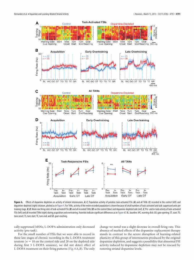
Learning-related changes in ensemble activity of striatal FSIsare severely disrupted by dopamine depletionThe local dopamine depletion in the sensorimotor striatum al-most fully blocked the development of the task-bracketing activ-ity of the FSI populations during learning. This pattern did formon the control side (ANOVA comparing peri-event firing rates inthe late overtraining phase, p � 0.02), as we have reported previ-ously for normal animals (Kubota et al., 2009). FSI ensembleactivity recorded in the dopamine-depleted striatum was strongthroughout the runs, with no hint of decreases in mid-run activ-ity (Fig. 6A,B,E). Even in the late stages of overtraining (stages8 –10), during which there was slight reduction in firing ratesaround run start and before tone onset, differences in firing ratesacross peri-event windows did not reach significance (p � 0.4).Despite these marked changes in the in-task firing of the FSIensemble, their baseline firing rates were unaffected (Fig. 6B,E).Similar results were found for rats with short and long delaysbetween 6-OHDA lesions and training onset, indicating that theeffects of dopamine depletion were unlikely to reflect time-dependent compensatory changes alone (data not shown). These
Hernandez et al. • Dopamine and Learning-Related Striatal Activity J. Neurosci., March 13, 2013 • 33(11):4782– 4795 • 4787
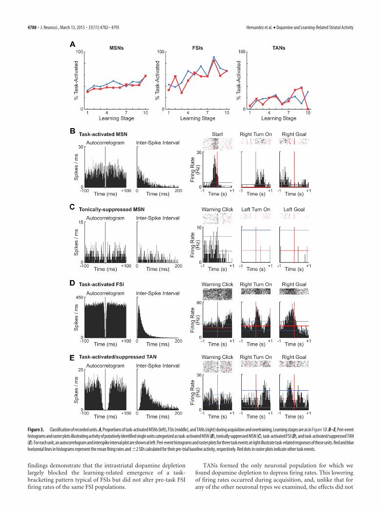
findings demonstrate that the intrastriatal dopamine depletionlargely blocked the learning-related emergence of a task-bracketing pattern typical of FSIs but did not alter pre-task FSIfiring rates of the same FSI populations.
TANs formed the only neuronal population for which wefound dopamine depletion to depress firing rates. This loweringof firing rates occurred during acquisition, and, unlike that forany of the other neuronal types we examined, the effects did not
Figure 3. Classification of recorded units. A, Proportions of task-activated MSNs (left), FSIs (middle), and TANs (right) during acquisition and overtraining, Learning stages are as in Figure 1D. B–E, Peri-eventhistograms and raster plots illustrating activity of putatively identified single units categorized as task-activated MSN (B), tonically suppressed MSN (C), task-activated FSI (D), and task-activated/suppressed TAN(E).Foreachunit,anautocorrelogramandinterspikeintervalplotareshownatleft.Peri-eventhistogramsandrasterplotsforthreetaskeventsatright illustratetask-relatedresponsesoftheseunits.Redandbluehorizontal lines in histograms represent the mean firing rates and �2 SDs calculated for their pre-trial baseline activity, respectively. Red dots in raster plots indicate other task events.
4788 • J. Neurosci., March 13, 2013 • 33(11):4782– 4795 Hernandez et al. • Dopamine and Learning-Related Striatal Activity

Figure 4. Selective disruption of activity of striatal projection neurons by dopamine depletion. A, C, Ensemble activity of putative task-activated (A) and task-suppressed (C) MSNs recorded in thecontrol (left) and dopamine-depleted (right) dorsolateral striatum. Firing rate of each unit during �200 ms around each of seven task events (as labeled on x-axis) were normalized to itsminimum-to-maximum scale (0 –1) and activity of individual units were averaged for each training stage (stages 1–10 on y-axis). Color scale is shown at right. Numbers of units for the stage are indicatedto the right of each row. B, D, Mean raw firing rates of task-activated (B) and task-suppressed (D) MSNs on the control (blue) and dopamine-depleted (red) sides during acquisition (stages 1– 4), earlyovertraining (5–7), and late overtraining (8 –10), plotted across task time. Shading represents SEM. Task-suppressed units were further classified to tonically suppressed (TS) and (Figure legend continues.)
Hernandez et al. • Dopamine and Learning-Related Striatal Activity J. Neurosci., March 13, 2013 • 33(11):4782– 4795 • 4789

depend on task time (Fig. 6C–E). Theseeffects were similar for the short and longdelay subgroups, again suggesting thatthey were related not to time elapsed afterdopamine depletion, but to T-maze learn-ing (data not shown). We found only asmall number of TANs that exhibited thecharacteristic pause in spiking at differenttask events (Fig. 3E). These units werefound both on the control and dopamine-depleted sides in comparable proportions(23 and 26%, respectively; p � 0.5, � 2
test).
L-DOPA treatment induces majorchanges in the firing patterns of striatalMSNs in the dopamine-depletedstriatumAfter completion of the overtraining pe-riod, we pretreated 14 rats with daily in-jections of L-DOPA 30 min before theyperformed the T-maze task (6 mg/kg �benserazide 15 mg/kg, i.p., for 9 –10 d).This procedure was designed to mimic thedopamine replacement therapy com-monly given to patients with Parkinson’sdisease, and to test its effects on task per-formance and on neuronal activity in thedopamine-depleted sensorimotor stria-tum. The dose levels were kept low toavoid L-DOPA-induced abnormal move-ments. Before and/or after the series ofL-DOPA sessions, a subset of the rats (n �9) also received 1–2 additional sessionswith saline injections (1 ml/kg, i.p.) as acontrol procedure.
In a separate set of rats (n � 3), wetested whether L-DOPA treatment in-creased striatal dopamine release inducedby MFB stimulation. We found that it did both on the lesion side(294% increase, p � 0.0094, t test) and on the control side (510%increase, p � 0.054), in accordance with previous observations(Keller et al., 1988).
The L-DOPA treatment did not affect the percent-correct per-formance of the animals (Fig. 7A, left). However, the rats ap-peared less motivated to run, potentially due to the rewardingeffects of the drug, and their average reaction times increasedfrom 1.5 s for the last three sessions of overtraining to 3.7 s for thefirst three L-DOPA sessions (Fig. 7A, middle; p � 0.05, t test).Reaction times slowly decreased as the L-DOPA administrationwas continued, but they remained higher than those before thestart of treatment in 5 of the 14 animals. The total running times
oftheanimalsalsotendedtoincreaseduringL-DOPAsessions(fromanaverage of 2.8 s for the last three overtraining sessions to 4.7 s for the firstthree L-DOPA sessions), but these changes did not reach statistical sig-nificance (Fig. 7A, right). Saline injection did not induce significantchanges in behavioral performance (Fig. 7A) or in ensemble activity ofdifferent neuronal populations.
L-DOPA almost completely reversed the heightened pre-startfiring rates of MSNs induced by dopamine depletion while pre-serving pre-start firing rates in the nondepleted striatum (Fig.7B–G). This normalizing effect of the L-DOPA therapy was evi-dent for both the task-activated and the tonically task-suppressedMSN populations in the dopamine-depleted striatum (ANOVAcomparing pre-task firing rates between the last five overtrainingsessions vs the first five L-DOPA sessions, p � 0.005 and p �0.001, respectively). Pre-task activity was not affected by the dailyL-DOPA treatment in the control striatum (p � 0.4 and p � 0.6for task-activated and tonically task-suppressed MSNs, respec-tively). In addition, L-DOPA administration increased neuronalfiring of task-activated MSNs in the dopamine-depleted striatumduring maze runs (p � 0.05), principally around run start andbefore goal-reaching (Fig. 7B,C). Again, firing rates in the non-depleted hemisphere were left nearly unchanged (p � 0.4). Thus,for the task-activated MSNs, L-DOPA treatment both decreased(pre-task) and increased (in-task) activity, whereas for the toni-
4
(Figure legend continued.) dynamically suppressed (DS) subpopulations. Arrow indicatesincreased pre-run firing rate in the dopamine-depleted side for TS units. Task events are asfollows: baseline (BL; 1.9 –1.4 s before warning click); warning click (WC), gate opening (GO),start (ST), tone onset (TO), turn start (TS), turn end (TE), and goal-reaching (GR). E, Averagepre-task (from 2 s before warning click to gate opening) and in-task (from start to goal reaching)firing rates of task-activated (left); tonically suppressed (middle); and dynamically suppressed(right) MSNs during acquisition, early overtraining, and late overtraining phases. Asterisks in-dicate significant differences in overall firing rates (black) and in period-specific firing rates(orange) between two sides (*p � 0.05; **p � 0.01; ***p � 0.0001). Error bars indicate SEM.
Figure 5. Learning- and time-dependent effects of dopamine depletion on tonically suppressed MSNs. A, Raw firing rates oftonically suppressed MSNs recorded in the control (blue) and dopamine-depleted (red) striatum of rats with short (top) and long(bottom) delays between 6-OHDA lesions and the onset of T-maze training. Plots are made as in Figure 4B for the entire task periodafter warning click. B, Activity of tonically suppressed MSNs, plotted as in A for the pre- and in-task periods, including pre-trialbaseline period. Orange line indicates warning click signaling task start. BL, baseline; WC, warning click; GO, gate opening; ST, start;TO, tone onset; TS, turn start; TE, turn end; and GR, goal-reaching.
4790 • J. Neurosci., March 13, 2013 • 33(11):4782– 4795 Hernandez et al. • Dopamine and Learning-Related Striatal Activity
cally suppressed MSNs, L-DOPA administration only decreasedactivity (pre-task).
For the small number of FSIs that we were able to record inthese late stages of chronic recording in the L-DOPA treatmentsessions (n � 10 on the control side and 28 on the depleted sideduring first 5 L-DOPA sessions), we did not detect effect ofL-DOPA treatment on their firing patterns (Fig. 8A,B). The only
change we noted was a slight decrease in overall firing rate. Thisabsence of marked effects of the dopamine replacement therapystands in contrast to the severe disruption of learning-relatedplasticity of this group of interneurons produced by the originaldopamine depletion, and suggests a possibility that abnormal FSIactivity induced by dopamine depletion may not be rescued byrestoring striatal dopamine levels.
Figure 6. Effects of dopamine depletion on activity of striatal interneurons. A, C, Population activity of putative task-activated FSIs (A) and all TANs (C) recorded in the control (left) anddopamine-depleted (right) striatum, plotted as in Figure 4. For TANs, activity of the entire recorded population is shown because of small numbers of task-activated and task-suppressed units pertraining stage. B, D, Mean raw firing rates of task-activated FSIs (B) and all recorded TANs (D) on the control (blue) and dopamine-depleted side (red). E, Pre- and in-task activity of task-activatedFSIs (left) and all recorded TANs (right) during acquisition and overtraining. Asterisks indicate significant differences as in Figure 4E. BL, baseline; WC, warning click; GO, gate opening; ST, start; TO,tone onset; TS, turn start; TE, turn end; and GR, goal-reaching.
Hernandez et al. • Dopamine and Learning-Related Striatal Activity J. Neurosci., March 13, 2013 • 33(11):4782– 4795 • 4791
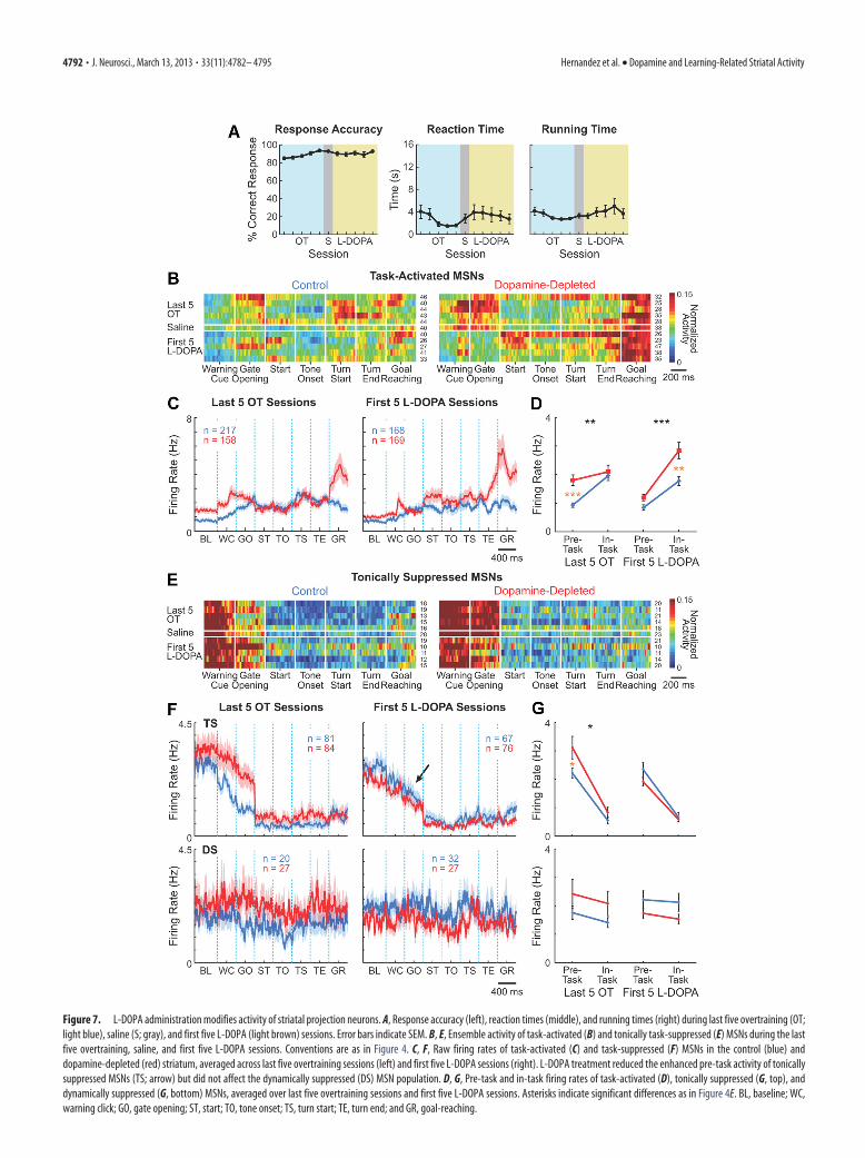
Figure 7. L-DOPA administration modifies activity of striatal projection neurons. A, Response accuracy (left), reaction times (middle), and running times (right) during last five overtraining (OT;light blue), saline (S; gray), and first five L-DOPA (light brown) sessions. Error bars indicate SEM. B, E, Ensemble activity of task-activated (B) and tonically task-suppressed (E) MSNs during the lastfive overtraining, saline, and first five L-DOPA sessions. Conventions are as in Figure 4. C, F, Raw firing rates of task-activated (C) and task-suppressed (F) MSNs in the control (blue) anddopamine-depleted (red) striatum, averaged across last five overtraining sessions (left) and first five L-DOPA sessions (right). L-DOPA treatment reduced the enhanced pre-task activity of tonicallysuppressed MSNs (TS; arrow) but did not affect the dynamically suppressed (DS) MSN population. D, G, Pre-task and in-task firing rates of task-activated (D), tonically suppressed (G, top), anddynamically suppressed (G, bottom) MSNs, averaged over last five overtraining sessions and first five L-DOPA sessions. Asterisks indicate significant differences as in Figure 4E. BL, baseline; WC,warning click; GO, gate opening; ST, start; TO, tone onset; TS, turn start; TE, turn end; and GR, goal-reaching.
4792 • J. Neurosci., March 13, 2013 • 33(11):4782– 4795 Hernandez et al. • Dopamine and Learning-Related Striatal Activity
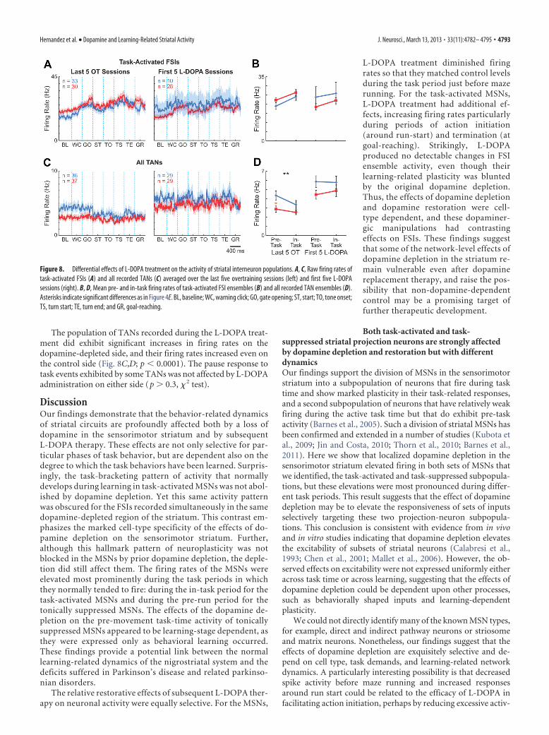
The population of TANs recorded during the L-DOPA treat-ment did exhibit significant increases in firing rates on thedopamine-depleted side, and their firing rates increased even onthe control side (Fig. 8C,D; p � 0.0001). The pause response totask events exhibited by some TANs was not affected by L-DOPAadministration on either side (p � 0.3, � 2 test).
DiscussionOur findings demonstrate that the behavior-related dynamicsof striatal circuits are profoundly affected both by a loss ofdopamine in the sensorimotor striatum and by subsequentL-DOPA therapy. These effects are not only selective for par-ticular phases of task behavior, but are dependent also on thedegree to which the task behaviors have been learned. Surpris-ingly, the task-bracketing pattern of activity that normallydevelops during learning in task-activated MSNs was not abol-ished by dopamine depletion. Yet this same activity patternwas obscured for the FSIs recorded simultaneously in the samedopamine-depleted region of the striatum. This contrast em-phasizes the marked cell-type specificity of the effects of do-pamine depletion on the sensorimotor striatum. Further,although this hallmark pattern of neuroplasticity was notblocked in the MSNs by prior dopamine depletion, the deple-tion did still affect them. The firing rates of the MSNs wereelevated most prominently during the task periods in whichthey normally tended to fire: during the in-task period for thetask-activated MSNs and during the pre-run period for thetonically suppressed MSNs. The effects of the dopamine de-pletion on the pre-movement task-time activity of tonicallysuppressed MSNs appeared to be learning-stage dependent, asthey were expressed only as behavioral learning occurred.These findings provide a potential link between the normallearning-related dynamics of the nigrostriatal system and thedeficits suffered in Parkinson’s disease and related parkinso-nian disorders.
The relative restorative effects of subsequent L-DOPA ther-apy on neuronal activity were equally selective. For the MSNs,
L-DOPA treatment diminished firingrates so that they matched control levelsduring the task period just before mazerunning. For the task-activated MSNs,L-DOPA treatment had additional ef-fects, increasing firing rates particularlyduring periods of action initiation(around run-start) and termination (atgoal-reaching). Strikingly, L-DOPAproduced no detectable changes in FSIensemble activity, even though theirlearning-related plasticity was bluntedby the original dopamine depletion.Thus, the effects of dopamine depletionand dopamine restoration were cell-type dependent, and these dopaminer-gic manipulations had contrastingeffects on FSIs. These findings suggestthat some of the network-level effects ofdopamine depletion in the striatum re-main vulnerable even after dopaminereplacement therapy, and raise the pos-sibility that non-dopamine-dependentcontrol may be a promising target offurther therapeutic development.
Both task-activated and task-suppressed striatal projection neurons are strongly affectedby dopamine depletion and restoration but with differentdynamicsOur findings support the division of MSNs in the sensorimotorstriatum into a subpopulation of neurons that fire during tasktime and show marked plasticity in their task-related responses,and a second subpopulation of neurons that have relatively weakfiring during the active task time but that do exhibit pre-taskactivity (Barnes et al., 2005). Such a division of striatal MSNs hasbeen confirmed and extended in a number of studies (Kubota etal., 2009; Jin and Costa, 2010; Thorn et al., 2010; Barnes et al.,2011). Here we show that localized dopamine depletion in thesensorimotor striatum elevated firing in both sets of MSNs thatwe identified, the task-activated and task-suppressed subpopula-tions, but these elevations were most pronounced during differ-ent task periods. This result suggests that the effect of dopaminedepletion may be to elevate the responsiveness of sets of inputsselectively targeting these two projection-neuron subpopula-tions. This conclusion is consistent with evidence from in vivoand in vitro studies indicating that dopamine depletion elevatesthe excitability of subsets of striatal neurons (Calabresi et al.,1993; Chen et al., 2001; Mallet et al., 2006). However, the ob-served effects on excitability were not expressed uniformly eitheracross task time or across learning, suggesting that the effects ofdopamine depletion could be dependent upon other processes,such as behaviorally shaped inputs and learning-dependentplasticity.
We could not directly identify many of the known MSN types,for example, direct and indirect pathway neurons or striosomeand matrix neurons. Nonetheless, our findings suggest that theeffects of dopamine depletion are exquisitely selective and de-pend on cell type, task demands, and learning-related networkdynamics. A particularly interesting possibility is that decreasedspike activity before maze running and increased responsesaround run start could be related to the efficacy of L-DOPA infacilitating action initiation, perhaps by reducing excessive activ-
Figure 8. Differential effects of L-DOPA treatment on the activity of striatal interneuron populations. A, C, Raw firing rates oftask-activated FSIs (A) and all recorded TANs (C) averaged over the last five overtraining sessions (left) and first five L-DOPAsessions (right). B, D, Mean pre- and in-task firing rates of task-activated FSI ensembles (B) and all recorded TAN ensembles (D).Asterisks indicate significant differences as in Figure 4E. BL, baseline; WC, warning click; GO, gate opening; ST, start; TO, tone onset;TS, turn start; TE, turn end; and GR, goal-reaching.
Hernandez et al. • Dopamine and Learning-Related Striatal Activity J. Neurosci., March 13, 2013 • 33(11):4782– 4795 • 4793

ity in “no-go” indirect pathway MSNs and by enhancing activityin “go” direct pathway MSNs. However, the rats actually hadslightly longer reaction times after the L-DOPA treatment, notshorter ones. This lengthening could have been related to otherpotential effects of the L-DOPA, including motivational changes,a change in context, or even drowsiness (Andreu et al., 1999),masking other potential reaction time effects.
Our findings indicate that the disruption of striatal activitypatterns that occurs following intrastriatal dopamine depletionemerges as animals learn new tasks. Our experimental designdoes not allow us entirely to exclude the possibility that theseapparent learning-related effects may be due to progressive com-pensatory responses to dopamine depletion. We found, however,nearly identical patterns of effects in rats that began training afterdifferent postlesion intervals, with the only exception being thepre-trial baseline activity of tonically suppressed MSNs. Thisworking hypothesis of a learning dependency of the depletioneffect is compatible with the wealth of evidence that dopamine isa critical part of nigrostriatal learning circuits (Aosaki et al.,1994a; Centonze et al., 1999; Schultz, 2007; Stalnaker et al., 2012).
What makes the observation of special interest is that we didnot find wholesale disruption of the learning-related plasticity inensemble activity known to occur during T-maze learning.Remarkably, the task-bracketing activity pattern that formsnormally among task-activated MSNs developed clearly in thedopamine-depleted striatum, indicating that dopamine-dependent plasticity in the sensorimotor striatum may be re-sponsible for only a subset of the learning-related changes innetwork activity patterns observed across learning. One possibil-ity is that, because we depleted dopamine only locally in the sen-sorimotor striatum, dopamine released in nearby striatal sitesmay have diffused into the depleted zone (Rice and Cragg, 2008),allowing the pattern formation. Dopamine in non-striatal sitescould have helped in shaping the relatively normal task-bracketing pattern. An interesting alternative is that non-dopaminergic influences could be more important for thisensemble-level plasticity in the sensorimotor striatum.
Dopamine depletion produces loss of task-related patterningof striatal FSI spike activityThe fast-spiking inhibitory interneurons of the striatum, thoughtto correspond mainly to the parvalbumin-containing GABAergicinterneurons (Kawaguchi, 1993), provide feedforward inhibitorysignals to striatal MSNs through excitatory corticostriatal projec-tions to these interneurons. Previous studies (Bennett andBolam, 1994; Parthasarathy and Graybiel, 1997; Berke, 2011;Howe et al., 2011) point to these striatal FSIs as key integrators ofstriatal circuit function, and dopamine depletion has been shownseverely to affect their firing properties in vitro (Salin et al., 2009;Gittis et al., 2011). Here we demonstrate that learning-relatedpatterning of FSI activity, clearly observed in simultaneous re-cordings made on the intact side, was severely disrupted on thedopamine-depleted side.
This deficit in FSI plasticity is striking considering the clearbeginning-and-end patterning of spike activity shown by the si-multaneously recorded task-activated MSNs. The contrast ex-tends to task-time patterning: the major depletion-inducedabnormalities in firing of the task-suppressed ensembles oc-curred during the pre-start periods, the periods not affected forthe FSI ensembles. Thus neither the firing patterns of the task-activated MSNs nor those of the task-suppressed MSNs could bepredicted by the marked changes in FSI ensemble activity. Thisresult indicates that there is not tight coupling between the plas-
ticity in firing patterns of FSIs and MSNs, a result supported byprevious studies (Salin et al., 2009; Lansink et al., 2010; Berke,2011).
Our recordings also suggest a sharp contrast between the re-sponsiveness of FSIs and MSNs to restoration of intrastriatal do-pamine levels. Some of the changes in MSN firing induced by theoriginal dopamine depletion were reversed by L-DOPA treat-ment, but we saw no trend for restoration of the task-bracketingactivity in the relatively small sample of FSIs that we were able torecord at that late stage of the chronic recordings.
The effect of dopamine depletion on TANs contrasted to thatof both the MSNs and FSIs in several ways. TANs formed the onlycell population characterized by decreased firing rates as a conse-quence of dopamine depletion; this effect was not task-time de-pendent; and it was strongest during the acquisition phase oftraining rather than later. In at least some of the putative TANs, atypical pause to salient rewarding events was found (Fig. 3E) aspreviously observed in primates (Apicella, 2007). However, inprimates, the dopamine depletion diminishes cue-related pauseresponses in most TANs (Aosaki et al., 1994a), whereas here, inour relatively small sample, we found pause responses in both thecontrol and dopamine-depleted striatum, perhaps due to otherinputs including those from the thalamus (Lacey et al., 2007;Goldberg and Reynolds, 2011; Threlfell et al., 2012).
Our findings suggest a startling level of selectivity in subcircuitdysfunction in the striatum following dopamine depletion, and acomparably striking selectivity in the restorative effects ofL-DOPA treatment. This inventory of differential effects shouldprovide valuable leads to developing equally selective therapeuticapproaches to Parkinson’s disease and related dopamine-depletion disorders.
ReferencesAndreu N, Chale JJ, Senard JM, Thalamas C, Montastruc JL, Rascol O (1999)
L-Dopa-induced sedation: a double-blind cross-over controlled studyversus triazolam and placebo in healthy volunteers. Clin Neuropharma-col 22:15–23. CrossRef Medline
Aosaki T, Graybiel AM, Kimura M (1994a) Effects of the nigrostriatal do-pamine system on acquired neural responses in the striatum of behavingmonkeys. Science 265:412– 415. CrossRef Medline
Aosaki T, Tsubokawa H, Ishida A, Watanabe K, Graybiel AM, Kimura M(1994b) Responses of tonically active neurons in the primate’s striatumundergo systematic changes during behavioral sensorimotor condition-ing. J Neurosci 14:3969 –3984. Medline
Apicella P (2007) Leading tonically active neurons of the striatum from re-ward detection to context recognition. Trends Neurosci 30:299 –306.CrossRef Medline
Barnes TD, Mao JB, Hu D, Kubota Y, Dreyer AA, Stamoulis C, Brown EN,Graybiel AM (2011) Advance cueing produces enhanced action-boundary patterns of spike activity in the sensorimotor striatum. J Neu-rophysiol 105:1861–1878. CrossRef Medline
Barnes TD, Kubota Y, Hu D, Jin DZ, Graybiel AM (2005) Activity of striatalneurons reflects dynamic encoding and recoding of procedural memo-ries. Nature 437:1158 –1161. CrossRef Medline
Bennett BD, Bolam JP (1994) Synaptic input and output of parvalbumin-immunoreactive neurons in the neostriatum of the rat. Neuroscience62:707–719. CrossRef Medline
Berke JD (2011) Functional properties of striatal fast-spiking interneurons.Front Syst Neurosci 5:45. Medline
Braak H, Braak E (2000) Pathoanatomy of Parkinson’s disease. J Neurol 247[Suppl 2]:II3-10. Medline
Brown P (2007) Abnormal oscillatory synchronisation in the motor systemleads to impaired movement. Curr Opin Neurobiol 17:656 – 664.CrossRef Medline
Calabresi P, Mercuri NB, Sancesario G, Bernardi G (1993) Electrophysiol-ogy of dopamine-denervated striatal neurons. Implications for Parkin-son’s disease. Brain 116:433– 452. CrossRef Medline
Cameron IG, Watanabe M, Pari G, Munoz DP (2010) Executive impair-
4794 • J. Neurosci., March 13, 2013 • 33(11):4782– 4795 Hernandez et al. • Dopamine and Learning-Related Striatal Activity
ment in Parkinson’s disease: response automaticity and task switching.Neuropsychologia 48:1948 –1957. CrossRef Medline
Centonze D, Gubellini P, Picconi B, Calabresi P, Giacomini P, Bernardi G(1999) Unilateral dopamine denervation blocks corticostriatal LTP.J Neurophysiol 82:3575–3579. Medline
Chen MT, Morales M, Woodward DJ, Hoffer BJ, Janak PH (2001) In vivoextracellular recording of striatal neurons in the awake rat following uni-lateral 6-hydroxydopamine lesions. Exp Neurol 171:72– 83. CrossRefMedline
Clark JJ, Sandberg SG, Wanat MJ, Gan JO, Horne EA, Hart AS, Akers CA,Parker JG, Willuhn I, Martinez V, Evans SB, Stella N, Phillips PE (2010)Chronic microsensors for longitudinal, subsecond dopamine detection inbehaving animals. Nat Methods 7:126 –129. CrossRef Medline
Cools R, Barker RA, Sahakian BJ, Robbins TW (2001) Mechanisms of cog-nitive set flexibility in Parkinson’s disease. Brain 124:2503–2512. CrossRefMedline
Day M, Wang Z, Ding J, An X, Ingham CA, Shering AF, Wokosin D, Ilijic E,Sun Z, Sampson AR, Mugnaini E, Deutch AY, Sesack SR, Arbuthnott GW,Surmeier DJ (2006) Selective elimination of glutamatergic synapses onstriatopallidal neurons in Parkinson disease models. Nat Neurosci 9:251–259. CrossRef Medline
Emondi AA, Rebrik SP, Kurgansky AV, Miller KD (2004) Tracking neuronsrecorded from tetrodes across time. J Neurosci Methods 135:95–105.CrossRef Medline
Faure A, Haberland U, Conde F, El Massioui N (2005) Lesion to the nigro-striatal dopamine system disrupts stimulus-response habit formation.J Neurosci 25:2771–2780. CrossRef Medline
Gerfen CR, Surmeier DJ (2011) Modulation of striatal projection systemsby dopamine. Annu Rev Neurosci 34:441– 466. CrossRef Medline
Gittis AH, Hang GB, LaDow ES, Shoenfeld LR, Atallah BV, Finkbeiner S,Kreitzer AC (2011) Rapid target-specific remodeling of fast-spiking in-hibitory circuits after loss of dopamine. Neuron 71:858 – 868. CrossRefMedline
Goldberg JA, Reynolds JN (2011) Spontaneous firing and evoked pauses inthe tonically active cholinergic interneurons of the striatum. Neurosci-ence 198:27– 43. CrossRef Medline
Henderson JM, Carpenter K, Cartwright H, Halliday GM (2000) Degener-ation of the centre median-parafascicular complex in Parkinson’s disease.Ann Neurol 47:345–352. CrossRef Medline
Howe MW, Atallah HE, McCool A, Gibson DJ, Graybiel AM (2011) Habitlearning is associated with major shifts in frequencies of oscillatory activ-ity and synchronized spike firing in striatum. Proc Natl Acad Sci U S A108:16801–16806. CrossRef Medline
Ingham CA, Hood SH, Arbuthnott GW (1989) Spine density on neostriatalneurones changes with 6-hydroxydopamine lesions and with age. BrainRes 503:334 –338. CrossRef Medline
Jin X, Costa RM (2010) Start/stop signals emerge in nigrostriatal circuitsduring sequence learning. Nature 466:457– 462. CrossRef Medline
Jog MS, Connolly CI, Kubota Y, Iyengar DR, Garrido L, Harlan R, GraybielAM (2002) Tetrode technology: advances in implantable hardware,neuroimaging, and data analysis techniques. J Neurosci Methods 117:141–152. CrossRef Medline
Jog MS, Kubota Y, Connolly CI, Hillegaart V, Graybiel AM (1999) Buildingneural representations of habits. Science 286:1745–1749. CrossRefMedline
Kawaguchi Y (1993) Physiological, morphological, and histochemical char-acterization of three classes of interneurons in rat neostriatum. J Neurosci13:4908 – 4923. Medline
Keller RW Jr, Kuhr WG, Wightman RM, Zigmond MJ (1988) The effect ofL-dopa on in vivo dopamine release from nigrostriatal bundle neurons.Brain Res 447:191–194. CrossRef Medline
Kubota Y, Liu J, Hu D, DeCoteau WE, Eden UT, Smith AC, Graybiel AM(2009) Stable encoding of task structure coexists with flexible coding oftask events in sensorimotor striatum. J Neurophysiol 102:2142–2160.CrossRef Medline
Lacey CJ, Bolam JP, Magill PJ (2007) Novel and distinct operational princi-ples of intralaminar thalamic neurons and their striatal projections.J Neurosci 27:4374 – 4384. CrossRef Medline
Lansink CS, Goltstein PM, Lankelma JV, Pennartz CM (2010) Fast-spikinginterneurons of the rat ventral striatum: temporal coordination of activitywith principal cells and responsiveness to reward. Eur J Neurosci 32:494 –508. CrossRef Medline
Lemaire N, Hernandez LF, Hu D, Kubota Y, Howe MW, Graybiel AM (2012)Effects of dopamine depletion on LFP oscillations in striatum are task-and learning-dependent and selectively reversed by L-DOPA. Proc NatlAcad Sci U S A 109:18126 –18131. CrossRef Medline
Mallet N, Ballion B, Le Moine C, Gonon F (2006) Cortical inputs and GABAinterneurons imbalance projection neurons in the striatum of parkinso-nian rats. J Neurosci 26:3875–3884. CrossRef Medline
Parthasarathy HB, Graybiel AM (1997) Cortically driven immediate-earlygene expression reflects modular influence of sensorimotor cortex onidentified striatal neurons in the squirrel monkey. J Neurosci 17:2477–2491. Medline
Picconi B, Piccoli G, Calabresi P (2012) Synaptic dysfunction in Parkinson’sdisease. Adv Exp Med Biol 970:553–572. CrossRef Medline
Rice ME, Cragg SJ (2008) Dopamine spillover after quantal release: rethink-ing dopamine transmission in the nigrostriatal pathway. Brain Res Rev58:303–313. CrossRef Medline
Salin P, Lopez IP, Kachidian P, Barroso-Chinea P, Rico AJ, Gomez-BautistaV, Coulon P, Kerkerian-Le Goff L, Lanciego JL (2009) Changes tointerneuron-driven striatal microcircuits in a rat model of Parkinson’sdisease. Neurobiol Dis 34:545–552. CrossRef Medline
Schultz W (2007) Multiple dopamine functions at different time courses.Annu Rev Neurosci 30:259 –288. CrossRef Medline
Stalnaker TA, Calhoon GG, Ogawa M, Roesch MR, Schoenbaum G (2012)Reward prediction error signaling in posterior dorsomedial striatum isaction specific. J Neurosci 32:10296 –10305. CrossRef Medline
Thorn CA, Atallah H, Howe M, Graybiel AM (2010) Differential dynamicsof activity changes in dorsolateral and dorsomedial striatal loops duringlearning. Neuron 66:781–795. CrossRef Medline
Threlfell S, Lalic T, Platt NJ, Jennings KA, Deisseroth K, Cragg SJ (2012)Striatal dopamine release is triggered by synchronized activity in cholin-ergic interneurons. Neuron 75:58 – 64. CrossRef Medline
Hernandez et al. • Dopamine and Learning-Related Striatal Activity J. Neurosci., March 13, 2013 • 33(11):4782– 4795 • 4795
Copyright © 2022 FDOKUMEN