Segmentation characteristics - [email protected]
32
Reference Text
-
Upload
khangminh22 -
Category
Documents
-
view
3 -
download
0
Transcript of Segmentation characteristics - [email protected]

Reference Text
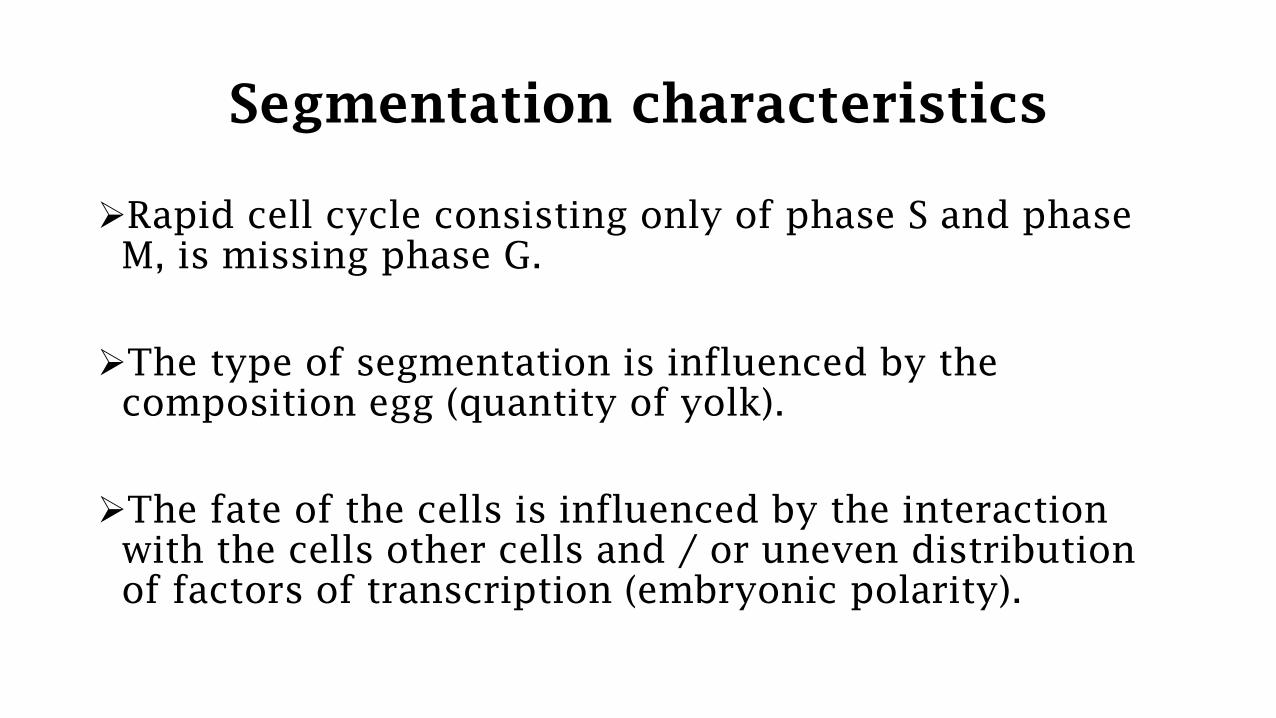
Segmentation characteristics
➢Rapid cell cycle consisting only of phase S and phase
M, is missing phase G.
➢The type of segmentation is influenced by the
composition egg (quantity of yolk).
➢The fate of the cells is influenced by the interaction
with the cells other cells and / or uneven distribution
of factors of transcription (embryonic polarity).
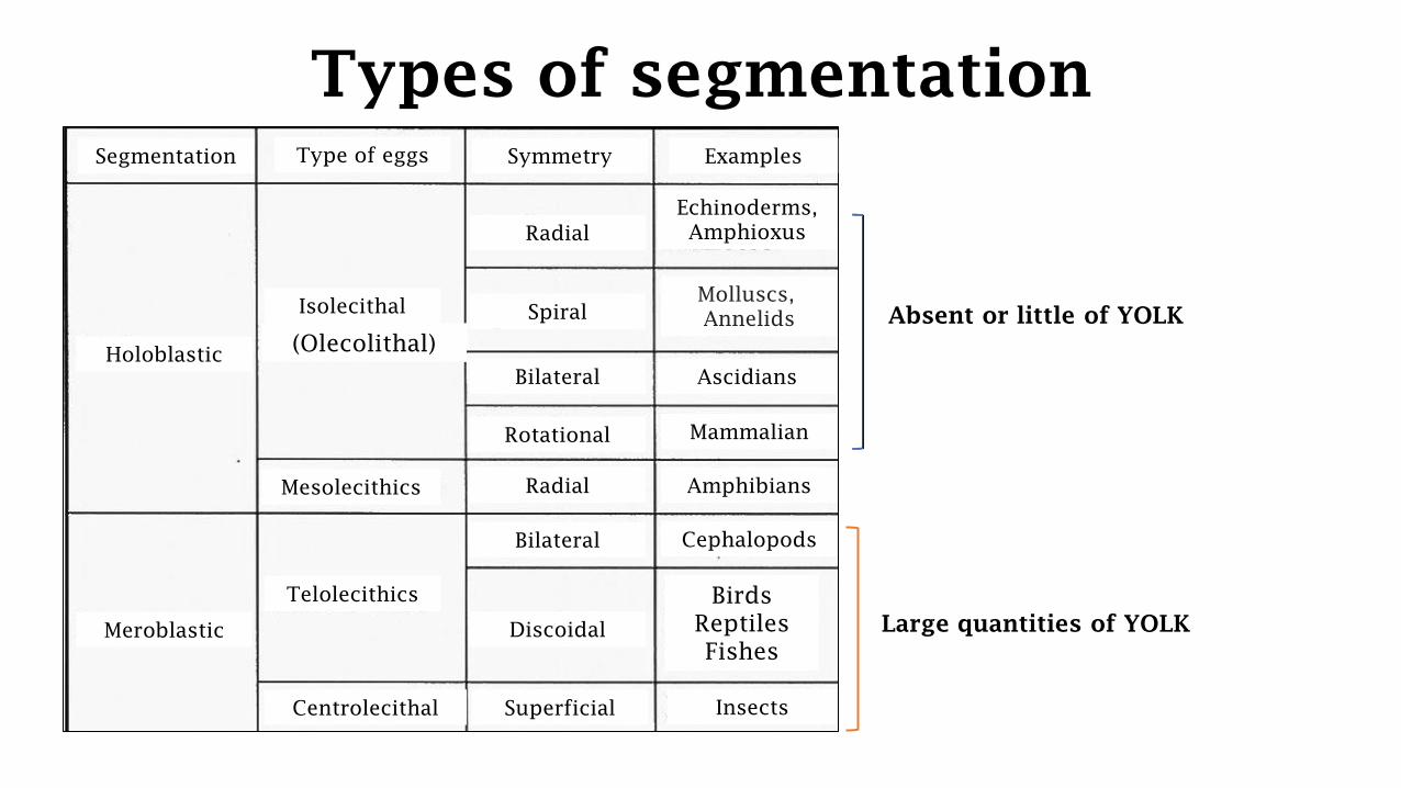
Types of segmentation
Segmentation Type of eggs Symmetry
Large quantities of YOLK
Examples
Holoblastic
Meroblastic
Radial
Spiral
Bilateral
Rotational
Radial
Bilateral
Discoidal
Superficial
Isolecithal
(Olecolithal)
Mesolecithics
Telolecithics
Centrolecithal
Mammalian
Amphibians
Cephalopods
Birds
Reptiles
Fishes
Insects
Molluscs,
Annelids
Ascidians
Echinoderms,
Amphioxus
Absent or little of YOLK
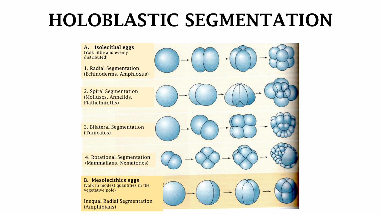
HOLOBLASTIC SEGMENTATION
B. Mesolecithics eggs
(yolk in modest quantities in the
vegetative pole)
Inequal Radial Segmentation
(Amphibians)
A. Isolecithal eggs
(Yolk little and evenly
distributed)
1. Radial Segmentation
(Echinoderms, Amphioxus)
2. Spiral Segmentation
(Molluscs, Annelids,
Plathelminths)
3. Bilateral Segmentation
(Tunicates)
4. Rotational Segmentation
(Mammalians, Nematodes)
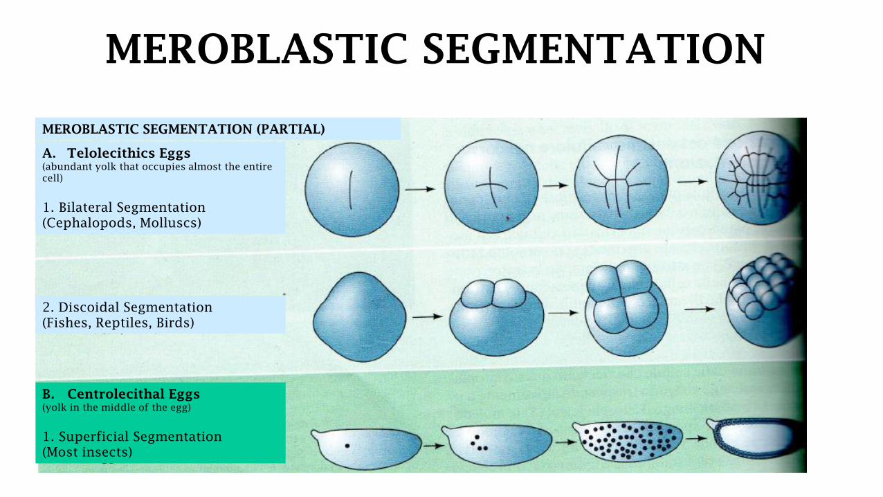
MEROBLASTIC SEGMENTATION
A. Telolecithics Eggs
(abundant yolk that occupies almost the entire
cell)
1. Bilateral Segmentation
(Cephalopods, Molluscs)
MEROBLASTIC SEGMENTATION (PARTIAL)
2. Discoidal Segmentation
(Fishes, Reptiles, Birds)
B. Centrolecithal Eggs
(yolk in the middle of the egg)
1. Superficial Segmentation
(Most insects)

Blastula types
Coeloblastula with central
Blastocoele
Amphioxus
Blastocyst
Mammalians
Coeloblastula with
eccentric Blastocoele
Amphibians
Discoid Blastula
Reptiles, Birds
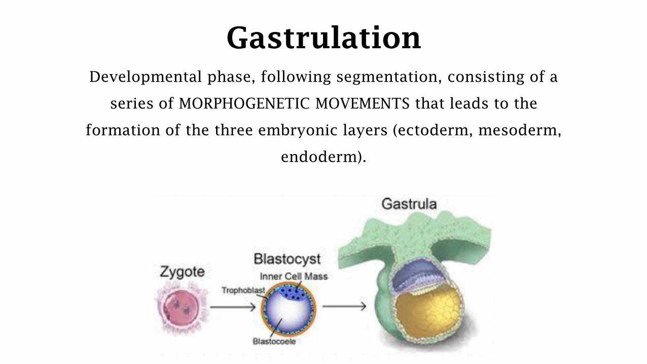
Gastrulation
Developmental phase, following segmentation, consisting of a
series of MORPHOGENETIC MOVEMENTS that leads to the
formation of the three embryonic layers (ectoderm, mesoderm,
endoderm).

Primary mechanisms of
gastrulation
• Epibolia
• Delamination
• Cell movements from the inside surface
- Intussusception
- Involution
- Entry or immigration
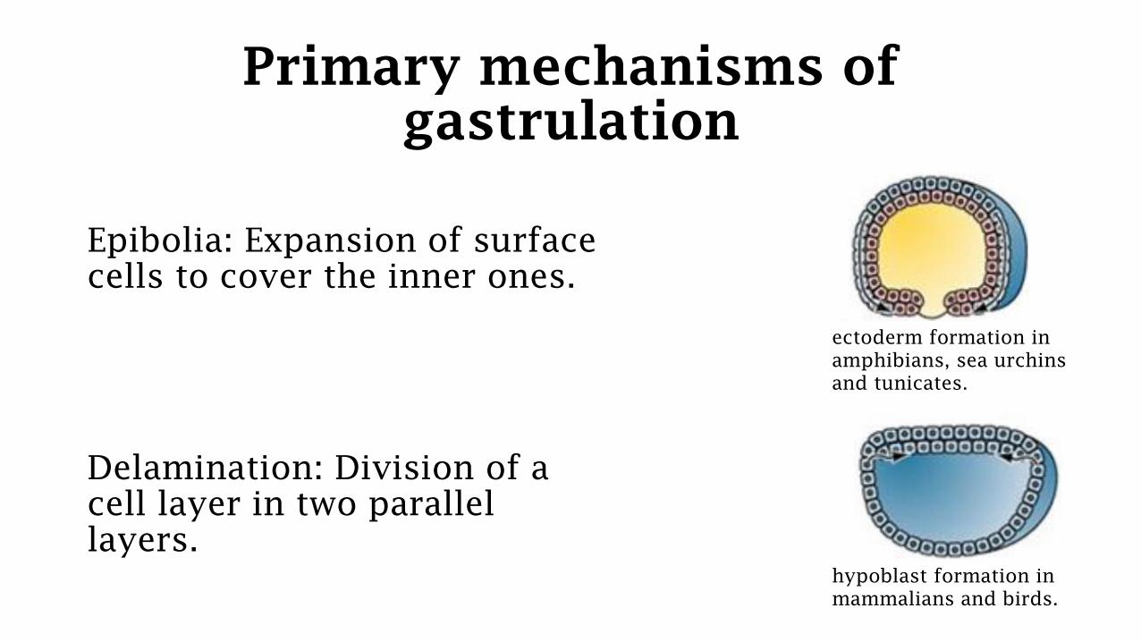
Primary mechanisms of
gastrulation
Epibolia: Expansion of surface
cells to cover the inner ones.
Delamination: Division of a
cell layer in two parallel
layers.
ectoderm formation in
amphibians, sea urchins
and tunicates.
hypoblast formation in
mammalians and birds.

Primary mechanisms of
gastrulation
Cell movements from the inner surface
Intussusception Involution Ingression
Folding of a
portion of
blastoderma
inwards.
Folding or
sliding of cells
so as to bear on
the inner surface
(EMBOLISM).
Migration of
cells from
external sheets
towards the
inside of the
embryo.
sea urchin’s endoderm Amphibians’ mesoderm sea urchin mesoderm,
drosophila neuroblasts
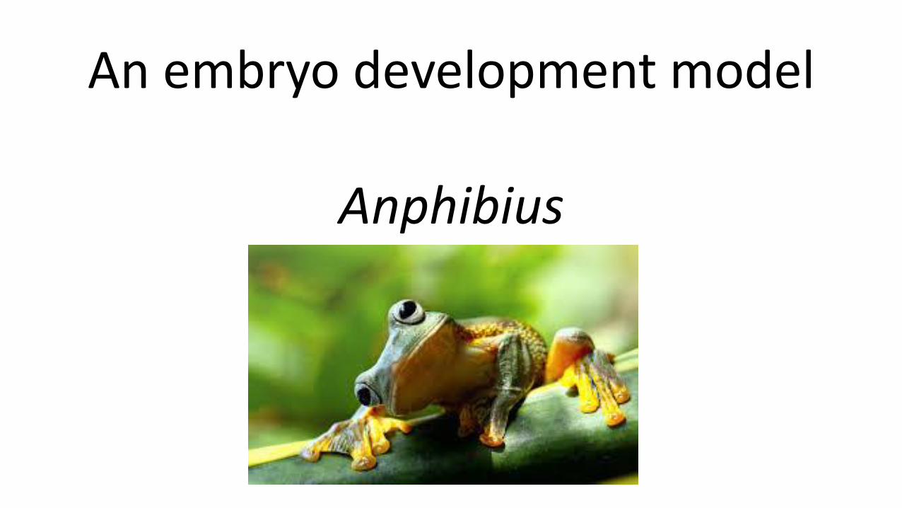
An embryo development model
Anphibius
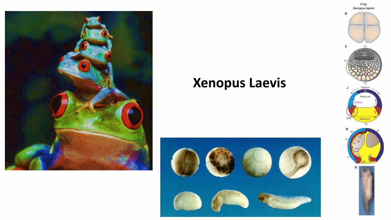
Xenopus Laevis
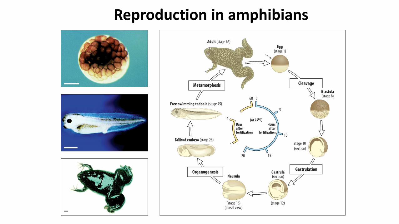
Reproduction in amphibians

Reproduction in amphibians can be internal or external

Mesolecitic egg Radial unequalholoblastic segmentation

Animal Pole
Segmentaziongroove
Micromeres
MacromeresVegetal Pole
16-64 CellsMorula
128 CellsBlastula
Radial unequal holoblastic segmentation
Crescent-grey

Xenopus Laevis Segmentation
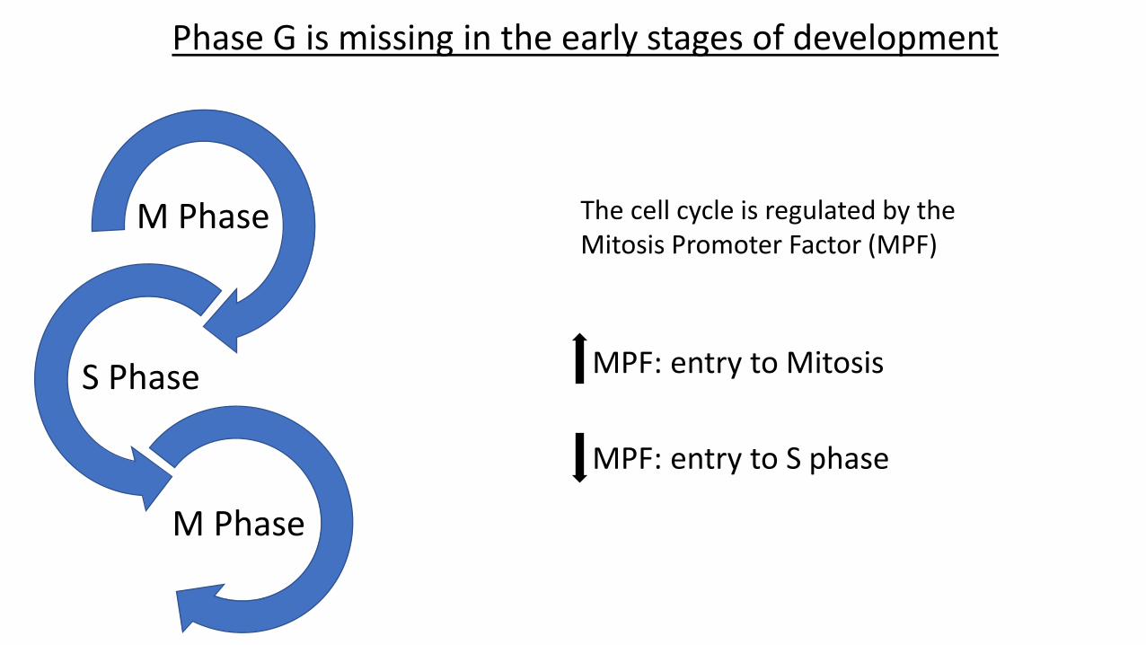
M Phase
M Phase
S Phase
Phase G is missing in the early stages of development
The cell cycle is regulated by the Mitosis Promoter Factor (MPF)
MPF: entry to Mitosis
MPF: entry to S phase
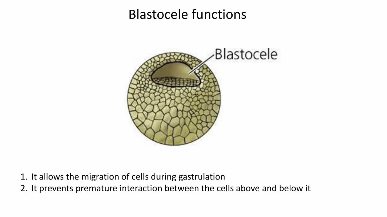
Blastocele functions
1. It allows the migration of cells during gastrulation2. It prevents premature interaction between the cells above and below it
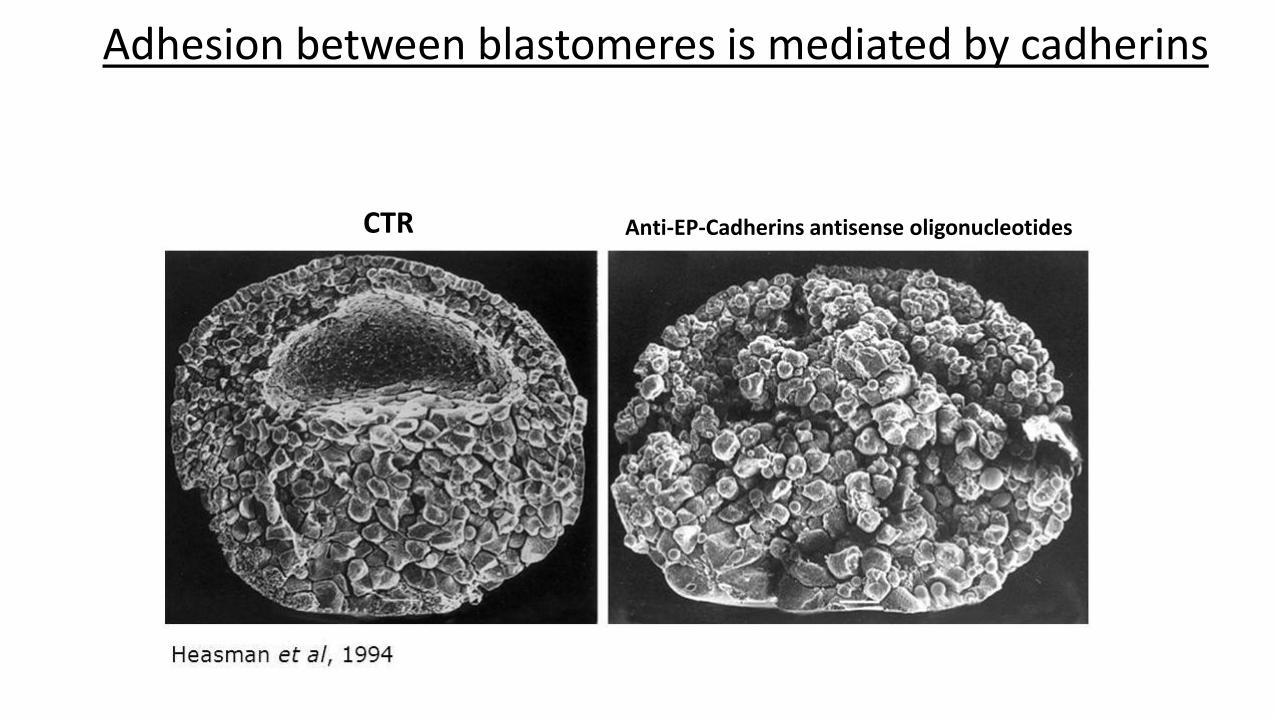
Adhesion between blastomeres is mediated by cadherins
CTR Anti-EP-Cadherins antisense oligonucleotides
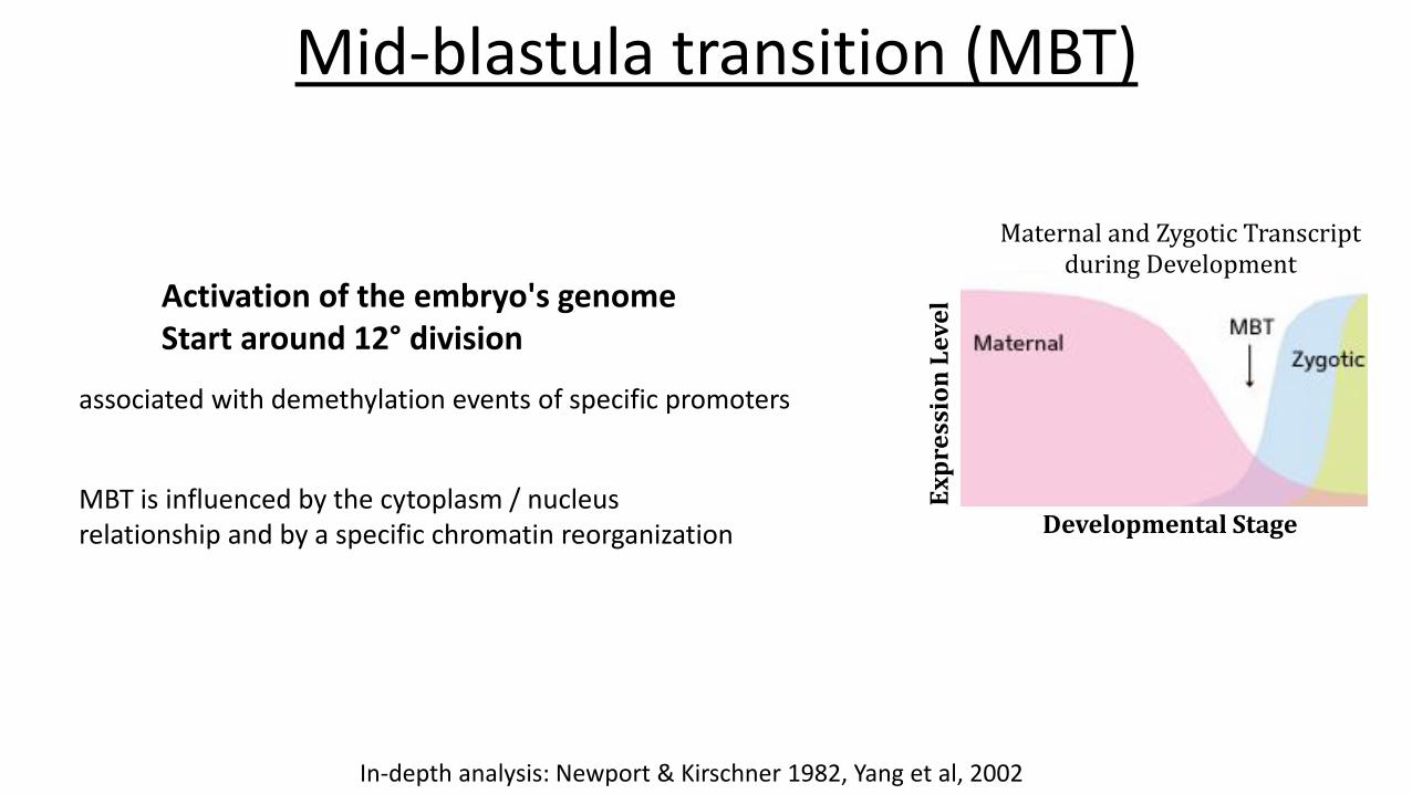
Mid-blastula transition (MBT)
Ex
pre
ssio
nL
ev
el
Developmental Stage
Maternal and Zygotic Transcriptduring Development
Activation of the embryo's genomeStart around 12° division
associated with demethylation events of specific promoters
MBT is influenced by the cytoplasm / nucleusrelationship and by a specific chromatin reorganization
In-depth analysis: Newport & Kirschner 1982, Yang et al, 2002

Embryogenesis time course of Xenopus laevis oocytes untilthe tadpole stage. Courtesy of Dr. Daniel Fisher, IGMM, Montpellier, France.
Gastrulation in Xenopus Laevis
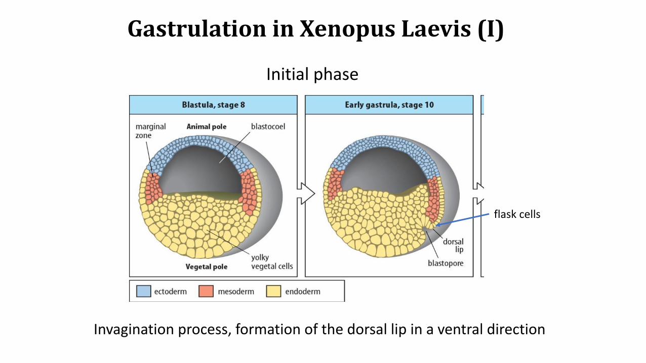
Gastrulation in Xenopus Laevis (I)
Invagination process, formation of the dorsal lip in a ventral direction
Initial phase
flask cells
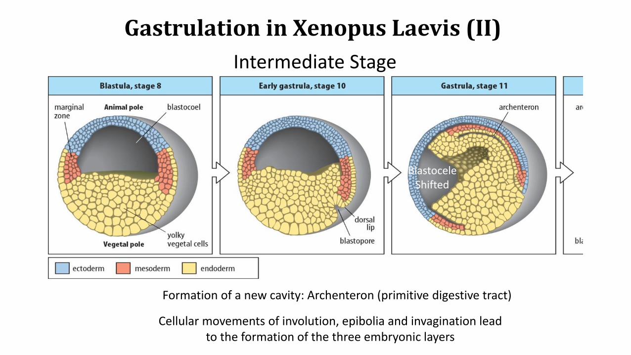
Intermediate Stage
Gastrulation in Xenopus Laevis (II)
Formation of a new cavity: Archenteron (primitive digestive tract)
Cellular movements of involution, epibolia and invagination leadto the formation of the three embryonic layers
BlastoceleShifted

Gastrulation in Xenopus Laevis (III)
Final Stage
The embryo is coated with the ectoderm, the endoderm hasbeen brought in, the mesoderm cells are arranged betweenectoderm and endoderm
The blastocele is reduced until it disappears
Notocord
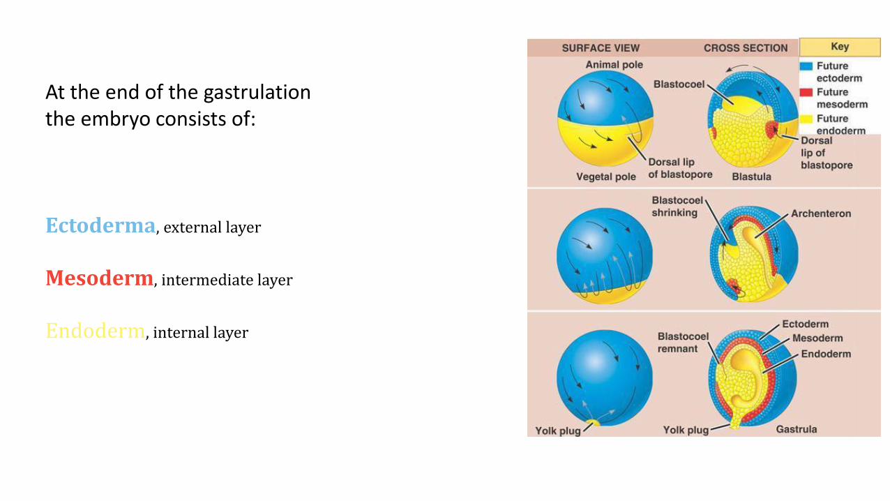
At the end of the gastrulationthe embryo consists of:
Ectoderma, external layer
Mesoderm, intermediate layer
Endoderm, internal layer

Determination of the axes
Axis
Dorsal – Ventral
Anterior – Posterior
Determined by sperm entry site
Regulated from the center of Nieuwkoop and specific proteins
Regulated by Spemann organizer
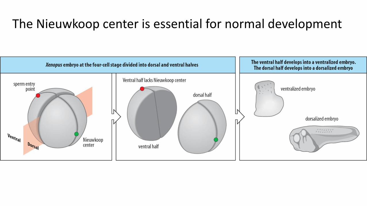
The Nieuwkoop center is essential for normal development

The transplantation of cells containing the center of Nieuwkoop in a receiving blastula determines the formation of two dorsal axes
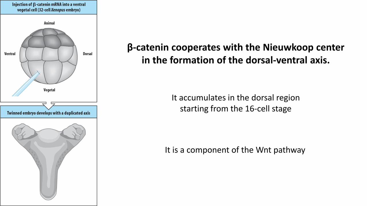
β-catenin cooperates with the Nieuwkoop center in the formation of the dorsal-ventral axis.
It accumulates in the dorsal regionstarting from the 16-cell stage
It is a component of the Wnt pathway
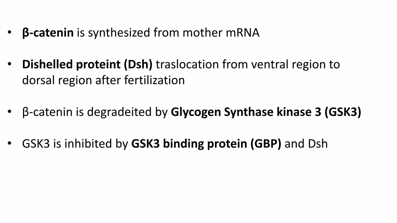
• β-catenin is synthesized from mother mRNA
• Dishelled proteint (Dsh) traslocation from ventral region to dorsal region after fertilization
• β-catenin is degradeited by Glycogen Synthase kinase 3 (GSK3)
• GSK3 is inhibited by GSK3 binding protein (GBP) and Dsh
?