Unit No. - Elearning
280
MICROBIOLOGY, MYCOLOGY AND PLANT PATHOLOGY BSCBO-102 UTTARAKHAND OPEN UNIVERSITY Page 1 Board of Studies Late Prof. S. C. Tewari Prof. Uma Palni Department of Botany Department of Botany HNB Garhwal University, Retired, DSB Campus, Srinagar Kumoun University, Nainital Dr. R.S. Rawal Dr. H.C. Joshi Scientist, GB Pant National Institute of Department of Environmental Science Himalayan Environment & Sustainable School of Sciences Development, Almora Uttarakhand Open University, Haldwani Dr. Pooja Juyal Department of Botany School of Sciences Uttarakhand Open University, Haldwani Programme Coordinator Dr. Pooja Juyal Department of Botany School of Sciences Uttarakhand Open University Haldwani, Nainital Unit Written By: Unit No. 1. Dr. Neeta Pandey 1 Associate Professor Department Of Botany MBPG College, Haldwani 2. Dr. Rajan Kumar Gupta 2 Associate Professor Department Of Botany PDBH Govt. P.G College, Kotdwar 3. Dr. Dinesh Giri 3, 5, 6, 7 & 8 Assistant Professor, Department of Botany, Garg PG College, Laksar, Haridwar
-
Upload
khangminh22 -
Category
Documents
-
view
3 -
download
0
Transcript of Unit No. - Elearning

MICROBIOLOGY, MYCOLOGY AND PLANT PATHOLOGY BSCBO-102
UTTARAKHAND OPEN UNIVERSITY Page 1
Board of Studies
Late Prof. S. C. Tewari Prof. Uma Palni
Department of Botany Department of Botany
HNB Garhwal University, Retired, DSB Campus,
Srinagar Kumoun University, Nainital
Dr. R.S. Rawal Dr. H.C. Joshi
Scientist, GB Pant National Institute of Department of Environmental Science
Himalayan Environment & Sustainable School of Sciences
Development, Almora Uttarakhand Open University,
Haldwani
Dr. Pooja Juyal
Department of Botany
School of Sciences
Uttarakhand Open University, Haldwani
Programme Coordinator
Dr. Pooja Juyal
Department of Botany
School of Sciences
Uttarakhand Open University
Haldwani, Nainital
Unit Written By: Unit No.
1. Dr. Neeta Pandey 1
Associate Professor
Department Of Botany
MBPG College, Haldwani
2. Dr. Rajan Kumar Gupta 2
Associate Professor
Department Of Botany
PDBH Govt. P.G College, Kotdwar
3. Dr. Dinesh Giri 3, 5, 6, 7 & 8
Assistant Professor,
Department of Botany,
Garg PG College, Laksar, Haridwar

MICROBIOLOGY, MYCOLOGY AND PLANT PATHOLOGY BSCBO-102
UTTARAKHAND OPEN UNIVERSITY Page 2
4. Dr. Pooja Juyal 4
Department of Botany
School of Sciences
Uttarakhand Open University
Haldwani, Nainital
5. Prof. Neerja Pande 9 & 12
Department of Botany, DSB Campus,
Kumaun University, Nainital
6. Dr. Anjala Durgapal 10 & 11
Associate Professor
Department Of Botany
MBPG College, Haldwani
Course Editor
Prof. N.S. Bisht
Head, Department of Botany
HNB Pauri Campus,
Garhwal Central University
Title : Phycology and Bryology
ISBN No. : 978-93-857-40-58-9
Copyright : Uttarakhand Open University
Edition : 2017
Published By: Uttarakhand Open University, Haldwani, Nainital-263139
MICROBIOLOGY, MYCOLOGY AND PLANT PATHOLOGY BSCBO-102
UTTARAKHAND OPEN UNIVERSITY Page 3
BSCBO-102
PHYCOLOGY AND BRYOLOGY
SCHOOL OF SCIENCES
DEPARTMENT OF BOTANY
UTTARAKHAND OPEN UNIVERSITY
Phone No. 05946-261122, 261123
Toll free No. 18001804025
Fax No. 05946-264232, E. mail [email protected]
htpp://uou.ac.in

MICROBIOLOGY, MYCOLOGY AND PLANT PATHOLOGY BSCBO-102
UTTARAKHAND OPEN UNIVERSITY Page 4
CONTENTS
BLOCK-1- ALGAE: GENERAL ACCOUNT PAGE NO.
Unit-1-General Characters and Life cycles in Algae 6-32
Unit-2-Important Classifications of Algae (Any three) 33-46
Unit-3-Range of Vegetative structure 47-66
Unit-4-Ecological and Economic importance of Algae 67-76
BLOCK-2- ALGAE: MAJOR GROUPS PAGE NO.
Unit-5-Occurrence, Structure of thallus and Mode of reproduction
in Cyanophyta and Bacillariophyta 78-92
Unit-6-Occurrence, Structure of thallus and Mode of reproduction
in Chlorophyta and Xanthophyta 93-121
Unit-7-Occurrence, Structure of thallus and Mode of reproduction
in Phaeophyta 122-138
Unit-8-Occurrence, Structure of thallus and Mode of reproduction
in Rhodophyta 139-154
BLOCK-3- BRYOPHYTES PAGE NO.
Unit-9-Habit, Distribution, Economic importance and Classification 156-182
Unit-10-Classification, Structure and Reproduction in Hepaticopsida 183-218
Unit-11-Classification, Structure and Reproduction in Anthocerotopsida 219-242
Unit-12-Classification, Structure and Reproduction in Bryopsida 243-280

MICROBIOLOGY, MYCOLOGY AND PLANT PATHOLOGY BSCBO-102
UTTARAKHAND OPEN UNIVERSITY Page 5
BLOCK-1- ALGAE: GENERAL ACCOUNT

MICROBIOLOGY, MYCOLOGY AND PLANT PATHOLOGY BSCBO-102
UTTARAKHAND OPEN UNIVERSITY Page 6
UNIT –1 – GENERAL CHARACTERS AND LIFE
CYCLES OF ALGAE
1.1 Objectives
1.2 Introduction
1.3 General Characteristics of algae
1.3.1 Occurrence and distribution
1.3.2 Organization of thallus
1.3.3 Prokaryotic cell
1.3.4 Eukaryotic cell
1.3.5 Nucleus
1.3.6 Golgi bodies
1.3.7 Mitochondria
1.3.8 Endoplasmic reticulum
1.3.9 Eye spot
1.3.10 Vacuoles
1.3.11 Pyrenoid
1.3.12 Flagella
1.3.13 Reserve food material
1.3.14 Pigmentation in algae
1.4 Reproduction in algae
1.4.1 Vegetative reproduction
1.4.2 Asexual reproduction
1.4.3 Sexual reproduction
1.5 Life cycle
1.5.1 Haplontic type
1.5.2 Diplontic type
1.5.3 Diplohaplontic type
1.5.4 Triphasic life cycle
1.6 Summary
1.7 Glossary
1.8 Self assessment questions
1.9 References
1.10 Suggested Readings
1.11 Terminal questions

MICROBIOLOGY, MYCOLOGY AND PLANT PATHOLOGY BSCBO-102
UTTARAKHAND OPEN UNIVERSITY Page 7
1.1 OBJECTIVES
After going through this unit you will able to know
General characteristics of algae
Various type of reproduction of algae
Life cycle patterns and alternation of generation in algae
1.2 INTRODUCTION
Before knowing about the details of algae, have you ever wondered where one could
find algae? What is the cell structure of algae? Whether they have prokaryotic or eukaryotic
structure? Are they good for environment? Can they be used as food? These are some
obvious questions that come in our mind when someone talks about algae. During rainy
season most of us have experienced the slippery green structure on the shady and moist
places, which is actually responsible for many accidents. All these structures about which we
are talking about are in fact the living organisms which we are going to study in the following
paragraphs. These organisms are commonly known as ‗algae‘- a Latin word which literally
means sea weeds. Phycologists however, use the term algae in common manner for all the
classes of algae. They are thalloid autotrophic organism which can synthesize their own
food by the process of photosynthesis in the presence of chlorophyll and sun light. The study
of algae is known as Phycology. Various Indian and international phycologists have worked
on algae. Amongst them R.N.Singh, M.O.P. Iyengar, H.D.Kumar, M.S.Randhawa (all are
Indians), F.E. Fritsch and G.M Smith are known for their valuable contributions. M.O.P.
Iyengar is known as ―father of Phycology‖ in India and F.E. Fritsch is known as “father
of Phycology”.
1.3 GENERAL CHARACTERSTICS OF ALGAE
Algae are chlorophyll bearing autotrophic organisms having a thalloid plant body
i.e., the plant body is not differentiated into root, leaf and stem. The thallus is non vascular
therefore have no element for the transport of fluids. Algae have simple reproductive
structure, sex organ are unicellular and if multicellular all the cells are fertile. Sex organs also
lack a sterile jacket of cells around the reproductive cell and no embryo is formed after the
fertilization.
They occur in a variety of habitat but mostly they are aquatic. They show distinct alteration
of generation.
1.3.1 Occurrence and distribution (Algal habitat)

MICROBIOLOGY, MYCOLOGY AND PLANT PATHOLOGY BSCBO-102
UTTARAKHAND OPEN UNIVERSITY Page 8
The algae are predominantly aquatic and are found in both fresh and marine water. Some are
terrestrial and can grow under the soil surface damp and shaded sides. So, on the basis of
habitat they may be classified in the following type.
A. Aquatic algae
B. Terrestrial algae
C. Algae of unusual habitats
A. Aquatic algae – Majority of algae are aquatic and found either in fresh water or in
salty or marine water. The aquatic algae are either free floating or attached to the
substratum with the help of holdfast.
1) Fresh water algae- These forms occur in fresh water of ponds, pools, lakes,
streams, river etc. these fresh water forms may be present in slow running water
e.g., Cladophora, Oedogonium, Chara or in stagnant or still water e.g.,
Hydrodictyon, Chlamydomanas.
2) Marine water algae-These algae occur in saline water of sea. Most of the
members of class Phaeophyceae and Rhodophyceae are found in marine water.
Marine algae are generally macroscopic having large thalli and commonly
known as ―sea weeds‖. Examples of marine forms are Ectocarpus, Sargassum,
Fucus, Laminaria.
3) Planktonic algae- Floating forms of algae are generally referred as planktonic
forms. These forms may be uniformly distributed in water or may be
discontinuous and patchy in pitches both horizontally and vertically. The
examples of fresh water planktonic algae are Chlorella, Hydrodictyon,
Chlamydomonas, Volvox, while Cyclotella, Hemidiscus, Fragillaria,
Trichodesmium, Ocillatoria are the example of marine water planktonic form.
The abundant growth of planktonic algae imparts color and odor to the water.
Such a phenomenon is called water- bloom or algal bloom. Formation of algal
blooms fairly depends upon the factors like temperature, longer days and
nutrient availability.
B. Terrestrial algae- Many algal genera are found on or beneath the moist soil surface
and are called terrestrial algae. The algal forms also occur on the surface of soil e.g.,
few species of Vaucheria, Botrydium, Fristchilla while some algae having
subterranean habit e.g., few species of Nostoc, Anabaena and Euglena and these are
known as Cryptophytes.
1) Aerophytes: Such algal forms are adapted for aerial mode of life and occur on the
tree trunks, moist walls, flower pots, rocks, fencing wires and get their water and carbon
dioxide requirement directly from atmosphere are called Aerophytes.
2) Cryophytes: Algae growing on the mountain peaks covered with snow are called
cryophytic algae. These algae impart attractive colours to the mountains.
Haematococcus nivalis gives red colour to the arctic and alpine regions while

MICROBIOLOGY, MYCOLOGY AND PLANT PATHOLOGY BSCBO-102
UTTARAKHAND OPEN UNIVERSITY Page 9
Chlamyodomanas yellowstonesis gives yellow green colour to the snow of the
mountains of European countries particularly in Arctic region.
3) Thermophytes: The algal genera occurring in hot springs at quite high temperature.
There are some algae which are known to tolerate the temperature upto 850 C.
Oscillatoria brevis, and Haplosiphon lignosum are example of thermophytes which
survive upto temperature of 700C at which plant life is not possible. Majority of
thermal algae belong to Myxophyceae.
4) Lithophytes: The algae growing attached to stone and rocky surface are called
lithophytes. Usually the members of Cyanophyceae grow on moist rock, wet wall and
other rocky surface e.g. Nostoc, Rivularia.
C. Algae of unusual form
1) Epiphytes- Such algal form which grow on the other plants are called epiphytic algae.
These algae do not obtain food from plants on which they grow, rather require support
only. Coleochaete in association with Chara and Nitella while Chaetophora on leaves of
Vallisnaria and Nelumbo, Oedogonium on Hydrilla are seen frequently growing in
natures as epiphytes.
2) Halophytes: Certain algae inhabit in water with high percentage of salts are called
halophytes. They include Chlamydomonas chrenbergii, Dunaliella and Stephanoptera
3) Symbionts: A large number of algae live in association with dissimilar organisms for
their mutual advantage and are called symbiotic algae. The common examples of such
association are the presence of Nostoc in Anthoceros, Anabaena in the coralloid root of
Cycas, Anabaena azollae in Azolla. Lichen is one of the best examples of symbiosis
where the association lies in between algae and fungi.
4) Epizoic: Many algae grow on the shells of Mollusces, turtles and fins of fishes and are
called epizoic algae. e.g., Cladophora is found on snails and shells of bivalves while
Protoderma and Basicladia grow on the back of turtle.
5) Endozoic: Algae which are found inside the body of aquatic animal is called endozoic
algae. Zoochlorellae occur in the coelom (body cavity) of hydra and several other
invertebrates. Zooxanthella lives in intimate association with coral community.
6) Parasitic algae: Few algae depend on other plants for obtaining food, these are termed as
parasitic algae. The common intercellular parasitic algae is Cephaleuros which grow on
the leaves of the tea plant (Thea sinesnsis) and cause the red rust disease of tea.
1.3.2 Organisation of thallus
Algal thalli range from unicellular microscopic structure to large sea weeds (macro algae)
such as giant kelp which is more than one hundred feet in length. The thallus organization
among algae shows a wide range of variation. One extreme is represented by a simple motile
unicell (Chlamydomonas) or non motile unicell (Chlorella), while in some, cells are grouped
into aggregations called colonies. (Volvox, Pediastrum). These colonies are again may be
motile or non motile (Hydrodictyon). If a colony has a definite shape it is known as
coenobium (Volvox). The filamentous habit is most elementary type of thallus and are

MICROBIOLOGY, MYCOLOGY AND PLANT PATHOLOGY BSCBO-102
UTTARAKHAND OPEN UNIVERSITY Page 10
multicellular. These filaments may be unbranched. e.g., Ulothrix or simple branched e.g.,
Cladophora or a highly complex e.g., Ectocarpus, polysiphonia, Sargassum, Laminaria. It is
noteworthy that no single type of thallus is restricted to any particular division and there
exists a striking parallelism among different division of algae. Detail of thallus organization
you will study in unit 3.
There are two types of cell structures present in algae-
A. Prokaryotic cell
B. Eukaryotic cell
1.3.3 Prokaryotic cell
The prokaryotic cell organization is found only in class Cyanophyceae (Mixophyceae). The
characteristic feature of prokaryotic algae is the presence of primitive or incipient nucleus, in
this type of nucleus nuclear membrane and histones (the basic protein) are absent. The DNA
consists of fibrils which may extend throughout the cell or concentrated in the central part.
The chlorophyll pigment is found in the photosynthetic lamellae or thylakoids which may
arrange in parallel layer in the periphery of the cytoplasm or form a network extending
throughout the cell cytoplasm. The chloroplast, mitochondria, Golgi body and endoplasmic
reticulum (the membrane bound organelle) are absent. The simple cells of blue green algae
which lack a nuclear membrane, mitochondria, plastids and do not divide by mitosis are
called prokaryotic. The cell wall is made up of mucopeptide, a specific strengthening
component not found in cell walls of other algae (Fig. 1.1).
Fig 1.1: A detailed structure of Prokaryotic cell (Cyanophycean cell)

MICROBIOLOGY, MYCOLOGY AND PLANT PATHOLOGY BSCBO-102
UTTARAKHAND OPEN UNIVERSITY Page 11
1.3.4 Eukaryotic cell
The eukaryotic cell is characterized by the presence of well organized nucleus and
membrane bounded organelles like plastids, mitochondria and golgi bodies (Fig 1.2).
Fig 1.2: A Typical Eukaryotic Cell of Algae
1.3.5 Nucleus
The nucleus is having a well developed nuclear membrane which separates it from
cytoplasm and it divides by mitosis. Algal cell may be uninucleated or multinucleated
(coenocytic). Each nucleus contains one or more dark stained nucleoli or endosomes. There
are four different types of nuclear structure found in algae are: a single nucleolus per nucleus,
two or more distinct nucleoli per nucleus, a complex nuclear mass and a linear association of
large number of small nucleoli.
1.3.6 Golgi bodies
Golgi bodies are composed of 2-20 flat vesicles. These are arranged in stacks
(Dictysomes). The Golgi bodies are associated with the synthesis of cell metabolites and have
also been shown to contribute to the plasma membrane as in higher plants.

MICROBIOLOGY, MYCOLOGY AND PLANT PATHOLOGY BSCBO-102
UTTARAKHAND OPEN UNIVERSITY Page 12
1.3.7 Mitochondria
The respiratory enzymes are located in mitochondria so act as respiratory center of the
cells. Mitochondria are also a site of enzyme action in protein synthesis and amino acid
conversion. Mitochondria are also known as power house of the cell.
1.3.8 Endoplasmic reticulum
The cytoplasm of the algal cell is traversed by a system of interconnecting tubules
called endoplasmic reticulum. The surface of endoplasmic reticulum has ribosomes, the sites
of protein synthesis in the cell.
1.3.9 Eye spot
The motile vegetative and reproductive cells of algae have a pigmented spot called
eye spot. The eye spot is considered to be light sensitive organelle which directs the
movement of swimming cell (Fig 1.3).
Fig 1.3: Eye spot in algae A- Chlamydomonas with thylakoids & B - Volvox
On the basis of their position and structure, there are five types of eye spots:
Type A- Eye spot is located in the chloroplast and it has no association with
flagella.
Type B- Eye spot is located in the chloroplast and associated with a swollen
flagellum.
Type C- Eye spot is an independent clusters of osmophilic granules and are
situated at anterior side of cell.

MICROBIOLOGY, MYCOLOGY AND PLANT PATHOLOGY BSCBO-102
UTTARAKHAND OPEN UNIVERSITY Page 13
Type D -Eye spot having osmophilic granular structure with membranous lamella.
It is present near flagellar bases.
Type E -Eye spot having a lens, retinoid and pigmented cup.
1.3.10 Vacuoles
The vacuole is bounded by a distinct membrane called tonoplast. There are two types
of vacuoles:
Simple or contractile vacuoles- These vacuoles show periodic contraction and
throughout waste product out of cell and it has a secretary function.
Complex vacuoles- A complex vacuole consists of a tube like cytopharynx, a large
reservoir, and a group of vacuoles of different sizes.
Vacuoles act as the main osmoregulatory organ in the cell and help in regulating the
absorption of water and solutes.
1.3.11 Pyrenoid
Pyrenoids are additional cell organelles present within or on the surface of chloroplast
or chromatophore. They are proteinaceous bodies made up of densely packed proteinaceous
fibrils and are the site of accumulation and synthesis of starch. In some algae the pyrenoids
are transient structure, found only in certain stages. There may be single pyrenoid as in
Chlamydomonas or more than one as in Oedogonium and Spirogyra. Pyrenoids are
invariably absent in class cyanophyceae.
1.3.12 Flagella
Flagella are (singular Flagellum) the thread like structure concerned with cell
movement and found in almost all the classes of algae except Rhodophyceae and
Cyanophyceae. Each flagellum consists of 2 central fibrils surrounded by 9 peripheral double
fibrils (9+2 arrangements) (Fig.1.4). A cell having flagella is also known as motile cell.
Fig.1.4: T.S of Flagella showing 9+2 arrangements of fibrils

MICROBIOLOGY, MYCOLOGY AND PLANT PATHOLOGY BSCBO-102
UTTARAKHAND OPEN UNIVERSITY Page 14
There are mainly two types of flagella found in algae
A. Whiplash or acronematic flagella-Flagella having a smooth surface (Fig 1.5 A &
B).
Fig.1.5: (A & B) Whiplash or acronematic flagella
B. Tinsel flagella or pleuromematic flagella-Here the surface of flagella is covered
with fine hair like appendages called mastigonemes. On the basis of arrangement of
mastigonemes tinsel flagella may be (i) pantonematic having two opposite rows of
mastigonemes (Fig 6 D) (ii) pantocronematic is having a terminal fibril (Fig 6 C)
and (iii) stichonematic flagellum in this mastigonemes develop only on one side of
flagellum (Fig.1.6 D).
Fig.1.6: A- Pleuronematic (Tinsel), B – Pantonematic Flagella, C – Pantocronematic Flagella
& D- Stichonematic Flagella
If the flagella of a cell are similar it is known as isoknot and when dissimilar, it is called
heteroknot.

MICROBIOLOGY, MYCOLOGY AND PLANT PATHOLOGY BSCBO-102
UTTARAKHAND OPEN UNIVERSITY Page 15
The size, number and arrangement of flagella are characteristic of specific class of
algae or genera.
The motile stages of Chlorophyceae possess two or four anteriorly inserted whiplash
flagella of equal length while the members of Phaeophyceae and Xanthophyceae have one
whiplash and one tinsel flagellum of unequal length.
1.3.13 Reserve food material
As we know that the algae are the autotrophic organisms which are capable of
synthesizing their food by the process of photosynthesis. The food synthesized during
photosynthesis is stored in different forms in various classes of algae which is known as
stored food or reserve food material which depends on the nature of pigmentation present
in that particular class. Reserve food in green algae is starch, in Cyanophyceae it is
cyanophycean starch, in Rhodophyceae it is floridean starch, while in Phaeophyceae it is
mannitol and laminarian starch.
1.3.14 Pigmentation in algae
The colour of thallus in algae is due to presence of pigments. Each pigment has its
own characteristics colour. Each algal division has its own particular combination of
pigments and a characteristic colour. In all there are four different kind of pigments found in
the Algae. These are chlorophylls, xanthophylls, carotenes and phycobilins. Usually the algal
pigments are located in plastids. Different forms of plastids are present in algae. They may be
cup shaped, in the form of parietal plate, lens- shaped, disc or network like or as an axial
band, or star shaped (stellate) or oval shaped, a lobed disc or in the form of a parietal ring.
The Cyanophyta lacks plastids and the pigments are located in the lamellae.
The chlorophylls
There are five known chlorophylls, namely, chlorophyll-a-b-c-d and –e. Of these
chlorophyll a occurs in all classes of algae and the chemical formula of chlorophyll a is C55
H72O5N4Mg. Chlorophyll a and b are found in Chlorophyta, Euglenophyta and Charophyta.
Chlorophyll-c occurs in Bacillariophyta, Pyrrophyta and Phaeophyta. Chlorophyll-d occurs
only in red algae, while Chlorophyll-e is found in Xanthophyta. The plastids containing both
chlorophyll a and b are called the Chloroplasts and those which lack Chlorophyll b and have
carotenoids in excess over the chlorophyll are usually called chromatophores. Chlorophylls
are fat soluble but insoluble in water. They absorb blue and red rays and are important
photosynthetic pigments.
The carotenoids (Carotenes + Xanthophylls)
It is a group of yellow, orange, red and brown pigments. About 60 different
carotenoids have been reported in plants. They are placed under two categories, the orange

MICROBIOLOGY, MYCOLOGY AND PLANT PATHOLOGY BSCBO-102
UTTARAKHAND OPEN UNIVERSITY Page 16
yellow carotenes and yellow or brown xanthophylls or carotenols. The carotenoids are
protective pigments functioning as screens to light. They absorb blue and green light waves.
(a) Carotenes – The Carotenes are linear unsaturated hydrocarbons represented by a
chemical formula C40H56. There are five carotenes so far known, namely Carotenes-a- B,-e,-
Y and lycopene. Carotenes are fats soluble pigments. They are insoluble in aqueous solution
but are soluble in lipid solvents such as ethyl alcohol, chloroform and carbon disulphide and
absorb blue and green light waves.
(b) Xanthophylls- They are yellow or brown pigments represented by the molecular
formula C40H56O2. They are closely related to the carotenes but contain oxygen in addition to
carbon and hydrogen. Both xanthophylls are insoluble in water but are soluble in chloroform.
Common xanthophylls are Zeaxanthin, Astaxanthin, lycopene, Diatoxanthin, Oscilloxanthin,
Fucoxanthin etc.
Fucoxanthin is characteristic pigment of the Phaeophyta imparting distinctive brown or olive
coloration to the thalli.
The Phycobilins
It is another group of pigments comprising the tetrapyrrolic compounds joined to the
globulin proteins. So far seven phycobilin pigments both blue and red have been enlisted.
They are pycoerythrin r,- c,- x,- b- and phycocyanin-r and -c-. Phycobilin are water soluble
pigment found in red and blue green algae. Of the seven phycobilins, r-phycoerythrin and r-
phycocyanin are common, the former absorb blue, green and sometime yellow rays whereas
the latter absorb green.
Chlorophyll-a is of prime importance in photosynthesis. The accessory pigments function
only indirectly. The wavelength of light which are not absorbed by chlorophyll are absorbed
by Phycocyanin and Phycoerythrin. The light energy trapped by the latter two pigments is
then transferred to chlorophyll–a which utilize it in photosynthesis.
1.4 REPRODUCTION IN ALGAE
Before going to the life cycle pattern among the algae it is necessary to know about
the process of reproduction in algae especially the process of sexual reproduction. Algae
reproduce by following three types: Vegetative, asexual and sexual.
1.4.1 Vegetative reproduction
Vegetative reproduction occurs generally under favourable conditions. Any portion of
the thallus plant body gets detached from parent and develops into new individual without
any apparent change in genetic constitution. It occurs through:
1. Cell division – It is the simplest method of propagation and usually common in unicellular
forms of algae e.g., Microcystis, chlorococcus. In this process algal cell divides mitotically
to form two daughter cells, each eventually grows into an independent organism.

MICROBIOLOGY, MYCOLOGY AND PLANT PATHOLOGY BSCBO-102
UTTARAKHAND OPEN UNIVERSITY Page 17
2. Fragmentation- This process is common in filamentous forms where the thallus breaks
into small fragments e.g., Spirogyra, Ulothrix. The process of fragmentation may be due to
mechanical pressure, accidental or due to the formation of separation disc.
3. Budding – In this bud like structures are formed due to prolification of vesicle which later
on get separated from the parent plant by the formation of the septum and develop into a new
plant. e.g., Protosiphon
4. Hormogonia – It is a specialized method of vegetative propagation characteristic of blue
green algae. The trichomes (filaments) of many filamentous genera regularly multiply by
breaking of their trichome into short fragments called hormogonia. The formation of
hormogone occurs either due to the formation of heterocyst or by formation of separation disc
e.g., Oscillatoria, Nostoc (Fig.1.7).
Fig.1.7: Hormogones
5. Tuber-Due to storage of food material some tuber like structures are formed on the
rhizoids and lower nodes of Chara .When detached from the parent plant, they produce
independent plant
6. Amylum star- Some nodal cells on the lower node of the plant proliferate and develop
special star shaped starch filled bodies. These are known as amylum stars (e.g. Chara) and
are capable of forming a new plant (Fig.1.8).
Fig.1.8: Amylum star

MICROBIOLOGY, MYCOLOGY AND PLANT PATHOLOGY BSCBO-102
UTTARAKHAND OPEN UNIVERSITY Page 18
7. Protonema – Secondary protonema develops either from the rhizoidal node of primary
protonema or from the basal node of primary rhizoid. Secondary protonema develops into
new plant just like primary protonema (Chara) (Fig.1.9).
Fig.1.9: Protonema
8. Adventitious branches –In some genera adventitious branches develop from the nodal
cell or storage part of thallus and on detachment from parent plant they are capable of
forming new plant e.g. Chara and Fucus.
1.4.2 Asexual reproduction
Generally the asexual reproduction also occurs in favourable conditions. In
prokaryotic algae (Cynophyceae), the sexual reproduction is absent and asexual reproduction
is the only means of reproduction. It is uniparental in which male and female two parents
are not required and mode of cell division is always mitotic. Asexual reproduction is the
process where the protoplast is released from the cell to form spore which develop into the
new individual. The cell which produces the spores is called sporangium. Ordinary vegetative
cell (e.g. Chlamydomonas) or any specially modified cell can become sporangium. Spores
present inside sporangia may be motile or non motile. Motile spores are called zoospores.
So, on the basis of structure, spores are of following type:
1. Zoospore- Zoospores are motile, naked structures with two, four or many flagella. These
flagella are usually inserted anteriorly, but are lateral in some brown algae. They are
produced in zoosporangium. These zoospores are formed both in unicellular as well as
filamentous algae e.g., Chlamydomonas, Ulothrix, Oedogonium, Ectocarpus etc (Fig
1.10 A & B). The zoospore of Xanthophyceae is known as synzoospore which is
multinucleated structure having numerous pairs of flagella. These motile spores are
altogether absent in class Cyanophyceae and Rhodophyceae.

MICROBIOLOGY, MYCOLOGY AND PLANT PATHOLOGY BSCBO-102
UTTARAKHAND OPEN UNIVERSITY Page 19
Fig 1.10 A: Zoospores Fig 1.10 B: Synzoospores
2. Aplanospore-These are non-motile spores, commonly found in terrestrial algae and in
some aquatic algae. Each cell may form a single aplanospore or its protoplast may divide
to form many aplanospores e.g., Ulothrix, Vaucheria etc (Fig.1.11).
Fig. 1.11 Aplanospore formation, liberation and germination
3. Hypnospore- Aplanospore of some algae secretes thick walls to overcome prolonged
period of desiccation. Such thick walled aplanospores are called hypnospore. Under
favourable conditions, hypnospore germinate and grow into new individuals or their
protoplast may form zoospores. The hypnospores of Chlamydomonas nivalis are red in
colour due to deposition of pigment, haematochrome, in their cell walls e.g.,
Chlamydomonas, Sphaerella etc (Fig.1.12).
Fig.1.12: Hypnospores

MICROBIOLOGY, MYCOLOGY AND PLANT PATHOLOGY BSCBO-102
UTTARAKHAND OPEN UNIVERSITY Page 20
4. Tetraspores- Diploid plants (sporophytic stage) of some algae form aplanospores which
are four in number and hence is called tetraspores. e.g., Polysiphonia (Fig.1.13).
Fig 1.13: Tetraspores
5. Autospore- The aplanospores when they are morphologically similar to the parent cell,
except in size are called autospores e.g., (Chlorella) (Fig.1.14).
Fig.1.14: Autospore
6. Akinete- In some algae vegetative cell develops into thick walled spore like structure
with the abundant food reserves. These are called akinetes. They are the resting cells
7. preferably meant for perennation rather than multiplication. Akinetes always have
additional wall layer. They are resistant to unfavourable environmental conditions
(Nostoc, Pithophora) (Fig.1.15).
Fig.1.15: Akinete

MICROBIOLOGY, MYCOLOGY AND PLANT PATHOLOGY BSCBO-102
UTTARAKHAND OPEN UNIVERSITY Page 21
8. Carpospores- In certain member of Rhodophyceae these are produced by division of
zygote while within the carposporangium, e.g., Batrachospermum, Porphyra etc.
9. Exospores- The exospores are formed externally. The protoplast of the cell comes out
through terminal pore and successively cut spherical spore e.g., Chamaesiphon
(Fig.1.16).
Fig1.16: Exospores
10. Endospore- Endospores are non motile and produced inside the sporangium by division
of protoplasts. These are formed in certain member of Cyanophyceae such as
Dermocarpa (Fig 1.17).
Fig 1.17: Endospores
1.4.3 Sexual Reproduction
Usually the sexual reproduction occurs during unfavorable conditions. Sexual
reproduction leads to creation of new combination of genes in the offspring. It involves
the fusion of two specialized reproductive cells called gametes. The fusing gametes may be
from same parent (monoecious) or from two different parents (dioecious). The process of
fusion of gamete is called fertilization and the product of fusion of gamete is called zygote (a
diploid structure). Sexual reproduction involves three phases:
(1) Plasmogamy i.e. fusion of cytoplasm (2) Karyogamy i.e. fusion of nuclei of two
different gametes / cells and (3) Meiosis i.e. meiotic division in zygote (a product of
karyogamy) to produce haploid cells.

MICROBIOLOGY, MYCOLOGY AND PLANT PATHOLOGY BSCBO-102
UTTARAKHAND OPEN UNIVERSITY Page 22
On the basis of the structure and physiological behaviour of sex organ and their complexity
the following types of sexual reproduction is reported in algae:
1. Isogamy: The simplest type of sexual reproduction in algae is isogamy (Fig.1.18). Iso
means similar, gamy means fusion i.e. the fusion of two morphological similar gametes
.Gamete is usually naked and always haploid. Gametes may be motile or nonmotile.
However, in some species of Chlamydomonas, the mature adult may directly function as
gamete (hologamy).
Fig 1.18 : Isogamy
2. Heterogamy: In this process the fusion occurs between morphologically as well as
physiologically different gametes. (Hetero= different, gamy= fusion) Heterogamy is of two
types (Fig 1.19).
Fig 1.19: Heterogamy /Anisogamy
(a) Anisogamy
(i) Anisogamy is fusion of dissimilar gametes where male is more active and smaller in
size (microgamete) while female is less active and bigger in size (macrogamete)
e.g., Chlamydomonas braunii

MICROBIOLOGY, MYCOLOGY AND PLANT PATHOLOGY BSCBO-102
UTTARAKHAND OPEN UNIVERSITY Page 23
(ii) Phyysiological anisogamy:- Sometimes the fusing gametes are morphologically
similar but physiologically different, they show physiological variation with one plus (+) and
other minus (-) strain.
(b) Oogamy is the most advance stage of sexual reproduction in which male gamete
develops within male gametangium / antheridium, the male gametes are active and
smaller in size, while the female gamete or egg is formed within oogonium which is
large and nonmotile. Male gamete (antherozoid) fuses with the egg to form zygote
e.g., Chara, Vaucheria (Fig 1.20).
Fig 1.20: Oogamy
1.5 LIFE CYCLE
Most of the algae found in nature are haploid (n) but diatoms are diploid (2n). The haploid
generation is commonly gametophytic generation is mainly known as gamete producing
phase while diploid generation (2n) or sporophytic generation is mainly spore producing
phase. Presence of both the phases is the integral part of life cycle. Now on the basis of
whether haploid phase is dominant or diploid phase we name them haplontic or diplontic
type of life cycle respectively. So the sequences of events through which an organism
passes from zygote to the zygote of next generation is called life cycle pattern. However
there are no regular and fixed alternation of generations are found in higher plants. Blue
green algae and certain Chlorophyceae (e.g. Protococcus) are the exception where sexual
reproduction is completely absent and they reproduce asexually only. So there is no
alternation of generation.
In algae there are following type of life cycle are present which we will study in detail:
1.5.1-Haplontic type
This is the simplest and most primitive type of life cycle. The other patterns of life cycle
have originated from this type. In most of member of Chlorophyceae haplontic type of life
cycle is present.

MICROBIOLOGY, MYCOLOGY AND PLANT PATHOLOGY BSCBO-102
UTTARAKHAND OPEN UNIVERSITY Page 24
The main plant body is thalloid and may be unicellular, multicellular or colonial. It bears
gametes in the gametangium and is thus called gametophyte (n).
In haplontic life cycle the major portion of life cycle is haploid while the diploid (2n) phase
is only represented by zygote which is formed by the fusion of gametes. The zygote
immediately divides meiotically or reduction division into 4 haploid zoospore or meiospore
which develop into individual plant. Example of haplontic type of life cycle is
Chlamydomonas (a unicellular alga) and most multicellular algae like Oedogonium,
Spirogyra, Chara etc (Fig 1.21).
1.5.2 Diplontic type
As the name indicate in diplontic type of life cycle the dominant phase of life cycle is diploid
or 2n or sporophyte. We can also say that this type of life cycle is reverse of haplontic type.
Here the somatic phase or main plant body is diploid (2n)
It bears sex organ (2n) which after reduction division produces gametes. Thus meiosis or
reduction division occurs at the time of differentiation of gametes in the sex organ. Therefore,
it is called as gametogenic meiosis. In these organisms the haploid condition is limited to the
gametes only. The zygote after mitotic division develops into the sporophyte. The adult or
main plant is sporophytic (2n). The sporophytic plant in the life cycle alternate with a few
Fig 1.21: Haplontic type

MICROBIOLOGY, MYCOLOGY AND PLANT PATHOLOGY BSCBO-102
UTTARAKHAND OPEN UNIVERSITY Page 25
haploid cells, the gametes. Such a life cycle is called diplontic. The characteristic of this life
cycle is the presence of gametogenic meiosis (Fig 1.22).
Fig 1.22: Diplontic type
Immediately after gametic union sporophytic phase or diploid phase is re-established. This
type of life cycle occurs in many diatoms (Bacillariophyceae) and other examples are
Fucus, Sargassam (Brown algae) etc.
According to Drew (1955) the haplontic and diplontic life cycle can be considered as
monomorphic or monogenic type- as the only one vegetative type of individual is
dominant as haploid or diploid in their life cycles.
1.5.3 Diplohaplontic life cycle
As you have studied in previous life cycles i.e. haplontic and diplontic, in both the case only
one vegetative phase either gametophytic or sporophytic is present in the life cycle but in
case of diplohaplontic life cycle there is alternation of two distinct vegetative individuals
having not only different chromosome number but different function as well. One of
these individual is haploid or gametophyte which is concern with sexual reproduction. The
other is diploid or sporophyte which after meiosis produces meiospore. This type of life cycle
which consist of the alternation of two vegetative individuals the gametophyte and the
sporophyte with sporogenic meiosis is called diplohaplontic life cycle. It is also called
diphasic life cycle.

MICROBIOLOGY, MYCOLOGY AND PLANT PATHOLOGY BSCBO-102
UTTARAKHAND OPEN UNIVERSITY Page 26
Diplohaplontic life cycle is also of two types-
(a) Isomorphic diplohaplontic life cycle
Isomorphic type: In this type of life cycle there is an alternation of two generation which are
exyernally similar but one is haploid (gametophyte) producing gametes and the other diploid
(sporophyte) producing Zoospores. The zygote germinates directly into 2n plant without
undergoing reduction division or meiosis and form a sporophytic plant which is
morphologically similar to gametophytic plant. Meiosis occurs in the sporongia present on 2n
plant. In case of Cladophorales and Ulvalves it occurs in zoosporangia while in case of
Ectocarpus it is unilocular or unicellular sporangia. This type of meiosis is also known as
sporogenic meiosis as it occurs in zoosporangia. The haploid zoospores thus formed grow
into new haploid plant. Sex organ (gametangia) develops on the haploid plant and these give
rise to haploid gametes. The haploid gametes fuse to form diploid zygote (e.g Ulva,
Cladophora, Ectocarpus, Dictyota etc) (Fig1.23).
Fig.1.23: Isomorphic Type
(b) Heteromorphic diplohaplontic life cycle
As the name indicates in this life cycle both sporophyte (2n) and gametophyte (n) plants
are morphologically distinct and they alternate to each other. Mostly the sporophytic plant
is large in comparision of gametophytic plant. In Laminaria the sporophyte is several metres
long which bears diploid sporangia .This macroscopic diploid plants bears zoosporangia
which after sporogenic meiosis produces haploid meiospores. These spores germinate into
minute gametophyte or haploid plant which produces gametes. These gametes after fusion
develop in to zygote which directly develops into diploid sporophytic plant (2n) (Fig 1.24).

MICROBIOLOGY, MYCOLOGY AND PLANT PATHOLOGY BSCBO-102
UTTARAKHAND OPEN UNIVERSITY Page 27
Fig. 1.24: Heteromorphic Type
1.5.4 Triphasic life cycle
Triphasic life cycle is a succession of three generations. This life cycle is again of two
types.
(a) Haplobiontic Life Cycle: The best examples of this life cycle is the members of
Rhodophyceae (Batarachospermum and Nemalion)where two well developed haploid
phases are present in the life cycle therefore it is called haplobiontic triphasic. The diploid
phase is represented by zygote only. The plant body of Batarachospermum is gametophyte
which bears sex organs (spematangium and carpogonia). Male gamete(spermatium) and
female gamete (egg) are formed in these sex organs which after fusion formed diploid zygote
while the basal portion of carpogonium gives rise to a haploid filament (gonimoblast
filament)The uppermost cell of these filaments function as carposporangium(n) which bear
haploid carpospores. The gonimoblast filament, carposporangia and carpospores covered by
sterile filments together represent carposporophyte generation (n). On liberation carpospores
germinate into heterotrichous chantransia stage. From chantrantia stage eventually arise the
normal gametophytic plant. There is thus alternation of three successive dissimilsr, somatic
haploid generation (carposporophyte, chantrantia stage and parent gametophyte) with a
diploid phase of short duration (Fig 1.25).

MICROBIOLOGY, MYCOLOGY AND PLANT PATHOLOGY BSCBO-102
UTTARAKHAND OPEN UNIVERSITY Page 28
Fig 1.25: Triphasic Haplobiontic Type
(b)Diplobiontic or Diplobiontic triphasic life: During diplobiontic life cycle
there are two distinct diploid phases i.e. carposporophyte and tetrasporophyte alternate with
haploid gametophytic phase and the best example is Polysiphonia of Rhodophyceae.The
male and female gametes are present on the their respective sex organs. These sex organs
develop on male and female gametophytic plant respectively. The product of fusion of male
and female gamete is zygote. The zygote divides mitotically and form small diploid
carposporophyte which remain attached to the gametophytic plant .The carposporophyte than
produce carposporangia having a single diploid carpospores. On liberation diploid
carpospores germinate to form a free living diploid tetrasporophyte. The adult
tetrasporophyte bears tetrasporangia (2n). Now meiosis occures in tetrasporangia and four
haploid tetraspore are formed. The tetraspore, on germination produces a free living
gametophytic plant (Fig 1.26). It is believed that the diplobiontic life cycle has evolved from
the haplobiontic by sudden mutation (Fritch, 1942b).
In general, we can conclude sexually producing algae complete their life cycle by passing
through two distinct phases (i) gametophytic phase (n) concerned with the production of
gametes and (ii) sporophytic phase (2n) concerned with the production of spores. These two
phases alternate each other in a regular sequence, and is known as alternation of
generation.

MICROBIOLOGY, MYCOLOGY AND PLANT PATHOLOGY BSCBO-102
UTTARAKHAND OPEN UNIVERSITY Page 29
Fig 1.26: Triphasic Diplobiontic Type
1.6 SUMMARY
Algae are the first chlorophyll bearing thalloid organisms which can synthesize their own
food by the process of photosynthesis. They exhibit a great diversity in their thallus
organization ranging from unicellular to parenchymatous. Flagella are present in all the
classes of algae except Cyanophyceae and Rhodophyceae. Algae are cosmopolitan in nature
and present almost every place where life is possible but predominantly they are aquatic.
The plant body of algae is thalloid having no differentiation into root, leaf and stems. Sex
organs are unicellular and if multicellular each cell is fertile.
The cell structure of algal thalli are basically of two kinds prokaryotic and eukaryotic. The
prokaryotic cell having incipient nucleus is present only in the class Cyanophyceae while all
the other classes of algae exhibit eukaryotic cell structure having a well developed nucleus
and all the membrane bounded cell organelle present in the cell.
The primary classification of algae is based on the habit and habitat, presence and ratio of
chlorophyll and other photosynthetic pigments, chemical nature of reserve food material,
presence or absence of flagella.

MICROBIOLOGY, MYCOLOGY AND PLANT PATHOLOGY BSCBO-102
UTTARAKHAND OPEN UNIVERSITY Page 30
The propagation of algae takes place by vegetative, asexual and sexual methods. Vegetative
reproduction occurs by the means of cell division, Hormogonia and adventious braches etc.
Asexual reproduction takes place with the help of Zoospore, Aplanospore, Hypnospore,
Tetraspore, Autospore, Akinetes etc. These are produced in sporangia.
Sexual reproduction involves the fusion of two gametes which come from same or different
parents/ gametangia. The sexual reproduction may be isogamous, anisogamous or oogamous
type.
Members of the sexually reproducing algae exhibit various life cycle pattern. They are
haplontic, diplontic, diplohaplontic (isomorphic or heteromorphic) or
haplobiontic/diplobiontic with triphasic life cycle.
1.7 GLOSSORY
Zoosore: Asexually produced motile spore
Aplanospore: Asexually produced non motile spore.
Heterogamy: In sexual reproduction when the two fusing gametes are morphologically
different
Oogamy: Fusion of motile sperm with non motile passive egg
Thallus: Plant body that is not differentiated into root leaf and stem
Hypnospore: Thick walled spore, meant for perennation
Phycology: Study of algae
Autotrophic: Plant that can make their own food by the process of photosynthesis
Haplontic life cycle: when the main plant body and the major portion of life cycle is haploid
and the zygote represents only diploid stage.
Diplontic life cycle: The plant body is diploid (sporophyte) and the major portion of life
cycle is diploid .The gametes represents only haploid stage.
Isomorphic life cycle: There is a alternation of two generation which are externally similar
but functionally different. One is gametophyte producing gametes while other is sporophyte
producing zoospore.
1.8 SELF ASSESSMENT QUESTIONS
1.8.1 One word Answer:
1. Name the class of algae where sexual reproduction is absent.
2. Give one example of algae having haplontic life cycle.
3. Which organelle is associated with storage of starch?
4. Name the causal organism of red rust of tea.
5. What are the characteristic pigments of class Chlorophyceae?

MICROBIOLOGY, MYCOLOGY AND PLANT PATHOLOGY BSCBO-102
UTTARAKHAND OPEN UNIVERSITY Page 31
6. What is the characteristic pigment of class Phaeophyceae?
7. What are phytoplanktons?
8. What we call an association of two dissimilar organisms for mutual benefit.
1.8.1- Answers: 1. Cyanophyceae 2. Chlamydomonas, 3. Pyrenoid, 4. Cephaleuroce,
5.Chlorophyll a and b, 6. Fucoxanthin, 7. Aquatic and floating algae, 8. Symbionts
1.8.2 True and False
1. The red and brown algae do not contain chlorophyll-b.
2. Motile reproductive bodies are completely absent in Rhodophyceae.
3. The spermatium is motile body.
4. Hypnospores are thick walled aplanospore.
5. Zoospore is non motile spore.
6. Polysiphonia is example of triphasic life cycle.
7. A colony having a definite shape is called coenobium.
8. M.O.P Iyenger is known as ― Father of Phycology in India‖.
1.8.2 Answers: 1. False, 2. True, 3. False, 4. True, 5. False, 6. True, 7. True, 8. True
1.8.3 Fill in the blanks-
1. …………… are the proteinaceous body found in chromatophore.
2. …………… is known as father of phycology.
3. The plant body of algae is ………………..
4. ………………… is a photoreceptive organ.
5. Study of algae is known as…………………
6. Sexual reproduction is absent in the members of class………….
7. All the classes except …………. include unicellular form.
8. Chlorophyll a and Chlorophyll b is present in class…………..only.
1.8.3 Answers: 1.Pyrenoids, 2.F.E.Fritch, 3.Thalloid, 4.Eye spot, 5.Phycology,
6.Cyanophyceae, 7.Phaeophyceae, 8.Chlorophyceae
1.9 REFERENCES
Chapman,D.J. and Chapman,V.J. 1961. Life histories in the algae.Ann.Bot.N.S.25:547-
561
Dixton, P.S.1963. Terminology and algal life histories with particular references to the
Rhodophyta. Ann. Bot.Lond.N.S.27: 353-355.
Drew, K.M.1955.Life histories in the algae with special reference to the Chlorophyta ,
Phaeophyta and Rhodophyta. Biot. Rev., 30: 343-390.

MICROBIOLOGY, MYCOLOGY AND PLANT PATHOLOGY BSCBO-102
UTTARAKHAND OPEN UNIVERSITY Page 32
Fritch, F.E.1942 b II. The algal life cycle, Ann Bot. NS 6: 533-563
1.10 SUGGESTED READINGS
B.R. Vashistha revised by Sinha A.K. and V.P. Singh. 2010. Algae. S. Chand and
Company LTD. Ram Nagar New Delhi 110055.
V. Singh, P. C. Pande and D. K. Jain. A Text book of Botany. Rastogi Publication.
V. Singh, P. C. Pande and D. K. Jain. Biodiversity of microbes and cryptogames.
Rastogi Publication.
B.P. Pande- College Botany. S. Chand publication.
S.N. Pandey and P.S. Trivedi. A Text book of Botany. Vol I. Eleventh ed.
H.C. Gangulee and A.K. Kar. College Botany. Vol II. New central book agency
V.P. Dube, M.U Charaya and Vasundhura Arora College Botany VOL I. Pragati
Prakashan.
1.11 TERMINAL QUESTIONS
1.11.1 Long answer type questions:
1. What are algae? Give the characteristic features of algae.
2. Describe the various pigments present in algae.
3. Give an illustrated account of various types of life cycle found in algae.
4. Describe the various modes of vegetative reproduction in algae.
5. Describe the various modes of asexual reproduction in algae.
6. Describe the basic types of cell organelle in algae.
7. Differentiate between isomorphic and heteromorphic types of life cycle.
8. Differentiate between prokaryotic and eukaryotic cell.
9. Give an account of asexual reproduction in algae studied by you.
1.11.2 Short answer type questions:
1. Algal habitat
2. Flagella in algae
3. Structure of prokaryotic cell
4. Triphasic life cycle in algae
5. Haplontic life cycle
6. Diplontic life cycle
7. Eye spot in algal cell
8. Types of chloroplast found in algae

MICROBIOLOGY, MYCOLOGY AND PLANT PATHOLOGY BSCBO-102
UTTARAKHAND OPEN UNIVERSITY Page 33
UNIT-2- IMPORTANT CLASSIFICATIONS
2.1-Objectives
2.2-Introduction
2.3-Important Classifications
2.3.1 Classification proposed by F.E. Fritsch
2.3.2 Classification proposed by Smith
2.3.3 Classification proposed by R.E. Lee
2.4- Summary
2.5- Glossary
2.6- Self assessment question
2.7-References
2.8-Suggested Readings
2.9-Terminal Questions

MICROBIOLOGY, MYCOLOGY AND PLANT PATHOLOGY BSCBO-102
UTTARAKHAND OPEN UNIVERSITY Page 34
2.1- OBJECTIVES
After reading this section you will know -
1. What are different algal classifications
2. Classification given by F.E. Fritsch.
3. Algal Classification given by Smith
4. Algal Classification given by R.E. Lee
2.2- INTRODUCTION
Algae possess diverse characters in their pigments, nature of reserve food, nature of cilia/
flagella etc. According to these morphological and physiological differences many
classifications have been proposed by phycologists.
Chlorophyll is present in all groups of algae but in some groups pigments other than
chlorophyll are dominant and mask the green colour of chlorophyll. On the basis of their
colour following four groups were recognized.
(i) Cyanophyceae (blue-green algae) dominant pigment – c- phycocyanin.
(ii) Chlorophyceae (green algae) dominant pigment - chlorophyll a and b.
(iii) Phaeophyceae (brown algae) dominant pigment - (fucoxanthin) Xanthophyllus.
(iv) Rhodophyceae (red algae) dominant pigment - (r-phycocrythrin) Phycobilin.
In the last few decades new informations have been reported on the structure, reproduction
and physiological processes of algae which has been used to develop more natural systems of
classification.
Although phycologists do not agree on the details of algal classification, However algae are
generally classified on the basis of the following characteristics:
1. Nature and properties of pigments.
2. Chemistry of reserve food products or assimilatory products of photosynthesis.
3. Type, number, insertion (point of attachment) and morphology of flagella.
4. Chemistry of cells and thalli.
5. Morphological characteristics of cells and thalli.
6. Life history pattern, reproductive structures and methods of reproduction.
2.3 IMPORTNAT CLASSIFICATIONS
2.3.1 Classification proposed by F.E. Fritsch
The first most comprehensive and authorative classification of algae was given by F.E Fritsch
(1935,1948) in his book, The Structure and Reproduction of the Algae. His classification
was based on the following criteria:
(i) Pigmentation

MICROBIOLOGY, MYCOLOGY AND PLANT PATHOLOGY BSCBO-102
UTTARAKHAND OPEN UNIVERSITY Page 35
(ii) Types of flagella
(iii)Assimilatory products
(iv) Thallus structure and
(v) Methods of reproduction.
F.E. Fritsch divided algae into the following 11 classes.
1. Myxophyceae (Cyanophyceae). Plants simple, no definite nucleus, absence of
chromatophores and motile cells; reproduction by fission; pigment c-phycocyanin in
addition to chlorophyll; commonly blue-green; products of photosynthesis sugar and
glycogen; sexual reproduction absent, e.g. Nostoc, Anabaena, Rivularia, etc.
2. Euglenophyceae. (Flagellates). Plants unicellular, combining characters of plants and
animals. Fresh- water or salt water, mostly solitary and free-swimming, but some
forms in gelatinous colonies and some are attached. Plants motile, green, with one or
two cilia, definite nucleus, contractile vacuole, chloroplasts and prominent eye spot;
reproduction by fission only, e.g. Euglena, Heteronema, etc.
3. Chlorophyceae. Plants variable in structure with definite nucleus, chloroplasts and
motile reproductive cells bearing variable number of flagella; commonly green due
to chlorophyll; products of assimilation starche and sugar; sexual reproduction ranges
from isogamy to anisogamy and oogamy, e.g. Volvox, Ulothrix, Spirogyra,
Vaucheria, etc.
4. Chloromonadineae. Plants bright - green with excess of chlorophyll; products of
assimilation fats; chloroplasts many, discoid; reproduction by longitudinal division of
individuals. Not much is known about the representatives of this class as yet.
5. Xanthophyceae (Heterokontae). Chloroplasts yellow-green owing to an excess of
xanthophyll; oil replaces starch; flagella two, of unequal lengths; sexual reproduction
rare, but isogamous; cell wall of two equal or unequal halves, overlapping each other,
e.g. Botrydium, Tribonema, etc.
6. Chrysophyceae. Plants primitive; chloroplasts brown or orange due to the presence
of accessory pigments such as phycochrysin; cell wall may or may not be present; fat
and leucosin (protein like substance) are usual forms of food storage; cysts silicified;
motile cells with one, two, rarely three equal flagella, rarely unequal; sexual
reproduction rare but isogamous when present, e.g. Chromulina, Chrysamoeba, etc.
7. Bacillariophyceae (Diatoms). Cell wall partly silicified and partly pectose,
symmetrical halves ornamented with delicate markings; chromatophores yellow or
golden-brown, one set of forms radially symmetrical, the other bilaterally
symmetrical; sexual reproduction isogamous or anisogamous, e.g. Pinnularia,
Navicula, Melosira, etc.
8. Cryptophyceae. Each cell with two large parietal chloroplasts with diverse colours
though frequently of a brown shade; starch as product of photosynthesis; motile cells

MICROBIOLOGY, MYCOLOGY AND PLANT PATHOLOGY BSCBO-102
UTTARAKHAND OPEN UNIVERSITY Page 36
with two unequal flagella; mostly flagellate forms; sexual reproduction isogamous in
one species only; cysts common and endogenous, e.g. Cryptomonas, Chilomonas, etc.
9. Dinophyceae (Peridineae). Most members are unicellular and motile with a tendency
towards filamentous habit: cell wall sculptured; chromatophores discoid, dark-yellow
or brown in colour; starche and fat are products of photosynthesis ; motile cells with a
longitudinal and transverse furrow, biflagellate; sexual reproduction rare, but
isogamous when present, e.g. Heterocapsa, Ceratium, Peridinium, etc.
10. Phaeophyceae. Mostly marine; colour brown due to the presence of a brown
pigment- fucoxanthin; products of photosynthesis alcohol, fat, polysaccharide and
sugar; plants filamentous or highly organized into large sea weeds with internal and
external differentiation; reproductive cells biflagellate, the flagella attached to one
side, one directed forward and the other backward, produced in uni or plurilocular
sporangia; sexual reproduction iso/aniso/or oogamous, e.g. Ectocarpus, Fucus,
Dictyota, Laminaria, etc.
11. Rhodophyceae. Mostly marine, few are fresh-water, coloured red or violet, due to
the presence of r- phycoerythrin and r-phycocyanin; food reserve is floridean starch;
reproductive cells non-flagellate; plants filamentous or highly organized showing
complex differentiation, though not as in phaeophyceae; protoplasmic connections
present between cells of all forms except proto-florideae; sexual reproduction
oogamous; male cells or spermatia carried by water currents to the trichogyne of the
female cell ; cystocarps produce carpospores which germinate to produce tetrasporic
diploid plants ; alternation of generations common, e.g. Nemalion, Batrachospermum,
Polysiphonia, etc.
Nematophyceae, a fossil group with two genera has also been suggested by Fritsch.
True affinities of this class are still doubtful; internal morphology similar to higher
Chlorophyceae, while spore tetrads are similar to Rhodophyceae.
2.3.1.1 Characters of different classes of algae given by F.E. Fritsch
1. Class: Chlorophyceae (Green Algae)
Occurrence: Most forms are fresh water and a few are marine.
Pigments: Chief pigments are chlorophyll a and b and carotenoids (yellow pigments)
Reserve food: Starch
Structure: Unicellular motile to heterotrichous filaments. Cell wall consists of cellulose.
Pyrenoids are commonly surrounded by starch sheath. Motile cells have equal flagella.
Reproduction: Sexual reproduction ranges from isogamous to advanced oogamous type.
Example: Chlamydomonas, Volvox, Chlorella, Scenedesmus, Pediastrum.

MICROBIOLOGY, MYCOLOGY AND PLANT PATHOLOGY BSCBO-102
UTTARAKHAND OPEN UNIVERSITY Page 37
Fig. 2.1 a. Desmotetra b. Stigeoclonium c. Desmodesmus
d. Chlamydomonas e. Pediastrum and f. Volvox
2. Class: Xanthophyceae (Yellow green algae)
Occurrence: Most forms are fresh water but a few are marine.
Pigments: Yellow xanthophyll is found abundantly.
Reserve food: oil
Structure: Unicellular motile to simple filamentous forms. Cell wall rich in pectic compounds
and composed of two equal pieces overlapping at their edges. Motile cells have two very
unequal flagella. Pyrenoids absent.
Reproduction: Sexual reproduction is rare and always isogamous, if present
Example: Vaucheria
3. Class: Chrysophyceae
Occurrence: Most forms occur in cold fresh water but few are marine.
Pigments: Chromatophores are brown or orange coloured. Phycochrysin serves as chief
accessory pigments.
Reserve food: Fat and leucosin.
1 3 5
2
4 6

MICROBIOLOGY, MYCOLOGY AND PLANT PATHOLOGY BSCBO-102
UTTARAKHAND OPEN UNIVERSITY Page 38
Structure: Plants are unicellular motile to branched filamentous forms. Flagella are unequal
attached at front end. Cells commonly contain one or two parietal chrmatophores.
Reproduction: Sexual reproduction seldom occurs but is of isogamous type when present.
4. Class: Bacillariophyceae (Diatoms)
Occurrence: In all kind of fresh water, sea, soil and terrestrial habitats.
Pigments: Chromatophores are yellow or golden brown. Nature of accessory pigments is not
very definite.
Reserve food: Fat and volutin.
Structure: All the members are unicellular or colonial. Cell wall is partly composed of silica
and partly of pectic substances. It consists of two halves and each has two or more pieces.
Cell wall is richly ornamented.
Reproduction: Forms are diploid. Sexual reproduction is special type, occurs by fusion of
protoplasts of the ordinary individuals.
Example: Pinnularia
5. Class: Cryptophyceae
Occurrence: Both in marine and fresh water
Pigments: Chromatophores show diverse pigmentation. It may be some shades of brown.
Chromatophores are usually parietal.
Reserve food: Solid carbohydrates or in some cases starch.
Structure: Represented by motile cells and most advanced forms are coccoid, flagella are
slightly unequal.
Reproduction: Isogamous in the reported cases.
Example: Chroomonas
6. Class: Dinophyceae
Occurrence: Plants occur widely as sea water planktons. A few may be fresh water forms.
Reserve food: Starch and oil
Pigments: Chromaophores are dark yellow, brown , etc., and contain a number of special
pigments.
Structure: plants are unicellular motile to branched filaments.
Reproduction: Sexual reproduction is of isogamous type. it is rare and not very definite.
Example: Dinoflagellate , Ceratium
7. Class: Chloromonadineae

MICROBIOLOGY, MYCOLOGY AND PLANT PATHOLOGY BSCBO-102
UTTARAKHAND OPEN UNIVERSITY Page 39
Occurrence: All plants are fresh water forms.
Pigments: Chromatophores are bright green in colour and contain an excess of chlorophyll.
Reserve food: Oil
Structure: The plants are motile, flagellate with two almost equal flagella.
8. Class: Euglenineae
Occurrence: Only fresh water forms are known
Pigments: Chromatophores are pure green. Each cell has several chromatophores.
Reserve food: Polysaccharide and Paramylon
Structure: Motile flagellates, flagella may be one or two arising from the base of canal like
invagination at the front end. Complex vacuolar system and a large prominent nucleus.
Reproduction: Sexual reproduction is not substantially known. It is isogamous type.
Example: Euglena
9. Class: Phaeophyceae (Brown algae)
Occurrence: Mostly marine
Pigments: chl a, c, carotenes, xanthophylls, chl b absent.
Reserve food: Mannitol as well as laminarin and fats.
Structure: The plants may be simple filamentous to bulky parenchymatous forms. Several
plants attain giant size, external and internal differentiation.
Reproduction: Sexual reproduction ranges isogamous to oogamous. Motile gametes have
two laterally attached flagella. Varied types of alternation of generation.
Example: Ectocarpus, Sargassum
10. Class: Rhodophyceae (Red algae)
Occurrence: Few forms are fresh water and others are marine.
Pigments: Chromatophores are red blue containing pigments like red r-phycoerythrin and
blue r-phycocyanin, Chl a, d, and carotenes.
Reserve food: Floridean starch
Structure: Simple filamentous forms, attaining considerable complexity of structure. Motile
structures are not known.
Reproduction: Sexual reproduction is advanced oogamous type. The male organ produces
non motile gametes and the female organ has a long receptive neck. After sexual
reproduction special spores (carpospores) are produced
Example: Batrachospermum, Polysiphonia

MICROBIOLOGY, MYCOLOGY AND PLANT PATHOLOGY BSCBO-102
UTTARAKHAND OPEN UNIVERSITY Page 40
11. Class: Myxophyceae (Cyanophyceae or Blue green algae)
Occurrence: Found in sea and fresh water, as well as moist places
Pigments: Chlorophyll, carotenes, xanthophylls, and c-phycocyain and c-phycoerythrin. The
ratio of last two pigments exhibits colour variation, commonly blue green.
Reserve food: Sugars and Glycogen
Structure: Simple type from cell to filamentous and some of the filamentous forms show
false or true branching, very rudimentary nucleus, no proper chromatophores, the
photosynthetic pigments being diffused throughout the peripheral position. No motile stages.
Reproduction: There is no sexual reproduction.
Example: Oscillatoria, Nostoc
2.3.2 Classification of algae given by Smith
Classification by Smith
The classification of algae proposed by Smith (1933, 1951, 1955) is based on the
physiological characteristics of vegetative cells and the morphology of motile reproductive
cells. He divided algae into seven divisions and then related classes were included in each
division. The classes which show close affinity have been placed under the same division.
For example, Xanthophyceae, Chrysophyceae and Bacillariophyceae show certain
resemblances in the structure and composition of the cell wall, flagellation and nature of food
reserves and despite differences in their pigment there is enough ground for placing them
together in the same division Chrysophyta. The seven divisions of algae recognized by Smith
are as follows:
Division 1. Chlorophyta
Class 1. Chlorophyceae (grass-green)
Class 2. Charophyceae
Division 2 Englenophyta
Class 1 Englenophyceae
Division 3 Pyrrophyta
Class 1 Desmophyceae (dinophysids)
Class 2 Dinophyceae (dimoflagelloids)
Division 4 Chrysophyta
Class 1 Chrysophyceae (golden brown)
Class 2 Xanthophyceae (yellow green)
Class 3 Bacillariophyceae (diatoms)

MICROBIOLOGY, MYCOLOGY AND PLANT PATHOLOGY BSCBO-102
UTTARAKHAND OPEN UNIVERSITY Page 41
Division 5 Phaeophyta (brown algae)
Class 1 Isogenerateae
Class 2 Heterogenerateae
Class 3 Cyclosporeae
Division 6 Cyanophyta (Blue Green algae)
Class 1 Myxophyceae
Division 7 Rhodophyta (Red Algae)
Class 1 Rhodophyceae
Algae of uncertain Systematic position
Chloromonadaceae
Cryptophyceae
2.3.3 Classification of algae proposed by R.E. Lee
Classification by R.E Lee
According to Lee there are 4 distinct groups within the algae.
Group 1 – It contains the only prokaryotic algae, the Cyanophyta or blue-green algae. It
forms a natural group by virtue of being the only prokaryotic algae.
Prokaryotic algae have an outer plasma membrane enclosing protoplasm containing
photosynthetic thylakoids, 70S ribosomes and DNA fibrils not enclosed within a separate
membrane. Chlorophyll a is the main photosynthetic pigment and oxygen is evolved during
photosynthesis.
Group 2 – It contains 1) Glaucophyta 2) Rhodophyta and 2) Chlorophyta. These form a
natural group of algae in that they have plastids surrounded by two membranes. The
evolutionary event that led to the chloroplast occurred as follows. The uptake of a
cyanobacterium by a protozoan into a food vesicle. This resulted in the establishment of an
endosymbiosis between the cyanobacterium and the protozoan. Through evolution, the
endosymbiotic cyanobacterium evolved into a chloroplast surrounded by two membranes of
the chloroplast envelope.
Group 3 -The Euglenophyta and Dinophyta are natural groupings in that this is the only
algal group to have one membrane of chloroplast endoplasmic reticulum. Chloroplast
endoplasmic reticulum resulted when a chloroplast from a eukaryotic alga was taken up to as
a food vesicle by a phagocytotic euglenoid or dinoflagellate. Initially a chloroplast was taken
up by a phagocytotic protozoan into a food vesicle. An endosymbiosis resulted, with the food
vesicle membrane eventually evolving a single membrane of chloroplast endoplasmic
reticulum surrounding the chloroplast.

MICROBIOLOGY, MYCOLOGY AND PLANT PATHOLOGY BSCBO-102
UTTARAKHAND OPEN UNIVERSITY Page 42
Group 4- Algae with two membranes of chloroplast endoplasmic reticulum (chloroplast ER)
has the inner membrane of chloroplast ER surrounding the chloroplast envelope. The other
membrane of chloroplast ER is continuous with the outer membrane of the nuclear envelope
and has ribosomes on the outer surface.
2.4- SUMMARY
F.E. Fritsch classified the whole of the algae into eleven classes on the basis of types
of pigments, nature of reserve food material, mode of reproduction etc. They are
Chlorophyceae, Xanthophyceae, Chrysophyceae, Bacillariophyceae, Cryptophyceae,
Dinophyceae, Chloromonodineae, Euglinineae, Phaeophyceae, Rhodophyceae and
Myxophyceae (Cyanophyceae). The classification is published in his book titled “The
Structure and Reproduction of Algae”.
The classification of algae proposed by Smith is based on the physiological characteristics of
vegetative cells and the morphology of motile reproductive cells. He divided algae into seven
divisions and then related classes were included in each division.
According to Lee there are 4 distinct groups within the algae.
2.5 GLOSSARY
Acronematic: Flagella with smooth surface and ending in a thin hair.
Akinete: Non motile resting spore.
Amyloplast: A colorless plastid
Anisogamy: Fusion of two dissimilar gametes.
Anisokont: Cell with flagella of dissimilar length
Aplanospore: Non motile spore.
Autospores: Aplanospore similar in shape to the parent cell
Axoneme: The shaft of the flagellum.
Coenobium: Colony consisting of a definite number of cells arranged in a definite manner.
Coenocyte: Multinucleate cell.
Cyanelle: Endosymbiotic cyanobacterium
Cyanome: Host cell containing cyanelle
Endophytic: Plant living within another plant.
Endosymbiosis: Term that describes an organism living inside a host in a mutually
beneficial relationship- symbiosis
Endozoic: Living within the tissues of animal but not parasitic.
Epizoic: Growing attached to the outer surface of animals.
Eye spot or Stigma: Pigmented area inside the algal cell composed of lipid droplets
associated with phototaxis.
Gametangium: Gamete bearing organ.
Heterokont: The presence of unequal flagella.
Heteromorphic alternation of generation: Alternation of morphologically dissimilar
generations

MICROBIOLOGY, MYCOLOGY AND PLANT PATHOLOGY BSCBO-102
UTTARAKHAND OPEN UNIVERSITY Page 43
Heterothallic (Dioecious): Producing male and female gametangia on different thalli
Heterotrichous: Thallus differentiated into prostate and erect systems.
Holophytic or autotrohic: Needing only light and inorganic substances for growth
Holozoic or phagocytosis: Absorbing food particles whole into food vesicles for digestion
Homothallic (Monoecious): Producing both male and female gametangia on the same
thallus (Self compatible)
Isokont: Cell with flagella of the same length
Isomorphic alternation of generation: Alternation of morphologically similar generations
Mastigoneme: Hair like appendage on flagella.
Oogamy: Fusion of motile/non motile male gamete with large non motile female gamete.
Palmelloid: Palmella like habit.
Pantonematic: Flagellum with surface covered with hairs.
Phycobilisome: Particles containing phycobiliproteins
Pseudoparenchymatous: Collection of cells, filaments forming tissue that resembles
parenchyma.
Rhizopodia: False feet for locomotion or attachment.
Siphonaceous: Tubular thallus in algae lacking septa or cross walls.
Stephanokont: Cell with a ring of flagella at one end
Thallus: Plant body in which root, stem and leaves cannot be differentiated.
Zoospore: Motile flagellated asexual cell.
2.6 SELF ASSESSMENT QUESTIONS
2.6.1 Long answer type questions:
(i) Give Classification given by F.E. Fritsch
(ii) Explain Classification given by Smith
(iii) Give Classification proposed by R.E. Lee
2.6.2 Short answer type questions:
(i) Name of the book in which Fritsch has given the algal Classification.
(ii) Mention the criteria on which algal classification proposed by Smith is based.
2.6.3 Multiple choice type questions:
(i) All algae possess
(a) Nuclei (b)Chloroplasts
(c) Both a and b (d)None of these
(ii) Kelps are algae found in
(a) Chlorophyta (b) Chrysophyta
(c) Phaeophyta (d) Pyrrophyta
(iii) Frustules made of silica are characteristic of

MICROBIOLOGY, MYCOLOGY AND PLANT PATHOLOGY BSCBO-102
UTTARAKHAND OPEN UNIVERSITY Page 44
(a) Euglenoids (b)Diatoms
(c) Desmids (d)Seaweeds
(iv) Which of the following best describe the algae known as diatoms?
(a) Cells have intricate shells of silicon dioxide with two halves
(b) Diatoms Cells have intricate shells of silicon dioxide with two halves
(c) Cells are encased in rigid walls composed of cellulose coated with silicon
(d) Cells have flagella and a light-detecting eye spot
(v) Chrysolaminarin is an energy storage material characteristic of
(a) Chlorophyta (b)Chrysophyta
(c) Phaeophyta (d)Rhodophyta
(vi) The ___________________ is the vegetative body of algae
(a) Mycelium (b) Plasmodium
(c) Pseudoplasmodium (d)Thallus
(vii) Chlamydomonas and Volvox are similar because
(a) They both are motile (b) They are members of the Chlorophyta
(c) Both (a) and (b) (d) None of these
(viii) Starch is an energy material characteristic of
(a) Chlorophyta (b) Chrysophyta
(c) Phaeophyta (d) Rhodophyta
2.6.3 Answers of Multiple choice questions-
(i) c (v) b
(ii) c (vi) d
(iii) b (vii) c
(iv) b (viii) a
2.7 REFERENCES
Lee, R.E. (2008). Phycology, Cambridge University Press, Cambridge. 4th edition.
Prescott, L.M., Harley J.P., Klein D. A. (2005). Microbiology, McGraw Hill, India. 6th
edition.
Kumar, H.D. (1999). Introductory Phycology. Affiliated East-West Press, Delhi.
Thomas G. Ebrey. 2002. A new type of photoreceptor in algae. PNAS. 99(13):
8463-8464.
Smith, G.M. 1955.Cryptogamic Botany,Vol I. Mc Graw Hill Book Co.New York.

MICROBIOLOGY, MYCOLOGY AND PLANT PATHOLOGY BSCBO-102
UTTARAKHAND OPEN UNIVERSITY Page 45
Van der Hoek, C., Mann, D.G. and Jahns, H.M. (eds.)1995. Algae: An Introduction
to Phycology , Cambridge University Press, Cambridge.
B.R. Vashishta, A.K.Singha, V.P. Singh (2013), Botany for Degree Students (Part-1)
Algae, S. Chand & Company Ltd. New Delhi
2.8 SUGGESTED READINGS
Textbook of Algae by H.D. Kumar.
Structure and Reproduction in Algae by F.E. Fritsch
The records of Botanical survey of India by K. Biswas
Cyanophyta by T.V. Desikachary
Charophyta by B. P. Pal, V.S. Sundaralingam & G.S. Venkataraman
Algae : Forms & function by G.S. Venkataraman, S.K. Goyal, B.K. Kaushi & P. Roy
choudhary
2.9 TERMINAL QUESTIONS
1. Who coined the term algae?
2. What is the study of algae called?
3. Give four most important characteristic features of algae.
4. Give one example each of unicellular, colonial, filamentous and parenchymatous type
of algae.
5. Differentiate between
a) Algae and fungi
b) Isogamy and Oogamy
c) Acronematic and pantonematic
d) Aplanospore and zoospore
6. Write short notes on:
a) Eye spot b) Pyrenoid c) Phycobiliproteins
7. Discuss briefly the different types of storage products found in various algal groups.
8. Discuss in brief, the classification given by R.E. Lee.
9. What are the main criteria for the classification of algae.
10. How are blue green algae distinguished from the other algae.
11. What is the name of the book written by Fritsch?
12. How would you justify the inclusion of the cyanophyta & Charophyta among algae.
13. What are the reserve food materials found in the cells of Rhodophyceae.
14. How many classes were recognized in algae by G.M Smith.
15. What are the common pigments present in algae. State the importance of these
pigments in the classification of algae.

MICROBIOLOGY, MYCOLOGY AND PLANT PATHOLOGY BSCBO-102
UTTARAKHAND OPEN UNIVERSITY Page 46
16. Give Systematic position of the following algae on the basis of G.M Smith
classification.
1) Nostoc 2) Chara
3) Ectocarpus 4) Polysiphonia.
17. What are the different types of chloroplasts found in Chlorophyceae.
18. Discuss the three types of sexual reproductions found in algae.
19. Discuss the four major groups of algae based on nature of the plastids and its
evolutionary origin according to Lee‘s classification.

MICROBIOLOGY, MYCOLOGY AND PLANT PATHOLOGY BSCBO-102
UTTARAKHAND OPEN UNIVERSITY Page 47
UNIT- 3- RANGE OF VEGETATIVE STRUCTURE IN
ALGAE
3.1-Objectives
3.2-Introduction
3.3-Organization of thallus
3.3.1-Chlamydomonas
3.3.2-Volvox
3.3.3-Oedogonium
3.3.4-Chara
3.3.5-Vaucheria
3.3.6-Polysiphonia
3.4- Summary
3.5- Glossary
3.6- Self assessment question
3.7-References
3.8-Suggested Readings
3.9-Terminal Questions

MICROBIOLOGY, MYCOLOGY AND PLANT PATHOLOGY BSCBO-102
UTTARAKHAND OPEN UNIVERSITY Page 48
3.1- OBJECTIVES
After reading this unit you will be able to:-
Know about thalloid plants.
Know the thallus organization in algae.
Understand the thallus structure of different forms of algae.
3.2- INTRODUCTION
As we know algae are chlorophyll containing autotrophic thalloid plants. They occur in a
variety of habitats, but majority of them are aquatic. Algae are placed in the division
thallophyta. The plant body is generally called thallus. Thallus is a plant body which is not
differentiated into root, stem and leaves. The vegetative structure of algae shows a wide
variety and it ranges from unicellular to complex multicellular thalli. The size of algal thalli
ranges from one micron to several meters. Multicelluar forms have been derived by repeated
division of unicellular forms. Filamentous algal thallus developed by repeated transverse
division of cells without separation of daughter cells. The daughter cells remain attached with
parental cell. A colonial type of thallus organization is formed by the aggregation of the
products of cell division within a mucilaginous mass. The siphonaceous types of thallus
organization developed by repeated nuclear divisions but without cross wall (septa) formation
except during formation of reproductive structure. Siphonaceous forms looks as a tube-like
multinucleate structure or a coenocyte. Parenchyma is a tissue composed of thin walled
closely associated cells which has arisen by the repeated division of a parent cell. Generally
parenchymatous thalli developed by the division of cells of a filament in two or more planes.
Some algal thalli are pseudoparenchymatous means false parenchymatous plant body. This
structure is secondary in development; close association of cells is a result of interweaving of
filaments. Thus there are great variations among the vegetative structure of algal thalli which
are as follows (Fig. 3.1).
1. Unicellual forms
2. Colonial forms
3. Filamentous form
4. Siphonaceous form
5. Pseudoparenchymatous form
6. Parenchymatous form

MICROBIOLOGY, MYCOLOGY AND PLANT PATHOLOGY BSCBO-102
UTTARAKHAND OPEN UNIVERSITY Page 49
Fig. 3.1 Different types of thallus organization in Algae
1. Unicellular Forms
Unicellular forms are quite common in all the groups of algae except Charophyceae and
Phaeophyceae. Unicellular forms function as a complete unit without any cellular
differentiation. The unicellular types may be amoeboid, motile or non-motile. Unicellular
types can be divided into following sub-groups-
(a) Unicellular rhizopodial form
(b) Unicellular motile form
Unicellular forms
e.g., Rhizochrysis, Rhizochloris,
Euglena, Chlorella, Pinnularia,
Microcystis, Chlamydomonas
Pseudoparenchymatous
forms
e.g., Polysiphonia,
Batrachospermum, Desmarestia
Siphoneous forms
e.g., Botrydium, Vaucheria,
Caulerpa, Codium, Halimeda
Filamentous forms
e.g., Spirogyra, Zygnema,
Oedogonium, Ulothrix, Oscillatoria,
Nostoc, Scytonema, Cladophora,
Fritschiella, Ectocarpus
Parenchymatous forms
e.g., Ulva, Sargassum, Scytosiphon,
Fucus, Porphyra
Colonial forms
e.g., Eudorina,
Pandorina, Volvox,
Hydrodictyon,
Tetraspora,
Chrysodendron
Thallus organization in
Algae

MICROBIOLOGY, MYCOLOGY AND PLANT PATHOLOGY BSCBO-102
UTTARAKHAND OPEN UNIVERSITY Page 50
(c) Unicellular filamentous form
(d) Unicellular non-motile
(a) Unicellular rhizopodial forms- The important features of these algae are
as follows:-
(i) These are also called amoeboid forms.
(ii) These algae lack flagella.
(iii) The movement in these algae takes place by cytoplasmic projections.
(iv) The rigid cell wall is absent in these algae.
(v) Examples- Chrysamoeba, Rhizochrysis (Fig. 3.2) etc.
(b) Unicellular Motile Forms- Following are the important features of these algae are
as-
(i) Flagella are present in all the groups of algae except Cyanophyceae, Phaeophyceae,
Rhodophyceae and Bacillariophyceae.
(ii) Flagella are the important character in algae.
(iii) The number of flagella is varying from species to species. For example in Chlorophyceae
have two Flagella while in Euglenophyceae there is only one Flagellum, inserted at the
anterior end of the thallus.
(iv) The size and nature of flagella is also varying from species to species, e.g.,
in Dinophyceae and Xanthophyceae there are two unequal flagella.
(v) Examples- Chlamydomonas, Euglena (Fig. 3.2) etc.
(c) Unicellular filamentous forms - These are mostly spiral or coiled structure.
Examples- Spirulina, Pinnularia (Fig. 3.2)
(d) Unicellular non-motile forms- The important features of these algae are as
follows:-
(i) Lack of flagella and eye spot.
(ii) These types of algae are called coccoid type it includes forms of diverse shape and size.
(iii) Examples- Chlorella (Fig. 3.2).

MICROBIOLOGY, MYCOLOGY AND PLANT PATHOLOGY BSCBO-102
UTTARAKHAND OPEN UNIVERSITY Page 51
D
Fig.3.2. Unicellular thallus organization in algae, A. Chrysamoeba, B. Euglena,
C. Chlamydomonas, D. Chlorella, E. Spirulina and F. Pinnularia
2. Colonial Forms
A colony is a group of separate cells generally similar in structure and function but
aggregated in a mucilaginous matrix. The members of some algal colony are connected with
each other by cytoplasmic connections so that they cannot break into small pieces or
segments (e.g. Volvox). On the basis of morphology there are following four types of colonial
organizations present in algae-
(a) Coenobial Colony
(b) Palmelloid Colony
(c) Dendroid Colony
E F

MICROBIOLOGY, MYCOLOGY AND PLANT PATHOLOGY BSCBO-102
UTTARAKHAND OPEN UNIVERSITY Page 52
(d) Rhizopodial Colony
(a) Coenobial Colony
A coenobium colony has a definite number of cells arranged in a particular manner which is
determined at the juvenile stage and does not increase during its subsequent growth even
though the cell enlarges. Coenobia may be motile or non-motile. In motile from flagella are
present e.g., Volvox and in non-motile, flagella are absent e.g. Hydrodictyon (Fig. 3.3).
(b) Palmelloid Colony
In palmelloid colony the number of cells, their shape and size is not definite. The cells remain
irregularly aggregated within a common mucilagenous mass, but they function
independently. Palmelloid stage may be temporary phase (e.g. Chlamydomonas) or it may be
a permanent feature (e.g. Tetraspora) (Fig. 3.3).
(c) Dendroid Colony
In Dendroid colonies, the cells are united together in a branching manner by a mucilagenous
thread. The colony looks like a tree in habit, also called microscopic tree. In this colony the
number, shape and size of the cells are also indefinite. Examples- Chrysodendron (Fig. 3.3).
(d) Rhizopodial Colony
In a rhizopodial colony the cells are united through rhizopodia. Example-
Chrysidiastrum.
A
Fig.3.3. Colonial type of thallus organization in algae:
A. Volvox, B. Hydrodictyon, C. Chrysodendron, D. Tetraspora
B
D C

MICROBIOLOGY, MYCOLOGY AND PLANT PATHOLOGY BSCBO-102
UTTARAKHAND OPEN UNIVERSITY Page 53
3. Filamentous Forms
As we know a filament is developed by repeated transverse division of cells. The filaments
may be branched or unbranched. Unbranched filaments are present in few groups of
algae. The filament may be attached to the substratum (e.g. Zygnema, Oedogonium), free
floating (e.g. Spirogyra) or form colony (e.g. Nostoc, Oscillatoria) (Fig. 3.4). Branched
Filaments is of two kinds- false and true. In false branching which occurs in the
scytonemataceae, the trichome generally fragments due to the degeneration of an intercalary
cell after which one or both of its ends adjacent to the dead cell grow out of the parent sheath,
giving the resemblance of branching (e.g. Scytonema). True branching results from repeated
transverse divisions of the lateral outgrowths produced by a few or many scattered cells of
the main filament. The true branched thalli are of two types-
(i) Simple Branched Filaments
(ii) Heterotrichous Filaments
Simple branched filaments remain attached to the substratum by a basal cell. In such filament
branches may arise from any cell except the basal cell. Example- Cladophora. The
heterotrichous habit is the most highly evolved type of filament, and differentiated into
prostrate and erect systems. This most highly evolved type of plant body showing a good
amount of division of labour, is characteristic of some groups like Chaetophorales, in many
Pheophyceae, Rhodophyceae, in some Chrysophyceae and Dinophyceae. This type of Algal
thallus is made up of two distinct parts- (i) a basal or prostrate creeping system and (ii) an
erect or upright system. The prostrate system is attached to the substratum, grows apically
and gives rise to numerous photosynthetic and rhizoidal filaments. In Fritschiella the rhizoid
filaments sometime penetrate the substratum. The erect system, develop from the prostrate
system and is composed of one or more and usually branched photosynthetic filaments.
Examples- Draparnaldiopsis, Ectocarpus, Sphacelaria, Chaetopeltis etc.
4. Siphonaceous Form
A Siphonaceous thallus is multinucleate and lacks septation (Septa) except during the
formation of reproductive organs. The simple organization is in the form of a small
unbranched vesicle. It contains a central vacuole with chloroplasts and nuclei in the
peripheral cytoplasm (Fig. 3.5).
Examples- Vaucheria, Botrydium, Caulerpa, Protosiphon etc.
5. Pseudoparenchymatous form
The term ‗pseudo‘ means false, the plant body gives the appearance of paranchymatous
construction. The pseudoparenchymatous thallus is a secondary development; close
association of cells is a result of interweaving of filaments. Secondary filamentous structure
develops in many genera. Pseudoparenchymatous thallus may be develop as (i) Uniaxial and
(ii) Multiaxial forms; In Uniaxial from, the thallus developes by the branching of only one
filament (e.g. Batrachospermum) while in multiaxial forms, branches of more than one
filaments are involved (e.g. Polysiphonia) (Fig. 3.6).

MICROBIOLOGY, MYCOLOGY AND PLANT PATHOLOGY BSCBO-102
UTTARAKHAND OPEN UNIVERSITY Page 54
A
Fig.3.5. Siphonaceous type of thallus organization. A. Botrydium, B. Vaucheria
Fig.3.4. Filamentous thallus organization: A. Oedogonium, B. Spirogyra
C. Ulothrix, D. Oscillatoria, E. Nostoc, F. Fritschiella G. Ectocarpus
B

MICROBIOLOGY, MYCOLOGY AND PLANT PATHOLOGY BSCBO-102
UTTARAKHAND OPEN UNIVERSITY Page 55
6- Parenchymatous Forms
Parenchyma is a simple permanent tissue composed of thin walled closely associated cells
which has arisen by the cell division of a common parent cell. Parenchymatous thallus
Fig. 3.6 Pseudoparenchymatous type of thallus organization. A & B. Batrachospermum
part of thallus and magnified view C&D. Polysiphonia Part of thallus and thallus apex
C
apical cell
D
A
node
carposporangia
branches of
limited growth B
glomerule
internod
e

MICROBIOLOGY, MYCOLOGY AND PLANT PATHOLOGY BSCBO-102
UTTARAKHAND OPEN UNIVERSITY Page 56
organization also is a modification of the filamentous habit, with cell division in more than
one plane. The parenchymatus thalli may be leaf like or foliose, tubular or highly developed
structure. Flat, foliose or tubular thalli is develop by the division of the cells in two or three
planes (e.g. Ulva, Porphyra). The example of tubular thallus is Scytosiphon, and of the
complex thallus is Sargassum. Sargassum thallus is diploid and sporophytic and
differentiated into holdfast and the main axis. The holdfast helps in attachment of thallus to
substratum. The internal structure is also made up of parenchymatous tissue (Fig. 3.7).
Examples- Ulva, Porphyra, Scytosiphon, Sargassum, Fucus, Dictyota, Laminaria etc.
Fig.3.7. Parenchymatous type of thallus organization, A. Sargassum thallus, B. T.S. of main axis
C. vertical section of leaf
3.3- ORGANIZATION OF THALLUS
3.3.1- Chlamydomonas
Chlamydomonas is a motile unicellular green alga. The thallus is represented by a single cell.
The cell is biflagellate (two flagella) spherical, ellipsoidal or pear shaped; about 30 m in
length and 20 m in diameter. The pyriform or pear shaped thalli are common; they have
narrow anterior end and a broad posterior end (Fig. 3.8 A). In some species of
Chlamydomonas the posterior end is pointed (e.g. Chlamydomonas caudata). Two, rarely
more, contractile vacuoles are found near the base of each flagellum, and a prominent cup
meristoderm
cortex
medulla
B
epidermis
cortex
medulla
sterile conceptacle
paraphysis
C
leave
receptacle
main axis
holdfast
air bladder
A

MICROBIOLOGY, MYCOLOGY AND PLANT PATHOLOGY BSCBO-102
UTTARAKHAND OPEN UNIVERSITY Page 57
shaped chloroplast is present in each cell. Cellulose is the main structural component of the
cell wall. In some species the cellulose wall is surrounded by a gelatinous sheath. Most of the
species of Chlamydomonas have a massive cup-shaped parietal chloroplast. Besides cup-
shaped chloroplast following types of chloroplast are also present in different species of
Chlamydomonas-
'H' shaped chloroplast - Chlamydomonas biciliata
discoid chloroplast - Chlamydomonas alpina
reticulate chloroplast - Chlamydomonas reticulacta
ridged chloroplast - Chlamydomonas stenii
axile chloroplast - Chlamydomonas eradians
Pyrenoid a proteinaceous body is present in chloroplast. Pyrenoids are concerned to
with the synthesis and storage of starch. The thallus contains single large, dark nucleus lying
inside the cavity of the cup shaped chloroplast. The flagella are present in the anterior end of
the cell. Flagella are equal in length and whiplash or acronematic type. The flagella are
mostly longer to the thallus but in some species it may be shorter or equal to the thallus. Each
flagellum originates from a basal granule or blepharoplast and comes out through a fine canal
in cell wall. The main function of the vacuole is excretion or osmoregulation. A pigmented
spot known as eye-spot or stigma is located at the anterior part of the cell. The shape and
position of the eye spot varies in different species. The eye spot has a colourless biconvex
photosensitive lens and a curved pigmented plate. It is photoreceptive organ and functions as
a primitive eye. (Fig 3.8 B)
Fig 3.8 Thallus organization of Chlamydomonas A. Cell structure, B. Structure of eye spot

MICROBIOLOGY, MYCOLOGY AND PLANT PATHOLOGY BSCBO-102
UTTARAKHAND OPEN UNIVERSITY Page 58
3.3.2- Volvox
Volvox is a colonial green alga. The habit of thallus is called coenobium. The colonies are
oval or spherical in shape having 500-60000 cells in each coenobium. The cells are
biflagellate and are arranged in a single layer within the periphery of the gelatinous colonial
envelop (Fig 3.9 A). The movement is brought about by the joint action of the flagella of
individual cells. The cells are connected to each other by cytoplasmic strands. The cells of
anterior end possess bigger eye spots than the cell of posterior end. The cells of posterior end
become reproductive at maturity. The cells of volvox colony are Chlamydomonas type. The
cells of colony are usually pyriform with narrow anterior end and broad posterior end. Two
flagella of equal length are present in each cell and are whiplash type. The protoplasm of cell
is enclosed within plasma membrane. Each cell contains one nucleus, a cup shaped
chloroplast, pyrenoid, an eye spot and two contractile vacuoles. The eye spot is towards the
external face of the cell. It has a colourless biconvex photosensitive lens and a curved
pigmented plate (Fig 3.9 B). Most cells of a colony are vegetative only a few are
reproductive. The flagella are absent in reproductive cells. Each cell of the colony is
independent for various functions.
Fig. 3.9: Volvox A. a colony, B. An eye spot
3.3.3- Oedogonium
Oedogonium is the filamentous and multicellular fresh water green alga. The filaments are
nbranched and consist of cylindrical cells except the basal cell which is modified into a
holdfast. The basal cell, which acts as holdfast is devoid of chloroplast. The terminal cells of
the filaments are generally rounded, elongated or acuminate and the intercalary cell shows an
apical basal polarity. A characteristics feature of this alga is the presence of distinct
transverse bands at the distal ends of some cells. The band formed at the time of cell division
is called apical cap, and the cell with apical cap is known as cap cell (Fig. 3.10).

MICROBIOLOGY, MYCOLOGY AND PLANT PATHOLOGY BSCBO-102
UTTARAKHAND OPEN UNIVERSITY Page 59
Fig.3.10. Oedogonium, A. a filament B. cell structure
3.3.4- Chara
Chara is a multicellular, highly advanced and macroscopic alga. Mostly Chara is 6-10 inches
tall and differentiated into main axis and rhizoids. The main axis and banches of Chara are
differentiated into nodes and internodes. Each node bears a whorl of several branches and
consists of a pair of central cells surrounded by a peripheral group of cells. The internode is
composed of a single elongated cell surrounded by elongated narrow cells forming cortex.
Two types of branches i.e. branches of limited growth and branches of unlimited growth arise
from node. The branches of limited growth arise in whorls of 6-20 from peripheral cells of
the nodes of main axis or on branches of unlimited growth. These branches are also known as
branchlets or branches of first order or primary laterals or leaves. The branches of unlimited
growth arise from the axils of the branches of limited growth hence these are also called
axillary branches or long laterals. The basal node of the branches of limited growth develops
short, oval, pointed single cell outgrowths called stipulodes. Reproductive organs of Chara
are highly advanced and complex amongst algae. The sex organs are borne on the branches of
limited growth (primary laterals). The male sex organ is called globule or antheridium and
the female sex organ is called nucule or oogonium. Development of globule and nucule is
almost simultaneous, but in some species globule mature before nucule. The growth of main
axis takes place by a dome- shaped apical cell. (Fig. 3.11).
Apical caps
cell wall
cytoplasm
nucleus
B

MICROBIOLOGY, MYCOLOGY AND PLANT PATHOLOGY BSCBO-102
UTTARAKHAND OPEN UNIVERSITY Page 60
Fig. 3.11, Chara, A. Thallus, B. well developed reproductive organ, C. V.S. of apical part
3.3.5- Vaucheria
The thallus of Vaucheria is sparingly branched, coenocytic and siphonaceous type. The
thallus is made up of long, cylindrical well branched filaments. The filament is aseptate,
multinucleate (coenocytic) structure. The thallus is attached to substratum by means of
branched rhizoids. The thallus contains an outer cellulosic cell wall, a central vacuole which
runs continuously from one end of the thallus to the other. The filaments are non-septate, the
protoplasm is continuous along the entire length of thallus making it a siphanaceous structure.
The male sex organ is called antheridium and female sex organ is called oogonium (Fig.
3.12). The septa formation occurs only in sex organs. The cell wall is made up of two layers,
the outer layer is pectic and the inner layer is cellulosic. The growth of filament is apical; the
filament increases in length by apical growth of all the branches.
Shoot of
limited growth
Shoot of unlimited
growth
node
internode
rhizoids
A
nucule
globule
node
B
apical cell
nodal initial
intermodal
initial
node
corticating
filament
internode
C

MICROBIOLOGY, MYCOLOGY AND PLANT PATHOLOGY BSCBO-102
UTTARAKHAND OPEN UNIVERSITY Page 61
A
Fig. 3.12: Vaucheria, A. Thallus structure B. T.S. of vegetative filament
3.3.6- Polysiphonia
Polysiphonia is a marine red alga. The thallus of Polysiphonia is heterotrichous type. The
thallus is multiaxial or polysiphonous. The plant body is differentiated into a basal prostrate
and an erect aerial system (heterotrichous). The prostrate system creeps over the substratum.
The prostrate system helps in anchoring the plant to the substratum with the help of unicellar
elongaed rhizoids. However, in some species (e.g. P. elongate and P. violacea) multiaxial
prostrate system is absent. The erect aerial system arises from the prostrate system. It is made
up of central axial cells (siphon), surrounded by pericentrd cells (siphon) of variable number
(multiaxial). Cells are connected to each other by cytoplasmic connection. The thallus is
dichotomously or laterally branched with two kinds of branches. The branches of unlimited
growth, which are made up of central and pericentral siphons; and the branches of limited
growth, known as trichoblasts. The thallus grows by means of an apical cell which by
repeated divisions forms a row of axial cells. A branch of unlimited growth may sometimes
arise in the axial of a trichoblast in which case its basal cell serves as the branch initial (Fig.
3.13).
Fig. 3.13. Polysiphonia, A. thallus, B. thallus apex, C. Cystocarp
3.4 – SUMMARY
In this unit we have discussed the simple thalloid plants of the sub-division Algae.
Algae range in size from minute unicellular plants (less than 1micron in diameter in some
plankton) to very large highly differentiated multicelluar forms. They are autotrophic in
A
B C
cystocarp
carposporangia
stalk
cell

MICROBIOLOGY, MYCOLOGY AND PLANT PATHOLOGY BSCBO-102
UTTARAKHAND OPEN UNIVERSITY Page 62
nature i.e., they synthesize their own food. Algae are of universal occurrence and they are
found in variety of habitats, such as freshwater, sea water, on snow, on rocks and on/or within
the plant and animal bodies. The algal thallus shows different forms. Their forms may be
unicellular, colonial, filamentous, siphoneous and parenchymatous. Unicelllar forms are quite
common in all the classes of algae except Charophyceae and Phaeophyceae. Unicellular
forms function as complete unit without any cellular differentiation. The Unicellular types
may be amoeboid, motile or non-motile. A colony is a group of separate cells generally
similar in structure and function and aggregated in a mucilaginous matrix. The members of
algal colony are connected with each other by cytoplasmic connections so that they cannot
break into small pieces or segments. Filamentous thallus may be branched or unbranched.
Unbranched filaments are present in a few groups of algae. The filaments may be attached
to the substratum (e.g. Zygnema, Oedogonium), free floating (e.g. Spirogyra) or form colony
(e.g. Nostoc, Oscillatoria).. A ‗tube- like multinucleate structure (coenocytes) is the
characteristic feature of Siphonaceous type of thallus. A Siphonaceous thallus lacks
septation (Septa) except during the formation of reproductive organs. The simple
organization is in the form of a small unbranched vesicle. It contains a central vacuole with
chloroplasts and nuclei in the peripheral cytoplasm. The pseudoparenchymatous thallus is a
secondary development; close association of cells is a result of interweaving of filaments.
Parenchymatous thallus organization also is a modification of the filamentous habit, with cell
division in more than one plane. The parenchymatus thalli may be leaf like or foliose, tubular
or highly developed structure.
3.5- GLOSSARY
Anterior - In or towards front portion of thallus
Antheridium - Male gametangium
Blepharoplast - Granule lying at base of flagellum; gives rise to one
flagellum
Coccoid - Pertaining to habit; non-motile spherical unicells
Coenobium - Colony consisting of definite number of cells arranged
in specific manner.
Coenocytic - Multinucleate aseptate structure.
Colonial - Habit showing number of cells held together within a
mucilaginous envelope.
Colony - Group of similar cells which have developed together
from single, original parent plant or cell. Each cell is
potentially capable of carrying out life activities
independent of others in the colony.
Contractile vacuoles. - Organelles of osmoregulation; also thought to play
role in excretion of waste material.

MICROBIOLOGY, MYCOLOGY AND PLANT PATHOLOGY BSCBO-102
UTTARAKHAND OPEN UNIVERSITY Page 63
Corticated - Outer layer of small cells covering the central part and
produced directly by apical cell.
Eyespot - Red-coloured photo receptive spot (stigma) generally
believed to have visual function
False branching. - Branching resulting from degeneration of cell in loop
or from growth of free ends of trichome through
filament sheath, as in some blue-green algae.
Flagella - Fine, thread-like structures and by the activity of
which algal cells move.
Gas vacuoles - Gas-filled cavities in cells of certain blue-green algae
which disappear when subjected to pressure. These are
also known as pseudovacuoles
Globule - Male reproductive organ of order Charales having
jacket of sterile cells around fertile cells; analogous to
antheridium.
Heterotrichous - Thallus differentiated into prostrate and erect system
of branching filaments.
Holdfast - Single cell or group of cells that acts as organ of
attachment.
Heterocyst - A specialized cell found in certain blue-green algae.
Intercalary - Growth pattern in which newly formed cells are
produced between two existing cells of filament.
Internode - Space between two joints (nodes); in a filament.
Motile - Capable of independent movement by means of
flagella or some other device
Multiaxial - Formation of main axis of thallus by group of
filaments
Multiseriate - Having more than one row of cells
Node - Point or area of axis where branching or leafing
occurs. In filament, it is location of septum
Nucule - Female reproductive organ of Charales
Palmella stage - Temporarily non-motile sedentary stage in life history
of certain motile algae; cells remain passive and
embedded in gelatinous matrix
Palmelloid - Palmella-like habit
Pseudoparenchymatous - Collection of cells, filaments or hyphae forming tissue
that resembles parenchyma
Rhizopodia - Unicellular organisms capable of forming
pseudopodia or false feet for locomotion or anchorage
Rhizopodial - Type of habit in which unicellular organisms form
pseudopodia as locomotory organs

MICROBIOLOGY, MYCOLOGY AND PLANT PATHOLOGY BSCBO-102
UTTARAKHAND OPEN UNIVERSITY Page 64
Siphoneous - Tubular thallus in algae lacking septa or cross walls
during vegetative phase of growth
Substratum - Surfaces or object upon or within which organism is
growing
Thalloid/Thallus - Plant body that is not differentiated into roots, stem,
and leaves
True branching - Branched by lateral division of cell in main filament
Uniseriate - Arranged in single row or series
3.6- SELF ASSESSMENT QUESTIONS
3.6.1-Long answer type question:
Q.1- What is a thalloid plant? Give an account of range of thallus organization in algae.
3.6.2-Short answer type questions:
Q 1. Write short notes on colonial forms.
Q 2. Write short notes on parenchymatous forms.
Q 3. Define heterotrichous type of thallus.
Q 4. Describe thallus of Chlamydomonas.
Q 5. Define filamentous type of thallus.
Q 6. Write comments on reproductive organs of Chara.
3.6.3-Fill in the blanks:
Q 1. The flagella in Chlamydomonas are…………………….type.
Q 2. Pyrenoids are concerned to be associated with the …………………………
Q 3. The……… …………………………. habit is the most highly evolved type of filament,
and differentiated into prostrate and erect systems.
Q 4. The ………………………….. thalus organization developed repeated nuclear divisions
without cross wall (septa) formation.
Q 5. A colony has a definite number of cells arranged in a particular manner is called
………………………..colony.
Q 6. The…………….. colony looks like a tree in habit, also called microscopic tree.
Q 7. Sargassum is an example of ……………………….…….type of thallus.
Q 8. Batrachospermum is the best example of …………………………type of thallus.
Q 9. Cap cells are present in ………………………………….
Q 10. The thallus of ……………… is sparingly branched, coenocytic and siphonaceous type.
Q 11. The thallus of Polysiphonia is …………………………. type.
Q 12. In Chara the …………. sex organ is called globule.

MICROBIOLOGY, MYCOLOGY AND PLANT PATHOLOGY BSCBO-102
UTTARAKHAND OPEN UNIVERSITY Page 65
3.6.4-True and false
Q 1. Algae are placed in the division thallophyta. (T/F)
Q 2. Chlamydomonas is a multicellular quadriflagellate alga. (T/F)
Q 3. Unicelllar forms are quite common in all the groups of algae except Charophyceae and
Phaeophyceae. (T/F)
Q 4. Spirulina,is a multicelluar colonial algae. (T/F)
Q 5. Chrysamoeba and Rhizochrysis are the examples of Unicellular rhizopodial algae.
(T/F)
Q 6. The heterotrichous habit does not differentiate into prostrate and erect systems. (T/F)
Q 7. In the Botrydium the thallus is multinucleate and lacks septation (septa). (T/F)
Q 8. The functions of contractile vacuoles are excretion or osmoregulation. (T/F)
Q 9. Volvox colony is called palmelloid colony. (T/F)
Q 10. In the coenobium colony, each cells of colony are independent for its various functions.
(T/F)
Q 11. In Chara, female sex organ is called nucule. (T/F)
Q 12. In Polysiphonia the erect system of filaments anchoring the plant to the substratum
with the help of unicellar elongaed rhizoids. (T/F)
Answer Keys:
3.6.3-FILL IN THE BLANKS:
1.whiplash or
acronematic type
4. siphonaceous type 7. Parenchymatous thallus 10. Vauncheria
2. synthesis and
storage of starch
5. A coenobium colony 8. Pseudoparenchymatous
thallus
11. Heterotrichous
3. heterotrichous
habit
6. Dendroid Colony 9. Oedogonium 12. male sex organ
3.6.4-TRUE FALSE
1. T 4. F 7. T 10. T
2. F 5. T 8. T 11. T
3. T 6. F 9. F 12. T

MICROBIOLOGY, MYCOLOGY AND PLANT PATHOLOGY BSCBO-102
UTTARAKHAND OPEN UNIVERSITY Page 66
3.7- REFERENCES
Vashishta BR, Sinha AK and Singh VP (2013). Algae. S.Chand & Company Ltd. ISBN-
81-219-3521-0
Singh V, Pande PC and Jain DK (2003). A text book of BOTANY. Rastogi Publication,
Meerut. ISBN- 81-7133-584-5.
Kumar S (2014). A text Book of Botany. Pragati Prakashan, Meerut. ISBN- 978-93-
5140-101-8.
3.8- SUGGESTING READINGS
Fritsch, F.E. (1935). The Structure and Reproduction of the Algae. Volume I. Cambridge
University Press, Cambridge
Sambamurty AVSS (2010). A text book of Algae. IK International Pvt. Ltd., NewDelhi.
ISBN- 81-88237-44-2.
Kumar HD (2011). Introductory Phycology. East West Press Pvt. Ltd., New Delhi.
ISBN- 81-85938962.
Vashishta BR, Sinha AK and Singh VP (2013). Algae. S.Chand & Company Ltd. ISBN-
81-219-3521-0
Singh V, Pande PC and Jain DK (2003). A text book of BOTANY. Rastogi Publication,
Meerut. ISBN- 81-7133-584-5.
3.9- TERMINAL QUESTIONS
Q 1. Give an account of range of thallus organization in algae with the help of suitable
examples.
Q 2. In Chara the sex organs are highly advanced. Explain it with the help of suitable
diagrams.
Q 3. How the filamentous type of thallus developed? Describe it in detail.

MICROBIOLOGY, MYCOLOGY AND PLANT PATHOLOGY BSCBO-102
UTTARAKHAND OPEN UNIVERSITY Page 67
UNIT-4- ECOLOGICAL AND ECONOMIC
IMPORTANCE OF ALGAE
4.1-Objectives
4.2-Introduction
4.3-Ecological importance
4.4-Economic importance
4.5- Summary
4.6- Glossary
4.7- Self assessment question
4.8-References
4.9-Suggested Readings
4.10-Terminal Questions

MICROBIOLOGY, MYCOLOGY AND PLANT PATHOLOGY BSCBO-102
UTTARAKHAND OPEN UNIVERSITY Page 68
4.1 OBJECTIVES
After going through this unit you will be able to know
How algae is ecologically important for living beings.
Economic importance of algae
Harmful effects of algae
4.2 INTRODUCTION
Since from ancient times, algal species are intimately connected with human beings as direct
source of food, medicine, etc. Algae are a very large and diverse group of oxygenic
phototrophic organisms. Previous chapters gave you the knowledge of structure, habit, habitat
and reproduction of Algae. Now in this chapter we will study the economic and ecological
importance of algae and its negative effects. Products made from algae are the natural
solution to the energy, food, economic and climate challenges facing our earth today.
Most of the algae are eukaryotic which means they have a true nucleus. They are oxygenic
phototrophs, meaning they use sunlight as their energy source for growth and produce oxygen
as a byproduct. In general algae can be referred to as plant-like organisms but what
distinguishes algae from plants is that algae do not have true roots, stems, leaves and vascular
tissue and have simple reproductive structures. There are about 30,000 species of algae.
Algae are the fastest- growing plants in the world. Almost everywhere on our planet e.g., in
marine water, fresh water, on the rocks, snow, in the soil and plants. They vary from
unicellular forms such as chlorella and diatoms to complex multicellular forms, such as the
giant kelps, a large brown alga that may grow upto 50 meters in length. Products made from
algae are the natural solution to the energy, food requirement and climate challenges facing
our world today. Algae have the power to put fuels in our vehicles, recycle CO2, and provide
nutrition for human beings and animals.
4.3 ECOLOGICAL IMPORTANCE
Algae are very important ecologically because they are the bases of the aquatic food
chain. Algae are a vital food source for marine organisms. Phytoplankton, usually unicellular
algae, are consumed by small, grazing animals called zooplankton, most of which are
crustaceans that drift near the surface of the sea. The zooplanktons are in turn fed upon by
larger zooplanktons and small fishes. Larger fishes eat the smaller ones. At the top of the
open water food web may be fish eating birds, whales, seals, very large fish such as tuna,
sharks, bluefine and human being.

MICROBIOLOGY, MYCOLOGY AND PLANT PATHOLOGY BSCBO-102
UTTARAKHAND OPEN UNIVERSITY Page 69
Algae are macroscopic too and some of these algae are very huge and provide shelter for
fishes and other aquatic animals.
Algae in sewage disposal: Sewage disposal is an aerobic process and requires oxygen which
is released by algae. Algae like Chlorella, Euglena, Chlamydomonas, Scendesmus,
Spirullina, etc are grown in sewage oxidation ponds or tanks along with suitable bacteria
(algal-bacterial-systems). They provide surplus oxygen for aerobic decomposition. Algae
take up nitrates and phosphates from the shallow tanks of effluents for its metabolism
liberating oxygen in photosynthesis. It helps the aerobic bacteria to decompose raw sewage,
thus purifying these wastes.
Role of Algae in Pollution Control: The photosynthesis done by algae is very important to
the biosphere because it reduces the amount of carbon dioxide and increase the amount of
oxygen in the atmosphere. Microalgae are capable of fixing CO2 in the atmosphere, thus
facilitating the reduction of increasing atmospheric CO2 levels. About 50% of total CO2
fixation on earth is carried out by algae through photosynthesis. Algae bioreactors are used
by some power plants to reduce CO2 emissions. The CO2 can be pumped into a shallow pond
or tank on which the algae feed. Carbon dioxide and water are the basic requirements for alga
growth and this in turn will release oxygen as a by-product.
Algae as a pollution indicator: One can identify the level of pollution in water by observing
the composition and growth pattern of the algal flora in a water body. According to Kolkwitz
and Marsson (1909), there are three zones in a polluted river, each zone having its
characteristic alga flora.
1-Mesosaprobic Zone: Algae like Oscilltoria, Phormidium, Ulothrix etc present in this zone,
organic waste is less and little quantity of O2 is available.
2-Polysaprobic zone: Euglena and Oscillatoria hardly grow in this zone. There is high
content of decomposable organic matter, but oxygen is deficient.
3-Oligosaprobic zone: Algae like Calothrix, Cladophora, Meridian, and Batrachospermum
grow where there is little or no decomposable organic matter and water is rich in O2.
Diatoms are highly sensitive to pH and different species of diatoms are found at different pH
values of water body. The excessive growth of some algae like Stigeoclonium, Cladophora
etc in a water body indicates pollution due to heavy metals because these type of algae
accumulate and absorb many heavy metals from the water. According to Patrick (1956)
diatoms are the best suited to be the algal indicators of water pollution.
Water Blooms (Algal Blooms): Excessive growth of algae which forms thick floating mats
on water surface is called water blooms or algal blooms. Only Planktonic algae form blooms.
Alga blooms severely affect the aquatic ecosystems in which they occur. Luxuriant growth of
some marine algae causes red tides that turn the surrounding sea a deep red

MICROBIOLOGY, MYCOLOGY AND PLANT PATHOLOGY BSCBO-102
UTTARAKHAND OPEN UNIVERSITY Page 70
colour. Water blooms cause oxygen depletion in water reservoirs thus causing death of many
fishes by suffocation. Water blooms may be temporary or may be permanent in some lakes as
the Sambhar Lake in Rajasthan has a permanent bloom of Anabenopsis.
4.4 ECONOMIC IMPORTANCE
Some most useful aspects of algae are mentioned below:
1-Algae as a Food for Man: From ancient times large numbers of green, brown and red
algae are used as source of food by human beings. They are rich in carbohydrates, proteins,
vitamins and minerals. People of coastal countries like China and Japan are using sea weeds
and certain other algae as a source of food. Many kind of seaweed are edible and rich in
vitamins and iodine. In many countries like Thailand, Malaya, China, Japan, Burma and
Indonesia, the seaweeds are consumed on large scale. Porphyra has 25-30% protein, vitamin
B and C, minerals like iodine. Porphyra is considered to be a tasteful dish in England. In
Japan a soup is prepared from boiling plants of Porphyra. A Japanese delicacy kombu is
eaten in Japan which is prepared from the stipes of Laminaria. Ulva in Europe is called sea
lettuce and it is used as food. In Scotland Ulva lactuca was used in preparation of salad and
soups. The Chlorella is high in protein and lipid contents, therefore used as substitute food by
Astronauts and Cosmonauts in space. In India a few species of Oedogonium and Spirogyra
are used as food in South India. Iodine is manufactured from Laminaria. Algae are also used
to decorate cakes, pastries, sandwiches in Japan. Rhodymenia which is popularly known as
dillis in Ireland, Dulse in Scottland, and Sol in Iceland is in great demand as a food.
2-Algae as Fodder: Algae are nutritious, high in protein and a low cost food option for
animals. Kelps (Brown algae) are used as fodder for cattle and chopped for sheep and
chickens in Great Britain, France etc. Rhodymenia is used for cattle food in Norway and
France. The diets of dairy cows and pigs can be supplemented with algal food. In China
Sargassum is used as fodder. Adding algae to the diet of cows resulted in a natural
breakdown of unsaturated fatty acids and a higher concentration of these beneficial
compounds in milk and meat. The milk yielding capacity and number of eggs of the poultry
increased by using kelp.
Fodder for aquatic animals: Algae form the base of the aquatic food chains that produce the
food resources that fish are adapted to consume. According to Chacko (1970), ―Oscillatoria
is the most favoured blue-green alga consumed by 56 species of fishes. Others in order of
preference are Spirulina, Anabaena, Microcystis, Lyngbya and Merismopedia‖. Certain types
of algae are used in aquarium, fisheries etc. Snails, Tadpoles of frog and crabs etc also feed
on algae. Micro algae are a natural component of the diet of many larval fishes. Diatoms form
a permanent food of many aquatic animals along with some fishes.

MICROBIOLOGY, MYCOLOGY AND PLANT PATHOLOGY BSCBO-102
UTTARAKHAND OPEN UNIVERSITY Page 71
3-Algae as Medicine: Algae have been used for centuries, as a remedy to prevent or cure
various diseases. Researchers found that algae are beneficial for human health. A few algae
yield antibiotis. Antibiotic chlorellin is obtained from green alga Chlorella, which inhibit the
growth of certain bacteria. Chlorellin is effective against a number of Pathogenic bacteria. An
antibiotic is obtained from a diatom Nitzschia palea which is effective against Escherichia
coli due to high iodine contents. Many seaweeds are used in manufacture of various goiter
medicines because of the high percentage of iodine content in them. Seaweeds have
beneficial effect on thyroid glands, gall bladders, kidneys, uterus and pancreas. Kelpeck is
prepared from kelps which is useful in the treatment of Goiter and other glandular troubles.
An effective and important algal product is Agar-Agar which is used in the manufacture of
ointments and tablets. Certain species of Polysiphonia produce anti bacterial substances
which are effective against both gram-positive and gram-negative bacteria. Sea weed
consumers are immune to hay fever. Chara and Nitella are used as mosquito repellents.
Chara is useful in the destruction of mosquito larvae. In Japan Spirogyra is used in the
manufacture of lens paper.
4-Algae in Nitrogen Fixation: It has been observed that species of blue -green algae are able
to fix atmospheric nitrogen in the soil. Species of Anabena, Nostoc, Calothrix, Scytonema,
Aulosira, Stigonema, Tolypothrix, Gleotrichia etc are the common nitrogen fixing blue green
algae. It has been found that about 60 species belongs to blue green algae are capable of
nitrogen fixation. Increase in amount of nitrogen makes the soil fertile. Blue green algae are
used for nitrogen fixation in rice fields. Many countries like Japan, China, Philippines,
Thailand and India have practiced the use of blue green in rice cultivation.
5-Algae in reclamation of Soil: The blue-green algae can also be used in the reclamation of
barren alkaline soils. It has been observed that some blue-green algae form a thick stratum on
the surface of the saline usar soils during the rainy season. These algae can be of used in the
reclamation of the ‗usar‘ lands. During the rainy season various species of Scytonema,
Nostoc, Anabaena, Aulosira etc grow in plenty. Gradually they decrease the alkalinity of soil
and increase the nitrogen, phosphorus and organic content in the soil thus converts it into a
fertile land after sometime.
Binding of Soil Particles: Algae play an important role as binding agent on the surface of the
soil. The sea weeds have the properties of soil binding. The concentrated extract of seaweeds
are sold as liquid fertilizer and added to lands.
6-Algae as Fertilizers: Since ancient times, most of the coastal countries of the world used
the sea weeds as fertilizers. Sea weeds are rich in Potassium chloride (KCl), Phosphorous,
calcium, some trace elements and growth substances. Oscillatoria, Spirulina, Scytonema etc
are used in rice fields. To overcome calcium deficiency Chara is used in the fields. Fucus is
used as common manure in Ireland in the cultivation of tuber crops. Scientists have been
successful in preparing a liquid fertilizer from a brown seaweed Sargassum which contains

MICROBIOLOGY, MYCOLOGY AND PLANT PATHOLOGY BSCBO-102
UTTARAKHAND OPEN UNIVERSITY Page 72
micronutrients required for plants. The large brown and red algae are used as organic
fertilizers due to the presence of potassium. Green algae also increase the soil fertility.
7-Algae in Industry: Algae yield certain chemical products which are extensively used in
various industries. The four major products derived commercially from algae are- Diatomite,
agar, carrageenin and alginates.
Diatomite: Diatomite is a soft, powdery, highly porous, friable light coloured sedimentary
rock formed by the accumulation of the amorphous silica remains of dead diatoms in marine
sediments. The fossil remains consist of a pair of symmetrical shells or frustules. Diatomite,
also known as diatomaceous earth, has various applications. It is used in the preparation of
Dynamite in ancient time. Alfred Nobel used the properties of diatomaceous earth in the
manufacture of dynamite as an absorbent for nitroglycerin. Due to its highly absorbent and
fire proof properties it is used in filters in brewing industries, sugar refineries etc. In paints,
diatomite alters glass and sheen and in plastic it works as an antiblocking agent. It is used as
insulator in furnaces and pipes. Diatomite has been used in toothpaste, metal polishes and in
some facial scrubs. Due to its abrasive and physico-sorptive properties it is used as an
insecticide.
Kelps are a rich source of soda, iodine, potash and aliginic acid. Japan produces about 100
tons of iodine per year from kelps. Potash and soda from seaweeds are used in the
manufacture of alum, soap, glassware etc. In Japan, Funori is a type of glue obtained from red
alga Gleopeltis furcata. It is used for sizing paper and cloth. It is also used as an adhesive.
Agar-agar: Agar-agar is dried, non-nitrogenous, jelly like substance extracted from different
species of red algae. Japan produces the largest quantity of agar and exports it to other
countries. Agar-Agar is used as a culture medium in laboratories for culturing
microorganisms because of its ability to afford good range of temperature for culturing. Its
melting point is between 90 and 1000
F. At lower temperature it changes into a solid. Agar is
employed as thickening material in the preparation of ice-cream, jellies, deserts, melted milk,
candies, pasteries, sauces, soups etc. It is also used for making moulds for artificial leg,
artificial silk and leather. Agar is used as a lubricant for photographic films. In the medicine,
agar is used as a laxative.
Alginates: Alginates or alginic acid was discovered in sea weeds and isolated from
Laminaria. Alginates are the salts of alginic acid present in the cell wall of Phaeophyceae. It
is insoluble in water and hard when dry but can absorb water 200-300 times its weight. They
are usually extracted from the middle lamella and primary walls of the brown and red algae.
Alginates are used in the preparation of flame-proof fabrics, water proofing concrete,
production of non-inflammable wrapping film, in surgical dressing, ice-creams etc. Sodium
alginate is used in sizing material for water proof articles dyes etc.
Carragenin: Carragenin, usually extracted from cell wall of red algae like Chondrus crispus
and Gigartina. It is a polysaccharide esterfied with sulphate. Carragenin is extracted by
boiling algae with 100 parts of water. When Carragenin dissolves in water, it is mixed with

MICROBIOLOGY, MYCOLOGY AND PLANT PATHOLOGY BSCBO-102
UTTARAKHAND OPEN UNIVERSITY Page 73
active charcoal and filtered. At last the gel obtained is carragenin. It is used as emulsifier in
pharmaceutical industry. Carragenin is also used as a remedy for cough. It is used in
stabilizing and gelling foods, leather industry, brewing industries and textile manufacture.
Algae as fertilizers: Blue-green algae treated as bio-fertilizers from ancient time. A bio-
fertilizer is a substance which contains microorganisms which restore the soils natural
nutrient cycle and build soil organic matter. Cynophyaceae (Blue-green algae) act as bio-
fertilizers. They have the capacity to accumulate mineral such as sulphur, calcium, potassium,
zinc, magnesium, copper, iodine, boron, lead, nickel, antimony, arsenic, manganese, cobalt,
molybdenum. Sea weeds are used as fertilizers in many countries due to the presence of
Potassium chloride. The red algae and larger brown algae are rich in potassium. They are also
used as organic fertilizers. Concentrated extract of sea weeds is sold as liquid fertilizers.
Algae as a source of biofuel: Alga fuel or alga biofuel may provide an alternative to fossil
fuel. Algae based biofuels is a new energy source. Algal biofuels use algae as a source of
natural oils. The oils can be extracted from the harvest and then refined into biodiesel,
gasoline, diesel or even jet fuels. Algal based biofuels is non-toxic, biodegradable and
contains no sulphur. It can reduce CO2 emissions. Scientists are exploring more possibilities
of using algae to make gasoline, diesel and other fuels.
Negative effects of algae:
Besides many useful uses of algae there are certain types of algae which create different types
of problems. Some of the harmful effects of algae are:
1. Some blue green algae like Anabaena, Microcyctis, etc have been reported poisonous.
They cause death of aquatic animals and fish by suffocation. Harmful effects as
weakness, loss of weight, abortion etc have seen who consume such contaminated
water.
2. Paralytic shell-fish poisoning: Dinoflagellates like secrete toxins called Saxitoxin
which is fatal to large number of marine fishes and invertebrates, as well as humans
who eat shellfish containing the toxins. Consumption of Dinoflagellates has been
reported to cause paralytic shell fish poisoning (PSP) and neurotoxic shell fish
poisoning (NSP).
3. Fouling of Ships: The luxuriant growth of large marine algae may slowdown the
smooth movement of boats and ships. Sargassum causes hindrance for the smooth
sailing of ships. The submerged metallic parts of ships carrode by algae.
4. Blocking of Photosynthesis: The epiphytic algae growing on plants and trees can
block photosynthesis process and damage the plants. Cephaleuros virescens, grows as
parasite on tea leaf causing red rust of tea. It is a great economic loss to Assam and
Darjeeling tea.

MICROBIOLOGY, MYCOLOGY AND PLANT PATHOLOGY BSCBO-102
UTTARAKHAND OPEN UNIVERSITY Page 74
5. Contamination of water supply: The excessive growth of plankton algae in
reservoirs and ponds make them unfit for water supply by their over abundant growth.
Excessive growths of many species of blue-green and green algae choke the pipelines,
water tanks etc and make the quality of water poor, unpleasant in smell, colouration of
water, fishy in taste and unfit for drinking.
4.5-SUMMARY
Algae are extremely important species on this earth. These are very important form economic
and ecological point of view. Algae are economically very significant as it provides food,
medicine, fodder for animals. Algae also have many commercial and industrial uses, in
addition to their ecological roles as oxygen producers and as the food base for aquatic
animals. Another potential use is in the production of bio-fuels. Algae are indicators of
ecosystem pollution. Besides so many uses of algae certain types of algae can be harmful in
some aspects. Algae as Anabaena, Microcyctis etc. are poisonous, while some algae form the
water blooms.
4.6-GLOSSARY
Zooplankton: Microscopic animals that are suspended in the water reservoirs
Algin: the soluble sodium salt of alginic acid
Alginate: the salt form of alginic acid
Diatomaceous Earth: Siliceous deposits made up of the sedimentary buildup of diatom
frustules
Alginic acid: A viscous gum that is abundant in the cell walls of brown algae
Agar-Agar: Gelatinous product of certain sea weeds, it is used as a base for bacterial culture
media and as a food additive
Diatom: A unicellular alga encased in siliceous shell or frustules a member of the class
Bacillariophyceae
Green Alga: A member of the Chlorophyaceae.
Algal bloom: high concentrations or densities of algae.
Dinoflagellate: A single-celled organism found in fresh and marine waters.
Laminaria is a genus of 31 species of brown algae commonly called ―Kelp‖.
Chryophyceae: unicellular golden brown algae that inhabit fresh and salt water
environments.
4.7-SELF ASSESSMENT QUESTION
4.7.1 Objective Type Questions:

MICROBIOLOGY, MYCOLOGY AND PLANT PATHOLOGY BSCBO-102
UTTARAKHAND OPEN UNIVERSITY Page 75
1- ―Red tides are produced by massive blooms of:
(a) Red algae (b) Brown algae
(c) Dinoflagellates (d) None
2- Agar- agar is extracted from:
(a) Brown algae (b) Blue-green algae
(c) Red algae (d) Green algae
3- Iodine is obtained from:
(a) Laminaria (b) Oedogonium
(c) Ulothrix (d) Ectocarpus
4- Ability to fix atmospheric nitrogen is found in:
(a) Marine red algae (b) Chlorella
(c) Blue-green algae (d) None
5- Paralytic shellfish poisoning can be caused by the toxins produced by:
(a) Yellow green algae (b) Euglena species
(c) Dinoflagellates (d) All of these
6- Kelpeck is effective in:
(a) Kidney problem (b) Goiter and other glandular troubles
(c) Hay fever (d) Typhoid
7- Which of the following is a parasitic algae:
(a) Cladophora (b) Sargassum
(c) Cephaleuros (d) Oedogonium
4.7.1 Answers: 1-(c), 2- (c), 3- (a), 4- (c), 5-(c), 6- (b), 7- (c)
4.8-REFERENCES
Dr. S. Sundara Rajan. (2013), College Botany Vol.-1, Himalaya Publishing House
Dr. K.A. Siddiqui, (2013) A Text Book of Botany (Diversity of Algae, Lichens and
Bryophytes), KITAB MAHAL
Dr. B.P. Pandey, (2015) Botany for Degree Students, S.Chand & Company Pvt. Ltd,
New Delhi
Dr. A.K.Sharma & Dr. Rajeshwari Sharma,(2011). Algae, Bryophyta and Pteridophyta,
Jagdamba Publishing CO. New Delhi

MICROBIOLOGY, MYCOLOGY AND PLANT PATHOLOGY BSCBO-102
UTTARAKHAND OPEN UNIVERSITY Page 76
4.9-SUGGESTED READINGS
Vashishta BR, Sinha AK and Singh VP (2013). Algae. S.Chand & Company Ltd. ISBN-
81-219-3521-0
Singh V, Pande PC and Jain DK (2003). A text book of BOTANY. Rastogi Publication,
Meerut. ISBN- 81-7133-584-5.
Kumar S (2014). A text Book of Botany. Pragati Prakashan, Meerut. ISBN- 978-93-5140-
101-8.
4.10-TERMINAL QUESTIONS
1- Discuss in detail the economic importance of Algae.
2- Describe the role of algae as a medicine.
3- What are the negative effects of Algae.
4- What is the role of Algae in Nitrogen fixation?
5- Write the ecological importance of Algae.

MICROBIOLOGY, MYCOLOGY AND PLANT PATHOLOGY BSCBO-102
UTTARAKHAND OPEN UNIVERSITY Page 77
BLOCK- 2- ALGAE: MAJOR GROUPS
MICROBIOLOGY, MYCOLOGY AND PLANT PATHOLOGY BSCBO-102
UTTARAKHAND OPEN UNIVERSITY Page 78
UNIT- 5- OCCURRENCE, STRUCTURE OF THALLUS
AND MODE OF REPRODUCTION IN CYANOPHYTA
AND BACILLARIOPHYTA
5.1-Objectives
5.2-Introduction
5.3-Cyanophyta
5.3.1-Oscillatoria
5.3.1.1-Occurrence
5.3.1.2-Structure of thallus
5.3.1.3-Mode of reproduction
5.3.2-Nostoc
5.3.2.1-Occurrence
5.3.2.2-Structure of thallus
5.3.2.3-Mode of reproduction
5.4- General account of Bacillariophyta
5.5-Summary
5.6- Glossary
5.7- Self assessment question
5.8-References
5.9-Suggested Readings
5.10-Terminal Questions

MICROBIOLOGY, MYCOLOGY AND PLANT PATHOLOGY BSCBO-102
UTTARAKHAND OPEN UNIVERSITY Page 79
5.1 OBJECTIVES
After reading this unit you will be able to:-
Know about general characteristic features of division Cyanophyta or blue green
algae.
Understand the thallus organization and mode of reproduction in blue green algae
like Oscillatoria, Nostoc
Understand the general account of diatoms and how these differ from other algae.
5.2 INTRODUCTION
As we know algae are green in colour due to the presence of chlorophyll but some
algae are bluish green in colour. These blue algae are placed in the division Cyanophyta.
The members of division Cyanophyta (Myxophyta) are commonly known as Blue green
algae. There are about 160 genera and 1500 species in this division. In India there are about
98 genera and 833 species. c-Phycocyanin is the major pigment in these algae. Beside c-
Phycocyanin they also contain chlorophyll a, β- carotene and c-phycoerythrin. The members
of this group show typical prokaryotic nature. The nucleus is of primitive type which lacks
nuclear membrane and nucleolus. Flagella are altogether absent in the members of this
division. Blue green algae have also been named as Cyanobacteria. There are many
similarities between bacteria and blue green algae. Mostly the members of this division are
fresh water in habitat. A few species are found in marine habitat. Some species like Nostoc
and Oscillatoria grow in terrestrial habitats. Some members are growing as endophytes
inside roots of Cycas, leaves of Azolla and thalli of Anthoceros.
Bacillariophyta, a group of microscopic algae have some unique features which differentiate
it from other division of algae. These are beautiful golden brown algae. This is a large group
of algae, consisting of over 10,000 species belonging to 200 genera. The members are
popularly known as diatoms. Fine geometrical expressions and intricate sculpturing in the
cell wall make these organisms one of the most beautiful microscopic objects. They occur in
almost every aquatic habitat as free-living photosynthetic autotrophs, or photosynthetic
symbionts. Diatoms are also found in terrestrial habitats and form a substantial proportion of
the soil flora. In tropical rain forests, they grow in association with blue-green algae on
leaves of trees. In India the group is represented by 569 species distributed over 92 genera.
5.3- CYANOPHYTA
Following are some important characteristics features of blue green algae.
The division is also known as myxophyta
The members of this division are commonly called blue green algae.

MICROBIOLOGY, MYCOLOGY AND PLANT PATHOLOGY BSCBO-102
UTTARAKHAND OPEN UNIVERSITY Page 80
c-phycocyanin, chlorophyll a and c- phyoerythrin are principal pigments of this
division. Due to these pigments these look bluish green.
Pigments are embedded within lamellae called thylakoids.
The thallus organization is very simple. These are unicellular (e.g. Chroococcus),
colonial (e.g. Gloeocapsa), chain of cells called trichomes (e.g. Oscillatoria,
Scytonema) or unbranched filamentous form (e.g. Nostoc, Anabaena).
Cell wall is made up of mucopeptides as in case of bacterial cell wall.
The cell structure is prokaryotic in nature. It lack membrane bound organelles and the
nucleus is of incipient poorly developed type.
The cell organelles like chloroplast, mitochondria, endoplasmic reticulum are
altogether absent.
Flagella are also absent.
Some members of this division show gliding or jerky movement (Oscillatoria).
Reserve food material is cyanophycean starch.
In some members of this division a special type of cell known as heterocyst is present.
Heterocysts are found in the members of Nostocales (except Oscillatoriaceae) and
Stigonematales.
Heterocysts are somewhat enlarged vegetative cells. These may be terminal or
intercalary.
Heterocysts considered as nitrogen fixing in blue green algae.
Reproduction takes place by vegetative and asexual means.
The sexual reproduction is completely absent.
5.3.1- Oscillatoria
5.3.1.1- Occurrence
This is a fresh water blue green alga, represented by 76 species. Commonly found in
fresh and polluted water of ponds, pools, drains, streams and also in damp soils and
rocks. These form bluish scums on water surface or at pond-bottom. O. princeps
grows in sea water and sub-aerial habitats. O. brevis can bear a temperature of -160C
while O. terebriformis occurs in hot water springs (thermal algae). Some species are
saprophytic and found in the digestive and respiratory tracts of the animals.
5.3.1.2- Structure of thallus
It is an unbranched, long, flat thread like filamentous made up of numerous cells
filamentous alga. Filaments also called trichome. Each trichome is made up of many
cells, arranged in uniseriate manner. Filaments may be either attached or free floating
and rarely occur singly. In majority of the species they form compact tangled mass or
spongy sheets. The filaments may be interwoven or arranged in parallel rows. The
trichome is slightly different at the anterior end and usually smooth but sometimes
constricted at the cross walls. The cells are broader than long in their length and show

MICROBIOLOGY, MYCOLOGY AND PLANT PATHOLOGY BSCBO-102
UTTARAKHAND OPEN UNIVERSITY Page 81
prokaryotic organization. In some species a thickened membrane is present in the
apical cell known as calyptra (Fig. 5.1).
All cells in the trichomes are similar in structure. Reserve food material is in
the form of cyanophycean starch, lipid, globules and cyanophycin. Planktonic species
of Oscillatroia possess gas vacuoles or pseudovacuoles which are devoid of any
membrane. It is made of a number of 'hexagonal' structures called 'gas vesicles‘.
Oscillatoria exhibits intercalary growth. All the cells of trichome are capable of
division. Plasmodesmata connect the two adjacent cells.
The name Oscillatoria (oscillare, to swing) is given to this alga due to the peculiar
movement shown by the trichome. It is called 'oscillatory movement'. These are the
jerky, pendulum like movements of the apical region of the trichome. Two types of
movements are commonly found i.e. gliding or creeping movement and Oscillatory
movement (Fig. 5.2). Gliding movement takes place within mucilaginous sheath and
there is no change in the shape of the organism. This movement is always along the
longitudinal axis. In the oscillatory movement the trichomes move left and right of the
axis.
Fig. 5.1 Oscillatoria different types of filaments
Fig. 5.2 Oscillatoria Movement, A. Gliding movement, B. Oscillatory movement
A B
A B C D
apical cell
separation
disc
hormogonium
mmm
calyptra

MICROBIOLOGY, MYCOLOGY AND PLANT PATHOLOGY BSCBO-102
UTTARAKHAND OPEN UNIVERSITY Page 82
5.3.1.3- Mode of reproduction
Reproduction takes palace only by vegetative methods. There are following two modes of
vegetative reproduction:-
1. By fragmentation
2. By Hormogonia
1. By fragmentation
In fragmentation, the trichomes break into small pieces due to injury. This injury may
be due to biting of some insects or any mechanical injury. Each of these fragments is
capable of developing into a new thallus.
2. By Hormogonia
Hormogonia or hormogones are short segments of trichome which consist of few
cells. These are formed due to formation of separation discs. These discs are
mucilaginous, pad like and biconcave in shape. These are formed by death of one or
more cells of the filament. These mucilage filed dead cells are also called necridia.
These hormogonia are capable of movement, and by repeated cell division they
develop into new trichomes.
5.3.2- Nostoc
5.3.2.1- Occurrence
Nostoc occurs in freshwater as well as in terrestrial habitats. Macroscopic colonies are
formed by fresh water species on the surface of water which vary from few
millimeters to few centimeters. Each colony is bounded externally by a common
membrane. Some species of this genus occur in terrestrial habitat in alpine region for
example N. commune. Some species of this genus are endophytic in nature i.e. they
occur in symbiotic association within other plants like N. punctiforme is found in the
coralloid roots of Cycas. Besides Cycas this is also reported in the cavity of some
bryophyte (Anthoceros) thallus. Some species of Nostoc help in increasing fertility of
the soil in rice field because they are capable to fix atmospheric nitrogen.
In India there are some important species of this genus reported by several workers
i.e. N. calcicola, N. endophyllum, N. ellipsoporum, N. muscorum and N. punctiforme.
5.3.2.2- Structure of thallus
Nostoc thallus is uniseriate and trichome or filament like. Each trichome is
surrounded by a gelatinous mucilaginous sheath. Usually many trichomes aggregate
together, and their gelatinous envelopes dissolve to form colonies of various shapes
and sizes. Some times it looks like a ball called a Nostoc ball which contain numerous
filaments (Fig.5, A). The important feature of this alga is the presence of specialized

MICROBIOLOGY, MYCOLOGY AND PLANT PATHOLOGY BSCBO-102
UTTARAKHAND OPEN UNIVERSITY Page 83
cell, heterocyst (Fig. 5.3, B & C). These heterocysts are somewhat enlarged
vegetative cells. These may be terminal or intercalary. The intercallary heterocysts
have two polar nodules and the terminal ones have only one (basal) polar nodule. The
cell wall is made up of mucopolymeric substance.
5.3.2.3- Mode of reproduction
Only vegetative reproduction is reported in this genus. Sexual reproduction is
completely absent. The vegetative propagation takes place by the following methods.
1. By fragmentation
2. By Hormogonia
3. By akinetes
4. By heterocysts
5. By endospores
1. By fragmentation
This is one of the common methods of vegetative reproduction in this genus.
Sometime the colony may break into small fragments due to mechanical,
physiological or other factors. After fragmentation each fragment has the
capability to develop into a new colony.
2. By hormogonia
The Nostoc filaments break into small segments due to the degeneration of
intercalary vegetative cells or because of the presence of intercalary heterocysts.
The multicellular cell structures developed after breaking are called hormogonia.
Generally hormogonia come out of the gelatinous mass of the colony, grow
rapidly and form new colonies (Fig. 5.3).
3. By akinetes
Akinetes develop during unfavourable conditions. Under unfavourable conditions,
some cells of the trichome are transformed into thick wall cells or resting spores
called akinetes. These akinetes develop new colonies when conditions become
favorable (Fig 5.3). During favourable conditions, the protoplasm becomes active
and breaks the thick outer wall and forms a new trichome or filament (Fig. 5.4).
4. By heterocysts
Heterocysts also participate in vegetative reproduction in some species of Nostoc
(e.g. N. commune). The protoplasm of heterocyst become active and germinates
to form a new trichome. The thick wall of the heterocyst ruptures and develops
into a new filaments or trichome (fig 5.5).
5. By endospore

MICROBIOLOGY, MYCOLOGY AND PLANT PATHOLOGY BSCBO-102
UTTARAKHAND OPEN UNIVERSITY Page 84
In some species of Nostoc the protoplasm of the heterocyst divides successively to
form endospores (e.g. N. commune, N. microscopicum). Enospores are thin
walled. Disintegration of the heterocyst wall results in their liberation, and the
rounded spores later germinate to form new trichomes.
B
Fig. 5.3. Nostoc . A. Colony, B. Filament, C&D. Heterocyst
nostoc ball
substratum
A

MICROBIOLOGY, MYCOLOGY AND PLANT PATHOLOGY BSCBO-102
UTTARAKHAND OPEN UNIVERSITY Page 85
Fig. 5.4. Nostoc, Reproduction by hormogonia
Fig. 5.5: Nostoc, Reproduction by heterocyst
5.4- GENERAL ACCOUNT OF BACILLARIOPHYTA
This is a large group of algae, consisting of over 10,000 species belonging to 200
genera. The members are popularly called diatoms, comprising a homogenous
assemblage of unicellular and colonial forms. In India, the group is represented by
569 species distributed over 92 genera. Pinnularia graciloides, Niedium gracile,
Cymbella affinis, Eunotia pectinalis, and Cosinodiscus radiatus are some common
Indian species of diatoms. Diatoms are differing from other algae in possessing highly
sculptured and symmetrically ornamented cell wall. This unique feature makes these

MICROBIOLOGY, MYCOLOGY AND PLANT PATHOLOGY BSCBO-102
UTTARAKHAND OPEN UNIVERSITY Page 86
organism one of the most beautiful microscopic objects. Followings are some unique
features of diatoms which differentiate them from other division of algae:-
a. Vegetative cells are diploid in nature.
b. The presence of chlorophyll-a and –c, together.
c. Cell wall is silicified which consists of two highly perforated overlapping pieces.
d. The stored food material is oil and chrysolaminarin but not starch.
Followings are some important features of division Bacillariophyta:-
Diatoms are cosmopolitan in distribution. These form a major component of
planktonic vegetation. Mostly they are fresh water in habitat.
Some species grow epiphytically on other freshwater algae such as Cladophora and
Oedogonium.
Benthic diatoms occur on rocks, sand or mud, or be epiphytic, epizoic or endozoic
(e.g. Licmophora) (Fig. 5.6).
Diatoms are also found in terrestrial habitats and form a substantial proportion of the
soil flora. In tropical rain forests, they grow in association with blue-green algae on
leaves of trees.
Diatoms are mostly unicellular but some colonial species form filaments, loose chains
or mucilagenous colonies.
Unicellular diatoms are divided into two orders, the pinnate diatoms (Pennales) and
the centric diatoms (Centrales).
The cell shows isobilateral symmetry (e.g. Pinnularia) or radial symmetry (e.g.
Cyclotella) (Fig. 5.7).
Colonial diatoms are organized into uniseriate filaments, as in some species of
Melosira.
Cell wall is silicified, it shows characteristics secondary structures, which are called
frustule.
Motile diatoms are characterized by the presence of raphe.
The cell wall (frustule) of the diatoms consists of two overlapping halves. The upper
half is larger, called epitheca and lower smaller half is called hypotheca.
The cells are microscopic and are variable in shape. They may be triangular (e.g.
Triceratium), boat shaped (e.g. Gyrosigma, Cymbella), rod shaped (e.g. Bacillaria),
oval (e.g. Cocconeis placenula) or spherical (e.g., Coscinodiscus excentricus).
Bacillariophyta are commonly called golden brown algae. These looks golden brown
because of their characteristics pigments which include carotenoids, fucoxanthin,
diatomin, besides chlorophyll-a and chlorophyll-c.

MICROBIOLOGY, MYCOLOGY AND PLANT PATHOLOGY BSCBO-102
UTTARAKHAND OPEN UNIVERSITY Page 87
The stored food material is oil and chrysolaminarin but not starch.
Reproduction takes place by cell division and auxospore formation.
Motile cells (antherozoids) have one pantonematic flagellum.
The cell shows gliding movements.
The zygote formed as a result of gametic union develops into a special type of spore,
called auxospore.
Life cycle of the diatom is predominantly diploid, predominant, without a well
marked alternation of generation.
Fig. 5.6. Common benthic diatoms, A. Licmophora, B. Melosira, C. Isthmia
Fig. 5.7, Diatoms, A. Triceratium, B. Navicula, C. Pinnularia

MICROBIOLOGY, MYCOLOGY AND PLANT PATHOLOGY BSCBO-102
UTTARAKHAND OPEN UNIVERSITY Page 88
5.5- SUMMARY
In this unit we have discussed the characteristic features of division Cyanophyta and
Bacillariophyta. The members of division Cyanophyta are commonly known as blue
green algae. c-phycocyanin, chlorophyll a and c- phyoerythrin are principal pigments
of this division. Due to these pigments these look bluish green. Pigments are
embedded within thylakoids. These are unicellular, colonial, chain of cells- trichomes
or unbranched filamentous form. Cell wall is made up of mucopeptides. The cell
structure is prokaryotic in nature. The cell organelles like chloroplast, mitochondria,
endoplasmic reticulum are altogether absent. Flagella are altogether absent. Some
members of this division show gliding or jerky movement. Reserve food material is
cyanophycean starch. In some members terminal or intercalary heterocyst are present.
These heterocysts are found in the members of Nostocales (except Oscillatoriaceae)
and Stigonematales. Heterocysts considered as nitrogen fixing in blue green algae.
Reproduction takes place by vegetative and asexual means. The sexual reproduction is
completely absent.
Oscillatoria is unbranched filamentous blue green alga. It is commonly found in fresh
and polluted water of ponds, pools, drains, streams and also in damp soils and rocks.
The name Oscillatoria is given to this alga due to the peculiar oscillatory movement
shown by the trichome. Reproduction takes place by fragmentation and hormogonia.
Nostoc is another important member of division Cyanophyta. The thallus of Nostoc is
uniseriate and filament like. Usually many trichomes aggregate together, and their
gelatinous envelopes dissolve to form colonies of various shapes and sizes. Some time
it is look like a ball called a Nostoc ball which contain numerous filaments. The
important feature of this alga is presence of specialized cell, heterocyst. Heterocysts
considered as a site of nitrogen fixation in blue green algae. Only vegetative
reproduction is reported in this genus.
We also learned that the members of Bacillariophyta (golden brown algae) are
popularly called diatoms; these comprise a homogenous assemblage of unicellular and
colonial forms. These look golden brown because of their characteristics pigments
which include carotenoids, fucoxanthin, diatomin, besides chlorophyll-a and
chlorophyll-c. Diatoms are differing from other algae in possessing highly sculptured
and symmetrically ornamented cell wall. Diatoms are entirely unique among the other
division of algae because of (i) The diploid nature of vegetative cells (ii) the presence
of chlorophyll-a and –c, (iii) cell wall is silicified which consists of two highly
perforated overlapping pieces and (iv) the storage food material is oil and
chrysolaminarin. Reproduction takes place by cell division and auxospore formation.

MICROBIOLOGY, MYCOLOGY AND PLANT PATHOLOGY BSCBO-102
UTTARAKHAND OPEN UNIVERSITY Page 89
5.6- GLOSSARY
Anterior - In or towards front
Antheridium - Male gametangium
Akinete - Vegetative cell that becomes converted into thick walled
non-motile resting spore; wall of cell becomes wall of spore.
Auxospore - Cell that enlarges greatly to offset diminution by vegetative
cell division; in most cases, a zygote.
Carotenoids - General name for pigments of carotene and xanthophyll
type; yellow, orange, or red pigments composed of eight
linearly joined isoprenoid units, wide-spread among all
organisms.
Chloroplast - Double membrane-bounded semiautonomous organelle of
cytoplasm of eucaryotes and characterized by internal
chlorophyll containing lamellar structure (thylakoids)
embedded in protein-rich stroma.
Coccoid - Pertaining to habit; nonmotile unicells
Colonial - Number of cells held together within envelope.
Colony - Loose organization of similar cells which have developed
together from single, original parent plant or cell. Each cell is
potentially capable of life activities independent of others in
the colony.
Cyanophycin
granules
- Proteinacesous food reserve occurring in granular form in
cells of blue-green algae.
Endospore - Internally formed thin-walled spores of Cyanophyta,
analogous to aplanospores.
Exospores - Spores produced externally or outwardly as in Cyanophyta;
analogous to aplanospores.
Flagella - Fine, thread-like structures helps in movement.
Heterocyst - A specialized cell found in certain blue-green algae.
Filamentous - Thread-like photosynthetic plants.
Intercalary - Growth pattern in which newly formed cells are produced
between two existing cells, e.g. of filament.
Motile - Capable of independent movement by means of flagella or
some other device
Pantonematic - Flagellum in which surface is covered with hair-like
appendages.
Substratum - Surfaces or object upon or within which organism is growing
Thalloid - Like plant or alga that is not differentiated into roots, stem,
and leaves
Thallus - Relatively undifferentiated plant body that is not divided into

MICROBIOLOGY, MYCOLOGY AND PLANT PATHOLOGY BSCBO-102
UTTARAKHAND OPEN UNIVERSITY Page 90
roots, stem, and leaves
Trichome - Row of cells without inveting sheath (as in Cyanophyta); any
sterile filamentous branch arising from thallus (as in algal
divisions other than Cyanophyta).
Thylakoid - Structural unit of lamellar system forming double membrane
disc.; photosynthetic lamella
Uniseriate - Arranged in single row or series
Vegetative - Formation of plant body lacking reproductive structures or
organs.
5.7- SELF ASSESSMENT QUESTIONS
5.7.1 Long answer type questions:-
Q 1. Describe the general characteristic features of blue green algae.
Q 2. Describe the occurrence and thallus organization of Oscillatoria.
Q 3. Describe the mode of reproduction in Nostoc.
Q 4. Give an account of characteristics features of diatoms with the help of suitable diagram.
5.7.2 Short answer type of questions:-
Q 1. Write a short note on movement in Oscillatoria.
Q 2. Write a short note on thallus structure in Nostoc.
Q 3. Write a short note on Heterocyst.
Q 4. Mention the unique features of diatoms which differentiate them from other divisions
of algae.
Q 5. What are Hormogonia.
Q 6. Write a short note on Akinetes.
5.7.3 Fill in the blanks.
Q 1. The members of division Cyanophyta are commonly known…………...….algae.
Q 2. The members of division………………….are popularly known as diatoms.
Q 3. The cell structure of Cyanophyta is………………………….
Q 4. Blue-green algae Nostoc is an unbranched ………………………alga.
Q 5. In the ………………… movement the trichomes move left and right of the axis.
Q 6. Generally akinetes are developed during …………………………. conditions.
Q 7. Hormogonia formation is the mode of ………………………reproduction.
Q 8. The reserve food material in the members of cyanophyta is………………….
Q 9. The function of Heterocysts is……………………………… in blue green algae.

MICROBIOLOGY, MYCOLOGY AND PLANT PATHOLOGY BSCBO-102
UTTARAKHAND OPEN UNIVERSITY Page 91
Q 10. Pinnularia graciloides is the member of division…………………………….
Q 11. Cell wall of diatom is called…………………………….
Q 12. In the members of diatoms, the storage food material is ………………………..
5.7.4 True and false
Q 1.The members of division Cyanophyta (Myxophyta) are commonly known as Blue green
algae. (T/F)
Q 2. The members of Bacillariophyta are popularly known as diatoms. (T/F)
Q 3. Flagella are present in the members of blue green algae. (T/F)
Q 4. O. terebriformis occurs in hot water springs. (T/F)
Q 5. Generally Akinetes develope during favourable conditions. (T/F)
Q 6. The filaments of Oscillatoria are uniseriate and unbranched. (T/F)
Q 7. Oscillatoria reproduce vegetatively by fragmentation and hormogonia. (T/F)
Q 8. Unicellular diatoms are divided into two orders, the pinnate diatoms (Pennales) and the
centric diatoms (Centrales). (T/F)
Q 9. Bacillariophyta are commonly called red algae. (T/F)
Q 10. In the members of diatoms the storage food material is starch. (T/F)
Q 11. Heterocysts is concerned with nitrogen fixation in blue green algae. (T/F)
Q 12. In Nostoc reproduction takes place by auxospore formation. (T/F)
ANSWERS:
5.7.3 Fill in the blanks.
1. Blue green algae 4. Filaments 7. Vegetative 10. Bacillariophyta
2. Bacillariophyta 5. Oscillatory 8. Cyanophycean starch 11. Frustules
3. Prokaryotic 6. Unfavourable 9. Nitrogen fixing 12. Chrysolaminarin
5.7.4 True and false
5.8- REFERENCES
1. T 4. T 7. T 10. F
2. T 5. F 8. T 11. T
3. F 6. T 9. F 12. F
MICROBIOLOGY, MYCOLOGY AND PLANT PATHOLOGY BSCBO-102
UTTARAKHAND OPEN UNIVERSITY Page 92
Vashishta BR, Sinha Ak and Singh VP (2013). Algae. S.Chand & Company Ltd. ISBN-
81-219-3521-0
Singh V, Pande PC and Jain DK (2003). A text book of BOTANY. Rastogi Publication,
Meerut. ISBN- 81-7133-584-5.
Kumar HD (2011). Introductory Phycology. East West Press Pvt. Ltd., New Delhi.
ISBN- 81-85938962.
5.9- SUGGESTING READINGS
Fritsch, F.E. (1935). The Structure and Reproduction of the Algae. Volume I. Cambridge
University Press, Cambridge
Sambamurty AVSS (2010). A text book of Algae. IK International Pvt. Ltd., New Delhi.
ISBN- 81-88237-44-2.
Kumar HD (2011). Introductory Phycology. East West Press Pvt. Ltd., New Delhi.
ISBN- 81-85938962.
Vashishta BR, Sinha AK and Singh VP (2013). Algae. S.Chand & Company Ltd. ISBN-
81-219-3521-0
Singh V, Pande PC and Jain DK (2003). A text book of BOTANY. Rastogi Publication,
Meerut. ISBN- 81-7133-584-5.
5.10- TERMINAL QUESTIONS
1. Describe the thallus organization and reproduction of Oscilltoria in detail.
2. Describe the thallus organization and reproduction of Nostoc.
3. Describe the general characteristic features of diatoms in detail.

MICROBIOLOGY, MYCOLOGY AND PLANT PATHOLOGY BSCBO-102
UTTARAKHAND OPEN UNIVERSITY Page 93
UNIT- 6- OCCURRENCE, STRUCTURE OF THALLUS
AND MODE OF REPRODUCTION IN
CHLOROPHYTA AND XANTHOPHYTA
6.1- Objectives
6.2-Introduction
6.3-Chlorophyta
6.3.1-Chlamydomonas
6.3.1.1-Occurrence
6.3.1.2-Structure of thallus
6.3.1.3-Mode of reproduction
6.3.2-Volvox
6.3.2.1-Occurrence
6.3.2.2-Structure of thallus
6.3.2.3-Mode of reproduction
6.3.3- Oedogonium
6.3.3.1-Occurrence
6.3.3.2-Structure of thallus
6.3.3.3-Mode of reproduction
6.4- Xanthopyta
6.4.1-Vaucheria
6.4.1.1-Occurrence
6.4.1.2-Structure of thallus
6.4.1.3-Mode of reproduction
6.5- Summary
6.6- Glossary
6.7- Self assessment question
6.8-References
6.9-Suggested Readings
6.10-Terminal Questions

MICROBIOLOGY, MYCOLOGY AND PLANT PATHOLOGY BSCBO-102
UTTARAKHAND OPEN UNIVERSITY Page 94
6.1- OBJECTIVES
After reading this unit you will be able to:-
Know the general characteristics features of division Chlorophyta.
Know the general characteristics features of division Xanthophyta.
Understand the thallus organization and mode of reproduction of
Chlamydomonas, Volvox and Oedogonium.
Understand the thallus organization and mode of reproduction of Vaucheria.
6.2- INTRODUCTION
In this unit we will discuss about the thallus organization and mode of reproduction of
some important genera belonging to division Chlorophyta and Xanthophyta. The
division Chlorophyta is commonly known as green algae. This is a large division of
algae and is represented by about 429 genera and 6500 sepcies. The members of
Chlorophyta are mainly fresh water algae (about 90 per cent species are fresh water
and 10 per cent marine). The freshwater species are commonly found in ponds, pools,
lakes, ditches, water tanks, rivers and canals. There are some species of this division
which are marine and found in the sea. Some species of this division are found in both
types of habitat i.e. fresh water and marine e.g. Chaetophorales and Cladophorales.
Many members are epiphytic in nature. Many species of Cladophora and Characium
are epizoic algae. Some green algae like Trebouxia, Chlorella form symbiotic
associationship with animals like Zoochlorella and Hydra. Some green algae form
symbiotic association with fungi to form lichens. Cephaleuros is parasitic algae on
leaves of tea, coffee, piper and magnolia plants. Cephaleuros causes red rust of tea.
Chlamydomonas nivalis causes red snow and Clamydomanas yellowstonensis causes
green snow. Some Chlamydomonas species are thermophilic.
The members of another division Xanthophyta are commonly yellow green in
colour. This division includes 375 species and 75 genera. Mostly these are fresh
water algae. The thallus organization exhibits morphological diversity. Mostly the
members of this division are motile, coccoid, filamentous and Siphoneous forms. The
order Heterosiphonales also called Vaucheriales includes the coenocytic siphoneous
forms.
6.3- CHLOROPHYTA
Important features of Chlorophyta
The division Chlorophyta is commonly known as green algae.

MICROBIOLOGY, MYCOLOGY AND PLANT PATHOLOGY BSCBO-102
UTTARAKHAND OPEN UNIVERSITY Page 95
The cells are eukaryotic and contain mitochondria, Golgi bodies, plastids,
endoplasmic reticulum and ribosome.
The cell wall is made of two layers, the inner layer mainly consisting of cellulose and
the outer layer consisting of pectic substances.
The chloroplasts are well organized, the main pigments are chlorophyll -a and –b and
the other pigments are carotenes and xanthophylls.
The shape of the chloroplast is variable. It may be cup shaped e.g. Chlamydomonas,
girdle shaped e.g., Ulothrix, reticulate e.g. Cladophora, stellate e.g. Zygnema, spiral
e.g. Spirogyra, discoid e.g., Chara.
The reserve food is in form of starch and its formation is associated with pyrenoids.
The motile reproductive structures i.e. zoospores and gametes have 2 or 4 flagella that
are apical or subapical, and equal in size and acronematic (whiplash) type.
The sexual reproduction is isogamous, anisogamous or oogamous type.
6.3.1- Chlamydomonas
6.3.1.1- Occurrence
Chlamydomonas is a large genus and is found almost in all places. It is represented by
about 400 species. This is simple, unicellular, motile fresh water alga. Mainly found
in fresh water which is rich in nitrogen salts and organic matter. It is also found in
stagnant water of ponds, pools, ditches, water tanks, sewage tanks and in slow
running water. Chlamydomonas is a planktonic alga and makes surface of water
appear green. Some species of Chlamydomonas are terrestrial, growing on moist soil
surface, in rice fields and on banks of rivers and lakes. Palmella sages of genus make
scum on soil surfaces. Some species are found in salty brackish water.
Chlamydomonas is also found as cryophytes i.e., growing on snow e.g., C. nivalis
causes red snow due to presence of red pigment haematochrome and C.
yellowstonenris imparts green colour to snow.
6.3.1.2- Structure of thallus
Chlamydomonas is a motile unicellular green alga. The thallus is represented by a
single cell. The cell is biflagellate (with two flagella) spherical, ellipsoidal or pear
shaped, about 30 m in length and 20 m in diameter. The pyriform or pear shaped
thalli are common, they have narrow anterior end a broad posterior end (Fig. 6.1). In
some species of Chlamydomonas the posterior end is pointed (e.g. Chlamydomonas
caudata). Two, rarely more, contractile vacuoles are found near the bases of the
flagella, and a prominent cup shaped chloroplast is present in each cell. Cellulose is
the main structural component of the cell wall. In some species the cellulose wall is

MICROBIOLOGY, MYCOLOGY AND PLANT PATHOLOGY BSCBO-102
UTTARAKHAND OPEN UNIVERSITY Page 96
surrounded by a gelatinous sheath. Besides cup-shaped chloroplast following types of
chloroplast are also present in some species of Chlamydomonas-
'H' shaped chloroplast - Chlamydomonas biciliata
discoid chloroplast - Chlamydomonas alpina
reticulate chloroplast - Chlamydomonas reticulacta
ridged chloroplast - Chlamydomonas stenii
axile chloroplast - Chlamydomenas eradians
Pyrenoids are proteinaceous bodies present in chloroplast. Pyrenoids are
concerned with the synthesis and storage of starch. The thallus contains single large,
dark nucleus lying inside the cavity of the cup shaped chloroplast. The flagella are
present at the anterior end of the cell. Flagella are equal in length and whiplash or
acronematic type. The flagella are mostly longer than the thallus but in some species
it may be shorter or equal than the thallus. Each flagellum originates from a basal
granule or blepharoplast and comes out through a fine canal in cell wall. Two
contractile vacuoles are present at the base of flagella. The main functions of these
vacuoles are excretion or osmoregulation. A pigmented spot known as eye-spot or
stigma located in the anterior part of the cell. The shape and position of the eye spot
varies in different species. The eye spot has a colourless biconvex photosensitive lens
and a curved pigmented plate. It is photoreceptive organ and functions as a primitive
eye.
Fig. 6.1. Thallus organization of Chlamydomonas, A. Cell structure, B. Structure of eye spot
flagellum
blepharoplast
apical papilla
contractile vacuole
eye spot
cytoplasm
nucleus
pyrenoid
cell wall
A

MICROBIOLOGY, MYCOLOGY AND PLANT PATHOLOGY BSCBO-102
UTTARAKHAND OPEN UNIVERSITY Page 97
6.3.1.3- Mode of reproduction
The reproduction in Chlamydomonas is both asexual and sexual.
1. Asexual Reproduction
Asexual reproduction takes place by following types of spores:
A. By zoospores
B. By aplanospores
C. Palmella stage
A. By zoospores
Zoospore development is the common feature of Chlamydomonas. These zoospores
develop during favourable conditions. In the zoospore formation the protoplast
contracts and gets detached from the cell wall. The parent cell loses flagella. The
contractile vacuoles and the neuromotor appearatus disappear. The protoplasm divides
longitudinally by simple mitotic division forming two daughter protoplasts. The
second longitudinal division of protoplasm takes place at right angle to the first, thus
making four daughter chloroplasts. Each daughter cell develops cell wall, flagella and
transforms into zoospore. The zoospores are liberated from the parent cell or
Reproduction in Chlamydomonas
Asexual reproduction Sexual reproduction
By zoospores
By aplanospores
Palmella stage
Isogamous
e.g. C. debaryanum, C. longistima, C. reinhardii
Anisogamous
e.g. C. braunii, C. suboogama
Oogamous
e.g. C. coccifera, C. ooganum

MICROBIOLOGY, MYCOLOGY AND PLANT PATHOLOGY BSCBO-102
UTTARAKHAND OPEN UNIVERSITY Page 98
zoosporangium by gelatinization or rupture of the parent cell wall. The zoospores are
identical to the parent cell in structure but smaller in size. The zoospores simply
enlarge to become mature Chlamydomonas (Fig 6.2).
B. By aplanospores
The aplanospores are formed under unfavourable conditions. During unfavourable
conditions the parent cell loses flagella. The protoplast divides into daughter
protoplasts. The protoplast rounds off and secretes a thin wall outside but does not
develop flagella. These non-motile structures are called aplanospores. Under
favourable conditions aplanospores may germinate either directly or divide to produce
zoospores. e.g. C. nivalis.
C. Palmella Stage
The palmella stage also formed under unfavorable conditions as shortage of water,
excess of salts etc. The protoplast of parent cell divides to make many daughter
protoplasts but they do not form zoospores. The parent cell wall gelatinizes to make
mucilaginous sheath around daughter protoplasts. The daughter protoplasts also
develop gelatinous wall around themselves without developing flagella. These
protoplast segments are called palmellospores. The division and redivision of the
protoplast ultimately forms amorphous colony with indefinite number of spores and it
is called palmella stage .When favorable conditions return, the gelatinous wall
dissolves, palmella spores develop flagella, and these spores are released to make new
thalli. This stage is a non-motile and temporary phase (Fig 6.2).

MICROBIOLOGY, MYCOLOGY AND PLANT PATHOLOGY BSCBO-102
UTTARAKHAND OPEN UNIVERSITY Page 99
Fig. 6.2. Asexual reproduction, A. Palmella stage, B. formation of zoospores
1. Sexual reproduction
The sexual reproduction in Chlamydomonas takes place by following methods:-
A. Isogamous
B. Anisogamous
C. Oogamous
A. Isogamy
Most of the Chlamydomonas species are isogamous in nature. In isogamous
reproduction the fusion of gametes, which are similar in size, shape and structure,
takes place. These gametes are morphologically similar but physiologically dissimilar.
The two gametes come close to each other by their anterior ends and later fusion
proceeds to lateral sides. The fusion product is quadriflagellate and binucleate
structure with two pyrenoids and two eye spots. The quadriflagellate zygote remains
motile for several hours to few days (Fig. 6.3). The two nuclei ultimately fuse forming
zygote.
B. Anisogamy

MICROBIOLOGY, MYCOLOGY AND PLANT PATHOLOGY BSCBO-102
UTTARAKHAND OPEN UNIVERSITY Page 100
In anisogamous reproduction the fusing gametes are unequal in size. The male gamete
is smaller called microgamete and female gamete is larger and called macrogamete.
The macrogametes are formed in female gametangium in which the protoplast divides
to make 2 to 4 gametes only. The microgametes are formed in male gametangium
where the protoplast divides to make 8-16 gametes. The microgametes are more
active than macrogametes. The microgametes come close to the macrogamete, the
protoplast of microgamete enters into macrogamete and after fusion a diploid zygote
is formed (Fig. 6.4).
C. Oogamy
The oogamous sexual reproduction takes place in few species of this genus like C.
coccifera and C. ooganum. The vegetative thallus functioning as female cell
withdraws its flagella and directly functions as non-motile macrogamete or egg. The
female gamete contains many pyrenoids. The microgametes are formed by four
divisions of protoplast as in case of anisogamous reproduction. The microgamete
reaches the female gamete and unites by anterior ends followed by the fusion and
diploid zygote is formed (Fig. 6.5).
Zygote and its development
The zygote is a diploid spore. The zygote survives for a long period of unfavourable
conditions and germinates on approach of favorable conditions (Fig. 6.6). Zygote
divides by reduction division developing four daughter protoplasts. Each protoplast
converts into biflagellate zoospore which then grows into a mature thallus.

MICROBIOLOGY, MYCOLOGY AND PLANT PATHOLOGY BSCBO-102
UTTARAKHAND OPEN UNIVERSITY Page 101
Fig.6.3. Chlamydomonas: Isogamous sexual reproduction. A. vegetative cell, B. Isogametes,
C-E Fusion of gametes, F-G. Zygote, H. Germination of zygote.

MICROBIOLOGY, MYCOLOGY AND PLANT PATHOLOGY BSCBO-102
UTTARAKHAND OPEN UNIVERSITY Page 102
Fig.6.4 Chlamydomonas: Anisogamous type of sexual reproduction

MICROBIOLOGY, MYCOLOGY AND PLANT PATHOLOGY BSCBO-102
UTTARAKHAND OPEN UNIVERSITY Page 103
Fig. 6.5 Chlamydomonas: Oogamous sexual reproduction
microgamete macrogamete
zygote
A B
C
D
E

MICROBIOLOGY, MYCOLOGY AND PLANT PATHOLOGY BSCBO-102
UTTARAKHAND OPEN UNIVERSITY Page 104
Fig. 6.6 Chlamydomonas: Germination of zygote
6.3.2- Volvox
6.3.2.1- Occurrence
Volvox is a free floating fresh water green colonial alga. It has about 20 species
which grows as planktons on surface of water bodies like temporary and permanent
ponds, and water tanks. During rainy season due to its fast growth the surface of water
bodies become green. The Volvox colonies appear as green rolling balls on surface of
water. Some important species in India are V. globator, V. aureus, V. prolificus, V.
africanus and V. rousselettii.
6.3.2.2- Structure of thallus
Volvox is a motile colonial alga with definite shape and number of cells and called
coenobium. The colonies are oval or spherical in shape having 500-60000 cells in
each colony (Fig. 6.7). The cells are biflagellate and are arranged in a single layer
meiosis formation of
haploid
nuclei
development of
four zoospores
biflagellate zoospores
zygote
A B C
D E

MICROBIOLOGY, MYCOLOGY AND PLANT PATHOLOGY BSCBO-102
UTTARAKHAND OPEN UNIVERSITY Page 105
within the periphery of the gelatinous colonial envelop. The movement is brought
about by the joint action of the flagella of individual cells. All the cells are connected
to each other by cytoplasmic strands. The cells of anterior end possess bigger eye
spots than those of the cells of posterior end. The cells of the posterior ends take part
in reproduction at maturity.
The cells of Volvox colony are similar to Chlamydomonas. The cells of colony
are usually pyriform with narrow anterior end and broad posterior end. Two flagella
are present in each cell which is equal in size and whiplash type. The protoplasm of
cell is enclosed within plasma membrane. Each cell contains one nucleus, a cup
shaped chloroplast, pyrenoids, an eye spot and two contractile vacuoles. The eye spot
is towards the external face of the cell. It has a colourless biconvex photosensitive
lens and a curved pigmented plate. Most cells of a colony are vegetative only a few
are reproductive. The flagella are absent in reproductive cells. Each cell of colony is
independent for various functions (Fig. 6.7).
6.3.2.3- Mode of reproduction
In Volvox the cells of posterior part of colony take part in reproduction. These
reproductive cells can be recognized by their larger size, prominent nuclei, dense granular
cytoplasm, more pyrenoids and absence of flagella. Reproduction is of following types-
I. Asexual reproduction
II. Sexual reproduction
Fig. 6.7. Volvox A. a colony, B. An eye spot
lens
photosensitive region
pigmented
plate
B
daughter colony
flagella
A

MICROBIOLOGY, MYCOLOGY AND PLANT PATHOLOGY BSCBO-102
UTTARAKHAND OPEN UNIVERSITY Page 106
I. Asexual Reproduction
The asexual reproduction takes place under favourable conditions during spring and early
summer. Followings are some important steps (Fig. 6.8) of asexual reproduction in this
genus:-
In the asexual reproduction some cells of the posterior end of the colony become
reproductive. These cells called gonidia or parthenogonidia.
Gonidia are larger than the vegetative cells. Flagella and eye spots are absent in
gonidia. Pyrenoids increase in number.
These gonidia are pushed towards interior of the colony. The protoplast of the
gonidium divides and forms daughter colonies.
The first division of gonidium is longitudinal to the plane of coenobium and this
forms two cells.
The second division is also longitudinal and at right angle to the first, forming 4.
By third longitudinal division all the four cells divide to make 8 cells of which 4
cells are central and 4 are peripheral. These 8 cells are arranged in curved plate-
like structure and it is called plakea stage.
Each of these 8 cells divides by longitudinal division forming 16 cells arranged
in the form of a hollo sphere.
The sphere is open on exterior side as a small aperture called phialopore.
The cells at this stage continue to divide till the number of cells reaches the
characteristic of that species.
The cells at this stage are naked and in close contact with each other. The pointed
anterior end of cells is directed towards inside.
The next step is called inversion of colony. As cells become opposite in
direction, their anterior pointed end has to face the periphery of colony.
The inversion of colony starts with formation of a constriction opposite to
phialopore. The cells of posterior end along with constriction are pushed inside
the sphere, till the whole structure comes out of the phialopore.
After inversion, the cells develop cell wall, flagella and eye spot. The cells
become seperated due to development of gelatinous sheath around each cell. This
newly developed colony is called daughter colony.
The daughter colonies initially remain attached to gelatinized wall of parent
colony and later become free in gelatinous matrix of parent colony.

MICROBIOLOGY, MYCOLOGY AND PLANT PATHOLOGY BSCBO-102
UTTARAKHAND OPEN UNIVERSITY Page 107
Fig. 6.8. Volvox. Asexual reproduction: development of daughter colony
II. Sexual Reproduction
Oogamous type of sexual reproduction takes place in this genus. The species of Volvox
are either monoecious (e.g., V. globator) or dioecious (e.g. V. aureus. Monoecious species
are usually protandrous i.e. antheridia mature before oogonia. Usually a colony involved
in sexual reproduction does not have asexual daughter colonies. There are followings
developmental stages (Fig. 6.9&10) of sexual reproduction in Volvox:-
Reproductive cells mostly differentiate in the posterior part of colony. These cells
enlarge, lose flagella and are called gameteangia.
The male reproductive cells are called antheridia or androgonidia and female
reproductive cells are called oogonia or gynogonidia.
In the posterior ends of the colony the development of antheridium starts with
formation of antheridial initial or androgonidial cell.
8-celled stage
(plakea stage)
D
phialopore
E
inversion of colony
F
G
young daughter
colony
4-celled stage
B
C
gonidium
vegetative cell
2-celled
stage A

MICROBIOLOGY, MYCOLOGY AND PLANT PATHOLOGY BSCBO-102
UTTARAKHAND OPEN UNIVERSITY Page 108
The initial cells enlarge, lose flagella, protoplasm becomes dense and nucleus
becomes larger. This cell undergoes simple divisions several times developing many
daughter cells.
Each cell develops into a naked, biflagellate and fusiform male reproductive cell
called antherozoid.
The antherozoid is spindle shaped, elongated, biflagellated structure containing two
contractile vacuoles, nucleus, cup shape chloroplast, pyrenoid and eye spot. It is pale
yellow or green in colour.
The antherozoids are released individually or sometimes in groups (Fig. 6.9).
The oogonium is larger than other cells of the colony.
The mature oosphere or ovum is round or flask shaped structure.
The egg is uninucleate struture, the beak of flask shape oogonium functions as
receptive spot (Fig. 6.10).
After liberation from antheridium, the antherozoids swim freely on surface of water.
Due to chemotactic response the antherozoids reach the oogonia. Some antherozoids
enter each oogonium. Only one antherozoid enters inside the oogonium through
receptive spot.
After this plasmogamy i.e. fusion of male and female cytoplasm and karyogamy i.e.
fusion of male and female nuclei take place. This results in formation of diploid
zygote.
The diploid zygote secretes a thick wall. It may remain dormant for some time. The
dormant zygote germinates on approach of favourable climatic conditions, by
undergoing reduction division. Three out of four daughter cells degenerate and only
one survives. The surviving cell becomes zoospore. This zoospore divides mitotically
again and again developing into a colony.
Fig. 6.9. Volvox: Development of antherozoids

MICROBIOLOGY, MYCOLOGY AND PLANT PATHOLOGY BSCBO-102
UTTARAKHAND OPEN UNIVERSITY Page 109
Fig. 6.10 Volvox, A-B Development of oogonium, C. Fertilization, D. Zygote
6.3.3- Oedogonium
6.3.3.1. Occurrence
This is a freshwater unbranched filamentous alga usually present in permanent water
bodies like lakes, tanks and ponds. Some species are terrestrial (e.g. O.terrestris, O.
randhawe) and found in moist soil. There are about 200 species reported in India.
6.3.3.2. Structure of thallus
Oedogonium is the filamentous and multicellular alga. The filaments of Oedogonium
are unbranched and consist of cylindrical cells except in the basal cell which is
modified into a holdfast. The basal cell, which acts as holdfast is devoid of
chloroplast. The terminal cells of the filaments are generally rounded, elongated or
acuminate. A characteristics feature of this alga is the presence of distinct transverse
bands at the distal ends of some cells. The band formed at the time of cell division is
called apical cap, and the cell with apical cap is known as cap cell (Fig. 6.11).
oogonium mature egg
antherozoid
zygote A
B C
D

MICROBIOLOGY, MYCOLOGY AND PLANT PATHOLOGY BSCBO-102
UTTARAKHAND OPEN UNIVERSITY Page 110
6.3.3.3. Mode of reproduction
Reproduction takes place by following three methods-
I. Vegetative reproduction
II. Asexual reproduction
III. Sexual reproduction
I. Vegetative reproduction
The vegetative multiplication takes place by following means-
1. Fragmentation
2. Akinetes
1. Fragmentation
Like many other algae, small fragments of Oedogonium filament have the
capacity to grow independently and develop into a new thallus. The fragmentation
of thallus may be due to the mechanical pressure or dissolution of transverse
walls.
2. Akinetes
Akinetes are formed during unfavorable conditions. Cells become thick walled
reddish or brownish structures at the advent of unfavorable conditions and formed
cap cells
cell wall
cytoplasm
nucleus
B
Fig. 6.11 Oedogonium, A. A filament, B. Cell structure

MICROBIOLOGY, MYCOLOGY AND PLANT PATHOLOGY BSCBO-102
UTTARAKHAND OPEN UNIVERSITY Page 111
in small chains. These akinetes germinate under favourable conditions and each
akinete forms new filaments.
II. Asexual reproduction
The asexual reproduction takes place by means of zoospores. The zoospores are
multiflagellate formed singly in the intercalary cap cell. Usually the newly formed
cap cell functions as the zoosporangium. After maturation, the wall of the
zoosporangium splits near the apical region and liberates the zoospore. The
mature zoospores are ovoid, spherical or pyriform. These are uninucleate and
contain a chloroplast. After liberation these zoospores swims and then settles on
substratum with its anterior end downwards. The apical cell divides repeatedly to
form a new filament.
III. Sexual reproduction
An advanced oogamous type of sexual reproduction reported in this genus. It
takes place with the help of male and female gametes. The male gametes
(antherozoids) are produced in antheridia and the female gametes (eggs) are
produced in oogonia. The genus Oedogonium exhibits sexual dimorphism as both
gametes differ morphologically and physiologically. There are followings some
important developmental steps (fig. 6.12) of sexual reproduction in this genus-
On the basis of distribution of sex organs the genus is divided into two types of
species i.e. Macrandrous species and Nannandrous species.
In macrandrous species the antheridia are born on the filaments of normal size.
In some species antheridia and oogonia develop on same filament are called
macrandrous monocious. Examples– O. nodulosum and O. fragile.
In some macrandrous species the antheridia and oogonia born on different
filaments, known as macrandrous dioecious species. Examples- O. crassum and
O. aquaticum.
In the nannandrous species, filamentous bearing antheridia and oogonia show
morphologically distinction. The male filaments are much smaller than the
female filaments are called dwarf male or nannandrium. The dwarf males
develop from androspores, formed in the androsporangia.
The antherozoids are unicellular, uninucleate and multiflagellate structures.
The development of oogonia is similar in both macrandrous and nannandrous
species.
During fertilization mostly a single antherozoid enters into the oogonium
through the opening present on the oogonial wall. Fertilization takes place and
diploid zygote is formed.
Mostly zygote undergoes a period of rest. The germination of zygotes takes
place under favorable conditions. Zygote divides undergoing reduction division
forming four daughter cells which converts into zoospores.

MICROBIOLOGY, MYCOLOGY AND PLANT PATHOLOGY BSCBO-102
UTTARAKHAND OPEN UNIVERSITY Page 112
Zoospores are liberated after the rupture of the zygote wall.
These zoospores germinate and develop new haploid filaments.
H I J
Fig 6.12 Oedogonium: Different stages of sexual reproduction. A. a developed antheridium,
B. liberation of antherozoid, C. an oogonium, D-J Fertilization and germination of zygote.
antherozoid
dwarf male
supporting cell
B
egg nucleus
mucilage
receptive spot
C
antherozoids
developing antheridia
A
egg
oogonium
male nucleus zygote
Meiosis
(reduction division)
zoomeiospore
ruptured zygote wall
deflagellated
zoomeiospore
young filament (haploid) male nucleus
D F G

MICROBIOLOGY, MYCOLOGY AND PLANT PATHOLOGY BSCBO-102
UTTARAKHAND OPEN UNIVERSITY Page 113
6.4 XANTHOPHYTA
Important features of Xanthophyta
The members of this division are called yellow green algae.
The plastids are yellow green and contain chlorophyll a chlorophyll-e and β-carotene.
The amount of carotenoids is usually large.
The chromatophores are discoid and many in each cell.
Reserve food materials are oil, fat and leucosin or chrysolaminarin.
The cell wall is silicified in a few species.
The motile forms have two unequal flagella. These flagella are inserted at the anterior
end. The tinsel flagellum is longer and whiplash flagellum is shorter.
Sexual reproduction is rare but in Vaucheria it is oogamous.
Mostly the members of this division are fresh water forms but a few species are
subaerial and terrestrial
The order vaucheriales includes the Coenocytic siphoneous forms. e. g. Vaucheria
The order Vaucheriales or Heterosiphonales comprises of two families, Botrydiaceae
and Vaucheriacea.
6.4.1- Vaucheria
6.4.1.1- Occurrence
Vaucheria is represented by 54 species of which about 19 species occur in India.
Mostly the species are fresh water in habitat but few species are marine ( e.g., V.
poloboloides) and some are terrestrial (e.g., V. sessilis, V. hamata and V. terrestris).
The terrestrial species are found as green mats on moist soil at shady places. V.
amphibia is reported as amphibious species. The common Indian species of
Vaucheria are V. amphibia, V. geminata, V. polysperma, V. sessilis and V. uncinata
etc.
6.4.1.2- Structure of thallus
The thallus is sparingly branched, coenocytic and siphonaceous type. The thallus is
made up of long, cylindrical well branched filaments (Fig. 6.13). The filament is
aseptate, coenocytic structure. The thallus is attached to substratum by means of
branched rhizoids. The thallus contains an outer cellulosic cell wall, a central vacuole
which runs continuously from one end of the thallus to the other. The filaments are
non-septate, the protoplasm with many nuclei is continuous, along the entire length of

MICROBIOLOGY, MYCOLOGY AND PLANT PATHOLOGY BSCBO-102
UTTARAKHAND OPEN UNIVERSITY Page 114
thallus making the thallus (coenocytic) making the thallus a siphonaceous structure.
The male reproductive sex organ is antheridium and female is oogonium (Fig. 6.13).
Septa or cross wall formation occurs at the time of reproduction. The cell wall is made
up of two layers, the outer layer is pectic and the inner layer is cellulosic. The growth
of filament is apical; the filament increases in length by apical growth of all the
branches. The reserve food material in Vaucheria is oil instead of starch.
Fig. 6.13. Vaucheria A. Thallus structure B. T.S. of vegetative filament
6.4.1.3- Mode of reproduction
There are following three modes of reproduction in Vaucheria-
I. Vegetative reproduction
II. Asexual reproduction
III. Sexual reproduction
I. Vegetative reproduction
Vegetative reproduction takes place by fragmentation. The thallus may be braek into
small segments due to the mechanical injury. The broken fragment develops thick
wall and later on develops into new thallus.
antheridium
oogonium
rhizoid
vacuole
A
filament wall
discoid
chromatophores
Central vacuole
cytoplasm
nuclei
B

MICROBIOLOGY, MYCOLOGY AND PLANT PATHOLOGY BSCBO-102
UTTARAKHAND OPEN UNIVERSITY Page 115
II. Asexual reproduction
Asexual reproduction takes place by following types of spores-
1. Zoospores
2. Aplanospores
3. Akinetes
1- Zoospores
Asexual reproduction by means of zoospores is quite common in aquatic species of
Vaucheria. These zoospores formed under favorable conditions within elongated club
shaped zoosporangium. The culb shaped zoosrangium is cut off by a septum at the
base. The sporangial protoplast shrinks slightly and a pair of flagella, which are of
unequal length, develops opposite each nucleus. This multinucleate and
multiflagellate sporangial mass behave as zoospores. These spores are entirely
different from those of other green algae due to their multiflagellate nature. After
liberation, these zoospores come to rest. Flagella disappear and secrete a thin
mucilaginous mass around itself. Under favourable condition these spores germinate
and develop new thalli (Fig.6.14).
Fig. 6.14. Vaucheria, Asexual reproduction: Development of Zoospore A-Zoosporangium, B-
C. Liberation of zoospore, D- Synzoospore, E- T.S. of synzoospore
2- Aplanospores
These spores are generally formed in terrestrial species. Aplanospores develop singly
within aplanosporangium at the terminal end. The aplanospores are non-motile in
nature. In Vaucheria uncinata alpanospores are spherical in shape and liberate by
rupture of the sporangial wall. These aplanospores after liberation germinate into new
thalli (Fig. 6.15 A).
3- Akinetes
Akinetes are thick walled multinucleate segments also called cysts or hypnospores.
These akinetes develop when conditions are not favorable. When many akinetes
zoosporangium
A
synzoospore
B C D
E

MICROBIOLOGY, MYCOLOGY AND PLANT PATHOLOGY BSCBO-102
UTTARAKHAND OPEN UNIVERSITY Page 116
remain attached to the parent thallus, the thallus gives the appearance of another alga
Gongrosira. Hence this stage of Vaucheria is also called Gongrosira stage. These
spores under favourable conditions develop into new thalli (Fig.6.15, B).
Fig.6.15. Vaucheria, Asexual reproduction: A. aplanospore, B Akinetes
III. Sexual reproduction
Sexual reproduction in Vaucheria is advanced and oogamous type. The male and
female sex organs are called antheridia and oogonia, respectively. Mostly all the
species are homothallic or monoecious, but few are heterothallic or dioecious (e.g.
V. dichotoma, V. litorea). In homothallic species antheridia and oogonia are borne
adjacent to one another on a common lateral branch (Fig. 6.16). Followings are
some important developmental stages (Fig. 6.17) of sexual reproduction:-
The mature antheridium may be cylindrical, tubular, straight or curved. The
antheridium may be sessile (without stalk) arising directly from main branch.
The young antheridium is usually green in colour. It contains cytoplasm,
nuclei and chloroplasts.
The anthrozoids do not have chloroplast and eyespot. The biflagellate
antherozoids are liberated through an aperture at the anterior end of the
antheridium.
The mature oogonim is spherical or sub-spherical with an apical beak. It is
uninucleate structure. The nucleus of oogonium with protoplasm develops
into a single egg.
The oogonium secretes a gelatinous material through a pore near the beak. A
large number of liberated antherozoids stick this gelatinous material and only
one antherozoid enters into the oogonium.
The nucleus of the antherozoid increases in size and fuse with the egg nucleus
to make zygote (Fig. 6.17), in the process of fertilization.
A
aplanosporangiu
mmm
aplanospore
germinating
aplanospore
B
akinetes
oil droplets

MICROBIOLOGY, MYCOLOGY AND PLANT PATHOLOGY BSCBO-102
UTTARAKHAND OPEN UNIVERSITY Page 117
The zygote secretes a thick layer around itself. Initially the zygote green in
colour but turns red due to degeneration of chlorophyll. It is dormant for a
few months before germination.
After germination the zygote develops new coenocytic thallus (Fig 6.17).
Fig. 6.16 Vaucheria: Reproductive organs. Mature antheridium surrounded by many oogonia
Fig.6.17. Vaucheria, Sexual reproduction and germination of zygote
z
y
g
o
t
e
antheridium
oogonia
antheridium
oogonium
septa formation antheridium
A
antherozoid
B
zygote
D
fusion of male and
female nuclei
C
E F G
zygote germinating zygote
young
coenocytic
thallus

MICROBIOLOGY, MYCOLOGY AND PLANT PATHOLOGY BSCBO-102
UTTARAKHAND OPEN UNIVERSITY Page 118
6.5- SUMMARY
In this unit we have discussed general characteristics features of Chlorophyta and
Xanthophyta. The division Chlorophyta is commonly known as green algae. This is a
large division of algae and is represented by about 429 genera and 6500 sepcies. The
members of Chlorophyta are mainly fresh water (about 90 percent species are fresh
water and 10 percent marine). The fresh water species are commonly found in ponds,
pools, lakes, ditches, water tanks, rivers and canals. Some species of this division are
marine and found in the Sea. Some species of this division are fresh water as well as
marine. Many green algae grow as epiphytic or epizoic algae. Some grow in
symbiotic association with animals like Zoochlorella and Hydra. Some green algae
form symbiotic association with fungi to form lichens. Cephaleuros is a parasitic alga
Some Chlamydomonas species are thermophilic. Reproduction takes place by
vegetative, asexual and sexual methods.
The members of division Xanthophyta are yellow green in colour. Mostly
these are found in fresh water. The thallus organization exhibits morphological
diversity. Mostly the members of this division are motile and coccoid forms,
filamentous and Siphoneous forms. The order Heterosiphonales also called
Vaucheriales which includes the coenocytic siphoneous form. Vaucheria is an
important genus of this division.
6.6- GLOSSARY
Blepharoplast - Granule lying at base of flagellum; gives rise to one
flagellum
Coccoid - Non-motile unicells
Coenobium - Colony consisting of definite number of cells arranged
in specific manner.
Coenocytic - Multinucleate aseptate structure.
Colonial - Habit showing number of cells held together within
envelope.
Colony - Loose organization of similar cells and parent plant
each cell is potentially capable of life activities
independent of others in the colony.
Contractile vacuoles. - Organelles of osmoregulation; also thought to play
role in excretion of waste material.
Eyespot - Red-coloured spot (stigma) generally believed to
have visual function
Flagella - Fine, thread-like structures by activity of which algal
cells move.
Gas vacuoles - Gas-filed cavities in cells of certain planktonic blue-
green algae which disappear when subjected to

MICROBIOLOGY, MYCOLOGY AND PLANT PATHOLOGY BSCBO-102
UTTARAKHAND OPEN UNIVERSITY Page 119
pressure. Also known as pseudovacuoles
Holdfast - Single cell or group of cells that act as an organ of
attachment.
Motile - Capable of movement by means of flagella or some
other devices
Palmella stage - Temporarily non-motile sedentary stage in life history
of certain motile algae.
Plakea Stage - Curved plate-like, eight-celled stage in development
of coenobium.
Palmelloid - Palmella-like habit
Siphoneous - Tubular thallus in alga lacking septa or cross walls
during vegetative phase of growth
Substratum - Surfaces or object upon which organism is growing
6.7- SELF ASSESSMENT QUESTIONS
6.7.1-Long answer type questions:-
Q 1. Describe the habitat and thallus organization of Chlamydomonas in detail.
Q 2. Write an account of reproduction in Chlamydomonas.
Q 3. Describe the habitat and mode of reproduction in Volvox.
Q 4. Give an account of habitat, thallus organization and sexual reproduction in Oedogonium.
Q 5. Describe the habitat and thallus organization of Vaucheria.
6.7.2-Short answer type of questions:-
Q 1. Write a short note on characteristics features of division Chlorophyta.
Q 2. Write a short note on eye spot of Chlamydomonas
Q 3. Write a short note on palmella stage of Chlamydomonas.
Q 4. Write comments on Volvox colony.
Q 5. Write a short note on plakea stage of Volvox.
Q 6. Write a short note on thallus structure of Oedogonium.
Q 7. Write a short note on vegetative reproduction found in Oedogonium.
Q 8. Enumerate the characteristics features of division Xanthophyta.
Q 9. What is synzoospore.
Q 10. Describe Aplanospores of Vaucheria& their function.
.
6.7.3-Fill in the blanks
Q 1. The members of division Chlorophyta are commonly known…………...algae.
Q 2. In Chlamydomonas the reserve food is in the form of ………………..
Q 3. The flagella are present at the …………………end of the Chlamydomonas cell.

MICROBIOLOGY, MYCOLOGY AND PLANT PATHOLOGY BSCBO-102
UTTARAKHAND OPEN UNIVERSITY Page 120
Q 4. Zoospores developed during ………………..conditions in Chlamydomonas.
Q 5. In isogamous reproduction the fusion of gametes are …………….in size, shape and
structure
Q 6. Volvox is free floating fresh water green …………………. alga.
Q 7. In Volvox reproductive cells mostly developed in the …………… part of colony.
Q 8. The genus Oedogonium exhibits sexual …………………… as both gametes are differ
morphologically and physiologically.
Q 9. In Vaucheria the thallus is attached to substratum by means of ………………..
Q 10. Akinetes are thick walled multinucleate segments also called ………………….
Q 11. Sexual reproduction in Vaucheria is an …………………… type
Q12. Aplanospores are generally formed by …………………….species
6.7.4-True and false
Q 1. The members of Chlorophyta are mainly fresh water algae. (T/F)
Q 2. The members of division Xanthophyta are commonly called yellow green algae (T/F)
Q 3. Cephaleuros causes red rust of tea. (T/F)
Q 4. The reserve food is in form of manitol in the division of Chlorophyta. (T/F)
Q 5. This palmella stage is a motile and temporary phase. (T/F)
Q 6. In Volvox the reproductive cells are called gonidia or parthenogonidia. (T/F)
Q 7. Sexual reproduction in Vaucheria is advanced and oogamous type. (T/F)
Q 8. In Vaucheria the aplanospores are motile in nature. (T/F)
ANSWERS:
6.7.3-Fill in the blanks
1. Green algae 4. Favorable 7. Posterior 10. cysts or hypnospores
2. Starch 5.Equal 8. Dimorphism 11. Oogamous
3. Anterior 6. Colonial 9. branched rhizoids 12. terrestrial
6.7.4-True and false
1. T 3. T 5. F 7. T
2. T 4 .F 6. T 8. F
6.8- REFERENCES
Vashishta BR, Sinha Ak and Singh VP (2013). Algae. S. Chand & Company Ltd. ISBN-
81-219-3521-0
Singh V, Pande PC and Jain DK (2003). A text book of BOTANY. Rastogi Publication,
Meerut. ISBN- 81-7133-584-5.

MICROBIOLOGY, MYCOLOGY AND PLANT PATHOLOGY BSCBO-102
UTTARAKHAND OPEN UNIVERSITY Page 121
Kumar S (2014). A text Book of Botany. Pragati Prakashan, Meerut. ISBN- 978-93-5140-
101-8.
6.9- SUGGESTING READINGS
Fritsch, F.E. (1935). The Structure and Reproduction of the Algae. Volume I. Cambridge
University Press, Cambridge
Sambamurty AVSS (2010). A text book of Algae. IK International Pvt. Ltd., NewDelhi.
ISBN- 81-88237-44-2.
Kumar HD (2011). Introductory Phycology. East West Press Pvt. Ltd., New Delhi. ISBN-
81-85938962.
Vashishta BR, Sinha AK and Singh VP (2013). Algae. S.Chand & Company Ltd. ISBN-
81-219-3521-0
Singh V, Pande PC and Jain DK (2003). A text book of BOTANY. Rastogi Publication,
Meerut. ISBN- 81-7133-584-5.
6.10- TERMINAL QUESTIONS
Q 1. Give an account of habitat thallus organization and sexual reproduction of green alga
that you have studied.
Q 2. Discuss the reproduction of Volvox.
Q 3. Compare the mode of sexual reproduction in macrandrous and noannandrous species of
Oedogonium.
Q 4. Describe the thallus organization and mode of reproduction of Vaucheria.

MICROBIOLOGY, MYCOLOGY AND PLANT PATHOLOGY BSCBO-102
UTTARAKHAND OPEN UNIVERSITY Page 122
UNIT- 7- OCCURRENCE, STRUCTURE OF THALLUS
AND MODE OF REPRODUCTION IN PHAEOPHYTA
7.1- Objectives
7.2-Introduction
7.3- Phaeophyta
7.3.1-Ectocarpus
7.3.1.1-Occurrence
7.3.1.2-Structure of thallus
7.3.1.3-Mode of reproduction
7.3.2-Sargassum
7.3.2.1-Occurrence
7.3.2.2-Structure of thallus
7.3.2.3-Mode of reproduction
7.4-Summary
7.5-Glossary
7.6-Self assessment question
7.7-References
7.8-Suggested Readings
7.9-Terminal Questions

MICROBIOLOGY, MYCOLOGY AND PLANT PATHOLOGY BSCBO-102
UTTARAKHAND OPEN UNIVERSITY Page 123
7.1 OBJECTIVES
After reading this unit you will be able to:-
Know the general characteristics features of division Phaeophyta.
Understand the habitat, thallus organization and mode of reproduction of
Ectocarpus
Understand the habitat, thallus organization and mode of reproduction of
Sargassum
7.2- INTRODUCTION
The members belonging to Phaeophyta are commonly called "brown algae". There are
about 250 genera and 1500 species included in this division. Most of the members of
this division are marine in habitat. Mostly brown algae are lithophytes growing in
rocky sea coastal region of oceans. They are prominently found in colder regions of
Arctic and Antarctic oceans. In India brown algae are commonly found on western
and southern coasts. Members of Ectocarpales, Dictyotales and species of Sargassum
grow in warm waters. In the tropics they are most abundant in the Sargasso Sea of the
Atlantic. There are few fresh water species also. Smith (1935) divided this division
into three divisions i.e. Isogeneratae, Heterogeneratae and Cyclosporeae. Members of
isogenerate are characterized by alternation of two isomorphic generations. The
members of heterogeneratae show alternation of heteromorphic generations. There is
no alternation of free-living multicelluar generations in the cyclosporeae.
7.3- PHAEOPHYTA
The important characteristic features of this division are as follows-
Thallus organization ranges from simple to complex parenchymatous structure. They
include Heterotrichous forms (e.g., Ectocarpus), Uniaxial pseudoparenchymatous
forms, Mutiaxial forms and Parenchymatous forms.
The thallus is differentiated into holdfast, stipe and blade.
These algae are attached to rock with the help of discoid or branched holdfast.
Cell structure is eukaryotic type.
The cell wall is made of two or more layers. The inner layer is made up of cellulose
and the outer mucilaginous layer is of pectin containing alginic and fucinic acid.
The cell contains a single large nucleus.
The chromatophores are usually pariental and the number varies from one to many.
The characteristic brown colour is due to the dominance of carotenoid pigment-
fucoxanthin. The other pigments are chlorophyll a, chlorophyll c, and xanthophylls.
Single, stalked or projected pyrenoid like body is present in cells.
The cytoplasm contains a number of small colourless vacuoles known as fucosan
vesicles which appear in cells as metabolic by-product.

MICROBIOLOGY, MYCOLOGY AND PLANT PATHOLOGY BSCBO-102
UTTARAKHAND OPEN UNIVERSITY Page 124
The characteristic food reserve is Laminarin, which is long term storage product.
However the accumulation product of photosynthesis is mannitol, a sugar alcohol is
also a reserve food material.
The flagellated structures have two unequal flagella. At the anterior end pantonematic
flagellum is present whereas at the posterior end acronematic flagellum is present.
In the male gamete an eye spot is present which is attached to chromatophores.
Reproduction takes place by vegetative, asexual and sexual methods.
Vegetative reproduction is mostly by fragmentation or by propagules. The fragments
or propagules grow into new thalli when detached from parent thallus.
Asexual reproduction takes place by zoospores except the members of Dictyotales
and Fucales.
Sexual reproduction is be isogamous, anisogamous and oogamous types.
In most brown algae fertilization is external. The gametes fuse outside the
gametangium in water.
There is no meiosis or reduction division during the germination of zygote.
After germination of zygote, a diploid thallus is formed.
The life cycle of brown algae may be isomorphic, heteromorphic, or diplontic.
7.3.1- Ectocarpus
7.3.1.1- Occurrence
Ectocarpus is worldwide in distribution particularly in colder regions of sea water of
temperate and polar regions. In India alga is commonly found on the western coastal
regions, growing epiphytically on sea plants or attached to rock. There are about 13
species reported from India. Ectocarpus indicus, E. coniferus, E. geminifructus and E.
dermonematus are some common Indian species.
7.3.1.2- Structure of thallus
The thallus is typically heterotrichous and differentiated into (a) creeping or prostrate
rhizoidal system and (b) an erect branched system. In some species one of the two
systems may be reduced. In epiphytic forms the prostrate system is well developed
than the erect system. In many species the thallus is sparingly to profusely branched,
the cells are uniseriate and joined end to end in a row (Fig 7.1). In some species, the
older portion of the main branch is corticated by a layer of descending rhizoidal
branches. In many species the terminal portion of a branch may end in a colourless
hair with a basal meristem. The erect part of thallus is usually irregularly branched.
The filaments are made up of uninucleate rectangular cells. Cell wall is made up of
two layers, an outer gelatinous layer and an inner cellulose layer. The outer gelatinous
layer is made up of algin and fucoiden. Chlorophyll-a, chlorophyll-c, carotene
(fucoxanthin) and xanthophylls are major algal pigments of this genus. The
chromatophores contain large amount of xanthophylls in addition to chlorophyll.

MICROBIOLOGY, MYCOLOGY AND PLANT PATHOLOGY BSCBO-102
UTTARAKHAND OPEN UNIVERSITY Page 125
Fucosan vesicles or granules are present in large number along with pyrenoid like
bodies in the cell. The reserve food material is in the form of laminarin and mannitol.
Thallus grows by the apical growth in prostrate part and intercalary growth in erect
part.
Fig. 7.1, Ectocarpus; A- Thallus organization, B- Unilocular sporangium and C-
Plurilocular sporangium
7.3.1.3- Mode of reproduction
Reproduction is of following type-
1. Asexual reproduction
2. Sexual reproduction
1- Asexual Reproduction
The asexual reproduction takes place with the help of zoospores. These zoospores are
biflagellate in nature and develop in sporangia. The sporophytic (diploid) plant
produces two types of sporangia: (i) unilocular zoosporangia, and (ii) plurilocular or
neutral zoosporangia. Both types of zoosporangia borne on the same plant or on
different plants. The unilocular sporangia form haploid zoospores and the plurilocular
sporangia develop diploid zoospores. Unilocular sporangia develop singly at the tips
of small branchlets (Fig. 7.1). The terminal cell of the branchlet enlarges in size and
functions as sporangial initial. Subsequently, it undergoes a series of divisions, first
being meiotic followed by mitotic divisions, producing several hundred small cubical
cells. Each cubical cell is haploid and forms a biflagellate zoospore. These zoospores
liberated through a terminal or lateral pore (Fig. 7.2). After liberation these zoospores
C
plurilocular
sporangium
unilocular
sporangium
plurilocular
sporangium
A B

MICROBIOLOGY, MYCOLOGY AND PLANT PATHOLOGY BSCBO-102
UTTARAKHAND OPEN UNIVERSITY Page 126
are settled on solid substratum with their anterior end and grow into a haploid or
gametophytic thallus.
Plurilocular sporangium has many locules and protoplast of each locule develop into a
diploid zoospore, diploid zoospore on liberation settles down and develop into a
diploid or sporophytic thallus which again bears uni & plurilocular sporangia.
Fig. 7.2 Ectocarpus: Liberation of zoospores
2- Sexual Reproduction
Sexual reproduction may be isogamous, anisogamous or oogamous, but most of the
species of Ectocarpus are anisogamous. The ansiogamy may be physiological or
morphological. The gametes develop in gametophytic thallus which develop
plurilocular gametangia that are similar to plurilocular sporangia. The zoogametes
produced singly from each locule and equal in size and are morphologically identical
to the zoospores. In which shows morphological ansiogamy, two types of gametangia
are produced. The megagametangium with larger locules and larger gametes, and the
microgametangium with smaller locules and smaller gametes. Mostly male gametes
are active and motile and female gametes are passive and sluggish. A large number of
male gametes cluster around female gamete to make clump formation (Fig. 7.3). One
male gamete fuses with the female and form zygospore or zygote. The zygote
germinates directly into new diploid thallus. During germination of zygote no meiosis
occurs. The new developed diploid thallus bears both unilocular and plurilocular
sporangia.
Fig. 7.3 Ectocarpus; Sexual reproduction
female gamete
male gametes
clump
formation
A B
zoospores

MICROBIOLOGY, MYCOLOGY AND PLANT PATHOLOGY BSCBO-102
UTTARAKHAND OPEN UNIVERSITY Page 127
Fig. 7.3 Ectocarpus; Sexual reproduction
7.3.2- Sargassum
7.3.2.1- Occurrence
This genus is represented by about 150 species, widely distributed in warmer regions
of tropical and sub-tropical seas of the southern hemisphere. The Atlantic Ocean of
the African continent called Sargasso Sea because of huge and abundant presence of
this species. In India the genus is represented by 16 species which occur along the
western and southern coastal areas. Some common Indian species of this genus are: S.
tenerrium, S. carpophyllum, S. duplicatum, S. plagiophyllum and S. wightii.
7.3.2.2- Structure of thallus
The thalli are highly advanced parenchymatous structure and show bilateral or radial
organization. The thallus is sporophytic (diploid) and differentiated into holdfast and
the main axis. The holdfast helps in attachment of thallus to substratum. In some
species the holdfast is stolon like and in some species holdfast is absent in case of free
floating species.
The main axis, also called stipe or stem is erect, elongated or flat upto 30 cm in
length. However, the length of thallus varies from species to species. The main axis
bears large number of primary laterals of unlimited growth. The main axis as well as
the primary laterals bear flat leaf-like branches, known as secondary laterals or leaves.
These leaf like structures are flat and simple with blade, veins and petiole like
structure. The mid-rib is present in the leaf except in S. enerve. Air bladders are also
present which help in floating of plants by increasing buoyancy. In some species air
bladders terminate into leaf like structure. Receptacles arise from branches which bear
reproductive organ in special flask-shaped structure, known as conceptacles (Fig. 7.4
A).
Anatomically the main axis is differentiated into three distinct parts as meristoderm,
cortex and medulla (Fig. 7.4, B). The outermost layer functions as epidermis and
fusion of
gametes
zygote
C
D
E
F

MICROBIOLOGY, MYCOLOGY AND PLANT PATHOLOGY BSCBO-102
UTTARAKHAND OPEN UNIVERSITY Page 128
meristematic in nature. The cortex part is made up of narrow, elongated
parenchymatous tissue. The cortex region contains reserve food material and acts as
storage region of main axis. The central part medulla functions in induction to
transport water and metabolites to different parts of the thallus. Internally the leaf is
also differentiated into three distinct regions as meristoderm, cortex and medulla (Fig
7.4, C). The outermost is epidermis followed by cortex present between meristoderm
and medulla. The function of medulla is conduction. Many unbranched filaments arise
from the wall of conceptacles, called paraphyses. The thallus grows by a quadrangular
apical cell.
Fig. 7.4 Sargassum: A- Thallus, B- Transverse section (T.S.) of main axis, C- Vertical section
(V.S.) of leaf.
7.3.2.3- Mode of reproduction
The reproduction takes place vegetative and sexual reproduction. Asexual reproduction
is completely absent.
Vegetative reproduction
Sargassum multiplies by fragmentation of thallus. Due to injury, death and decay
occur in older part of the, thallus which breaks into small segments. These segments
grow into new thallus.
meristoderm
cortex
medulla
B
epidermis
cortex
medulla
sterile conceptacle
paraphysis
C
leave
receptacle
main axis
holdfast
air bladder
A

MICROBIOLOGY, MYCOLOGY AND PLANT PATHOLOGY BSCBO-102
UTTARAKHAND OPEN UNIVERSITY Page 129
Sexual reproduction
Sexual reproduction is oogamous type. The reproductive organs develop in special
flask-shaped cavities, known as conceptacles. The male sex organ is called
antheridium and the female oogonium. The male and female sex organs develop in
separate conceptacles. The important developmental stages of Sargassum sexual
reproduction are as followings-
The conceptacle develops from a single cell called initial cell (Fig. 7.5). The
initial cell divides by transverse division into two cells. The lower cell is called
basal cell and the upper cell is called tongue cell. The tongue cell elongates and
divides transversely to form a small filament which soon disappears.
The fertile layer of the conceptacle develops from the basal cell. The sex
organs develop from the cells of this layer.
Antheridium develops from the fertile layer of the conceptacle. The antheridial
initial cell divides into two, lower stalk cell and an upper antheridial cell (Fig.
7.6). Upper cell develops into antheridium.
The mature antheridium is an oval structure surrounded by a two layered wall.
The outer wall is called the exochite and inner wall layer, the endochite. (Fig.
7.6,D, E). The antheridium attached to the base of the conceptacle with the help
of a stalk cell. Antheridium produces 64 pear shaped biflagellate antherozoids
(Fig. 7.6, E)
After maturation these antheridia are detached from the stalk and come out of
the conceptacle through the ostiole (Fig. 7.6,F).
Any cell of the fertile layer of the female conceptacle can function as oogonial
initial (Fig 7.7, A). The oogonial initial divides by a transverse division to
make very small lower stalk cell and the large, upper oogonial cell (Fig. 7.7 C).
Oogonia are almost sessile.
The oogonial cell gradually become spherical having dense cytoplasm. The
diploid nucleus of the oogonial cell undergoes a meiotic division (reduction
division) followed by two simple divisions and eight haploid nuclei are formed.
Out of these, only one nucleus function as the egg nucleus and remaining
degenerate (Fig. 7.7 E-H).
The mature oogonia are discharged from the conceptacle but remain attached to
the conceptacle wall by means of a long gelatinous stalk (Fig 7.7 I and J).
A large number of antherozoids surround the oogonium and attach to oogonial
wall with the help of anterior flagellum. Only one antherozoid penetrates the
oogonial wall (Fig. 7.8,A).

MICROBIOLOGY, MYCOLOGY AND PLANT PATHOLOGY BSCBO-102
UTTARAKHAND OPEN UNIVERSITY Page 130
The male and female nuclei fuse together and form a diploid zygote (Fig. 7.8,
B).
After fertilization zygote germinates immediately. The zygote first divides by
transverse division to make a lower and an upper cell. The lower cell develops
rhizoids, whereas the upper cell develops a new diploid thallus after anticlinal
and periclinal divisions (Fig. 7.8, C-G).
The life cycle of Sargassum is diplontic type and there is no alternation of
morphological generations.
Fig. 7.5 Sargassum: Development of conceptacle
initial cell
tongue
cell
ostiol
e
basal cell
A B C
D E
F
G
tongue
cell

MICROBIOLOGY, MYCOLOGY AND PLANT PATHOLOGY BSCBO-102
UTTARAKHAND OPEN UNIVERSITY Page 131
Fig. 7.6, Sargassum: Development of antheridium; A-C. Initial stages of antheridial
development, D- Mature antheridium, E- Antherozoid (a male gamete), F- Vertical section of a
male conceptacle
paraphysis
C
stalk cell
A
antheridial initial cell
B
antheridial cell
stalk cell
D
exochite
endochite
antherozoid
s
stalk cell
E
F
ostiole
male conceptacle
stalk of antheridium
antheridium paraphysis
receptacle

MICROBIOLOGY, MYCOLOGY AND PLANT PATHOLOGY BSCBO-102
UTTARAKHAND OPEN UNIVERSITY Page 132
Fig. 7.7 Sargassum; different developmental stages of an oogonium (female sex organ)
A
oogonial initial cell ostiole
C D E
diploid
nucleus
oogonial
cell
stalk cell
2-nucleate
stage
F G H
functional nucleus
degenerating
nuclei 8-nucleate stage
4-nucleate
stage
I
ostiole
oogonium
stalk
J
exuded oogonium
gelatinous stalk

MICROBIOLOGY, MYCOLOGY AND PLANT PATHOLOGY BSCBO-102
UTTARAKHAND OPEN UNIVERSITY Page 133
Fig.7.8. Sargassum; fertilization and germination of zygote
antherozoid
ovum
A zygote
zygote nucleus
(diploid)
B
upper cell
lower cell
C
4-cell stage
D
rhizoidal cell
E
F
G
rhizoidal cell

MICROBIOLOGY, MYCOLOGY AND PLANT PATHOLOGY BSCBO-102
UTTARAKHAND OPEN UNIVERSITY Page 134
7.4- SUMMARY
In this unit we have discussed about brown algae. Mostly brown algae are lithophytes
growing in rocky sea coastal region of oceans. They are prominently found in colder
regions of Arctic and Antarctic oceans. In India brown algae are commonly found on
western and southern coasts. Species of Sargassum grow in warm waters. In the
tropics they are most abundant in the Sargasso Sea of the Atlantic. Mostly all the
members of this division are marine in habitat. Few fresh water species of this
division are also reported. The thallus organization of these algae is highly advanced.
These look like an angiospemic plant through truly not angiosperms as they lack
complex tissue organization. The plant body is made up of parenchymatous type of
tissue and differentiated into main axis, leaves and rhizoids. Thallus organization
includes Heterotrichous forms, Uniaxial pseudoparenchymatous structure, Mutiaxial
forms and Parenchymatous forms. The brown colour of these algae is due to the
accessory carotenoid pigment fucoxanthin. Other pigments are chlorophyll a,
chlorophyll c, and xanthophylls.
Ectocarpus, an important genus of the division is found in colder regions of
sea water. In India the alga commonly found on the western coastal region. There are
about 13 species reported from India. The thallus is typically heterotrichous and
differentiated into (a) creeping or prostrates rhizoidal system and (b) an erect
branched system. Reproduction takes place by asexual and sexual means. The asexual
reproduction takes place with the help of zoospores which develops in sporangia. The
sporophytic (diploid) plant produces two types of sporangia: (i) unilocular
zoosporangia, and (ii) plurilocular or neutral zoosporangia. Both types of
zoosporangia borne either on the same plant or on different plants. Sexual
reproduction may be isogamous, anisogamous or oogamous, but in most of the
species of Ectocarpus it is anisogamous type. After fertilization, the zygote
germinates directly into new diploid thallus without undergoing any reduction
division (meiosis).
Sargassum, the another member is widely distributed in warmer regions of
tropical and sub-tropical seas of the southern hemisphere. In India there about 16
species of this genus which occurs along the western and southern coastal areas. The
thallus of Sargassum is highly advanced and made up of parenchymatous type of
tissue. The thallus is diploid, and differentiated into main axis and holdfast. The
internal structure of the main axis and leaf is advanced and differentiated into
meristoderm, cortex and medulla. So such advanced tissue differentiation is a unique
feature of this alga. The reproduction takes place vegetative and sexual means.
Asexual reproduction is completely absent in Sargassum. Vegetative reproduction
takes place by fragmentation. Sexual reproduction is an oogamous type. The sex
organs developed in special flask-shaped cavities, known as conceptacles. The male
and female sex organs develop in separate conceptacles. After fertilization zygote

MICROBIOLOGY, MYCOLOGY AND PLANT PATHOLOGY BSCBO-102
UTTARAKHAND OPEN UNIVERSITY Page 135
germinates immediately, undergoing mitotic (simple) division producing diploid
plant. There is no morphological alternation of generations.
7.5- GLOSSARY
Algin - One or more polysaccharides in intercellular spaces of tissues
of larger Phaeophyceae.
Alginate - General term for salts of alginic acid, especially Na salts.
Antheridium - Male gametangium
Anisogamy - Fusion between two morphologically dissimilar gametes.
Antherozoids - Sperms; male gametes.
Anticlinal.
- Cell division which occurs in direction perpendicular to
circumference of surface.
Carotenoids - General name for pigments of carotene and xanthophyll type.
Chloroplast - Double membrane-bounded semiautonomous organelle of
cytoplasm of eucaryotes and characterized by internal
chlorophyll containing lamellar structure (thylakoids)
embedded in protein-rich stroma.
Heteromorphic - Morphologically dissimilar thalli
Heterotrichous - Thallus differentiated into prostrate and erect system of
branching filaments
Holdfast - Single or group of cells that acts as organ of attachment
Filamentous - Thread-like photosynthetic plants.
Intercalary - A cell between two cells a filament.
Isogamy - Fusion between morphologically similar gametes.
Pantonematic - Flagellum in which surface is covered with hair-like
appendages.
Oogamy - Fusion of motile sperm with large passive non-motile egg
Parenchymatous - Tissue composed of thin-walled, living cells
Substratum - Surfaces or object upon or within which organism grows
Thallus - The plant body that is not differentiated into root, stem, and
leaf.
Uniseriate - Arranged in single row or series
7.6- SELF ASSESSMENT QUESTIONS
7.6.1-Long answer type questions:-
Q 1. Write a general account of division Pheaophyata with the help of suitable
examples.
Q 2. Describe the occurrence and thallus organization of Ectocarpus.
Q 3. Describe the mode of reproduction in Ectocarpus.

MICROBIOLOGY, MYCOLOGY AND PLANT PATHOLOGY BSCBO-102
UTTARAKHAND OPEN UNIVERSITY Page 136
Q 4. Describe the occurrence and thallus organization of Sargassum in detail.
Q 5. Describe the mode of reproduction in Sargassum.
7.6.2-Short answer type of questions:-
Q 1. Write a short note on thallus structure of Ectocarpus.
Q 2. Write a short note on zoospore development in Ectocarpus.
Q 3. Draw labeled diagrams of internal structure of Sargassum main axis and leaf.
Q 5. Write comments on development of conceptacle in Sargassum.
Q 6. Write a short note on clump formation.
Q 6. Describe development of antheridium of Sargassum.
Q 7. Discuss germination of zygote of Sargassum.
7.6.3-Fill in the blanks:
Q 1. The members of division Phaeophyta are commonly known as………...…. algae.
Q 2. Mostly all the members of division Phaeophyta are …………… in habitat.
Q3. Brown algae are attached to rock with the help of discoid or branched ………
Q4. The characteristic brown colour of brown algae is due to the accessory carotenoid
pigment ………
Q5. In Ectocarpus the asexual reproduction takes place with the help of ………
Q6. The internal structure of Sargassum leaf differentiated into three distinct regions
as meristoderm, cortex and ………………
Q7. The vegetative reproduction of Sargassum takes place by ………of thallus
Q8. In Ectocarpus, antheridia are detached from the stalk and come out of the
conceptacle through the ………………
Q9. Members of isogenerate are characterized by alternation of two
………….generations.
Q10. Life cycle of Sargassum is ……………. type.
7.6.4-True and False (T/F):
Q 1.The members of division Pheophyta are commonly known as Red algae. (T/F)
Q 2. The air bladders are present in the thallus of Sargassum. (T/F)
Q 3. Ectocarpus indicus ,is an Indian species. (T/F)
Q 4 In most brown algae fertilization is external, the gametes fuse outside the
gametangium in water. (T/F)

MICROBIOLOGY, MYCOLOGY AND PLANT PATHOLOGY BSCBO-102
UTTARAKHAND OPEN UNIVERSITY Page 137
Q 5. In brown algae meiosis division occur during the germination of zygote. . (T/F)
Q 6. In Sargassum, air bladder helps in floating of thallus by increasing buoyancy.
(T/F)
Q 7. Asexual reproduction is present in Sargassum. (T/F)
Q 8. During fertilization of Sargassum, only one antherozoid penetrates the oogonial
wall (T/F)
ANSWERS
7.6.3-Fill in the blanks
1. brown 3. holdfast 5. zoospores 7. fragmentation 9. isomorphic
2. marine 4. fucoxanthin 6. medulla 8. ostiole 10. diplontic
7.6.4-True and false (T/F):
1. F 3. T 5. F 7. F
2. T 4. T 6. T 8. T
7.7- REFERENCES
Vashishta BR, Sinha Ak and Singh VP (2013). Algae. S. Chand & Company Ltd. ISBN-
81-219-3521-0
Singh V, Pande PC and Jain DK (2003). A text book of BOTANY. Rastogi Publication,
Meerut. ISBN- 81-7133-584-5.
Kumar S (2014). A text Book of Botany. Pragati Prakashan, Meerut. ISBN- 978-93-5140-
101-8.
7.8- SUGGESTING READINGS
Fritsch, F.E. (1935). The Structure and Reproduction of the Algae. Volume I. Cambridge
University Press, Cambridge
Sambamurty AVSS (2010). A text book of Algae. IK International Pvt. Ltd., NewDelhi.
ISBN- 81-88237-44-2.
Kumar HD (2011). Introductory Phycology. East West Press Pvt. Ltd., New Delhi. ISBN-
81-85938962.
Vashishta BR, Sinha AK and Singh VP (2013). Algae. S.Chand & Company Ltd. ISBN-
81-219-3521-0
Singh V, Pande PC and Jain DK (2003). A text book of BOTANY. Rastogi Publication,
Meerut. ISBN- 81-7133-584-5.

MICROBIOLOGY, MYCOLOGY AND PLANT PATHOLOGY BSCBO-102
UTTARAKHAND OPEN UNIVERSITY Page 138
7.9- TERMINAL QUESTIONS
Q 1. Write a general account of important characters of division Pheophyta .
Q 2. Describe the thallus structure and reproduction in Ectocarpus.
Q 3. Describe the external and internal thallus organization of Sargassum with the help of
suitable diagram.
Q 4. Describe the development of male and female sex organs of Sargassum with the help of
suitable diagram.

MICROBIOLOGY, MYCOLOGY AND PLANT PATHOLOGY BSCBO-102
UTTARAKHAND OPEN UNIVERSITY Page 139
UNIT – 8- OCCURRENCE, STRUCTURE OF
THALLUS AND MODE OF REPRODUCTION IN
RHODOPHYTA
8.1- Objectives
8.2-Introduction
8.3- Rhodophyta
8.3.1-Polysiphonia
8.3.1.1-Occurrence
8.3.1.2-Structure of thallus
8.3.1.3-Mode of reproduction
8.3.2-Batracospermum
8.3.2.1-Occurrence
8.3.2.2-Structure of thallus
8.3.2.3-Mode of reproduction
8.4-Summary
8.5- Glossary
8.6- Self assessment question
8.7-References
8.8-Suggested Readings
8.9-Terminal Questions

MICROBIOLOGY, MYCOLOGY AND PLANT PATHOLOGY BSCBO-102
UTTARAKHAND OPEN UNIVERSITY Page 140
8.1- OBJECTIVES
After reading this unit you will be able to:-
Know the general characteristic features of division Rhodophyta
Understand the occurrence, thallus organization and mode of reproduction of
Polysiphonia.
Understand the thallus organization and mode of reproduction of Batracospermum
8.2- INTRODUCTION
Rhodophyta is a large group of algae represented by about 831 genera and 5,250
species. The members of this division are commonly called red algae. Most of these
algae are marine in habitat with uniaxial or multiaxial thalli except few fresh water
species. The fresh water species are found either in fast flowing streams (e.g.,
Batrachospermum) or grow in stagnant water (e.g., Compsopogon). Generally the
larger and fleshy forms of the algae are found in the cool temperate regions while
small and filamentous forms occur in the tropical seas. One important feature
regarding the members of Rhodophyta is their ability to live at greater depths in the
ocean than members of other algal groups. Thallus organization ranges from
unicellular to multicelluar complex structure. The best example of unicellular red
algae is Porphyridium, found in terrestrial habitats. The red algae also exhibit a high
degree of epiphytism and parasitism with considerable specificity. Some species are
epiphytic growing on other red algae. Generally the cell wall is made up of two
layers, the outer layer is pectic and inner cellulosic in nature.
8.3- RHODOPHYTA
The important characteristic features of division Rhodophyta are as follows:-
Mostly red algae are beautiful, soft and slimy.
Thallus organization ranges from simple unicellular to complex multiaxial forms.
The thallus organization is more advanced and complex in the members of sub-
class Florideae which is divided into two types i.e. Uniaxial thallus and
multiaxial thallus.
The uniaxial thallus is characterized by the presence of a single central or axial
filament which is usually corticated by many well branched laterals. Example-
Batrachospermum.

MICROBIOLOGY, MYCOLOGY AND PLANT PATHOLOGY BSCBO-102
UTTARAKHAND OPEN UNIVERSITY Page 141
The multiaxial thallus is a mass of central or main filaments and each central
filament gives out lateral branches. Examples- Polysiphonia, and
Helminthocladia.
The cell wall is made up of two layers of which outer layer is pectic and inner
layer is cellulosic.
The mucilaginous material of the outer pectic layer mainly consists of agars and
carrageenans.
Cells are generally uninucleate and in some genera they may be multinucleate.
The cell is eukaryotic in nature contains organelles like endoplasmic reticulum,
mitochondria and dictyosomes.
The photosynthesis pigments present in the chromatophore include chlorophyll a
and d, xanthophylls and carotenes (biliproteins) such as r-phycoerythrin and r-
phycocyanin.
The pigments r-phycoerythrin and r-phycocyanin are responsible for red colour of
the thallus.
β-Phycoerythrin is present in the primitive red alga, Porphyridium.
The reserve food material is stored in the form of floridean starch.
There is complete absence of motile stage either in asexual or sexual phase of life
cycle.
The reproduction takes place mainly by asexual and sexual methods.
Asexual reproduction takes place by aplanospores (monospores, neutral spores,
carpospores and tetraspores).
The sexual reproduction is of oogamous type.
The non-motile male gametes are called spermatia which are produced in
spermatangia.
The female reproductive organ is called procarp which consists of carpogonium
and trichogyne.
In the sub-class Florideae the fertilization is followed by the production of
specialized filaments, called gonimoblasts.
Fritsch (1935) classified this division into two major sub-classes, Bangioideae and
Florideae.
Sub-class Bangioideae includes primitive‘s forms of Rhodophyta. They lack pit
connection and apical growth.

MICROBIOLOGY, MYCOLOGY AND PLANT PATHOLOGY BSCBO-102
UTTARAKHAND OPEN UNIVERSITY Page 142
Sub-class Florideae have uni-or multiaxial thalli which show apical growth. The
reproductive organs are more developed and complex. The zygote on
germination forms gonimoblast filaments and the terminal cell of these filaments
develops into a carposporangium. Example- Polysiphonia.
8.3.1- Polysiphonia
8.3.1.1- Occurrence
Polysiphonia is a marine genus comprising about 150-200 species. In India there are
16 species found on the Southern and Western coasts. Most of the species of this
genus are lithophytic (growing on rocks). However, some species are epiphytic
growing on other algae. The plants grow in dense tufts.
8.3.1.2- Structure of thallus
The thallus of Polysiphonia is heterotrichous type. The thallus is multiaxial or
polysiphonous. The main axis and its branches have central axial cell (siphon)
surrounded by pericentral cells (siphon) of variable number. The cells are connected
to each other through cytoplasmic connections and each cell is uninucleate with many
disc shaped plastids. The plant body is differentiated into a basal prostrate and an
erect aerial system. The prostrate system creeps over the substratum. The prostrate
system of filaments is anchored the thallus to the substratum with the help of
unicellular elongated rhizoids. However, in some species (e.g. P. elongate and P.
violacea) multiaxial prostrate system is absent. The erect aerial system arises from the
prostrate system. It is made up of multiaxial branched filaments. The thallus is
dichotomously or laterally branched with two kinds of branches. The branches of
unlimited growth are made up of central and pericentral siphons; and the branches of
limited growth, known as trichoblasts. The thallus grows by means of an apical cell
which by repeated divisions forms a row of axial cells. A branch of unlimited growth
may sometimes arise in the axis of a trichoblast in which case its basal cell serves as
the branch initial.

MICROBIOLOGY, MYCOLOGY AND PLANT PATHOLOGY BSCBO-102
UTTARAKHAND OPEN UNIVERSITY Page 143
Fig 8.1 Polysiphonia; A: Thallus organization, B: A portion of aerial axis
Polysiphonia is mainly heterothallic and following three types of thalli are found:
1. The gametophytic thalli which are haploid, free living and dioecious. The male and
female sex organs develop on different thalli. The male sex organs spermatangia are
formed on male plant and the female sex organs carpogonia develop on female plant
(Fig 8.2, A&B).
2. The carposporophyte develops by mitotic division of zygote and diploid in nature. It
is dependent upon the female gametophyte and bears carpospores.
3. The asexual thallus or tetrasporophyte develops from diploid carpospores and bear
tetrasporangia. Tetraspores are haploid in nature which again give rise to male and
female gametophytic plants.
8.3.1.3- Mode of reproduction
In the life cycle of Polysiphonia the reproduction takes place both asexual and sexual means.
Followings are some important points of reproduction in Polysiphonia-
The asexual reproduction takes place by means of tetraspores. These tetraspores are
formed in tetrasporangia and haploid in nature (Fig. 8.2, C&D).
After liberation, out of the four tetraspores in a sporangium, two develop male
gametophytes and two female gametophytes.
The sexual reproduction is oogamous type. Male and female reproductive organs
develop on different thalli.
The male sex organs are called spermatangia or antheridia. The spermatangia develop
on fertile trichoblasts present at the tip of the male gametophyte. Spermatangia are

MICROBIOLOGY, MYCOLOGY AND PLANT PATHOLOGY BSCBO-102
UTTARAKHAND OPEN UNIVERSITY Page 144
short stalked, spherical or oval structure. Each spermatangium produces a single male
gamete or spermatium.
The spermatium is liberated through a narrow slit present at the tip of the
spermatangium.
The female sex organ is called carpogonium. Carpogonia also develop formed on the
trichoblast present on the female gametophyte. A flask shaped carpogonium develops
at the tip of carpogonial filament. Carpogonium has a swollen base and a narrow
elongated part known as trichogyne.
During fertilization the spermatia are carried to the trichogyne of carpogonium by
water current. The spermatium adheres to the trichogyne by the mucilage around it.
The male protoplasm enters carpogonium through trichogyne and fuses with the egg
nucleus. After fertilization, a diploid zygote is formed.
After fertilization the supporting cells of carpogonia filament cuts off an auxillary cell
from its apex and a tubular connection is established between auxillary cell and
carpogonium.
The diploid zygote nucleus divides mitotically into two daughter nuclei, one of which
remains within the carpogonium and the other passes into the auxillary cell through
the tubular connection. The haploid nucleus of the auxillary cell degenerates and
contains diploid nucleus only.
The diploid nucleus of the auxillary cell divides mitotically and gonimoblast filaments
develop from auxillary cell.
The apical cell of each gonimoblast filament develops into a carposporangium. The
protoplasm of carposporangium develops a single diploid carpospore.
The sterile cells close to the carpogonium grow out to form sterile filament forming a
protective layer.
The gonimoblast filaments along with carposporangia get enclosed within a sheath
and form a large urn-shaped body, called cystocarp or carposporophte (Fig 8.2, E).
The carpospores (diploid) are liberated through the ostiole of carposporophyte.
The carpospores germinates and form a diploid asexual thallus called tetrasporophyte.
The tetrasporophytes are free living diploid plants in the life cycle which are
morphologically similar to haploid gametophytic plants.
Tetrasporangia develop in the central cells (central siphon) of the axis.

MICROBIOLOGY, MYCOLOGY AND PLANT PATHOLOGY BSCBO-102
UTTARAKHAND OPEN UNIVERSITY Page 145
The diploid nucleus of tetrasporangium divides meiotically forming four haploid
nuclei. The four uninucleate segments then develop into four haploid tetraspores.
These tetraspores are arranged tetrahedrally and also called meiospores (Fig. 8.2 D).
After liberation these tetraspores develop haploid gametophytic thalli, two develop
male and two develop female plants.
Polysiphonia thus exhibits the triphasic alternation of generation. In the life cycle of
this genus two diploid phases (tetrasporophyte and carposporophyte) alternate with
one haploid (gametophytic) phase.
Fig. 8.2 Polysiphonia; A. male gametophyte, B. female gametophyte, C. section through apex of
tetrasporophyte. D. tetraspores, E. mature carposporophyte (cystocarp)

MICROBIOLOGY, MYCOLOGY AND PLANT PATHOLOGY BSCBO-102
UTTARAKHAND OPEN UNIVERSITY Page 146
8.3.2- Batracospermum
8.3.2.1- Occurrence
This is a freshwater red alga. It grows in slow moving water of streams, lakes and
ponds in the tropical and temperate regions. Usually thallus grows in deep and shady
ponds and lakes. Commonly alga is found in well aerated waters. The thallus is blue-
green, olive-green, violet and reddish in colour. However, the colour varies as a result
of the differences in light intensity. The species which grow in deep water are reddish
or violet in colour whereas the species growing in shallow water are olive-green in
colour.
8.3.2.2- Structure of thallus
The thallus is profusely branched filamentous and gelatinous structure. The thallus is
haploid (gametophytic). Thallus organization is differentiated into a prostrate and an
erect system. The prostate system anchors the thallus to the substratum. Many species
are attached by rhizoids. The primary main axis of thallus is made up of a uniseriate
row of large cells and differentiated into nodes and internodes (Fig. 8.3,). Two types
of lateral branch‘s develop from the nodal regions of the thallus i.e. branches of
limited growth and branches of unlimited growth. The branches of limited growth
arise in whorls just below the septa of the axial filament. The basal cells of the lateral
branches grow into narrow threads. These threads grow downwards forming an
envelope around the main axis concealing it and giving corticated appearance. The
whorl of branches of limited growth present at a node is known as glomerule. The
branches of unlimited growth develop from the nodal cells of the main axis. The main
pigments are chlorophyll a, chlorophyll d, and dominant pigments are r-phycoerythrin
and r-phycocyanin. Cell structure is eukaryotic type. Floridean starch is the reserve
food. The cells of the axial filament are interconnected through pit connection.
Thallus grows by means of hemispherical apical cell. The apical cell, by repeated
divisions, gives rise to a series of cells towards the posterior ends.

MICROBIOLOGY, MYCOLOGY AND PLANT PATHOLOGY BSCBO-102
UTTARAKHAND OPEN UNIVERSITY Page 147
Fig. 8.3, Batrachospermum; A. Thallus organization. B An enlarge view of glomerule
8.3.2.3- Mode of reproduction
In Batrachospermum, reproduction takes place by asexual and sexual means (fig. 8.4)-
Reproduction
Asexual reproduction
Sexual reproduction
Monospores
oogamous type
male reproductive structure
female reproductive
structure
Spermatangia or
antheridia
carpogonia

MICROBIOLOGY, MYCOLOGY AND PLANT PATHOLOGY BSCBO-102
UTTARAKHAND OPEN UNIVERSITY Page 148
Fig. 8.4, Batrachospermum; types of reproduction
Asexual reproduction
Followings are some important points of asexual reproduction-
1. Asexual reproduction takes place by means of monospores formed singly in the
monosporangia (Fig. 8.6, B).
2. Monospores are uninucleate, haploid and non-motile.
3. Monospores develop in the erect portion of heterotrichous filaments of ‗Chantransia‘ stage
which is produced during post fertilization stage of the sexual reproduction.
4. The monospore gives rise to haploid gametophyte of Batrachospermum.
Sexual reproduction
Followings are some important points of sexual reproduction-
1. The sexual reproduction in this genus is highly advanced oogamous type. Thallus may be
monoecious or dioecious.
2. The male reproductive organ is called spermatangium or antheridium (Fig 8.5, A).
3. The spermatangia are unicellular, uninucleate, spherical or globose and colourless
structure.
4. Spermatngia develop at the distal ends of the branches of limited growth.
5. Each spermatangium bears a single spermatium.
6. The female reproductive organ is called carpogonium.
7. The carpogonium is a flask shaped structure, differentiated into a basal swollen egg cell
and a narrow neck called trichogyne (Fig 8.5, B).
8. The carpogonia develops on special lateral branches, known as carpogonial branches.
9. The spermatia liberated from the spermatangium reach to the trichogyne of the
carpogonium with the help of water currents.
10. The male and female nuclei fuse together and form a diploid zygote. Trichogyne part
disappears gradually.
11. The zygote undergoes meiotic division; resulting into four haploid nuclei. These nuclei
divide repeatedly forming many daughter nuclei. At this stage, many outgrowths arise from
the basal swollen part of the carpogonium. These outgrowths with haploid nuclei are called
gonimoblast initials.

MICROBIOLOGY, MYCOLOGY AND PLANT PATHOLOGY BSCBO-102
UTTARAKHAND OPEN UNIVERSITY Page 149
12. Repeated transverse divisions of gonimobast initials give rise to a number of small,
unbranched or branched gonimoblast filaments (Fig 8.5, C).
13. The terminal cell of gonimoblast filaments function as carposporangium. Each
carposporangium develop a single carpospores. Numerous sterile threads develop from cells
below carpogoninm, forming an ecvelop around gonimoblast filaments.
14. Collectively the carposporangia, carpospores and gonimoblast filaments along with sterile
filaments is known as cystocarp or carposporophyte.
15. After liberation, carpospores form a protonema like structure which eventually develop
into a heterotrichous structure, called chantransia stage or juvenile stage (Fig. 8.6, A).
16. The life cycle of this genus is called triphasic haplobiontic. The diploid phase (zygote) is
short-lived.
Fig. 8.5, Batrachospermum; Different stages of fertilization

MICROBIOLOGY, MYCOLOGY AND PLANT PATHOLOGY BSCBO-102
UTTARAKHAND OPEN UNIVERSITY Page 150
Fig. 8.6: Batrachospermum; A. Juvenile stage (Chantransia stage), B. Monosporangium
8.4- SUMMARY
In this unit we have learned about the division Rhodophyta. This is a large group of
algae. The members of this division are commonly called red algae. Mostly these are
marine in habitat except few species which are fresh water in habitat. Generally the
cell wall is made up of two layers, the outer cell wall is pectic and inner cellulosic.
The members of this division are eukaryotic in nature. The photosynthetic pigments
present in the chromatophore include chlorophyll a and d, carotenes, xanthophylls and
biliproteins such as r-phycoerythrin and r-phycocyanin. The red colour of these algae
is due to the dominant pigments r-phycoerythrin and r-phycocyanin. The reserve food
material is stored in the form of floridean starch. One important point is the absence
of motile stage. The reproduction takes place mainly by asexual and sexual methods.
Asexual reproduction takes place by aplanospores (monospores, neutral spores,
carpospores and tetraspores). The sexual reproduction is highly advanced and
oogamous type. In this unit we learned about Batrachospermum (fresh water alga)
and Polysiphonia (marine alga).
Batrachospermum is a freshwater genus of division Rhodophyta. The thallus
is profusely branched filamentous and haploid. The thallus organization also
advanced like Polysiphonia. It is differentiated into a prostrate and an erect system.
Main axis of thallus is made up of a uniseriate row of large cells and differentiated
into nodes and internodes. This is also a unique feature of this alga. Reproduction
takes place by asexual and sexual means. Asexual reproduction takes place by means
of monospores formed singly in the monosporangia. Sexual reproduction is advanced
oogamous type. The life cycle consists of two gametophytic phases

MICROBIOLOGY, MYCOLOGY AND PLANT PATHOLOGY BSCBO-102
UTTARAKHAND OPEN UNIVERSITY Page 151
(Batrachospermum and chantransia stage) alternating with one short lived sporophytic
(zygote) phase. The life cycle of this genus is triphasic & called haplobiontic. The
diploid phase (zygote) is short-lived.
Polysiphonia is an important marine genus of this division. The thallus
organization is also advanced type. The thallus is multiaxial or polysiphonous. The
plant body is differentiated into a basal prostrate and an erect aerial system. The
prostrate system creeps over the substratum. The prostrate system is anchored to the
substratum with the help of unicellular elongated rhizoids. The reproduction takes
place both by asexual and sexual means. The asexual reproduction takes place by
means of tetraspores. The sexual reproduction is advanced, oogamous type. Male and
female reproductive organs develop on different male and female gametophytic thalli.
This genus exhibits the triphasic alternation of generation. In the life cycle two diploid
phases (tetrasporophyte and carposporophyte) alternate with one haploid
(gametophytic) phase.
8.5- GLOSSARY
Antheridium - Male gametangium
Corticated - Outer layer of small cells covering main axis.
Carpogonium - Female reproductive part of red algae.
Carpospore - Spore produced within carposporangium of red algae.
Heterotrichous - Thallus differentiated into prostrate and erect system
of branching filaments.
Internode - Space between two joints or points of attachment.
Meiospore - Spores formed after meiosis.
Multiseriate - Having more than one row of cells
Node - Point or area of axis where branching or leafing
occurs.
Spermatium - Non-flagellated naked male gamete of red algae.
Substratum - Surfaces or object upon or within which organism is
growing
Uniseriate - Arranged in single row or series
8.6- SELF ASSESSMENT QUESTIONS
8.6.1-Short answer type of questions:-
Q 1. Write a short note tetrasporophyte.
Q 2. Write a short note on cystocarp.
Q 3. Describe thallus structure of Batrachospermum..

MICROBIOLOGY, MYCOLOGY AND PLANT PATHOLOGY BSCBO-102
UTTARAKHAND OPEN UNIVERSITY Page 152
Q 4. Write comment on the thallus structure of Polysiphonia.
Q 5. Write short notes on Chantransia stage.
Q 6. Write short notes on monosporangium.
8.6.2- Fill in the blanks:
Q 1. The members of division Rhodophyta are commonly known…………...….
algae.
Q 2. In Rhodophyta, the tip of the carpogonium is prolonged into a structure
called…………
Q 3. The female reproductive structure in Polysiphonia is called……………………..
Q4. Chantransia stage is a juvenile stage in the life history of
…………………………
Q 5. In Polysiphonia, the tetraspores are produced after…………… cell division.
Q 6. The pigments ………………are responsible for red colour of the thallus.
Q7. In the Rhodophyta the reserve food material is stored in the form of ………starch
Q 8. There is a complete absence of …………….. in the life cycle of Rhodophyta.
Q 9. In Rhodophyta, the sexual reproduction is mostly ……………..type
Q 10. The carposporophyte depends upon the …………………gametophyte.
8.6.3-True and false
Q 1. Batrachospermum is a fresh water alga (T/F)
Q 2. The thallus of Polysiphonia is heterotrichous type. (T/F)
Q 3. In Polysiphonia the branches of unlimited growth is known as trichoblasts (T/F)
Q 4 Tetraspores are diploid in nature (T/F)
Q 5. Thallus of Batrachospermum is multiaxial (T/F)
Q 6. The carposporangium, bears a single haploid carpospores (T/F)
Q 7. The juvenile stage of Batrachospermum is known as ‗Chantransia stage‘ (T/F)
Q 8. Monospores are multinucleate and motile in nature (T/F)
Q 9. Tetraspores are also called meiospores which are arranged tetrahedrally (T/F)
Q 10. The life cycle of Batrachospermum is called triphasic diplobiontic (T/F).

MICROBIOLOGY, MYCOLOGY AND PLANT PATHOLOGY BSCBO-102
UTTARAKHAND OPEN UNIVERSITY Page 153
Q 11. Thallus of Polysiphonia is uniaxial (T/F).
Q 12. In Batrachospermum, the branches of unlimited growth developed from the
internodal cells of the main axis (T/F).
Answer Keys:
8.6.2-Fill in the blanks
1. red 3. carpogonium 5. meiotic 7. floridean 9. oogamous
2. trichogyne 4. Batrachospermum 6. r-phycoerythrin
and r-phycocyanin
8. motile 10. female
8.6.3-True and false
1. T 4. F 7. T 10. F
2. T 5. F 8. F 11. F
3. F 6. T 9. T 12. F
8.7- REFERENCES
Vashishta BR, Sinha AK and Singh VP (2013). Algae. S.Chand & Company Ltd. ISBN-
81-219-3521-0
Singh V, Pande PC and Jain DK (2003). A text book of BOTANY. Rastogi Publication,
Meerut. ISBN- 81-7133-584-5.
Kumar S (2014). A text Book of Botany. Pragati Prakashan, Meerut. ISBN- 978-93-5140-
101-8.
www.onlinelibrary.wiley.com
8.8- SUGGESTING READINGS
Fritsch, F.E. (1935). The Structure and Reproduction of the Algae. Volume I. Cambridge
University Press, Cambridge
Sambamurty AVSS (2010). A text book of Algae. IK International Pvt. Ltd., NewDelhi.
ISBN- 81-88237-44-2.
Kumar HD (2011). Introductory Phycology. East West Press Pvt. Ltd., New Delhi. ISBN-
81-85938962.
Vashishta BR, Sinha AK and Singh VP (2013). Algae. S.Chand & Company Ltd. ISBN-
81-219-3521-0
Singh V, Pande PC and Jain DK (2003). A text book of BOTANY. Rastogi Publication,
Meerut. ISBN- 81-7133-584-5.

MICROBIOLOGY, MYCOLOGY AND PLANT PATHOLOGY BSCBO-102
UTTARAKHAND OPEN UNIVERSITY Page 154
8.9- TERMINAL QUESTIONS
Q.1. Polysiphonia is a heterothallic genus. Discuss it with the help of suitable diagram.
Q.2. Describe the mode of reproduction in Batrachospermum with the help of suitable
diagrams.
Q 3. Write a general account of division Rhodophyta with the help of suitable examples.
Q 4. Give an account of the thallus organization and reproduction of Polysiphonia.
Q5. Describe the occurrence, thallus organization and mode of reproduction in
Batrachospermum.
nucleus
gonimoblast
filament
C

MICROBIOLOGY, MYCOLOGY AND PLANT PATHOLOGY BSCBO-102
UTTARAKHAND OPEN UNIVERSITY Page 155
BLOCK – 3- BRYOPHYTES

MICROBIOLOGY, MYCOLOGY AND PLANT PATHOLOGY BSCBO-102
UTTARAKHAND OPEN UNIVERSITY Page 156
UNIT-9 HABIT, DISTRIBUTION, CLASSIFICATION
AND ECONOMIC AND ECOLOGICAL IMPORTANCE
9.1- Objectives
9.2-Introduction
9.3-Habit
9.4-Distribution
9.5-Classification
9.6-Economic and ecological importance
9.7- Summary
9.8- Glossary
9.9- Self assessment question
9.10-References
9.11-Suggested Readings
9.12-Terminal Questions
MICROBIOLOGY, MYCOLOGY AND PLANT PATHOLOGY BSCBO-102
UTTARAKHAND OPEN UNIVERSITY Page 157
9.1 OBJECTIVES
After reading this unit
To know about the bryophytes.
To study the nature (habit) of these plants.
To know about their disttibution in world with particular reference to India.
To study the classification of bryophytes.
To know whether the bryophytes are economically important or not.
How are the bryophytes useful ecologically?
9.2 INTRODUCTION
What Are Bryophytes?
The bryophyes are simple and primitive members of the plant kingdom. They are small
(largest Dawsonia, may reach a height of 40 to 70 cm), inconspicuous green plants. They
usually grow in tufts and cushions and contribute green colour to the mountains, forests and
moors in rainy season. They are the simplest, truly land-inhabiting plants and restricted to
moist and shady places. They are regarded as incompletely adapted to land conditions, because
almost all of them still require water for the act of fertilization. Most of them also require
sufficient moisture for vigorous growth. Because of the requirement of water to complete their
life cycle, they are called ―Amphibians‖ of plant kingdom.
9.3 HABIT
The plant body of bryophyte is broadly divided into two types. It may be a simple thallus
(thallose) or a leafy shoot (foliose). Sometimes partly thallose or partly foliose forms are also
found.
Thallose Forms: The gametophyte is a thallus. The thalloid forms are not differentiated into
stem and leaves. The thallus is usually flat, green, prostrate, dorsi-ventral and dichotomously
branched (Fig.9.1). It has a distinct midrib (Riccia and Marchantia) or midrib may be absent
(Anthoceros). Thallus is generally fixed on the soil by means of rhizoids and in many cases
possesses scales on the ventral surface.

MICROBIOLOGY, MYCOLOGY AND PLANT PATHOLOGY BSCBO-102
UTTARAKHAND OPEN UNIVERSITY Page 158
Fig. 9.1 Thalloid forms
Leafy Forms: The gametophyte of leafy liverworts has more or less prostrate leafy axis and
have three rows of leaves; two rows of dorsal leaves which are placed laterally, one on each
side of the stem and a third ventral row of smaller leaves, which are present on the underside
of the stem (Porella). The leaves are always without a midrib (Fig 9.2 A). The rhizoids are not
septate. The protonema is small and short- lived.
In mosses, gametophytic plant body is a leafy shoot consisting of main axis (stem), phylloids
(leaves) and rhizoids (Fig.9.2 B, C). The leaves are small, simple sessile and are spirally

MICROBIOLOGY, MYCOLOGY AND PLANT PATHOLOGY BSCBO-102
UTTARAKHAND OPEN UNIVERSITY Page 159
arranged on the stem in three vertical rows. The leaves generally possess a midrib of variable
size. Branching is lateral (Funaria and Polytrichum) and pinnate type. Rhizoids are branched
and transversely septate. Protonema a juvenile stage in mosses is well developed, filamentous
and branched. Sometimes thalloid protonema is also found.
Fig. 9.2 Leafy forms
9.4 DISTRIBUTION
Bryophytes tend to show wider distributions than flowering plants. Many families are found
throughout the world. Several genera, like Polytrichum, Grimmia, Bryum and Brachythecium
among mosses; Plagiochila, Lophpcolea, Radula and Frullania among liverworts are
worldwide in distribution. Some of the species are known as international weed species as
they are cosmopolitan in distribution, for example, Funaria hygrometrica, Tortula muralis
etc.
Herzog (1926) indicated certain patterns of distribution that was repeated again and again.
Therefore, groups of families and genera that showed circumboreal, Mediterranean, Pan
tropical, bipolar and other kinds of distribution can be observed. Endemism, among
bryophytes is of two main kinds. First, there are forms which are evolved recently and lacked
the time to achieve wider distribution. Second, those ancient species which vanished from
their stations elsewhere; and now they have become restricted to a single country. Some of
them evolved far back in time but because of geographical barriers remained endemic.

MICROBIOLOGY, MYCOLOGY AND PLANT PATHOLOGY BSCBO-102
UTTARAKHAND OPEN UNIVERSITY Page 160
In Indian context, Pande (1958) and Kachroo (1969) divided the Indian subcontinent into six
bryo-geographical units each one with distinctive vegetation (See Map).
Fig. 9.3 Map of India showing Bryogeographical units
I. The Western Himalayan Territory: It extends from the western boundary of Nepal
to Kashmir. In this area rain fall is less. The most luxuriant vegetation occurs between
altitudes 6000-8000 ft. The vegetation is characterized by some endemic genera and arctic
species such as Sauteria alpina and S. spongiosa. Some temperate or cosmopolitan elements
common to Europe are also represented. An interesting plant is the genus Delavayella with
restricted distribution in Almora, and Darjeeling.
II. Eastern Himalayan Territory: It comprises the eastern part of mountains of Indian
Territory that separates the India proper from Burma and China and includes a vast area of
Assam: The rain fall in this area is fairly heavy. Some plants confined entirely to this territory
in India and with restricted distribution in other parts of the world, present interesting
distribution patterns. Conocephalum supradecompositum is known from subtropical regions
of Japan,the province of Shensi in China and from Darjeeling, Monoselenium tenerum is
known from China, Japan and Assam; Jackielia Schiffn. has its 5 species distributed in Japan,

MICROBIOLOGY, MYCOLOGY AND PLANT PATHOLOGY BSCBO-102
UTTARAKHAND OPEN UNIVERSITY Page 161
Java, Ceylon, Sumatra, Singapore, Tahiti and Caroline Islands and among these J. javanica
var. carvifolia Schiffn. is known from Darjeeling; Schiffneria St., represented by a species in
Japan (S. viridis St.), another in Batjan (S. hyalina St.) is also represented in Indian flora by S.
levieri Schiffn. from Darjeeling and Megaceros stahlii is distributed only in Java and
Darjeeling.
III. The Punjab and the west Rajasthan plains: This area with very low and
inconsistent precipitation is not well-suited for hepatic growth. Only some xeromorphic
forms such as species of Asterella, Plagiochasma, Riccia and Targionia grow in favourable
habitats.
IV. Central India and the Gangetic plain: In this part of the country, although the rain
fall is not very heavy (average 70-80 inches) the hepatic vegetation is comparatively more
luxuriant. About 40 species are known, of which 26 are common to the western Himalayas,
18 to the eastern Himalayas and 24 to South India. The area is a meeting ground for the
hepatics of northern and southern parts of India. Some of the interesting plants known from
this zone are Anthoceros crispulus, Riccia curtisii and Riella affinis.
V. The west Coast region: This region lies between the crests of the Western Ghats and
the Arabian Sea. Places such as Augumbe, Kunduremukh and Dodabetta, with heavy
rainfall, support luxuriant hepatic vegetation and particularly Augumbe (rainfall ca. 35inches)
abounds in a number of epiphyllous liverworts. Several of these, viz., Cololejeunea,
Diplasiolejeunea, Leptocolea, Microlejeumea, Rectolejeunea, etc. strongly resemble their
African allies and also to that of Mouflong forest in Assam and Sikkim Himalayas.
Leptocolea has an interesting distribution pattern. L. himalayensis, described from the
western Himalayas has been reported from South Africa and probably also occurs in South
India and the eastern Himalayas and L. marginata and L. ocellata known from America and
Japan occur also in South India (Chopra, 1938).
VI. The East Coast region and Deccan plateau: This zone consists of the Eastern Ghats,
the Nilgiris and the Deccan plateau. It has about 31 species of liverworts common to Indo-
Malayan countries including Java, Formosa, Sumatra, Philippines, Luzon, Borneo, Siam,
Caroline Islands, Nicobar, etc. The Deccan plateau has no distinctive flora and shows plants
common to the western and Eastern Ghats and is a meeting ground for the vegetation of these
two areas.
9.4.1 General Characters of Bryophytes
The term Bryophyta was introduced by Braun (1864), wherein he included Algae, Fungi,
Lichens and Mosses. Schimper (1879) formed the division Bryophyta. The meaning of this
word is moss like plants (Gr. Bryon = Moss; Phyton = plant).
Bryophytes as Amphibians of Plant Kingdom
MICROBIOLOGY, MYCOLOGY AND PLANT PATHOLOGY BSCBO-102
UTTARAKHAND OPEN UNIVERSITY Page 162
The bryophytes occupy the position intermediate between the green Thallophyta (Algae) and
the vascular cryptogams (Pteridophyta). The plants that grow in water are called aquatics and
others are terrestrial. In Aquatics you must have studied algae. In land plants you will study
the seed bearing plants (spermatophytes).
Between the land and water, a transitional zone is present, where plants can grow on both the
habitats (water and land). These plants are known as bryophytes and they have successfully
adapted to land as well as water. But, these may be regarded as incompletely adapted to land
conditions, because all of them require water for the act of fertilization. Most of them require
sufficient moisture for vigorous vegetative growth and they are unable to grow actively
during dry periods. On account of their complete dependence on external water for
completing their life cycle, the bryophytes are known as the amphibians of the plant
kingdom.
9.4.2 Salient Features in the Life Cycle of Bryophytes
The gametophyte is the conspicuous and dominant phase of the life cycle as compared with
the sporophytic generation. The gametophyte is small, highly developed with tissue
differentitation and is an independent plant.
1. The gametophyte plant body is rootless either a simple flattened thallus or a definite
rootless leafy shoot (leafy form; Fig. 9.1 and 9.2).
2. The thalloid plant body is not differentiated into root, stem and leaves. It grows
prostrate on the ground and is attached by branched or unbranched unicellular or
multi cellular hair like structure, the rhizoids (Fig. 9.4). In leafy forms that is, in
mosses the plant body is erect. It consists of central axis which bears leaf like
expansions.
3. Like the thallophytes, the dominant phase of the life cycle of bryophytes is the
gametophyte. It is independent and related to sexual reproduction.
4. In general, Bryophytes does not possess vascular tissues (xylem and phloem) in their
sporophytes and gametophytes, which is present in fern and other higher plants, and
therefore also called as Atracheata by Tippo (1942).

MICROBIOLOGY, MYCOLOGY AND PLANT PATHOLOGY BSCBO-102
UTTARAKHAND OPEN UNIVERSITY Page 163
Fig.9.4 Rhizoids; A. Tuberculate rhizoid in surface view, B&C Smooth walled rhizoid
and tuberculate rhizoid as under microscope
9.4.3 Reproduction
9.4.3.1 Vegetative reproduction:
Bryophytes have ability to reproduce by vegetative means. It takes place by various methods
such as, by death and decay of the older parts of the plants, by growing apices, by
adventitious branches, by fragmentation, by tubers, by gemmae (Fig.9.5), by bulbils, by
primary protonema and by secondary protonema.
Fig. 9.5 Gemmae

MICROBIOLOGY, MYCOLOGY AND PLANT PATHOLOGY BSCBO-102
UTTARAKHAND OPEN UNIVERSITY Page 164
9.4.3.2 Sexual reproduction
In bryophytes sex is determined either by morphologically distinct sex chromosomes (Sphaerocarpos
donellii) or by environmental factors.
Sexual reproduction is oogamous type. The sex organs are jacketed and multicellular. Archegonium:
Female sex organ is called archegonium which is first reported in bryophytes (Fig. 9.6A).
Antheridium: The male sex organ is called antheridium. It is stalked globose or somewhat elliptical
structure. It has an outer sterile one cell thick jacket, which surrounds the fertile mass of androcytes.
The androcytes metamorphoses into motile biflagellate antherozoids (Fig. 9.6 B & C).
C
Fig. 9.6 Archegonium (A), Antheridium (B) and antherozoid (C)
9.4.3.3 Fertilization
It takes place in the presence of water (Fig.9.7).
The fertilized egg (zygote) remained within the venter of the archegonium.

MICROBIOLOGY, MYCOLOGY AND PLANT PATHOLOGY BSCBO-102
UTTARAKHAND OPEN UNIVERSITY Page 165
Fig.9.7 Diagram illustrating fertilization
Sporophyte
Zygote: The zygote does not pass the resting period but immediately undergoes simple
(miotitic) division (Fig. 9.8).
Embryo: It undergoes the repeated division to form an undifferentiated, multicellular
structure called embryo.
Sporogonium: The embryo by further cell divisions and differentiation forms the
sporophyte. It consists of a foot, a seta and a capsule (Fig. 9.9).
The sporophyte is attached to the parent gametophyte throughout its life and is dependent on
it partially or wholly for nutrition.
The sporogonium is concerned with asexual reproduction. It produces non motile haploid
meiospores which disseminate by wind.
All meiospores are morphologically same hence, bryophytes are homosporous.
The unicellular haploid spores after falling on suitable substratum germinate and give rise to
gametophytic plant either directly as in Riccia and Marchantia or indirectly to produce a
juvenile filamentous or thalloid protonema as in mosses.
The life cycle of bryophytes is diplo-haplontic type, where diploid (Sporophytic) phase is
followed by haploid (Gametophytic) phase.

MICROBIOLOGY, MYCOLOGY AND PLANT PATHOLOGY BSCBO-102
UTTARAKHAND OPEN UNIVERSITY Page 166
Fig. 9.8 Stages of zygote development (A-F)
Fig.9.9 Sporophytes: A. Marchantia B. Riccia C. Corsinia
9.4.3.4 Alternation of Generations
The alternating individuals in the life cycle are morphologically distinct. This kind of
alternation of generation is called heterologous or heteromorphic. The difference in two
generations is due to the different modes of life. The gametophyte is independent, while the
sporophyte is partially or wholly dependent on the gametophyte (Fig. 9.10).

MICROBIOLOGY, MYCOLOGY AND PLANT PATHOLOGY BSCBO-102
UTTARAKHAND OPEN UNIVERSITY Page 167
Fig. 9.10 Alternation of generation in bryophytes
Similarities between the Bryophyta and Algae
Presence of thalloid plant body is a feature of both Bryophyta and Algae.
In both groups, the dominant phase of the life cycle is gametophyte.
In both groups, plants are autotrophic in nature.
In both groups, chloroplast contains chlorophyll a, chlorophyll b, alpha and beta carotene,
lutein, violaxanthin and xeoxanthin.
In Chlorophyceae (green algae) and Anthocerotales plastids with pyrenoids are present.
In both groups starch is the reserved food material
In both groups, vascular tissue is absent and Cellulose is the chief constituent of cell wall.
MICROBIOLOGY, MYCOLOGY AND PLANT PATHOLOGY BSCBO-102
UTTARAKHAND OPEN UNIVERSITY Page 168
In both groups, motile and flagellate antherozoids with whiplash type of flagella are
present.
A filamentous protonema, formed in the juvenile stage of mosses, resembles with algal
filaments in structure.
Differences between the Bryophyta and Algae
Bryophytes are generally terrestrial growing on shady and moist habitats, whereas most
of the algae are aquatic.
In bryophytes plant body is multicellular thalloid or leafy in form differentiated into
rhizoids, axis and lateral appendages, while in Algae plant body is unicellular,
multicellular, filamentous or pseudo parechymatous.
In Bryophytes, sexual reproduction is oogamous type while in algae it is isogamous,
anisogamous and oogamous type.
In Bryophytes, female sex organ is Archegonium while in algae it is oogonium.
In Bryophytes, sex organ are covered by a sterile jacket while in algae it is not covered by
a sterile jacket.
In Bryophytes, zygote remained enclosed in the archegonium while in algae zygote is
liberated from the plant.
In Bryophytes, zygote develops into embryo while in algae zygote never develops into
embryo. Hence embryo stage is present in Bryophytes and absent in algae.
In Bryophytes, sporophyte is dependent on gametophyte while in algae it is independent
of gametophyte.
In Bryophytes, sporophyte is differentiated into foot seta and capsule while in algae no
such differentiation is seen in sporophyte.
In Bryophytes, mitospores are absent while in algae mitospores are usually present
In Bryophytes, alternation of generation is heteromorphic while in algae alternation of
generation is isomorphic type.
Similarities between the Bryophyta and Pteridophyta
Simple leafless and rootless sporophytes of certain primitive pteridophyte (members of
Psilophytales) can be compared with the sporophytes of bryophytes.
In both groups plants are archegoniate, and the structure of archegonium is similar.
Antheridium in both the groups is surrounded by a sterile jacket.
In both the groups, antherozoids are flagellate.
In both the groups, water is necessary for fertilization.
In both the groups, zygote develops into embryo.
The terminal sporangia with columella of Psilophytales are similar to moss capsules
Both groups are characterized by the presence of heteromorphic alternation of generation.
Differences between Bryophytes and Pteridophytes

MICROBIOLOGY, MYCOLOGY AND PLANT PATHOLOGY BSCBO-102
UTTARAKHAND OPEN UNIVERSITY Page 169
In bryophytes, dominant phase of life cycle is gametophyte whereas in Pteridophyta,
sporophyte is the dominant phase in the life cycle.
In bryophytes, vascular tissue is absent whereas in Pteridophyta, vascular tissue is
present.
In bryophytes sporophyte is completely dependent upon the gametophyte whereas in
Pteridophyta, sporophyte is autotrophic and independent.
9.5 CLASSIFICATION OF BRYOPHYTA
The bryophytes have been classified differently by different authors. Conventionally, they are
divided into two classes, the Hepaticae and Musci. These are further divided into the orders
as follows:
Bryophyta
Class I: Hepaticae or Liverworts
Order: 1. Marchantiales
2. Jungermanniales
3. Anthocerotales
Class II: Musci or Mosses
Order: 1. Sphagnales
2. Andreales
3. Bryales
Howe (1899) raised Anthocerotales to the status of class called Anthocerotes. He thus divided
bryophytes into three classes, Hepaticae, Anthocerotes and Musci. Campbell, Smith and
Takhtajan supported this system but they called Anthocerotae instead of Anthocerotes.
Rothmaler (1951) suggested the class names as follows:
1. Hepaticopsida for Hepaticae
2. Anthocerotopsida for Anthocerotes
3. Bryopsida for Musci
The new names suggested by Rothmaler have been recognised by the International Code of
Botanical Nomenclature. Proskauer (1957) suggested the name Anthocerotopsida for
Anthoceropsida. Presently, Bryophyta is classified into the following three classes:
1. Hepaticopsida
2. Anthocerotopsida
3. Bryopsida
Salient features of classes and orders

MICROBIOLOGY, MYCOLOGY AND PLANT PATHOLOGY BSCBO-102
UTTARAKHAND OPEN UNIVERSITY Page 170
1. Class Hepaticopsida: Gametophyte is dorsiventral either thallus or leafy axis (foliose),
develops directly from spores, sporophyte without any meristematic tissue, sporogenous
tissue is endothecial in origin, columella absent.
It is divided into the following orders:
Order1. Sphaerocarpales
2. Marchantiales
3. Metzgeriales
4. Jungermanniales
5. Calobryales
Order: Sphaerocarpales: Thallus is without any internal differentiation of tissues, each sex
organ is surrounded by involucres, archegonial neck is composed of 6 vertical rows of cells,
capsule wall one cell in thickness.
Order: Marchantiales: Thallus flat, dichotomously branched, internally differentiated into
dorsal region of air chambers and ventral region of parenchymatous storage zone, archegonial
neck composed of 6 vertical rows of cells, capsule with a jacket of sterile cells and one cell
thick.
Order: Jungermanniales: Plant body foliose, no internal differentiation of tissues
archegonial neck made up of 5 vertical rows of cells, apical cell is used up in the formation of
archegonium, capsule wall more than one layer in thickness.
Order: Metzgeriales: Plant body thallose, rarely foliose, if foliose, then dorsiventral and
prostrate at least in certain stages of development, apical cell not used up in the formation of
archegonium, jacket of the capsule 2-5 layered thick, involucres of sex organs not formed of
leaves.
Order Calobryales: Plant body erect, leafy with leaves in three vertical rows, neck consists
of 4 vertical rows of cells, capsule wall one cell in thickness.
2. Class Anthocerotopsida: Plant body simple, lobed thallus with or without midrib,
dorsiventral, without internal tissue differentiation, archegonium develops from the
superficial cell, antheridium from hypobasal cell, sporophyte long lived ,between cylinrical
capsule and foot meristematic zone is present, seta absent, archesporium amphithecial in
origin and is dome shaped arching over columella.
Order Anthocerotales: characters are similar to the class Anthocerotopsida.
3. Class Bryopsida: Members commonly known as mosses.The predominant phase of life
cycle is gametophyte. The plant body is radially symmetrical and differentiated into stem and
leaf like structures. The stem shows a little tissue differentiation into cortex and conducting
strand. The gametophyte is in two forms, an initial filamentous protonemal stage followed by
the leafy gametophyte form. The branching is monopodial .Moss leaf has a midrib (costa),
the rhizoids are multi cellular and branched with oblique septa. Sex organs are stalked.
Sporophyte is complex and elaborate with high degree of specialisation and sterilization.

MICROBIOLOGY, MYCOLOGY AND PLANT PATHOLOGY BSCBO-102
UTTARAKHAND OPEN UNIVERSITY Page 171
Sporogonium elevated on a seta. Spore sac is in the form of hollow cylinder around the
columella, Capsule opens by a lid. Calyptra well developed and peristome is present.
Remiers (1954) recognized five sub classes:
(i) Sphagnidae
(ii) Andreaeidae
(iii) Bryidae
(iv) Buxbaumidae
(v) Polytrichidae
Subclass Sphagnidae: Order Spagriales, it has Thallose protonema, globular sporogonium
elevated on non leafy gametophytic shoot the pseudopodium, seta absent, columella roofed
by the dome shaped spore sac, capsule opens by the separation of lid, calyptra present,
peristome absent.
Subclass Andreaeidae: Order Andeales, Ribbon shaped protonema,elongated sporogonium
elevated on a pseudopodium,columella roofed over by the spore sac, calyptra massive,capsule
dehisces by longitudinal slits but the valves remain united at the tip, peristome absent.
Subclass Bryidae: There are 12 orders, Filamentous protonema, Sporogonium elevated on a
seta.Spore sac is in the form of hollow cylinder around the columella, Capsule opens by a lid.
Calyptra well developed.
Subclass Buxbaumidae: Order Buxbaumiales, Gametophyte is very small and partially
saprophytic .Sporophyte is fully developed. Plants are dioecious .Female plant is larger than
the male plant. Capsule is massive and oblique. .
Subclass Polytrichidae: Order Polytricales, Dawsoniales, Gametophores are tall and
perennial. The leaf has a green, narrow limb with a broad colourless sheathing base. The
capsule has hood shaped or cucculate calyptra. Operculum has a distinct beak. Archesporium
is surrounded by a double air space system. The peristome consists of a ring of 32 or 64
pyramidal, solid teeth with their tips joined above to a thin, pale membrane- the epiphragm.
9.6 ECONOMIC AND ECOLOGICAL IMPORTANCE OF
BRYOPHYTES
Bryophytes are the simplest and primitive land plants which are one of the first colonizers of
the terrestrial habitat. Taxonomically, these plants are placed between algae and Pteridophyta.
They are generally represented by about 21000 species (Schofield, 1985) with a worldwide
distribution. As a group, bryophyte is divided into three classes: , Hepaticapsida (liverworts,
6000 species) , Anthocerotopsida (hornworts, 300 species) and. Bryopsida, (mosses,14000
species). Owing to their potential to live on a variety of habitats, they are exposed to
differential degree of biotic and environmental hazards. To cope up with these adverse
MICROBIOLOGY, MYCOLOGY AND PLANT PATHOLOGY BSCBO-102
UTTARAKHAND OPEN UNIVERSITY Page 172
conditions, numerous secondary metabolites of several types are synthesized in their tissues
as a defense system (Herout, 1990). A lack of commercial value, small size, and
inconspicuous place in the ecosystem has made the bryophytes to be of no use to general
public. But still bryophytes have been useful in many ways to the mankind.
9.6.1 Ecological Uses of Bryophytes Both liverworts and mosses are often good indicators of environmental conditions. The terrestrial
bryophytes and other plants are used to characterize forest types.
Soil Conditioning
Mosses are often used in conditioning of soil. Coarse textured mosses increase water-storage
capacity, whereas fine-textured mosses provide air spaces Mosses accumulate potassium,
magnesium, and calcium from rainfall. These trapped nutrients may then be released slowly
to soil.
Erosion control
It was found that Barbula, Bryum, and Weissia were important pioneers on new road banks,
helping to check soil erosion before the establishment of larger plants.In Japan, it was
observed that Atrichum, Pogonatum, Pohlia, Trematodon. Blasia, and Nardia play an
important role in preventing erosion of river banks.
Nitrogen Fixation
Nitrogen is generally a limiting nutrient for plant growth. Bryophyte crusts, enriched with
nitrogen fixing Cyanobacteria can contribute considerable soil nitrogen, particularly to dry
range land soil. Some of these Cyanobacteria live symbiotically with Anthoceros thalli.
In Pollution studies
Bryophytes play a major role in monitoring changes in the Earth‘s atmosphere. In Finland,
Hylocomium splendens was used as moss bags to monitor heavy metals around coal-fired
plants.
UV Radiation
The moss Bryum argenteum is being used to monitor the thickness of the ozone layer over
Antarctica (Hedenas 1991). There is, increased exposure to UV radiation with the decrease of
ozone layer and UV radiation stimulate production of flavonoids in this species.
Radioactivity Indicators
Because of the ability of bryophytes to trap minerals without any harm to thalli, they are good
indicators of accumulated radioactivity. Because of its cation exchange activity, Fischer et al.
(1968) suggested that Sphagnum could be used to decontaminate water containing
radioactive materials.

MICROBIOLOGY, MYCOLOGY AND PLANT PATHOLOGY BSCBO-102
UTTARAKHAND OPEN UNIVERSITY Page 173
Indicators
Aquatic Bioindicators
Bryophytes are also useful as monitors in aquatic habitats. The death of bryophytes is slow
and the release of accumulated substances permits the bryophytes to retain their toxic load
after death (Pakarinen 1977).
At low concentrations of phenol (50 mg phenol dm-3
), Fontinalis antipyretica can
decompose 32-43% of the phenol, and Platyhypnidium riparioides decomposes 20-27%
(Samecka-Cymerman, 1983).
Other Indicator Species
Both liverworts and mosses are often good indicators of environmental conditions Copper
mosses grow almost exclusively in areas high in copper, particularly when copper value is
ranging from 30-770 ppm, Some of the copper mosses are Mielichhoferia elongata,
M.mielichhoferi and Scopelophila sp.
Bryophytes sequester both metals and nutrients by cation exchange to cell walls of leaves.
Sphagnum exchanges hydrogen ion in the water Hydrogen ions make the water more acidic,
hence, Sphagnum is a reliable indicator of acid conditions.
It was found that several other bryophytes also indicate other soil conditions. For example,
Ceratodon purpureus suggests good drainage and high amounts of nitrogen, whereas
Pogonatum alpinum and Pogonatum urnigerum signal less nitrogen, at least in Iceland.
Bryophytes could be used as indicators of soil quality in steppe forests. Crum (1973)
Polytrichum is a good acid indicator; its ability to live on acidic soils. The rhizoids at the
base of this moss probably enchance uptake of water and nutrients from soil.
SO2 and Acid Rain:
It was t found that SO2 could limit distribution, reproductive success, and capsule formation
in mosses. Grimmia pulvinata was used as an indicator of SO2 in England. .Acid rains due to
SO2 emissions, can actually improve conditions for Pleurozium schreberi in some Jack pine
(Pinus banksiana) forests.
Bog Succession
Peat mosses on the banks of lakes and water bodies extend inwards and grow over the surface
of water and form thick mats. This moss mat because of the moisture and humus forms a
suitable substratum for the germination of seeds of various species and with time the mosses
and herbaceous vegetation is replaced by higher plants.
As Rock Builders
Certain mosses growing in association with other plants bring about decomposition of
bicarbonate ions and liberate carbon di oxide if the underlying rock is rich in calcium. This
precipitates as calcium carbonate around the plants which gradually hardens. These deposits
continue to grow and are used as building stones.

MICROBIOLOGY, MYCOLOGY AND PLANT PATHOLOGY BSCBO-102
UTTARAKHAND OPEN UNIVERSITY Page 174
9.6.2 Economic Uses of Bryophytes
Peat Formation
Sphagnum and other mosses are the chief constituent of Peat. Peat is the partially
decomposed plant material in the bogs which is gradually compressed and carbonized under
pressure of the overlapping deposits of decaying material. This is hardened by the weight of
fresh deposits in due course of time and attains considerable thickness .The compact, partially
decomposed and carbonized dead plant deposits are called peat, and used in various ways.
Cleaning Agent
Bryophytes have been used for cleaning up toxic wastes. At some places sewage waste has
been diverted through peat land for cleaning as well as to clean up factory effluents
containing acid and toxic heavy metal discharge, detergents, and dyes. Microorganisms have
also been removed by Sphagnum perhaps due to the antibiotic properties of the peat.
In Horticulture
In horticulture practices bryophytes have been used traditionally as soil additives for, ground
cover, as wll as for dwarf plants, greenhouse crops, potted ornamental plants, and even for
seedling beds. Sphagnum is used in making small poles to support climbing plants and moss-
filled wreaths. Other horticultural uses of bryophytes include making baskets and covering
flower pots and containers for floral arrangements. Wet Sphagnum is used typically for
shipping live plants.
Mycorrhizal Association
Cryptothallus mirabilis,s a colourless bryophyte that lives at the expense of mycorrhizal
fungi It is observed that the association of bacteria with moss is necessary before the bud
induction. The association of bryophytes with bacteria is attributed to the capability of
bryophytes to retain moisture.
As Medicine
Sphagnum was used as a surgical dressing during world war I. Sphagnum is superior to
cotton dressings in a number of ways. It absorbs three to four times liquid and three times
faster, necessitating less frequent change. It is also cooler, softer less irritating and retards
bacterial growth.
Bryophytes are traditionally used in North America, Europe, China, and India as herbal
medicine to treat illness of cardiovascular system, bronchitis, skin diseases, burns, boils
bruises and external wounds.
About 30-40 species of bryophytes are used as herbal medicine. Chinese people used
Fissidens sp. as an antibacterial agent for swollen throats and other symptoms of arterial
infections. Marchantia polymorpha is used to treat liver ailments like jaundice and

MICROBIOLOGY, MYCOLOGY AND PLANT PATHOLOGY BSCBO-102
UTTARAKHAND OPEN UNIVERSITY Page 175
externally to reduce inflammation. The Chinese also use Polytrichum commune as a
detergent diuretic, laxative and hemostatic agent.
In India, people of Kumaun Himalaya used Marchantia polymarpha and M.palmata to cure
boils, abscesses and to reduce pus formation, while paste of Riccia sp. is applied on the ring
worm disease of skin. Simlarily Plagiochasma appendiculatum is used for treating skin
disease by Gaddi tribe in Kangra Valley.
Anti-tumor Properties
The extracts of Polytrichum juniperinum had anticancerous activity against Sarcoma 37 in
mice.
Several compounds, like, Marchantin from Marchantia palacea, M. plymorpha, and M.
tosana, show cytotoxic activity.
Medicinal uses of some bryophytes
Species Physiological activities and effects
Concephalum conicum Antimicrobial, antifungal, antipyretic, antidotal activity, for
cuts, burns, fractures, poisonous snake bites, gallstones.
Marchantia polymorpha Antipyretic, Antiseptic, antidotal, diuretic activity, for cuts
burns, poisonous snake bites, open wounds.
Bryum argenteum Antidotal, antipyretic, antithinitic, for bacteriosis
Ditrichum palladium For convulsions, convulsions of infants.
Leptodictyum riparium Antipyretic, uropathy.
Mnium cuspidatum For haemostasis, external wounds, epilepsia.
Rhodobryum giganteum Antipyretic, diuretic, cuts, antihypertension, for sedative
cardiotherapy, expansion of blood vessel of heart.
Production of useful compounds
Ther are about 25 monoterpenes, 172 sesquiterpenes, 44 diterpenoids, 33 steroids and several
other compounds have been reported from liverworts. Recent studies indicate that most of the
hepaticopsids contain mainly lipophilic mono sesqui-diterpenoids, aromatic compounds and
acetogenins which constitute the oil bodies. The biological activities of bryophytes are
mainly due to the presence of these compounds.
Bedding, stuffing and caulking:
In ancient times (A.D 90 to 120) people covered the floors of some of the buildings with
thick layers of fern straw and mosses. Eighty five per cent mosses belonged to two species
Hylocomium splendens and Rhytidiadelphus squarrosus. Ancient man also packed mosses
between the wall timbers in their house. A very important use of moss was as caulking
material between the planks of boats built by both Bronze Age and Iron Age man.

MICROBIOLOGY, MYCOLOGY AND PLANT PATHOLOGY BSCBO-102
UTTARAKHAND OPEN UNIVERSITY Page 176
Moss Gardens
In Japan, mosses are used in developing gardens instead of grass to prepare lawns. Moss
gardens are often associated with Buddhist temples the most famous of which is Kyoto‘s
Kokedera which is also called as mosses temple‖ Pogonatum and Polytrichum species are
among the most-often used mosses for gardens. There are some other species also that are
used in moss gardens which grow as cushions, creating landscape resembling miniature hills.
Fuel
In Canada there appears to be more energy in native peat deposits than in forests and natural
gas reserves. Mosses are important sources of fuel in northern Europe, especially in Finland,
Germany, Ireland, Poland, Russia, and Sweden. 25% of the fuel in Ireland is moss-based.
Construction
In countries where bryophytes are common, they have been important constituent in
construction of houses, furnishings, boats, and other items and are still used today, especially
in construction of log cabins.
Household Use
Mosses are widely used for decoration in store windows and displays, Christmas tree and toy
train yards, floral arrangements, and Christmas ornaments.
Clothing
In Germany, Sphagnum is used to line hiking boots where it absorbs moisture and odor.
Women in the villages of Kumaun, India, stuff mosses into cloth sacks to make head
cushions (sirona) that also absorb leaking water as they carry water vessels.
As food source
The Chinese consider mosses to be a famine food.
Flavouring
Mosses have been used for flavouring. Sphagnum contributes to flavour the Scotch
whisky. In a drink of wine, Marchantia polymorpha soaks up the wine and makes a tasty,
crunchy treat drink.
9.7 SUMMARY
1. The habit of the bryophyte plant is either thalloid or leafy in nature.
2. In thalloid forms, there is no differentiation into leaf, stem and roots, while leafy forms
are differentiated into leaf ,stem and root like structures.

MICROBIOLOGY, MYCOLOGY AND PLANT PATHOLOGY BSCBO-102
UTTARAKHAND OPEN UNIVERSITY Page 177
3. Generally bryophytes are world- wide in distribution, but some of the genera or species
show restricted distribution and may be endemic.
4. In Indian context, the distribution of bryophytes can be studied by dividing the Indian sub
continent into 6 bryo- geographical units.
5. Bryophytes are broadly classified into 3 classes viz. i. Hepaticopsida ii. Anthocerotopsida
and iii. Bryopsida. Each class is further divided into a number of orders.
6. Since ancient times, bryophytes are variously used. Ecologically, these plants help in soil
formation and conservation, as well as in development of vegetation cover, in succession, as
a rock builder, seed bank, pollution indicator etc.
7. Bryophytes are little known for their economic importance. However , they are being used
varioully now a days, for example, they are used in peat formation, medicinal use of species
of Marchantia, Polytrichum etc, antibiotic activities against microorganisms, as fuel, food,
packing material, in horticulture and moss gardens etc.
9.8 GLOSSARY
Amphibians- Plants that is adapted to live in land and water both.
Antheridium- The male sex organ of the cryptogams.
Antidotal- That related to counteract the harmful effects.
Antipyretic-The drug that reduces fever
Archegonium- The female sex organs of bryophytes, containing the egg inside a cellular
jacket.
Archesporium-The first cell generation of sporogenous tissue, or the cell or group of cells
from which the spores of a sporangium are ultimately derived.
Bacteriosis- Any infection by bacteria
Boreal- Pertaining to North
Calyptra-A covering developed from the venter of the archegonium in bryophytes, which
surrounds the young sporophyte.
Capsule - The part of the sporogonium containing spores.
Cardiotherapy-Treatment of heart diseases
Caulking- A sealing material used to seal joints between heterogenous materials.
Circumboreal- The distribution pattern of organisms around the high latitudes of the
northern hemisphere of boreal zone. .
Convulsions- An intense involuntary muscular contraction
Crusts- A solid hard layer on the surface
Cyanobacteria– Photosynthetic prokaryotic microorganisms (blue green algae)
Dichotomous- The state of equal division of the growing point to form two equal branches.
Dioecious- Having unisexual male and female sexual reproductive organs borne on different
thalli.

MICROBIOLOGY, MYCOLOGY AND PLANT PATHOLOGY BSCBO-102
UTTARAKHAND OPEN UNIVERSITY Page 178
Diploid- Nuclei having double the number of chromosomes (2n).
Diploid generation- The sporophyte.
Diuretic-That increases the frequency of urination
Dorsiventral- An organ having distinct dorsal and ventral surfaces which show difference in
colour.
Elaters- Hygroscopic structures helping to disperse spores.
Embryo- Young plant developed within the archegonium from zygote.
Endemic – Restriction of a species or a taxonomic group to a particular geographic region.
Endothecium- The inner layer of a young sporogonium in bryophytes.
Foliose- Having a leafy shoot.
Habit- General form and aspect of a plant.
Habitat- A place or condition suitable for an organism to live in.
Haploid- Having a single set of unpaired or reduced number of chromosomes in each nucleus.
Homosporous- Producing one kind of spores.
Hygrophilous- Plants which need a large amount of moisture for their growth.
Involucres- A protective envelope enclosing the reproductive organs.
Mediterranean- A relatively small region, being restricted to Iberia and Mediterranean coast.
Pan tropical– The distribution pattern of organisms that occur more or less throughout
tropics.
Peristome – The ring of hygroscopic teeth round the mouth of mature capsule.
Pharmacology- The study of science of drugs.
Pollution –Contamination of environment by harmful agents.
Prostrate –Trailing on the ground
Protonema–The early filamentous stage produced on germination of the spore in some
bryophytes.
Rhizoids- Single or many celled hair like structures for the attachment of gametophyte in
bryophytes and perform the function of roots
Sedative-A drug that have a soothing effect
Seta- The stalk of sporogonium in liverworts and mosses.
Sporogonium- The spore producing structure of bryophytes
Terrestrial- Plants living on ground.
Thallose –Having a form of a thallus.
Wreaths-- something intertwined or curled
9.9 SELF ASSESSMENT QUESTION
9.9.1 Fill up the blanks:
(i) The gametophyte of Riccia is -------- in habit.
(ii) Porella plant represents the ----------- habit
(iii) In Funaria, the gametophytic plant body is divided into------, ------,and----
(iv) The leaves are laterally placed in--------.

MICROBIOLOGY, MYCOLOGY AND PLANT PATHOLOGY BSCBO-102
UTTARAKHAND OPEN UNIVERSITY Page 179
(v) Indian subcontinent is divided into -------- bryo geographical units.
(vi) The richest bryo-geographical unit is--------
(vii) Scanty rainfall generally occurred in---------.
(viii) The---------- species of liverworts of central India are common with western
Himalayan species.
(ix) Funaria hygrometrica is known as an -------------------- species.
(x) Bryophyta are divided into -------- classes.
(xi) Class Hepaticopsida is characterised by the presence of--------.
(xii) Mosses are kept in the class---------.
(xiii) Bryophytes include-------- and ----------.
(xiv) The class-------- is represented by maximum number of orders.
9.9.1 Answer Key: (i)- Thallose, (ii) Foliose, (iii) Stem, leaf and rhizoids, (iv) Funaia, (v) Six,
(vi) East Himalayan, (vii) Punjab & West Himalayan, (viii) 26, (ix) Cosmopolitan, (x) Three,
(xi) Absence of columella, (xii) Bryopsida, (xiii) Thallose & foliose form, (xiv) Bryopsida
9.9.2 State True or False
1. The sporophyte generation is dominant in bryophytes.
2. In Moss, the plant body is differentiated into root, stem and leaves.
3. Bryophytes generally prefer to grow in dry places.
4. In Funaria rhizoids are branched and unseptate.
5. The habit of a moss plant is Thallose.
6. Riccia is not a leafy form.
7. Bryophytes are amphibians of plant kingdom.
8. Moss protonema is filamentous and branched.
9. Endemic species have world wide distribution.
10. Funaria is a weedy species.
11. The richest liverwort territory in India is Eastern Himalaya.
12. India is divided into 5 bryogeographical units.
13. Rajasthan represents the area where the xeromorphic species of bryophytes are found.
14. Bryophyta is divided into three classes.
15. Class Bryopsida includes all mosses.
16. Presence of meristematic zone between foot and capsule is not a feature of class
Anthocerotopsida.
17. Sphagnum is used as a surgical pads.
18. Peat is formed by compression of Marchantia.
19. Anthoceros can increase the fertility of soil.
9.9.2 Answers key:
1. False 2. True 3. False 4. False 5. False 6. True 7. True 8. True 9. False 10. True 11. True 12.
False 13. True 14. True 15. True 16. False 17. True 18. False 19. True

MICROBIOLOGY, MYCOLOGY AND PLANT PATHOLOGY BSCBO-102
UTTARAKHAND OPEN UNIVERSITY Page 180
9.9.3 Very Short Answer Type Questions:
i. Name a moss used in flavouring the Scotch whisky. (Sphagnum)
ii. Name a moss used to line hiking boots. (Sphagnum)
iii. Name the mosses used as pioneers on road banks, helping to control erosion.
(Barbula, Bryum, and Weissia)
iv. Name a moss used to make wreaths and crosses. (Climacium americanum)
v. Name a moss that in the Himalayan Highlands, shepherds use as chinking in temporary
summer homes. (Actinothuidium hookeri, Anomodon minor)
vi. Name two mosses which are among the most-often used species for moss gardens.
(Pogonatum and Polytrichum)
vii. Name a species which is used for treatment of fractures and poisonous snake bites.
(Concephalum conicum)
viii. Name a species whose extract had anticancer activity against Sarcoma 37 in mice.
(Polytrichum juniperinum)
ix. Name the species that people of Kumaun Himalaya used to cure boils, abscesses and
to reduce pus formation. (Marchantia polymarpha and M. palmata)
x. Name a species whose paste is applied on the ring worm disease of skin. (Riccia)
xi. Name a colourless bryophyte that lives at the expense of mycorrhizal fungi.
(Cryptothallus mirabilis)
xii. Name a moss which is used in making small poles to support climbing plants.
(Sphagnum)
xiii. Name a moss which is the chief constituent of Peat (Sphagnum)
xiv. Name a moss which is a SO2 indicator in England. (Grimmia pulvinata)
xv. Which moss is a reliable indicator of acidic conditions. (Sphagnum)
xvi. Name the mosses which signal less nitrogen, at least in Iceland.
(Pogonatum alpinum and Pogonatum urnigerum)
xvii. Name a moss which .is being used to monitor the thickness of the ozone layer over
Antarctica. (Bryum argenteum)
xviii. Name a moss which has a role of soil binder. ( Polytrichum juniperinum)
9.10 REFERENCES
Asakawa, Y. (2007). Biologically active compounds from bryophytes.Pure Applied
Chemistry 7: 22-29.
Belkin M., Fitzgerald D.B. and Felix, M. D. (1952). Tumor damaging capacity of
plant materials .II. Plants used as diuretics. J. Nat. Cancer Inst. 13:741-744.
Bland, J. (1971). Forests of Lilliput. Prentice-Hall,Englewood Cliffs,N.J. 210 Pp.

MICROBIOLOGY, MYCOLOGY AND PLANT PATHOLOGY BSCBO-102
UTTARAKHAND OPEN UNIVERSITY Page 181
Chopra, R.S. (1938). Notes on Indian Hepatics.I. South India. Proc .Indian Acad.
Sci. 7B: 239-251.
Crum, H. (1973). Mosses of great lakes forest. Contributions from the University of
Michigan Herbarium. Vol.10. 404 Pp.
Ding, H. (1982). Medicinal spore-bearing plants of China. 409 Pp. Shanghai.
Glime, J.M. and Saxena, D. (1991) Uses of Bryophytes.Pp.100. Today & Tomorrow‘s
Printers & Publishers, New Delhi.
Herzog, Th. (1926). Geographie der Moose. Jena
Hotson, J. W. (1918). Sphagnum as a surgical dressing. Science, N.S. 48: 203-208.
Hotson, J. W. (1919). Sphagnum from bog to bandage. Puget Sound Biol.St.Bull. 2:
211-247.
Hotson, J. W. (1921). Sphagnum as a surgical dressing in Germany during the world
war. Bryologist 24: 74-78; 89-96.
Howe, M.A. (1899). The Hepaticae and Anthocerotes of California, Mem, Torrey
Bot. Club, 7:1-208.
Kachroo, P. (1969). Hepaticae of India—A Taxonomic survey and census.I. Floristic
and Taxonomic considerations.Kashmir J. Sci. 6:39-55
Pande, S.K. (1958). Some aspects of Indian Hepaticology. (Presendial address). J.
Indian bot. Sci. 37: 1-26.
Pande, S.K. (1958). Some aspects of Indian Hepaticology. (Presendial address). J.
Indian bot. Sci. 37: 1-26.
Pande, S.K. and Bharadwaj, D.C. (1949). Studies in Indian Hepaticae. IV. On some
liverworts new to Indian flora. J. Indian bot. Soc. 28: 15-27.
Pande, S.K. and Bharadwaj, D.C. (1952). The present position of Indian Hepaticology
with a note on the hepatic vegetation of the country .Palaeo-bot. India 1: 368-381.
Proskauer, J. (1957). Studies in Anthocerotales. V.Phytomorphology 7: 113-135.
Rayemaekers, G. (1987). Effects of simulated acidic rain and lead on the biomass and
nutrient status, and heavy metal content of Pleurizium schreberi (Brid.) Mitt. J.
Hattori Bot. Lab. 63: 219-230
Remiers, H. (1954). Bryophyta, in A. Engler, H. Melchior and E. Werdermann,
Syllabus der Pflanzenfamilien, 12 Auft. Bd., I Pp. 218-268
Richardson, D.H.S. (1981). The Biology of Mosses. Oxford 220 Pp.
Rothmaler, W. (1951). Die Abteilungen and Klassen der Pflanzen, Feddes Repert.,
54:256-266.
Simon, T. (1975). Mosses as indicator organisms for soil conditions in steppe- forest
ecosystems. XII Int. Bot.Cong.Abstr., Lenningrad, 1:87.
9.11 SUGGESTED READINGS

MICROBIOLOGY, MYCOLOGY AND PLANT PATHOLOGY BSCBO-102
UTTARAKHAND OPEN UNIVERSITY Page 182
Parihar, N.S. (1955). An Introduction to Embryophyta (Vol.I) Bryophyta. Central Book Depot,
Allahabad. Pp 377.
Vashistha, B.R., Sinha, A. K. Dr.and Kumar, Adarsh. Dr. (2014). Botany for Degree Students:
Bryophyta. S. Chand & Company Pvt. Ltd. New Delhi
Glime, J.M. and Saxena, D. (1991). Uses of Bryophytes. Pp.100. Today & Tomorrow‘s Printers
& Publishers, New Delhi.
9.12 TERMINAL QUESTIONS
1. Give a brief account of the classification of bryophyte.
2. Describe the characteristic features and classification of liverworts.
3. List the salient features of class Hepaticopsida and Bryopsida.
4. Differentiate between the class Hepaticopsida and Anthocerotopsida.
5. Describe the habit of the liverworts and mosses.
6. Give an account on the distribution of bryophytes in India.
7. How are bryophytes useful in soil formation, soil conservation and succession?
8. Give an account on the role of bryophytes as pollution indicators.
9. What are the medicinal uses of bryophytes?
10. What is Peat? Describe importance of peat.
11. Describe the antibiotic activities of bryophytes.
12. What is a moss garden? Describe.
13. Discuss the role of bryophytes as food, flavouring agent and clothing.
14. Describe the role of bryophytes in construction and horticulture.
15. Describe the anti tumor properties of bryophytes.

MICROBIOLOGY, MYCOLOGY AND PLANT PATHOLOGY BSCBO-102
UTTARAKHAND OPEN UNIVERSITY Page 183
UNIT-10-CLASSIFICATION, STRUCTURE AND
REPRODUCTION IN HEPATICOPSIDA
10.1- Objectives
10.2-Introduction
10.3- Hepaticopsida
10.3.1- Riccia
10.3.1.1- Classification
10.3.1.2- Structure
10.3.1.3- Reproduction
10.3.2- Marchantia
10.3.2.1- Classification
10.3.2.2- Structure
10.3.2.3- Reproduction
10.4- Summary
10.5- Glossary
10.6- Self assessment question
10.7-References
10.8-Suggested Readings
10.9-Terminal Questions

MICROBIOLOGY, MYCOLOGY AND PLANT PATHOLOGY BSCBO-102
UTTARAKHAND OPEN UNIVERSITY Page 184
10.1- OBJECTIVES
After reading this section you will know:
What are hepatics?
Systematic position of Riccia and Marchantia
Thallus structure of Riccia and Marchantia
Structure and development of sex organs
Development and structure of mature sporophyte of Riccia and Marchantia
Difference between Riccia and Marchantia
10.2-INTRODUCTION
In the previous unit you have studied the characteristic features of the bryophytes. This
group includes non - vascular cryptogams in which zygote divides mitotically and
develops embryo. Sporophyte (Embryo) depends on gametophyte for nutrition and
support. These are the amphibians of the Plant Kingdom.
The name Hepaticopsida has been derived from the Latin word Hepatica, which
means liver. In the Medieval period these plants were used in treating liver
disorders, hence the members of this class are commonly known as hepatics or
liverworts.
In India, liverworts are widely distributed in the Himalayas. They flourish in heavy
rainfall areas. The number of species and individuals of a species are directly proportional
to the amount of rainfall.
10.3- GENERAL CHARACTERS OF HEPATICOPSIDA
The class includes 6 orders, about 280 genera and 9,500 species. The characteristic
features of the group are as follows:
(1) The main plant body is gametophyte which is independent, dorsiventral and thalloid
or foliose.
(2) In thalloid forms, the plant body is prostrate, lobed and dichotomously branched. In
foliose forms central axis bears leaf like structure in 2 or 3 rows. These flat leaf like
structures.
(3) The ventral surface of the thallus bears many unicellular, unbranched, smooth walled
and tuberculate rhizoids and scales.
(4) The anatomy of the gametophyte is either simple or composed of various tissues.
Photosynthetic cells contain numerous chloroplasts without pyrenoids.
(5) The sex organs occupy dorsal or terminal position on the thallus. They develop from
a single superficial cell. In thalloid liverworts, the sex organs are either embedded in

MICROBIOLOGY, MYCOLOGY AND PLANT PATHOLOGY BSCBO-102
UTTARAKHAND OPEN UNIVERSITY Page 185
the thallus (e.g., Riccia) or are stalked (e.g., Marchantia). In foliose forms, they are
always stalked.
(6) The sporophyte may be simple, represented by capsule only (e.g., Riccia) or
differentiated into foot, seta and capsule (e.g., Marchantia).
(7) In each case the sporogenous cells develop from the endothecium. The sporogenous
tissue either forms only spore mother cells (e.g., Riccia) or becomes differentiated
into fertile spores mother cells and sterile elater mother cells (e.g.,Marchantia, Pellia
).
10.3.1- Riccia
10.3.1.1 Classification:
Division-Bryophyta (i) True roots are absent (ii) Presence of multicellular antheridia and
archegonia (iii) Vascular tissue is absent. (iv) Embryo stage is present in the life cycle.
Class-Hepaticopsida (i) Rhizoids unicellular (ii) Chloroplasts without pyrenoids (iii)
Capsule lacks columella
Order- Marchantiales (i) Presence of scales (ii) Occurrence of two types of rhizoids
Family- Ricciaceae (i) Air pores are simple, (ii) Sex organs are present in the mid- dorsal
groove, (iii) Sporophyte composed of capsule only, while foot and seta are absent, (iv)
Almost all cells of sporogenous tissue form spores. Elaters are absent
Genus - Riccia (i) Scales on the margins, (ii) Assimilatory filaments are unbranched and
vertical
There are about 130 species in this genus, which are widely distributed in both tropical
and temperate regions of the world. The thallus grows predominantly on damp soil, moist
and shady rocks. Riccia fluitans is an aquatic species floating on stagnant or slowly
running water. About 33 species of Riccia have been reported from India. The species
have been recorded from the hills as well as from plains. Riccia billardieri occur up to an
altitude of 900 meters in East and Western Himalayas. R. fluitans, an aquatic species is
reported from different part of Uttrakhand. R. pathankotensis, R. discolor, R.
melamspora, R. robusta and R. cruciate are endemic to India.
Gametophyte
10.3.1.2 External structure of the thallus:
The gametophyte is flat, prostrate, rosette like, dichotomously branched and dorsi-ventral
structure which is deep green in colour and occur on miost places or damp soil. The
branches of thallus are linear to wedge shaped. In terrestrial forms, the plant usually takes
a typical rosette form due to presence of several dichotomies close to each other
(Fig.10.1). The thallus is approximately 5-7 mm in length and 1-3 mm in width. A
longitudinal groove is found on the dorsal side of each branch of the thallus. Each branch

MICROBIOLOGY, MYCOLOGY AND PLANT PATHOLOGY BSCBO-102
UTTARAKHAND OPEN UNIVERSITY Page 186
of the thallus has a midrib on the dorsal surface (Fig.10.1). It usually extends from the
base to the tip of the thallus. The midrib ends in a depression, known as apical notch.
The growing point is situated in this notch. The ventral surface of the thallus bears a row
of the one celled thick scales. The scales are pink or violet, multicellular and arranged
in a single row along the margin of the thallus (Fig.10.3). Their pink or violet color is due
to the presence of anthocyanin pigments. In the apical region, scales overlap each other
and protect the growing point. The degree of persistence and size of the scales is
dependent upon the habitat. Thallus growing in moist terrestrial habitats usually have
small and ephemeral scales, whereas those of dry habitats have large and persistent
scales. In R. crystalline the scales are either absent or rudimentary. The rhizoids are also
found on the ventral surface of the thallus. The rhizoids help in anchorage and absorb the
nutrients and water from the substratum, this way they perform the function of the roots.
The rhizoids are of two types: (i) smooth walled rhizoids, their outer as well as inner
walls are stretched and smooth, and (ii) tuberculate rhizoids, the inner wall of these
rhizoids grows into peg like ingrowths (Fig.10.2). In each case the rhizoids are
unicellular and unbranched.
Fig.10.1 Rosette Fig.10.2 Riccia: Smooth walled & tuberculated rhizoids
Fig. 10.3, Riccia: Gametophytic thalli (A) Female, (B) and Male (C) plants, (D) Ventral surface
of same showing scales and rhizoids, (E) Longitudinal sectional view of a tuberculate rhizoid

MICROBIOLOGY, MYCOLOGY AND PLANT PATHOLOGY BSCBO-102
UTTARAKHAND OPEN UNIVERSITY Page 187
The aquatic species, R. fluitans has long, narrow, flat, light green, ribbon- like,
dichotomously branched thallus. It is about 30-50mm in length and 1mm in width. It lacks
scales and rhizoids.
Internal structures of the thallus
To study the anatomy of the thallus of Riccia, one has to cut the thin transverse section of
the thallus. Anatomically, the thallus is differentiated into an upper or dorsal
photosynthetic and a lower or ventral storage region (Fig. 10.3).
a. Photosynthetic region consists of vertical rows of unbranched photosynthetic
filaments. All the cells of photosynthetic filament, except the uppermost one, are similar.
These cells contain numerous discoid chloroplasts. The terminal cells are somewhat
larger and colorless, and form an ill-defined loose and discontinuous upper epidermis.
Fig.10.4, Riccia: T.S. of thallus A. Diagrammatic (B) Cellular part in detail

MICROBIOLOGY, MYCOLOGY AND PLANT PATHOLOGY BSCBO-102
UTTARAKHAND OPEN UNIVERSITY Page 188
The photosynthetic filaments are separated from each other by air chambers. These air
chambers open to the exterior through simple air-pores on the dorsal surface of the
thallus. These pores help in gaseous exchange. The epidermis is single layered. However,
the epidermis is continuous in aquatic species, i.e. R. fluitans, but in terrestrial forms each
air chamber opens by a very small pore.
b. Storage region: Ventral region of the thallus consists of compactly arranged
parenchymatous cells lies below the photosynthetic region. These cells are colorless and
intercellular spaces are not found (Fig.10.4).They contain starch as reserve food .The
lower rmost layer of the storage region forms the lower epidermis. Some cells of the
lower epidermis give rise to unicellular rhizoids and multicellular scales.
Apical Growth: In Riccia, the apical growth of the thallus takes place by means of 3-5 or
more apical cells. They are arranged in a horizontal row. These cells are situated in the
apical notch. The apical cells form derivatives on dorsal and ventral sides alternately.
Most part of the thallus is derived from the cells cut off toward the dorsal side. The
ventral derivatives of the apical cell form only the lower epidermis, rhizoids and scales.
10.3.1.3 Reproduction
Reproduction in Riccia takes place by means of (1) vegetative and (2) sexual methods.
Vegetative reproduction
This type of reproduction takes place by the following methods.
(a) By the death and decay of older parts of the thallus: Thallus of Riccia is
dichotomously branched. The older parts of the thallus gradually die and the
process of death and decay reachs the dichotomy, the young lobes become
separated. Each of these by apical growth grows into a new thallus (Fig. 10.5).
(b) By tubers. It has been reported that R. discolor, R. vescata, and R. bulbifera,
develop tubers at the apices of the thallus lobes at the end of the growing season
(Fig.10.5). Tubers survive during adverse conditions, and develop into new thalli
on the approach of favorable conditions.
(c) By adventitious branches. In several species of Riccia, the adventitious
branches are produced on the ventral surface of the thallus (Fig. 10.5). These
branches, on separation from the parent thallus, grow into new gametophyte.
(d) By thick apices. In certain species like R. himalayensis, at the end of growing
season the apex of the thallus grows downward into the soil and become thick. This
thick apex survives in the soil and develops into a new plant on the resumption of
favorable conditions.

MICROBIOLOGY, MYCOLOGY AND PLANT PATHOLOGY BSCBO-102
UTTARAKHAND OPEN UNIVERSITY Page 189
Fig.10.6 Riccia: A. Mature
antheridium; B. An antherozoid
Fig.10.5, Riccia: Different modes of vegetative reproduction. A – C. Fragmentation in R.
fluitans; D – E. thallus of R. discolor with tubers; F. Riccia thallus with adventitious
branches.
Sexual reproduction
Sexual reproduction takes place by means of well-developed sex organs. The male and
female reproductive organs are known as antheridia and archegonia, respectively.
Most of the species of Riccia (e.g. R. crystalliana, R. cruciate, and R. glauca) are
homothallic or monoecious, i.e. they bear male and female sex organs on the same
thallus. The heterothallic or dioecious species (R. discolor, R. frostii and R. personii)
are also common. In such species the antheridia and archegonia develop on different
thalli. The sex organs are situated on the dorsal
surface of the mature gametophyte in acropetal
succession, i.e., the mature sex organs are present at
the posterior end and younger towards the apex of
the thallus. In monoecious species, several alternate
groups of antheridia and archegonia occur along the
midrib.
(a) Antheridium
Structure: The mature antheridium is an elongated
structure. It consists of an ovoid or pear shaped body
seated on a short stalk. Antheridium is attached to the
bottom of antheridial chamber by means of its
multicellular stalk. Each antheridial chamber opens at
the upper surface of the thallus by a narrow pore called
MICROBIOLOGY, MYCOLOGY AND PLANT PATHOLOGY BSCBO-102
UTTARAKHAND OPEN UNIVERSITY Page 190
ostiole. The body of the antheridium has an outer jacket, made up of single layer of
sterile cells. It is called the antheridial wall. The antheridial wall is one cell in thickness
and protective in function. It encloses a mass of small, fertile cubical cells called the
androcyte mother cells. Each androcyte mother cell has a denser cytoplasm and a
relatively larger nucleus. It divides diagonally to form two sperm cells known as the
spermatids or androcytes (Fig.10.6 A). The protoplast of each androcyte gets
metamorphosed into a single sperm or anthrozoid. In this way several hundred
biflagellate sperms are produced in each antheridium. Each sperm is a minute, slender,
curved structure. It is furnished with a pair of whiplash flagella at its anterior end (Fig.
10.6 B).
Development: Each antheridium develops from a single superficial cell called the
antheridial initial (Fig.10.7 A). It lies on the dorsal surface of the thallus which is
situated two or three cells back of the apical cell. The antheridial initial divides
transversely into an upper cell and a lower cell (Fig.10. 7 B).The lower cell undergoes
a few divisions to form the embedded portion of the antheridial stalk. The upper or
outer cell, develops into the main antheridium. The upper cell enlarges and divides by a
transverse wall into an upper primary antheridial cell and a lower primary stalk cell
(Fig.10.7 C). Both these undergo a transverse cleavage. The young antheridium at this
stage consists of a row of four cells (Fig. 10.7 D -E).The two lower cells of this row
function as the stalk cells. They undergo a few further divisions to form the stalk of
antheridium. The two upper cells of the row function as antheridial cells. Each
antheridial cell divides by two successive vertical divisions at right angles to each other
forming four cells (Fig. 10.7 F).

MICROBIOLOGY, MYCOLOGY AND PLANT PATHOLOGY BSCBO-102
UTTARAKHAND OPEN UNIVERSITY Page 191
Fig.10.8 Riccia: A – F.
Metamorphosis of androcyte into
antherozoid.
The body of the antheridium at this stage consists of two tiers
of four cells each. Periclinal divisions now appear in all the
cells of both tiers (Fig.10.7 H). The young antheridium is now
differentiated into an outer layer of eight sterile jacket or
wall initials enclosing the eight fertile inner cells. The inner
fertile cells are called the primary androgonial cells. The
eight primary androgonial cells divide repeatedly in all
possible planes and form mass of sperm mother cell or
spermatocytes or androcyte mother cell (Fig.10.7 I-M ).
Each androcyte mother cell divides diagonally forming two
triangular sperm cells or androcytes, enclosed within a
common parent wall (Fig.10. 8 A-F). Each of these gives
rise to a minute, biflagellate sperm.
Fig.10. 7, Riccia: A-M Stages in the development of the antheridium

MICROBIOLOGY, MYCOLOGY AND PLANT PATHOLOGY BSCBO-102
UTTARAKHAND OPEN UNIVERSITY Page 192
b. Archegonium
Structure: Archigonium is a flask shaped organ. It consists of two parts, the basal swollen
portion called the venter and a long, slender neck. Archegonium is directly attached to the
tissue of the thallus. There is usually no visible stalk. The neck part encloses neck canal cells
which are surrounded by a layer of sterile cells (neck cells) forming a protective jacket. The
jacket or neck cells are arranged in six longitudinal rows. Each row is 6-9 cells in height.
The tip of the neck is made up of four specialized large cap or cover or lid cells with greater
diameter than the neck cells. The venter part also has a jacket of sterile cells continuous
above with the jacket of the neck. It is venter wall. The venter wall is also one cell in
thickness. Venter encloses the venter cavity which is filled with two cells, the lower larger
egg cell (female gamete) and the upper smaller ventral canal cell. Each archegonium lies
within a cavity. The distal portion of the archegonial neck, projects above the surface of the
thallus (Fig.10. 8).
Development: Like antheridium, the archegonium also develops from single dorsal
superficial cell, the archegonial initial which lies close to the apex of thallus. The
archegonial initial grows and project above the surface of the thallus (Fig 10.9 A). It
divides by a transverse wall, separating a lower basal cell from the upper outer cell
(Fig.10.9 B). The lower cell takes no further part in the development of archegonium. The
upper cell functions as an archegonial mother cell. It enlarges and divides by three
successive vertical intersecting walls which separate three peripheral initials
surrounding a central axial cell (Fig.10.9 C). The latter functions as the primary axial
cell. Each of the three peripheral initial divides longitudinally to form six jacket initials
or envelop cells. At this state a transverse division appears in the six jacket initials and
this division differentiates the jacket initials into two tiers of six cells each. The six jacket
cells of the upper tier function as neck initials and those of the lower tier as venter
initials. The neck initials divide transversely to form the neck of the archegonium. The
neck consists of six vertical rows of neck cells with six to nine cells in each row. The
venter initials divide repeatedly and eventually form the wall of swollen venter. The
primary axial cell now undergoes a transverse division. This separates an upper
primary cover cell from an inner central cell (Fig.10.9 F). The primary cover cell
forms a rosette of four cover cells by two vertical divisions at right angles to each
other. A transverse wall divides the central cell into an upper primary neck canal cell
and the primary ventral cell (Fig.10.9 G). The former gives rise to four to six neck
canal cells by repeated transverse divisions. The venter cell divides transversely into a
small ventral canal cell and a large egg cell (Fig. 10.9 J-K).

MICROBIOLOGY, MYCOLOGY AND PLANT PATHOLOGY BSCBO-102
UTTARAKHAND OPEN UNIVERSITY Page 193
Fig. 10.9 Riccia: A-L. Successive stages in the development of archegonium
Fertilization
It takes place in the presence of water. Usually rain or dew water accumulated in the
form of a thin layer in the dorsal surface helps in fertilization. Water is essential for (i)
the liberation of antherozoids from the antheridium, (ii) the transfer of antherozoids to the
archegonium, (iii) disintegration of neck canal cells and venter canal cell.
Presence of moisture is essential for the dehiscence of a mature antheridium. The wall of
the anderocytes have dissolved and the sperms lie free in the viscous fluid in the cavity
of antheridium surrounded by the jacket layer. Water enters the ostiole. Sterlie jacket cells
of the apical region of the antheridium are softened and eventually disintegrate to form a
distal pore. The antherozoids are thrown out of the antheridium by imbibitional force.
Here

MICROBIOLOGY, MYCOLOGY AND PLANT PATHOLOGY BSCBO-102
UTTARAKHAND OPEN UNIVERSITY Page 194
they swim freely in a thin film of water in the dorsal furrow. At the same time the neck
canal cell and the ventral canal cell of the mature archegonia degenerate to form a mass of
mucilage (Fig. 10.10 A). The mucilage absorbs water and swells. The imbibitional force
thus built within the neck results in separation of four cover cells from one another. In
this way a passage is formed for the entry of antherozoids in the archegonium. The
exuded mucilage contains certain chemical substances such as malic acid, soluble
proteins and inorganic salts of potassium which attract the freely swimming antherozoids
towards the open neck and then direct them towards the egg (Fig. 10.10 B). This
phenomenon of chemical attraction of athrozoids is known as chemotaxis or chemotactic
response. Although many antherozoids may enter the neck, usually one, probably the first
to arrive, penetrate and fuses with the egg to form a diploid zygote. The act of
fertilization ends the gametophytic phase.
Sporophyte
Zygote is the fusion product formed by the union of sperm with the egg and is the pioneer
structure of the sporophytic phase. It secretes a wall around it and enlarges in size. The
zygote differs from the unfertilized egg in the following two aspects:
a) Zygote has a diploid nucleus, while egg before syngamy has a haploid nucleus.
b) Zygote has a cellulose cell wall around it and egg is naked.
The zygote lying in the venter undergoes repeated cell division and cell enlargement. The
act of fertilization also stimulates the cells of venter wall. They divide anticlinally as well
Fig. 10.10 Riccia: A. Mature archegonium; B. Diagram illustrating fertilization

MICROBIOLOGY, MYCOLOGY AND PLANT PATHOLOGY BSCBO-102
UTTARAKHAND OPEN UNIVERSITY Page 195
as periclinally and eventually form a two layered calyptra around the young sporophyte.
The neck of the archegonium later on withers and disappear.
Development of Sporophyte
Fig. 10.11 Riccia: A-H. Successive stages in the development of sporophyte
The first division of the zygote is by a horizontal wall (Fig. 10.11 C). The next division is at
right angles to the first. The young embryo now consists of four cells. It is the quadrant
stage of the embryo (Fig 10.11 D). Another vertical division at right angles to the preceding
one giving rise to an eight celled embryo (octant stage). The octant stage is followed by
several irregular divisions and a 20 to 30 celled embryo is formed. At this stage in the
outermost layer there is a periclinal division and the embryo is differentiated into two
regions. The outer layer is amphithecium and the inner mass of cells is endothecium (Fig.
10.11 F).
The cells of the amphithecium divide only anticlinally and form a single layered sterile
jacket of the sporogonium. Amphithecium is protective in nature. The endothecium behaves
as archesporium and divides repeatedly giving rise to mass of sporogenous cells. All the
cells of the last generation of sporogenous tissue are called the sporocytes or spore mother
cells (Fig. 10.11 G-H). All spore mother cells are fertile and have the potential to form spore
tetrads. But in some species of Riccia (R. crystalliana) a few spore mother cells are supposed
to disintegrate to form a nutritive fluid. These are known as the nurse cells. Some bryologists

MICROBIOLOGY, MYCOLOGY AND PLANT PATHOLOGY BSCBO-102
UTTARAKHAND OPEN UNIVERSITY Page 196
consider the nurse-cells as the forerunners of the elaters of the more advanced members of
the Marchantiales.
Sporogenesis is the process of formation of spores from spore mother cell. Spore mother
cells increase in size and the diploid nucleus undergo meiosis. This division involves two
steps. In the first step, the chromosome number becomes half of the somatic number and in
the second step, the two daughter nuclei divide mitotically. After the first step, the wall
formation is partial but towards the end of the second step walls are laid down simultaneously
around each haploid nucleus. Thus a tetrad of four spores is resulted. In the beginning all the
four spores of a tetrad remain adhered to each other and surrounded by a common sheath
(Fig. 10.11 I). The spores become separated from each other only on maturation. In R.
crustisii the spores are united in permanent tetrads even after maturation. The spores are
haploid.
Structure of the mature sporophyte
The sporophyte or sporogonium of Riccia is the simplest among the liverworts. It lacks
both the foot and the seta. It is just a spore sac or capsule spherical in outline, embedded on
the dorsal surface of the gametophyte. The spore mother cells are enveloped by a single
layered jacket, the capsule wall. The capsule has a protective covering of two layered
calyptra which is a part of the gametophyte. Before the spore mother cells divide to form
spores, the single layered wall of the sporangium disintegrates (Fig. 10.12). Later the inner
layer of calyptra also break down. The mature spores lie free in a cavity or sac surrounded by
the outer layer of calyptra. There are no elaters. The mature sporogonium of Riccia thus has
no diploid or sporophytic structures at this stage. Thus at this stage the spores of Riccia,
which represents the new gametophyte generation, are housed in a cavity or sac provided
by the parent gametophyte. This is an anomaly in which the new gametophyte is enclosed
within the old gametophyte.
Fig.10.12 Riccia: Mature sporophyte or sporogonium

MICROBIOLOGY, MYCOLOGY AND PLANT PATHOLOGY BSCBO-102
UTTARAKHAND OPEN UNIVERSITY Page 197
Dehiscence of spore sac
The capsule of Riccia never dehisces. The sporophyte does not have any mechanism for
the dehiscence and dispersal of spores. It is primitive as compared to other members of
Hepaticopsida. The spores are liberated by the decay of surrounding outer layer of
the calyptra and thallus tissue. The spores remain on the soil. In this condition they may
be dispersed by the wind. They remain alive for some time. Finally they germinate with
the onset of favorable conditions for growth.
Spore: Each spore is uninucleate and haploid structure. The food is largely stored in the
form of oil globules. The mature spore is three layered. The outermost cutinized layer is
exosporium. The middle layer is mesosporium, which is thick walled and the innermost
homogenous layer is endosporium. The endosporium is composed of callose and
pectose. The complete spore wall which surrounds the spore is irregularly thickened and
protective is known as the sporoderm. There is a tri-radiate mark present at one end of
the spore, spore wall is thin at this mark.
Germination of spore and formation of young gametophyte
Spore is the first cell of the gametophytic generation (Fig. 10.13A). Presence of moisture
is an important and essential factor for spore germination. The exosporium and the
mesosporium rupture at the tri- radiate ridge and the endosporium comes out in the form
of tubular outgrowth, the germ tube (Fig.10. 13 B). The germ tube elongates rapidly and
forms a club shaped structure. Most of the protoplasm which contains oil globule and
chloroplasts has been shifted to the terminal end. A transverse wall appears separating a
large, terminal cell at the distal end of the tube (Fig.10.13 C). Meanwhile the first rhizoid
appears at its base near the point of its emergence from the spore. The large cell at distal
end undergoes two vertical divisions at right angles to each other, followed by a
transverse division. As a result giving rise to two tiers of four cells each (Fig.10.13 D).
One of the four cells of the upper tier becomes an apical cell (Fig.10.13 E). This apical
cell possesses two cutting faces which regularly cut off cells on the dorsal and ventral
sides. These derivatives form a new thallus. As the new thallus grows at the tip of the
germ tube, a large number of rhizoids develop from the ventral epidermis of the thallus
(Fig.10.13 F-G). In this way new gametophyte of Riccia develops completing its life
cycle.
Fig. 10.13 Riccia, A- G: Successive stages in germination of spore

MICROBIOLOGY, MYCOLOGY AND PLANT PATHOLOGY BSCBO-102
UTTARAKHAND OPEN UNIVERSITY Page 198
10.3.2 Marchantia
10.3.2.1 Classification
Division-Bryophyta
Class- Hepaticopsida
Order- Marchantiales
Family-Marchantiaceae i) Sex organs borne on stalked receptacles (ii) Barrel shaped
air pores (iii) Elaters present in the capsule
Genus - Marchantia (i) Assimilatory filaments branched (ii) Scales ligulate and
appendiculate both (iii) Gemma cups present but not semicircular shaped.
Marchantia is a genus of cosmopolitan distribution with about 65 species. About 11
species are known from India occurring in the Himalaya and all the hills in India. The
species grow best in cool, moist and shady places. M. polymorpha grows best in the
burnt soil. They commonly grow as a pioneer after the forest fires in the burnt soil. This
species reported from Mukteshwar (Nainital) an altitude of 2175 m on moist rocks. M.
palmata reported from Kumaun, Kashmir, Punjab, Assam and South India. M. nepalensis
reported from Nainital, Kausani, Ranikhet, Mussoorie, Dhanolti and Punjab plains. M.
nepalensis and M. simlana are exotic to India. The thalli with gemma cups are found
throughout the year, whereas the plants with sex organs occur abundantly during
February –March in Himalayas and October –November in the hills of South India.
Gametophyte
10.3.2.2 External structure of the thallus: The thallus of Marchantia is prostrate,
dichotomously branched and dorsiventral (Fig.10. 14 A). The rhomboidal or polygonal
small areas are found on the dorsal surface of thallus which demarcates the outline of
underlying air chambers (Fig.10.14 B ). Each polygonal area has a pore at its center
through which the air chamber is connected to the external environment.
Fig.10. 14 Marchantia : A. Thalli showing growth habit; B. Part of thallus; C. Gemma cup on
thallus

MICROBIOLOGY, MYCOLOGY AND PLANT PATHOLOGY BSCBO-102
UTTARAKHAND OPEN UNIVERSITY Page 199
The apex of each branch of the thallus bears a notch in which the growing point is
situated. Each branch of the thallus grows indefinitely by means of growing point. On the
dorsal surface of the thallus, the gemma cups are found along the mid-rib. The margins of
the gemma cups are smooth or spiny. The gemma cups contain the vegetative bodies, the
gemmae helping in vegetative reproduction as they give rise to new gametophytic plants
(Fig. 10.14C).
On the ventral surface of the thallus, on either side of the mid-rib two or more rows of the
pinkish, multicellular and one cell thick scales are present( Fig.10. 14 B). Violet color of
scales is due to the presence of anthocyanin pigments in their cytoplasm.
The scales in Marchantia are of two types: (i) appendiculate –these are characterized by
having a narrow construction dividing the scale into two parts, the body and the
appendage (Fig. 13 B); they usually form the inner row of scales, close to the midrib, and
(ii) ligulate – these are relatively small without construction and do not have any
appendages (Fig. 10.15 A). The primary function of scale is to protect the growing point.
Moreover, they also retain some water by capillary action. The colorless, unicellular and
unbranched rhizoids are found on the ventral surface. There are three or four rows of
rhizoids on either side of the midrib. They are of two types: smooth walled and
tuberculate (Fig.10.15 C - D).The thallus remains attached to the substratum by means of
these rhizoids. They help in absorbing water and nutrients.
Fig.10.15, Marchantia: A. Simple scale; B. Appendiculate scale; C. Smooth walled rhizoid;
D. Tuberculate rhizoid.
In Marchantia, mature thalli produce special erect and up-right sexual branches at their
growing apices. The special branches bear the male and female sex organs and are called
the antheridiophore and archegoniophore respectively, which are produced on different
thalli.

MICROBIOLOGY, MYCOLOGY AND PLANT PATHOLOGY BSCBO-102
UTTARAKHAND OPEN UNIVERSITY Page 200
Internal structure of the thallus: A transverse section of the thallus shows three distinct
regions: (i) epidermal region, (ii) photosynthetic region, and (iii) storage region
(Fig.10. 15 C - D).
Fig. 10.16, Marchantia: A. Dorsal surface of thallus; B. Ventral surface of thallus; C. T.S. of
thallus (diagrammatic); D. T.S. of thallus ( cellular); E. Air pore as seen from below; D. Air
pore as seen from the upper surface.
i. epidermal region: It includes upper and lower epidermis. The upper or dorsal
epidermis is single layered, Cells are square, thin walled, arranged close to each other and
contain less number of chloroplasts. It is interrupted by many barrel shaped air-pores
that open internally into air chambers. Each pore is usually surrounded by four to eight
super imposed tiers of cells and each tier consists of a ring of four to five cells and thus in
total 16-40 cells are there around pore arranged in collar. The cells of the inner most tier
project inward in the pores, and opening of the pore looks star like in its surface view
(Fig.10.16E). Half of the tiers project above the level of the dorsal epidermis and the
other half protrude into the air chamber below the epidermis. Apparently the pore
resembles to the stomata of higher plants but the cells around the air-pore do not
regulate the opening as guard cells do in the stomata. The lowermost layer of the
storage region forms the lower epidermis. From some cells of this lower epidermis,
rhizoids and scales are known to develop.

MICROBIOLOGY, MYCOLOGY AND PLANT PATHOLOGY BSCBO-102
UTTARAKHAND OPEN UNIVERSITY Page 201
ii. Photosynthetic region: Beneath the upper epidermis are the air chambers. The air
chambers are uniform in shape and are arranged in a single horizontal row. Air chambers
are formed by the splitting of the cell wall (schizogenously). These chambers are
separated from each other by a single layered partition wall. The partitions are three or
four cells in height. Each air chamber communicates with the exterior through pore. From
the floor of each chamber arise short, simple or branched filaments known as the
assimilatory or photosynthetic filaments. The filaments are composed of chloroplast
containing cells (Fig. 10.16 D).
iii. Storage region: It lies just below the photosynthetic region as a zone of several layers
of polygonal parenchymatous cells which usually lack chloroplast and are compactly
arranged, no intercellular spaces in between them. It is thickest in the center. Towards the
margins it is reduced to 3-4 layers of cells in the thickness. Most of the cells contain
starch and protein grains. A few cells are filled with oil bodies and mucilage.
10.3.2.3. Reproduction: Marchantia reproduces by vegetative and sexual methods.
1. Vegetative reproduction. The thallus reproduces vegetatively under favorable
condition by the following methods:
(a) By progressive death and decay of older parts of the thallus (Fragmentation): It
is brought about by the ageing of the vegetative cells. It takes place in similar way as
we have studied in case of Riccia. The aged cells in the basal part of the thallus rot
and disintegrate. When this decay of cells reaches dichotomy the two apical lobes or
parts of thallus separate and develop into two independent thalli (Fig. 10.17 A-C).
Fig. 10.17 Marchantia : Vegetative reproduction: A-C. By death and decay of the older parts
of the thallus; D. By adventitious branches; E- F: By gemmae.

MICROBIOLOGY, MYCOLOGY AND PLANT PATHOLOGY BSCBO-102
UTTARAKHAND OPEN UNIVERSITY Page 202
(b) By adventitious branches: These may develop from any part of the thallus
particularly from its ventral surface (Fig. 10.17 D). In M. palmata development of
adventitious branches from the stalk and disc of the female gametophore was reported by
Kashyap. These branches when detached from the parent plant by the decay of the
connecting tissue develop into new individuals.
(c) By gemmae: The most common method of vegetative reproduction in Marchantia
takes place by means of especially designed vegetative bodies, the gemmae. The gemmae
develop inside little, shallow, cup-like growth with fringed margins, known as gemma
cups, present on the dorsal surface of the thallus in the mid rib region (Fig.10.17 E-F
).The gemma cup is approximately 2 mm in diameter and 3 mm in height. Each gemma
cup contains a large number of gemmae in it (Fig. 10.17 A-D).
Cups are formed a short distance back from the growing point. Each gemma develops
from a single superficial cell present on the floor of gemma cup. Any of the superficial
cells protrude out from the floor and act as a gemma initial. The gemma initial divides by
a transverse wall into a lower and an upper cell. The lower cell does not divide further. It
forms the one celled stalk. The upper cell divides further by transverse, longitudinal and
periclinal divisions giving rise to the gemma proper. In the beginning the gemma is flat
and one cell in thickness, but at the later stage, due to further periclinal divisions, it
becomes three or four celled in thickness at the center (Fig.10.18 C).
The gemma at maturity is discoid, biconvex and multicellular structure. It is several cells
thick in the median portion and deeply notched on the lateral margins opposite to each
other. In each marginal notch lies the growing point. The growing point consists of
meristematic cells. All the cells of gemma contain abundant chloroplast and are called the
chlorenchymatous cells. There are certain isolated cells, here and there on the gemma,
which contain oil in them and called the oil cells. Both the surfaces possess isolated,
superficial colorless, somewhat larger cells are known as the rhizoidal cells. The
cytoplasm of these cells is dense and granular. These cells give rise to rhizoids on
germination. Some minute club-shaped mucilage hairs also arise from the floor of
gemma cup. The mucilage secreted by these hairs imbibes water and the imbibitional
force thus generated helps in dissemination of gemmae from the gemma cup
The gemmae are bilaterally symmetrical and are not differentiated into dorsal and ventral
surfaces. The mature gemmae fall on the ground and if conditions are favorable their
germination starts immediately. The surface of the gemma which comes in contact of the
soil gives out many rhizoids. This surface eventually becomes the lower or ventral surface
of the thallus. From each growing point, a young thallus develops. This way, two young
thalli develop in opposite directions from a single gemma. Eventually, the central
part of gemma dies and the two thalli are separated, which grow in opposite
directions.

MICROBIOLOGY, MYCOLOGY AND PLANT PATHOLOGY BSCBO-102
UTTARAKHAND OPEN UNIVERSITY Page 203
Sexual reproduction: In Marchantia, the sex organs are borne on the special erect
stalked branches, called antheridiophore and arhegoniophore (Fig. 10.19 A-B),
respectively. The sexual branches are apical or terminal in position.
Fig.10.18 Marchantia: A. Gemma cup ; B. Gemma cup in vertical section; C. Gemma; D.
V.T.S. of thallus passing through a gemma cup.
Fig. 10.19, Marchantia : A. Antheridiophore ; B. Archegoniophore

MICROBIOLOGY, MYCOLOGY AND PLANT PATHOLOGY BSCBO-102
UTTARAKHAND OPEN UNIVERSITY Page 204
In their formation, the growing points of the lobes are fully utilized, therefore, after the
formation of gametophores the vegetative growth of the thallus ceases completely. Thus
each upright sexual branch is a direct continuation of the prostrate thallus lobe from
which it arises. The thallus like nature of the gametophore is the presence of rhizoids and
scales in the groove on the ventral side. The dorsal side of it possesses the air chambers
with photosynthetic filament. The development of the reproductive branches depends on
several environmental factors. In Marchantia the male and female reproductive branches
develop on different thalli, i.e. the genus is strictly heterothallic (dioecious). In some
specimens of M. palmata and M. polymorpha, both antheridia and archegonia have been
found on the same receptacle. Such receptacles are known as androgynophore.
Antheridiophore: It is 1-3 cm. long stalk that bears a slightly convex 8-lobed peltate disc
at its apex. At the tip of each lobe a growing point is situated. Each lobe represents a
branch. In certain species, the number of lobes is reduced to four. The rhizoids and scales
are present in the grooves situated on the ventral surface of antheridiophore. The internal
structure of the peltate disc is similar to that of the thallus. The upper epidermis is
interrupted by several barrel shaped air pores. These pores open in the air chambers,
containing photosynthetic filament in them. The antheridial cavities or chambers alternate
in position with the air chambers. Each antheridial chamber contains a single antheridium
and opens externally by a pore, the ostiole (Fig. 10.20 A). Antheridia arise in acropetal
succession, i.e., the oldest being near the center and young ones towards the margin of the
disc.
Fig.10.20, Marchantia: A. Longitudinal section of antheridiophore with antheridia; B. A
mature stalked antheridium; C. A mature biflagellate sperm.
The development of antheridium in Marchantia is similar to that of Riccia.
Mature antheridium: The mature antheridium is a globular or pear shaped structure,
attached to the floor of antheridial chamber by a multicellular stalk (Fig. 10.20 B).
The body of the antheridium has a single layered sterile jacket enclosing a mass of
anderocyte which eventually metamorphose into antherozoids. Each androcyte mother

MICROBIOLOGY, MYCOLOGY AND PLANT PATHOLOGY BSCBO-102
UTTARAKHAND OPEN UNIVERSITY Page 205
cell divides diagonally to form two triangular androcyte cells, which are enclosed
within a common parent wall. Each androcyte cell metamorphoses into an
antherozoid. The antherozoid is a minute rod like biflagellate structure (Fig. 10.20 C).
The antherozoid swim in the water with the help of their flagella.
Dehiscence of antheridium: Water collected on the peltate disc or in the rhizoidal
grooves of antheridiophore enters into antheridial chambers through the ostiole. Some
cells at the distal end of the antheridial jacket disintegrate when they come in contact
with water. The antheridial wall thus ruptures and a large mass of androcytes comes
out of the ostiole just like a smoke column. The androcytes spread on the surface of
the disc, it breaks up into individual androcyte. Very soon, the antherozoids are
liberated from the androcytes.
Archegoniophore or carpocephalum: The archegoniophore arises at the apical
notch of the female thallus. It becomes specially modified like antheridiophore. It
consists of a stalk bearing lobed disc at its distal end. In its external and internal
structure the stalk resembles that of antheridiophore. The stalk of the
archegoniophore is slightly longer (2-5 cm) than that of antheridiophore. The
terminal disc, which is formed by three successive dichotomies of the apex of the
young archegoniophore, is usually 8-lobed (7 -9 lobed in M. nepalensis and 7-11 in
M. palmata). The lobes are dorsiventral and a growing point is located at the tip of
each lobe.
Position of archegonia on archegoniophore: The archegonia in very young
receptacle are borne on the upper surface of the receptacle with their necks directed
upwards. They also develop in acropetal order i.e., the mature archegonia are
towards the center and young ones towards the margin of the disc (Fig. 10.21). There
is one row to each lobe and 12-14 archegonia in each row.
Fig.10.21. Marchantia: A. Position of archegonia on archegoniophore before fertilization; B-D.
Stages in inversion of archegonia after fertilization.

MICROBIOLOGY, MYCOLOGY AND PLANT PATHOLOGY BSCBO-102
UTTARAKHAND OPEN UNIVERSITY Page 206
The archegonia are sub-sessile. The fertilization takes place at this stage. After
fertilization the stalk of the archegoniophore elongates and the central portion of the
the disc on the dorsal side grows more rapidly than the ventral surface. As a result of
this differential growth of the two surfaces, the marginal region of the disc bearing
archegonia is pushed downward and inward (Fig. 10.21 C). Consequently thus the
archegonia become transferred to the lower surface. In a mature female receptacle
they are pendulous, hanging downwards from the under surface of the disc with the
youngest now near the stalk (Fig. 10.21 D).The inversion of the archegonia is
accompanied by the development of plate like tissue on either side of each row of
archegonia. It is the parechaetium or the involucre. It is a single layer of cells in
thickness. Thus in a mature archegoniophore, archegonia of a lobe are separated from
those of the adjacent one by perichaetium ( Fig.10.22). In the meanwhile from the
upper surface or margin of the disc develop long, stout and green finger like
projections between the groups of archegonia. These appendages, which are usually
nine in number, are known as rays. In young stage they give the archegonial disc an
umbrella like appearance. At maturity they spread widely apart giving the female
receptacle a stellate or star shaped appearance.
The mature female receptacle has the same internal structure as the male. There is the
upper epidermis with air pores embedded in it. Beneath it are the air chambers
arranged in a single layer. From the floor of the air chambers arise the assimilatory
filaments. There are no pits alternating with air chambers as are found in the male
receptacle.
Development of archegonium. The archegonium arises from a single superficial
dorsal cell. It lies just close to the apical cell of each lobe of the female receptacle. It
is called the archegonial initial. The archegonial initial enlarges. It then divides into
an inner primary stalk cell and an outer primary archegonial cell. Development of
archegonium from primary archegonial cell in Marchantia is similar as in Riccia.

MICROBIOLOGY, MYCOLOGY AND PLANT PATHOLOGY BSCBO-102
UTTARAKHAND OPEN UNIVERSITY Page 207
Fig.10.22 Marchantia: A. A mature archegonium; B. Longitudinal section of
archegoniophore.
Structure of archegonium: The mature archegonium is pendulous and attached to
the ventral surface of the archegonial disc by a short stalk. The archegonium consists
of an elongated neck and bulbous venter. The neck consists of six vertical rows of
neck cells, which encloses 4-8 neck canal cells. The venter is surrounded by single
layered sterile jacket. It contains a large egg and the ventral canal cell. The egg is
also called oosphere. At the top of neck there are four cover cells, which give way for
the entrance of the antherozoids at the time of fertilization.

MICROBIOLOGY, MYCOLOGY AND PLANT PATHOLOGY BSCBO-102
UTTARAKHAND OPEN UNIVERSITY Page 208
Fig. 10.23 Marchantia: A. Young archegonium; B. Mature archegonium.
Fertilization
The sex organs in Marchantia develop in long receptacles. Most of the species of
Marchantia are dioecious or heterothallic and receptacles borne on different thalli.
These facts present difficulties in the way of fertilization. Because of uncertainty of
fertilization, reproduction by vegetative methods is very common, efficient and
effective in Marchantia. Fertilization is dependent on the presence of water and is
possible when the male and female plants grow together. It takes place when the
plants are wet with rain or dew. Fertilization takes place at a stage when archegonia
are placed upright on the dorsal of the disc of the archegoniophore. Just before
fertilization, the neck canal cells and ventral canal cell of the archegonium degenerate
and form mucilaginous substance. The pressure built by the imbibition of water by
this substance results in the separation of cover cells. Thus a free passage is formed
for the entry of antherozoids in the archegonium. The antherozoids , which swim
freely on the surface of the antheridial disc, are splashed by rain water on the disc of
the archegoniophore. The free-swimming antherozoids are attracted towards the
mouth of the archegonium, because of the presence of certain chemicals, which ooze
out from the mouth. In other words the antherozoids are positively chemotactic to
these chemical substances. Theses chemical substances are certain proteins, malic
acid and inorganic salts of potassium. Although many antherozoids may enter the
archegonium, only one fuses with the egg. At the time of fertilization the archegonia
have their necks directing upward.
Post fertilization stages

MICROBIOLOGY, MYCOLOGY AND PLANT PATHOLOGY BSCBO-102
UTTARAKHAND OPEN UNIVERSITY Page 209
The diploid zygote (oospore) is the first cell of the sporophytic generation. As soon
as the fertilization is over the fertilized egg begins to enlarge in size. secretes a
cellulose wall around it, and ultimately fills the cavity of the venter. Immediately after
fertilization many changes take place. There is an elongation of the archegoniophore.
It is accompanied by the rapid growth of the central part of the archegonial disc that
ultimately leads to inversion of archegonia. The act of fertilization stimulates the
cells of the venter wall which divide periclinally and give rise to a 2-3 layered
calyptra. The calyptra forms a protective covering around the young sporophyte.
Some of the cells, at the base of the venter also become stimulated by fertilization.
These cells divide repeatedly giving rise to a one celled thick collar – like
perigynium or pseudoperianth. Thus three layers develop around the sporophyte,
viz., the perichaetium, perigynium and calyptra.
Sporophyte
The zygote enlarges in size and fills the cavity of venter (Fig. 10.25 A). The zygote
divides by transverse wall, resulting in an outer epibasal cell and an inner hypobasal
cell (Fig. 10.25 B ). The next division is at right angles to the first. The globular
embryo at this stage consists of four cells. It is the quadrant stage (Fig. 10.25 C). In
M. chenopoda and some other species, the second division of the zygote is parallel to
the first division and it results in the formation of 3-celled filamentous embryo. In
quadrant embryo, the epibasal cells form capsule and a part of seta, whereas the
hypobasal cells form the remaining part of seta and foot. In a filamentous embryo the
epibasal cell forms capsule, the middle cell seta and the hypobasal cell develops into
foot. The four-celled embryo divides further by vertical wall intersecting at right
angles and giving rise to eight celled embryo, i.e., the octant stage. Now, the embryo
elongates to some extent. The cells of the hypobasal region divide repeatedly giving
rise the parenchymatous bulbous mass of the cells, the foot.
Fig. 10.24, Marchantia: Post fertilization changes

MICROBIOLOGY, MYCOLOGY AND PLANT PATHOLOGY BSCBO-102
UTTARAKHAND OPEN UNIVERSITY Page 210
The outermost cells destined to form capsule divide periclinally. Thus a peripheral
single layered amphithecium becomes distinct from a central many celled
endothecium (Fig. 10.25 D). The cells of the amphithecium divide only anticlinally
and form a single layered wall of the capsule. As the sporophyte matures, the cells of
the capsule wall develop annular thickenings. The endothecium develops into the
archesporium. The cells of this region divide repeatedly giving rise to sporogenous
tissue. Initially all cells of this tissue are almost of the same shape and size but
subsequently about half of the sporogenous cells, by repeated transverse divisions,
forms vertical rows of more or less cuboidal spore mother cells or sporocytes. The
number of spore mother cells derived from a sporogenous cells varies in different
species. In M. polymorpha each sporogenous cell undergoes five successive divisions
giving rise to 32 spore mother cells, whereas in some other species a sporogenous cell
divides 3-4 times forming 8-16 spore mother cells. Each spore mother cell divides
meiotically to form a spore tetrad. Half of the or remaining sporogenous cells which
do not divide become elongated and give rise to sterile elaters. The elaters are
tapering at both the ends and possess spiral thickening on their walls. The elaters are
diploid cells and they have many spiral thickening bands. They are hygroscopic in
nature and their coiling and uncoiling movements help in scattering of the spores. In
Marchantia polymorpha to each elater there are 32 sporocytes, which give rise to 128
spores. This way, there are 128 spores to each elater. Cells destined to form seta
divide slowly and simultaneously forming a short structure connecting foot and
capsule. Cells of seta are arranged in vertical rows with the formation of spore tetrad
in capsule, seta elongate slightly.
Fig. 10.25, Marchantia: Successive stages in the development of sporophyte

MICROBIOLOGY, MYCOLOGY AND PLANT PATHOLOGY BSCBO-102
UTTARAKHAND OPEN UNIVERSITY Page 211
Structure of mature sporophyte
The sporophyte of Marchantia is differentiated into foot, seta and capsule (Fig.
10.26 A).
1. Foot. This is the bulbous part which penetrates the disc of the archegoniophore
and composed of parenchymatous cells. The foot also absorbs nutrition from the
gametophyte.
2. Seta. It is a short and stout stalk that connects foot to the capsule. It is made up of
parenchymatous cells. As the capsule matures, these cells undergo repeated
transverse divisions and elongation. Thus the seta increases in length, and pushes
the
capsule out of the three layers, the calyptra, perigynium and perichaetium. At
maturity, the cells of seta become vacuolated.
3. Capsule. The mature capsule is yellow colored oval structure. It is covered by a
single layered jacket and has annular thickening bands. The capsule contains
spores and spindle shaped, sterile and hygroscopic elaters ( Fig. 10.26 C).
The spores are small and spherical. They range from 12-30µ in diameter. Each spore
is covered by two layers. The outer thick layer of the spore is exospore and the inner
relatively thin endospore (Fig. 10.26 B). In some species an additional layer, known
as perisporium, is present outside the exospore. The spore contains vacuolated
granular cytoplasm with a distinct haploid nucleus. M. polymorpha has approximately
3, 00, 000 spores per capsule.
Dehiscence of capsule
With the ripening of spores there is slight elongation of the seta. The capsule breaks
through the calyptra. It projects beyond the perigynium and the perichaetium. The
single layered capsule wall then splits and open along four to six lines. Each split
starts from the apex to about the middle of the capsule (Fig.10.27 A). The annular
thickening in the jacket cells cause the valve to roll backward exposing the spores and
elaters. The jerky movements of the elaters, due to their hygroscopic nature, assist in
the loosening up of the spore mass and scattering the spores in the air.

MICROBIOLOGY, MYCOLOGY AND PLANT PATHOLOGY BSCBO-102
UTTARAKHAND OPEN UNIVERSITY Page 212
Fig.10.26, Marchantia : A. Mature sporophyte in longitudinal section; B. Spores; C. Elater.
Fig. 10.27, Marchantia: A. V.L.S. of archegoniophore after the formation of capsule; B-
C. Dehiscence of capsule

MICROBIOLOGY, MYCOLOGY AND PLANT PATHOLOGY BSCBO-102
UTTARAKHAND OPEN UNIVERSITY Page 213
Germination of spores
The spores of Marchantia remain viable for about a year. If the conditions are
favorable they absorb moisture from the substratum and germinate immediately. The
increase in size of the spore‘s protoplast accompanied by the reappearance of
chlorophyll. The germinating spore immediately produces a long irregular filament of
6 to 8 cells. At the apex it is two celled wide. One of the cells act as an apical cell
with two cutting faces. This cell cuts alternatively 5 to 7 segments both on right and
left faces and contribute to the development of a new thallus (Fig. 10.28 A-G).
Marchantia is a dioecious plant. Two spores of a tetrad grow into male and the other
two into female thalli.
Fig. 10.28, Marchantia: Successive stages in the germination of spores
10.4 SUMMARY
The life cycle of Hepaticopsida consists of two phases: (i) Gametophye (ii)
Sporophyte. Total number of species of Riccia is about 130 of which about 33 are
reported from India. Marchantia with about 65 species and 11 are reported from
India. In Riccia, the sporophyte is the simplest known among the Hepaticopsida. It is
simply a sac of spores and incapable of nutrition. On the other hand the sporophyte of
Marchantia is complex one and also completely dependent on the gametophyte for its
nutrition. There is gradual and progressive sterilization of the fertile cells of the
sporogenous tissue in the sporophyte.
The differences between Riccia and Marchantia are as follows:
S. No. Riccia Marchantia
1 (i)
(ii)
Scales arranged in single row, near
margin
Scales are one type (ligulate)
Scales arranged in two to four rows on
each side of the midrib, Scale are of
two types, ligulate and appendiculate.

MICROBIOLOGY, MYCOLOGY AND PLANT PATHOLOGY BSCBO-102
UTTARAKHAND OPEN UNIVERSITY Page 214
2 Gemma cups are absent.
Gemma cups are present on the dorsal
surface of the thallus
3 Pores simple and narrow vertical air
chambers, photosynthetic tissue
consist of chlorophyll containing
one cell thick photosynthetic
unbranched filaments.
Pores compound, composed of 4 to 8
superimposed tiers of cells, each tier
consists of 4-5 cells, single horizontal
layer of air chambers separated by
partitions, the photosynthetic tissue
consists of branched or unbranched
filaments of chlorophyllous cells
arising from the floor of air chambers.
4 Monoecious or dioecious Marchantia is dioecious as the
antheridiophores and archegoniophores
are borne on different thalli.
5 The antheridia are situated on dorsal
surface of thallus and remain
immersed in antheridial chambers.
The antheridia are borne in groups on
the upper surface of the disc of
antheridiophore, and remain immersed
in antheridial chambers.
6 The archegonia are situated on
dorsal surface of thallus; archegonia
are protected by archegonial
chambers, usually 4 neck canal cells
are present.
The archegonia are situated on the
dorsal surface of disc of stalked
archegoniophore; archegonia are
protected by perichaetium and rays.
Generally 4-6 neck canal cells are
present.
7 The jacket of sporophyte develop
from amphithecium ; the
endothecium gives rise to
archesporium; archesporium gives
rise to sporocytes and abortive
nurse cells; the foot and seta are
absent; sporophyte is globose ; wall
of sporophyte is single-layered and
abortive; elaters are absent; spores
are liberated by the decay of thallus
tissue.
The amphethecium gives rise to the
jacket of capsule, the endothecium
gives rise to archesporium ;
archesporium gives rise to sporocytes
and elaters; the foot is bulbous ; seta
elongates rapidly after the maturity of
spores; capsule is nearly spherical
structure; wall of capsule is single
layered and persistent; long, narrow,
unicellular diploid,spindle shaped
elaters with two spiral thickenings; the
wall of capsule splits longitudinally
into four to six lobes and elaters
become helpful in spore dispersal

MICROBIOLOGY, MYCOLOGY AND PLANT PATHOLOGY BSCBO-102
UTTARAKHAND OPEN UNIVERSITY Page 215
10.5 GLOSSARY
Amphibians: Adapted for life either on land or water
Androcyte: A cell arising by growth from an antheridium and giving rise to
antherozoids
Antheridiophore: A gametophore bearing antheridia
Anticlinal: In anticlinal cell division the plane of division is at right angles to the or
the periphery
Archesporium: A cell or mass of cells dividing to form sporogenous tissue
Bulbous: like a bulb
Cryptogams: Plants without flower & seeds and reproduce by forming spores
Dehiscence: Release of spores by splitting of capsule
Dichotomy: Is a partition of a whole (or a set) into two parts (subsets) or repeated
forking
Dorsiventral: Thallus where upper and lower surfaces are distinct
Gametophyte: The gamete forming phase responsible for sexual reproduction
Groove: furrow or depression
Growing point: A part of plant body at which cell division is localized generally
terminal and composed of meristematic cells
Nurse cells: Cells providing nourishment to the developing spores in the sporangium
Perichaetium: Membranous envelop on either side of archegonial row
Periclinal: Parallel to the periphery or surface of an organ
Perigynium: Membranaous envelop of archegonium in liverworts
Sporogenous: Spore producing
Sporophyte: The diploid (2n) spore-producing phase responsible for production of
haploid spore (meiospore)
10.6 SELF-ASSESSMENT QUESTIONS
10.6.1 Very Short answer type questions:
(1) Name an aquatic species of Riccia.
(2) Marchantia is monoecious or dioecious plant?
(3) Name the protective layer of sporogonium of Riccia.
(4) In Marchantia specialized branch bearing female sex organs are called?
(5) How many protective layers surround the sporophyte of Marchantia from
outside to inside?
(6) In Riccia nurse cells develop from?
(7) Name the bryophyte in which appendiculate scales are present.
(8) The most primitive type of sporogonium is present in which bryophyte.
10.6.1 Answer Key:

MICROBIOLOGY, MYCOLOGY AND PLANT PATHOLOGY BSCBO-102
UTTARAKHAND OPEN UNIVERSITY Page 216
(1) Riccia fluitans (2) dioecious (3) calyptra (4) archegoniophore/carpocephalum
(5) three (6) endothecium (7) Marchantia (8) Riccia
10.6.2 Multiple choice type questions:
(1) Elaters are seen in
(a) Antheridium (b) Archegonium
(c) Sporogonium (d) None of the above
(2) The nurse cells are present in the sporogonium of
(a) Riccia (b) Marchantia
(c) Porella (d) Anthoceros
(3) The most primitive type of sporogonium is seen in
(a) Anthoceros (b) Marchantia
(c) Porella (d) Riccia
(4) A sporophyte of Marchantia is enveloped by
(a) Two protective sheath (b) One protective sheath
(c) Three protective sheath (d) Four protective sheath
(5) Spores are liberated from the sporogonium only by the decay of the outer layer
of calyptras and thallus tissue in
(a) Marchantia (b) Riccia
(c) Funaria (d) Anthoceros
(6) In archegonium the cell above the egg cell is
(a) Neck - canal cell (b) Neck cell
(c) Cover cell (d) Ventral – canal cell
(7) Barrel shaped pores are found in
(a) Riccia (b) Anthoceros
(c) Marchantia (d) Funaria
(8) A dioecious gametophyte is seen in the case of
(a) Funaria (b) Riccia
(c) Marchantia (d) Anthoceros
10.6.2 Answers: (1) c, (2) a, (3) d, (4) c, (5) b, (6) d, (7) c, (8) c
10.6.3 Fill in the blanks:

MICROBIOLOGY, MYCOLOGY AND PLANT PATHOLOGY BSCBO-102
UTTARAKHAND OPEN UNIVERSITY Page 217
(1) In Marchantia the endothecium ultimately give rise to ……and…….
(2) The nurse cells are present in the sporogonium of……….
(3) Appendiculate scales are present in………………..
(4) In Marchantia vegetative reproduction is brought about by special bodies called…..
(5) The elaters are hygroscopic in nature and so serve in the dispersal of………
(6) Spores are liberated from the sporogonium only by the decay of gametophyte
in……
(7) The true elaters are found in the sporophyte of………………….
(8) The most primitive type of sporogonium is seen in…………………..
10.6.3 Answers: (1) spores and elaters (2) Riccia (3) Marchantia (4) Gemma
(5) spores (6) Riccia (7) Marchantia (8) Riccia
10.6.4 True or False statement:
1. The sporogonium of Riccia consists of foot, seta and capsule.
2. In Marchantia archesporium is derived from the amphithecium.
3. Vegetative propagation in Marchantia takes place by the gemmae.
4. The nurse cells are present in the sporogonium of Riccia.
5. In Riccia the sex organs are borne on special sexual branches of the thallus.
6. In Riccia the antherozoids are elongated curved and biflagellate.
7. The archegoniophore of Marchantia is also known as carpocephalum.
8. The true elaters are present in capsule of Riccia.
10.6.4 Answers: (1) False (2) False (3) True (4) True (5) False (6) True (7) True (8) False
10.7 REFERENCES
V. Singh, P.C. Pande & D.K. Jain (2013) A Text Book of Botany. Rastogi
Publications , Meerut, New Delhi
B. P. Pandey (2011) College Botany Volume I. Published by S. Chand &
Company Ltd. Ramnagar, New Delhi.
B. R. Vashishta, A.K. Sinha & Adarsh Kumar (2008) Botany for degree
students bryophyta. S. Chand & Company Ltd. Ramnagar, New Delhi.
H. C. Gangulee & A.K. Kar (1989) College Botany, Volume II. Published by
New Central Book Agency (P) Ltd. Kolkata.
10.8 SUGGESTED READING
V. Singh, P.C. Pande & D.K. Jain (2013) A Text Book of Botany. Rastogi
Publications , Meerut, New Delhi
B. P. Pandey (2011) College Botany Volume I. Published by S. Chand &
Company Ltd. Ramnagar, New Delhi.

MICROBIOLOGY, MYCOLOGY AND PLANT PATHOLOGY BSCBO-102
UTTARAKHAND OPEN UNIVERSITY Page 218
B. R. Vashishta, A.K. Sinha & Adarsh Kumar (2008) Botany for degree
students bryophyta. S. Chand & Company Ltd. Ramnagar, New Delhi.
H. C. Gangulee & A.K. Kar (1989) College Botany, Volume II. Published by
New Central Book Agency (P) Ltd. Kolkata.
10.9 TERMINAL QUESTIONS
10.9.1 Short answer type questions
1. Why the sporophyte of Riccia is considered to be primitive?
2. Describe briefly the structure of gemma cup of Marchantia.
3. Draw the transverse section of Marchantia thallus and label the parts.
4. Draw labelled diagram of longitudinal section of sporophyte of Marchantia.
5. Describe the sporophyte of Riccia.
6. How will you differentiate between the antheridiophore and archegoniophore
of Marchantia.
10.9.2 Long answer type questions:
1. With the help of labelled diagrams illustrate the life cycle of Riccia.
2. Describe the structure of the sporophyte of Riccia and compare it with that of
Marchantia.
3. Describe the morphology and anatomy of Marchantia thallus and compare it
with the thallus of Riccia.
4. Describe the structure of antheridiophore of Marchantia and how it is different
from the archegoniophore.
5. Describe the post- fertilization changes in the female receptacle of Marchantia.
6. Give a comparative account of the position and structure of the archegonia of
Riccia and Marchantia.

MICROBIOLOGY, MYCOLOGY AND PLANT PATHOLOGY BSCBO-102
UTTARAKHAND OPEN UNIVERSITY Page 219
UNIT–11 CLASSIFICATION, STRUCTURE AND
REPRODUCTION IN ANTHOCEROTOPSIDA
11.1- Objectives
11.2-Introduction
11.3- Anthocerotopsida
11.3.1-Anthoceros
11.3.1.1-Classification
11.3.1.2-Structure
11.3.1.3-Reproduction
11.3.2-Notothylas
11.3.2.1-Classification
11.3.2.2-Structure
11.3.2.3-Reproduction
11.4- Summary
11.5- Glossary
11.6- Self assessment question
11.7-References
11.8-Suggested Readings
11.9-Terminal Questions

MICROBIOLOGY, MYCOLOGY AND PLANT PATHOLOGY BSCBO-102
UTTARAKHAND OPEN UNIVERSITY Page 220
11.1 OBJECTIVES
After reading this unit you will know:
What are hornworts
Salient features of Anthocerotopsida
Gametophyte of Anthoceros and Notothylas
Structure and development of sex organs in Anthoceros and Notothylas
Reproduction in Anthoceros and Notothylas
Structure and development of sporophyte in Anthoceros and Notothylas
11.2-INTRODUCTION
As in the previous units you have studied the characteristic features of the bryophytes. The
Bryophytes generally form a group where the common plant body is a gametophyte. The
sporophyte is a very rudimentary structure, completely parasitic on the gametophyte and
devoid of common organs as leaf, stem and root. Vascular tissues are absent. This much you
have learnt so far. In this unit we will study about Anthocerotopsida. This group differs in
many respects from the other Bryophytes. Anthocerotopsida occupies an intermediate
position between the two important classes, the Hepaticopsida and Bryopsida.
The members of this group are commonly known as hornworts because capsules arise from
the thalli in the form of small horn like structures.
11.3 GENERAL CHARACTERS OF ANTHOCEROTOPSIDA
The class Anthocerotopsida is represented by 6 genera and 300 species. According to Muller
(1940), Reimers (1954) and Proskauer (1951) the order Anthocerotales includes two families
(i) Anthocerotaceae and (ii) Notothylaceae. The characteristic features of Anthocerotopsida
are as follows:
1-The gametophytic plant body is simple, thalloid and dorsiventral.
2- The rhizoids are simple and smooth walled. Tuberculate rhizoids and ventral scales are
absent.
3-The tissue of the thallus is uniform, not differentiated into photosynthetic and storage
region. Air chambers and air-pores are absent but intercellular mucilage cavities are present
which open on the ventral surface by slit like structure called slime pore. Each cell of the
thallus possesses a large chloroplast and a visible pyrenoid within it.
4 Sex organs are embedded in the thallus. The antheridia are endogenous i.e. they develop on
the dorsal surface from the hypodermal cells of the thallus. The antheridia develop within the
antheridial chambers, singly or in groups.
5-The archegonia are found in sunken condition on the dorsal surface of the thallus.

MICROBIOLOGY, MYCOLOGY AND PLANT PATHOLOGY BSCBO-102
UTTARAKHAND OPEN UNIVERSITY Page 221
6- Sporophyte is elongated and cylindrical structure. It consists of foot, meristematic region
and capsule. It possesses intercalary meristem, and continues its growth throughout the
growing season. The wall of sporogonium contains chlorophyll hence sporophyte is partially
independent. The central sterile portion is columella, which is surrounded by sporogenous
tissue and spores. Sporogenous tissue develops from the amphithecium.
7- Elaters do not have thickening bands and are known as pseudoelaters.
11.3.1 Anthoceros
About 200 species of Anthoceros are found throughout the world in temperate and
tropical regions. The genus is represented in India by about 25 species. The plants grow
mostly in very shady and moist places. They are found in the crevices of the moist rocks in
dense patches and some species are growing on the decaying wood. The three species viz.
Anthoceros himalayensis, A. erectus and A. chambensis are common in various hilly regions
of Mussoorie, Kumaon, Chamba valley and other places ranging from 5,000 to 8,000 feet.
Anthoceros crispulus reported from Munsyari and Dharchula (Uttarakhand). Some species
have been reported from South India. The species of Anthoceros may be annual (e.g., A.
erectus, A. punctatus) or perennial (e.g., A. fusiformis, A. himalayensis).
11.3.1.1 - Classification
Division-Bryophyta (1) True roots are absent and rhizoids are present (2) No
vascular strand.
Class - Anthocerotopsida (1) Thalloid (2) Rhizoids are without septa (3) Each cell of the
thallus has generally a single large chloroplast, with pyrenoid. Order-Anthocerotales (1)
Internal structure of the thallus is homogenous (2) Only smooth walled rhizoids are present
(3) Scales and tuberculate rhizoids are absent.
Family–Anthocerotaceae (1) Capsule is erect and cylindrical (2) Capsule wall has stomata
(3) Archesporium develops from amphithecium (4) Elaters are without thickening bands
and multicellular.
Genus –Anthoceros (1) Capsule is cylindrical and erect (2) Capsule wall has stomata
11.3.1.2 – Structure of gametophyte
External Structure
The plant body is small, dark green, dorsiventral and prostrate thallus. The thallus is lobed
and the lobes are somewhat divided (Fig.1A, B, C). The thallus is thick in the middle but it is
without sharply defined mid rib. The dorsal surface of thallus of A. laevis is smooth, whereas
it is velvet like in A. crispulus or rough in A. fusiformis.
The ventral surface bears unicellular smooth walled rhizoids along the median line (Fig.1D,
G, H). Tuberculate rhizoids and scales are altogether absent. On the ventral side of the
thallus, a few bluish green spots are seen indicating the presence of blue green alga (viz.
Anabaena or Nostoc). Spots may be easily seen with the help of lens on the underside of the
thallus.

MICROBIOLOGY, MYCOLOGY AND PLANT PATHOLOGY BSCBO-102
UTTARAKHAND OPEN UNIVERSITY Page 222
Internal structure
The internal structure of the thallus is simple. The thallus tissue shows little or no
differentiation (Fig.11.1 F, G). The thallus is composed of uniform, thin walled
parenchymatous cells which are many cells thick in the middle (Fig. 1G). There is a distinct
and continuous epidermis, made up of comparatively small cells. Each cell of the thallus
contains a single oval chloroplast with a central pyrenoid resembling some green alga like
Coleochaete (Fig.1I). The nucleus remains very near to the chloroplast. The chloroplasts of
the superficial cells are longer than the chloroplasts of the other cells. Such a chloroplast with
pyrenoid is unknown in the whole group of embryophyta except in Selaginella. Air chambers
and air pores are absent but the lower ventral surface of the thallus shows small, rounded,
bluish green spots which may be detected with hand lens. These spots are cavities filled with
mucilage and opening on the ventral side by means of narrow slits called slime pores
(Fig.1G). These pores are formed by the partial separation of adjacent cells of the epidermis.
These mucilage cavities always contain colonies of endophytic Nostoc. There is no symbiotic
relationship between these Nostoc colonies and thalli. Nostoc lives in thalli of Anthoceros
possibly as a space parasite.
Fig.11.1, Anthoceros: Thallus structure; Dorsal surface A. A. crispulus B. A. erectus C.
A.himalayensis D. Ventral surface, E. Thallus with tubers, F. Transverse section of thallus
(Diagramatic), G. T. S. of thallus showing cellular details, H. Smooth walled rhizoids, I. Cells of
thallus

MICROBIOLOGY, MYCOLOGY AND PLANT PATHOLOGY BSCBO-102
UTTARAKHAND OPEN UNIVERSITY Page 223
Apical growth
The thallus usually grows by a single apical cell with four cutting faces – two laterals, a
dorsal and a ventral. The derivatives of the dorsal and ventral faces contribute to the
thickness of the thallus, whereas those from the lateral faces contribute to its lateral
expansion. A. erectus and A. himalayensis have been shown to grow by a group of apical
cells.
11. 3.1.3 Reproduction
The reproduction takes place by means of (1) vegetative and (2) sexual methods.
1. Vegetative reproduction: The vegetative reproduction takes place by various ways:
(a) By progressive death and decay of thallus: The vegetative propagation is by
progressive death and decay of the posterior part of thallus which reaches to the point of
dichotomy, the lobes get separated and each lobe grows into an independent thallus. But this
method is less common in Anthoceros as compared to Riccia and Marchantia.
(b) By tubers:
In many species of Anthoceros ( A. laevis, A, tuberosus, A hallii, A. pearsoni and A.
himalayensis), the thallus becomes thickened at several places on the margin under
unfavorable condition (Fig.1E). Such marginal thickenings are called the tubers. These tubers
are perennating structures. They survive in the drought conditions. Each tuber germinates into
a thallus on the return of the favorable conditions.
(c) By gemmae:
In some of the species of Anthoceros, the gemmae are known to develop on the margin of the
thallus. The gemmae have been reported from the species, A. glandulosus, A. formosae, etc.
These gemmae germinate into new gametophyte as in Marchantia.
(d) By persistent growing apices:
Species like A. pearsoni and A. fusiformis completely dry up in summers, leaving growing
apices with adjacent tissues. These apices face the drought conditions. On the approach of
favorable conditions, these apices develop into new thalli.
2. Sexual reproduction
The species of Anthoceros may be homothallic or monoecious (A. fusiformis, A. punctatus)
or heterothallic or dioecious (A. himalayensis).The monoecious species are protandrous, i.e.,
the antheridia mature before archegonia. The sex organs, i.e., antheridia and archegonia are
found embedded on the dorsal side of the thallus.
Antheridium
Development of antheridium:

MICROBIOLOGY, MYCOLOGY AND PLANT PATHOLOGY BSCBO-102
UTTARAKHAND OPEN UNIVERSITY Page 224
The antheridia are produced singly or in groups within closed cavities, known as antheridial
chambers. The development is endogenous. A dorsal superficial cell of the thallus, situated
near the growing apex divides periclinally giving rise to two daughter cells. The upper
daughter cell acts as roof initial and the lower one acts as an antheridial initial. The
antheridial initial is thus endogenous rather than superficial as in other bryophytes (Fig.11.2
A-B). Eventually, a mucilage filled space appears in between the roof initial and antheridial
initial (Fig.11.2C). This mucilage cavity enlarges in size and ultimately becomes the
antheridial chambers. The roof initial is nothing to do with the development of an
antheridium. It divides and re- divides several times anticlinally and periclinally giving rise
to a two layered roof of the antheridial chamber (Fig.11.2 D-H). Simultaneously, the
antheridial initial develops into a single antheridium or in a group of antheridia. A single
antheridium develops in A. pearsoni and sometimes in A. himalayensis. According to Mehra
and Handoo (1953), in A. erectus a number of antheridia develop in an antheridial chamber.
Here, the antheridial initial divides many times anticlinally producing many cells and each
cell thus produce, an antheridium. The further development of an antheridium is as follows:
The antheridial initial divides twice by vertical walls intersecting each other at right angles,
giving rise to four cells (Fig.11.2D). This is followed by another transverse division giving
rise to two tiers of four cells each (Fig. 11.2E). The cells of the lower tier are called stalk
cells, they divide by transverse walls and form a multicellular stalk of the antheridium. The
cells of the upper tier form the body of the antheridium. The four cells of the upper tier divide
transversely giving rise to eight cells, the octant stage (Fig. 11.2F).All of the cells of octant
stage divide periclinally giving rise to eight outer primary jacket cells and eight inner primary
androgonial cells (Fig.11.2G). Outer primary jacket cells divide anticlinally forming single
layerd jacket. The primary androgonial cells divide repeatedly to form a mass of androcyte
mother cells (Fig.11.2 H). Each androcyte mother cell divides diagonally producing two
androcytes. Each androcyte metamorphoses into a spindle like biciliate antherozoid (Fig.11.
2K).

MICROBIOLOGY, MYCOLOGY AND PLANT PATHOLOGY BSCBO-102
UTTARAKHAND OPEN UNIVERSITY Page 225
Fig.11.2, Anthoceros: A-H.Sequential stages in development of antheridium; I. Mature
antheridium; J. Mature dehisced antheridium releasing out androcyte mass; K. Biflagellate
antherozoid with long anterior flagella
Structure of mature antheridium and its dehiscence:
The mature antheridium is stalked and club shaped (Fig.11.2 I). The stalk of the antheridium
may consist of the mass of cells (A. laevis) or it may consist of the four rows of the cells (A.
erectus and A. punctatus). The antheridium proper is covered by a single layered jacket.
Inside the jacket, there are numerous androcytes which metamorphose into antherozoids.
On the maturation of an antheridium, roof of the antheridial chamber disintegrates, with the
result the antheridia are exposed to outside. Soon after, the antheridia absorb water and burst
at their apical ends, giving way to the antherozoids to move outside. The androcytes come
out in the form of a smoky mass at the opening of an antheridium. Within few minutes
they metamorphose into the antherozoids (Fig.11.2 J). The antherozoid is spindle like and
biciliate. The cilia are attached to the anterior end of the body (Fig.11.2 K). The antherozoids
swim in the water with the help of their flagella.
Archegonium
Development of archegonium:
Archegonia arise in an acropetal sequence on the dorsal surface of the thallus. The
development of archegonium also begins from a single superficial cell. This cell becomes
prominent and acts as archegonial initial. This archegonial initial divides by a transverse wall
to form an outer primary archegonial cell and an inner primary stalk cell. But according to
Mehra and Handoo (1953), the archegonial initial functions directly as a primary
archegonial cell. The primary archegonial cell, by three intersecting periclinal divisions,

MICROBIOLOGY, MYCOLOGY AND PLANT PATHOLOGY BSCBO-102
UTTARAKHAND OPEN UNIVERSITY Page 226
producing three jacket initials which surround an axial cell (Fig. 11.3 A-C). The axial cell
divides transversely to form two cells of almost equal size. The lower one becoming the
primary ventral cell and the upper cell divides again forming a top cover initial and a lower
primary neck canal cell. The cover initial forms a rosette of four cover cells while the primary
neck canal cell divides repeatedly, producing a linear row of 4-6 neck canal cells. The
primary venter cell divides transversely once, giving rise to two cells, upper a ventral canal
cell and lower an egg (oosphere). The three jacket initials also divide by transverse wall,
forming two tiers, and three cells in each tier (Fig.11.3 D-G). The cells of the upper tier
divide anticlinally and form six cells. The neck wall or neck is formed by six vertical rows of
cells. The further development of the jacket is difficult to follow as the archegonium is
embedded in the tissue of gametophyte. The neck cells are indistinguishable from the
surrounding vegetative cells of the thallus. The cells of the lower tier, by transerve and
vertical divisions form the venter wall.
Structure of mature archegonium:
The mature archegonium is embedded in the dorsal surface of the thallus, but the cover cells
protrude above the surface of the thallus. The neck is composed of six vertical rows of neck
cells and the narrow cavity of the neck is occupied by an axial row of 4-6 or more neck canal
cells. The lower swollen venter contains a large egg and relatively small ventral canal cell
(Fig. 11.3 H).

MICROBIOLOGY, MYCOLOGY AND PLANT PATHOLOGY BSCBO-102
UTTARAKHAND OPEN UNIVERSITY Page 227
Fig. 11.3, Anthoceros: A- G. Successive stage in the development of archgonium H. Mature
archegonium
On the maturation of the archegonium, the venter canal cell and neck canal cells become
gelatinized. Thus a mature archegonium is flask-like in shape, without neck canal cells,
venter canal cell and with an egg (oosphere) in its venter. At the tip of an archegonium there
are four cover cells.
Fertilization (syngamy):
Prior to fertilization, the cover cells present at the tip of archegonium get separated. The neck
canal and the venter canal cell become gelatinized forming a mucilagenous mass. The
mucilage collects at the tip of archegonium. The antherozoids are attracted chemotactically.
Through the medium of water, many antherozoids enter the mouth of an archegonium.
Ultimately, one antherozoid penetrates the egg, and the fertilization takes place. The male and
female nuclei unite to each other, producing a zygote (oospore), zygote is diploid, and
this is the beginning of the sporophytic stage.
Sporophyte
Development of sporophyte:
After fertilization, the zygote secretes a cellulose wall around it and enlarges, and completely
fills the venter of the archegonium. The first division of the zygote is vertical. But in certain
cases it may be transverse.With the result of the first vertical division, two almost equal
daughter cells are produced. Two cells now divide transversely forming a four celled embryo.
These four cells may be equal in size or the upper two are larger. These cells again divide
vertically, developing eight celled embryo, the octant. In the octant stage cells are arranged in
two tiers of, four cells each (Fig.11. 4 A-D).
Further development of the sporophyte shows variation in different species. In A. erectus the
octant does two tiered only, the lower tier forms the foot while capsule develops from the
upper tier. In A. gemmulosus the cells of one of the two tiers divide by a transverse wall and
as such three tiers of four cells each are formed. The lower most tier forms the foot and the
upper two tiers develop the capsule. In A. crispulus the cells of both the tiers of octant divide
transversely thus forming a four tiered embryo. The cells of upper two tiers give rise to the
capsule, while those of the lower two tier, the foot.
The cells of embryo destined to give rise to foot divide regularly or irregularly many times,
producing the bulbous foot which is made up of thin walled vacuolated cells. The lower most
cells of the foot grow short rhizoid – like projection increasing the absorptive surface for
sucking food from the gametophyte for the developing sporophyte.
The cells of the upper tier or tiers of an embryo, which are destined to give rise to capsule,
divide periclinally, separating out the central part, the endothecium forms the outer, the
amphithecium (Fig.11.4E). The whole endothecium gives rise to the sterile columella. In the

MICROBIOLOGY, MYCOLOGY AND PLANT PATHOLOGY BSCBO-102
UTTARAKHAND OPEN UNIVERSITY Page 228
young sporophyte, the columella consists of four vertical rows of the sterile cells, but later on
it is made up of sixteen rows of cells. According to Bhardwaj (1958) in A. gemmulosus the
columella consists of 36 to 49 vertical rows of the cells. The amphithecium divides
periclinally producing an outer sterile layer of the jacket initials and an inner fertile layer of
archesporium
Fig.11. 4, Anthoceros : A – F. Successive stages in the development of sporophyte; G .
Sporophyte showing line of dehiscence; H. Dehisced capsule.
The jacket initials divide again and again periclinally producing the 4 to 6 layered wall of the
capsule. The outermost layer develops into the single layered epidermis. The epidermal cells
are cutinized. Regular stomata with guard cells occur on the epidermis. The inner cells of the
jacket are parenchymatous and usually contain two chloroplast in place of the single
chloroplast of the gametophytic cells, but this number may vary. These chlorophyllous cells,
however, help in synthesizing the food. The inner cells of the amphithecium become the
archesporium, which overarches the rounded apex of the columella and may or may not
extend to the base of the columella. Subsequently, the archesporial layer divide periclinally
and thus the mature capsule has two or more layered archesporium or sporogenous tissue. In
some species ( A. erectus),the archesporium remains single layered throughout, in A. pearsoni
and A. himalayensis it may become two, three or even four cells in thickness. In younger
stages all the cells of sporogenous tissue are small, rectangular and densely
cytoplasmic. They mature in basipetal sequence, i.e., the cells of apical part mature earlier
than the basal cells. At maturity the archesporium or sporogenous tissue differentiate into
two types of cells, i.e., ( i ) sporocytes (spore mother cells), these are large, spherical or oval
fertile cells with dense cytoplasm and larger nucleus (ii) sterile cells (elater mother cell)
which are smaller in size with comparatively small nucleus. The two types of cells are almost
MICROBIOLOGY, MYCOLOGY AND PLANT PATHOLOGY BSCBO-102
UTTARAKHAND OPEN UNIVERSITY Page 229
equal in number and are arranged in regular alternate rows in the young capsule. But they
intermingle as the capsule matures. The spore mother cells undergo the reduction division,
each producing a spore tetrad. The sterile cells soon divide obliquely or transversely
producing 1 - 4 celled simple or branched elaters, which lose their protoplasmic contents
when mature and remain thin walled. The elaters of Anthoceros do not have thickening bands
and that is why called pseudoelaters. The pseudoelaters help in the dehiscence of the capsule
and behave like true elaters. In earlier stages their function seems to be nutritive. The apical
growth of the sporophyte ceases after the differentiation of jacket, archesporium and
columella in the capsule. At this stage, an intercalary meristem differentiates at the
base of the capsule and this meristem is responsible for further growth of the capsule.
The young sporophyte of Anthoceros is covered by a protective sheath, the calyptras or
involucre. It develops mainly from the tissue of gametophyte surrounding the archegonium.
In early stages, growth of the calyptra keeps pace with that of the sporophyte and the young
sporophyte is completely surrounded by the involucre. Subsequently, the sporophyte grows
faster and pushes up through the involucre. In this way the calyptra or involucre occurs as a
collar at the base mature sporophyte.
Structure of mature sporophyte
External structure
The mature sporophyte is an elongated structure, present on the dorsal surface of the thallus.
The capsules arise from the thalli in the form of small horny structures. Usually they are two
to three centimeters long. But in some species they range from five to fifteen centimeters in
their height and because of their horny appearance, the species are called ‗hornworts‘. The
collar like involucre surrounds the base of the sporophyte. The young sporophyte is green in
color, but at maturity it gradually turns dark yellow or black from the apex to base. The
mature sporophyte consists of a bulbous foot and a slender and erect capsule. There is no
distinct seta but an intercalary meristematic zone is present in between the foot and the
capsule. This meristem continuously adds new tissues to the capsule from the base.
Internal structure
Basal bulbous part of the sporophyte is composed of vacuolated parenchymatous cells and
known as foot. In some species the superficial cells of the foot are palisade like, whereas in
others some of these cells elongate and grow as haustoria in the adjoining tissue of the
gametophyte. The foot absorbs water and nutrients from the gametophyte for the developing
sporophyte (Fig.11.5A). Capsule wall is composed of 4-6 layers of parenchymatous cells.
The outermost layer, which forms an epidermis, is composed of elongated cells. On the outer
side the epidermal cells are cutinized. Epidermis is interrupted by stomata at several places.
These stomata have guard cells like those of the higher plants. The stomata open in the
intercellular spaces of the chlorophyllous cells. Usually each cell possesses two chloroplasts.
Thus the sporophyte is capable of synthesizing food by itself. However, the sporophyte

MICROBIOLOGY, MYCOLOGY AND PLANT PATHOLOGY BSCBO-102
UTTARAKHAND OPEN UNIVERSITY Page 230
remains dependent on the thallus for the supply of water and other nutrients throughout its
life. The apical region of the capsule has two or four shallow grooves in the epidermis. The
capsule dehisces along these grooves. In between the jacket and the columella is the
cylindrical archesporial zone or sporogenous tissue. In contrast to liverworts, the sporogenous
tissue in Anthoceros overarches the columella like a dome. Archesporium matures in a
basipetal sequence (Fig.11.5A). The archesporium is single layered and is undifferentiated at
the base of the capsule, but in the upper region the spore tetrad and pseudoelaters are
arranged in regular alternate blocks (Fig.11.5D,E). Elaters are simple or branched and are
composed of 1-4 cells of irregular shape. In some species, elaters are unicellular. The elaters
of Anthoceros are characterized by the absence of thickening bands (Fig.11.6 D). Columella
is the central sterile part of the capsule extending almost to entire length. It is composed of 16
vertical rows of thick walled elongated cells. The main function of columella is to provide
mechanical support to tender and elongated capsule. It also helps in the dispersal of spores.
Under certain conditions the elongated cells of columella also function as water conducting
tissue.
Fig. 11.5 Anthoceros: A. Longitudinal section of mature sporophyte, B-E. Transverse sections of
sporophyte at different levels
Dehiscence of the capsule:

MICROBIOLOGY, MYCOLOGY AND PLANT PATHOLOGY BSCBO-102
UTTARAKHAND OPEN UNIVERSITY Page 231
On maturation, the tip of the sporogonium becomes yellow, black or dark-brown in color.
The dehiscence of the capsule is more or less dependent upon the loss of water. This way the
dry atmosphere helps in the dehiscence of the capsule. The dehiscence begins from the tip
region of the capsule. At first a small longitudinal slit appears, which widens, enlarges and
extend towards the base. Thus capsule wall splits into two or four valves depending on the
species. Initially, the valves remains attached at the tip but as the valves dry and twist, they
separate from each other, exposing the spores and elaters. Hygroscopic movement of the
pseudoelaters release the mature spores at the top. The liberated spores are dispersed by wind
from one place to another.
The spore and its germination:
The haploid spore is the mother cell of the gametophytic generation. In earlier stages the
spores are found to be arranged in tetrads. After being separated from each other they are
dispersed. Each spore is somewhat spherical and possesses two wall layers. The outer wall
layer is exine and the inner wall layer is intine. The intine is smooth and thin, whereas the
exine is somewhat thick and ornamented (Fig.11.6 C). The color of the mature spores varies
from species to species; this may be yellow, brown, dark brown or black. Each spore
possesses a single nucleus, a colorless plastid, few oil droplets and food material within it.
The spore germinates either immediately after their liberation or undergoes a period of rest
prior to germination which ranges from few weeks to few months. The exine (exospore)
ruptures along the tri-radiate ridge and the intine (endospore) comes out in the form of a long
germ tube through the slit thus formed (Fig. 11.7A- B).
Fig.11.6, Anthoceros: A- Archisporial tissue in young sporophyte. B- Spore tetrad. C- Spore.
D- Pseudoelaters.

MICROBIOLOGY, MYCOLOGY AND PLANT PATHOLOGY BSCBO-102
UTTARAKHAND OPEN UNIVERSITY Page 232
Development of young gametophyte:
The contents of the spore migrate into the germ tube. The colorless plastid of the spore turns
green in the germ tube. Two successive transverse divisions differentiate two cells at the tip
of the germ tube (Fig.11.7 D). Both these cells divide by two vertical walls, at right angles to
each other forming an 8- celled octant at the tip of the germ tube (Fig. 11.7 E). This 8-celled
structure is known as sporeling. The four distal cells of the octant function as apical
meristem. The activity of these cells results in the formation a new gametophyte (Fig.11.7 F).
Fig.11.7, Anthoceros: Spore and its germination. A. Spore with triradiate ridge, B.
Germinating spore producing germ tube, C-E. Further stages of spore germination, F.
Development of new thallus and first rhizoid.
11.3.2- Notothylas
11.3.2.1Classification
Division- Bryophyta
Class – Anthocerotopsida
Family – Notothylaceae (1) The capsule is cylindrical and horizontal (2) The capsule wall
does not contain stomata (3) Elaters have irregular thickening bands.
Genus- Notothylas (1) Capsule is cylindrical and horizontal (2) Capsule wall does contain
stomata.
There are 13 species in this genus. Five of them are reported from India, viz. N. indica,
N.levieri, N. pandei, N. chaudhurii and N. javanicus. This genus is widely distributed in the
tropical and temperate parts of the world. The plants grow in moist and shady places, on
rocks or on the floors and walls of old buildings.
11.3.2.2 Structure of gametophyte

MICROBIOLOGY, MYCOLOGY AND PLANT PATHOLOGY BSCBO-102
UTTARAKHAND OPEN UNIVERSITY Page 233
External structure
The gametophytic plant body is thin, delicate, thalloid, dorsiventral, prostrate somewhat
lobed. The thallus is light green or yellowish green in color and has a characteristic lobed
appearance. The lobes of the thallus have serrated or entire margins (Fig.11.8 A,B).
Numerous smooth walled unicellular rhizoids are present on the ventral surface of the thallus
(Fig.11.8 C,F). The thallus remains attached to the substratum by means of rhizoids. The
tuberculate rhizoids and scales are altogether absent.
Fig.11.8, Notothylus: A. Gametophyte of N. orbicularis with sporophyte, B.
Gametophyte of N.levieri, C. A lobe of the gametophyte (ventral view), D. Diagramatic
representation of transverse section of the thallus, E. T.S. of thallus showing cellular details, F.
Smooth walled rhizoids
Internal Structure
The thallus, when examined in its cross section shows no internal differentiation of tissues
(Fig.11.8, D-E). It is 6-8 cells thick in the middle, gradually thinning out towards the margins
and only one cell thick at the extreme margins. The cells in the middle region are twice big as
compared to the cells of the upper and lower epidermal layers. The chloroplasts of superficial
cells are usually larger than those in other cells and are lens shaped. Each chloroplast
possesses a single large pyrenoid. All species of Notothylas possesses mucilage- filled
intercellular cavities which often contain a blue green alga, Nostoc. These cavities open to the
ventral surface by narrow slits. However, the thallus of N. javanicus is solid and does not

MICROBIOLOGY, MYCOLOGY AND PLANT PATHOLOGY BSCBO-102
UTTARAKHAND OPEN UNIVERSITY Page 234
contain any colonies of blue green alga. The apical growth of a thallus is initiated by a single
apical cell.
11.3.2.3 – Reproduction: It takes place by vegetative and sexual methods.
Vegetative reproduction:
The methods of vegetative reproductions are same as we have studied in Anthoceros. The
vegetative reproduction takes place by progressive death and decay of posterior or older parts
of the thallus. When the process of decay reaches near the apex of the thallus, the apical lobes
separate and each grow into a new individual. Sometimes the margins of the gametophytes
become thickened and the superficial cells develop into a corky layer. These structures are
called tubers which remain viable during drought period when other portions of a thallus die.
On the approach of favorable conditions each tuber develops into a new thallus.
Sexual reproduction
Species may be homothallic (monoecious) or heterothallic (dioecious). N. indica, N.
chaudhurii and N. levieri are monoecious and protandrous (i.e. antheridia mature before
archegonia).
Antheridia
The antheridia develop endogenously on the dorsal surface of thallus and these are formed
near the growing point. The development of antheridium in Notothyla is similar to that of
Anthoceros. They are formed in groups of 2-6 and are enclosed within antheridial chamber
(Fig.11.9 A-B). The antheridium is oval or globose steructure, attached to the floor of the
antheridial chamber by a short multicellular stalk (Fig.11.9 C). The body of the antheridium
has a single layered sterile jacket, enclosing a mass of anderocyte. Each androcyte
metamorphoses into uninuleate and biflagellate antherozoids.
Fig.11.9, Notothylus : Antheridium ; A. Transverse section of thallus showing two antheridia in
an antheridial chamber, B. Transverse section of thallus showing opening of antheridial
chamber, C. A mature antheridium

MICROBIOLOGY, MYCOLOGY AND PLANT PATHOLOGY BSCBO-102
UTTARAKHAND OPEN UNIVERSITY Page 235
Archegonia
Archegonia develop from the superficial cells of the thallus close to the growing apex.
Archegonia are sessile and are embedded in the dorsal surface of the thallus. The mature
archegonium similar to that of Anthoceros consists of a neck and a venter. The neck is
composed of six vertical rows of neck cells which enclose 3-5 neck canal cells. A rosette of
four cover cell is present at the tip of the neck, which protrudes out on the dorsal surface of
the thallus. The venter contains a ventral canal cell and an egg. The development of
archegonium is similar to that of Anthoceros (Fig.11.10 A-G).
Fig.11.10. Notothylas: Successive stages of the development of archegonium
Fertilization
Water is essential for fertilization. Through the medium of water, the antherozoids enter
through the separated cover cells into the archegonim. Ultimately one of the antherozoids
penetrates the egg and fertilization takes place. The male nucleus fuses with the female
forming a zygote (oospore).
Sporophyte
The zygote, formed after the fusion of antherozoid with the egg, enlarges in size and occupies
almost the entire venter cavity. The development and structure of mature sporophyte varies in
different species. The first division of the zygote may be vertical or transverse. It is vertical in
N. javanicus, N. levieri and N. breutelli. In N. indica and N. orbicularis, the division of
zygote is transverse. If the first division is vertical it is followed by a transverse division and
four cells are formed. The third division of the zygote is vertical, at right angles to the first
vertical division. Thus an eight –celled embryo is formed with two tiers of four cells each. A
transverse division in the cells of the upper tier results in the three tiered embryo. In both
types, whether the first division is vertical or transverse, of the three tiers, the uppermost tier

MICROBIOLOGY, MYCOLOGY AND PLANT PATHOLOGY BSCBO-102
UTTARAKHAND OPEN UNIVERSITY Page 236
forms the capsule and seta or meristematic zone while the lower two tiers forms, the foot. The
cells of the uppermost tier divide periclinally resulting in the formation of a central
endothecium and the peripheral amphithecium. Further development of the sporophyte varies
and the species of Notothylus can be placed in the following three categories:
A. Columellate species: In N. indica and N. orbicularis the amphithecium divides
periclinally forming inner and outer layer. The inner layer forms the archesporium and the
outer layer gives rise to the wall of capsule. The entire endothecium gives rise to the
columella. This way, in these species the sporogenous tissue develops exclusively from
amphithecium like that of Anthoceros (Fig.11.11 A).
B. Intermediate species: These species form a connecting link between the columellate
and non-columellate species. For example, in N. javanicus (a columellate species) the
endothecium usually develops into a normal columella, but sometimes it is much reduced and
confined to the basal part of the capsule and in the upper part the endothecium forms fertile
sporogenous cells. In N. breutellii (a non-columellate species) though the endothecium
develops into archesporium, but in many cases the endothecium forms sterile tissue towards
the end of development, which give rise to a reduced columella(Fig.11.11 B).
C. Non-columellat species- In N. chaudhurii, N. flabellata and N. levieri columella is
absent. The amphithecium is entirely utilized in the formation of wall layers of the capsule
and the endothecium forms the fertile part, the archesporium (Fig.11.11C).
Fig. 11.11, A-C .Notothyla: Various type of sporophytes ; A. Columellate (N. indica), B.
Intermediate (N. javonicus), C. Non-columellate (N. chaudhurii)

MICROBIOLOGY, MYCOLOGY AND PLANT PATHOLOGY BSCBO-102
UTTARAKHAND OPEN UNIVERSITY Page 237
Structure of mature sporophyte
The fully mature sporophyte is cylindrical and 2 to 3 mm. long. The sporophytes are found
along the margin of the thallus in between the lobes. They are tapering at both the ends and
usually found horizontally on the thallus. In N. levieri, sporophytes arise in pairs. Usually
sporophyte is completely enclosed with in membranous involucres, but in N. indica and in N.
levieri it projects slightly beyond the involucre. The sporophyte is differentiated into foot,
seta and capsule. The foot is more or less triangular and much smaller than that of
Anthoceros. The seta is represented by a small narrow intercalary meristematic zone which
shows limited meristematic activity. The capsule wall is made up of four layers. The
outermost layer, the epidermis has thick brown colored cells. There are no pores or stomata.
The three sub-epidermal layers have comparatively thin walled cells. There is little or no
chloroplast in the wall layers and thus the capsule is completely dependent upon the parent
thallus for nutrition and water. The capsule wall encloses the sporogenous tissue surrounding
the columella, whereas in non-columellate species, sporogenous tissue completely occupies
the cavity of capsule. Sporogenous tissue, at maturity differentiates into fertile spore mother
cells and sterile cells. These two types of cells are arranged in alternate transverse rows. The
spore mother cells undergo meiosis to form spore tetrads. The sterile cells form short
unicellular elaters, which are irregular in shape, and have thickenings on their walls
(Fig.11.12 A-B).
Fig.11.12 A-B. Notothyla: Structure of sporophyte; A. Longitudinal section of young
columellate sprophyte, B. Longitudinal section of mature columellate sporophyte

MICROBIOLOGY, MYCOLOGY AND PLANT PATHOLOGY BSCBO-102
UTTARAKHAND OPEN UNIVERSITY Page 238
Dehiscence of capsule
The capsule ruptures lengthwise, generally by one suture only. In N. orbicularis, the capsule
splits into two to four valves. The dispersal of spores takes place by air currents.
Spore germination and young gametophyte
The spores are dark brown and each spore remains surrounded by two layers, inner thin
endospore and outer thick exospore. The spores usually germinate immediately after their
liberation, if they get suitable substratum. Prior to germination, the spore absorbs water, with
the result the exospore ruptures and the endospore comes out in the form of a small papilla.
The germ cell or papilla divides by repeated transverse and longitudinal divisions, forming a
cellular mass which differentiates into a new gametophyte. Thus young gametophyte
develops in the same manner as we studied in case of Anthoceros.
11.4 - SUMMARY
Anthoceros is represented in India by 25 species and Notothylas by 5 species. Thallus is thin,
dorsiventral with an indistinct midrib; tendency towards dichotomous branching;
rhizoids-unicellular, smooth walled and unbranched; tuberculate rhizoids and scales are
absent. All cells are photosynthetic, each cell of the thallus possesses a large
chloroplast and a visible pyrenoid within it. Species of Anthoceros and Notothylas are
monoecious as well as dioecious. Antheridia are endogenous, developing inside closed
antheridial cavity on the dorsal surface of the thallus, stalk is massive or slender, jacket of
body is single layered, antherozoid is biciliate. Archegonia are embedded in the dorsal
surface of the thallus, its neck is made up of 6 vertical rows of cells, venter wall is single
layered thick, in Anthoceros 4-6 or more neck canal cells while 3-5 in Notothylas. In
Anthoceros amphithecium gives rise to wall of capsule and archesporium and endothecium
gives rise to columella. In case of Notothylas there is the formation of amphithecium and
endothecium, but the further development of sporophyte varies and the species may be
columellate or non-columellate. Archesporium gives rise to sporocytes and pseudoelaters,
foot is bulbous, seta is absent; capsule is long and cylindrical and its wall is made up of 4-6
layers; pseudoelaters help in spore dispersal. Difference between Anthoceros and Notothyla
are following:
Anthoceros
1- The capsule is erect and cylindrical
2- Capsule wall has stomata
3-Archesporium develops from
amphithecium
4- Elaters are without thickening bands
(pseudoelaters)
Notothylas
1-The capsule is cylindrical and
horizontal
2-capsule wall without stomata
3-Archesporium develops from the
amphithecium in some species and from
endothecium in others

MICROBIOLOGY, MYCOLOGY AND PLANT PATHOLOGY BSCBO-102
UTTARAKHAND OPEN UNIVERSITY Page 239
4-Elaters have thickening bands (elaters)
11.5 - GLOSSARY
Archegonium: a multicellular female reproductive structure.
Antheridium: a multicellular male reproductive structure.
Capsule: the sporangium of bryophytes.
Diploid: having two sets of chromosomes; characteristic of the sporophytic generation
Dioecious: having male and female reproductive structures on different thalli.
Globose: spherical
Haploid: having one set of chromosomes; characteristic of the gametophytic generation.
Protandrous: when male reproductive part develops before female parts.
Rhizoid: a thread like structure present on the lower surface in bryophytes; important for
anchorage and absorption.
Sexual reproduction: the fusion of gametes followed by meiosis.
Sporocyte: a diploid cell that divides by meiosis to produce four spore cells
Tetrad: group of four; that cluster together as a group.
Vegetative: asexual parts of the thallus.
Venter: swollen basal portion of archegonium, containing egg
Ventral canal cell: cell just above the egg in the vetnter of archegonium which disintegrates
before fertilization
Zygote: product of fusion of two gametes or the fertilized egg.
11.6– SELF-ASSESSMENT QUESTIONS
11.6.1 Very short answer type questions
1. In which bryophyte pyrenoids are present in chloroplast?
2. Name the tissue which gives rise to columella in Anthoceros.
3. What is the function of pseudoelaters in Anthoceros?
4. Name non- collumelate species of Notothylas.
5. Name the tissue which gives rise to spore mother cells in Anthoceros.
6. The members of which class are commonly known as hornworts.
7. Name any bryophyte where pseudoelaters are found.
8. Name the bryophyte in which mucilage cavities are present.
11.6.1 Answers
1. Anthoceros 2.Endothecium 3.Help in the dispersal of spores 4. N. chaudhurii
5.amphithecium 6.Anthocerotopsida 7.Anthoceros 8.Anthoceros

MICROBIOLOGY, MYCOLOGY AND PLANT PATHOLOGY BSCBO-102
UTTARAKHAND OPEN UNIVERSITY Page 240
11.6.2 Multiple choice type questions:
1. Meristematic tissue is present in the sporophyte of
(i)Riccia (ii)Marchantia
(iii)Anthoceros (iv) Funaria
2. Sporogenous tissue in Anthoceros is derived from
(i) Endothecium (ii) Amphithecium
(iii) Gametophyte (iv) Tissue of columella
3. Development of antheridia in Anthoceros is of ……………….type
(i) Endogenous (ii) Exogenous
(iii) Endosporic (iv) Exosporic
4. Pseudoelaters without thickening bands occur in:
(i)Marchantia (ii) Anthoceros
(iii) Porella (iv) None of these
5. In the presence of pyrenoids in the cells of the thallus, Notothylus resembles
(i) Algae (ii) Fungi
(iii) Bryophytes (iv) Pteridophytes
6. Like Anthoceros, Notothylas thallus also contains
(i) Fungi (ii) Green algae
(iii) Red algae (iv) Blue green algae
7. Stomata are present on the capsule wall of the genus
(i)Riccia (ii) Marchantia
(iii) Anthoceros (iv) Porella
8. Scales and tuberculate rhizoids are absent in:
(i) Riccia (ii) Marchantia
(iii) Anthoceros (iv) none of the above
11.6.2 Answers:
(1) iii (2) ii (3) i (4) ii (5) i (6) iv (7) iii (8) iii
11.6.3 Fill in the blanks:
1. Nostoc colonies are present in the thallus of……………………
2. In the genus………………stomata are present on the capsule walls.

MICROBIOLOGY, MYCOLOGY AND PLANT PATHOLOGY BSCBO-102
UTTARAKHAND OPEN UNIVERSITY Page 241
3. In Anthoceros columella develops from……………………………….
4. Thallus cells have chloroplasts with pyrenoids in………
5. Pseudoelaters occur in……………………
6. The bryophyte in which the sporophyte has an intercalary meristematic zone is ………
7. Vegetative reproduction in Anthoceros takes place by…………………….
8. Anthocerotopsida differ from the Hepticopsida by the absence of…….……and………
11.6.3 Answers
(1) Anthoceros (2) Anthoceros (3) Endothecium (4) Anthoceros (5)Anthoceros (6)
Anthoceros (7) Tubers (8) Tuberculate rhizoids and scales.
11.6.4 True and false statements:
(a) Tuberculate rhizoids are present in Notothylas.
(b) The columella of Anthoceros sporophyte corresponds to the vascular tissue of some
primitive higher plants.
(c) Anthoceros is commonly known as hornwort.
(d) In Anthoceros, the antheridia develop endogenously.
(e) The sporophyte of Anthoceros grows continuously for a long period due to basal
meristematic tissue.
(f) In Anthoceros pseudoelaters are found in sporogonium.
(g) The rhizoids are always smooth walled in Anthocerotopsida.
(h) The antheridial jacket is two layered and has stomata in Anthoceros.
11.6.4 Answers:
(a) False (b) True (c) True (d) True (e) True (f) True (g) True (h) False
11.7 REFERENCES
Singh , P.C. Pande and D.K. Jain (2013) A Text Book of Botany. Rastogi
Publications , Meerut, New Delhi
P. Pandey (2011) College Botany Volume I. Published by S. Chand & Company
Ltd. Ramnagar, New Delhi.
B.R.Vashishta, A.K. Sinha and Adarsh Kumar (2008) Botany for Degree
Students Bryophyta. Published by S. Chand & Company Ltd. Ramnagar, New
Delhi.
4 - H. C. Gangulee and A.K. Kar (1989) College Botany, Volume II.Published by
New Central Book Agency (P) Ltd. Kolkata.
11.8 SUGGESTED READING

MICROBIOLOGY, MYCOLOGY AND PLANT PATHOLOGY BSCBO-102
UTTARAKHAND OPEN UNIVERSITY Page 242
Singh , P.C. Pande and D.K. Jain (2013) A Text Book of Botany. Rastogi
Publications , Meerut, New Delhi
P. Pandey (2011) College Botany Volume I. Published by S. Chand & Company
Ltd. Ramnagar, New Delhi.
B.R.Vashishta, A.K. Sinha and Adarsh Kumar (2008) Botany for Degree
Students Bryophyta. Published by S. Chand & Company Ltd. Ramnagar, New
Delhi.
4 - H. C. Gangulee and A.K. Kar (1989) College Botany, Volume II.Published by
New Central Book Agency (P) Ltd. Kolkata.
11.9 TERMINAL QUESTION
11.9.1 Long answer type questions:
1. Give an account of the gametophyte of Anthoceros with necessary diagrams.
2. Give an illustrated account of sporophyte of Anthoceros and compare it with that of
Marchantia.
3. Describe the structure and development of sex organs in Anthoceros.
4. Explain the external and internal features of the gametophyte of Notothylas.
5. With the help of labelled diagrams describe the structure of sporophyte of Notothylas.
6. Give a comparative account of the sporophyte of Anthoceros and Notothylas.
11.9.2 Short answer type questions:
1. What do you understand by hornworts?
2. Write important features of class Anthocerotopsida.
3. Describe male reproductive structure of Anthoceros.
4. What variations are found in the structure of sporophyte in different species of
Notothylas.
5. Describe various types of vegetative reproduction in Anthocerotopsida.
6. Draw T.S. of Anthoceros thallus (Cellular).

MICROBIOLOGY, MYCOLOGY AND PLANT PATHOLOGY BSCBO-102
UTTARAKHAND OPEN UNIVERSITY Page 243
UNIT: 12 CLASSIFICATION, STRUCTURE AND
REPRODUCTION IN BRYOPSIDA
12.1- Objectives
12.2-Introduction
12.3- Bryopsida
12.3.1-Funaria
12.3.1.1-Classification
12.3.1.2-Structure
12.3.1.3-Reproduction
12.3.2-Polytricum
12.3.2.1-Classification
12.3.2.2-Structure
12.3.2.3-Reproduction
12.4- Summary
12.5- Glossary
12.6- Self assessment question
12.7-References
12.8-Suggested Readings
12.9-Terminal Questions

MICROBIOLOGY, MYCOLOGY AND PLANT PATHOLOGY BSCBO-102
UTTARAKHAND OPEN UNIVERSITY Page 244
12.1-OBJECTIVES
This unit focuses on the structure and reproduction in Funaria and Polytrichum.
After reading this unit student will be able:
To know about mosses.
To know about the distribution and habitat of the moss Funaria and Polytrichum
To get an idea of the morphological characters of Funaria and Polytrichum
gametophytes.
To study the anatomical features of the leaf, axis of Funaria and Polytrichum and
rhizome of Polytrichum.
To study the vegetative reproduction in Funaria and Polytrichum.
To know about the sexual reproduction in Funaria and Polytrichum by studying
the structure of sex organs (antheridium and archegonium) and fertilization
process.
To study the structure of sporogonium, seta, foot, spore, spore germination and,
protonema formation.
12.2 – INTRODUCTION
In unit – 9 you must have studied the general characteristics of bryophytes. I hope that you
will be aware of the term Moss. The moss is a bryophyte with a leafy axis with an upright or
prostrate shoot. In this unit we will study the structure and reproduction of two mosses viz.,
Funaria and Polytrichum. The genus Funaria is a member of order Funariales. It is a weedy
species and mostly found in nitrogen rich soil, in burnt areas of the forest. The important
features of this genus are: the gametophyte is the dominant phase of the life cycle .It has two
stages, the juvenile, short –lived filamentous phase, the protonema and the leafy
gametophores –the moss plant. It has multicellular , branched rhizoids with oblique septa.
The sex organs (antheridia and archegonia) are borne in separate branches of the leafy
gametophyte. The sporophyte grows by two growing points. The mature sporogonium is
divided into foot, seta and capsule.
The genus Polytrichum belongs to the order Polytrichales. This genus is much advanced with
respect to structure. It has a prominent underground rhizome from which aerial shoots arise.
The foliage leaves are large and has two distinct parts, the sheathing base and the blade.
Multicellular rhizoids form a wick like structure that help in conduction of water.
Anatomically, stem rhizome show presence of well developed conducting strand carrying
hydroids (water conducting elements) and leptoids (sieve tube like elements )the apical cell is
not used up in the formation of antheridia ,therefore the shoot may resume growth in the next
year. On drying, the calyptra becomes brown and appears like a hairy hood covering the
capsule, therefore, the moss is named as Hair Cap Moss.

MICROBIOLOGY, MYCOLOGY AND PLANT PATHOLOGY BSCBO-102
UTTARAKHAND OPEN UNIVERSITY Page 245
12.3 –BRYOPSIDA
Class -Bryopsida is represented by 5sub classes with about 660 genera and about 14500
species. The characteristic features of this class are:
The plants have well differentiated plant body constisting of axis, leaves and multi
cellular rhizoids.
The leaves are arranged in 3-8 rows on the axis, Generally, the leaf has a midrib(costa).
The sex organs are terminal in position and develop at the tip of the leafy axis.
The sporophyte is differentiated into foot, seta and capsule.
The sporogenous mass or archesporium develops from the outer layer of the
endothecium and columella.
The capsule wall is made up of several layers of chlorophyllous cells and stomata.
The peristome teeth are present in one or two rows and surround the terminal opening
of the capsule. The peristome teeth are absent in few cases.
Order- Funariales
The broad and spathulate leaves form terminal rosette like structure.
The capsule is wide without beak like operculum.
The calyptra is usually distended.
The peristome is usually double, the endostome (inner peristome) processes
opposite to exostome(outer peristome).
The endostome lacks a basal membrane and the cilia are absent.
Order- Polytrichales
The gametophore is tall and perennial.
The leaves are narrow. The longitudinal lamellae are on the upper surface of the
midrib.
The capsule is erect to horizontal.
The peristome arises from a single annual series of cells, in the inner zone of the
amphithecium forming a ring of 32 or 64 short pyramidal teeth.
The tip of the peristome teeth are joined above to a thin membrane, the
epiphragm, which covers the mouth of the capsule
The capsule is cucullate, and is either smooth, spinulose or hairy.
12.3.1 -Funaria
12.3.1.1- Classification
Division - Bryophyta
Class - Bryopsida
Order - Funariales
Family - Funariaceae
Genus - Funaria

MICROBIOLOGY, MYCOLOGY AND PLANT PATHOLOGY BSCBO-102
UTTARAKHAND OPEN UNIVERSITY Page 246
Habitat: It usually grows on the ground in close tufts, frequently on recently burnt lands
where ashes of plants are present. Sometimes it also occurs on rocks, walls or crevices.
Distribution: It is a very common moss and widely distributed throughout the world.
12.3.1.2- Structure
This moss plant has a dominant gametophyte phase and a dependent diploid sporophyte
phase.
Gametophyte
The gametophyte phase of Funaria consists of two stages.
1. The juvenile stage, represented by the protnema.
2. The leafy stage, represented by the adult leafy gametophores.
Adult gametophores
External features
The mature plant of Funaria is a small gametophore, about 1 to 3 cm high, and consists of a
slender, erect, radial central axis(stem) .It is covered with small, simple leaves which are
spirally arranged on the axis (Fig.12.1).The stem is branched. Branching is monopodial. The
branch originates below a leaf, that is, extra-axillary. The branches are also erect and grow in
a vertical direction like the main stem. At the base of the gametophores, numerous branched,
slender, multi cellular rhizoids arise. Each rhizoid arises from a superficial cell on the lower
part of the stem and consists of a branching row of cells with oblique septa. The rhizoids are
colourless in the young stage, but become red or brown at maturity. The colour of the
rhizoids is due to the coloured walls. They usually contain oil and when exposed to light, they
develop chlorophyll.
Fig.12.1, Funaria hygrometrica, A leafy gametophore

MICROBIOLOGY, MYCOLOGY AND PLANT PATHOLOGY BSCBO-102
UTTARAKHAND OPEN UNIVERSITY Page 247
Leaves
The leaves are spirally arranged on the stem. The younger leaves at the apex are arranged in
three vertical rows corresponding to the three cutting faces of the apical cell of the
gametophores. The lower leaves are smaller and scattered, while the upper ones are larger
and crowded at the apex of the stem. The leaves are sessile, ovate in form with a pointed apex
and entire margin. They are attached to the stem by broad base. The leaves have a distinct
midrib. The apical growth of the shoot takes place by means of a single pyramidal
(tetrahedral) apical cell with three cutting faces.
Anatomy
Stem
The transverse section of stem shows a very simple internal structure. The cells exhibit a
well- marked differentiation of tissues into a central cylinder, cortex and epidermis (Fig.12.2
A&B).The central cylinder consists of long, narrow, thin walled, colourless cells without
protoplasm. The central cylinder is surrounded by the cortical tissue. The cells of the cortex
in the young part contain chloroplast. In the mature stem the outer cells of the stem have the
thicker walls and are reddish brown in colour. The cells of the inner cortex are thin walled.
The epidermis is one cell thick and contain chlorophyll. The stomata are absent. The
chlorophyll- containing epidermal cells perform the photosynthesis.
Fig. 12.2 Funaria hygrometrica, Transverse sections of the young(A) and mature (B) stem
The Leaf
The leaf has a well defined midrib, which is several cell thick. The wing on either side of the
midrib is single layered.(Fig.12. 3 B) The centre of the midrib is occupied by a small central
strand of narrow thin walled cells forming a simple type of conducting strand. This is
surrounded by a sheath of narrow thick walled cells and on the surface above and below there
is a sheath of green cells. The cells of the thin marginal part of the wing are polygonal
(parenchymatous) and contain numerous large and prominent chloroplasts. The chloroplasts
are peculiar as they continue to multiply by division even after the cells are fully mature.

MICROBIOLOGY, MYCOLOGY AND PLANT PATHOLOGY BSCBO-102
UTTARAKHAND OPEN UNIVERSITY Page 248
Fig.12. 3, Funaria hygrometrica
(A. A leaf as seen under microscope, B. Vertical cross-section of leaf.)
Apical growth
The stem grows by means of a single pyramidal (tetrahedral) apical cell with three cutting
faces, which cut off segments parallel to the three sides. Each segment divides by a periclinal
wall into an inner and outer cell. The inner cells by repeated divisions give rise to the inner
tissue of the stem .The outer cells give rise to three leaves, the outer part of the stem and to
the lateral branches. The growth of the leaf is carried out by the three sided apical cell.
12.3.1.3 - Reproduction
Vegetative Reproduction
The gametophyte of Funaria multiplies vegetatively by following means:
Multiplication of Protonema: The primary protonema multiplies vegetatively either by the
breaking up of branches or small terminal groups of cells, which may grow into new
protonema. In Funaria, certain separation cells, which have colourless contents, are formed
by intercalary division. These cells break up the protonema into single celled or many celled
filaments.These filaments grow into new protonema thus giving rise to a fresh crop of leafy
gametophores.
By Gemmae: In F. hygrometrica gemmae were formed from the terminal cells of the
protonemal branches by division in various planes. These gemmae are thin walled, with many
chloroplasts and readily regenerat to protonemata. During unfavourable conditions, the
gemmae develop on leafy plants on the axis and leaves.

MICROBIOLOGY, MYCOLOGY AND PLANT PATHOLOGY BSCBO-102
UTTARAKHAND OPEN UNIVERSITY Page 249
By Bulbils: The bulbils, resting buds, are formed on the rhizoids. During favourable
conditions they produce protonema.
By Secondary Protonema: The protonema may also be formed by other methods, other
than the spores. These are known as secondary protonema, and are similar to primary
protonema. Various methods of the formation of secondary protonema are: (i) the rhizoids of
a leafy gametophores, exposed to light in moist conditions develop protonema.(ii) any
detached living part of the gametophores (stem, leaves, antheridium, archegonium,
paraphysis or the sterile cells of capsule or seta) may develop protonema
Sexual Reproduction
The Sex Organs The sex organs develop in terminal clusters and limits the growth of the
axis that bears it. Funaria hygrometrica is a monoecious, and autoicous (i.e., the antheridia
and archegonia are borne on separate branches on the same plant).The main shoot of the
gametophores bears a group of antheridia at its apex, and acts as a male branch. The female
branch develops later as a lateral branch from the base of the male branch, but eventually
grows higher than the male branch.
The Male Branch: The antheridia are formed in groups at the apex of the male shoots which
are about one centimetre in height. The leaves on the lower part of the male shoot are small
and scattered but at the top they are crowded together to form a rosette of spreading leaves
(the perigonial leaves), thus simulating a ―flower‖(not homologous with the flower of
angiosperms).The central part of the rosette is of reddish colour. At the convex expanded
apex of the male branch, within the rosette, a large number of antheridia are formed in long
continued succession without any fixed order, with the result all the stages in development
can be traced in a single head (Fig.12.4A).
Fig. 12. 4 Funaria hygrometrica.

MICROBIOLOGY, MYCOLOGY AND PLANT PATHOLOGY BSCBO-102
UTTARAKHAND OPEN UNIVERSITY Page 250
(A. Longitudinal section of the tip of male shoot (adapted from Parihar. N.S.) B. A mature
antheridium)
The Mature Antheridium: It has a short massive stalk and a body about 0.25mm.in length
.The body is club-shaped. The body has a single layered outer jacket formed of polyhedral
flattened cells containing numerous chloroplasts when young, and which often turn orange or
red at maturity (Fig.12. 4 B) .The apex of the jacket is closed by one or two large colourless
opercular cells with thicker walls. Within the jacket a dense central mass of numerous
androcytes is present. On the rounded apex of the male branch there are a number of upright
multicellular hairs which are intermingled with the antheridia. These are known as
paraphyses. Each paraphysis is composed of a single row of 4 to 5 cells .The cells at the base
are narrow, but the distal cells are greatly enlarged and almost globular in form. All the cells
of the paraphyses contain chloroplasts .The function of paraphyses is not definitely known.
They probably help in ensuring sufficient moisture for the developing antheridia, and thus
prevent drying of these organs, either by holding water between them by capillarity, or by
secreting water. The enlarged sub-spherical terminal cells of the paraphyses meet over the
antheridia .The cells of the paraphyses being rich in chloroplasts may also help in
photosynthesis, though to a limited extent.
Dehiscence of Antheridium: Dehiscence of mature antheridium takes place when water is
collected in the cup formed by the perigonial leaves as a result of rain or dew. According to
Muggoch and Walton (1942) if water comes in contact with the anteridium the inner surface
of the outer wall of the opercular cell becomes mucilaginous and swells .The inner wall of the
opercular cell then bursts inwards and the whole content of the opercular cell slips into the
antheridial cavity at the top of the androcyte mass before actual dehiscence of antheridium
occurs. The outer wall of the opercular cell also ruptures shortly after and the mass of the
androcytes comes out in the form of stream throuth it. As this mass reaches the surface of
water in the perigonial cup, the androcytes separate and spread out in a film. The androcyte
masses break up and the individual androcytes spread apart on the surface of water. The
antherozoids are liberated from the androcytes in a few minutes.
The Female shoot: Archegonia are found at the apex of the female branch. The leaves
surrounding the archegonial group are known as perichaetial leaves. These are similar to
ordinary foliage leaves and within the leaves there are a large number of archegonia and
paraphyses. The axis of the female branch has a limited growth as the apical cell itself is used
up in the formation of archegonium (Fig.12.5 A).
The Mature Archegonium: The archegonium has a relatively long and massive stalk, a
slightly enlarged venter and a long neck. The jacket of the archegonium is double –layered in
the region of the venter but the neck usually consists of only a single layer made up of six
ventral rows of cells, somewhat obliquely placed. Inside the jacket there is the central row of
cells, consisting of egg, and ventral canal cell in the venter, and six or more neck canal cells

MICROBIOLOGY, MYCOLOGY AND PLANT PATHOLOGY BSCBO-102
UTTARAKHAND OPEN UNIVERSITY Page 251
in the long neck. When the archegonium is mature, the neck canal cells and the ventral canal
cells disintegrate forming a mucilaginous substance, and the terminal cells of the neck
separate widely from each other, leaving a passage leading to oosphere (Fig. 12.5 B).
Fig. 12.5 Funaria hygrometrica
(A. Longitudinal section of the female shoot (adapted from Parihar, N.S.) B. A mature
archegonium)
Fertilization: The presence of water is essential for the antherozoids to swim upto the
archegonium. The splashing of rain drops from the heads containing mature antheridia to
those with archegonia may facilitate the transit of antherozoids to the archegonium. After the
antherozoids reach near the archegonium, they are attached towards the neck of the
archegonium on account of some chemotactic substances, possibly sugars, emitted by the
archegonium. Many antherozoids pass through the fluid remains of the neck canal cells and
ventral canal cell, but only one fuses with the egg.
The Sporophyte
After fertilization the zygote is formed which secretes a wall and inceases in size. The zygote
divides by a transverse wall into an upper epibasal cell and a lower hypobasal cell (Fig. 12.6
B). In the epibasal cell two successive oblique walls are laid down, thus forming a two sided
apical cell .In hypodermal cell, apical cell is also formed similar to the epibasal cell. In this
way, in the early developmental stages of sporogonium with two growing points are formed.

MICROBIOLOGY, MYCOLOGY AND PLANT PATHOLOGY BSCBO-102
UTTARAKHAND OPEN UNIVERSITY Page 252
The upper apical cell cuts off segments right and left alternately and finally these cells form
the capsule and upper part of the seta.
Fig.12.6 Funaria hygrometrica, Early developmental stages of sporogonium
The lower apical cell cuts off segments into irregular fashion and give rise to foot and lower
part of seta. The apical growth goes on and the young sporogonium becomes cylindrical in
shape.
The Mature Sporogonium
A mature sporogonium is composed of three parts: (i) foot (ii) seta and (iii) capsule.
Foot: The foot is poorly developed. It is small dagger-like conical structure which is
embedded in the apex of the archegonial branch. From the archegonial branch, it absorbs
water and mineral nutrients for the developing sporogonium.
Seta: The seta is long, slender, twisted structure and bears the capsule at its upper end. The
axial tissue of the seta forms a strand of elongated cells, which is continued upwards into the
tapered base of the capsule. The axial cylinder is surrounded by a thick-walled cortex and
epidermis. The strand has the function of conduction of water. It also functions as a
mechanical tissue.
Capsule: The mature capsule is a pear shaped slightly oblique structure (Fig.12.7). Its
upperportion is covered b a conical hood or cap, the calyptras. The capsule is divided into
three distnict regions: (i) a sterile basal region, the apophysis, (ii) the central fertile region,
the theca proper, (iii) upper region, the operculum and peristome.
The Apophysis

MICROBIOLOGY, MYCOLOGY AND PLANT PATHOLOGY BSCBO-102
UTTARAKHAND OPEN UNIVERSITY Page 253
The seta extends above to form the basal region of the capsule, called apophysis. In the centre
of the apophysis conducting strand is present .It is in continuation with the central strand of
the seta. The central tissue is surrounded by a zone of spongy green palisade like tissue with
intercellular space. The epidermis has true stomata which are formed by the division of the
epidermal cell and at early stage the stoma has two guard cells. In the later stages, there is
single annular guard cell with two nuclei surrounding the pore. The stomata are oriented with
their long axis parallel to the long axis of the sporogonium.
Fig. 12.7 Funaria hygrometrica, A longitudinal section of capsule
The Fertile Region (The Theca Proper)
In the centre of the fertile region, there is a sterile column of thin-walled parenchymatous
cells, the columella. The upper end of the columella is cone-shaped, and extends upwards
into the concave inner portion of the operculum, while its basal tapered end is connected with
the central tissue of the apophysis by filaments of cells.
The columella is surrounded by a barrel shaped spore sac. In L.S. of the capsule, the spore
sac appears U-shaped, which is interrupted at its base by columella. The spore sac has an
outer wall of 3 to 4 layers of cells, and inner wall of one layer of cells. Between these are the
spore mother cells, each of which divides to form four spores by the usual meiotic division.
Outside the spore sac there is a wide cylindrical air space, which is traversed by filaments of
2 to 4 narrow green cells, joined externally to the inner surface of the capsule wall. The
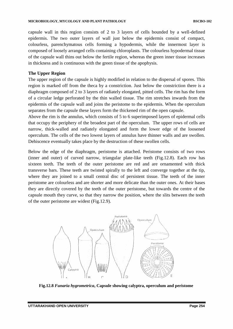
MICROBIOLOGY, MYCOLOGY AND PLANT PATHOLOGY BSCBO-102
UTTARAKHAND OPEN UNIVERSITY Page 254
capsule wall in this region consists of 2 to 3 layers of cells bounded by a well-defined
epidermis. The two outer layers of wall just below the epidermis consist of compact,
colourless, parenchymatous cells forming a hypodermis, while the innermost layer is
composed of loosely arranged cells containing chloroplasts. The colourless hypodermal tissue
of the capsule wall thins out below the fertile region, whereas the green inner tissue increases
in thickness and is continuous with the green tissue of the apophysis.
The Upper Region
The upper region of the capsule is highly modified in relation to the dispersal of spores. This
region is marked off from the theca by a constriction. Just below the constriction there is a
diaphragm composed of 2 to 3 layers of radiately elongated, pitted cells. The rim has the form
of a circular ledge perforated by the thin walled tissue. The rim stretches inwards from the
epidermis of the capsule wall and joins the peristome to the epidermis. When the operculum
separates from the capsule these layers form the thickened rim of the open capsule.
Above the rim is the annulus, which consists of 5 to 6 superimposed layers of epidermal cells
that occupy the periphery of the broadest part of the operculum. The upper rows of cells are
narrow, thick-walled and radiately elongated and form the lower edge of the loosened
operculum. The cells of the two lowest layers of annulus have thinner walls and are swollen.
Dehiscence eventually takes place by the destruction of these swollen cells.
Below the edge of the diaphragm, peristome is attached. Peristome consists of two rows
(inner and outer) of curved narrow, triangular plate-like teeth (Fig.12.8). Each row has
sixteen teeth. The teeth of the outer peristome are red and are ornamented with thick
transverse bars. These teeth are twisted spirally to the left and converge together at the tip,
where they are joined to a small central disc of persistent tissue. The teeth of the inner
peristome are colourless and are shorter and more delicate than the outer ones. At their bases
they are directly covered by the teeth of the outer peristome, but towards the centre of the
capsule mouth they curve, so that they narrow the position, where the slits between the teeth
of the outer peristome are widest (Fig.12.9).
Fig.12.8 Funaria hygrometrica, Capsule showing calyptra, operculum and peristome

MICROBIOLOGY, MYCOLOGY AND PLANT PATHOLOGY BSCBO-102
UTTARAKHAND OPEN UNIVERSITY Page 255
Fig.12.9: Funaria hygrometrica, Capsule showing calyptra, operculum and peris
The mouth of the capsule is closed by the operculum which consists of about three layers of
small thin-walled cells bounded by a superficial epidermal layer, with thickened outer walls.
Dehiscence of Capsule
At maturity the capsule begins to dry up, and the columella along with other thin walled
tissues looses water. They shrivel up, tearing open the space in which the spores lie. The
annulus proper, which consists of peculiarly elastic and hygroscopic cells, aids in the removal
of the operculum. When moisture is available the mucilaginous walls of cells of annulus
proper swell rapidly with the result that the annulus breaks free from the rim of the capsule
and suddenly rolls back, that results into throwing off the operculum. Thereafter, the
peristome teeth by their hygroscopic movements assist in emptying the spores from the
capsule. Each tooth of the outer peristome is a two ply structure and is composed of two
layers joined together, one forming the outer surface and the other the inner surface. The
outer layer of each tooth lengthens when wet and shortens when dry, but the inner layer is not
affected by changes in moisture content. When the outer peristome teeth absorb water, the
outer layer increase in length to a greater extent than the inner and they curve together
inwards and the dome is reconstituted, resulting into the opening of the capsule. The inner
peristome teeth do not show hygroscopic movements. In dry weather, the outer peristome
teeth loose water and their outer layers shorter more than the inner, with the result that they
bend outwards, with jerky movements as they separate from one another and the slits
between the outer teeth become wider, and the inner peristome teeth act as a sieve allowing
only gradual discharge of the spores, a few at a time (Fig.12.10).

MICROBIOLOGY, MYCOLOGY AND PLANT PATHOLOGY BSCBO-102
UTTARAKHAND OPEN UNIVERSITY Page 256
Fig.12.10 Funaria hygrometrica, Spore dispersal
The long slender seta of ripe capsule is wavy and twisted and also shows the hygroscopic
movements. When moistened the seta becomes untwisted, swinging round. As it dries up its
movements are reversed. Thus by the combined effort of the hygroscopic seta and peristome,
the spores are liberated in smaller amounts over a longer period.
The Young Gametophyte
The Spores
The spores are more or less spherical with a smooth surface. Spore diameter ranges from
0.012 to 0.020 mm. The spore has two wall layers, an inner hyaline endosporium; and a
coloured but smooth exosporium. The spore has single nucleus, numerous oil globules and
chloroplasts.
Spore Germination and Protonema formation
Under favourable conditions, spore begins to germinate within a few days. Heitz (1942)
found that they remain viable at least for two years. Upon absorbing moisture, spore increases
in size and the exospore bursts. The endospore protrudes out as one or two germ tubes,
partitioned by cross walls. These cells divide to form a branched, filamentous multi-cellular
structure, the primary protonema. The protonema grows apically by the divisions of the
apical cell. The filaments of protonema grows in two different directions and form two types
of branches. The branches that grow prostrate on the surface of the substratum and upright
branches the chloronemal branches (Fig.12.11).

MICROBIOLOGY, MYCOLOGY AND PLANT PATHOLOGY BSCBO-102
UTTARAKHAND OPEN UNIVERSITY Page 257
Fig.12.11. Funaria hygrometrica, Stages of Spore germination and protonema formation
The chloronemal branches are thick, with hyaline cell walls and which are divided by
transverse walls. The cells are chlorophyllous (many discoid small chloroplasts) thus
photosynthetic.
The rhizoidal branches are thin, with brown outer walls, and with oblique cross walls. The
cells either contain small chloroplasts or leucoplasts. The main function of these branches is
attachment, but in juvenile stage they also absorb water and minerals. These branches may
develop chlorophyll on exposure to light and transformed into chloronemal branches.
Sironval (1947) studied the development of the protonema in Funaria hygrometrica in
culture and opined that it consisted of two clear-cut stages, the chloronema and caulonema.
Accordingly, the chloronema is formed immediately on spore germination and differs from
the caulonema, which succeeds it, in morphological characters.
The chloronema has the following features: (i) it has sparse and irregular branching; (ii) the
cross walls are at right angles to the long axis of the protonemal thread; (iii) the cell walls are
colourless; (iv) numerous chloroplasts, circular in outline and evenly distributed; (v) nucleus
is complete, not visible in light microscope;(vi) positively geotropic; (vii) does not form
buds; (viii) it forms a thermolabile growth promoting substance.
According to Sironval the chloronema persists for 20 days thereafter most of its cells
degenerate, but certain apical cells persists and give rise to the second stage, the caulonema.
The caulonema has the following features: (i) it is profusely and regularly branched; (ii) the
cross walls are oblique to the long axis of the filaments;(iii) the cell walls are brownish; (iv)
the chloroplasts are fewer, spindle shaped and less evenly distributed; (v)the nucleus shows
degeneration of nucleolus and easily visible in light microscope;(vi) negatively geotropic;

MICROBIOLOGY, MYCOLOGY AND PLANT PATHOLOGY BSCBO-102
UTTARAKHAND OPEN UNIVERSITY Page 258
(vii) the buds of the gametophores are formed only on the branches of caulonema; (viii) in
caulonema, after about 10 days, a substance is formed which is thermostable and functions as
a growth inhibitor (Bopp, 1963).
Bud Formation
The bud arises as a lateral swelling from a cell of a parent filament, just behind a cross wall.
The main position of the bud is near the base of the principal filament of the aerial erect
branch system, but sometimes develop directly on the brown filaments. The lateral protrusion
after cutting off one or two stalk cells, swells at the end and divides by three oblique walls to
form the terminal tetrahedral cell of the future gametophores, with three cutting faces. It
begins to function as the apical cell of the young gametophores and cuts off three sets of
lateral segments. The first few segments of the apical cell do not form any leaf, but contribute
only to the base of the leafy gametophores; and rhizoids may also grow from this region
before any leaves are formed. Later on the segments give rise to the leaves and the tissue of
the stem. After the establishment of the leafy gametophores with several leaves and rhizoids,
the primary protonema withers and disappears and the several gametophores borne on a
protonema become independent.
Life Cycle of Funaria hygrometrica
In a single life cycle, two distinct vegetative individuals are seen.Such a life cycle is called
diplobiontic (Fig.12.12).One of the vegetative individual is the leafy shoot and the other is the
sporogonium. The green leafy shoot represents the dominant phase and is gametophytic in
nature It is related with sexual reproduction.This is the gametophytic or haploid (n) generation
of the Funaria plant. It begins with the formation of spores and ends with the gametes (egg
and antherozoids). The second individual is the sporophyte, which represents the sporophytic
or diploid (2n) generation. It begins with the formation of zygote. The zygote, the embryo and
the sporogonium( foot, seta and capsule) form the sporophytic generation.

MICROBIOLOGY, MYCOLOGY AND PLANT PATHOLOGY BSCBO-102
UTTARAKHAND OPEN UNIVERSITY Page 259
Fig.12.12.Graphic representation of the life cycle of Funaria hygrometrica
Alternation of generation
The alternating individuals in the life cycle are morphologically different. This kind of
alternation of generation is called heterologous or heteromorphic. The difference in two
generations is due to the different modes of life .The gametophyte is independent, while the
sporophyte is partially dependent on the gametophyte.(Fig.12.13)

MICROBIOLOGY, MYCOLOGY AND PLANT PATHOLOGY BSCBO-102
UTTARAKHAND OPEN UNIVERSITY Page 260
Fig.12.13 Diagrammatic representation of the life cycle of Funaria hygrometrica

MICROBIOLOGY, MYCOLOGY AND PLANT PATHOLOGY BSCBO-102
UTTARAKHAND OPEN UNIVERSITY Page 261
12.3.2. Polytrichum
12.3.2.1-Classification
Divison- Bryophyta
Class - Bryopsida (musci)
Order - Polytrichales
Family - Polytrichaceae
Genus - Polytrichum
Distribution: It is a large genus of about 100 species which are worldwide in distribution,
but mainly found in cool temperate and tropical regions. P. commune is a cosmopolitan
species. The common species of Polytrichum in India are P.densifolium, P. xanthofolium and
P.juniprerinum. Polytrichum is commonly called as hairy root moss.
Habitat
Polytrichum grows in a variety of habitats, such as sandy ground, on banks and heaths, dry
and stony places, dry woods, marshy moors, peat bogs, and damp soil along the margin of
lakes and ponds.
12.3.2.2- Structure
The Gametophyte
The gametophore of Polytrichum is differentiated into two regions, the horizontal
underground rhizome and the branched erect leafy stems. The aerial leafy stems may attain a
height up to 20 centimetres or more in some species of Polytrichum, for example P.
commune. (Fig.12.14)
Fig. 12.14. Polytrichum, gametophores (A) and foliage leaf (B)

MICROBIOLOGY, MYCOLOGY AND PLANT PATHOLOGY BSCBO-102
UTTARAKHAND OPEN UNIVERSITY Page 262
In P. commune between the rhizome portion and the upper leafy shoot, a transitional middle
region is present which is brown in colour, and three sided. At the base and on the lower side
of every 12th
leaf, bud initials are present throughout the axis. These initials along the leafy
shoot usually remain dormant but may grow if the tips of the main shoots are cut or if shoots
are kept in a moist atmosphere (Wigglesworth, 1959).
Leaves
The leaves on the rhizome and the middle transitional region are brown or colourless and
sealy. They are borne in three vertical rows with a divergence of 1/3. The leaves on the aerial
leafy shoot are relatively large, thick and rigid and are arranged on the stem in a complicated
spiral manner.
Each leaf has a broad, colourless membranous unistratose sheathing base, which narrows
above into a `limb‘ which is lanceolate to linear lanceolate. The limb is occupied mainly by a
broad midrib (costa) and the rudimentary wing .The wing is developed slightly at the extreme
margin of the limb which is entire to coarsely toothed. The midrib has close set rows of
longitudinal plates of green tissue (lamellae) of one cell thick on its upper surface. These
lamellae are attached to the surface of the midrib by one edge. As a result the midrib becomes
firm, opaque and dark green (Fig.12.14).
Rhiozoids
The numerous rhizoids are borne on the rhizome of the gametophores. The rhizoids are long,
thick-walled, multicellular and with oblique septa. The rhizoids coil round one another to
form dense tangled mass looking like a badly twisted string, as thick as half the diameter of
the rhizome, or sometimes more. The water besides passing through the lumen of the rhizoids
also passes upwards through the wick-like strands of rhizoids by external capillarity and,
therefore, many species of Polytrichum are able to live in relatively dry places. In species
which attain sufficient height, these strands of rhizoids provide mechanical support also.
Anatomy
The internal structure of the subterranean rhizome and the aerial leafy stem are different in
anatomical features.
The Rhizome
In transverse section (Fig.12.15) the rhizome is triangular with round corners and slightly
convex sides but may be roughly circular in outline, e.g. in P. formosum.
Epidermis: On the outside there is a superficial layer of thin walled or thick walled cells
bearing a large number of rhizoids which form a dense tangled mass.
Cortex: Below the superficial layer, there are two or three layers of living cortical
parenchyma.

MICROBIOLOGY, MYCOLOGY AND PLANT PATHOLOGY BSCBO-102
UTTARAKHAND OPEN UNIVERSITY Page 263
Fig.12.15 Polytrichum: T.S. of rhizome diagrammatic (A); A portion enlarged (B);
T.S. of aerial shoot diagrammatic (C)
Radial Strands
This tissue is interrupted opposite the three ridges by the sclerenchymatous cells of three
hypodermal strands .The cells of the hypodermal strands are distinctly prosenchymatous with
pointed ends. They are living cells sometimes containing starch. Passing radially inwards
from the hypodermal strands there are cells of greater diameter but with thinner and lignified
walls .They show no sharp line of demarcation from those of the hypodermal strands, and
form together with a wedge-shaped mass of tissue gradually narrowing inwards and are
called the radial strands.
Endodermis: At the inner limit of the cortex is a layer of radially elongated cells with
thickenings and suberization on their radial and horizontal walls. This layer resembles the
endodermis of roots, but is discontinuous and consists of three arcs separated by the larger
cells of the radial strands.
Pericycle: In most cases the three-lobed central strand is surrounded by an interrupted
pericycle composed of two to three layers of cells
Central Cylinder: The central cylinder consists of an inner compact mass of tissue with
three broad projecting regions and three narrower furrows opposite its angles.

MICROBIOLOGY, MYCOLOGY AND PLANT PATHOLOGY BSCBO-102
UTTARAKHAND OPEN UNIVERSITY Page 264
Stereom: The central mass consists mainly of very thick-walled elongated living cells often
with oblique end walls with little starch. These are steroids, as a whole being called stereom.
Hydrom: Scattered among these are elements of about same diameter, but quite destitute of
contents. These are often united in bands of 2 or 3, the cells of each band being separated by
extremely delicate cellulose walls; which are really the very oblique walls of the cells. For
these elements, which are exactly like and continuous with those of central strand of the
aerial stem, these named are hydroids, the water conducting tissue and as a whole being
called hydrom.
Leptoids: In the furrows, the centre of the bay is occupied by a group of 6 to 8 polygonal
cells, containing thick granular contents of protein nature. These sieve tube like cells are
known as leptoids, as a whole being called leptom (Tansley and Chick (1901).
Amylom: The leptoids are surrounded by starchy parenchyma which was named amylom.
The amylom is situated between hydrom and leptom.
Aerial Leafy Stem
The outline of the section is irregular. It shows the following structures (Fig.12.15 C).
Epidermis: The superficial layer is single layered but not clearly defined as an epidermal
layer.
Cortex: Cortex is well defined. The peripheral cortical region consists of compact elongated
prosenchymatous cells which gradually merge into the compact parechymatous cells of the
inner cortex. The cortical cells contain starch. In the cortical region the leaf traces are seen
passing through it to join the central cylinder.
Pericycle: Inside the inner cortex, a layer with abundant starchy contents represents the
rudimentary pericycle interrupted by incoming leaf traces.
Leptome: Internal to this is an irregular zone of typical sieve tube like cells, interrupted by
starchy cells, with protoplasm but no starch. This is the leptom mantle. This is equivalent to
the phloem of vascular plants.
Hydrom Sheath: Inside this is the hydrom sheath (amylom layer) composed of one or two
layers of cells with dark brown suberized walls and copious starchy content.
Hydrome Mantle: Inside the hydrom sheath is the hydrom mantle composed of thin-walled
cellswithout contents.
Water conducting tissue: In the centre of the stem is the hydrom cylinder composed of
thick walled cells. This tissue is concerned with the conduction of water and is equivalent to
the xylem of vascular plants physiologically.
The Leaf

MICROBIOLOGY, MYCOLOGY AND PLANT PATHOLOGY BSCBO-102
UTTARAKHAND OPEN UNIVERSITY Page 265
In the T.S. of the limb (Fig 12.16) the broad midrib is several cells thick in the centre and
gradually merges into the rudimentary wing (lamina) at the margins. On the lower surface
there is a well defined epidermis made up of large cells whose outer walls are strongly
thickened. Within the epidermis are one or two layers of small, sclerenchymatous elongated
cells, in which the lumen is almost obliterated.
Fig. 12.16 Polytrichum, V.T.S. of leaf (limb)
The central tissue of the leaf is formed by large thin-walled parenchymatous cells, among
which are scattered here and there small groups of sclerenchymatous cells. On the upper
surface there is layer of large cells from which arise numerous parallel thin, longitudinal
plates of chlorophyll containing cells, which represent the transverse sections of the
longitudinal plates or lamellae observed on the upper surface of the leaf in a surface view.
Each plate is 5 to 8 cells high and the terminal cell of each plate is wider or papillose, and
differs from rest of the cells. The slightly enlarged terminal cells of the adjoining plates
almost touch one another. The lamellar outgrowths of the midrib are the chief photosynthetic
tissue of the leaf, and they compensate for the reduction of the ordinary assimilatory tissue of
the reduced wing. These lamellae besides serving as organs of assimilation, also act in a
capillary manner and hold water between them, as they stand very close to each other.
In Polytrichum, the wing of the leaf usually consists of hyaline cells and in some species they
bend over the assimilatory portion of the leaf (midrib) when the air is dry. This feature is
marked in P. piliferum and P. juniperinum which grow in comparatively dry or exposed
places.
Conduction of Water
Bowen (1931, 1933) is of the opinion that the main water supply of P. commune and many
other mosses passes up` over the external surface of the plant in the form of a series of
capillary films between the closely adherent sheathing leaf bases and the stem. This water is

MICROBIOLOGY, MYCOLOGY AND PLANT PATHOLOGY BSCBO-102
UTTARAKHAND OPEN UNIVERSITY Page 266
then absorbed through the unthickened external walls which are generally to be found in the
cells aggregated at the apex of the plant and in the leaves and branches. The central
conducting strand in the stem takes very little part, if any, in the upward conduction of water.
On the other hand, Blaikley (1932) and Magdefrau (1935), however, hold that there is a high
rate of internal conduction of water through the central conducting strand.
12.3.2.3- Reproduction
Vegetative reproduction
Wigglesworth (1947) reported that the rhizoid system is important in vegetative reproduction.
These wick-like rhizoid strands bear buds which develop into leafy axes. In these buds, food
is stored in the form of starch to be used when required. Intact and unwounded leaves of P
commune may be induced to initiate growth leading to production of new and separate plants
when placed on suitable media, in sterile culture and under high humidity (Ward, 1960) (Fig.
12.17).
Fig.12.17 Polytrichum rhizoids with gemma
Sexual reproduction
Polytrichum is usually dioecious and the archegonia and antheridia are borne in terminal
clusters at the apex of separate gametophores.
Antheridial Head
The terminal part of the male shoot forms a conspicuous almost flower-like open cup. This
cup is formed by perigonial leaves which are different in form and colour from the other
vegetative leaves on the gametophores. These leaves are red-brown, or dull red in colour, and
each consists of a very broad sheathing base and a short bristle point (Fig.12.18).
The growth of the male shoot is not stopped by the development of the antheridia, as the
apical cell of the shoot is not used up in the formation of an antheridium. After the antheridia
have been formed and matured, the vegetative axis may grow out in the following year
through the antheridial group and produce a new shoot (proliferation) which may bear
antheridia at its apex. This proliferation of the vegetative shoot upwards through the

MICROBIOLOGY, MYCOLOGY AND PLANT PATHOLOGY BSCBO-102
UTTARAKHAND OPEN UNIVERSITY Page 267
antheridial head may be repeated several times and it gives the appearance of an axis through
a series of antheridial cups.
The antheridia are produced in groups at the base of each perigonial leaf in the position of
lateral buds. According to Leitgeb, each group of antheridia represents a shortened lateral
branch, and the whole anheridial head is a compound structure, in contrast to the simple male
head of Funaria.
Mature Antheridium
The mature antheridium consists of a short stalk and a club shaped body. The body has a
single-layered jacket surrounding a central mass of androcytes. Androcytes metamorphose
into biflagellate atherozoids at maturity. Associated with the antheridia there are paraphyses.
Some of these are simple filamentous, others are broadened at their tips into a spathulate
single-celled thick mass of cells (Fig.12.18).
Fig.12.18 Polytrichum male shoot (A); L.S. of male shoot showing antheridia (B); A
perigonial leaf (C); An antherozoid
The Archegonial Head
The archegonia are borne in terminal clusters at the apex of the leafy shoot of female
gametophore. In P. commune there are usually three archegonia in each cluster. The apical
cell of the leafy shoot itself forms the initial of an archegonium with the result that the further
growth in length of the shoot stops with the formation of a sporogonium (Fig.12.19). The
archegonium in structure it is more or less similar to that of Funaria.

MICROBIOLOGY, MYCOLOGY AND PLANT PATHOLOGY BSCBO-102
UTTARAKHAND OPEN UNIVERSITY Page 268
Fig.12.19, Polytrichum, (A) female shoot showing archegonia; (B) An argegonium
Fertilization
Is accomplished by the union of male (antherozoid) and female (egg) gametes with the aid of
water. The diploid zygote thus formed does not pass the resting period but undergoes several
divisions and forms the sporophytic plant-the sporogonium.
The Sporophyte
The mature sporogonium consists of a foot, a long seta and a capsule (Fig. 12.20).
Foot: The foot is embedded in the tissue of the apex of an archegonial branch and consists of
thin-walled parenchymatous cells.
Seta: Just above the foot and continuous with it is the long slender seta, which supports the
capsule at its apex. In T.S. of the seta, there is a superficial layer of thick-walled cells. Inside
it is a band of brown sclerenchymatous cells, which merges internally into thin-walled green
parenchymatous cells with intercellular spaces. The axial tissue of the seta forms a central
strand, without intercellular spaces and consists of cells essentially similar to those in the
gametophores axis, but usually of much simpler structure.
At the base of the capsule the seta enlarges to form the apophysis which is marked off from
the sporogenous portion (theca proper) by a groove. The apophysis has a distinct epidermis
with stomata followed by a mass of chlorophyllose tissue. The stomata are usually restricted
to the groove and are long pored and range in number from 20 in P. alpstre to nearly 200 in

MICROBIOLOGY, MYCOLOGY AND PLANT PATHOLOGY BSCBO-102
UTTARAKHAND OPEN UNIVERSITY Page 269
P. formosum. The stomata are large and are raised above the level of the epidermis. The
number of guard cells is usually two, but occasionally single annular guard cells are seen
(Paton, 1957).
Capsule: The capsule is angular and shows a polygonal outline in a cross section. The wall
of the capsule is composed of several layers of chlorophyllose cells, the outermost layer of
which is differentiated into an epidermis with thick external layer. Inside the wall there is an
outer space (air space) filled with filaments of chlorophyll-containing cells, in a radial
direction, which are connected internally with the outer wall of the spore sac. The spore sac is
limited internally by an inner lacuna which is again bridged over by filaments of cells
connecting the spore sac with the central columella (Fig.12. 21).
The spore sac is bounded on both the sides by two layers of thin walled cells. According to
the age of the sporogonium the archesporium consists of one layer (in young), or 4 to 6 layers
of cells (in adults). All cells of the sporogenous tissue give rise to spores. In the centre is the
columella.
Fig.12. 20, Polytrichum sporophytes

MICROBIOLOGY, MYCOLOGY AND PLANT PATHOLOGY BSCBO-102
UTTARAKHAND OPEN UNIVERSITY Page 270
Fig.12.21, Polytrichum: (A) L.S of sporophyte; (B) T.S. of capsule and (C)Peristome teeth
Operculum: At the top of the capsule there is an operculum which appears as a conical lid
with a beak or rostrum. A distinct annulus is not present in Polytrichum though the thickened
diaphragm (rim) is present.
Epiphragm: At the base of the operculum there is a transparent membranous tissue
composed of pale compressed cells without intercellular spaces. This tissue stretches like a
drum- head (tympanum) over the opening of the capsule and closes it. This is the epiphragm.
Peristome: The peristome consists of bundles of mostly curved crescent-shaped thickened
fibrous cells. The ends of the crescent are directed upwards and join the ends of the adjacent
horse shoe-shaped bundles. At maturity the peristome is composed of 32 or 64 short

MICROBIOLOGY, MYCOLOGY AND PLANT PATHOLOGY BSCBO-102
UTTARAKHAND OPEN UNIVERSITY Page 271
pyramidal teeth, connected below with the wall of the capsule, and united above with the
margin of the epiphragm by their tips (Fig 12.21. C).
Dispersal of spores: the capsule matures, operculum goes off and the cells beneath may dry
up. By drying up of the thin walled cells between the teeth, small openings are formed in the
margin of the epiphragm through which the small spores sift out, a few at a time, as the
capsule sways by the forces of wind (censer mechanism).
Like Funaria, the calyptra is ruptured by the continuous growth of the sporogonium, and the
upper part is separated as a cap. It is carried upwards on the top of the capsule forming a dry
fibrous hood covering the apex of the capsule. The calyptra consists externally of dry,
branched hypha-like filaments loosely mated together.
The Gametophyte
The Spores: The spores represent the gametophytic phase. They are uninucleate small in size
ranging from 0.005 to 0.01 millimetres in diameter. They remain viable for a long time. The
spores have two layers, outer exospore and inner smooth endospore. Spores start germination
under favourable conditions.
Germination of Spore and Formation of Protonema
Wigglesworth (1947) observed the spore germination in P. commune. The yellow spore soon
turns green due to the formation of chloroplasts (Fig.12. 22). The exospore ruptures and the
spore protoplast bound by endospore protrudes as one or more germ tubes. The germ tube
enlarges rapidly and soon gives rise to separate branched filamentous protonema by repeated
transverse divisions. The growth of the filament is apical. Some filaments grow upwards and
turn green while others penetrate the substratum remaining colourless. These latter filaments
are of small diameter and with cross wall oblique to the axis. Later on buds appear near the
base of the upright filaments. Eventually a pyramidal apical cell of the future shoot is
differentiated, which cut off segments and an axis bearing leaves is formed. This young shoot
bears two kinds of rhizoids at the base. The mature type of leaves does not appear on the
young plant until the third year.

MICROBIOLOGY, MYCOLOGY AND PLANT PATHOLOGY BSCBO-102
UTTARAKHAND OPEN UNIVERSITY Page 272
Fig. 12.22. Polytrichum stages of germination of spores
Life cycle of Polytrichum
Like Funaria, the life cycle of Polytrichum also exhibits, two distinct vegetative individuals in
a single life cycle. Such a life cycle is called diplobiontic (Fig.12.23). One of the vegetative
individual is the leafy shoot and the other is the sporogonium. The green leafy shoot represents
the dominant phase and is gametophytic in nature It is related with sexual reproduction. This is
the gametophytic or haploid (n) generation of the plant. It begins with the formation of spores
and ends with the gametes (egg and antherozoids). The second individual is the sporophyte,
which represents the sporophytic or diploid (2n) generation. It begins with the formation of
zygote. The zygote, the embryo and the sporogonium (foot, seta and capsule) form the
different phases of sporophytic generation.
Fig. 12.23. Diagrammatic representation of the life cycle of Polytrichum
Alternation of generation

MICROBIOLOGY, MYCOLOGY AND PLANT PATHOLOGY BSCBO-102
UTTARAKHAND OPEN UNIVERSITY Page 273
The alternating individuals in the life cycle are morphologically different. This kind of
alternation of generation is called heterologous or heteromorphic. The difference in two
generations is due to the different modes of life .The gametophyte is independent, while the
sporophyte is partially dependent on the gametophyte (Fig.12.24).
Fig.12.24, Graphic representation of the life cycle of Polytrichum
12.4 SUMMARY
(A) Life cycle of Funaria
A. Gametophyte.
1. It consists of (a) a branched, green, alga-like filament, the protonema and (b)
a leafy gametophores, the moss plant. The protonema is short lived.

MICROBIOLOGY, MYCOLOGY AND PLANT PATHOLOGY BSCBO-102
UTTARAKHAND OPEN UNIVERSITY Page 274
2. The leafy gametophore consists of a slender, stem-like central axis bearing
rhizoids at its base and leaf like expansions throughout its length. It is independent.
3. The so called leaf is traversed by a definite midrib more than one cell in
thickness.
4. The rhizoids are branched, septate and multicellular. The septa between the
cells are obliquely placed.
5. The sex organs (antheridia and archegonia) are borne in terminal clusters at
the tips of separate branches of the leafy gametophores.
6. The mature antheridium consists of a short multicellular stalk and the globose
body.The body has a one celled thick jacket wall. Within the jacket, androcytes are
present which give rise to biflagellate sperms. The mature archegonium is flask
shaped borne on a short multicellular stalk. The neck is long with six rows of neck
cells. They enclose 6 or more neck canal cells, the venter wall is two layer thick and
has a vetral canal cell and an egg.
7. The antherozoids after liberation from the antheridium splashed out of the
perichaetial leaves by rain drops on the arcegonial cluster and swim down to the
open necks of arhegonia and fertilize the egg.
B. Sporophyte
1. The zygote represents the first cell of sporophyte and diploid in nature. The
zygote divides and forms an embryo with two growing points at each end.
2. The growth of the embryo is apical and by cutting off the segments it
becomes a long slender structure.
3. The mature sporogonium is differentiated into a foot, a seta and a pear-
shaped capsule of great complexity.
4. The seta is long and carries the capsule far above the leafy gametophores.
5. The foot is embedded in the tissues of female branch and functions as an
anchoring and absorbing organ.
6. The mature capsule is differentiated externally into apophysis, theca and
operculum regions.
7. The apophysis is a swollen sterile basal portion. It is associated with
photosynthesis and conduction. It has well defined epidermis with functional stomata
connected with substomatal air space.
8. The theca has centrally located thin walled sterile parenchyma cells, the
columella. On either side of columella spore sac is located which does not arch over
the columella. Outside the spore sac, there is a wide air space traversed by green
filaments, the trabeculae. The spore sac contains the spore mother cells which
undergo meiotic division to form haploid spores. Outside the air space is the wall of
the theca, It is made up of one to two layer thick spongy layer followed by two layers
thick hypodermis which is bounded by a single layered epidermis.
9. The apical portion above the theca is operculum. A transverse ring of modified
epidermal cells separates the theca with operculum. This is called annulus. Below the

MICROBIOLOGY, MYCOLOGY AND PLANT PATHOLOGY BSCBO-102
UTTARAKHAND OPEN UNIVERSITY Page 275
operculum a double row of conical teeth is present which collectively form the
peristome. In each row 16 teeth are present. The teeth are hygroscopic and they play
an important role in spore dispersal.
(B) Life cycle of Polytrichum
A. Gametophyte.
1. It is a perennial plant. It consists of (a) a short lived, branched, green
protonema and (b) leafy gametophores.
2. The leafy gametophore arises from an underground rhizome. The gametophore
has a slender, stem-like central axis bearing leaf like expansions throughout
its length. It is independent.
3. Rhizoids arise from the rhizome and often entangle with each other and form
wick like structure. These rhizoids help in retention in water and give
mechanical strength to the plant. The rhizoids are branched, septate and
multicellular. The septa are oblique.
4. The rhizoid system is important in vegetative reproduction. These wick-like
rhizoid strands bear buds which develop into leafy axis.
5. The leaves on the rhizome and the middle transitional region are brown or
colourless .They are borne in three vertical rows with a divergence of 1/3.
6. The leaves on the aerial leafy shoot are relatively large, thick and rigid and
are arranged on the stem in a complicated spiral. Each leaf has a broad,
colourless membranous unistratose sheathing base, which narrows above into
a `limb‘ which is lanceolate to linear lanceolate
7. In Transverse section the rhizome is triangular with round corners and slightly
convex. The conducting tissue is advanced containing water conducting
(hydroids), and food conducting (leptoids) tissues.
8. Plant is dioecious and the archegonia and antheridia are borne in terminal
clusters at the apex of separate gametophores.
9. The terminal part of the male shoot forms a conspicuous almost flower-like
open cup. This cup is formed by perigonial leaves which are different in form
and colour from the other vegetative leaves on the gametophores The growth
of the male shoot is not stopped by the development of the antheridia, as the
apical cell of the shoot is not used up in the formation of an antheridium.
10. The mature antheridium consists of a short stalk and a club –shaped body. The
body has a single-layered jacket surrounding a central mass of androcytes
11. The archegonia are borne in terminal clusters at the apex of the leafy shoot.
The apical cell of the leafy shoot itself forms the initial of an archegonium
with the result that the further growth in length of the shoot stops with the
formation of a sporogonium.
B. Sporophyte

MICROBIOLOGY, MYCOLOGY AND PLANT PATHOLOGY BSCBO-102
UTTARAKHAND OPEN UNIVERSITY Page 276
1. The mature sporogonium consists of a foot, a long seta and a capsule.
2. The foot is embedded in the tissue of the apex of an archegonial branch and
consists of thin-walled parenchymatous cells.
3. Seta is continuous with foot andt is long and slender, which supports the
capsule at its apex.
4. At the base of the capsule the seta enlarges to form the apophysis which is
marked off from the sporogenous portion (theca proper) by a groove.
5. The capsule is angular and shows a polygonal outline in a cross section
6. The apophysis has a distinct epidermis with stomata followed towered by
mass of chlorophyllose tissue.
7. The spore sac with the central columella is present in the ca region.
According to the age of the sporogonium the archesporium consists of one
layer (in young), or 4 to 6 layers of cells (in adults) .
8. All cells of the sporogenous tissue give rise to spores.
9. At the top of the capsule there is an operculum which appears as a conical
lid with a beak or rostrum.
10. At the base of the operculum there is a transparent membranous tissue
composed of pale compressed cells without intercellular spaces. This is the
epiphragm.
11. At maturity the peristome is composed of 32 or 64 short pyramidal teeth,
connected below with the wall of the capsule, and united above with the
margin of the epiphragm by their tips.
12. The capsule matures, operculum goes off and the cells beneath may dry up.
13. By drying up small openings are formed in the margin of the epiphragm
through which the small spores sift out, a few at a time, as the capsule sways
by the forces of wind( censer mechanism
14. The spore represents the gametophytic phase. They are uninucleate small in
size.
15. The spore germinates and gives rise to filamentous protonema the
gametophyte.
16. Buds appear near the base of upright filament of protonema, leafy
gametophores develops from the buds.
12.5 GLOSSARY
Abaxial surface: The side of the lateral organ (leaf), away from the axis (dorsal).
Acrocarpus: Of mosses, bearing the archegonia at the tip off stem or main branches.
Acropetal: Development of organs towards the apex, the oldest at the base, youngest towards
the apex.
Adaxial: The side of the lateral organ (leaf), nearest to the main axis (ventral).
Androcyte: Antherozoid mother cell.
Antheridium: The male sex organ of the cryptogams.

MICROBIOLOGY, MYCOLOGY AND PLANT PATHOLOGY BSCBO-102
UTTARAKHAND OPEN UNIVERSITY Page 277
Antherozoid: Small, motile male gamete with flagella.
Anticlinal: Perpendicular to the surface.
Apophysis: The swollen sterile tissue at the base of capsule, of some mosses, next to seta and
below the spore sac.
Archegonium: The female sex organs of bryophytes.
Archesporium: Sporogenous tissue or group of cells from which the spores ultimately
derived.
Basipetal: Development of organs in which the youngest structures are at the base and the
oldest at the apex.
Calyptra: A covering developed from the venter of the archegonium in bryophytes.
Capsule: The part of the sporogonium containing spores.
Chlorophyllose: Containing chlorophyll.
Cortex: The region of a vascular plant that lies between the epidermis and the stele.
Diploid generation: The sporophyte.
Dorsiventral: An organ having distinct dorsal and ventral surfaces.
Elaters: Hygroscopic twisted structures helping to disperse spores.
Epibasal: Forming the upper part of the embryo.
Epidermis: The outer most layer of cells of a plant body.
Epiphragm: A membrane which closes the opening of the theca in some mosses, e,g.,
Polytrichum.
Epiphyllous: Growing on leaves.
Epiphyte: A plant growing on another plant.
Exine: The outer layer of the cell wall of spore.
Gametophyte: The haploid or gamete producing generation of any plant.
Gemma: Organ of vegetative reproduction, consisting of one to many cells, that becomes
detached from parent plant and develops into a new plant.
Germ tube: The first tubular outgrowth from a germinating spore.
Guard cells: Specialized chlorophyllous cells which bound the stoma and regulate its
opening.
Habit: General form and aspect of a plant.
Habitat: The place where an organism lives.
Haploid generation: Gametophyte generation.
Haustorium: The absorbing organ of a parasite.
Homosporous: Producing one kind of spores.
Hygrophilous: Plants which need a large amount of moisture for their growth.
Hypobasal: Forming the lower part of the embryo.
Peristome: The ring of hygroscopic teeth round the mouth of dehiscent capsule in mosses,
concerned with spore dispersal.
Protonema: The early filamentous stage produced on germination of spore in some
bryophytes.
Perigonial leaves or bracts: Leaves surrounding the antheridia.
Perichaetial leaves or bracts: Leaves surrounding the archegonia.

MICROBIOLOGY, MYCOLOGY AND PLANT PATHOLOGY BSCBO-102
UTTARAKHAND OPEN UNIVERSITY Page 278
12.6 SELF ASSESSMENT QUESTIONS
12.6.1 Fill in the blanks:
(i) Rhizoids in Polytrichum are multicellular, branched and-------------.
(ii) The number of peristome teeth in Polytrichum is--------.
(iii)Polytrichum is commonly called---------.
(iv) The horizontal underground part of the gametophores of Polytrichum is called-------.
(v) The rhizoids are borne on the -------of the gametophores.
12.6.2 Multiple choice Questions:
1. Plants of Funaria generally grow
(a) In pairs (b) Solitary
(c) In aggregates (d) As saprophyte
2. The middle sterile part of Funaria capsule is known as
(a) Columella (b) Apophysis
(c) Operculum (d) Spore sac
3. In Funaria the leaves are arranged on the stem
(a) Oppositely (b) Spirally
(c) Alternately (d) All of the above
4. Which part of the Funaria capsule is haploid
(a) Annulus (b) Columella
(c) Operculum (d) Calyptra
5. Leptome mantle in Polytrichum is equivalent to:
(a) Rudimentary pericycle (b) Starchy layer
(c) phloem (d) xylem
6. Number of peristome teeth in Polytrichum are
(a) 8-16 (b) 32-64
(c) 72-128 (d) more then 128
7. Hair cap moss is the name given to
(a) Polytrichum (b)Pogonatum
(c) Funaria (d) None of the above
8. In Polytrichum the spore liberation is regulated by

MICROBIOLOGY, MYCOLOGY AND PLANT PATHOLOGY BSCBO-102
UTTARAKHAND OPEN UNIVERSITY Page 279
(a) Operculum (b) Columella
(c) Peristome (d) Annulus
9. In Polytrichum the leaves are arranged in
(a) Two rows (b) Three rows
(c) Four rows (d) Six rows
12.6.2 Answer keys:
1.(c), 2. (a), 3.(b), 4. (d), 5. (c), 6.(b), 7.(a), 8. (c), 9.(b)
12.7 REFERENCES
Allsopp, A. and Mitra, G. C. (1958). The morphology of protonema and bud formation in
the Bryales. Ann. Bot.N.S. 22: 95-115
Berkley, E.E (1941). Gemmae of Funaria hygrometrica. Trans. Illinois State Acad. Sci.
24:102-104
Blaikley, N.M. (1932). Absorption and conduction of water and transpiration in
Polytrichum commune Ann. Bot. 46:289-300.
Bowen, E.J (1932).The mechanism of water conduction in the Musci, considered in
relation to habitats. Ann. Bot. 47: 401-422; 889-912.1933.
Bowen, E.J. (1931). Water conduction in Polytrichum commune Ann. Bot. 45:175-200.
Parihar, N.S. (1955). An introduction to embryophyta (vol.I) BRYOPHYTA. Central
Book Depot, Allahabad. Pp 377.
Paton, J.A. (1957). The occurrence, structure and function of the stomata in British
Bryophytes I. Occurrence and structure. Trans. Brit. Bryol. Soc. 3: 228 -242.
Sironval, C. (1947). Experiences sur les stades de developmente de la forme filamentuse
en culture de Funaria hygrometrica. Bull. Soc. Royl. Bot. Belgique. 79: 48-72.
Tansley, A.G. and Chick, E. (1901). Notes on the conducting tissue system in Bryophyta.
Ann. Bot. 15: 1-38.
Ward, M. (1960). Vegetative propagation from intact leaves of Polytrichum commune.
Phytomorphology. 10: 325 -329.
Wigglesworth, G. (1947). Reproduction in Polytrichum commune. Trans. Brit. Bryol.
Soc. 1:4-13.
Wigglesworth, G. (1956). Further notes on Polytrichum commune. Trans. Brit. Bryol.
Soc. 3: 115- 120.
12.8 SUGGESTED READINGS
College Botany .Volume I By B.P.Pandey. 2010 Pp.221. S.Chand & Company Ltd. New
Delhi

MICROBIOLOGY, MYCOLOGY AND PLANT PATHOLOGY BSCBO-102
UTTARAKHAND OPEN UNIVERSITY Page 280
Botany for Degree Students BRYOPHYTA By B.R.Vashistha, Dr.A.K.Sinha & DR.
Adarsh Kumar. 2014 Pp.517.S.Chand & Company Ltd. New Delhi
A Text Book of Botany By V. Singh, P.C. Pande & D.K.Jain.2013. Rastogi Publications
Meerut.
A Text Book of Botany Volume II By S.N. Pandey.P.S.Trivedi & S.P.Misra, 2004. Vikas
Publishing House Pvt. Ltd. New Delhi
A Text Book of Botany Bryophyta By N.S. Parihar 2006. Pp.327 Central Book Depot.
Allahabad .
12.9 TERMINAL QUESTIONS
1. Give an illustrated account of the sporophyte of Funaria or Polytrichum.
2. Draw the labelled Diagrams of the following:
(i) L.S. of Funaria capsule (ii) T.S. of Rhizome of Polytrichum.
3. Describe the Structure of the leaf and the axis of Polytrichum.
4. Write Short notes on:
(i) Salient features of Bryopsida (ii) Sex organs of Funaria (iii) Vegetative Reproduction in
Funaria (iv) Rhizoids
5. Describe the life cycle of Funaria or Polytrichum by making suitable diagrams.