Isolation and characterization of chitinase genes from pitchers ...
Upload
independentCategory
view
0download
0
Segmental Allergen Challenge Enhances Chitinase Activity andLevels of CCL18 in Mild Atopic Asthma
Monica L Gavala1, Elizabeth A. B. Kelly2, Stephane Esnault2, Sandeep Kukreja2, Michael DEvans3, Paul J Bertics1,*, Geoffrey L Chupp4, and Nizar N Jarjour2
1Department of Biomolecular Chemistry, University of Wisconsin School of Medicine and PublicHealth, Madison, WI 53792, USA2Allergy, Pulmonary and Critical Care Medicine, Department of Medicine, University of WisconsinSchool of Medicine and Public Health, Madison, WI 53792, USA3Biostatistics and Medical Informatics, University of Wisconsin School of Medicine and PublicHealth, Madison, WI 53792, USA4Section of Pulmonary and Critical Care Medicine, Yale University School of Medicine, NewHaven, CT, USA
AbstractBackground—Allergic airway inflammation contributes to the airway remodeling that has beenlinked to increased obstruction and morbidity in asthma. However, the mechanisms by whichallergens contribute to airway remodeling in humans are not fully established. CCL18,chitotriosidase (CHIT1) and YKL-40 are readily detectable in the lungs and contribute toremodeling in other fibrotic diseases, but their involvement in allergic asthma is unclear.
Objective—We hypothesized that CCL18, YKL-40, and CHIT1 bioactivity are enhanced inallergic asthma subjects after segmental allergen challenge and are related to increased profibroticand Th2-associated mediators in the lungs.
Methods—Levels of CCL18 and YKL-40 protein and CHIT1 bioactivity in bronchoalveolarlavage (BAL) fluid, as well as CCL18, YKL-40, and CHIT1 mRNA levels in BAL cells, wereevaluated in patients with asthma at baseline and 48 h after segmental allergen challenge. We alsoexamined the correlation between CCL18 and YKL-40 levels and CHIT1 activity with the levelsof other profibrotic factors and chemokines previously shown to be up-regulated after allergenchallenge.
Results—CHIT1 activity and YKL-40 and CCL18 levels were elevated after segmental allergenchallenge and these levels correlated with those of other profibrotic factors, T cell chemokines,and inflammatory cells after allergen challenge. CCL18 and YKL-40 mRNA levels also increasedin BAL cells after allergen challenge.
Corresponding author: Nizar N Jarjour, Allergy, Pulmonary and Critical Care Medicine, University of Wisconsin School of Medicineand Public Health, 600 Highland Ave, CSC K4/914, Madison, WI 53792-9988 Phone: 608-263-9344; FAX: (608) 263-3746;[email protected].*Deceased
The authors have no conflicts of interest to report.
Authors’ contributions to the study: All authors contributed to the conception and design of the study, interpretation of the data, andrevision of the manuscript and provided final approval of the submitted version. NNJ, EABK, PJB, and GLC were responsible forobtaining funding. MLG drafted the manuscript. EABK, SE, JDP, SK, and MLG contributed to data acquisition, analysis, andinterpretation.
NIH Public AccessAuthor ManuscriptClin Exp Allergy. Author manuscript; available in PMC 2013 April 11.
Published in final edited form as:Clin Exp Allergy. 2013 February ; 43(2): 187–197. doi:10.1111/cea.12032.
NIH
-PA Author Manuscript
NIH
-PA Author Manuscript
NIH
-PA Author Manuscript

Conclusions & Clinical Relevance—Our results suggest that CCL18 and YKL-40 levels andCHIT1 activity are enhanced in allergic airway inflammation and thus may contribute to airwayremodeling in asthma.
Keywordsasthma; segmental bronchoprovocation; CHIT1; YKL-40; CCL18
INTRODUCTIONAsthma is marked by airway obstruction, hyperresponsiveness, inflammation, andremodeling [1, 2]. Allergen exposure up-regulates the adaptive and innate immuneresponses, leading to the production of proinflammatory and profibrotic factors that mayultimately contribute to airway remodeling [2–4]. However, the relationship betweeninflammatory mediators and the observed structural changes seen in asthma is still unclear.
Chitinases have been linked to fibrotic diseases and Th2-mediated inflammation and areassociated with asthma severity. Human chitinases, such as acidic mammalian chitinase(AMcase) and chitotriosidase (CHIT1), are evolutionarily conserved glycoproteins thatcleave polysaccharide chitin found in several lower life forms including fungi, parasites, andcrustaceans [5, 6]. Chitinase-like proteins, such as YKL-40, are structurally similar proteinsthat do not possess chitinase activity. Both chitinases and chitinase-like proteins havediverse immunological functions especially within the realm of Th2-mediated inflammation[7]. Increased YKL-40 levels in plasma and its presence in the lungs correlate with diseaseseverity and degree of airway remodeling in asthma [8, 9]. Furthermore, polymorphisms inCHI3L1, the gene encoding YKL-40, have been associated with asthma [10]. Of the twoknown active human chitinases, CHIT1 is the most predominant in the lungs and its activityhas been linked to other pulmonary disorders such as chronic obstructive pulmonary disease(COPD) and sarcoidosis [11, 12]. An association between atopy and CHIT1 activity isbeginning to emerge [13, 14], but it is unclear how allergens affect the activity of CHIT1 inhuman lungs.
There have been links between CHIT-1 and the expression of a chemokine stronglyexpressed in the lungs, Pulmonary and Activated Regulated Chemokine (PARC or CCL18),in the context of sarcoidosis and Gaucher’s disease [12, 15]. Increased levels of CCL18 havebeen reported in fibrotic lung diseases, including hypersensitivity pneumonitis, sarcoidosis,and idiopathic pulmonary fibrosis, with many of these studies implicating alveolarmacrophages as the primary source of this chemokine [16–19]. CCL18 mRNA has beenreported to be increased in the sputum of subjects with asthma compared to atopic and non-atopic controls [20]. Furthermore, basal CCL18 concentrations were higher in the serum andBAL fluid of allergic asthma patients who were untreated compared to those who eitherreceived inhaled corticosteroids or were non-allergic control subjects [21]. In a primatemodel, CCL18 mRNA expression in the lungs was increased after allergen inhalation [22],supporting a role for antigen-mediated up-regulation of CCL18. Furthermore, CCL18expression can be induced in peripheral blood mononuclear cells (PBMCs) by allergenexposure ex vivo [21]. However, it has not been determined how in vivo exposure toallergen affects airway expression of CCL18 in asthma.
There is emerging evidence for a role of CCL18 and chitinases in asthma pathophysiology.Given evidence of their involvement in allergic asthma individually, the objective of thisstudy was to elucidate the relationship between CHIT1 activity, YKL-40 and CCL18 levels,in the context of Th2-mediated inflammation and factors associated with remodeling, which
Gavala et al. Page 2
Clin Exp Allergy. Author manuscript; available in PMC 2013 April 11.
NIH
-PA Author Manuscript
NIH
-PA Author Manuscript
NIH
-PA Author Manuscript

was investigated using an established model of segmental airway allergen challenge inallergic asthmatic patients. (PROOFED)
METHODSSubjects
BAL fluid analysis was performed on 18 atopic subjects (skin prick test positive) with ahistory of mild asthma (median FEV1 was 97% of predicted and median methacholine PC20was 5.7 mg/ml), including 9 males and 9 females (ages 19 to 32) (See Table 1). Analysis ofBAL cell mRNA was performed on an additional group of four females and three males(ages 19–27) with a history of mild allergic asthma (mean ± SEM for FEV1 was 92 ± 4% ofpredicted and methacholine PC20 was 7.6 ± 2.9 mg/ml). All subjects were non-smokers, didnot have a respiratory infection or asthma exacerbations within 30 days of the study, and hadnot received long-acting β-agonists within 2 days, antihistamines or leukotriene antagonistswithin 7 days, or corticosteroids within 30 days of study enrollment. The University ofWisconsin-Madison Health Sciences Human Subjects Committee approved the study, andinformed consent was obtained from all subjects.
Segmental Bronchoprovocation with Antigen (SBP-Ag) and BALSBP-Ag and BAL (D0) were performed in two different bronchopulmonary segments aspreviously described [23]. Allergens used in SBP-Ag were cat antigen (Fel d1, BayerAllergy Products, Spokane, WA, USA), ragweed (GS Ragweed mix, Greer Labs, Lenoir,NC, USA) and house dust mite (HDM, Dermatophagoides farinae, Miles Allergy Products,Spokane, WA, USA) based on each subject’s skin test reactivity and correlative history. Theallergen dose for SBP-Ag was based on the subject’s physiological response to a previousinhaled allergen challenge (i.e., the provocative dose of allergen required to achieve a 20%fall in FEV1 (AgPD20)). A total dose of 30% of the subject’s AgPD20 was administeredincrementally to insure subject’s safety: 10% of the AgPD20 in the first segment and, whenthis dose was well tolerated, 20% in the second segment. A second bronchoscopy with BAL(D2) was performed 48 h later. A total of 160 ml of sterile 0.9% NaCl warmed to 37°C wasinstilled in each segment in 4 aliquots. BAL fluid recovered from the two segments waspooled for analysis.
Analysis of BAL fluidCells were recovered by centrifugation and total and differential cell counts were performed[24]. BAL fluid was kept on ice throughout processing and stored at −80°C before analysis.
CHIT1 activity was measured utilizing the fluorogenic substrate 4-methylumbelliferyl-β-D-N,N′,N″-triacetylchitotrioside (Sigma-Aldrich, St. Louis, MO, USA) in McIlvain buffer(100 mmol/L citric acid and 200 mmol/L sodium phosphate, pH 7.0). This assay wasperformed under optimal conditions for CHIT1, versus AMCase (whose optimal enzymaticactivity is at pH of 4.0 [11]). Substrate was incubated with BAL fluid for 15 minutes at37°C. The reaction was stopped with 2 ml of 0.3 mol/L glycine-NaOH buffer (pH 10.6). Thesubstrate hydrolysis product 4-methylumbelliferone was quantified with a fluorometer usingan excitation wavelength of 366 nm and an emission wavelength of 446 nm, and the resultswere compared to a standard 4-methylumbelliferone calibration curve. CHIT1 activity isexpressed as nanomoles of substrate hydrolyzed per hour per milliliter of BAL fluid (nmol/ml/h). Thrombin activity was measured utilizing the chromogenic substrate H-D-hexahydrotyrosol-alanyl-arginine-para-nitroanilide diacetate (American Diagnostica,Greenwich, CT, USA) as previously described [25]. Thrombin activity is expressed asNational Institutes of Health (NIH) U/ml of 1x BAL fluid.
Gavala et al. Page 3
Clin Exp Allergy. Author manuscript; available in PMC 2013 April 11.
NIH
-PA Author Manuscript
NIH
-PA Author Manuscript
NIH
-PA Author Manuscript

“In-house” sandwich ELISAs were utilized as previously described [26] with the followingmodifications. After thawing, 1 ml aliquots of BAL fluid in Eppendorf tubes werecentrifuged at 5,000 x g for 5 min to remove any precipitates or debris. BAL fluid wasdiluted in LowCross-Buffer (Boca Scientific, Boca Raton, FL, USA) to reduce nonspecificlow-affinity binding. Four serial dilutions of each BAL fluid were analyzed on the sameplate and the dilutions that resulted in identical calculated values were averaged for theabsolute concentration. The antibodies (ab) used for YKL-40 include a mouse monoclonalcapture ab and a biotinylated goat polyclonal detection ab from an R&D Systems Duoset kit(R&D Systems, Minneapolis, MN, USA). The capture ab for CCL18 (clone 64507),interferon-induced protein of 10kDa (IP-10)/CXCL10 (clone 33036.211), matrixmetalloproteinase (MMP) 9 (clone 36020), and thymus and activation-regulated chemokine(TARC)/CCL17 (clone 54026) were all mouse monoclonal ab from R&D Systems. Thedetection ab were biotinylated goat polyclonal detection ab from R&D Systems. Ab forfibronectin included mouse monoclonal capture ab, clone 1601 (Biodesign International,Saco, ME, USA), and a horseradish peroxidase (HRP)-conjugated rabbit polyclonaldetection ab (DakoCytomation, Carpinteria, CA, USA). Insulin-like growth factor bindingprotein-3 (IGFBP-3) was measured using a commercial kit from R&D Systems. Theminimum detection limits were as follows: FN ≥ 30 ng/ml, CCL18 and YKL-40 ≥ 30 pg/ml,IP-10 and TARC ≥ 12 pg/ml, and MMP9 ≥ 3 pg/ml.
Real-time PCR of BAL cellsTotal RNA was extracted from BAL cells isolated from BAL fluid before (D0) and 48 hafter SBP-Ag (D2) using the RNeasy Mini kit (Qiagen, Valencia, CA, USA). The reversetranscription reaction was performed using the Superscript III system (Invitrogen/LifeTechnologies, Carlsbad, CA, USA). YKL-40 (forward: attgacagcagctatgacattgc, reverse:aaatcgtaggtcatgatgctaatgaa), CCL18 (forward: ccccaagccaggtgtcatc, reverse:tgtatttctggacccacttcttattg), CHIT1 (forward: ggcctgggcaggtttcat, reverse:tgaagtagcagaccagttttgca), IP-10 (forward: gatttgctgccttatctttctgact, reverse:caggtacagcgtacagttctagagaga), and TARC (forward: agcacatccacgcagctcga, reverse:tgaagtactccaggcagcactccc) specific primers were designed with Primer Express 3.0 (AppliedBiosystems/Life Technologies, Carlsbad, CA, USA) and blasted against the human genometo determine specificity using: http://www.ncbi.nlm.nih.gov/tools/primer-blast. Expressionswere determined by quantitative real-time PCR (qPCR) using SYBR Green Master Mix(SABiosciences/Qiagen, Frederick, MD, USA). The housekeeping g e n e β-glucuronidase(GUSB) (forward: caggacctgcgcacaagag, reverse: tcgcacagctggggtaag) was used tonormalize the samples. Standard curves were performed and efficiency was determined foreach set of primers. Efficiency was typically above 94%. The data are expressed as foldchange using the comparative cycle threshold (ΔΔCT) method (ΔCt = Ct gene − Ct GUSBand ΔΔCt = ΔCt of BAL cells at D0 − ΔCt of BAL cells at D2). Values shown are foldchange 48 h after SBP-Ag (2−ΔΔCt) with the level in BAL cells before challenge fixed at 1.
Statistical AnalysisBaseline and post-SBP-Ag challenge measurements were compared using the paired t-test.Measurements were log-transformed where appropriate. Associations among chemokinesand between chemokines and cell counts/percentages were examined using the Spearman’srank correlation coefficient. Measurements are summarized using the median and 1st and 3rd
quartiles. A two-sided p-value < 0.05 was regarded as statistically significant.
Gavala et al. Page 4
Clin Exp Allergy. Author manuscript; available in PMC 2013 April 11.
NIH
-PA Author Manuscript
NIH
-PA Author Manuscript
NIH
-PA Author Manuscript

RESULTSSBP-Ag increases YKL-40 and CCL18 levels and CHIT1 activity in BAL fluid
General characteristics of the subjects are shown in Table 1. YKL-40 and CCL18 in BALfluid quantified by ELISA are shown in Figure 1A. YKL-40 levels significantly increasedfrom a median of 23 (1st and 3rd quartiles 16, 30) ng/ml at baseline to 150 (105, 354) ng/ml48 h after antigen challenge (p < 0.0001). CCL18 levels also significantly increased afterchallenge (1.2 (0.7, 1.9) ng/ml at baseline to 16 (12, 21) ng/ml, p < 0.0001). The increasedrelease of YKL-40 and CCL18 were allergen-specific, as challenge with antigensignificantly increased the release of these cytokines compared to donor-matched segmentsthat were saline-challenged (see Supplemental Figure 1).
Chitinase activity in BAL fluid, which is due to CHIT1 [11], increased significantly from0.1 (0.1, 0.2) nmol/ml/h at baseline to 1.3 (0.5, 2.7) nmol/ml/h after SBP-Ag (p < 0.0001).Higher levels of YKL-40 at 48 h correlated with both higher CCL18 levels (r=0.51, p=0.03)and greater CHIT1 activity (r=0.71, p=0.001) (Figure 1B). Higher CCL18 levels tended tobe associated with greater CHIT1 activity 48 h post-SBP-Ag (r=0.46, p=0.06).
CHIT1 activity and YKL-40 and CCL18 levels correlate with levels of remodeling factors inBAL fluid 48 h after SBP-Ag
As shown in Table 2, thrombin activity and the levels of fibronectin (FN), insulin-likegrowth factor binding protein-3 (IGFBP-3), and matrix metalloproteinase 9 (MMP9) aresignificantly higher in BAL fluid 48 h post-SBP-Ag, which is consistent with previousstudies [24, 25, 27, 28]. Because CCL18 and chitinases have been implicated in otherfibrotic diseases, we also explored the correlation of various fibrotic factors with CCL18/YKL-40 levels and CHIT1 activity in BAL fluid (Figure 2).
FN levels are correlated with those of CCL18 (r=0.47, p=0.05) and YKL-40 (r=0.75,p<0.001) as well as with CHIT1 activity (r=0.54, p=0.02). These relationships weredependent on allergen challenge, as none of these factors correlate to basal (D0) fibronectinlevels in the BAL fluid (data not shown). Likewise, IGFBP-3 levels in BAL fluid onlycorrelated with CCL18 levels (r=0.64, p=0.004), YKL-40 levels (r=0.75, p<0.001) andCHIT1 activity (r=0.50, p=0.04) 48 h post-SBP-Ag. MMP9 levels correlated with YKL-40levels (r=0.79, p<0.001) and CHIT1 activity (r=0.58, p=0.01) but not significantly withCCL18 levels (r=0.43, p=0.08) 48 h post-SBP-Ag. Thrombin activity correlated withYKL-40 levels (r=0.55, p=0.03) and CHIT1 activity (r=0.53, p=0.03) but not significantlywith CCL18 levels (r=0.44, p=0.08) 48 h post-SBP-Ag. Interestingly, a relationship betweenthrombin and CHIT1 activity in BAL fluid was also observed at baseline prior to SBP-Ag(r=0.68, p=0.004).
CHIT1 activity and CCL18/YKL-40 levels correlate with levels of inflammatory cytokines inBAL fluid after SBP-Ag
We and others have previously reported increased chemokines after SBP-Ag [29–31]. Todetermine if CCL18/YKL-40 levels after SBP-Ag correlated with the levels of selectedinflammatory mediators, the levels of thymus and activation-regulated chemokine (TARC)/CCL-17 and interferon-γ-inducible protein-10 (IP-10) were determined and compared to thelevels of CCL18/YKL-40 and CHIT1 activity. As reported previously [30, 31], IP-10 andTARC levels in BAL fluid significantly increased after SBP-Ag (Table 2). In addition, 48 hafter SBP-Ag, TARC and IP-10 levels in BAL fluid correlated with YKL-40 levels (r=0.68,p = 0.002 vs. TARC; r=0.71, p<0.001 vs. IP-10) and CHIT1 activity (r=0.62, p=0.006 vs.TARC; r=0.47, p = 0.05 vs. IP-10), but only TARC, not IP-10, correlated with CCL18 levels(r=0.82 p<0.001 vs. TARC; r=0.33, p=0.18 vs. IP-10) (Figure 3).
Gavala et al. Page 5
Clin Exp Allergy. Author manuscript; available in PMC 2013 April 11.
NIH
-PA Author Manuscript
NIH
-PA Author Manuscript
NIH
-PA Author Manuscript

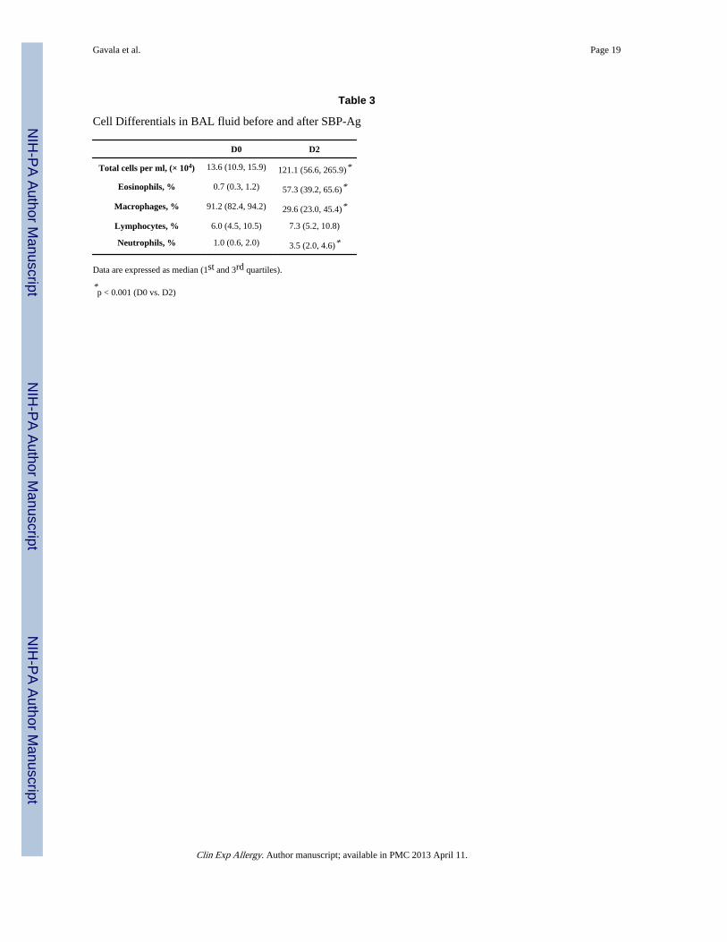
Relationship between BAL cell composition and allergen-elicited mediators in BAL fluidAlveolar macrophages are a major source of CCL18 and CHIT1 in the lungs [12, 32–34],whereas YKL-40 is expressed by alveolar macrophages, neutrophils, and epithelial cells [8,35, 36]. To assess the relationship between allergen challenge-induced CCL18/YKL-40levels or CHIT1 activity with different cell populations, we determined the cellular profileof the BAL fluid before and after SBP-Ag (Table 3). Before SBP-Ag, CCL18 and YKL-40levels were detectable in the BAL fluid, but did not correlate with BAL cells. Conversely,CHIT1 activity did correlate with both the total number of BAL cells recovered (r = 0.39, p= 0.04) and the number of macrophages present (r = 0.40, p = 0.04) in the BAL fluid. It wasalso observed that IP-10 levels in BAL fluid were inversely correlated with total BAL cells(r=−0.51, p=0.03) and total macrophage counts (r=−0.59, p = 0.01). Basal levels of IP-10were also inversely correlated with the percentage of macrophages (r=−0.55, p=0.02) butcoordinately correlated with the percentage of lymphocytes (r=0.66, p=0.003) present inBAL fluid prior to SBP-Ag. Basal IGFBP-3 levels also correlated with the percentage ofeosinophils in the BAL fluid (r=0.47, p=0.05), whereas basal MMP9 levels correlated withboth total neutrophil numbers (r=0.52, p=0.03) and the percentage of neutrophils (r=0.51,p=0.03) in the BAL fluid.
In contrast, the levels of many of the measured mediators in BAL fluid correlatedsignificantly with BAL cells 48 h post-SBP-Ag (Table 4). As shown in Table 4, CCL18levels after allergen challenge significantly correlated with the number of macrophages,lymphocytes and eosinophils present. Furthermore, YKL40 levels correlated withlymphocyte and eosinophil numbers, and CHIT1 activity correlated with lymphocyte andneutrophil counts. Overall, most of the factors measured (except CHIT1 activity and MMP9and IP-10 levels) correlated with cellular influx into the antigen challenged segment (i.e.,fold change in total BAL cell numbers), supporting that these recruited cells are related tothe increased presence of factors in the lungs. When considering the concentration of cellsper milliliter of BAL fluid, CHIT1 activity also correlated with the increased number ofBAL cells. Many of the other examined factors also correlated with the concentrations ofeosinophils, macrophages, and lymphocytes present in the BAL. Of note, besides CHIT1activity, only MMP9 levels in BAL fluid correlated with the number of neutrophils presentin the BAL fluid 48 h after SBP-Ag, and IP-10 was the only factor that did not significantlycorrelate with any of the BAL leukocytes.
To further support that BAL cells are a potential source of these up-regulated mediators,mRNA was extracted from isolated BAL cells before and after SBP-Ag and the mRNAexpression of CCL18, YKL-40, CHIT1, TARC, and IP-10 was determined. As shown inFigure 4, mRNA levels for CCL18, YKL-40, and TARC are significantly increased in BALcells after SBP-Ag. Consistent with the lack of BAL cell correlations, IP-10 expression didnot significantly change after SBP-Ag. Interestingly, mRNA expression for CHIT1, theactive chitinase in human lungs [11], was decreased in BAL cells after SBP-Ag.
Overall, these data support a relationship between leukocytes in the airway and the releaseof profibrotic factors and proinflammatory cytokines after allergen challenge in the lungs.
DISCUSSIONUsing an established in vivo model for allergic airway inflammation in asthma, wedemonstrated that BAL fluid levels of CCL18 and YKL-40 and CHIT1 activity increasesignificantly 48 h following allergen segmental airway challenge. Furthermore, theindividual increases in these factors not only correlate with each other, but also with otherprofibrotic and inflammatory mediators after allergen challenge, suggesting a related role in
Gavala et al. Page 6
Clin Exp Allergy. Author manuscript; available in PMC 2013 April 11.
NIH
-PA Author Manuscript
NIH
-PA Author Manuscript
NIH
-PA Author Manuscript

allergic asthma pathophysiology. Moreover, we established that BAL cells express CCL18,YKL-40, and CHIT-1 mRNA, suggesting that they are a potential source of these factors.
CCL18 is a chemokine unique to primates and does not yet have a known receptor [17]. It isconstitutively expressed in the lungs and is primarily produced by alveolar macrophages anddendritic cells [21]. It is chemotactic for lymphocytes, including Th2 polarized cells andnaïve T and B cells, and immature dendritic cells [21, 37]. Earlier reports have notedincreased CCL18 expression in diseases associated with Th2 inflammation, such as atopicdermatitis [38, 39]. Our data also reveal a strong correlation between CCL18 levels and theTh2-associated chemokine TARC in the BAL fluid after allergen challenge.
Nadaï et al. [21] previously suggested the involvement of CCL18 in allergic asthma. Theyfound that PBMCs treated in vitro with patient-specific allergens release CCL18, and non-medicated allergic asthmatics have elevated CCL18 levels in their BAL fluid/macrophagecontent versus non-allergic or steroid-treated allergic asthmatics. Our study demonstrates forthe first time that CCL18 can be induced in human lungs following in vivo allergenchallenge, further providing evidence of its role in allergic asthma and Th2-mediatedinflammation.
Levels of CCL18 have been shown to be increased in scleroderma lung disease,hypersensitivity pneumonitis, idiopathic pulmonary fibrosis, and sarcoidosis fibrosis [16–19]. CCL18 has also been shown to promote collagen production from pulmonaryfibroblasts using in vitro models [40, 41]. CCL18 has been shown to increase collagenproduction in human lung fibroblasts [42–44], and to be a sensitive indicator of activepulmonary fibrosis [45]. Furthermore, CCL18 production from BAL cells has is enhanced inpatients with pulmonary fibrosis [33]. Our data further support a relationship betweenCCL18 and fibrotic factors, as levels of fibronectin and IGFBP-3 in the BAL fluid afterallergen challenge correlated with the levels of CCL18.
Both chitinases and chitinase-like proteins have diverse immunological functions [7]. Theseevolutionarily conserved enzymes undoubtedly have a role in the anti-parasitic responses ofthe human immune system, but growing evidence also suggests that they might have otherimportant immunomodulatory roles, especially in the context of unregulated inflammation.Although the exact function of chitinases as immunomodulators is still being elucidated, agrowing body of literature shows involvement of chitinases in a variety of disease processes,including COPD, Gaucher disease, chronic inflammation (e.g., sarcoidosis, arthritis), andinfections [12, 15, 34, 46].
Although the major chitinases in murine lungs are AMcase and YM1/2, CHIT1 is thepredominant active chitinase in humans [11, 47]. Subjects homozygous for CHIT1deficiency are less likely to suffer from atopic conditions [13]. Furthermore, increased levelsof CHIT1 mRNA and protein are detectable in nasal mucosa from allergic donors comparedwith normal donors [14]. The role of CHIT1 in human asthma is unclear, but it has beenargued that only smokers with COPD, but not asthmatics, have higher chitinolytic activity,increased levels of CHIT1 in BAL fluid, and more CHIT1-positive cells in bronchialbiopsies [11, 35]. However, it is important to note that these studies did not screen for the 24base pair duplication of CHIT1, which renders CHIT1 inactive. Therefore, the expression/activity of CHIT1 in asthmatics may have been masked in prior studies. It is also possiblethat there is sufficient active AMcase expression in the lungs to lead to the increasedchitinase activity after allergen challenge, although it has been reported that all of thedetectable AMcase in human lungs is in an inactive form [11].
Our data are the first to show that CHIT1 activity is increased in human lungs after in vivoallergen exposure and support the notion that allergen challenge leads to increased CHIT1
Gavala et al. Page 7
Clin Exp Allergy. Author manuscript; available in PMC 2013 April 11.
NIH
-PA Author Manuscript
NIH
-PA Author Manuscript
NIH
-PA Author Manuscript

activity and correlates with the increased presence of profibrotic factors in the lungs.Interestingly, although CHIT1 activity increases, the levels of CHIT1 mRNA detected fromBAL cells decreases 48 h after allergen challenge. There are many potential explanations forthis decrease in CHIT1 mRNA: 1) CHIT1 mRNA may be more transient than the otherfactors measured; therefore, its mRNA accumulation may diminish while protein andactivity is still present; 2) the cellular source of CHIT1 may be exclusively alveolarmacrophages [11, 48], which are a lower percentage of the cells after allergen challenge,thus comparing the levels of CHIT1 before and after challenge is skewed against observingmacrophage-derived endpoints; 3) the increased CHIT1 activity observed may not be linkedto changes in CHIT1 expression. During post-translational processing, a fraction of CHIT1is intracellularly converted by carboxyl-terminal proteolytic processing into a 39-kDaisoform that accumulates in lysosomes and has no chitinase activity [48]. Allergen challengemay block the accumulation of CHIT1 in the lysosome, resulting in increased levels ofsecreted active CHIT1 and 4) other factors besides CHIT1 are involved in the increasedCHIT1 activity seen after allergen challenge. Regardless, CHIT1 activity is potentiated afterallergen challenge and likely plays a role in the up-regulation of Th2-mediatedinflammation.
YKL-40, a chitinase-like protein, has been shown to be increased in the serum and lungs ofallergic asthmatics, and these YKL-40 levels correlate with severity of disease, asthmaexacerbations, eosinophil levels, and degree of airway remodeling measured as the thicknessof the subepithelial basement membrane [8, 49]. In addition, genetic polymorphisms in thechitinase 3-like 1 gene encoding for YKL-40 are associated with increased YKL-40 levels,bronchial hyperresponsiveness, risk for asthma, and reduced lung function [6, 9].
YKL-40, which is also increased in the BAL fluid and serum of COPD patients, canpromote the release of proinflammatory mediators and remodeling factors, such as MMP9from alveolar macrophages, suggesting a role for YKL-40 in airway inflammation andremodeling [35]. Our data show a strong correlation between YKL-40 and MMP9 levels(and all of the other fibrotic factors tested) after an in vivo allergen challenge of atopicasthmatics, further supporting that these two factors are linked in pulmonary inflammatorydiseases.
In a letter to the editor, Kuepper et al. reported increased serum and BAL YKL-40 levels 24h following a segmental allergen challenge in allergic asthma patients [50]. Leukocyterecruitment/composition in the lungs varies over time, with the peak of total leukocytes andalveolar macrophages occurring after two days [51], so our detection of YKL-40 levels after48 h captured the peak of leukocyte recruitment to the lungs. Our data extend Kuepper’sobservations by demonstrating an increase in YKL-40 48 h after allergen challenge, andthey also reveal new correlations between YKL-40 levels in the lungs with other allergen-elicited mediators and leukocytes recruited to the site of allergen challenge.
There are some limitations to our study. First, these patients may not represent the fullspectrum of asthma patients; to ensure patient safety, only patients with mild asthma wereselected for SBP-Ag. Therefore, it is not known if our findings can be directly extrapolatedto patients with more severe disease. Second, our model is based on allergic inflammatoryresponse, thus the findings are not applicable to patients with non-allergic asthma or to non-allergic stimuli such as viral respiratory tract infections. Third, with segmental airwayallergen challenge, we only have lung samples at two time points. While in theory, morefrequent sampling might be feasible, subject safety and comfort precluded obtaining morefrequent samples. Since the inflammatory process occurs along a time continuum, it ispossible that the levels of some of the cytokines would be different either before or after the48 h time point when the second BAL sampling is performed. Therefore, our sampling may
Gavala et al. Page 8
Clin Exp Allergy. Author manuscript; available in PMC 2013 April 11.
NIH
-PA Author Manuscript
NIH
-PA Author Manuscript
NIH
-PA Author Manuscript

have missed peaks of some of the measured factors. Finally, we did not examine tissuebiopsies in the current study, thus we can’t ascertain if remodeling process was initiatedfollowing antigen challenge. However, the increase in soluble matrix protein (i.e., FN) thatwe were able to demonstrate suggests that there is a potential for such outcome.
In conclusion, we provide new evidence for the involvement of CCL18, YKL-40, andCHIT1 in allergic airway inflammation and suggest that these factors might be linked toairway remodeling, which is an important component of asthma pathophysiology. Betterunderstanding of how these factors relate to each other and to the recruited inflammatorycells in the airway could provide necessary rationale for new therapeutic approaches toprevent structural airway changes that are potentially linked to allergen exposure. Suchadvances could also have implications to other Th2 inflammatory-fibrotic conditions.
Supplementary MaterialRefer to Web version on PubMed Central for supplementary material.
AcknowledgmentsThis manuscript is dedicated to the memory of our colleague, Dr. Paul Bertics, who made many contributions tothis publication and to our research programs over 25 years as a member of the University of Wisconsin facultyprior to his untimely death in December 2011. The authors wish to thank our Research Coordinators, Mary JoJackson, R.N., Michele Wolff, R.N., and Holly Eversoll, R.N. for patient recruitment and screening; Keith Meyer,M.D., and Richard Cornwell, M.D. for assistance with bronchoscopies; Larissa DeLain, B.S., for laboratoryassistance, and Jami Hauer, B.S. for laboratory and editorial assistance.
This work was supported by NIH grants HL088594, HL069116, UL1RR025011L (NNJ), HL69174, HL56396(PJB), HL-095390, and HL093017 (GLC). It was also supported in part by grants 1UL1RR025011 andULRR024146 from the Clinical and Translational Science Award (CTSA) program of the National Center forResearch Resources, National Institutes of Health.
References1. Busse WW, Lemanske RF Jr, Asthma. N Engl J Med. 2001; 344:350–62. [PubMed: 11172168]
2. Jarjour NN, Kelly EA. Pathogenesis of asthma. Med Clin North Am. 2002; 86:925–36. [PubMed:12428538]
3. Kim HY, DeKruyff RH, Umetsu DT. The many paths to asthma: phenotype shaped by innate andadaptive immunity. Nat Immunol. 2010; 11:577–84. [PubMed: 20562844]
4. Humbles AA, Lloyd CM, McMillan SJ, Friend DS, Xanthou G, McKenna EE, Ghiran S, Gerard NP,Yu C, Orkin SH, Gerard C. A critical role for eosinophils in allergic airways remodeling. Science.2004; 305:1776–9. [PubMed: 15375268]
5. Bussink AP, van Eijk M, Renkema GH, Aerts JM, Boot RG. The biology of the Gaucher cell: thecradle of human chitinases. Int Rev Cytol. 2006; 252:71–128. [PubMed: 16984816]
6. Ober C, Chupp GL. The chitinase and chitinase-like proteins: a review of genetic and functionalstudies in asthma and immune-mediated diseases. Curr Opin Allergy Clin Immunol. 2009; 9:401–8.[PubMed: 19644363]
7. Elias JA, Homer RJ, Hamid Q, Lee CG. Chitinases and chitinase-like proteins in T(H)2inflammation and asthma. J Allergy Clin Immunol. 2005; 116:497–500. [PubMed: 16159614]
8. Chupp GL, Lee CG, Jarjour N, Shim YM, Holm CT, He S, Dziura JD, Reed J, Coyle AJ, Kiener P,Cullen M, Grandsaigne M, Dombret MC, Aubier M, Pretolani M, Elias JA. A chitinase-like proteinin the lung and circulation of patients with severe asthma. N Engl J Med. 2007; 357:2016–27.[PubMed: 18003958]
9. Ober C, Tan Z, Sun Y, Possick JD, Pan L, Nicolae R, Radford S, Parry RR, Heinzmann A,Deichmann KA, Lester LA, Gern JE, Lemanske RF Jr, Nicolae DL, Elias JA, Chupp GL. Effect ofvariation in CHI3L1 on serum YKL-40 level, risk of asthma, and lung function. N Engl J Med.2008; 358:1682–91. [PubMed: 18403759]
Gavala et al. Page 9
Clin Exp Allergy. Author manuscript; available in PMC 2013 April 11.
NIH
-PA Author Manuscript
NIH
-PA Author Manuscript
NIH
-PA Author Manuscript

10. Zheng T, Liu W, Oh SY, Zhu Z, Hu B, Homer RJ, Cohn L, Grusby MJ, Elias JA. IL-13 receptoralpha2 selectively inhibits IL-13-induced responses in the murine lung. J Immunol. 2008;180:522–9. [PubMed: 18097054]
11. Seibold MA, Donnelly S, Solon M, Innes A, Woodruff PG, Boot RG, Burchard EG, Fahy JV.Chitotriosidase is the primary active chitinase in the human lung and is modulated by genotypeand smoking habit. J Allergy Clin Immunol. 2008; 122:944–50. e3. [PubMed: 18845328]
12. Boot RG, Hollak CE, Verhoek M, Alberts C, Jonkers RE, Aerts JM. Plasma chitotriosidase andCCL18 as surrogate markers for granulomatous macrophages in sarcoidosis. Clin Chim Acta.2010; 411:31–6. [PubMed: 19808030]
13. Lee P, Waalen J, Crain K, Smargon A, Beutler E. Human chitotriosidase polymorphisms G354Rand A442V associated with reduced enzyme activity. Blood Cells Mol Dis. 2007; 39:353–60.[PubMed: 17693102]
14. Cho WS, Kim TH, Lee HM, Lee SH, Yoo JH, Kim YS. Increased expression of acidic mammalianchitinase and chitotriosidase in the nasal mucosa of patients with allergic rhinitis. Laryngoscope.2010; 120:870–5. [PubMed: 20422678]
15. Boot RG, Verhoek M, de Fost M, Hollak CE, Maas M, Bleijlevens B, van Breemen MJ, van MeursM, Boven LA, Laman JD, Moran MT, Cox TM, Aerts JM. Marked elevation of the chemokineCCL18/PARC in Gaucher disease: a novel surrogate marker for assessing therapeutic intervention.Blood. 2004; 103:33–9. [PubMed: 12969956]
16. Prasse A, Probst C, Bargagli E, Zissel G, Toews GB, Flaherty KR, Olschewski M, Rottoli P,Muller-Quernheim J. Serum CC-chemokine ligand 18 concentration predicts outcome inidiopathic pulmonary fibrosis. Am J Respir Crit Care Med. 2009; 179:717–23. [PubMed:19179488]
17. Schutyser E, Richmond A, Van Damme J. Involvement of CC chemokine ligand 18 (CCL18) innormal and pathological processes. J Leukoc Biol. 2005; 78:14–26. [PubMed: 15784687]
18. Mrazek F, Sekerova V, Drabek J, Kolek V, du Bois RM, Petrek M. Expression of the chemokinePARC mRNA in bronchoalveolar cells of patients with sarcoidosis. Immunol Lett. 2002; 84:17–22. [PubMed: 12161279]
19. Pardo A, Smith KM, Abrams J, Coffman R, Bustos M, McClanahan TK, Grein J, Murphy EE,Zlotnik A, Selman M. CCL18/DC-CK-1/PARC up-regulation in hypersensitivity pneumonitis. JLeukoc Biol. 2001; 70:610–6. [PubMed: 11590198]
20. Kim HB, Kim CK, Iijima K, Kobayashi T, Kita H. Protein microarray analysis in patients withasthma: elevation of the chemokine PARC/CCL18 in sputum. Chest. 2009; 135:295–302.[PubMed: 19017877]
21. de Nadai P, Charbonnier AS, Chenivesse C, Senechal S, Fournier C, Gilet J, Vorng H, Chang Y,Gosset P, Wallaert B, Tonnel AB, Lassalle P, Tsicopoulos A. Involvement of CCL18 in allergicasthma. J Immunol. 2006; 176:6286–93. [PubMed: 16670340]
22. Zou J, Young S, Zhu F, Gheyas F, Skeans S, Wan Y, Wang L, Ding W, Billah M, McClanahan T,Coffman RL, Egan R, Umland S. Microarray profile of differentially expressed genes in a monkeymodel of allergic asthma. Genome Biol. 2002; 3:research0020. [PubMed: 12049661]
23. Liu LY, Sedgwick JB, Bates ME, Vrtis RF, Gern JE, Kita H, Jarjour NN, Busse WW, Kelly EA.Decreased expression of membrane IL-5 receptor alpha on human eosinophils: I. Loss ofmembrane IL-5 receptor alpha on airway eosinophils and increased soluble IL-5 receptor alpha inthe airway after allergen challenge. J Immunol. 2002; 169:6452–8. [PubMed: 12444154]
24. Kelly EA, Busse WW, Jarjour NN. Inhaled budesonide decreases airway inflammatory response toallergen. American journal of respiratory and critical care medicine. 2000:883–90. [PubMed:10988100]
25. Terada M, Kelly EA, Jarjour NN. Increased thrombin activity after allergen challenge: a potentiallink to airway remodeling? Am J Respir Crit Care Med. 2004; 169:373–7. [PubMed: 14630620]
26. Kelly EA, Rodriguez RR, Busse WW, Jarjour NN. The effect of segmental bronchoprovocationwith allergen on airway lymphocyte function. Am J Respir Crit Care Med. 1997; 156:1421–8.[PubMed: 9372655]
Gavala et al. Page 10
Clin Exp Allergy. Author manuscript; available in PMC 2013 April 11.
NIH
-PA Author Manuscript
NIH
-PA Author Manuscript
NIH
-PA Author Manuscript

27. Meerschaert J, Kelly EA, Mosher DF, Busse WW, Jarjour NN. Segmental antigen challengeincreases fibronectin in bronchoalveolar lavage fluid. Am J Respir Crit Care Med. 1999; 159:619–25. [PubMed: 9927382]
28. Veraldi KL, Gibson BT, Yasuoka H, Myerburg MM, Kelly EA, Balzar S, Jarjour NN, PilewskiJM, Wenzel SE, Feghali-Bostwick CA. Role of insulin-like growth factor binding protein-3 inallergic airway remodeling. Am J Respir Crit Care Med. 2009; 180:611–7. [PubMed: 19608721]
29. Bochner BS, Hudson SA, Xiao HQ, Liu MC. Release of both CCR4-active and CXCR3-activechemokines during human allergic pulmonary late-phase reactions. J Allergy Clin Immunol. 2003;112:930–4. [PubMed: 14610482]
30. Pilette C, Francis JN, Till SJ, Durham SR. CCR4 ligands are up-regulated in the airways of atopicasthmatics after segmental allergen challenge. Eur Respir J. 2004; 23:876–84. [PubMed:15219001]
31. Liu L, Jarjour NN, Busse WW, Kelly EA. Enhanced generation of helper T type 1 and 2chemokines in allergen-induced asthma. Am J Respir Crit Care Med. 2004; 169:1118–24.[PubMed: 15001464]
32. Hieshima K, Imai T, Baba M, Shoudai K, Ishizuka K, Nakagawa T, Tsuruta J, Takeya M, SakakiY, Takatsuki K, Miura R, Opdenakker G, Van Damme J, Yoshie O, Nomiyama H. A novel humanCC chemokine PARC that is most homologous to macrophage-inflammatory protein-1 alpha/LD78 alpha and chemotactic for T lymphocytes, but not for monocytes. J Immunol. 1997;159:1140–9. [PubMed: 9233607]
33. Prasse A, Pechkovsky DV, Toews GB, Jungraithmayr W, Kollert F, Goldmann T, Vollmer E,Muller-Quernheim J, Zissel G. A vicious circle of alveolar macrophages and fibroblastsperpetuates pulmonary fibrosis via CCL18. Am J Respir Crit Care Med. 2006; 173:781–92.[PubMed: 16415274]
34. Letuve S, Kozhich A, Humbles A, Brewah Y, Dombret MC, Grandsaigne M, Adle H, Kolbeck R,Aubier M, Coyle AJ, Pretolani M. Lung chitinolytic activity and chitotriosidase are elevated inchronic obstructive pulmonary disease and contribute to lung inflammation. Am J Pathol. 2010;176:638–49. [PubMed: 20042671]
35. Letuve S, Kozhich A, Arouche N, Grandsaigne M, Reed J, Dombret MC, Kiener PA, Aubier M,Coyle AJ, Pretolani M. YKL-40 is elevated in patients with chronic obstructive pulmonary diseaseand activates alveolar macrophages. J Immunol. 2008; 181:5167–73. [PubMed: 18802121]
36. Park JA, Drazen JM, Tschumperlin DJ. The chitinase-like protein YKL-40 is secreted by airwayepithelial cells at base line and in response to compressive mechanical stress. J Biol Chem. 2010;285:29817–25. [PubMed: 20650887]
37. Wimmer A, Khaldoyanidi SK, Judex M, Serobyan N, Discipio RG, Schraufstatter IU. CCL18/PARC stimulates hematopoiesis in long-term bone marrow cultures indirectly through its effect onmonocytes. Blood. 2006; 108:3722–9. [PubMed: 16888095]
38. Nomura I, Goleva E, Howell MD, Hamid QA, Ong PY, Hall CF, Darst MA, Gao B, BoguniewiczM, Travers JB, Leung DY. Cytokine milieu of atopic dermatitis, as compared to psoriasis, skinprevents induction of innate immune response genes. J Immunol. 2003; 171:3262–9. [PubMed:12960356]
39. Gunther C, Bello-Fernandez C, Kopp T, Kund J, Carballido-Perrig N, Hinteregger S, Fassl S,Schwarzler C, Lametschwandtner G, Stingl G, Biedermann T, Carballido JM. CCL18 is expressedin atopic dermatitis and mediates skin homing of human memory T cells. J Immunol. 2005;174:1723–8. [PubMed: 15661937]
40. Prasse A, Pechkovsky DV, Toews GB, Schafer M, Eggeling S, Ludwig C, Germann M, Kollert F,Zissel G, Muller-Quernheim J. CCL18 as an indicator of pulmonary fibrotic activity in idiopathicinterstitial pneumonias and systemic sclerosis. Arthritis Rheum. 2007; 56:1685–93. [PubMed:17469163]
41. Pochetuhen K, Luzina IG, Lockatell V, Choi J, Todd NW, Atamas SP. Complex regulation ofpulmonary inflammation and fibrosis by CCL18. Am J Pathol. 2007; 171:428–37. [PubMed:17569779]
42. Luzina IG, Tsymbalyuk N, Choi J, Hasday JD, Atamas SP. CCL18-stimulated upregulation ofcollagen production in lung fibroblasts requires Sp1 signaling and basal Smad3 activity. Journal ofcellular physiology. 2006; 206:221–8. [PubMed: 16021625]
Gavala et al. Page 11
Clin Exp Allergy. Author manuscript; available in PMC 2013 April 11.
NIH
-PA Author Manuscript
NIH
-PA Author Manuscript
NIH
-PA Author Manuscript

43. Luzina IG, Highsmith K, Pochetuhen K, Nacu N, Rao JN, Atamas SP. PKCalpha mediates CCL18-stimulated collagen production in pulmonary fibroblasts. Am J Respir Cell Mol Biol. 2006;35:298–305. [PubMed: 16601239]
44. Atamas SP, Luzina IG, Choi J, Tsymbalyuk N, Carbonetti NH, Singh IS, Trojanowska M, JimenezSA, White B. Pulmonary and activation-regulated chemokine stimulates collagen production inlung fibroblasts. Am J Respir Cell Mol Biol. 2003; 29:743–9. [PubMed: 12805086]
45. Kodera M, Hasegawa M, Komura K, Yanaba K, Takehara K, Sato S. Serum pulmonary andactivation-regulated chemokine/CCL18 levels in patients with systemic sclerosis: a sensitiveindicator of active pulmonary fibrosis. Arthritis Rheum. 2005; 52:2889–96. [PubMed: 16142750]
46. Lee CG, Da Silva CA, Dela Cruz CS, Ahangari F, Ma B, Kang MJ, He CH, Takyar S, Elias JA.Role of chitin and chitinase/chitinase-like proteins in inflammation, tissue remodeling, and injury.Annu Rev Physiol. 2011; 73:479–501. [PubMed: 21054166]
47. Zhu Z, Zheng T, Homer RJ, Kim YK, Chen NY, Cohn L, Hamid Q, Elias JA. Acidic mammalianchitinase in asthmatic Th2 inflammation and IL-13 pathway activation. Science. 2004; 304:1678–82. [PubMed: 15192232]
48. Renkema GH, Boot RG, Strijland A, Donker-Koopman WE, van den Berg M, Muijsers AO, AertsJM. Synthesis, sorting, and processing into distinct isoforms of human macrophage chitotriosidase.Eur J Biochem. 1997; 244:279–85. [PubMed: 9118991]
49. Tang H, Fang Z, Sun Y, Li B, Shi Z, Chen J, Zhang T, Xiu Q. YKL-40 in asthmatic patients, andits correlations with exacerbation, eosinophils and immunoglobulin E. Eur Respir J. 2010; 35:757–60. [PubMed: 20356987]
50. Kuepper M, Bratke K, Virchow JC. Chitinase-like protein and asthma. N Engl J Med. 2008;358:1073–5. author reply 75. [PubMed: 18322291]
51. Lommatzsch M, Julius P, Kuepper M, Garn H, Bratke K, Irmscher S, Luttmann W, Renz H, BraunA, Virchow JC. The course of allergen-induced leukocyte infiltration in human and experimentalasthma. J Allergy Clin Immunol. 2006; 118:91–7. [PubMed: 16815143]
Gavala et al. Page 12
Clin Exp Allergy. Author manuscript; available in PMC 2013 April 11.
NIH
-PA Author Manuscript
NIH
-PA Author Manuscript
NIH
-PA Author Manuscript

Figure 1. BAL fluid concentrations of YKL-40 and CCL18 and CHIT1 activity are increased 48h after SBP-Ag(A) Data represent the concentrations of specific analytes in BAL fluid obtainedimmediately before (D0) and 48 h after (D2) SBP-Ag. Each symbol represents the sameindividual subject across assays. (B) Correlations between YKL-40, CCL18 levels andCHIT1 activity in BAL fluid 48 h post SBP-Ag were plotted together and correlations weredetermined. rs = Spearman’s correlation coefficients. Each symbol represents the sameindividual subject across assays (n = 18).
Gavala et al. Page 13
Clin Exp Allergy. Author manuscript; available in PMC 2013 April 11.
NIH
-PA Author Manuscript
NIH
-PA Author Manuscript
NIH
-PA Author Manuscript

Figure 2. Associations between concentrations of remodeling factors with CHIT1 activity andYKL-40 and CCL18 concentrations in BAL fluidrs = Spearman’s correlation coefficients. Specific symbols represent individual subjectsacross correlations (n = 18).
Gavala et al. Page 14
Clin Exp Allergy. Author manuscript; available in PMC 2013 April 11.
NIH
-PA Author Manuscript
NIH
-PA Author Manuscript
NIH
-PA Author Manuscript

Figure 3. Associations between concentrations of Th-associated chemokines with CHIT1 activityand YKL-40 and CCL18 concentrations in BAL fluidrs = Spearman’s correlation coefficients. Specific symbols represent individual subjectsacross correlations (n = 18).
Gavala et al. Page 15
Clin Exp Allergy. Author manuscript; available in PMC 2013 April 11.
NIH
-PA Author Manuscript
NIH
-PA Author Manuscript
NIH
-PA Author Manuscript

Figure 4. Mediator mRNA expression by BAL-derived cells before and after SBP-Ag(A) YKL-40, (B) CCL18, (C) CHIT1, (D) TARC, and (E) IP-10 mRNA expression in BALcells immediately before (D0) and 48 h after (D2) SBP-Ag (n = 8, except CCL18 (n = 7)).
Gavala et al. Page 16
Clin Exp Allergy. Author manuscript; available in PMC 2013 April 11.
NIH
-PA Author Manuscript
NIH
-PA Author Manuscript
NIH
-PA Author Manuscript

NIH
-PA Author Manuscript
NIH
-PA Author Manuscript
NIH
-PA Author Manuscript
Gavala et al. Page 17
Table 1
Characteristics for subjects in the BAL fluid study*
Number 18
Sex 9 F, 9 M
Age (yrs) 22 (20, 23)
Baseline FEV1 (% predicted) 97 (90, 102)
Methacholine PC20 (mg/ml) 1.7 (0.8, 5.2)
*median (1st and 3rd quartiles)
Clin Exp Allergy. Author manuscript; available in PMC 2013 April 11.

NIH
-PA Author Manuscript
NIH
-PA Author Manuscript
NIH
-PA Author Manuscript
Gavala et al. Page 18
Table 2
Select profibrotic and BAL fluid (BALF, 1x) protein profile before and 48 h after SBP-Ag
D0 D2
CHIT1 activity (nmol/ml/h) 0.1 (0.1, 0.2) 1.3 (0.5, 2.7)*
YKL-40, ng/ml 22.7 (15.6, 29.8) 150.1 (104.8, 354.4)*
CCL18, ng/ml 1.2 (0.7, 1.9) 16.0 (11.9, 21.4)*
FN, μg/ml 1.0 (0.5, 2.4) 36.4 (15.4, 108.0)*
IGFBP3, ng/ml 0.7 (0.5, 2.0) 27.1 (13.2, 64.6)*
MMP9, ng/ml 0.2 (0.1, 0.3) 2.1 (1.3, 6.4)*
thrombin activity, NIH U/ml 0.4 (0.3 0.5) 1.1 (0.7, 1.5)*
TARC, pg/ml 0 (0, 0) 1867 (987, 4310)*
IP10, pg/ml 35 (15, 42) 176 (80, 245)*
Data are expressed as median (1st and 3rd quartiles).
*p < 0.001 (D0 vs. D2)
Clin Exp Allergy. Author manuscript; available in PMC 2013 April 11.
NIH
-PA Author Manuscript
NIH
-PA Author Manuscript
NIH
-PA Author Manuscript
Gavala et al. Page 19
Table 3
Cell Differentials in BAL fluid before and after SBP-Ag
D0 D2
Total cells per ml, (× 104) 13.6 (10.9, 15.9) 121.1 (56.6, 265.9)*
Eosinophils, % 0.7 (0.3, 1.2) 57.3 (39.2, 65.6)*
Macrophages, % 91.2 (82.4, 94.2) 29.6 (23.0, 45.4)*
Lymphocytes, % 6.0 (4.5, 10.5) 7.3 (5.2, 10.8)
Neutrophils, % 1.0 (0.6, 2.0) 3.5 (2.0, 4.6)*
Data are expressed as median (1st and 3rd quartiles).
*p < 0.001 (D0 vs. D2)
Clin Exp Allergy. Author manuscript; available in PMC 2013 April 11.

NIH
-PA Author Manuscript
NIH
-PA Author Manuscript
NIH
-PA Author Manuscript
Gavala et al. Page 20
Tabl
e 4
Cor
rela
tion
s be
twee
n B
AL
Cel
lula
r an
d P
rote
in F
luid
Lev
els
48 h
aft
er S
BP
-Ag
The
num
ber
of le
ukoc
ytes
(#)
col
lect
ed in
the
BA
L f
luid
was
com
pare
d to
the
leve
ls o
f se
lect
ed p
rote
ins
mea
sure
d 48
h a
fter
seg
men
tal a
llerg
ench
alle
nge.
The
Spe
arm
an’s
ran
k co
rrel
atio
ns a
nd c
orre
spon
ding
p-v
alue
s ar
e di
spla
yed.
The
box
es in
whi
te in
dica
te s
igni
fica
nt c
orre
latio
ns a
nd th
e bo
xes
in g
rey
indi
cate
no
sign
ific
ant c
orre
latio
n. C
ell.f
old:
fol
d ch
ange
in c
ell n
umbe
rs f
rom
D0
to D
2; m
ac: m
acro
phag
e; ly
m: l
ymph
ocyt
e; p
mn:
poly
mor
phon
ucle
ar (
neut
roph
il); e
os: e
osin
ophi
ls.
#cel
lce
ll.fo
ld#m
ac#l
ym#p
mn
#eos
Chi
tina
se A
ct0.
480.
450.
450.
570.
640.
42r s
0.04
0.06
0.06
0.01
0.00
50.
08p
valu
e
YK
L40
0.52
0.51
0.31
0.59
0.44
0.51
r s
0.03
0.03
0.20
0.00
90.
060.
03p
valu
e
CC
L18
0.66
0.64
0.55
0.72
0.35
0.68
r s
0.00
30.
004
0.02
0.00
080.
160.
002
p va
lue
FN
0.76
0.70
0.65
0.82
0.43
0.73
r s
0.00
020.
001
0.00
3<
0.00
010.
070.
0006
p va
lue
MM
P9
0.42
0.45
0.30
0.57
0.56
0.40
r s
0.08
0.06
0.22
0.01
0.02
0.10
p va
lue
TA
RC
0.75
0.74
0.65
0.86
0.47
0.73
r s
0.00
030.
0005
0.00
4<
0.00
010.
050.
0006
p va
lue
IP-1
00.
180.
250.
080.
340.
410.
16r s
0.47
0.32
0.75
0.17
0.09
0.52
p va
lue
IGF
BP
30.
760.
730.
630.
860.
440.
74r s
0.00
030.
0006
0.00
5<
0.00
010.
060.
0004
p va
lue
Thr
ombi
n A
ct0.
760.
610.
810.
760.
400.
66r s
0.00
070.
010.
0001
0.00
060.
130.
005
p va
lue
Clin Exp Allergy. Author manuscript; available in PMC 2013 April 11.
Copyright © 2022 FDOKUMEN