The peanut allergy epidemic: allergen molecular characterisation and prospects for specific therapy
18
The peanut allergy epidemic: allergen molecular characterisation and prospects for specific therapy Maria P. de Leon 1 , Jennifer M. Rolland 1 and Robyn E. O’Hehir 1,2, * Peanut (Arachis hypogaea) allergy is a major cause of food-induced anaphylaxis, with increasing prevalence worldwide. To date, there is no cure for peanut allergy, and, unlike many other food allergies, it usually persists through to adulthood. Prevention of exposure to peanuts is managed through strict avoidance, which can be compromised by the frequent use of peanuts and peanut products in food preparations. Conventional subcutaneous- injection allergen immunotherapy using crude peanut extract is not a recommended treatment because of the risk of severe side effects, largely as a result of specific IgE antibodies. Consequently, there is an urgent need to develop a suitable peanut allergen preparation that can induce specific clinical and immunological tolerance to peanuts in allergic individuals without adverse side effects. This requires detailed molecular and immunological characterisation of the allergenic components of peanut. This article reviews current knowledge on clinically relevant peanut allergens, in particular Ara h 1, Ara h 2 and Ara h 3, together with options for T-cell-reactive but non-IgE-binding allergen variants for specific immunotherapeutic strategies. These include T-cell-epitope peptide and hypoallergenic mutant vaccines. Alternative routes of administration such as sublingual are also considered, and appropriate adjuvants for delivering effective treatments at these sites examined. Allergy to peanuts (Arachis hypogaea) is a burgeoning health problem. Approximately 1% of the population suffers from peanut allergy (Ref. 1), but there is evidence that the prevalence is increasing (Ref. 2). For many peanut-allergic individuals, exposure to minute quantities of peanut can lead to severe reactions including anaphylaxis, which can sometimes prove fatal. Currently, there is no cure for this condition and strategies for the prevention of severe reactions 1 Department of Immunology, Monash University, Melbourne, Victoria 3004, Australia. 2 Department of Allergy, Immunology and Respiratory Medicine, Alfred Hospital, Melbourne, Victoria 3004, Australia. *Corresponding author: Robyn E. O’Hehir, Department of Allergy, Immunology and Respiratory Medicine, Alfred Hospital, Commercial Road, Melbourne, Victoria 3004, Australia. Tel: +61 3 9276 2251; Fax: +61 3 9207 1692; E-mail: [email protected] expert reviews http://www.expertreviews.org/ in molecular medicine 1 Accession information: DOI: 10.1017/S1462399407000208; Vol. 9; Issue 1; January 2007 & 2007 Cambridge University Press The peanut allergy epidemic: allergen molecular characterisation and prospects for specific therapy
-
Upload
independent -
Category
Documents
-
view
1 -
download
0
Transcript of The peanut allergy epidemic: allergen molecular characterisation and prospects for specific therapy
The peanut allergy epidemic: allergen
molecular characterisation and
prospects for specific therapy
Maria P. de Leon1, Jennifer M. Rolland1 and Robyn E. O’Hehir1,2,*
Peanut (Arachis hypogaea) allergy is a major cause of food-inducedanaphylaxis, with increasing prevalence worldwide. To date, there is no curefor peanut allergy, and, unlike many other food allergies, it usually persiststhrough to adulthood. Prevention of exposure to peanuts is managed throughstrict avoidance, which can be compromised by the frequent use of peanutsand peanut products in food preparations. Conventional subcutaneous-injection allergen immunotherapy using crude peanut extract is not arecommended treatment because of the risk of severe side effects, largely as aresult of specific IgE antibodies. Consequently, there is an urgent need todevelop a suitable peanut allergen preparation that can induce specific clinicaland immunological tolerance to peanuts in allergic individuals without adverseside effects. This requires detailed molecular and immunologicalcharacterisation of the allergenic components of peanut. This article reviewscurrent knowledge on clinically relevant peanut allergens, in particular Ara h 1,Ara h 2 and Ara h 3, together with options for T-cell-reactive but non-IgE-bindingallergen variants for specific immunotherapeutic strategies. These includeT-cell-epitope peptide and hypoallergenic mutant vaccines. Alternative routes ofadministration such as sublingual are also considered, and appropriateadjuvants for delivering effective treatments at these sites examined.
Allergy to peanuts (Arachis hypogaea) is aburgeoning health problem. Approximately 1%of the population suffers from peanut allergy(Ref. 1), but there is evidence that the prevalenceis increasing (Ref. 2). For many peanut-allergic
individuals, exposure to minute quantities ofpeanut can lead to severe reactions includinganaphylaxis, which can sometimes prove fatal.Currently, there is no cure for this condition andstrategies for the prevention of severe reactions
1Department of Immunology, Monash University, Melbourne, Victoria 3004, Australia.
2Department of Allergy, Immunology and Respiratory Medicine, Alfred Hospital, Melbourne,Victoria 3004, Australia.
*Corresponding author: Robyn E. O’Hehir, Department of Allergy, Immunology and RespiratoryMedicine, Alfred Hospital, Commercial Road, Melbourne, Victoria 3004, Australia. Tel: +61 3 92762251; Fax: +61 3 9207 1692; E-mail: [email protected]
expert reviewshttp://www.expertreviews.org/ in molecular medicine
1Accession information: DOI: 10.1017/S1462399407000208; Vol. 9; Issue 1; January 2007
&2007 Cambridge University Press
The
pea
nutallergyep
idem
ic:a
llergen
molecu
lar
charac
terisa
tionan
dprosp
ects
forsp
ecifictherap
y

are limited to avoidance of exposure topeanuts and administration of adrenaline as anemergency treatment after inadvertent contact.There is a clear need for a safe and effectivespecific therapy for patients with peanut allergy.
Molecular characterisation of allergeniccomponents of peanuts and elucidation ofimmune mechanisms that determine clinicalresponses are required for the development ofspecific therapies and improved managementfor peanut allergy. This review begins bysummarising the clinical features of peanutallergy and the mucosal immune response tofood allergens, and then discusses the molecularcharacterisation of peanut and crossreactiveallergens, and how this knowledge is beingutilised for the development of potentialtherapeutic modalities for the treatment andprevention of peanut allergy.
Clinical features of peanut allergyPeanut allergy generally develops early in lifeand is commonly associated with other atopicdisorders such as asthma, eczema and rhinitis.Sensitisation is thought to occur through theconsumption of foods such as peanut butter or,in some cases, the use of topical preparationscontaining peanut oil (Ref. 5). Exposure can alsooccur in utero (Ref. 6) or through breastmilk (Ref. 7), although the importance of thisroute for sensitisation has been contentious(Ref. 5). It is becoming apparent that theincreasing consumption of peanuts togetherwith other, unidentified factors is leading toan increase in prevalence of peanutallergy, particularly in children (Refs 1, 2). Thisis likely to flow through to a higher prevalencein adults.
Allergic symptoms following the ingestionof peanuts occur from within minutes to a fewhours, and manifestations range from oralpruritus, nausea, vomiting, urticaria andangioedema to bronchospasm (Ref. 8). In severecases, anaphylaxis with angioedema, respiratorycompromise and hypotension can prove fatalwithout the prompt administration of adrenaline.Peanuts account for the majority of food-relatedanaphylaxis in children, adolescents and adults(Refs 3, 4). A characteristic of peanut allergy isits tendency to persist through to adulthood,with only 21.5% of peanut-allergic individualsexperiencing resolution of this type of foodallergy with increasing age (Refs 9, 10). Thus,
lifelong vigilance is essential for the majority ofsufferers of peanut allergy.
The mucosal immune response topeanut allergens
Exposure of the immune system to a foodallergen such as peanut generally occurs at themucosal surface of the gut. Peanut allergens aretaken up by specialised epithelial cells called Mcells, and transferred to antigen-presenting cellssuch as dendritic cells where they are processedinto peptide fragments and presented on thecell surface in the context of class II majorhistocompatibility complex (MHC) molecules(Refs 11, 12) (Fig. 1). These peptides arepresented to naive T helper (Th) cells viainteraction of the MHC–peptide complex withthe T-cell receptor, resulting in Th-cell primingand activation, which triggers the humoraland cellular events associated with allergicinflammation.
In atopic individuals, the activation of Thcells results in the secretion of cytokines thatstimulate B cells to synthesise IgE antibodiesspecific to the allergen. Th cells are polarisedinto two subgroups, defined by the dominantpattern of cytokine secretion (Ref. 13). Th1-type cells mainly secrete interleukin 2 (IL-2),interferon g (IFN-g) and tumour necrosis factor a(TNF-a). By contrast, Th2-type cells secrete IL-4,IL-5, IL-9 and IL-13. Th2 cells play a pivotal rolein the allergic response as it is their activationand secretion of IL-4 and IL-13 that drivesallergen-stimulated B-cell differentiation intoIgE-secreting plasma cells.
IgE antibodies are bound by high-affinitysurface IgE receptors (Fc1RI) present on effectorcells such as mast cells and basophils. In peanut-allergic individuals, subsequent exposure topeanut allergens induces inflammatory reactionslargely governed by mast cells, basophils andeosinophils. Adjacent IgE antibodies bound byFc1RI on the surface of mast cells and basophilsare crosslinked by peanut allergens, resulting inthe release of inflammatory mediators such ashistamine, prostaglandins, leukotrienes, heparinand platelet-activating factor. Additionalcytokine production occurs during this phase,most notably IL-4 and IL-13 by mast cells andbasophils, further augmenting Th2-celldifferentiation and IgE synthesis. Th2 cells andmast cells also produce TNF-a, IL-5 andchemokines, which results in the recruitment of
expert reviewshttp://www.expertreviews.org/ in molecular medicine
2Accession information: DOI: 10.1017/S1462399407000208; Vol. 9; Issue 1; January 2007
&2007 Cambridge University Press
The
pea
nutallergyep
idem
ic:a
llergen
molecu
lar
charac
terisa
tionan
dprosp
ects
forsp
ecifictherap
y
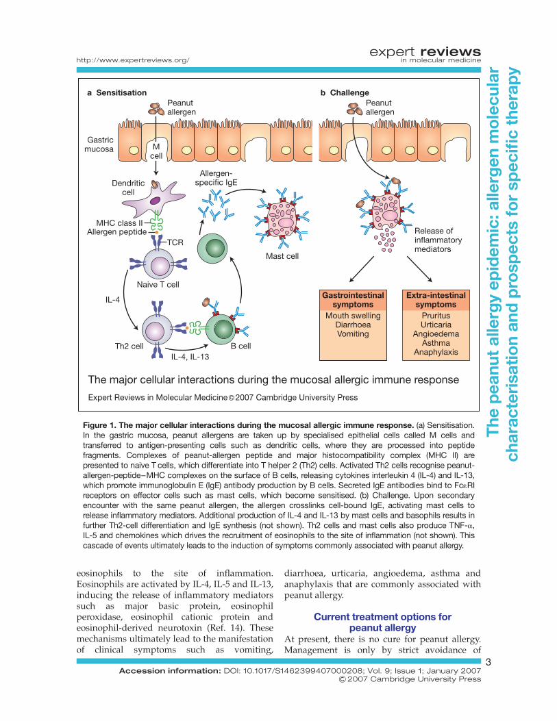
eosinophils to the site of inflammation.Eosinophils are activated by IL-4, IL-5 and IL-13,inducing the release of inflammatory mediatorssuch as major basic protein, eosinophilperoxidase, eosinophil cationic protein andeosinophil-derived neurotoxin (Ref. 14). Thesemechanisms ultimately lead to the manifestationof clinical symptoms such as vomiting,
diarrhoea, urticaria, angioedema, asthma andanaphylaxis that are commonly associated withpeanut allergy.
Current treatment options forpeanut allergy
At present, there is no cure for peanut allergy.Management is only by strict avoidance of
Peanutallergen
Dendriticcell
Release ofinflammatorymediators
Allergen-specific IgE
Mcell
Naive T cell
Th2 cell B cell
MHC class II
TCR
Th2
B
IL-4, IL-13
IL-4
Mast cell
Peanutallergen
Extra-intestinalsymptoms
PruritusUrticaria
AngioedemaAsthma
Anaphylaxis
b Challenge
The major cellular interactions during the mucosal allergic immune response
Expert Reviews in Molecular Medicine C 2007 Cambridge University Press
Gastrointestinalsymptoms
Mouth swellingDiarrhoeaVomiting
a Sensitisation
Gastricmucosa
Allergen peptide
Figure 1. The major cellular interactions during the mucosal allergic immune response. (a) Sensitisation.In the gastric mucosa, peanut allergens are taken up by specialised epithelial cells called M cells andtransferred to antigen-presenting cells such as dendritic cells, where they are processed into peptidefragments. Complexes of peanut-allergen peptide and major histocompatibility complex (MHC II) arepresented to naive T cells, which differentiate into T helper 2 (Th2) cells. Activated Th2 cells recognise peanut-allergen-peptide–MHC complexes on the surface of B cells, releasing cytokines interleukin 4 (IL-4) and IL-13,which promote immunoglobulin E (IgE) antibody production by B cells. Secreted IgE antibodies bind to Fc1RIreceptors on effector cells such as mast cells, which become sensitised. (b) Challenge. Upon secondaryencounter with the same peanut allergen, the allergen crosslinks cell-bound IgE, activating mast cells torelease inflammatory mediators. Additional production of IL-4 and IL-13 by mast cells and basophils results infurther Th2-cell differentiation and IgE synthesis (not shown). Th2 cells and mast cells also produce TNF-a,IL-5 and chemokines which drives the recruitment of eosinophils to the site of inflammation (not shown). Thiscascade of events ultimately leads to the induction of symptoms commonly associated with peanut allergy.
expert reviewshttp://www.expertreviews.org/ in molecular medicine
3Accession information: DOI: 10.1017/S1462399407000208; Vol. 9; Issue 1; January 2007
&2007 Cambridge University Press
The
pea
nutallergyep
idem
ic:a
llergen
molecu
lar
charac
terisa
tionan
dprosp
ects
forsp
ecifictherap
y
the offending food, and administration ofadrenaline as an emergency treatment.Avoidance can be difficult as peanuts are widelyused as additives in different foods, and somefoods are inadequately labelled. Furthermore,contamination of foods with peanut proteins canoccur inadvertently during the manufacturingprocess, posing the threat of ‘hidden allergens’within these foods. Foods cooked using crude orcold-pressed peanut oil, which have been shownto contain peanut allergens (Refs 15, 16), mightalso elicit an allergic reaction in sensitiveindividuals. Given the risks associated withpeanut allergy, it is not surprising that accidentalexposures are quite common for many patients,further emphasising the need to maintainvigilance.
Allergen-specific immunotherapy, commonlyused as a therapeutic strategy for environmentalallergies such as those to house dust miteand grass pollen, is not currently available forpeanut allergy. This form of therapy usuallyinvolves subcutaneous injections of graduallyincreasing doses of allergen extract during aninduction or updosing phase. Subsequently, amaintenance phase is typically given withstable doses at fixed intervals for 3 to 5 yearsin order to achieve clinical ‘tolerance’ uponsubsequent exposure to the same allergen.Successful allergen-specific immunotherapyresults in the modulation of several T-cell andB-cell responses. There is often an increase inthe ratio of Th1 cytokines to Th2 cytokines,induction of T-cell anergy, the generation ofallergen-specific T-regulatory cells and anincrease in production of regulatory cytokines(Refs 17, 18, 19, 20). B cells have also beenshown to produce allergen-specific IgGantibodies that effectively compete with IgEantibodies for binding to allergens, thusblocking the downstream events associatedwith allergic inflammation (Ref. 21).
There are a few reports where peanut-specificallergen immunotherapy using crude peanutextracts has been explored. In one case study,successful desensitisation was performed forpeanut allergy (Ref. 22) but this involved apatient with only relatively mild gastrointestinalmanifestations and no data are available onduration of efficacy. In another study, thepotency of crude peanut extract was highlightedwhen an attempt was made to desensitisepeanut-allergic patients by traditional injection
rush (accelerated) immunotherapy using a crudepeanut extract (Ref. 23). The rate of systemicreactions was 13.3% and the study wasprematurely terminated after one participantsuffered a fatal anaphylactic reaction through anadministration error of active extract to a controlpatient (Ref. 23).
Conventional immunotherapy for peanutallergy using crude peanut extracts is thereforenot recommended currently because of theunacceptably high risk of anaphylaxis. A safehypoallergenic formulation is needed to allowdevelopment of novel treatments for peanutallergy and perhaps for use as preventativeagents in high-risk infants. For this, a detailedknowledge of immunoreactive components ofpeanut is required.
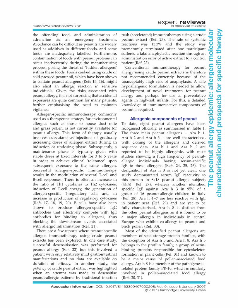
Allergenic components of peanutTo date, eight peanut allergens have beenrecognised officially, as summarised in Table 1.The three main peanut allergens – Ara h 1,Ara h 2 and Ara h 3 – are well characterised,with cloning of the allergens and derivedsequence data. Ara h 1 and Ara h 2 arereported to be highly allergenic, with moststudies showing a high frequency of peanut-allergic individuals having serum-specificIgE to these allergens (Refs 24, 25, 26). Thedesignation of Ara h 3 is not yet clear: onestudy demonstrated serum IgE reactivity tothis protein in 8/18 peanut allergic subjects(44%) (Ref. 27), whereas another identifiedspecific IgE against Ara h 3 in 95% of agroup of 16 peanut-allergic children in Italy(Ref. 28). Ara h 4–7 are less reactive with IgEin patient sera (Ref. 29) and are yet to befully characterised. Ara h 8 is distinct fromthe other peanut allergens as it is found to bea major allergen in individuals in centralEurope who exhibit co-allergy to peanut andbirch pollen (Ref. 30).
Most of the identified peanut allergens aremembers of seed storage protein families, withthe exception of Ara h 5 and Ara h 8. Ara h 5belongs to the profilin family, a group of actin-binding proteins responsible for cytoskeletonformation in plant cells (Ref. 31) and known tobe a major cause of pollen-associated foodallergy. Ara h 8 is a member of the pathogenesis-related protein family PR-10, which is similarlyinvolved in pollen-associated food allergy(Refs 30, 31).
expert reviewshttp://www.expertreviews.org/ in molecular medicine
4Accession information: DOI: 10.1017/S1462399407000208; Vol. 9; Issue 1; January 2007
&2007 Cambridge University Press
The
pea
nutallergyep
idem
ic:a
llergen
molecu
lar
charac
terisa
tionan
dprosp
ects
forsp
ecifictherap
y

Table
1.Molecu
larch
arac
teristicsofpea
nutallergen
s
Pea
nutallergen
Ara
h1
Ara
h2
Ara
h3
Ara
h4
Ara
h5
Ara
h6
Ara
h7
Ara
h8
Molec
ular
mas
s(kDa)
63.5
(Ref.2
4),
68(Ref.2
6)17
(Ref.2
5),
17.5
(Ref.4
3),
17.3
(Ref.2
9)
57 (Ref.2
9)35
.9(Ref.2
9)14 (Ref.2
9)14
.5(Ref.2
9)15
.8(Ref.2
9)16
.9(Ref.3
0)
Protein
family
Vicilin(Ref.2
6)Con
glutin
(Ref.4
3)Glycinin
(Ref.2
7)Le
gumin
(Ref.2
7)
Glycinin
(Ref.2
9)Profilin
(Ref.2
9)Con
glutin
(Ref.2
9)Con
glutin
(Ref.2
9)PR-10
protein
(Ref.3
0)
Allergen
icity
100%
(Ref.2
4),
94%
(Ref.2
6),6
5%(Ref.2
9)
100%
(Ref.2
5),
85%
(Ref.2
9)44
%(Ref.2
7),
95%
(Ref.2
8)53
%(Ref.2
9)13
%(Ref.2
9)38
%(Ref.2
9)43
%(Ref.2
9)75
–85
%(Ref.3
0)
Gen
bank
Acc
ession
no.
L344
02L7
7197
AF0
9354
1AF0
8682
1AF0
5961
6AF0
9284
6AF0
9173
7AY
3280
88
B-celle
pito
pes
reported
Yes
[L(Ref.4
2),
C(Ref.4
5)]
Yes
[L(Ref.4
3)]
Yes
[L(Ref.2
7)]
No
No
No
No
No
T-ce
llep
itopes
reported
No
Yes(Ref.4
7)No
No
No
No
No
No
Hyp
oallergen
icreco
mbinan
tmutan
tproduc
ed?
Yes
(Ref.7
2)Ye
s(Refs69
,70)
Yes
(Ref.7
1)No
No
No
No
No
Abbreviations
:C,c
onform
ationa
l;L,
linea
r.
expert reviewshttp://www.expertreviews.org/ in molecular medicine
5Accession information: DOI: 10.1017/S1462399407000208; Vol. 9; Issue 1; January 2007
&2007 Cambridge University Press
The
pea
nutallergyep
idem
ic:a
llergen
molecu
lar
charac
terisa
tionan
dprosp
ects
forsp
ecifictherap
y

Biochemical properties of peanut allergensFood allergens and their corresponding IgE-binding epitopes are thought to possessphysicochemical properties that conferresistance to digestive enzymes and thermalprocessing. This increases their allergenicity byenhancing their ability to reach the intestinalmucosa. Ara h 1 is capable of forming stabledimers, trimers and larger complexes uponheating without affecting IgE reactivity (Ref. 32).Treatment of Ara h 1 with gastrointestinal enzymessuch as pepsin, trypsin and chymotrypsinproduces large proteolytic fragments thatretain binding affinity for serum IgE frompeanut-allergic individuals (Ref. 33). Theresistance of Ara h 1 to degradation followingheating and treatment with digestive enzymesmay be related to its stable, homotrimericstructure (Ref. 33). This monomer–monomerinteraction reduces access to the catalytic siteswithin the protein, allowing Ara h 1 to surviveintact during food processing or passage alongthe digestive tract, thus contributing to itspotency as an allergen.
Similarly, Ara h 2 and Ara h 6 retainallergenicity following proteolytic digestion aswell as heat treatment (Refs 34, 35). Structuralstudies revealed that following digestion, bothallergens yielded immunologically active corestructures that were able to induce release ofinflammatory mediators even in the context ofdecreased serum IgE binding (Ref. 35). Ara h 2shares sequence homology with trypsininhibitors and can act as a weak trypsin inhibitorwith increased activity following roasting, thusprotecting itself as well as Ara h 1 from trypsindigestion (Ref. 36). This function of Ara h 2may contribute to the overall allergenicproperties of peanut proteins through increasedresistance to digestive enzymes. By contrast,Ara h 8 has low stability to heat and gastricdigestion (Ref. 30).
The Maillard reaction is also a contributingfactor to the allergenicity of peanuts (Refs 37, 38,39). This is a nonenzymatic reaction between aprotein and a reducing sugar that occurs duringthermal processing and cooking (Ref. 40). Theamino groups of proteins become glycosylatedto form Amadori products, which degrade intodicarbonyl intermediates. These intermediarycompounds react with amino groups of proteinsto form stable end products known as advancedglycation end products. Chung and Champagne
(Ref. 37) demonstrated that advanced glycationend products, formed by heating a previouslynonallergenic peanut lectin protein in thepresence of sugars, can effectively compete withuntreated peanut allergens for serum IgEantibodies, suggesting that the Maillard reactioncan convert a nonallergenic protein intoa potentially allergenic protein. Similarly,Maleki and colleagues observed that roastedpeanut proteins inhibited IgE binding toraw peanut proteins more effectively (90-foldhigher) than raw peanut proteins (Ref. 38).Both studies concluded that the presence ofadvanced glycation end products in heat-treatedpeanuts contributed to their overall heightenedallergenicity.
Epitope mappingIdentification of suitable peanut allergenpreparations for specific immunotherapyrequires detailed characterisation of sites thatinteract with antibodies/B cells and T cells.Antibodies and B cells interact with nativeallergens in their conformational state, whereasT cells interact with linear peptides presented inthe context of MHC molecules on antigen-presenting cell surfaces. Conformationalepitopes recognised by antibodies comprisediscontinuous short linear sequences that areclosely associated in the correctly foldedmolecule, but some antibody epitopes are longerlinear sequences. It has been suggested thatchildren with milk allergy whose predominantIgE reactivity is against conformational epitopesare more likely to develop tolerance to milk thanthose who react against linear epitopes (Ref. 41),but whether this relationship occurs for peanutallergens is not known.
B-cell epitopesTo date, most studies on peanut IgE reactivityhave focused on the more readily analysedlinear epitopes. The linear IgE-binding epitopesof Ara h 1, Ara h 2 and Ara h 3 were mappedusing synthetic peptides (Table 1). Typicallythese epitopes ranged from six to ten aminoacids in length, and in all cases the epitopescould be rendered nonreactive to IgE by alaninesubstitution of a single amino acid residue(Refs 27, 42, 43). Structural studies that generatedthe tertiary structure of Ara h 1, using the highlyhomologous phaseolin protein in homology-based modelling, allowed the positional
expert reviewshttp://www.expertreviews.org/ in molecular medicine
6Accession information: DOI: 10.1017/S1462399407000208; Vol. 9; Issue 1; January 2007
&2007 Cambridge University Press
The
pea
nutallergyep
idem
ic:a
llergen
molecu
lar
charac
terisa
tionan
dprosp
ects
forsp
ecifictherap
y

identification of the linear IgE-binding epitopesof Ara h 1 within its native conformation.The majority of amino acid residues identifiedas critical for binding to IgE were distributedon the surface of the molecule in epitopeclusters (Refs 44, 45). The presentation ofclustered epitopes to mast cells and basophilsmay result in a more efficient release ofmediators, which may explain the severe clinicalsymptoms associated with peanut allergy. Asmentioned previously, Ara h 1 is capable ofhigher-order aggregation, forming a stabletrimeric complex through interactions betweenhydrophobic amino acid residues (Refs 33, 44).Structural analysis of the Ara h 1 monomeridentified hydrophobic regions at each end ofthe molecule acting as contact points fortrimer formation, with most of the IgE-bindingepitopes clustered around these contact points(Fig. 2).
Diversity of IgE recognition of linear epitopesof peanut allergens has been shown to correlatewith the severity of clinical symptoms. Using20-mer peptides corresponding to the entireprimary sequences of Ara h 1, Ara h 2 and Ara h3, it was found that peanut-specific serum IgEfrom subjects with a history of severe systemicallergic reactions displayed a higher degree ofepitope diversity in comparison to those whoexperienced only cutaneous reactions (Ref. 46).Analysis of individual patients revealed nodistinct pattern of linear-epitope recognition,confirming the diversity of the IgE response topeanut allergens between peanut-allergicindividuals.
T-cell epitopesBy contrast to B-cell epitopes, data on the T-cellepitopes of peanut allergens are extremelylimited. To date, dominant sites of T-cellreactivity have been reported for only onepeanut allergen, Ara h 2, and the core T-cellepitopes within these sites have not yet beenidentified. Glaspole et al. identified two highlyimmunogenic T-cell-reactive regions: Ara h 2(19–47) and Ara h 2 (73–119). Peptides spanningthese two regions induced strong T-cellproliferation associated with a Th2-typecytokine response (Ref. 47). Knowledge of thedominant T-cell epitopes of allergens is criticalinformation for the development of a T-cell-targeted vaccine for peanut-specific allergenimmunotherapy.
Peanut allergen crossreactivityCrossreactivity between peanutand tree nutsClinical studies indicate that peanut-allergicsubjects, particularly adults, usually exhibitmultiple sensitivities to both peanut and treenuts. In specialist allergy clinics, the prevalenceof co-allergy to peanut and tree nuts wasreported as 35–40% (Refs 48, 49), although inour experience up to 80% of adult peanut-allergic patients are clinically allergic to one ormore tree nuts (R. O’Hehir, unpublished).Sicherer and colleagues examined the serologyof peanut-allergic children and found significantcorrelations between the level of peanut-specificIgE and tree-nut-specific IgE antibodies. Inparticular, peanut-specific IgE levels correlatedwith IgE levels for hazelnut, Brazil nut andalmond (Ref. 49). Given this, several studieshave investigated the presence of crossreactiveallergens in peanut and various tree nuts.
Molecular model of the Ara h 1 trimer
Expert Reviews in Molecular Medicine 2007Published by Cambridge University Press
Figure 2. Molecular model of the Ara h 1 trimer.A space-filled, homology-based model of theAra h 1 trimer shows that the majority of IgE-binding epitopes (red) are located in areas close tomonomer–monomer contact. Reprinted fromRef. 33 (& 2000 The American Association ofImmunologists, Inc.), with permission from TheJournal of Immunology and Gary Bannon(Monsanto, St Louis, MO, USA).
expert reviewshttp://www.expertreviews.org/ in molecular medicine
7Accession information: DOI: 10.1017/S1462399407000208; Vol. 9; Issue 1; January 2007
&2007 Cambridge University Press
The
pea
nutallergyep
idem
ic:a
llergen
molecu
lar
charac
terisa
tionan
dprosp
ects
forsp
ecifictherap
y

IgE crossreactivity was demonstrated betweenpeanut and the tree-nuts pistachio, macadamia,almond, Brazil nut and hazelnut (Refs 50, 51,52). In the majority of these cases the identity ofthe crossreacting allergen(s) was not established,with the exception of Ara h 2, which shares IgE-binding epitopes with almond and Brazil nutallergens (Ref. 53).
Although peanut is a legume and istaxonomically distantly related to tree nuts(Fig. 3), they are both defined as ‘edible’ seedsand perform similar functions in plantdevelopment. Indeed, several known peanutand tree-nut allergens belong to the sameprotein families. The major peanut allergen,
Ara h 1, is a member of the vicilin family ofseed storage proteins (Ref. 24) together withallergens from walnut, cashew and hazelnut(Refs 54, 55, 56). Also, 2S albumin seed storageproteins, implicated as allergens in almond,Brazil nut, hazelnut and walnut (Refs 56, 57, 58,59), are related to conglutin seed storageproteins, which include allergens from almond(Ref. 57) and peanut – namely Ara h 2, Ara h 6and Ara h 7 (Refs 25, 29). Legumins or 11Sglobulins are seed storage proteins shown to beallergenic in peanut (Ara h 3), cashew, hazelnutand walnut (Refs 27, 56, 60, 61, 62). Therefore,it is highly likely that a number of peanutallergens are responsible for the observed
Fabales(Order)
Proteales(Order)
Rosales(Order)
Sapindales(Order)
Arachishypogaea(Peanut)
Macadamiaintegrifolia
(Macadamia nut)
Prunus dulcis(Almond)
Ivesiapityocharis(Pine nut)
Corylusavellana
(Hazelnut)
Juglansregia
(Walnut)
Lecythidales(Order)
Plantae(Kingdom)
Tracheobionta(Subkingdom)
Spermatophyta(Superdivision)
Magnoliophyta(Division)
Magnoliopsida(Class)
Fagales(Order)
Juglandales(Order)
Fabacae(Pea family)
Proteaceae(Protea family)
Rosaceae(Rose family)
Betulaceae(Birch family)
Juglandaceae(Walnut family)
Anacardiaceae(Sumac family)
Lecythidaceae(Brazil nut family)
Anacardiumoccidentale
(Cashew nut)Pistacia vera
(Pistachio nut)
Bertholletiaexcelsa
(Brazil nut)
Rosidae(Subclass)
Hamamelidae(Subclass)
Dilleniidae(Subclass)
Taxonomic classification of peanut and tree nuts
Expert Reviews in Molecular Medicine © 2007 Cambridge University Press
Figure 3. Taxonomic classification of peanut and tree nuts. Many people suffering from peanut allergyare also allergic to one or more tree nuts. Although peanut and a number of tree nuts belong to differentplant families, they arise from the same class, namely Magnoliopsida, and therefore may containhomologous proteins that play a role in allergenic crossreactivity. Tree nuts that have been shown to shareIgE-binding epitopes with peanuts include almond, Brazil nut, hazelnut, macadamia and pistachio.The classification shown is based on information at the website of the US Department of Agriculture(http://plants.usda.gov/).
expert reviewshttp://www.expertreviews.org/ in molecular medicine
8Accession information: DOI: 10.1017/S1462399407000208; Vol. 9; Issue 1; January 2007
&2007 Cambridge University Press
The
pea
nutallergyep
idem
ic:a
llergen
molecu
lar
charac
terisa
tionan
dprosp
ects
forsp
ecifictherap
y
IgE crossreactivity between peanut and certaintree nuts.
Clinical and biological relevance of IgEcrossreactivityWhether or not in vitro allergen crossreactivitytranslates into clinical hypersensitivity iscontroversial. In addition to peanut, the legumefamily includes soybean, pea, lima bean andgreen bean; however, although these foodsare closely related taxonomically, clinicalhypersensitivity to more than one legume is rare(Refs 63, 64). van der Veen and colleaguesobserved that a third of patients sensitised tograss pollen (including timothy and orchardgrass as confirmed by serum specific IgE test)had significant serum levels of peanut-specificIgE antibodies but no clinical symptoms(Ref. 65). This phenomenon was attributedto crossreactive carbohydrate determinants(CCDs) – N-linked carbohydrate groups ofglycoprotein allergens that induce theproduction of crossreactive IgE antibodies tofood and grass pollen allergens (Refs 66, 67).Subjects with true peanut allergy had minimallevels of IgE antibodies specific for CCDs, incontrast to those with false-positive skin pricktests to peanut (Ref. 65).
IgE antibodies to CCDs also appear to havenegligible biological activity. In the same studyby van der Veen and colleagues, higherconcentrations of peanut extract (�1000-fold)were required for basophil histamine release inpatients with high levels of anti-CCD IgEantibodies with no peanut allergy comparedwith peanut-allergic patients, further validatingthe assertion that crossreactive IgE antibodiesdirected at CCDs are not indicative of clinicalsensitivity. However, biological activity ofpeanut-specific IgE antibodies that crossreactwith tree-nut allergens was demonstrated bybasophil-activation assays (Ref. 68). Humanbasophils passively resensitised with peanut-specific serum IgE antibodies from peanut- andtree-nut-allergic subjects become activatedfollowing incubation with almond and Brazil-nut extracts, as indicated by detection of CD63surface expression by flow cytometry (Ref. 68).Although the threshold doses of peanut, almondand Brazil-nut extracts required for basophilactivation were not compared, it is highlylikely that crosslinking of peanut-specific IgEantibodies present on effector cells by tree-nut
allergens is clinically relevant given the highincidence of cosensitisation to peanut and treenuts in peanut-allergic subjects.
Clinical implications:immunotherapeutic options
There is a critical need for a safe and effectivetreatment for peanut allergy given the severityand life-threatening nature of this condition.Currently, several immunotherapeutic methodsare being developed as proposed treatments forpeanut allergy, including the use of allergenderivatives as vaccines (Fig. 4). Conventionalsubcutaneous immunotherapy using crudeallergen extracts in order to modify the immuneresponse and obtain clinical tolerance to theallergen is not feasible for peanut allergybecause of the high risk of severe systemic sideeffects including anaphylaxis and death(Ref. 23), as discussed earlier in this review.
Modified peanut allergens with reducedIgE reactivityGiven the high-affinity IgE binding of peanutallergens, there is a need to develop suitableallergen preparations that do not crosslink IgEantibodies bound to effector cells. One approachis the engineering of recombinant allergens withconserved T-cell reactivity and decreased IgEbinding. Knowledge of the IgE-binding epitopesof peanut allergens has allowed preparation ofhypoallergenic variants with decreased IgEbinding (Refs 69, 70).
PCR mutagenesis targeting linear IgE-bindingepitopes was utilised to produce a hypoallergenicAra h 2 mutant; however, only 75% efficacy wasachieved, with serum IgE from 12/16 patientsdemonstrating a considerable decrease inreactivity to the Ara h 2 mutant in comparisonwith the wild-type allergen (Ref. 69). Similarly,another hypoallergenic Ara h 2 mutant wasproduced but this variant also retained IgEreactivity in a small cohort of peanut-allergicsubjects and induced significant mediator releasefrom basophils passively sensitised by peanut-allergic sera (Ref. 70).
Site-directed mutagenesis was utilised in thedevelopment of a hypoallergenic variant ofAra h 3. A 40 kDa acidic subunit of Ara h 3containing the four identified Ara h 3 linearIgE-binding epitopes was expressed using abacterial system, with the critical residues forIgE binding targeted for point mutations by
expert reviewshttp://www.expertreviews.org/ in molecular medicine
9Accession information: DOI: 10.1017/S1462399407000208; Vol. 9; Issue 1; January 2007
&2007 Cambridge University Press
The
pea
nutallergyep
idem
ic:a
llergen
molecu
lar
charac
terisa
tionan
dprosp
ects
forsp
ecifictherap
y

substitution with an alanine residue (Ref. 71).The resulting modified protein was still IgE-reactive although binding was substantially
decreased in comparison with the wild-typeprotein (Ref. 71). A hypoallergenic form of Ara h1 with T-cell reactivity has also been produced
Mutagenesis of IgE-binding sites
Dominant T-cell-epitope peptidesHypoallergenic variant
N
Allergen identification by serumIgE reactivity with peanut extract
IgE
Cloning of major allergens and recombinant-protein expression
a Identification, cloning and expression of peanut allergens
b Identification of IgE-binding sites c Identification of dominant T-cell-reactive sites
T-ce
ll re
spon
se
Overlapping allergen peptides
Potential specific immunotherapeutic strategies for the treatment of peanut allergy
Expert Reviews in Molecular Medicine C 2007 Cambridge University Press
C
Figure 4. Potential specific immunotherapeutic strategies for the treatment of peanut allergy. Theidentification and characterisation of peanut allergens allows for the development of vaccines that couldpotentially be used for the treatment of peanut allergy. (a) Peanut allergens can be identified using serum IgEfrom peanut-allergic individuals and subsequently cloned, sequenced and expressed as recombinantproteins. (b) Following the identification of the IgE-binding sites on the peanut allergens, mutations can beintroduced into the peanut allergen gene to render these sites nonreactive to IgE. Expression of the mutatedpeanut allergen construct will result in the production of hypoallergenic variants that can be used forimmunotherapy. (c) Alternatively, overlapping peptides spanning the entire protein sequence of peanutallergens can be used in T-cell assays to identify peptides encoding the dominant T-cell epitopes that canalso be utilised in a vaccine. Ultimately the aim of using hypoallergenic peanut-allergen variants and/orT-cell-epitope peptide vaccines is to induce tolerance to peanut allergens in susceptible individuals withoutthe IgE-mediated side effects.
expert reviewshttp://www.expertreviews.org/ in molecular medicine
10Accession information: DOI: 10.1017/S1462399407000208; Vol. 9; Issue 1; January 2007
&2007 Cambridge University Press
The
pea
nutallergyep
idem
ic:a
llergen
molecu
lar
charac
terisa
tionan
dprosp
ects
forsp
ecifictherap
y

(Ref. 72) but the characterisation of this modifiedallergen has been limited.
The retention of IgE reactivity with the Ara h 2and Ara h 3 variants following mutation ofthe linear IgE-binding epitopes suggests thatconformational B-cell epitopes should also bedisrupted to avoid IgE-mediated adversereactions in patients. In view of the multiplesensitivities to peanut allergens in most patients(Ref. 73), it is likely that a cocktail ofhypoallergenic mutants would be required in avaccine.
T-cell-epitope peptide vaccinesAn alternative approach to specificimmunotherapy uses short T-cell-epitopepeptides that lack IgE binding. This strategyshows promise in the treatment of certainallergies and is particularly attractive becausethe peptides are too short to crosslink effector-cell-bound IgE antibodies. In preclinical studies,the administration of peptides based ondominant T-cell epitopes of allergens from catand house dust mites induced specific T-celltolerance in mice (Refs 74, 75). This approachhas been encouraging in clinical trials usingallergen peptides for cat allergy (Refs 76, 77) andbee venom allergy (Ref. 78), and experimentaldata with a potent allergen of natural rubberlatex have shown some promise (Ref. 79).Treatment with multiple doses of catallergen (Fel d 1) peptides inhibited early- andlate-phase allergic reactions to the wholeallergen, with an associated decrease in Th1 andTh2 responses and an increase in IL-10production by peripheral blood mononuclearcells (Ref. 76). Furthermore, this treatmentimproved nasal symptom scores in subjectswith allergic rhinitis (Ref. 77). In the case ofallergy to bee venom, immunotherapy usingT-cell-epitope peptides of the major beevenom allergen, phospholipase A2 (PLA2),protected patients from challenge with PLA2
(Ref. 78). Concurrent decreases in PLA2-allergen-induced T-cell proliferation andcytokine secretion were detected, consistentwith T-cell anergy.
A similar therapeutic approach for peanutallergy requires full characterisation of thedominant T-cell epitopes of the major peanutallergens. While dominant T-cell epitopes of Arah 2 have been reported (Ref. 47), there is noinformation available on Ara h 1 T-cell epitopes.
In addition, if Ara h 3 is also confirmed as amajor peanut allergen, T-cell epitopes of Ara h 3will also need definition to ensure that thenecessary dominant T-cell-reactive sites areincluded in a potential vaccine.
Nonspecific immunotherapeutic strategiesNon-allergen-specific strategies have also beenconsidered for the treatment of peanut allergy.Anti-human-IgE therapy showed promise instudies of asthma and allergic rhinitis. Onepreparation used in such therapy is TNX-901, ahumanised IgG1 monoclonal antibody specificfor an epitope in the CH3 region of IgE – theregion that binds to the high-affinity Fc1RIreceptor on mast cells and basophils (Ref. 80). Inone study on peanut-allergic patients, TNX-901was administered subcutaneously every 4 weeksfor 16 weeks and subsequent peanut foodchallenges demonstrated a significant increasein the threshold dose of peanut flour elicitingsymptoms (the new threshold dose wasequivalent to approximately six to eightpeanuts) (Ref. 80). Although this treatment wasnot curative, the risk of a severe or fatal reactionafter accidental ingestion was diminished. Thenonspecific nature of this form of treatment maybe advantageous in patients with food allergieswhere a strict diet is difficult to manage andspecific immunotherapy is not available, as isthe case with peanut allergy. Conceptually, it hasappeal for patients who are extremely sensitiveto more than one allergen from any givensource. However, high patient commitment andcompliance would be needed and the costof such therapy is considerable. Currently, analternative humanised anti-IgE antibody isbeing tested for peanut allergy for futuretherapeutic use (Ref. 81). Other nonspecifictherapies reported to suppress peanut-inducedanaphylaxis in murine studies include theadministration of IL-12 (Ref. 82) and treatmentwith a Chinese herbal formula, FAHF-1 (Ref. 83),but efficacy and safety in humans have not beenshown.
Humanised anti-IgE antibody treatmentmight be useful as an adjunct during theinduction phase of conventional injectionallergen-specific immunotherapy. Intuitively, thehigh potency of crude peanut allergens suggeststhat hypoallergenic extracts or T-cell-epitopepeptide preparations would still be preferablefor safety because of the difficulty in delineating
expert reviewshttp://www.expertreviews.org/ in molecular medicine
11Accession information: DOI: 10.1017/S1462399407000208; Vol. 9; Issue 1; January 2007
&2007 Cambridge University Press
The
pea
nutallergyep
idem
ic:a
llergen
molecu
lar
charac
terisa
tionan
dprosp
ects
forsp
ecifictherap
y

the optimal dose of anti-IgE therapy that wouldgive a satisfactory level of protection fromadverse reactions for every peanut-allergicindividual. Nevertheless, the possibilities ofsublingual routes (see next section) with theirapparently decreased adverse events remain tobe fully explored.
Research in progress and outstandingresearch questions
Sublingual immunotherapy – analternative route?Currently, much of the research on peanutallergy is being driven by the need to providean effective immunotherapy for this life-threatening food allergy. As discussed above,several immunotherapeutic interventions havebeen proposed for peanut allergy, with theultimate goal of inducing tolerance to peanuts.In general, the route of administration forconventional immunotherapy is subcutaneous;however, local routes, most notably sublingual,are being investigated as an alternative.Sublingual immunotherapy (SLIT) has raisedsubstantial interest because it is noninvasive andis reported to be efficacious with minimaladverse events. In this immunotherapy regimen,allergen extract is placed under the tonguefor 1–2 min and then either swallowed or spatout (Ref. 84). While SLIT has been utilisedsuccessfully for environmental allergens(Refs 85, 86), it is conceptually difficult toimagine that it can be used for peanut-allergicindividuals, given that accidental ingestion ofpeanut extract can lead to life-threateninganaphylactic reactions. However, SLIT usingstandardised hazelnut extract to desensitisepatients who were allergic to hazelnut showedpromising results. Following treatment, therewas a significant increase in the threshold doseof hazelnut that elicited objective symptoms,together with an increase in the level ofhazelnut-specific IgG4 (Ref. 87). A low rate ofside effects was reported in the study (threereactions out of 1466 doses), which includedsubjects who previously experienced both localand systemic reactions after hazelnut ingestion(Ref. 87). Although complete tolerance tohazelnut was not achieved, the higher thresholddose after treatment would nevertheless providea level of protection in cases of accidentalingestion. It is yet to be seen if similar results canbe achieved with SLIT using peanut extract,
although the primary concern would be therisk of extreme side effects or the developmentof apparent initial benefit followed bytachyphylaxis (resistance to benefit) with therisk of a subsequent severe reaction.
An alternative strategy to avoid IgE-mediatedreactions is to use hypoallergenic preparationsfor SLIT. However, whether IgE-mediateduptake of the allergen at the oral mucosa isimportant for tolerance induction remains to beresolved (Ref. 84). The need for IgE-reactiveallergen preparations for successful SLIT couldpose difficulties in the treatment of peanutallergy, especially in extremely sensitivepatients. A high level of caution would beneeded during clinical investigation withperhaps an emphasis on the desirability of goodanimal models for proof of concept studies.
Burks and colleagues are currentlyinvestigating oral immunotherapy in childrenwith peanut allergy (http://www.foodallergyproject.org/Duke-Burks.pdf). Thechallenge will be to determine the duration oftreatment required (3 years, 5 years or lifelong)and a safe dosing interval. Again, complianceconcerns with chronic therapy and risks oftachyphylaxis will need careful considerationwith the use of crude extracts, because a lapse ofregimen could result in a heightened risk of acatastrophic allergic reaction.
Identification of crossreactive peanutand tree-nut allergensWith the observation that cosensitisation topeanuts and tree nuts might be due to allergeniccrossreactivity, there is a need to identify thecrossreactive components. Such information willimprove the diagnosis and management ofthese allergies. The seed storage proteins, whichare conserved throughout different plantfamilies, are strong candidates for mediating theobserved crossreactivity. Membership of thesame protein family, however, does notnecessarily translate into allergeniccrossreactivity. For example, no crossreactivitywas demonstrated between Ara h 1 and anothermember of the vicilin family in walnut, Jug r 2(Ref. 54). The cashew allergen Ana o 1, anothervicilin protein, also showed no common IgE-binding epitopes despite sharing 45% aminoacid sequence similarity with Ara h 1 (Ref. 55). Itis likely that 50–70% amino acid sequencehomology is required for immunological B-cell
expert reviewshttp://www.expertreviews.org/ in molecular medicine
12Accession information: DOI: 10.1017/S1462399407000208; Vol. 9; Issue 1; January 2007
&2007 Cambridge University Press
The
pea
nutallergyep
idem
ic:a
llergen
molecu
lar
charac
terisa
tionan
dprosp
ects
forsp
ecifictherap
y
crossreactivity to occur between proteins (Ref. 88).Alternatively, homology between two proteinslimited to a small stretch of amino acids mightresult in crossreactivity if there are similaritiesin the tertiary structure (Ref. 89). Furthercharacterisation of potential crossreactivepeanut and tree-nut allergens at the level ofsecondary and tertiary molecular structure wouldprovide valuable insight into the molecularcharacteristics required for immunologicallyrelevant crossreactivity to occur. Evaluation usingfunctional assays such as basophil-activationassays will provide convincing evidence forclinically relevant crossreactivity.
Transgenic plants for the productionof hypoallergenic peanutsA novel strategy for decreasing the incidence ofpeanut allergy and, most significantly, avoidingthe risk of exposure to peanut allergens, is theengineering of transgenic plants to producehypoallergenic peanuts. This may be achievedby introducing antisense RNA copies of theallergen gene into the plant to suppress allergengene expression (Ref. 90). Using this approach,expression of a 16 kDa allergen was decreasedin rice seeds (Ref. 91) and expression of themajor allergen Gly m Bd 30 K was silencedin soybean seeds (Ref. 92). Importantly,suppression of the allergen in transgenicsoybeans did not interfere with the normal plantlife cycle, with no obvious differences in growth,development, reproduction and seed maturationcompared with wild-type soybeans (Ref. 92).
An alternative strategy, post-transcriptionalgene silencing (PTGS) by RNA degradation, isbeing trialled for the production ofhypoallergenic peanuts (Ref. 93). For this, anextra copy of an endogenous gene is introducedinto plants, leading to the degradation of RNAencoded by both the transgene and homologousendogenous gene (Ref. 94). Preliminary studiesshowed that transformation of peanut embryoswith a fragment of Ara h 2 mRNA results in theintegration of the Ara h 2 transgene into thepeanut genome followed by stable expressionthroughout plant development (Ref. 93). It ishypothesised that the truncated Ara h 2 mRNAwill be synthesised in the peanut seeds andsubsequently trigger the specific degradation ofendogenous Ara h 2 mRNA (Ref. 93). Thesuccess of this strategy is yet to be seen as noresults have been reported on the production of
peanuts lacking the Ara h 2 protein, if it isindeed possible. For clinical effect, it is likelythat peanuts also lacking Ara h 1 and Ara h 3would need to be engineered as minimumrequirements for decreased allergenicity in thepeanut-allergic population. However, thedeletion of at least three genes from the peanutgenome might have deleterious effects on plantgrowth and development and thus hamper theproduction of a hypoallergenic peanut.
Concluding remarksAn optimal strategy for the treatment andprevention of peanut allergy is currently lacking.Potential specific and nonspecific strategies havebeen considered and their combined use may bewarranted. A T-cell-targeted approach offersprospects for effective and safe specifictreatment of peanut allergy but requires furtherelucidation of dominant T- and B-cell epitopesof peanut allergens. Optimal strategies fordownregulating adverse immune responses topeanut allergens then require careful evaluationin preclinical studies using either short linearpeptides based on the T-cell epitopes or T-cell-reactive hypoallergenic mutants. Hypoallergenicmutants have been proposed for specifictreatment of allergic disease arising from otherallergens, but in the case of the potent peanutallergens, careful consideration must be made ofthe possibility of severe side effects. The IgEreactivity of any peanut mutant would need tobe completely abolished before clinical use. Todate, this has not been achieved and a morecomplete understanding of IgE-reactive sites ofpeanut allergens is required. While linear IgEepitopes of some peanut allergens have beenidentified, there is scant information regardingthe conformational IgE epitopes of peanutallergens. This is important given that spatialclustering of conformational IgE-bindingepitopes on the allergen surface might be crucialfor allergenic activity and might influence theintensity of the allergic response (Ref. 95). This isparticularly relevant for peanut allergensgiven the high incidence of adverse allergicreactions with potentially fatal consequences.Therefore, further molecular characterisationof peanut allergens, coupled with improvedunderstanding of the immune mechanismsunderlying the allergic response to peanut, areessential components for the development ofimmunotherapeutic strategies.
expert reviewshttp://www.expertreviews.org/ in molecular medicine
13Accession information: DOI: 10.1017/S1462399407000208; Vol. 9; Issue 1; January 2007
&2007 Cambridge University Press
The
pea
nutallergyep
idem
ic:a
llergen
molecu
lar
charac
terisa
tionan
dprosp
ects
forsp
ecifictherap
y
Acknowledgements and fundingFinancial support from the National Healthand Medical Research Council of Australia, theAlfred Research Trusts and the Ilhan FoodAllergy Foundation is gratefully acknowledged.The authors thank the reviewers for their helpfulcomments.
References1 Sicherer, S.H., Munoz-Furlong, A. and Sampson,
H.A. (2003) Prevalence of peanut and tree nut
allergy in the United States determined by means
of a random digit dial telephone survey: a 5-year
follow-up study. J Allergy Clin Immunol 112,
1203-1207
2 Grundy, J. et al. (2002) Rising prevalence of allergy
to peanut in children: data from 2 sequential
cohorts. J Allergy Clin Immunol 110, 784-789
3 Sampson, H.A., Mendelson, L. and Rosen, J.P.
(1992) Fatal and near-fatal anaphylactic reactions
to food in children and adolescents. N Engl J Med
327, 380-384
4 Bock, S.A., Munoz-Furlong, A. and Sampson, H.A.
(2001) Fatalities due to anaphylactic reactions to
foods. J Allergy Clin Immunol 107, 191-193
5 Lack, G. et al. (2003) Factors associated with the
development of peanut allergy in childhood. N
Engl J Med 348, 977-985
6 Kaufman, H.S. (1971) Allergy in the newborn: skin
test reactions confirmed by the Prausnitz-Kustner
test at birth. Clin Allergy 1, 363-367
7 Vadas, P. et al. (2001) Detection of peanut allergens
in breast milk of lactating women. JAMA 285,
1746-1748
8 Sampson, H.A. (2002) Peanut allergy. N Engl J
Med 346, 1294-1299
9 Bock, S.A. and Atkins, F.M. (1989) The natural
history of peanut allergy. J Allergy Clin Immunol
83, 900-904
10 Skolnick, H.S. et al. (2001) The natural history of
peanut allergy. J Allergy Clin Immunol 107,
367-374
11 Prioult, G. and Nagler-Anderson, C. (2005)
Mucosal immunity and allergic responses: lack of
regulation and/or lack of microbial stimulation?
Immunol Rev 206, 204-218
12 Strobel, S. and Mowat, A.M. (2006) Oral tolerance
and allergic responses to food proteins. Curr Opin
Allergy Clin Immunol 6, 207-213
13 Mosmann, T.R. et al. (1986) Two types of murine
helper T cell clone. I. Definition according to
profiles of lymphokine activities and secreted
proteins. J Immunol 136, 2348-2357
14 Gleich, G.J. (2000) Mechanisms of eosinophil-
associated inflammation. J Allergy Clin Immunol
105, 651-663
15 Hourihane, J.O. et al. (1997) Randomised, double
blind, crossover challenge study of allergenicity of
peanut oils in subjects allergic to peanuts. BMJ
314, 1084-1088
16 Hoffmann, D.R. and Collins-Williams, C. (1994)
Cold pressed peanut oils may contain peanut
allergen. J Allergy Clin Immunol 93, 801-802
17 Secrist, H. et al. (1993) Allergen immunotherapy
decreases interleukin 4 production in CD4þ T cells
from allergic individuals. J Exp Med 178,
2123-2130
18 Gardner, L.M. et al. (2004) Induction of T
‘regulatory’ cells by standardized house dust mite
immunotherapy: an increase in CD4þ CD25þinterleukin-10þ T cells expressing peripheral
tissue trafficking markers. Clin Exp Allergy 34,
1209-1219
19 Bellinghausen, I. et al. (1997) Insect venom
immunotherapy induces interleukin-10
production and a Th2-to-Th1 shift, and changes
surface marker expression in venom-allergic
subjects. Eur J Immunol 27, 1131-1139
20 Jutel, M. et al. (2003) IL-10 and TGF-beta cooperate
in the regulatory T cell response to mucosal
allergens in normal immunity and specific
immunotherapy. Eur J Immunol 33, 1205-1214
21 Flicker, S. and Valenta, R. (2003) Renaissance of the
blocking antibody concept in type I allergy. Int
Arch Allergy Immunol 132, 13-24
22 Patriarca, G. et al. (2006) Oral rush desensitization in
peanut allergy: a case report. Dig Dis Sci 51, 471-473
23 Oppenheimer, J.J. et al. (1992) Treatment of peanut
allergy with rush immunotherapy. J Allergy Clin
Immunol 90, 256-262
24 Burks, A.W. et al. (1991) Identification of a major
peanut allergen, Ara h I, in patients with atopic
dermatitis and positive peanut challenges.
J Allergy Clin Immunol 88, 172-179
25 Burks, A.W. et al. (1992) Identification and
characterization of a second major peanut allergen,
Ara h II, with use of the sera of patients with
atopic dermatitis and positive peanut challenge.
J Allergy Clin Immunol 90, 962-969
26 Burks, A.W. et al. (1995) Recombinant peanut
allergen Ara h I expression and IgE binding in
patients with peanut hypersensitivity. J Clin Invest
96, 1715-1721
27 Rabjohn, P. et al. (1999) Molecular cloning and
epitope analysis of the peanut allergen Ara h 3.
J Clin Invest 103, 535-542
expert reviewshttp://www.expertreviews.org/ in molecular medicine
14Accession information: DOI: 10.1017/S1462399407000208; Vol. 9; Issue 1; January 2007
&2007 Cambridge University Press
The
pea
nutallergyep
idem
ic:a
llergen
molecu
lar
charac
terisa
tionan
dprosp
ects
forsp
ecifictherap
y
28 Restani, P. et al. (2005) Identification of the basic
subunit of Ara h 3 as the major allergen in a group
of children allergic to peanuts. Ann Allergy
Asthma Immunol 94, 262-266
29 Kleber-Janke, T. et al. (1999) Selective cloning of
peanut allergens, including profilin and 2S
albumins, by phage display technology. Int Arch
Allergy Immunol 119, 265-274
30 Mittag, D. et al. (2004) Ara h 8, a Bet v
1-homologous allergen from peanut, is a major
allergen in patients with combined birch pollen
and peanut allergy. J Allergy Clin Immunol 114,
1410-1417
31 Breiteneder, H. and Ebner, C. (2000) Molecular and
biochemical classification of plant-derived food
allergens. J Allergy Clin Immunol 106, 27-36
32 Koppelman, S.J. et al. (1999) Heat-induced
conformational changes of Ara h 1, a major peanut
allergen, do not affect its allergenic properties.
J Biol Chem 274, 4770-4777
33 Maleki, S.J. et al. (2000) Structure of the major
peanut allergen Ara h 1 may protect IgE-binding
epitopes from degradation. J Immunol 164,
5844-5849
34 Suhr, M. et al. (2004) Isolation and characterization
of natural Ara h 6: Evidence for a further peanut
allergen with putative clinical relevance based on
resistance to pepsin digestion and heat. Mol Nutr
Food Res 48, 390-399
35 Lehmann, K. et al. (2006) Structure and stability of
2S albumin-type peanut allergens: implications for
the severity of peanut allergic reactions. Biochem J
395, 463-472
36 Maleki, S.J. et al. (2003) The major peanut allergen,
Ara h 2, functions as a trypsin inhibitor, and
roasting enhances this function. J Allergy Clin
Immunol 112, 190-195
37 Chung, S.Y. and Champagne, E.T. (1999)
Allergenicity of Maillard reaction products from
peanut proteins. J Agric Food Chem 47, 5227-5231
38 Maleki, S.J. et al. (2000) The effects of roasting on
the allergenic properties of peanut proteins.
J Allergy Clin Immunol 106, 763-768
39 Chung, S.Y. et al. (2002) High-oleic peanuts are not
different from normal peanuts in allergenic
properties. J Agric Food Chem 50, 878-882
40 Namiki, M. (1988) Chemistry of Maillard reactions:
recent studies on the browning reaction
mechanism and the development of antioxidants
and mutagens. Adv Food Res 32, 115-184
41 Vila, L. et al. (2001) Role of conformational and
linear epitopes in the achievement of tolerance in
cow’s milk allergy. Clin Exp Allergy 31, 1599-1606
42 Burks, A.W. et al. (1997) Mapping and mutational
analysis of the IgE-binding epitopes on Ara h 1, a
legume vicilin protein and a major allergen in
peanut hypersensitivity. Eur J Biochem 245,
334-339
43 Stanley, J.S. et al. (1997) Identification and
mutational analysis of the immunodominant IgE
binding epitopes of the major peanut allergen
Ara h 2. Arch Biochem Biophys 342, 244-253
44 Bannon, G.A. et al. (1999) Tertiary structure and
biophysical properties of a major peanut allergen,
implications for the production of a hypoallergenic
protein. Int Arch Allergy Immunol 118, 315-316
45 Shin, D.S. et al. (1998) Biochemical and structural
analysis of the IgE binding sites on Ara h1, an
abundant and highly allergenic peanut protein.
J Biol Chem 273, 13753-13759
46 Shreffler, W.G. et al. (2004) Microarray
immunoassay: association of clinical history, in
vitro IgE function, and heterogeneity of allergenic
peanut epitopes. J Allergy Clin Immunol 113,
776-782
47 Glaspole, I.N. et al. (2005) Characterization of the
T-cell epitopes of a major peanut allergen, Ara h 2.
Allergy 60, 35-40
48 Ewan, P.W. (1996) Clinical study of peanut and nut
allergy in 62 consecutive patients: new features
and associations. BMJ 312, 1074-1078
49 Sicherer, S.H., Burks, A.W. and Sampson, H.A.
(1998) Clinical features of acute allergic reactions
to peanut and tree nuts in children. Pediatrics
102, e6
50 Parra, F.M. et al. (1993) Pistachio nut
hypersensitivity: identification of pistachio nut
allergens. Clin Exp Allergy 23, 996-1001
51 Sutherland, M.F. et al. (1999) Macadamia nut
anaphylaxis: demonstration of specific IgE
reactivity and partial cross-reactivity with
hazelnut. J Allergy Clin Immunol 104, 889-890
52 de Leon, M.P. et al. (2003) Immunological
analysis of allergenic cross-reactivity between
peanut and tree nuts. Clin Exp Allergy 33,
1273-1280
53 de Leon, M.P. et al. (2007) IgE cross-reactivity
between the major peanut allergen Ara h 2 and
tree nut allergens. Mol Immunol 44, 463-471
54 Teuber, S.S. et al. (1999) Identification and cloning
of a complementary DNA encoding a vicilin-like
proprotein, Jug r 2, from English walnut kernel
(Juglans regia), a major food allergen. J Allergy
Clin Immunol 104, 1311-1320
55 Wang, F. et al. (2002) Ana o 1, a cashew
(Anacardium occidental) allergen of the vicilin
expert reviewshttp://www.expertreviews.org/ in molecular medicine
15Accession information: DOI: 10.1017/S1462399407000208; Vol. 9; Issue 1; January 2007
&2007 Cambridge University Press
The
pea
nutallergyep
idem
ic:a
llergen
molecu
lar
charac
terisa
tionan
dprosp
ects
forsp
ecifictherap
y

seed storage protein family. J Allergy Clin
Immunol 110, 160-166
56 Pastorello, E.A. et al. (2002) Identification of
hazelnut major allergens in sensitive patients with
positive double-blind, placebo-controlled food
challenge results. J Allergy Clin Immunol 109,
563-570
57 Poltronieri, P. et al. (2002) Identification and
characterisation of the IgE-binding proteins 2S
albumin and conglutin gamma in almond (Prunus
dulcis) seeds. Int Arch Allergy Immunol 128,
97-104
58 Pastorello, E.A. et al. (1998) Sensitization to the
major allergen of Brazil nut is correlated with the
clinical expression of allergy. J Allergy Clin
Immunol 102, 1021-1027
59 Teuber, S.S. et al. (1998) Cloning and sequencing of
a gene encoding a 2S albumin seed storage protein
precursor from English walnut (Juglans regia), a
major food allergen. J Allergy Clin Immunol 101,
807-814
60 Wang, F. et al. (2003) Ana o 2, a major cashew
(Anacardium occidentale L.) nut allergen of the
legumin family. Int Arch Allergy Immunol 132,
27-39
61 Beyer, K. et al. (2002) Identification of an 11S
globulin as a major hazelnut food allergen in
hazelnut-induced systemic reactions. J Allergy
Clin Immunol 110, 517-523
62 Teuber, S.S. et al. (2003) Identification and cloning
of Jug r 4, a major food allergen from English
walnut belonging to the legumin group. J Allergy
Clin Immunol 111, S248
63 Bernhisel-Broadbent, J. and Sampson, H.A. (1989)
Cross-allergenicity in the legume botanical family
in children with food hypersensitivity. J Allergy
Clin Immunol 83, 435-440
64 Bernhisel-Broadbent, J., Taylor, S. and
Sampson, H.A. (1989) Cross-allergenicity in
the legume botanical family in children
with food hypersensitivity. II. Laboratory
correlates. J Allergy Clin Immunol 84, 701-709
65 van der Veen, M.J. et al. (1997) Poor biologic
activity of cross-reactive IgE directed to
carbohydrate determinants of glycoproteins.
J Allergy Clin Immunol 100, 327-334
66 Batanero, E. et al. (1996) Cross-reactivity between
the major allergen from olive pollen and unrelated
glycoproteins: evidence of an epitope in the glycan
moiety of the allergen. J Allergy Clin Immunol 97,
1264-1271
67 Petersen, A. et al. (1996) Ubiquitous structures
responsible for IgE cross-reactivity between
tomato fruit and grass pollen allergens. J Allergy
Clin Immunol 98, 805-815
68 de Leon, M.P. et al. (2005) Functional analysis of
cross-reactive immunoglobulin E antibodies:
peanut-specific immunoglobulin E sensitizes
basophils to tree nut allergens. Clin Exp Allergy
35, 1056-1064
69 Burks, A.W., King, N. and Bannon, G.A. (1999)
Modification of a major peanut allergen leads to
loss of IgE binding. Int Arch Allergy Immunol 118,
313-314
70 King, N. et al. (2005) Allergenic characteristics of a
modified peanut allergen. Mol Nutr Food Res 49,
963-971
71 Rabjohn, P. et al. (2002) Modification of peanut
allergen Ara h 3: effects on IgE binding and T cell
stimulation. Int Arch Allergy Immunol 128, 15-23
72 Bannon, G.A. et al. (2001) Engineering,
characterization and in vitro efficacy of the major
peanut allergens for use in immunotherapy. Int
Arch Allergy Immunol 124, 70-72
73 Lewis, S.A. et al. (2005) The promiscuity of
immunoglobulin E binding to peanut allergens, as
determined by Western blotting, correlates with
the severity of clinical symptoms. Clin Exp Allergy
35, 767-773
74 Briner, T. et al. (1993) Peripheral T cell tolerance
induced in naive and primed mice by
subcutaneous injection of peptides from the major
cat allergen Fel d 1. Proc Natl Acad Sci USA 90,
7608-7612
75 Hoyne, G.F. et al. (1993) Inhibition of T cell and
antibody responses to house dust mite allergen by
inhalation of dominant T cell epitope in naive and
sensitized mice. J Exp Med 178, 1783-1788
76 Oldfield, W.L., Larche, M. and Kay, A.B. (2002)
Effect of T-cell peptides derived from Fel d 1 on
allergic reactions and cytokine production in
patients sensitive to cats: a randomised controlled
trial. Lancet 360, 47-53
77 Alexander, C. et al. (2005) The effect of Fel d
1-derived T-cell peptides on upper and lower
airway outcome measurements in cat-allergic
subjects. Allergy 60, 1269-1274
78 Muller, U.R. et al. (1998) Successful
immunotherapy with T cell epitope peptides of
bee venom phospholipase A2 induces specific T
cell anergy in bee sting allergic patients. J Allergy
Clin Immunol 101, 747-757
79 Drew, A.C. et al. (2004) Hypoallergenic variants of
the major latex allergen Hev b 6.01 retaining
human T lymphocyte reactivity. J Immunol 173,
5872-5879
expert reviewshttp://www.expertreviews.org/ in molecular medicine
16Accession information: DOI: 10.1017/S1462399407000208; Vol. 9; Issue 1; January 2007
&2007 Cambridge University Press
The
pea
nutallergyep
idem
ic:a
llergen
molecu
lar
charac
terisa
tionan
dprosp
ects
forsp
ecifictherap
y
80 Leung, D.Y. et al. (2003) Effect of anti-IgE therapy
in patients with peanut allergy. N Engl J Med 348,
986-993
81 Teuber, S.S. and Beyer, K. (2004) Peanut, tree nut
and seed allergies. Curr Opin Allergy Clin
Immunol 4, 201-203
82 Lee, S.Y. et al. (2001) Oral administration of IL-12
suppresses anaphylactic reactions in a murine
model of peanut hypersensitivity. Clin Immunol
101, 220-228
83 Li, X.M. et al. (2001) Food Allergy Herbal Formula-
1 (FAHF-1) blocks peanut-induced anaphylaxis in
a murine model. J Allergy Clin Immunol 108,
639-646
84 Moingeon, P. et al. (2006) Immune mechanisms of
allergen-specific sublingual immunotherapy.
Allergy 61, 151-165
85 Dahl, R. et al. (2006) Efficacy and safety of
sublingual immunotherapy with grass allergen
tablets for seasonal allergic rhinoconjunctivitis.
J Allergy Clin Immunol 118, 434-440
86 Bousquet, J. et al. (1999) Sublingual-swallow
immunotherapy (SLIT) in patients with asthma
due to house-dust mites: a double-blind, placebo-
controlled study. Allergy 54, 249-260
87 Enrique, E. et al. (2005) Sublingual
immunotherapy for hazelnut food
allergy: a randomized, double-blind,
placebo-controlled study with a standardized
hazelnut extract. J Allergy Clin Immunol 116,
1073-1079
88 Aalberse, R.C. (2000) Structural biology of
allergens. J Allergy Clin Immunol 106, 228-238
89 Aalberse, R.C., Akkerdaas, J.H. and van Ree, R.
(2001) Cross-reactivity of IgE antibodies to
allergens. Allergy 56, 478-490
90 Moseley, B.E. (2001) How to make foods safer–
genetically modified foods. Allergy 56 Suppl 67,
61-63
91 Tada, Y. et al. (1996) Reduction of 14–16
kDa allergenic proteins in transgenic rice plants by
antisense gene. FEBS Lett 391, 341-345
92 Herman, E.M. et al. (2003) Genetic
modification removes an
immunodominant allergen from soybean. Plant
Physiol 132, 36-43
93 Dodo, H., Konan, K. and Viquez, O. (2005)
A genetic engineering strategy to eliminate
peanut allergy. Curr Allergy Asthma Rep 5,
67-73
94 Vaucheret, H., Beclin, C. and Fagard, M. (2001)
Post-transcriptional gene silencing in plants. J Cell
Sci 114, 3083-3091
95 Flicker, S. et al. (2006) Spatial clustering of the
IgE epitopes on the major timothy grass
pollen allergen Phl p 1: importance for
allergenic activity. J Allergy Clin Immunol 117,
1336-1343
Further reading, resources and contacts
The International Union of Immunological Societies Allergen (IUIS) Nomenclature Sub-committee websiteprovides the official list of allergens:
http://www.allergen.org
The Allergome website contains information and relevant publications of allergens that are officiallyrecognised by the IUIS Allergen Nomenclature Sub-committee
http://www.allergome.org
expert reviewshttp://www.expertreviews.org/ in molecular medicine
17Accession information: DOI: 10.1017/S1462399407000208; Vol. 9; Issue 1; January 2007
&2007 Cambridge University Press
The
pea
nutallergyep
idem
ic:a
llergen
molecu
lar
charac
terisa
tionan
dprosp
ects
forsp
ecifictherap
y

Features associated with this article
FiguresFigure 1. The major cellular interactions during the mucosal allergic immune response.Figure 2. Molecular model of the Ara h 1 trimer.Figure 3. Taxonomic classification of peanut and tree nuts.Figure 4. Potential specific immunotherapeutic strategies for the treatment of peanut allergy.
TableTable 1. Molecular characteristics of peanut allergens.
Citation details for this article
Maria P. de Leon, Jennifer M. Rolland and Robyn E. O’Hehir (2007) The peanut allergy epidemic: allergenmolecular characterisation and prospects for specific therapy. Expert Rev. Mol. Med. Vol. 9, Issue 1,January 2007, DOI: 10.1017/S1462399407000208
expert reviewshttp://www.expertreviews.org/ in molecular medicine
18Accession information: DOI: 10.1017/S1462399407000208; Vol. 9; Issue 1; January 2007
&2007 Cambridge University Press
The
pea
nutallergyep
idem
ic:a
llergen
molecu
lar
charac
terisa
tionan
dprosp
ects
forsp
ecifictherap
y