Schistosomula of Schistosoma mansoni use lysophosphatidylcholine to lyse adherent human red blood...
10
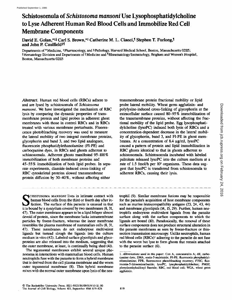
Schistosomula of Schistosoma mansoni Use Lysophosphatidylcholine to Lyse Adherent Human Red Blood Cells and Immobilize Red Cell Membrane Components David E. Golan,**ll Carl S. Brown,** Catherine M. L. Cianci,1 Stephen T. Furlong,1 and John P. Caulfield§1 Departments of* Medicine, *Pharmacology, and §Pathology, Harvard Medical School, Boston, Massachusetts 02115; U Hematology Division and Departments of II Medicine and ~ Rheumatology/Immunology, Brigham and Women's Hospital, Boston, Massachusetts 02115 Abstract. Human red blood cells (RBCs) adhere to and are lysed by schistosomula of Schistosoma mansoni. We have investigated the mechanism of RBC lysis by comparing the dynamic properties of trans- membrane protein and lipid probes in adherent ghost membranes with those in control RBCs and in RBCs treated with various membrane perturbants. Fluores- cence photobleaching recovery was used to measure the lateral mobility of two integral membrane proteins, glycophorin and band 3, and two lipid analogues, fluorescein phosphatidylethanolamine (FI-PE) and carbocyanine dyes, in RBCs and ghosts adherent to schistosomula. Adherent ghosts manifested 95-100% immobilization of both membrane proteins and 45-55 % immobilization of both lipid probes. In sepa- rate experiments, diamide-induced cross-linking of RBC cytoskeletal proteins slowed transmembrane protein diffusion by 30-40%, without affecting either transmembrane protein fractional mobility or lipid probe lateral mobility. Wheat germ agglutinin- and polylysine-induced cross-linking of glycophorin at the extracellular surface caused 80-95 % immobilization of the transmembrane proteins, without affecting the frac- tional mobility of the lipid probe. Egg lysophosphati- dylcholine (lysoPC) induced both lysis of RBCs and a concentration-dependent decrease in the lateral mobil- ity of glycophorin, band 3, and F1-PE in ghost mem- branes. At a concentration of 8.4 ~tg/ml, lysoPC caused a pattern of protein and lipid immobilization in RBC ghosts identical to that in ghosts adherent to schistosomula. Schistosomula incubated with labeled palmitate released lysoPC into the culture medium at a rate of 1.5 fmol/h per I(P organisms. These data sug- gest that lysoPC is transferred from schistosomula to adherent RBCs, causing their lysis. S CHISTOSOMA MANSONI lives in intimate contact with human blood cells from the third or fourth day after in- fection. The surface of this parasite is unusual in that it is bound by a syneytium covered by two membranes (8, 31, 47). The outer membrane appears to be a lipid bilayer almost devoid of protein, since the membrane lacks intramembrane particles by freeze-fracture, whereas the inner membrane resembles the plasma membrane of mammalian ceils (8, 31, 47). These membranes do not endocytose multivalent ligands but instead slough the ligands into the culture medium in vitro (42). Labeled surface glycolipids and glyco- proteins are also released into the medium, suggesting that the outer membrane, at least, is continually being shed (41). The tegumental membranes exhibit several unique phe- nomena in interactions with mammalian blood ceils. Human neutrophils fuse with the parasite to form a hybrid membrane that is derived from the cell plasma membrane and the worm outer tegumental membrane (8). This hybrid membrane mixes with the normal outer membrane upon lysis of the neu- trophil (9). Similar membrane fusions may be responsible for the parasite's acquisition of host membrane components such as murine histocompatibility antigens (23, 24, 43, 44) and membrane glycolipids (14, 15, 29). Further, human neu- trophils endocytose multivalent ligands from the parasite surface along with the surface components to which the ligands are bound (10). Paradoxically, the removal of these surface components does not produce structural alteration in the parasite membranes as seen by freeze-fracture or thin- section transmission microscopy. Unlike neutrophils, human red blood cells (RBCs) t adhering to the parasite do not fuse with the worm but lyse to form ghosts that remain attached to the parasite surface (6). 1. Abbreviations used in this paper: Con A, concanavalin A; diI, carbo- cyanine dyes; EMA, eosin-5-maleimide; F1-PE, fluorescein phosphatidyl- ethanolamine; FPR, fluorescence photobleaching recovery; FTSC, fluo- rescein-5-thiosemicarbazide; lysoPC, iysophosphafidylcholine; PMSF, phenylmethyisulfonyl fluoride; RBC, red blood cell; WGA, wheat germ agglutinin. © The RockefellerUniversity Press, 0021-9525/86/09/819/10 $1.00 The Journal of Cell Biology, Volume 103, September 1986 819-828 819 on February 24, 2016 jcb.rupress.org Downloaded from Published September 1, 1986
-
Upload
independent -
Category
Documents
-
view
0 -
download
0
Transcript of Schistosomula of Schistosoma mansoni use lysophosphatidylcholine to lyse adherent human red blood...
Schistosomula of Schistosoma mansoni Use Lysophosphatidylcholine to Lyse Adherent Human Red Blood Cells and Immobilize Red Cell Membrane Components David E. Golan,**ll Carl S. Brown,** Catherine M. L. Cianci,1 Stephen T. Furlong,1 and John P. Caulfield§1
Departments of* Medicine, *Pharmacology, and §Pathology, Harvard Medical School, Boston, Massachusetts 02115; U Hematology Division and Departments of II Medicine and ~ Rheumatology/Immunology, Brigham and Women's Hospital, Boston, Massachusetts 02115
A b s t r a c t . Human red blood cells (RBCs) adhere to and are lysed by schistosomula of Schis tosoma mansoni . We have investigated the mechanism of RBC lysis by comparing the dynamic properties of trans- membrane protein and lipid probes in adherent ghost membranes with those in control RBCs and in RBCs treated with various membrane perturbants. Fluores- cence photobleaching recovery was used to measure the lateral mobility of two integral membrane proteins, glycophorin and band 3, and two lipid analogues, fluorescein phosphatidylethanolamine (FI-PE) and carbocyanine dyes, in RBCs and ghosts adherent to schistosomula. Adherent ghosts manifested 95-100% immobilization of both membrane proteins and 45-55 % immobilization of both lipid probes. In sepa- rate experiments, diamide-induced cross-linking of RBC cytoskeletal proteins slowed transmembrane protein diffusion by 30-40%, without affecting either
transmembrane protein fractional mobility or lipid probe lateral mobility. Wheat germ agglutinin- and polylysine-induced cross-linking of glycophorin at the extracellular surface caused 80-95 % immobilization of the transmembrane proteins, without affecting the frac- tional mobility of the lipid probe. Egg lysophosphati- dylcholine (lysoPC) induced both lysis of RBCs and a concentration-dependent decrease in the lateral mobil- ity of glycophorin, band 3, and F1-PE in ghost mem- branes. At a concentration of 8.4 ~tg/ml, lysoPC caused a pattern of protein and lipid immobilization in RBC ghosts identical to that in ghosts adherent to schistosomula. Schistosomula incubated with labeled palmitate released lysoPC into the culture medium at a rate of 1.5 fmol/h per I(P organisms. These data sug- gest that lysoPC is transferred from schistosomula to adherent RBCs, causing their lysis.
S CHISTOSOMA MANSONI lives in intimate contact with human blood cells from the third or fourth day after in- fection. The surface of this parasite is unusual in that
it is bound by a syneytium covered by two membranes (8, 31, 47). The outer membrane appears to be a lipid bilayer almost devoid of protein, since the membrane lacks intramembrane particles by freeze-fracture, whereas the inner membrane resembles the plasma membrane of mammalian ceils (8, 31, 47). These membranes do not endocytose multivalent ligands but instead slough the ligands into the culture medium in vitro (42). Labeled surface glycolipids and glyco- proteins are also released into the medium, suggesting that the outer membrane, at least, is continually being shed (41).
The tegumental membranes exhibit several unique phe- nomena in interactions with mammalian blood ceils. Human neutrophils fuse with the parasite to form a hybrid membrane that is derived from the cell plasma membrane and the worm outer tegumental membrane (8). This hybrid membrane mixes with the normal outer membrane upon lysis of the neu-
trophil (9). Similar membrane fusions may be responsible for the parasite's acquisition of host membrane components such as murine histocompatibility antigens (23, 24, 43, 44) and membrane glycolipids (14, 15, 29). Further, human neu- trophils endocytose multivalent ligands from the parasite surface along with the surface components to which the ligands are bound (10). Paradoxically, the removal of these surface components does not produce structural alteration in the parasite membranes as seen by freeze-fracture or thin- section transmission microscopy. Unlike neutrophils, human red blood cells (RBCs) t adhering to the parasite do not fuse with the worm but lyse to form ghosts that remain attached to the parasite surface (6).
1. Abbreviations used in this paper: Con A, concanavalin A; diI, carbo- cyanine dyes; EMA, eosin-5-maleimide; F1-PE, fluorescein phosphatidyl- ethanolamine; FPR, fluorescence photobleaching recovery; FTSC, fluo- rescein-5-thiosemicarbazide; lysoPC, iysophosphafidylcholine; PMSF, phenylmethyisulfonyl fluoride; RBC, red blood cell; WGA, wheat germ agglutinin.
© The Rockefeller University Press, 0021-9525/86/09/819/10 $1.00 The Journal of Cell Biology, Volume 103, September 1986 819-828 819
on February 24, 2016
jcb.rupress.orgD
ownloaded from
Published September 1, 1986

The present studies investigate the mechanism of RBC ly- sis. Because cultures of schistosomula and RBCs contain a few adherent ghosts, many adherent and nonadherent RBCs, and an excess of parasite membrane, the membrane proper- ties of individual cells and ghosts must be measured in situ. Fluorescence photobleaching recovery (FPR) is ideal for this purpose, because it measures the dynamic properties of pro- tein and lipid in a single biological membrane without per- turbation of the membrane environment (3). In this tech- nique, a single cell membrane is observed in a fluorescence microscope, using a focused laser beam as the excitation source. A small area of the membrane is exposed to a brief, intense laser pulse, causing irreversible bleaching of the fluorophore in that area. Fluorescence recovery resulting from lateral diffusion of unbleached fluorophore into the bleached area is measured. Analysis of the fluorescence recovery curves yields the fraction of fluorescently labeled protein or lipid that is free to diffuse in the plane of the mem- brane (the mobile fraction, f ) , as well as the diffusion coefficient of the mobile fraction (D).
FPR is used to measure the lateral mobility of two integral membrane proteins, glycophorin and band 3, and two lipid probes, fluorescein phosphatidylethanolamine (FI-PE) and carbocyanine dyes (diD, in RBCs and ghosts adherent to schistosomula. These findings are compared with measure- ments obtained in RBCs subjected to a variety of membrane perturbants. Such experiments suggest that lysophospha- tidylcholine (lysoPC) is the agent responsible for RBC lysis on the parasite surface. In independent experiments, worms are labeled with 3H-palmitate to see whether they metabo- lize palmitate to lysoPC and, if so, whether the lysoPC is released into the culture medium, as predicted from the shedding of the outer surface membrane.
Materials and Methods
Reagents Fluorescein-5-thiosemicarbazide (FTSC), eosin-5-maleimide (EMA), and carbocyanine dyes [diI-Cn(3), diI-Ct4(3), diI-C18(3)] were purchased from Molecular Probes, Inc. (Junction City, OR). Diamide and poly-L-lysine were from Sigma Chemical Co. (St. Louis, MO). Wheat germ agglutinin 0VGA) and concanavalin A (Con A) were from Vector Laboratories (Burlin- game, CA). 9,10-3H-Palmitic acid (specific activity, 27.5 Ci/mmol) was from New England Nuclear (Boston, MA). FI-PE and phospholipids (egg phosphatidylcholine, egg lysoPC, egg phosphatidylethanolamine, bovine liver phosphatidylinositol, and bovine brain phosphatidylserine, all >99% pure) were from Avanti Polar-Lipids, Inc. (Birmingham, AL). The fatty acid composition of egg lysoPC is 70.5% palmitic acid, 25.7% stearic acid, 1.8 % uleic acid, and 0.7 % palmitoleic acid (Avanti Polar-Lipids, Inc., per- sonal communication). RPMI-1640 culture medium was from Gibco (Grand Island, NY). Sodium m-periodate and bovine serum albumin (essentially fatty acid and globulin free) (BSA) were from Sigma Chemical Co. EDTA was from Fisher Scientific Co. (Pittsburgh, PA). Phenylmethylsnlfonyl fluo- ride (PMSF) was from Eastman Organic Chemicals (Rochester, NY).
Fluorescent Labeling of Human RBCs and RBC Ghosts Glycophorin. RBC glycopborin was fluorescently labeled by conjugation of FTSC to glycophorin-linked (22) sialic acid moieties (27). Similar methods have been used by Abraham and Low (1) and by Cherry et al. (B) to label glycophorin selectively with other fluorescent probes. Fresh human blood was washed three times by centrifugation in phosphate-buffered saline (PBS)(128 mM sodium chloride, 10 mM sodium phosphate, pH 7.4) with 120 gM PMSF and 1 mM EDTA (PBS/PMSF/EDTA). Washed RBCs were stirred with an equal volume of 2 mM sodium periodate in PBS for 15 rain
at 0°C to oxidize selectively sialic acid exposed at the outer membrane sur- face (22). Unreacted periodate was removed by washing twice in 0.1 M glycerol in PBS (1) and once in PBS. The oxidized RBCs were incubated with an equal volume of FTSC, 0.5 mg/ml in PBS, for 60 rain at 0°C, then washed three times in PBS with 1% BSA (PBS/BSA) (1) and once in RPMI. Unless otherwise indicated, all operations were performed at 4°C in the dark. Under these labeling conditions 80% of the membrane-associated fluorescence co-migrated on SDS polyacrylamide gels with the sialoglyco- proteins PAS-1,2,3,4. Of this, 75% was coincident with glycophorin A (PAS-1,2) (21). The stoichiometry of labeling as determined by spec- trophotometry and RBC membrane protein assay was 1.9 fluorophores per glycophorin monomer, or 1 × 10⁵ fluorophores per RBC.
Band 3. RBC band 3 was fluorescently labeled by the technique of Nigg and Cherry (39). Briefly, fresh human RBCs washed in PBS/PMSF/EDTA were mixed with 0.2 mg/ml (final concentration) EMA in PBS with 10 mM glucose for 45 rain at room temperature. Excess fluorophore was removed by washing three times in PBS/BSA and once in RPMI, all at 4°C. Under these conditions >--80% of the membrane-associated fluorescence co- migrated on SDS polyacrylamide gels with band 3, as confirmed by selective extraction procedures on EMA-labeled RBC ghosts (39). The stoichiometry of labeling determined as above was 0.8 fluoropbores per band 3 monomer, or 1 x 10⁶ fluorophores per RBC.
FI-PE. A modification of the technique of Golan et al. (25) was used to incorporate FI-PE into the membranes of intact RBCs. FI-PE (2 mg) was dried from a stock solution in chloroform, 10 mi of PBS was added, and the mixture was bath sonicated at room temperature until clarified. 75 gl of the FI-PE mixture was added to 1.5 ml of washed, packed RBCs in 3.0 ml PBS and the mixture gently shaken at room temperature for 30 rain. La- beled cells were washed three times in PBS/BSA and once in RPMI. The stoichiometry of labeling, determined by comparing the average fluores- cence intensities of FTSC- and FI-PE-labeled RBCs, was 8 x 10⁵ fluoro- phores per RBC or 0.003 tool of FI-PE per mole of endogenous membrane lipid. This concentration of F1-PE does not perturb the dynamic behavior of RBC membrane lipid (unpublished observations). In some cases F1-PE- labeled RBCs were lysed in 40 vol of 5 mM sodium phosphate with 1 mM EDTA and 120 gM PMSF, pH 7.4, for 45 rain at 4°C. The resulting ghosts were pelleted by centrifugation and washed three to four times in the lysis buffer and once in RPMI, all at 4°C.
diI. 50 ttl of diI, 0.5 mg/mi in ethanol, was added to 0.5 ml of washed, packed human RBCs in 8.0 ml of PBS and the mixture gently shaken at 4°C for 30 rain. Excess fluorophore was removed by washing twice in PBS to which 1% BSA had been added (PBS/1% BSA)and once in RPMI/I% BSA. F1-PE-, FTSC-, and EMA-labeled RBCs each comprised a uniform popula- tion of fluorescent, intact discocytes, whereas diI labeled only 10% of the total RBCs and lysed ,x,50% of the RBCs.
Preparation of Schistosomula and Fluorescen t Labeling with Carbocyanine Dyes Schistosomnia were prepared as described (40, 42). 2,000 schistosomula were cultured overnight at 37°C and mixed in 1.5 mi PBS with 2 gl of diI, L0 mg/ml in ethanol, for 30 min at room temperature. The labeled somules were washed twice in PBS and once in RPMI/0.1% BSA.
Incubation of Schistosomula with RBCs 1,000 cultured schistosomula were mixed with 2 gl of fluorescenfly labeled RBCs in 100-500 ktl of RPMI/0.1% BSA and incubated at 37°C for 24-48 h. Controls were labeled RBCs or RBC ghosts incubated in the absence of somules. Immediately before FPR or photography, samples were treated with 10 mM eserine sulfate to immobilize the worms (42). This concentra- tion of eserine sulfate did not affect the lateral mobility of RBC membrane components in control RBCs or ghosts.
Diamide Treatment of RBCs RBCs were incubated with either 2 or 20 mM diamide in PBS at 37°C for 30 rain (30, 45), washed twice in PBS containing 10% glycerol, and washed once in PBS. Diamide treatment was performed either before or after fluorescent labeling. Both protocols yielded the same FPR results.
Incubation of RBCs on Poly-L-lysine-, WGA-, and Con A-Coated Slides FTSC-, EMA-, and Fl-PE-labeled RBCs were washed twice more in RPMI
The Journal of Cell Biology, Volume 103, 1986 820
on February 24, 2016
jcb.rupress.orgD
ownloaded from
Published September 1, 1986

and resuspended in 20 vol of this buffer. Poly-L-lysine was prepared as a 1.0 mg/ml solution in 1-120. WGA and Con A were prepared as 0.5 mg/ml solutions in PBS. 100-200 gl of these solutions was dropped on clean glass slides for 10-15 rain at room temperature and the slides washed with PBS. 5 ttl of fluorescenfly labeled RBC suspension was then dropped on the slides for 10 rain at room temperature. The slides were washed with RPMI and examined immediately.
Incubation of RBCs or Ghosts with LysoPC
25 ~tl of packed, lluorescently labeled RBCs or RBC ghosts were mixed with 12 ml of PBS and 0-20 ttg/ml of egg lysoPC from a freshly prepared stock solution of 10 mg/ml in H20. The mixture was vortexed for 1 rain at room temperature, washed twice in PBS and once in RPMI with 19[ BSA (RPMI/BSA). Samples were diluted in 10 vol of RPMI/BSA and 1-ttl ali- quots were sealed on RPMl/BSA-treated microscope slides for FPR experi- ments. Pretreatment with BSA prevented echinocyte formation in control RBC samples.
FPR
A 4-W argon ion laser (164-08, Spectra-Physics Inc., Mountain View, CA) tuned to 488 um was used as the excitation source for a fluorescence micro- scope (Leitz Orthoplan) equipped for incident-light (Ploem) illumination. The beam was focused to a waist at the secondary image plane of the micro- scope by a weak planoconvex lens ( f = 250, 350, or 500 ram) and to another waist at the specimen plane by a 1130 or 63 × phase fluorescence objective (Leitz). An interferometer (Ealing Corp., S. Natick, MA) placed in the ex- citation path was used to split the beam into measuring and bleaching paths, as described (25). The interferometer mirrors were fitted with remote- control linear actuators (Newport Corp., Fountain Valley, CA) to allow pre- cise alignment of bleaching and measuring beams at the sample plane. The Gaussian beam radius at the sample plane, as determined by a two- dimensional emission scan technique (Brown, C. S., A. H. Stolpen, and D. E. Golan, manuscript submitted for publication), was 0.53 + 0.02 ttm. Photobleaching power at the sample was '~2 roW. Bleaching times were typically 40 ms for protein diffusion measurements and 5 ms for lipid diffusion measurements. Measuring beam intensities were '~3 gW. The op- tical apparatus was mounted on a 4' x 6' research quality vibration isolation table (Newport Corp.). Sample temperatures were controlled to +0.1*C by using a thermal microscope stage (Leitz).
Emitted light was collected by the objective, filtered by the dichroic mir- ror (Leitz TK510) and suppression filter (Leitz K510), and directed through a series of lenses, beamsplitters, and mirrors (Leitz MPV-3) to an extended S-20 photomultiplier tube (9558QA; Thorn EMI Gencom Inc., Fairfield, NJ) in a thermoelectrically cooled housing (TE-104RF; Products for Research, Inc., Danvers, MA) driven by a stabilized high-voltage power supply (I-IVS- 1; Princeton Applied Research [PAR], Princeton, NJ). An adjustable field diaphragm placed in the image plane was used to discriminate against out- of-plane fluorescence. The photocurrent was fed into an amplifier/discrimi- nator (PAR l121A), which converted the signal into a series of single-photon pulses. The pulses were counted within specified time intervals (typically 50-200 ms) by a photon counter (PAR 1109), and the number of counts per interval stored in a computer (PDP 11/23; Digital Equipment Corp., Marl- boro, MA). The data could be converted into analog form for display on a cathode ray tube (4006-1;Tektronix, Inc., Beaverton, OR) and for hard- copy readout (Tektronix 4631), or plotted directly on an interactive digital plotter (Tektronix 4662/31). The computer was used to direct the sequence of opening and closing shutters for bleaching and measuring pulses, to gate the photon counter in step with the shutters, and to collect and analyze data. Data were fitted by nonlinear least squares analysis (4) to the approximate solution for fluorescence recovery after a "low percent" photobleach intro- duced by Yguerabide et al. (Eq. 12 of reference 50).
Unless otherwise indicated, samples were sealed on RPMIfl% BSA- treated microscope slides for FPR experiments. Blood was donated by four normal individuals. Experiments were performed two to five times and the results pooled, since there were no significant differences among results from different donors.
Incubation of Schistosomula with Labeled Palmitate
1 mCi of 9,10-3H-palmitic acid was dried under nitrogen, 200 gi of RPMI was added, and the mixture vortexed vigorously for 1 rain. 60,000 schistoso- mnla cultured overnight at 3-/*C were added to the radiolabel and the volume brought to 1.0 ml with RPMI/0.1% BSA. The incubation was carried out
for 90 rain at 37"C under a 5 % COz atmosphere. After incubation the schisto-~muia were harvested by centrifugation at 200 g, washed three times in RPMI/0.1% BSA, and resnspended to 1,200 worms/ml in fresh RPMI/0.1% BSA. Aliquots consisting of 1.0 ml of the worm suspension per 1.5-ml conical centrifuge tube were incubated under a 5% COz at- mosphere at 37"C for up to 12 h. Cultures were sampled at 0, 3, 6, and 9 h (two experiments) or 0, 6, and 12 h (one experiment). Either four (two ex- periments) or five (one experiment) tubes were pooled for each of three sep- arate determinations at each time point. Somuies were separated from the culture medium by centrifugation and both pellet and medium were ana- lyzed for phospholipid.
Phospholipid Analysis
Medium and pellet were extracted in chloroform/methanol (17) and the phases separated by centrifugation. The upper phase was discarded and the lower phase immediately dried under nitrogen. Phospholipids from the lower phase were separated immediately using high performance liquid chromatography on a Waters automated gradient control system (Waters In- struments, Inc., Rochester, MN) equipped with a model 510 pump, a "Z ~ module, a U6K injector, a 10-ttm Radial-Pak ttPorasil cartridge, and a model 441 fixed wavelength detector. Samples were suspended in hex- ane/isopropanol/water (3/4/1), injected, and eluted isocraticaily with ace- tonitriledmethanol/85 % phosphoric acid (130/6/1.5) (12). Radioactivity was quantitated by liquid scintillation counting. Retention times for the various phospholipids were compared with those of standard phospholipids detected at a wavelength of 214 urn.
Results
Adherence of Human RBCs to Schistosomula Leads to Lysis and Immobilization of Membrane Components
Incubation with schistosomula for 24--48 h at 3"/°C caused many human RBCs to adhere to the worm surface and un- dergo profound morphological change (Fig. 1). Two general types of adherent RBCs were recognized. First, there were intact RBCs, many of which were deformed and attached by membrane tethers to the parasite surface. A few attached cells were echinocytic. Second, there were lysed RBCs (ghosts) which were visualized best by fluorescence micros- copy (el. Figs. 1 and 2 of reference 6). After a 24-h incuba- tion three to five ghosts and 50-100 intact RBCs were at- tached to each organism. The ultrastructure of RBCs and ghosts adherent to schistosomula has been described (6).
The lateral mobilities of the major transmembrane pro- teins, band 3 and glycophorin, and a phospholipid analogue, F1-PE, were measured at 23°C in both adherent RBCs and ghosts. The diffusion coefficients of both transmembrane proteins and FI-PE were two- to threefold less in deformed RBCs adherent to the parasite surface than in control RBCs incubated under the same conditions. The fractional mobili- ties of deformed and control RBCs were not significantly different (Table I). RBC ghosts on the worm surface ex- hibited 100 % immobilization of both glycophorin and band 3 and ,o50% immobilization of F1-PE (Figs. 2 and 3; Table I). Since 100% of the membrane protein was immobilized, both adherent and non-adherent portions of the RBC ghost membranes must have been perturbed by the RBC-parasite interaction.
Hypotonically lysed RBC ghosts incubated in the absence of schistosomula for 24 h at 0°C manifested the slow protein diffusion and rapid lipid diffusion previously observed in in- tact RBCs and ghosts (5, 25, 28, 33, 34, 46). Unlike the ghosts found on the surface of schistosomula, these controls had fractional mobilities of"o60% for glycophorin, 20% for
Golar~ et al. S. mansoni Lyses Human RBCs with lysoPC 821
on February 24, 2016
jcb.rupress.orgD
ownloaded from
Published September 1, 1986

Figure L Fluorescence micrograph of a schistosomulum incubated with FTSC-labeled RBCs (R). Many of the adherent cells are deformed and attached to the parasite by tethers (arrows). Some cells are deformed into echinoc~tes (E) and others are lysed to form ghosts (G). Both echinocyte formation and lysis are observed after lysoPC treatment of intact RBCs. Bar, 10 Ixm.
band 3, and 100% for F1-PE (Table I). Incubation of ghosts for 24 h at 37°C, conditions which resembled those of the RBC-worm incubation, led to 50-fold increases in the diffusion coefficients of the transmembrane proteins and in- creases in the fractional mobilities to 100 % for glycophorin and 80% for band 3 (Table I). These increases are consistent with a relaxation of the cytoskeletal restrictions on the lateral mobility of transmembrane proteins seen in intact RBCs and ghosts incubated at 0°C (28, 34). In addition, these controls indicate that buffer and incubation conditions did not induce the immobilization of RBC membrane protein and lipid mea- sured in ghosts adherent to schistosomula.
Cross-linking Agents Do Not Immobilize an RBC Membrane Lipid Probe
Cross-linking agents were tested on RBCs for their ability to duplicate the pattern of protein and lipid immobilization ob-
served in ghosts adherent to schistosomula. Diamide at a concentration of 2 mM has been shown to cross-link the cytoskeletal protein spectrin into a high molecular weight complex (30) and to cause a slowing of transmembrane pro- tein diffusion in the RBC membrane (45). Both glycophorin and band 3 diffusion were slowed by 30-50 % after treatment with 2 mM diamide (Table II), in agreement with earlier findings (45). Diamide had no effect, however, on the rate of FI-PE diffusion or on the fractional mobility of either protein or lipid. Treatment with 20 mM diamide yielded identical FPR results (data not shown), suggesting that the effects of 2 mM diamide were maximal. Intracellular cross-linking of the RBC cytoskeleton is unlikely to cause the immobilization of transmembrane proteins and F1-PE seen in ghosts adher- ent to schistosomula.
In an attempt to mimic the attachment of RBCs to the sur- face of schistosomula, RBCs were incubated on glass slides
Table L Effect of Adherence to Schistosomula on Lateral Mobility of RBC Membrane Components
Glycophorin Band 3 FI-PE
D f n D f n D f n
Intact RBC control, 37°C 3.6 + 1.7" 56 5 :12 10 2.3 5 :2 .0 58 + 20 17 410 5:110 99 5 : 6 13 Adherent deformed RBC 1.4 5 :0 .5 71 5 :28 8 0.8 5 :0 .5 50 + 11 12 220 5 :80 85 5 :14 9 Adherent RBC ghost - 2 + 3 16 - 2 + 3 12 160 + 100 48 5 : 9 15 RBC ghost control, 37°C 47 5:21 98 + 6 12 79 5 :57 78 + 24 11 300 5 :90 95 5 :5 8 RBC ghost control, 0°C 1.I + 0.4 63 5 :20 15 1.4 + 2.3 18 + 19 8 340 5 :100 101 + 5 8
Glycophorin was labeled with FTSC, band 3 with EMA, and RBCs with F1-PE. Schistosomula were mixed with fluorescently labeled RBCs and incubated for 24 h at 37°C. As controls, intact RBCs and hypotonically lysed RBC ghosts were incubated in the absence of worms for 24 h at either 37°C or 0°C. Lateral mobility at 23°C was measured by FPR. D, diffusion coefficient, x 10" cm 2 s-'. f, fractional mobility, %. n, number of measurements. - , D cannot be deter- mined for f < 20 %. * Mean ± SD.
The Journal of Cell Biology, Volume 103, 1986 822
on February 24, 2016
jcb.rupress.orgD
ownloaded from
Published September 1, 1986

1300
1200
f lO0
1000
900
800
700
CO0
a
~ 4 0 0
1200
1000
800
600
40O
200
0
b
4'
I I I I I I00 200 300 400 500 6uO
TIME (SECONDS)
+
,o~ 2do 3oh ,oh sob odo TIME (SECONDS)
Figure 2. Representative FPR curves of glyco- phorin lateral mobility in intact control RBCs (a) and in RBC ghosts adherent to schistoso- mula (b). FTSC-labeled RBCs were incubated for 24 h at 37°C in the absence (a) and in the presence (b) of schistosomula. Crosses repre- sent fluorescence counts at various times before and after the photobleaching pulse, which oc- curs at time = 0. Recovery curves are fit by nonlinear least squares analysis to theoretical curves for diffusion with a Gauss±an laser beam. Adherent RBC ghosts exhibited complete im- mobilization of glycophorin. (a) Diffusion coefficient D = 2.95 x 10-" cm 2 s-t; frac- tional mobility f = 59.8%. (b) f = 0%.
coated with cross-linking reagents. Both polylysine, which primarily binds to glycophorin-linked sialic acid residues, and WGA, which primarily binds to glycophorin-linked N-acetylglucosamine residues (2), should cross-link trans- membrane glycophorin molecules on the external surface of the RBC membrane. Treatment with both of these agents
lysed all RBCs adherent to the slide. As shown in Table II, polylysine and WGA partially immobilized glycophorin and band 3, each of which manifested a fractional mobility of '~5-20%. FI-PE fractional mobility was unaffected. Con A, which primarily binds to band 3-1inked mannose residues (16, 20) and should cross-link transmembrane band 3 mole-
Table II. Effect of Cross-linking Agents on Lateral Mobility of RBC Membrane Components
Glycophorin Band 3 FI-PE
RBC Treatment D f n D f n D f n
Intact control 2.3 + 1.3" 82 ± 15 29 2.1 5:1.4 45 ± 17 31 300 ± 90 99 ± 5 16 Hypotonic lysis 1.8 + 0.6 62 ± 15 8 1.1 ± 1.0 47 ± 17 7 300 ± 90 95 ± 5 8 Diamide, 2 mM 1.8 f 0.7 92 ± 5 12 0.8 ± 0.4 52 ± 10 8 290 + 80 97 ± 2 8 Polylysine slide - 6 ± 10 7 - 8 ± 13 9 80 + 30 87 ± 13 7 WGA slide 12 5:7 21 + 7 7 7 ± 6 16 ± 15 8 290 ± 80 94 + 5 8 Con A slide 1.9 ± 0.4 65 ± 9 7 ND ND ND ND
Glycophorin was labeled with FTSC, band 3 with EMA, and RBCs with F1-PE. Labeled RBCs were incubated under various conditions, as described in Materials and Methods. Lateral mobility was measured by FPR. All FPR measurements were performed at 23°C except those for glycophorin and band 3 mobility in the intact control and diamide-treated samples, which were performed at 37°C. D, diffusion coefficient, × 10" cm 2 s -~. f , fractional mobility, %. n, number of mea- surements. ND, not done. - , D cannot be determined for f < 20%. * Mean 5: SD.
Golan et al. S. mansoni Lyses Human RBCs with lysoPC 823
on February 24, 2016
jcb.rupress.orgD
ownloaded from
Published September 1, 1986

F L U 0 R E S C E N C E
C 0 U N T S
1500
1400
1300
1200
1100
1000
900
800
700
600
500
a
8 0 0 0
4" 4,
~m_.._--.- - - ~
T I ~ (SECONDS)
I 2O
I 25
I 30
7000
6 0 0 0
5000
C 4000 0 U N T S 3000
.$
2000 I I I I
b 0 ~ 10 I~ 20 2s TXHE (SECONDS)
I 3o
Figure 3. Representative FPR curves of FI- PE lateral mobility in intact control RBCs (a) and in RBC ghosts adherent to schisto- somula (b). FI-PE-labeled RBCs were in- cubated for 24 h at 37°C in the absence (a) and in the presence (b) of schistosomula. Experimental data are presented and ana- lyzed as described in the legend to Fig. 2. Adherent RBC ghosts exhibited significant immobilization of FI-PE. (a) D = 4.21 × 10 -9 cm 2 s-l; f = 96.1% (b) D = 2.03 × 10⁻⁹ cm 2 s-l; f = 49.0%.
cules on the extracellular membrane surface, lysed 10-50% of RBCs. It had no effect on glycophorin diffusion (Table II). Extracellular cross-linking of RBC transmembrane proteins is unlikely to be the mechanism by which schistosomula in- duce protein and lipid immobilization.
Three Carbocyanine Dyes Are Immobilized in Ghosts Adherent to Schistosomula
Carbocyanine dyes have been used in FPR experiments on both the schistosome membrane (32) and the RBC mem- brane (5, 33, 46), and they have been shown to transfer from adherent RBCs to the parasite membrane (6). RBCs were la- beled with diIs having acyl chain lengths of 12, 14, and 18 carbons, and then incubated with schistosomula. All three dyes manifested fractional mobilities of 80-90% in de- formed RBCs and 40-50% in ghosts adherent to the parasite (Table HI). The fractional mobility of diI in control RBCs or RBC ghosts incubated in the absence of worms was 90-100% (Table HI). The fractional mobility of diI in both adherent and control RBCs and RBC ghosts was similar to
that of FI-PE (Table I). The diffusion coefficient of diI in RBC controls (Table HI) was between two- and fivefold greater than that of F1-PE in control cells (Table I). This difference may have been caused either by the different incu- bation times at 37°C of the two preparations (48 h and 24 h, respectively) or by the diI(3) probes themselves, which may disrupt membrane architecture (5, 33). The lateral mobility of diI was also measured in schistosome membranes in which dye had been acquired either through incubation with labeled RBCs or through direct labeling. The fractional mo- bility of all three diI species was 65-80%, and was indepen- dent of the method by which the parasite acquired the dye (Table HI). Similarly, the diffusion coefficients of diI-C14 and diI-C18 were not significantly different in the two schis- tosome preparations (Table m) .
LysoPC Induces Lysis o f RBCs and Causes a Concentration-dependent Immobilization of RBC Membrane Components Since intracellular and extracellular RBC membrane protein
The Journal of Cell Biology, Volume 103, 1986 824
on February 24, 2016
jcb.rupress.orgD
ownloaded from
Published September 1, 1986

Table IlL Effect of Adherence to Schistosomula on Lateral Mobility of Carbocyanine Dyes in RBCs
diI-C 12(3) diI-C 14(3) diI-C 18(3)
D f n D f n D f n
Intact RBC control Adherent deformed RBC Adherent RBC ghost RBC ghost control Schistosomula membrane (acq.) Schistosomula membrane (dir.)
20,1 + 10,5" 95 ± 7 3 ND ND 19.6 ± 5.4 94 ± 2 3 ND ND 7.4 ± 4.4 81 -1- 11 4 2.1 ± 1,6 89 + 7 5
5.2 ± 3.6 48 ± 13 6 2 .8 ± 3.7 54 ± 21 7 2 .4 ± 0 .8 44 + 12 6 7.2 5 : 1 . 1 102 ± 4 3 9.3 ± 3.2 95 ± 5 9 8.3 + 3.1 94 5 : 5 3
12.9 + 7.5 66 5 :1 3 3.5 ± 1.2 82 + 7 3 6 .4 -t- 4 .9 81 5 :11 3 ND ND 2 ,0 ± 0.9 69 -i- 15 42 1,1 + 0.8 67 ± 18 16
Schistosomula were mixed with dil-labeled RBCs and incubated for 48 h at 370C, Lateral mobility at 23°C was measured by FPR. The lateral mobility of dil acquired by the worm membrane during the incubation with labeled RBCs is shown (acq.), as well as that of diI incorporated directly into the worm membrane (dir.). D, diffusion coefficient, x 109 cm 2 s-*. f, fractional mobility, %. n, number of measurements. ND, not done. * Mean + SD.
PC LPC
10 +4
a
FRACTION
10-=,
5
b
FRACTION Figure 4. High performance liquid chromatography chromatograms of 3H-palmitate-labeled lipid from intact schistosomula (a) and culture medium (b). Labeled lipid was ehted from a 10-1am silica cartridge (ItPorasil) with acetortitrile/methanol/85% phosphoric acid (130/6/1.5) at a flow rate of 4.0 ml/min. Fractions were col- lected at 30-s intervals and radioactivity quantitated by liquid scin- tillation counting. Lipid standards were detected by ultraviolet ab- sorption at 214 nm. Schistosomula released significant quantifies of lysoPC into the incubation medium.
cross-linking techniques could not duplicate the immobiliza- tion of lipid probes in RBC ghosts adherent to schistoso- mula, we examined the effects of lysoPC on RBCs. This lyso- genie agent directly perturbs the RBC lipid bilayer and therefore might immobilize membrane lipid as well as trans- membrane protein. Low concentrations of egg lysoPC (1.0-1.5 ~tg/ml) caused lysis of 50% of intact RBCs after 1-min incubation at room temperature. Higher concentra- tions of egg lysoPC (>~4 l~g/ml) caused lysis of 100% of intact RBCs. In intact discocytes incubated in the absence of lysoPC, glycophorin and band 3 had identical diffusion coefficients, D = 1-2 x 10 -u cm 2 s -1, and fractional mobil- ities ( f ) of 68 and 44%, respectively. Control values for F1- PE mobility were D = 5 x 10 -9 cm 2 s -~ and f = 96% (Ta- ble IV). As the concentration of egg lysopc was increased from 0 to 8.4 I~g/mi, there was a monotonic decrease in the fractional mobility of glycophorin, without change in the dif- fusion coefficient of the mobile fraction. Complete immo- bilization of both glycophorin and band 3 ( f < 10%) was ob- served at a lysoPC concentration of 8.4 Ixg/ml (Table IV). Both the diffusion coefficient and the fractional mobility of FI-PE decreased monotonically over the same concentration range, although ,,040% of the lipid probe remained mobile at a lysopc concentration, 8.4 ~tg/ml, which had totally im- mobilized the transmembrane proteins. A higher concentra- tion of egg lysopc, 16.8 I~g/ml, induced complete immobili- zation of the lipid probe as well (Table IV). These effects were not a direct consequence of lysoPC-induced RBC lysis, since identical results were obtained upon lysoPC incubation of RBC ghosts which had been produced by osmotic lysis (data not shown).
Schistosomula Synthesize LysoPC and Release It into the Culture Medium
Independent experiments were performed to determine whe- ther schistosomula metabolize labeled palmitate to lyso- PC. Fig. 4, a and b show typical chromatograms of 3H- phospholipids in cultured schistosomula and in the culture medium, respectively, after incubation for 90 rain with 3H- palmitate and culture for 6 h in cold medium. In the worms one major pbospholipid peak was observed and this peak co- eluted with a PC standard. In the culture medium there were two major phospholipid peaks and these co-eluted with PC and lysoPC standards. At this time point lysoPC comprised <2% of the total labeled phospholipid in intact schisto- somula, while it was 34 % of the total labeled phospholipid in the culture medium. The rate of appearance of labeled
Golan et al. S. mansoni Lyses Human RBCs with lysoPC 825
on February 24, 2016
jcb.rupress.orgD
ownloaded from
Published September 1, 1986

Table IV. Effect of Egg LysoPC on Lateral Mobility of RBC Membrane Components
Glycophorin Band 3 FI-PE LysoPC #g/ml D f n D f n D f
0 1.2 :t: 0.3* 68 + 5 5 1.6 + 0.5 44 + 16 6 510 + 190 96 + 4 10 2.1 1.2 + 0.7 35 + 13 9 ND ND 310 + 100 87 + 9 8 4.2 1.2 + 0.6 22 + 6 6 ND ND 230 + 170 79 + 14 8 8.4 - 9 + 7 9 - 3 + 6 18 140 + 120 37 + 27 10
16.8 ND ND ND ND - 3 + 5 8
Egg lysoPC was incubated with fluorescently labeled RBCs for 1 rain at room temperature in RPMI, and lateral mobility at 23°C was measured by FPR. D, diffusion coefficient, x 10 tl cm 2 s -l. f, fractional mobility, %. n, number of measurements. ND, not done. - , D cannot be determined for f < 20%. * Mean + SD.
lysoPC in the culture medium was linear up to 12 h. In three separate experiments an average of 1.5 ± 0.3 fmol lysoPC/h per 103 organisms (mean ± SD) was released into the cup ture medium.
Discussion
The present studies demonstrate that RBC ghosts adherent to schistosomula exhibit complete immobilization of two in- tegral membrane proteins, glycophorin and band 3. Two lipid probes, fluorescein-phosphatidylethanolamine and diI, are partially immobilized. These findings are not duplicated by membrane protein cross-linking techniques. An identical pattern of protein and F1-PE immobilization is found in RBCs lysed with egg lysoPC at a concentration of 8.4 Ixg/ml. Finally, parasites incubated with labeled palmitate release lysoPC into the culture medium at a rate of 1.5 fmol/h per 103 organisms. Taken together, these data suggest that ly- soPC is transferred from schistosomula to adherent RBCs, causing their lysis.
Lysed RBCs adhere to schistosomula via 10-20-nm thick electron-dense plaques, but most of the ghost membrane is not directly attached to the parasite outer membrane (6). The finding that glycophorin and band 3 are completely immobi- lized in the adherent ghosts suggests that the entire RBC membrane is affected, in both attached and unattached regions. Further, the slowed diffusion of transmembrane proteins and FI-PE in deformed, adherent RBCs implies that the more extreme lateral mobility changes seen in adherent ghosts are the result of a progressive process initiated by con- tact between parasite and RBC. Possible mechanisms for such a process include transmembrane protein and lipid in- teractions with either eytoskeletal proteins or extracellular li- gands, transmembrane protein aggregation, and lateral phase separation of membrane lipid. Here, diamide-mediated cytoskeletal cross-linking does not immobilize RBC mem- brane protein or F1-PE, although it slows the lateral diffusion of both band 3 and glycophorin. Extracellular cross-linking ligands such as polylysine and WGA also do not decrease the fractional mobility of F1-PE, although they immobilize transmembrane proteins and slow the lateral diffusion of the lipid probe. In contrast, egg lysoPC induces a concentration- dependent immobilization of both transmembrane proteins and Ft-PE in RBC ghosts. LysoPC at a concentration of 8.4 ~tg/ml leads to ,,050% immobilization of F1-PE, duplicating the changes observed in ghosts adherent to schistosomula. Thus, lysoPC is most likely the agent responsible for RBC
membrane component immobilization and RBC lysis in the present study.
LysoPC is probably a component of the outer tegumental membrane of schistosomula which transfers directly into the plasma membrane of attached RBCs to produce lysis. Ex- change of lysoPC between membranes of different cell types has been observed in other systems (48). When RBC adher- ence to schistosomula was promoted with lectins, lysis oc- cuffed within 1 h (6). Close apposition of parasite and RBC membranes is apparently critical for lysis. In the present studies both lysis of RBCs and immobilization of RBC mem- brane components are seen only in attached RBCs, again im- plying that the critical interaction occurs at the parasite sur- face. Furthermore, while the rate of release of lysoPC by schistosomula is too low to attain lyric concentrations in the medium, it is adequate to achieve a lyric amount of lysoPC at the worm surface. In a 24-h incubation each somule releases ~2 x 10⁷ labeled lysoPC molecules into the medium. In comparison, ,,o2 x l0 T lysoPC molecules per RBC are required for lysis (49), and 1-10 x 106 lysoPC molecules per RBC are needed to produce echinocytes (18, 35, 38). Since the labeled palmitate experiments used a pulse rather than continuous labeling and are not corrected for pool size, it is likely that each somule produces enough lysoPC in a 24-h incubation to lyse three to five RBCs on its surface.
Labeled lysoPC is found in large amounts relative to PC in the culture medium, but in only trace quantities in total worm lipid. Labeled phospholipids in the culture medium may reflect the phospholipid composition of the outer (and, possibly, the inner) tegumental membrane, since surface la- beled glycoproteins and glycolipids are shed by schistoso- mula into the medium (41). Parasite glands may also dis- charge lipid into the medium, although the effects of such lipids would presumably be manifested in non-adherent as well as adherent RBCs. The metabolic labeling is therefore consistent with the notion that lysoPC is present in the outer membrane, from which it can be transferred into a juxta- posed membrane or shed into the medium.
The presence of lysoPC on the parasite surface may ex- plain interactions previously observed between schistosomes and mammalian cells. First, lysis is seen in other cells be- sides RBCs. Human eosinophils, which can kill the parasite by degranulation, often lyse on the parasite surface before they exocytose (11). Second, the membrane fusion that occurs between human neutrophils and the outer tegumental mem- brane (8-10) may be induced by lysoPC, which is a known
The Journal of Cell Biology, Volume 103, 1986 826
on February 24, 2016
jcb.rupress.orgD
ownloaded from
Published September 1, 1986

fusogen. Third, rat peritoneal mast cells adherent to schisto- somula by means of antibody or complement receptors do not degranulate (7). It has recently been shown that lysoPC linearly inhibits compound 48/80-induced degranulation of rat mast cells (37), although other parasite molecules may also inhibit mast cell secretion (36). Fourth, the detergent properties of lysoPC may explain how both the shedding and the endocytosis by neutrophils of parasite surface proteins occur without damage to the outer membrane (10, 42).
There are three generalizations that can be made concern- ing the role of lysoPC in immunological reactions directed against the parasite. First, lysoPC can cause lysis and can also alter the cell plasma membrane, thereby neutralizing at- tacking host cells. Second, lysoPC may promote membrane fusion with host cells resulting in the acquisition of host membrane components by the parasite. Finally, the presence of lysoPC may alter antigen presentation and immune recog- nition of parasite antigens. These three properties may repre- sent a central mechanism by which the parasite escapes host immune responses.
We thank Adam Abroms for expert technical assistance, Ann Hein and Drs. Donald Ham and Alan Kleinfeld for critical review of the manuscript, and Joanne Miccile for expert typing of the manuscript.
This work was supported by National Institutes of Health grants AI-15311, HL-32854, AI-19581, and AI-23083. D. E. Golan is a Fellow of The Medi- cal Foundation, Inc., Boston, Massachusetts. Portions of this work were presented at the 26th Annual Meeting of the American Society of Hematol- ogy (26) and the 70th Annual Meeting of the Federation of the American Societies for Experimental Biology (19).
This work is dedicated to the memory of Dr. William R. Veateh.
Received for publication 11 March 1986, and in revised form 19 May 1986.
References
1. Abraham, G., and P. S. Low. 1980. Covalent labeling of specific mem- brane carbohydrate residues with fluorescent probes. Biochim. Biophys. Acta. 597:285-291.
2. Adair, W. L., and S. Kornfeld. 1974. Isolation of receptors for wheat germ agglutinin and the Ricinus communis lectins from human erythrocytes using affinity chromatography. J. Biol. Chem. 249:4676-4704.
3. Axelrod, D., D. E. Koppel, J. Schlessinger, E. Elson, and W. W. Webb. 1976. Mobility measurement by analysis of fluorescence photobleaching recov- ery kinetics. Biophys. J. 16:1055-1069.
4. Bevington, P. R. 1969. Data Reduction and Error Analysis for the Physi- cal Sciences. McGraw-Hilt, Inc., New York. 336 pp.
5. Bloom, J. A., and W. W. Webb. 1983. Lipid diffusibility in the intact erythrocyte membrane. Biophys. J. 42:295-305.
6. Caulfield, J. P., and C. M. L. Cianci. 1985. Human erythrocytes adher- ing to schistosomula of Schistosoma mansoni lyse and fail to transfer membrane components to the parasite. J. Cell Biol. 101:158-166.
7. Caulfield, J. P., A. Hein, G. Moser, and A. Sher. 1981. Light and elec- tron microscopic appearance of rat peritoneal mast cells adhering to schistoso- mula of Schistosoma mansoni by means of complement or antibody. J. Parasitol. 67:776-783.
8. Caulfield, J. P., G. Korman, A. E. Butterworth, M. Hogan, and J. R. David. 1980. The adherence of human neutrophils and eosinophils to schistoso- mula: evidence for membrane fusion between cells and parasites. J. Cell Biol. 86:46-63.
9. Caulfield, J. P., G. Korman, A. E. Butterwogh, M. Hogan, and J. R. David. 1980. Partial and complete detachment of neutrophils and eosinophils from schistosomula: evidence for the establishment of continuity between fused and normal parasite membrane. J. Cell Biol. 86:64-76.
10. Caulfield, J. P., G. Korman, and J. C. Samuelson. 1982. Human neu- trophils endocytose multivalent ligands from the surface of schistosomula of S. mansoni before membrane fusion. J. Cell Biol. 94:370-378.
11. Caulfield, J. P., H. L. Lenzi, P. Elsas, and A. J. Dessein. 1985. Ultra- structure of the attack of eosinophils stimulated by blood mononuclear cell prod- ucts on schistosomula of Schistosoma mansoni. Am. J. Pathol. 120:380-390.
12. Chert, S. S., and A. Y. Kou. 1982. Improved procedure for the separation of phospholipids by high performance liquid chromatography. J. Chromatog. 227:25-31.
13. Cherry, R. J., E. A. Nigg, and G. S. Beddard. 1980. Oligosaccharide motion in erythrocyte membranes investigated by picosecond fluorescence
polarization and microsecond dichroism of an optical probe. Proc. Natl. Acad. Sci. USA. 77:5899-5903.
14. Dean, D. A. 1974. Schistosoma mansoni: adsorption of human blood group A and B antigens by schistosomula. J. Parasitol. 60:260-263.
15. Dean, D. A., and K. W. Sell. 1972. Surface antigens on Schistosoma mansoni. 1I. Adsorption of a Forssman-like host antigen by schistosomula. Clin. Exp. lmmunol. 12:525-540.
16. Findlay, A. 1974. The receptor proteins for concanavalin A and Lens culinaris phytohemagglutinin in the membrane of the human erythrocyte. J. Biol. Chem. 249:4378-4403.
17. Folcb, J., M. Lees, and G. H. Sloan-Stanley. 1957. A simple method for isolation and purification of total lipid from animal tissues. J. Biol. Chem. 226:497-509.
18. Fujii, T., and A. Tamura. 1983. Dynamic behavior of amphiphilic lipids to penetrate into membrane of intact human erythrocytes and to induce change in the cell shape. Biomed. Biochim. Acta. 11/12:$81-$85.
19. Furlong, S. T., and L P. Caulfield. 1986. Characterization of lipid syn- thesized by cultured schistosomula of Schistosoma mansoni. Fed. Proc. 45:623. (Abstr.)
20. Furthmayr, H., I. Kahane, and V. T. Marchesi. 1976. Isolation of the major intrinsic transmembrane protein of the human erythrocyte. J. Membr. Biol. 26:173-187.
21. Furthmayr, H., M. Tomita, and V. T. Marchesi. 1975. Fractionation of the major sialoglycopeptides of the human red blood cell membrane. Biochem. Biophys. Res. Coramun. 65:113-121.
22. Gahmberg, C. G., and L. C. Andersson. 1977. Selective radioactive labeling of cell surface sialoglycoproteins by periodate-tritiated borobydride. J. Biol. Chem. 252:5888-5894.
23. GiUer, B. D., and R. T. Damian. 1982. Murine alloantigen acquisition by schistosomula ofSchistosoma numsoni: further evidence for the presence of K, D, and I region gene products on the tegumental surface. Parasite lmmunol. (Oxf ). 4:383-393.
24. Gitter, B. D., S. L. McCormick, and R. T. Damian. 1982. Murine allo- antigen acquisition by Schistosoma mansoni: presence of H-2K determinants on adult worms and failure of allogeneic lymphocytes to recognize acquired MHC gene products on schistosomula. J. Parasitol. 68:513-518.
25. Golan, D. E., M. R. Alecio, W. R. Veatch, and R. R. Rando. 1984. Lateral mobility ofphospholipid and cholesterol in the human erythrocyte mem- brane: effects of protein-lipid interactions. Biochemistry. 23:332-339.
26. Golan, D. E., C. S. Brown, C. M. Cianci, and J. P. Caulfield. 1984. Schistosoma mansoni induces lysis of human erythrocytes and immobilization of glycophorin. Blood. 64:26a. (Abstr.)
27. Golan, D. E., P. A. Singer, and W. R. Veateh. 1984. Calcium-induced immobilization of glycophorin in the human erythrocyte membrane. Biophys. J. 45:202a. (Abstr.)
28. Golan, D. E., and W. Veatch. 1980. Lateral mobility of band 3 in the human erythrocyte membrane studied by fluorescence photobleaching recov- ery: evidence for control by cytoskeletal interactions. Proc. Natl. Acad. Sci. USA. 77:2537-2541.
29. Goldring, O. L., J. A. Clegg, S. R. Smithers, and R. J. Terry. 1976. Acquisition of human blood group antigens by Schistosoma mansoni. Clin. Exp. lmmunol. 26:181-187.
30. Haest, C. W. M., D. Kamp, G. Plasa, and B. Deuticke. 1977. Intra- and intermolecular cross-linking of membrane proteins in intact erythrocytes and ghosts by SH- oxidizing agents. Biochim. Biophys. Acta. 469:226--230.
31. Hockley, D. J., and D. J. McLaren. 1979. Schistosoma mansoni: changes in the outer membrane of the tegument during development from cer- carla to adult worm. Int. J. Parasitol. 3:13-25.
32. Johnson, P., P. B. Garland, P. Campbell, andJ. R. Kusel. 1982. Changes in the properties of the surface membrane of Schistosoma mansoni during growth as measured by fluorescence recovery after photobleaching. FEBS (Fed. Fur. Biochem. Soc.) Lett. 141:132-135.
33. Kapitza, H.-G., and E. Sackmann. 1980. Local measurement of lateral motion in erythrocyte membranes by photobleaching technique. Biochim. Bio- phys. Acta. 595:56-64.
34. Koppel, D. E., M. P. Sheetz, and M. Schindler. 1981. Matrix control of protein diffusion in biological membranes. Proc. Natl. Acad. Sci. USA. 78:3576-3580.
35. Lange, Y., and J. M. Slayton. 1982. Interaction of cholesterol and lysophosphatidylcholine in determining red cell shape. J. Lipid Res. 23: 1121-1127.
36. Mazingue, C., D. Camus, J. P. Dessaint, M. Capron, and A. Capron. 1980. In vitro and in vivo inhibition of mast cell degranulation by a factor from Schistosoma mansoni. Int. Arch. Allergy Appl. Immunol. 63:178-189.
37. Mio, M., A. Ikeda, M. Akagi, and K. Tasaka. 1985. Inhibitory effect of lysophosphatidylcholine on the histamine release from rat peritoneal mast cells. Agents Actions. 16:113-117.
38. Mohandas, N., A. C. Grcenquist, and S. B. Shohet. 1978. Bilayer bal- ance and regulation of red cell shape changes. J. Supramol. Struct. 9:453-458.
39. Nigg, E. A., and R. J. Cherry. 1979. Influence of temperature and cho- lesterol on the rotational diffusion of band 3 in the human erythrocyte mem- brane. Biochemistry. 18:357-365.
40. Ramalho-Pinto, F. J., G. Gazzinelli, R. E. Howells, T. A. Mota-Santos, E. A. Figueiredo, and J. Pelligrino. 1974. Schistosoma mansoni: defined sys- tem for a step-wise transformation of cercariae to schistosomula in vitro. Exp.
Golan et al. S. mansoni Lyses Human RBCs with lysoPC 827
on February 24, 2016
jcb.rupress.orgD
ownloaded from
Published September 1, 1986

Parasitol. 36:360-372. 41. Samuelson, J. C., and J. P. Caulfield. 1982. Loss of covalently labeled
glyeoproteins and glycelipids from the surface of newly transformed schistoso- mula of S. mansoni. J. Cell Biol. 94:363-369.
42. Samuelson, J. C., J. P. Caulfield, and J. R. David. 1982. Schistosomula of Schistosoma mansoni clear concanavalin A from their surface by sloughing. J. Cell Biol. 94:355-362.
43. Sher, A., B. F. Hail, and M. A. Vadas. 1978. Acquisition of murine ma- jor histocompatibility complex gene products by schistosomula of Schistosoma man~oni. J. Exp. Med. 148:46-57.
44. Simpson, A. J. G., D. Singer, T. F. McCutchan, D. L. Sacks, and A. Sher. 1983. Evidence that schistosome MHC antigens are not synthesized by the parasite but are acquired from the host as intact glycoproteins. J. lmmunol. 131:3315-3330.
45. Smith, D. K., and J. Palek. 1982. Modulation of lateral mobility of band 3 in the red cell membrane by oxidative cross-linking of spectrin. Nature
(Lond.). 297:424-425. 46. Thompson, N. L., and D. Axelrod. 1980. Reduced lateral mobility of a
fluorescent lipid probe in cholesterol-depleted erythrocyte membranes. Bio- chim. Biophys. Actu. 597:155-165.
47. Torpier, G., M. Capron, and A. Capron. 1977. Structural changes of the tegumental membrane complex in relation to developmental stages of Schisto- soma mansoni (Platyhelminthes: Trematoda). J. Ultrastruct. Res. 61:309-324.
48. Weltzien, H. U. 1979. Cytolytic and membrane-perturbing properties of lysophosphatidylcholine. Biochim. Biophys. Acta. 559:259-287.
49. Weltzien, H. U., B. Arnold, and R. Reuther. 1977. Quantitative studies on lysolecithin-mediated hemolysis: use of ether-deoxy lysolecithin analogs with varying aliphatic chain-lengths. Biochira. Biophys. Acta. 466:411-421.
50. Yguerabide, J., J. A. Schmidt, and E. E. Yguerabide. 1982. Lateral mo- bility in membranes as detected by fluorescence recovery after photobleaching. Biophys. J. 39:69-75.
The Journal of Cell Biology, Volume 103, 1986 828
on February 24, 2016
jcb.rupress.orgD
ownloaded from
Published September 1, 1986