sara busquets estruch - MPG.PuRe
285
CHARACTERIZATION OF TRANSCRIPTION FACTORS IN MONOGENIC DISORDERS OF SPEECH AND LANGUAGE sara busquets estruch
-
Upload
khangminh22 -
Category
Documents
-
view
3 -
download
0
Transcript of sara busquets estruch - MPG.PuRe

C H A R AC T E R I Z AT I O N O F T R A N S C R I P T I O N FAC T O R SI N M O N O G E N I C D I S O R D E R S O F S P E E C H A N D L A N G UAG E
sara busquets estruch

© 2018, Sara Busquets EstruchISBN: 978-90-76203-92-8Printed and bound by Ipskamp Drukkers

Characterization of transcription factorsin monogenic disorders of speech and language
Proefschriftterter verkrijging van de graad van doctoraan de Radboud Universiteit Nijmegen
op gezag van de rector magnificus prof. dr. J.H.J.M. van Krieken,volgens besluit van het college van decanen
in het openbaar te verdedigen op maandag 11 juni 2018om 14.30 uur precies
doorSara Busquets Estruch
geboren op 16 maart 1988te Barcelona (Spanje)

PromotorProf. dr. Simon E. Fisher
CopromotorDr. Sarah A. Graham (Birmingham Women’s and Children’s NHS FoundationTrust, Verenigd Koninkrijk)
ManuscriptcommissieProf. dr. Han G. BrunnerProf. dr. Gudrun Rappold (UniversitätHeidelberg, Duitsland)Prof. dr. Paul Coffer (UMC Utrecht)

Characterization of transcription factorsin monogenic disorders of speech and language
Doctoral Thesisto obtain the degree of doctor
from Radboud University Nijmegenon the authority of the Rector Magnificus prof. dr. J.H.J.M. van Krieken,
according to the decision of the Council of Deansto be defended in public on Monday, June 11, 2018
at 14.30 hours
bySara Busquets EstruchBorn on March 16, 1988
in Barcelona (Spain)

SupervisorProf. dr. Simon E. Fisher
CopromotorDr. Sarah A. Graham (Birmingham Women’s and Children’s NHS FoundationTrust, United Kingdom)
ManuscriptcommissieProf. dr. Han G. BrunnerProf. dr. Gudrun Rappold (University of Heidelberg, Germany)Prof. dr. Paul Coffer (UMC Utrecht)

Aprendre
Caminar.Caminar més de pressa.
Buscar.Palpar.Trobar.
Fugir.Perdre’s.
Tornar a caminar,però més a poc a poc.
by Pep Mita
To Learn
To walk.To walk faster.
To search.To palpate.
To find.To run away.
To get lost.To walk again,
but slower.
by Pep Mita


C O N T E N T S
1 introduction 111.1 Characterizing the genetic basis of human language . . . . . . . 111.2 Monogenic language-related disorders as a window into the molec-
ular networks underlying speech and language . . . . . . . . . . 171.3 Aims and scope of the thesis . . . . . . . . . . . . . . . . . . . . . 46
2 the language-related transcription factor foxp2 is post-
translationally modified with small ubiquitin-like modi-
fiers 492.1 Introduction . . . . . . . . . . . . . . . . . . . . . . . . . . . . . . 502.2 Material and Methods . . . . . . . . . . . . . . . . . . . . . . . . . 522.3 Results . . . . . . . . . . . . . . . . . . . . . . . . . . . . . . . . . 562.4 Discussion . . . . . . . . . . . . . . . . . . . . . . . . . . . . . . . 772.5 Supplementary Material . . . . . . . . . . . . . . . . . . . . . . . 81
3 assessment of foxp2-candidate protein interactions iden-
tified in previous mass spectrometry screens 853.1 Introduction . . . . . . . . . . . . . . . . . . . . . . . . . . . . . . 863.2 Material and Methods . . . . . . . . . . . . . . . . . . . . . . . . . 883.3 Results . . . . . . . . . . . . . . . . . . . . . . . . . . . . . . . . . 913.4 Discussion . . . . . . . . . . . . . . . . . . . . . . . . . . . . . . . 1003.5 Supplementary Material . . . . . . . . . . . . . . . . . . . . . . . 104
4 proteomic analysis of foxp proteins reveals interactions
between cortical transcription factors associated with
neurodevelopmental disorders 1094.1 Introduction . . . . . . . . . . . . . . . . . . . . . . . . . . . . . . 1104.2 Materials and Methods . . . . . . . . . . . . . . . . . . . . . . . . 1124.3 Results . . . . . . . . . . . . . . . . . . . . . . . . . . . . . . . . . 1164.4 Discussion . . . . . . . . . . . . . . . . . . . . . . . . . . . . . . . 1304.5 Supplementary Material . . . . . . . . . . . . . . . . . . . . . . . 135
5 functional characterization of bcl11a mutations in neu-
rodevelopmental disorders 143
9

10 contents
5.1 Introduction . . . . . . . . . . . . . . . . . . . . . . . . . . . . . . 1455.2 Material and Methods . . . . . . . . . . . . . . . . . . . . . . . . . 1505.3 Results . . . . . . . . . . . . . . . . . . . . . . . . . . . . . . . . . 1525.4 Discussion . . . . . . . . . . . . . . . . . . . . . . . . . . . . . . . 166
6 general discussion 1716.1 Summary . . . . . . . . . . . . . . . . . . . . . . . . . . . . . . . . 1716.2 Fine-tuning FOXP2 with post-translational modifications . . . . 1746.3 Novel functional regions in FOXP proteins . . . . . . . . . . . . . 1786.4 A transcription factor network underlying cortical development 1806.5 The expanding FOXP interactome . . . . . . . . . . . . . . . . . . 1866.6 The value of functional characterization studies in language-related
disorders . . . . . . . . . . . . . . . . . . . . . . . . . . . . . . . . 1896.7 The big picture of functional genomics in language-related dis-
orders. . . . . . . . . . . . . . . . . . . . . . . . . . . . . . . . . . 192
bibliography 197
Appendix I: Functional characterization of rare FOXP2 variants in
neurodevelopmental disorder 241
Nederlandse samenvatting 261
Summary 265
acknowledgements 269
biography 273
publications 275
mpi series in psycholinguistics 277

11I N T R O D U C T I O N
1.1 characterizing the genetic basis of human language
1.1.1 Human language and language disorders
Language is arguably the trait that most clearly distinguishes humans fromother species. While other species do communicate and some of them do sousing vocalizations, their communication systems are far simpler than humanlanguage (Levinson & Holler, 2014). Human language goes beyond transmit-ting simple and practical information about essential aspects of our lives, suchas food or imminent danger, it also enables us to share our thoughts, expressour feelings, and talk about even the most abstract matters we can imagine.Language allows us to exchange an unlimited number of ideas using a finite setof mental tools, thus it is not surprising that the mental processes underlyingthis trait are complex (Graham et al. , 2015). Yet, language comes naturally tous, and it is remarkable that, given the adequate environment, any normal childbecomes a proficient speaker in their native language without consciously mak-ing any effort or needing formal tuition (Kuhl, 2004). Language is not only auniquely human trait, but it also appears to be universal to all human beings(Fisher & Vernes, 2015). No human society that lacks complex language has yetbeen discovered (Hammarström, 2016). Moreover, language has been crucial tohuman cultural evolution, as it has enabled humans to cooperate and to shareknowledge that has been transmitted over many generations (Smith & Kirby,2008).
Our linguistic environment clearly plays a role in developing and shapingour language capacity, but there is also an innate component to these capabili-ties, as evident from heritability studies in twins (Bishop et al. , 1995; Lewis &Thompson, 1992). The first clues about the molecular basis of language camefrom looking at the genetic make-up of individuals with speech and languageimpairments. Individuals with speech and/or language impairment do not de-velop adequate skills in this area even if they are raised in a language-rich en-
11

1
12 introduction
vironment and receive appropriate input. Often in individuals with a general-ized neurodevelopmental disorder, language is impaired as a secondary conse-quence of other cognitive deficits, but in some other cases the only cognitiveaspect affected in that person is language itself, and those cases are defined as aspecific language disorder (Graham & Fisher, 2015). There are several differentkinds of developmental disorders that affect speech, language and/or reading(Table 1.1), all of which usually manifest in early childhood and continue intoadulthood, affecting many aspects of life, such as educational achievement orsocial and job opportunities. Because language deficits are often a core featureof neuropsychiatric disorders such as autism-spectrum disorder (ASD) or in-tellectual disability (ID) (Bishop, 2010; Pal, 2011), these neurodevelopmentaldisorders are also useful conditions to investigate in order to disentangle thegenetic basis of speech and language.
1.1.2 The discovery of FOXP2-related language disorder
The fact that language-related disorders typically cluster in families providedearly indirect evidence that genetic factors may contribute to language (Neils &Aram, 1986; Tomblin, 1989; Barry et al. , 2007; Lewis et al. , 2007). Moreover,twin studies showed that monozygotic twins presented higher rates of concor-dance for language traits and disorders than dizygotic twins (Lewis & Thomp-son, 1992; Bishop et al. , 1995; Bishop & Hayiou-Thomas, 2008), highlightingthe genetic contribution to linguistic abilities. Towards the end of the 1990s,with the rise of molecular genetic techniques, specific regions in the genomewere identified to be involved in the etiology of language impairment by linkagestudies (Fisher et al. , 2003), and followed up by targeted association studies ofthe specific linked regions and mutation screens of candidate genes (Newbury& Monaco, 2002).
A major breakthrough occurred in 2001, when geneticists identified the tran-scription factor FOXP2 as the first gene implicated in a language disorder, thr-ough investigation of a large multigenerational family (the KE family) with se-vere communication problems (Lai et al. , 2001). Half of the members in theKE family suffer from a rare form of speech and language impairment char-acterized by difficulties in coordinating the orofacial movements required forspeech (childhood apraxia of speech, CAS; also known as developmental ver-bal dyspraxia DVD). In addition to core deficits in orofacial motor control and

1
characterizing the genetic basis of human language 13
Table 1.1: Disorders of speech, language and/or reading mentioned in this thesis
Disorder Definition
Childhood apraxia ofspeech (CAS)
Deficits in planning and coordination of speech and soundsequences necessary for fluid speech. Also known as devel-opmental verbal dyspraxia (DVD)
Dysarthria Speech that may be abnormally slow, fast, weak or impre-cise caused by affections to muscles and nerves that controlspeech
Dyslexia A difficulty with reading and spelling that cannot be ex-plained by other causes such as low IQ, physical impairmentor lack of opportunity to learn
Specific language im-pairment (SLI)
Unexplained impairment in acquisition of spoken language,affecting one or more of morphology, syntax, semantics andpragmatics. Also known as developmental language disor-der (DLD)
Expressive languageimpairment
Type of SLI. Impaired ability to formulate ideas and mes-sages using words and
Receptive languageimpairment
Type of SLI. Impaired ability to understand messages en-coded in words and sentences
Stuttering Involuntary repetitions, prolongations of syllables, andpauses during speech
spoken language production, verbal fluency and language comprehension werealso impaired in the affected individuals (Vargha-Khadem et al. , 2005). Thepedigree of the KE family suggested a simple autosomal dominant inheritance;

1
14 introduction
and linkage analysis identified a genomic region on chromosome 7 (7q31) thatsegregated with the disorder in the family (Fisher et al. , 1998). It was an inde-pendent clinical case with a similar phenotype to the KE family and a chromo-somal translocation within the linked region that led researchers to the FOXP2gene (Lai et al. , 2000). Sequencing of FOXP2 in the KE family revealed thata missense mutation affecting this gene (p.R553H) was present in the affectedmembers of the family but not in the unaffected members, providing robustevidence for the implication of a single gene in a speech and language disor-der (Lai et al. , 2001). Follow-up studies on the function of this transcriptionfactor demonstrated that the p.R553H mutation severely disrupted the abilityof the FOXP2 protein to bind to DNA and regulate gene expression, as well asaffecting its subcellular localization (Vernes et al. , 2006). Since then, disrup-tions affecting FOXP2 have been found in multiple additional cases of CAS (forfurther details see section 1.2.3.) (MacDermot et al. , 2005; Turner et al. , 2013;Laffin et al. , 2012; Shriberg et al. , 2006; Feuk et al. , 2006; Zeesman et al. ,2006; Reuter et al. , 2016). This gene has thus become a fundamental startingpoint to expand our knowledge on the genetic and biological basis of languageimpairment, by studying its function and molecular networks in the context ofbrain development. Note that throughout this dissertation symbols for genesare italicized (e.g., FOXP2), whereas symbols for proteins are not italicized (e.g.,FOXP2); human protein and gene symbols are written all in uppercase letters;whereas when referring to murine orthologues only the first letter is uppercaseand the rest are all lowercase (e.g., Foxp2); and orthologues in other species arewritten with the first and the last letters in uppercase and the rest in lowercase(e.g., FoxP2).
1.1.3 The search for further language-related genes
The discovery of FOXP2 mutations causing a rare speech and language disorderwas an exceptional finding, as it is very rare to find a well-defined behaviouralphenotype with such a straightforward inheritance pattern. Identifying addi-tional language-related genes remains challenging despite the technological ad-vances that have emerged during the past decade. Language involves a highlycomplex set of skills, with a complex underlying genetic architecture, whichis likely to be grounded in small contributions exerted by myriads of differentgenes. That is probably the main reason why it has been difficult to pinpoint

1
characterizing the genetic basis of human language 15
and disentangle the contributing genetic factors that shape this trait. Neverthe-less, some notable progress has been made towards determining more of thegenetic risk factors underpinning language and language-related disorders.
Several linkage studies have been carried out aiming at identifying more chro-mosomal regions co-inherited by individuals with a language disorder withina particular family (Newbury & Monaco, 2010; Kang & Drayna, 2011; Readeret al. , 2014; Graham & Fisher, 2015). However, none of these has yet led to thediscovery of another simple monogenic form of impairment, like CAS in theKE family (Graham & Fisher, 2015). On the contrary, most of these studies havefound linkage of multiple genomic regions to the language impairment, suggest-ing a heterogeneous genetic basis for these disorders. Follow-up genetic associa-tion studies focused on the identified linkage intervals and provided a numberof candidate risk genes for different language impairments, all of which rep-resented common variants with small effect sizes. Examples of this are CMIP1and ATP2C2, single nucleotide polymorphisms (SNPs) in which were associ-ated with specific language impairment (SLI) risk; or KIAA0319, DCDC2 andMRPL19\C2ORF3, associated with dyslexia (Anthoni et al. , 2007; Francks et al., 2004; Meng et al. , 2005; Newbury et al. , 2009). DYX1C1 and ROBO1 areother candidate susceptibility genes for dyslexia, which were also identified byscreening of mapped linkage regions (Hannula-Jouppi et al. , 2005; Taipale et al., 2003). Like dyslexia, stuttering is also a very well-defined condition and anal-yses of a linkage region (12q) identified by studying various consanguineousfamilies led to the discovery of a coding variant in the gene GNPTAB that im-perfectly co-segregated with stuttering in one family (Kang et al. , 2010). Theputative risk variant altered a conserved residue of the protein and was identi-fied in a number of other cases, at a higher frequency than controls. Moreover,follow-up investigations screening for variants of GNPTAB as well as for closelyrelated genes of the same pathway (GNPTG and NAGPA) in case-control stud-ies identified several different rare coding variants in these genes that may beassociated with stuttering (Kang et al. , 2010).
In the last few years the first genome-wide association studies (GWAS) forlanguage-related disorders and language-related abilities in the general popu-lation have been carried out (Graham & Fisher, 2015). So far, this methodologyhas yielded only two genome-wide significant effects: one associated with so-cial abilities near the SCN11A gene, and one associated with infant expressivevocabulary near the ROBO2 gene (St Pourcain et al. , 2014a,b). The latter is

1
16 introduction
particularly intriguing given the association of its close paralogue ROBO1 withdyslexia and the well-established functions of ROBO proteins in brain devel-opment (St Pourcain et al. , 2014a). However, in most language-related GWASstudies, no variant has reached the threshold for genome-wide significance,probably because these efforts have been relatively underpowered. Larger-scaleGWASs and meta-analyses are promising strategies to circumvent this issue andthey may be able to provide genome-wide significant hits in the future (Readeret al. , 2014; Graham & Fisher, 2015).
The development of cheaper, more efficient and faster next generation se-quencing (NGS) technologies has enabled large-scale whole-exome sequencing(WES) and whole-genome sequencing (WGS), and consequently the genome-wide study of mutations has in recent years been extended to single-base res-olution (Stranneheim & Wedell, 2016). This has had a revolutionary impacton the discovery of genes underlying developmental disorders, for which hun-dreds of causal variants have been discovered by focusing on rare de novo vari-ants in severe, sporadic cases (Stranneheim & Wedell, 2016). Given that sub-jects with neurodevelopmental disorders typically present severe phenotypesin which the reproductive fitness is greatly reduced, it was long hypothesizedthat de novo variants might be particularly prevalent in this type of condition.That is unlikely to be the case for language-related disorders, which do notaffect reproductive fitness as much as neurodevelopmental disorders such assevere autism or ID (Graham & Fisher, 2015). Thus, language-related disordersare more likely to be caused by inherited variants rather than by de novo vari-ants. Nevertheless, a de novo frameshift mutation disrupting the FOXP2 genehas been reported in a child with CAS (Turner et al. , 2013) and a de novocopy-number variant (CNV) affecting ATP2C2, previously associated with SLI,was reported in a case of language delay and expressive language impairment(Smith et al. , 2015).
WES and WGS have not only been useful to uncover de novo variation, buthave also facilitated the identification of rare genetic variants in specific popu-lations and families. An interesting example is the case of the Robinson Crusoeisland, which has a population isolate in which the incidence of SLI is notablyhigh. Initial linkage analyses had indicated that multiple genomic regions wereimplicated in the etiology of SLI in this population, but it was the use of exomesequencing to study five members of the population that pinpointed the puta-

1
monogenic language-related disorders 17
tive transcription factor NFXL1 as a novel candidate gene for involvement inSLI risk (Villanueva et al. , 2015).
In a recent study, WES of 43 unrelated individuals affected by severe SLIidentified several potentially pathogenic inherited and de novo variants in sev-eral different genes (Chen et al. , 2017). They found variants in genes alreadyimplicated language-related disorders, such as ERC1, GRIN2A, ATP2C2, CNT-NAP2 or ROBO1 (see sections below) but also some others are novel candidategenes for language-related disorders, such as OXR1, SCN9A and KMT2D (Chenet al. , 2017).
1.2 monogenic language-related disorders as a window into the
molecular networks underlying speech and language
1.2.1 Insights into complex phenotypes from monogenic disorders
Neurodevelopmental conditions such as language disorders typically have ahighly complex genetic etiology that makes the understanding of their biol-ogy challenging. Rare monogenic cases may offer the best opportunities to fur-ther our insights into the molecular mechanisms underlying these disorders.In monogenic conditions the causality of any particular gene is of higher confi-dence than in oligogenic or polygenic phenotypes. The large effect that a geneticvariant has on a monogenic disorder provides the possibility to efficiently in-vestigate the biological and molecular impact that the variant might have usingcell and animal models. This greatly helps to clarify the potential causal connec-tions between mutation of that particular gene and the condition, strengthening(or not) the link between genotype and phenotype. By further investigating thefunctions of the gene of interest and its molecular networks, researchers canexpand the knowledge of the biological foundations of complex neurodevelop-mental disorders.
The disruption of FOXP2 in CAS cases is a paradigm of a monogenic lan-guage disorder. As discussed in the following sections, the investigation of thefunction and dysfunction of FOXP2 in cell and animal models has providedample insights into the biology underlying speech and language. To date, onlya few other cases of monogenic language disorders have been described or sug-gested. For instance, the ERC1 gene was found to be the smallest region of over-lap in probands with deletions in the 12p13.33 locus that suffered from CAS

1
18 introduction
(Thevenon et al. , 2013). But the link between ERC1 and language is not as clearas the one with FOXP2, as more genes are affected in the deletions and no func-tional studies have been performed to see the impact of the loss of function ofERC1 in model systems.
Conversely, in other neurodevelopmental conditions such as ASD or ID, whichcan take advantage of much larger cohorts, advances in sequencing technolo-gies such as NGS and WES have allowed the identification of hundreds of novelcandidate variants (Vorstman et al. , 2017; Hoischen et al. , 2014). ASD geneticstudies provide proof that the investigation of monogenic conditions can greatlyhelp narrowing the neurobiological pathways underlying heterogenic neurode-velopmental disorders. Genes implicated in ASD converge on various biologi-cal processes such as chromatin remodeling and transcriptional regulation, cellgrowth and proliferation, and neuronal processes such as synaptic activity andorganization, dendritic morphology and axonogenesis (Vorstman et al. , 2017).
Neurodevelopmental disorders frequently co-occur with speech and languagedifficulties, which suggests an overlapping etiology that probably includes sharedgenetic influences (Graham & Fisher, 2015). Therefore, investigating monogenicneurodevelopmental disorders in which language is severely impaired can alsobe of great help to unmask genetic factors underlying language impairment.Multiple language-related genes have been identified through studies involv-ing individuals that suffered from additional cognitive deficits other than justlanguage impairment. Relevant examples include mutations in GRIN2A, whichhave been linked to an epilepsy-aphasia disorder that includes speech apraxiaand dysarthria (Turner et al. , 2015); and de novo mutations in TBR1, a transcrip-tion factor recurrently mutated in sporadic ASD cases that include language dif-ficulties (Deriziotis et al. , 2014a). Initiatives like the Deciphering Developmen-tal Disorders (DDD) study (https://decipher.sanger.ac.uk/ddd#overview)or the SFARI database (https://gene.sfari.org) are valuable resources of ge-netic variants found in neurodevelopmental disorders and autism, respectively.As of September 2017, more than 1500 genes have been implicated in devel-opmental disorders by the DDD study and 900 genes have been implicated inASD and are reported in the SFARI database. Although the confidence for theseassociations is variable since the mere occurrence of a rare mutation in a genedoes not inevitably imply causality, a large number of them are supported bymultiple and/or strong sources of evidence. As mentioned above, functionalcharacterization of the putative risk variants can further support the role of the

1
monogenic language-related disorders 19
variant in the disorder as well as reveal novel insights of the basic biology thegene that may be relevant for the phenotype. For instance, the pathogenic roleof TBR1 variants in ASD was strongly confirmed with functional studies show-ing that patient mutations impaired core aspects of the biology of the encodedprotein such as its transcriptional activity (Deriziotis et al. , 2014a). Moreover,these findings led researchers to investigate the physical interaction betweenthe TBR1 and FOXP2 proteins, and to consequently discover that pathogenicmutations in both transcription factors disrupted the interaction. These find-ings revealed a relevant molecular link underlying distinct neurodevelopmen-tal disorders that include impaired language ability, hence providing anotherpiece in the intricate puzzle of genetics of language and brain development(Deriziotis et al. , 2014a).
Investigating the biology of language-related genes can uncover the networksin which they participate and can potentially feed back into the interpretationof NGS and GWAS studies. For instance, a number of FOXP2-related genes arealso implicated in cases of neurodevelopmental disorders. As aforementioned,TBR1 mutations lead to ASD with language delay (Deriziotis et al. , 2014a).FOXP1 is the closest paralogue of FOXP2 and the encoded proteins form het-erodimers to regulate gene expression; rare disruptions of FOXP1 result in asyndromic form of ID that includes language impairment (Bacon & Rappold,2012; Sollis et al. , 2016). Various FOXP2 downstream target genes have alsobeen connected to neurodevelopmental conditions such as ASD, schizophreniaor epilepsy (Song et al. , 2008; Roll et al. , 2006; Lambert et al. , 2014; Peng et al., 2013; Mukamel et al. , 2011; Walker et al. , 2012; Roll et al. , 2010).
Transcription-related genes have emerged as key determinants of the biologi-cal processes that lead to neurodevelopmental disorders (De Rubeis et al. , 2014).Several transcription-related genes have been found in monogenic cases thatpresented an impaired language together with other cognitive deficits. Due totheir roles in regulating the expression of large networks of genes, the neurode-velopmental conditions caused by mutations in transcription factors are oftendosage sensitive. For instance, haploinsufficiency of the transcription factorsFOXP1, TBR1, TCF4, FOXG1, SOX5 or SATB2 all lead to neurodevelopmentaldisorders that include language difficulties (Sollis et al. , 2016; Zarate & Fish,2016; Lamb et al. , 2012; Peter et al. , 2014; Ma et al. , 2016; Nesbitt et al. , 2015;Lennertz et al. , 2011; Deriziotis et al. , 2014a). The way language is affected ineach of these monogenic disorders is variable; probands carrying FOXP1 vari-

1
20 introduction
ants mostly show an affected expressive language, SOX5 and TBR1 mutationscause prominent language delays, and SATB2, FOXG1 and TCF4 mutation caseshave absent or limited speech development.
The following sections will (1) outline how the investigation of FOXP2 func-tion has opened up many paths into the molecular and neural basis of speechand language, and (2) introduce BCL11A, a transcription factor potentially rel-evant in neurodevelopmental language-related disorder.
1.2.2 The FOXP family of proteins
The forkhead (FOX) family of proteins includes over 40 members characterizedby the presence of a forkhead box (FOX) domain, a highly conserved sequencethat mediates binding of these proteins to DNA. FOX proteins play importantroles during embryonic development in the regulation of genes important for or-chestrating organogenesis (Hannenhalli & Kaestner, 2009; Jackson et al. , 2010;Golson & Kaestner, 2016). The FOX family is organized into 19 subfamiliesbased on phylogenetic hierarchy and sequence homology (Hannenhalli & Kaest-ner, 2009; Jackson et al. , 2010). FOXP2 belongs to the FOXP subfamily, whichcomprises FOXP1, FOXP2, FOXP3 and FOXP4, all of which generally act astranscriptional repressors (Golson & Kaestner, 2016; Li et al. , 2004; Shu et al., 2001). FOXP1, FOXP2 and FOXP4 play overlapping yet distinct functions inneurodevelopment and cooperative roles in mouse lung and esophagus devel-opment (Bowers & Konopka, 2012b; Li et al. , 2004). The involvement of Foxp2and Foxp4 in spinal cord development (Rousso et al. , 2012) has also been de-scribed. In contrast, FOXP3 is structurally and evolutionary divergent, and itsfunction and expression is restricted to the immune system, where it regulatesT-cell lymphocyte development (Fontenot et al. , 2003).
FOXP1, FOXP2 and FOXP4 are expressed in the brain, the heart and the lung(Shu et al. , 2001; Teufel et al. , 2003). They are all expressed in the cerebral cor-tex and in the striatum. FOXP1 and FOXP2 are also found in the thalamus andhypothalamus, and FOXP1 and FOXP4 overlap in the hippocampus. FOXP2 andFOXP4 are also specifically expressed in the Purkinje cells in the cerebellum. Inthe cerebral cortex, FOXP1 expression is confined to layers III-V; FOXP2, to lay-ers V-VI; and FOXP4 is expressed through all the cortical layers (Takahashi et al., 2003, 2008; Ferland et al. , 2003).

1
monogenic language-related disorders 21
Forkhead domain
NLS
Zinc finger Leucine zipper
Polyglutamine tracts Glutamine-rich
ZF LZ Q-rich
FOXP2 715
FOXP3 431
FOXP4 680
FOXP1 677 FOX
Figure 1.1: The FOXP family of proteins Schematic representation of the FOXP familyof proteins. Known domains are shown: the glutamine-rich region (Q-rich) is shaded inblack and white dashes; the polyglutamine tracts are shaded black; the zinc finger (ZF),in light blue; the leucine zipper (LZ), in green; the forkhead domain (FOX), in dark blue;and the nuclear localization signals (NLS), in pink. The number of amino acid residuesis indicated to the right of the schematic
The FOXP proteins share a conserved leucine zipper domain, a zinc fingermotif, a long glutamine-rich region (not present in FOXP3), and the FOX DNA-binding domain, which is the most evolutionary conserved part of all FOXPmembers (Li et al. , 2004; Wang et al. , 2003) (Figure 1.1). The FOX domainincludes two nuclear localization signals (NLS) (Mizutani et al. , 2007; Verneset al. , 2007). The FOXP proteins bind to the specific nucleotide sequence in theDNA 5’-TRTTKRY through the FOX domain, but sequence variation among thefamily members is thought to confer specificity to each protein in the recogni-tion of different targets (Vernes et al. , 2007; Stroud et al. , 2006; Wang et al. ,2003). The crystal structure of the FOX domain of FOXP2 bound to DNA fur-ther revealed key residues in the recognition helix (helix 3) that mediate thisinteraction (Stroud et al. , 2006).
The leucine zipper mediates the homo- and hetero-dimerization of the FOXPproteins, which is a unique characteristic of this subfamily. This property isneeded for them to bind DNA and regulate gene expression (Li et al. , 2004).A mutation in the leucine zipper of FOXP3 that disrupts its dimerization abil-ity results in an X-linked autoimmunity and allergic dysregulation syndrome(IPEX) (Chatila et al. , 2000). This mutation involves the deletion of a conservedglutamic amino acid in the leucine zipper (Chae et al. , 2006). When this con-

1
22 introduction
served residue was deleted in FOXP1, FOXP2 or FOXP4, it also impaired theirability to homo- and hetero-dimerize (Li et al. , 2004). FOXP family hetero-dimerization may offer another level of DNA site recognition, elegantly con-trolled by the expression or availability of different FoxP partners.
All members of the FOXP subfamily also have a C2H2 zinc finger domain. Insome proteins this kind of domain is involved in binding DNA (Wolfe et al. ,2003), but it may also mediate protein-RNA and protein-protein interactions.The function of the zinc finger domain in FOXP proteins has not been deter-mined yet. All FOXP proteins (except FOXP3) have a glutamine-rich region.FOXP2 has two polyglutamine tracts (polyQ) of 40 and 10 glutamines, neitherof which are present in human FOXP1 or FOXP4 (Estruch et al. , 2016a). Rarepolymorphisms of the polyQ tract have been identified in FOXP2 in individualswith neurodevelopmental disorders; for instance, an addition of 4 glutamineswas found in a case of CAS (MacDermot et al. , 2005). However, the causalrole of these variants in the disorder is unclear, as variations in the number ofglutamines in FOXP2 are also found in the general population (Exome Aggre-gation Consortium (ExAC), Cambridge, MA (http://exac.broadinstitute.org)). Unlike polyQs in triplet repeat diseases such as Huntington disease whichare encoded by long CAG repeats, FOXP2 polyQ tracts are encoded by a mixtureof CAAs and CAGs, which probably makes them more stable and less prone toexpansion (Butland et al. , 2007; Bruce & Margolis, 2002). One study suggesteda role in transcriptional repression for this region of the FOXP proteins, basedon the finding that mouse Foxp1 isoforms lacking the glutamine-rich regionshowed stronger repression than the isoforms that have it (Wang et al. , 2003).However, an effect of this kind was not observed in studies of FOXP2, whichfound that the protein repressed transcription at similar levels, regardless ofpresence or absence of the polyQ tract (Estruch et al. , 2016a).
1.2.3 FOXP2 mutations in childhood apraxia of speech
As introduced in section 1.1.2, heterozygous disruptions of the FOXP2 genecause a rare speech and language disorder, in which the most prominent fea-ture is childhood apraxia of speech (CAS), which is a motor speech disordercharacterized by difficulties in coordinating and articulating the rapid orofacialmuscle movements necessary for fluent speech (Lai et al. , 2001). Together withCAS, subjects with FOXP2 haploinsufficiency also present receptive and expres-

1
monogenic language-related disorders 23
sive language difficulties, IQ is usually (but not always) within the normal range(Morgan et al. , 2016), and fine motor skills may be impaired but gross motorskills are typically unaltered.
The first DNA sequence variant that was implicated in this speech and lan-guage disorder was a missense mutation in one copy of the FOXP2 gene whichyields an arginine-to-histidine substitution (p.R553H) in the highly conservedDNA-binding domain of the encoded transcription factor. The speech and mo-tor deficits were accompanied by other expressive and receptive problems inboth oral and written language, as well as poor processing and production ofgrammatical structures. Some of the affected KE family members also exhibitedlower IQ scores than unaffected members, but this did not entirely co-segregatewith the language disorder indicating that effects on non-verbal cognition wereunlikely to be central to the phenotype.
The underlying neuropathology of the disorder was studied using structuralmagnetic resonance imaging (MRI) and functional MRI (fMRI) techniques. To-gether, these approaches revealed that the affected KE family members pre-sented bilateral structural and functional abnormalities in several motor-relatedregions of the brain (Vargha-Khadem et al. , 1998; Watkins et al. , 2002; Liégeoiset al. , 2003, 2011). Affected KE family members exhibited significant bilateralreductions in grey matter density in the caudate nucleus of the basal ganglia,the ventral cerebellum and the inferior frontal gyrus of the cortex, which con-tains Broca’s area. Conversely, the putamen of the basal ganglia and severalother cortical regions including the Wernicke’s area presented increased levelsof grey matter. Interestingly, the caudate nucleus was smaller in individualswith CAS and its volume correlated significantly with the performance on atest of oral praxis (Vargha-Khadem et al. , 1998; Watkins et al. , 2002; Beltonet al. , 2003). Consistent with the morphological findings, fMRI during the per-formance of semantic language tasks revealed an under-activation of Broca’sarea and the putamen in the affected family KE members (Liégeois et al. , 2003,2011). All these data indicate that the frontostriatal and frontocerebellar net-works involved in learning, planning and execution of speech motor sequencescould be key circuits affected in impaired KE family members and FOXP2 mightplay an important role in the development of such circuits.
Since the discoveries of the mutation in the KE family and the translocationaffecting FOXP2 in a child with CAS, several etiological inherited and de novoFOXP2 variants in CAS have been identified, although overall disruption of this

1
24 introduction
gene remains a rare cause of language disorder. Various large 7q31 deletionsthat span FOXP2 have also been reported in individuals with CAS (Feuk et al. ,2006; Lennon et al. , 2007; Palka et al. , 2012; Rice et al. , 2012; Zeesman et al. ,2006; Zilina et al. , 2012). The deletions encompassed several genes other thanFOXP2, such that the phenotype of some these individuals is broader and moresevere than that of the KE family, but it is likely that disruption of FOXP2 is thecause of the speech and language problems of these individuals. Chromosomalrearrangements directly disrupting the FOXP2 gene in subjects with CAS fur-ther support its involvement in the etiology of the disorder (Feuk et al. , 2006;Lai et al. , 2000, 2001; Shriberg et al. , 2006; Tomblin et al. , 2009).
Most interestingly, several intragenic FOXP2 mutations affecting the amino-acid sequence of the encoded protein have been reported in cases of speech-related disorders (Figure 1.2). Based on the inheritance patterns, some of thevariants are clearly pathogenic, such as the variant of the KE family, p.R553H(Lai et al. , 2001). Other likely pathogenic variants include the nonsense vari-ants p.R328*, identified in two independent families with CAS, and p.R478*,found de novo in one proband with delayed speech development and also toco-segregate in another family with speech delay (MacDermot et al. , 2005;Reuter et al. , 2016). Mutations that are predicted to truncate FOXP2 protein,such as nonsense and frameshift variants, are usually etiological since they usu-ally lead to a complete loss of protein function. Examples of this are FOXP2variants p.R328* and p.R478* or de novo variants identified in a proband withCAS (p.Q390Vfs*7) and in individuals with mild ASD and speech impairment(p.R564* and p.F538Vfs*28) (Turner et al. , 2013; Reuter et al. , 2016). Of note,inherited and de novo intragenic mutations in FOXP2, which include some ofthe above described (p.R478* and p.F538Vfs*28), have been recently identifiedin 14 individuals from eight unrelated families. The subjects present variabledegrees and types of speech and language impairment and most of them alsoexhibited mild cognitive deficits, broadening the phenotypical and clinical spec-trum associated with FOXP2 disruptions (Reuter et al. , 2016).
Several reported missense variants of FOXP2 are of uncertain significance,since they either do not fully segregate with the typical disease phenotype inthe respective families in which they were reported or their inheritance patternis unknown (Graham & Fisher, 2015). For example, variant p.Q17L was identi-fied in a subject with CAS but not in his affected sibling, and variant p.M406Twas carried by a proband that suffered from epilepsy-aphasia spectrum disorder

1
monogenic language-related disorders 25
FOXP2
p.Q390Vf*7 p.R328* p.R478* p.R553H
p.P505L p.R538Vfs*28 p.R564*
Figure 1.2: FOXP2 causal variants Schematic representation of FOXP2 variants proba-bly pathogenic identified in cases of speech impairment. Known domains are shown: theglutamine-rich region (Q-rich) is shaded in black and white dashes; the polyglutaminetracts are shaded black; the zinc finger (ZF), in light blue; the leucine zipper (LZ), ingreen; the forkhead domain (FOX), in dark blue; and the nuclear localization signals(NLS), in pink. Variants that have been functionally characterized in cell models are inred.
but also by his unaffected siblings and father (MacDermot et al. , 2005; Roll et al., 2010). Variant p.N597H was identified in a proband with CAS but the parentalgenotypes were not determined (Laffin et al. , 2012) and variant p.R536P, foundin a child with severe speech delay and autistic traits, was inherited from hisunaffected father, who probably presented mosaicism for this variant, but theaffected brother was not sequenced (Reuter et al. , 2016). In the cases where theinformation on the pedigree is incomplete or where the variant does not clearlyco-segregate with the phenotype it is essential to determine with experimen-tal methods whether the variant has an impact on protein function in order toclarify putative causality. Bioinformatics algorithms, which are based on knowninformation on protein sequence and evolutionary history, can predict whetheran amino acid change may be damaging, but often provide false positives ornegatives. Functional characterization assays have proven to be fundamental indetermining the causality of FOXP2 variants in the disorder (Vernes et al. , 2006;Estruch et al. , 2016a). These have been done extensively for the mutation ofthe KE family p.R553H, which causes abnormal subcellular localization of theFOXP2 protein and, most notably, impairs its ability to bind DNA and regulategene expression (Vernes et al. , 2006; Estruch et al. , 2016a). Similarly, p.R328*and p.Q390Vfs*7 mutations impair subcellular localization and transcriptionalactivity of FOXP2, which is expected since these mutations result in a truncatedform of the protein that lacks the DNA-binding domain as well as the NLSs

1
26 introduction
(Vernes et al. , 2006; Estruch et al. , 2016a). In both cases, the functional assaysprovided further support for the pathogenic role of the variants in the etiologyof CAS. Conversely, the FOXP2 missense variants p.Q17L and p.M406T, whichdid not co-segregate with disorder in the respective families but were predictedto be damaging variants, displayed normal nuclear localization and DNA bind-ing, which suggests that they are incidental to the phenotype (Estruch et al. ,2016a). Similarly, FOXP2 variant p.N597H, for which the genotype of the par-ents of the affected proband was unknown, did not exhibit any altered proteinfunction in cellular assays (Estruch et al. , 2016a). These are examples that showhow functional characterization studies are critical to ascertain the significanceof FOXP2 variants for speech and language disorder.
1.2.4 FOXP1 variants in neurodevelopmental disorders
FOXP1 is the closest paraloguous protein to FOXP2, sharing 68% amino acididentity. The expression of FOXP1 significantly overlaps with FOXP2 in severalbrain structures (Teramitsu et al. , 2004; Ferland et al. , 2003). Moreover, thetwo proteins are known to functionally interact by forming hetero-dimers tocooperatively regulate shared downstream target genes (Li et al. , 2004; Shu et al., 2001). The close relationship of FOXP1 with FOXP2 suggested that mutationsaffecting FOXP1 might also be related to speech and language disorders.
The first evidence implicating FOXP1 in speech and language impairmentcame from patients that presented large deletions of chromosome 3p, whichspanned this gene, as well as other genes (Petek et al. , 2003; Pariani et al. ,2009). Although the phenotypes were understandably varied probably due toinvolvement of multiple genes, most of the subjects presented language difficul-ties. Subsequent studies of individuals with ID reported missense and truncat-ing variants or smaller deletions encompassing FOXP1, demonstrating that thedisruption of a single copy of the gene was sufficient to cause a neurodevelop-mental disorder characterized by global developmental delay and ID togetherwith moderate to severe speech impairment (Hamdan et al. , 2010; O’Roak et al., 2011; Srivastava et al. , 2014; Lozano et al. , 2015; Sollis et al. , 2016; Carr et al., 2010; Le Fevre et al. , 2013; Siper et al. , 2017; Meerschaut et al. , 2017). Autis-tic behaviours frequently accompany the FOXP1-related disorder (Siper et al., 2017; Meerschaut et al. , 2017). Recent large-scale exome sequencing effortshave identified FOXP1 as a gene with recurrent de novo mutations associated

1
monogenic language-related disorders 27
with ASD (Iossifov et al. , 2014). Also, distinctive facial features such as a highbroad forehead and a short nose are associated with FOXP1 mutations (Siperet al. , 2017; Meerschaut et al. , 2017). The language impairment seen in pa-tients with FOXP1 mutations consistently affects expressive skills more severelythan receptive skills. Although articulation problems were reported for a largenumber of cases, formal diagnoses of CAS have not been described (Bacon &Rappold, 2012). Moreover, no causative FOXP1 mutations were identified in anearly study that screened a cohort of 49 patients with CAS, which is perhapsnot surprising given that it is now known that the phenotype resulting fromhaploinsufficiency of this gene is typically more severe than that due to FOXP2mutations (Vernes et al. , 2009).
Notably, all of the causative FOXP1 mutations reported to date in which theinheritance could be determined have occurred de novo, consistent with the factthat this type of mutation represents the major genetic cause of severe forms ofsporadic neurodevelopmental disorders, such as ID, epilepsy and autism (Siperet al. , 2017; Veltman & Brunner, 2012). The disrupting effect of missense andtruncating variants on FOXP1 protein function was demonstrated in cell mod-els, which showed that etiological mutations disrupt nuclear subcellular local-ization and transcriptional repression activity (Sollis et al. , 2016). Interestingly,whilst most of the assessed variants showed impaired dimerization with FOXP1or FOXP2, some others retained the interaction, suggesting the possibility of adominant-negative effect, for example by preventing wild-type FOXP proteinfrom binding to DNA and regulating gene expression (Sollis et al. , 2016).
Given that the phenotype caused by FOXP1 mutations is more severe thanthat due to FOXP2 mutations, it may be that the former gene has broader effectson neurodevelopment than the latter. All patients carrying heterozygous FOXP1mutations present with problems in the language domain, but it is not knownto what extent these language deficits share molecular pathophysiology withFOXP2-related language disorder. It is interesting to speculate that the speechand language deficits in some of the reported FOXP1 mutation cases could beexacerbated through a potential dominant-negative effect on wild-type FOXP2protein (Bacon & Rappold, 2012; Sollis et al. , 2016).

1
28 introduction
1.2.5 Functions of FOXP2 in the developing and adult brain
The discovery of FOXP2 mutations in speech and language disorder put thistranscription factor in the spotlight and promoted many investigations to ex-plore its functions in the brain. FOXP2 is expressed in several brain areas, in-cluding the basal ganglia, the deep cortical layers, the thalamus and the cere-bellum (Lai et al. , 2003), sites in which structural and functional abnormalitieshave been reported in the KE family (Vargha-Khadem et al. , 1998; Watkins et al., 2002; Liégeois et al. , 2003, 2011), as described in section 1.2.3. The involve-ment of these FOXP2-expressing regions in neural processes such as learning,planning and executing movement sequences suggests a role for this gene inthe circuitry underlying speech and language.
Animal models, including knockouts as well as cell lines carrying etiologicalvariants, have been essential to further the knowledge on FOXP2 functions inthe developing brain. Human FOXP2 and mouse Foxp2 are expressed in thesame brain regions, including the cerebral cortex, the striatum and the cerebel-lum (French & Fisher, 2014). Homozygous disruption of murine Foxp2 resultsin mice with smaller cerebellums that die 3 to 4 weeks after birth (French &Fisher, 2014). Heterozygous knockouts of Foxp2 in mice have also been stud-ied to model the haploinsufficiency of humans with FOXP2-related CAS. Somestudies report that Foxp2 haploinsufficiency leads to striatal and cerebellarstructural defects together with motor impairments (Shu et al. , 2005). Like ho-mozygous knockouts, mice carrying two copies of Foxp2 p.R552H, the mousevariant analogous to the mutation found in the KE family, die soon after birthand have a cerebellum of reduced size with decreased foliation (Groszer et al. ,2008). Foxp2 p.R552H heterozygote mice (matching the heterozygous statusobserved in humans with CAS) are viable and healthy. Despite exhibiting anormal baseline motor performance, experiments using a voluntary running-wheel system and accelerating rotarods showed that these heterozygous micehave impaired motor-skill learning (Groszer et al. , 2008). For example, Foxp2p.R552H heterozygotes had significant delays in learning the necessary motorskills to use the wheel (Groszer et al. , 2008). This is in line with the problemsin fine motor control observed in some subjects with FOXP2 disruptions; suchunderlying difficulties might be particularly apparent for learning the motorsequences that are crucial for speech articulation (Alcock et al. , 2000; Palkaet al. , 2012; Turner et al. , 2013). Furthermore, electrophysiology experiments

1
monogenic language-related disorders 29
revealed abnormal synaptic plasticity for Foxp2 p.R552H heterozygous mice inthe striatum and cerebellum, brain regions implicated in motor learning andcontrol (Groszer et al. , 2008).
FOXP2 is among the most conserved genes in humans. Aside from variationin polyQ tracts, the mouse and human proteins differ in only three amino acidsand show conserved expression patterns. Two of the relevant substitutions oc-curred on the human lineage after splitting from chimpanzees. Researchers in-serted these two human substitutions into the endogenous Foxp2 gene in mice,creating a partially humanized version of the protein, and observed that stri-atal, cortical and thalamic neurons had significantly longer dendrites and in-creased synaptic plasticity (Enard et al. , 2009; Reimers-Kipping et al. , 2011).Notably, mice carrying one copy of the Foxp2 p.R552H mutation showed theopposite effects (Groszer et al. , 2008; Enard et al. , 2009). All together, thispoints towards an important role for Foxp2 in cortico-striatal circuits that reg-ulate motor-skill learning and function, and also suggests that this particularfunction might have been important for the evolution of speech and languagein humans (Enard et al. , 2009; Reimers-Kipping et al. , 2011). Further investiga-tions indicated that Foxp2 controls the development of cortico-striatal circuitsthrough directly repressing the expression of Mef2c, which is a regulator ofcortico-striatal synapse formation and striatal spinogenesis (Chen et al. , 2016b).
Independent investigations of downstream target genes of FOXP2 both in cul-tured neurons and human fetal brain further highlighted its role in regulatingmultiple aspects of neural plasticity. FOXP2 directly regulates genes involved insynaptic plasticity, neurite outgrowth and axon guidance (Spiteri et al. , 2007;Vernes et al. , 2007). Follow-up functional experiments demonstrated that inthe developing brain the KE mutation significantly impaired normal neuriteoutgrowth and branching of medium spiny neurons, a type of neuron thatrepresent 85% of all striatal neurons (Vernes et al. , 2011). Gain- and loss-of-function experiments indicated that FOXP2 promotes neuronal differentiationin neuronal-like cell lines and medium spiny neurons (Devanna et al. , 2014;Chiu et al. , 2014). Interestingly, this effect seems to be mediated by interactionof FOXP2 with retinoic acid signaling pathways, some members of which areregulated by FOXP2 (Devanna et al. , 2014).
Animal models have also been used to investigate the role of this transcrip-tion factor in the production of vocalizations. Wild-type mouse pups generateultrasonic vocalizations when separated from their mother, known as isolation

1
30 introduction
calls. These vocalizations are absent in Foxp2 complete knockouts and reducedin the heterozygote knockouts (Shu et al. , 2005). However, pups carrying Foxp2p.R552H heterozygotes produce similar numbers of isolation calls to wild-typelittermates (Groszer et al. , 2008). Ultrasonic vocalizations of mouse pups areentirely innate and cannot be considered a proxy to human speech or language.Conversely, the vocalizations of zebra finches have to be learned from an adulttutor. Juvenile male zebra finches learn complex vocalizations by imitation,which makes this animal a valuable model to study roles of FoxP2 in vocallearning. Additionally, the neural circuits that underlie vocal motor learning insongbirds show interesting parallels to the ones in humans (Fisher & Scharff,2009). In zebra finches, FoxP2 is expressed in a striatal brain structure of thesong system called Area X (Teramitsu et al. , 2004). Interestingly, the expressionof FoxP2 in Area X is increased during the period of song learning. Moreover,viral knockdown of FoxP2 in this structure results in an incomplete and inac-curate vocal imitation, which alters the ability of juvenile birds to learn songsfrom other individuals (Murugan et al. , 2013; Haesler et al. , 2007). Follow-upstudies found that knocking down FoxP2 affected the signaling between area Xand the connected cortical structures, in part through a decrease of dopaminelevels in these circuits (Murugan et al. , 2013). Together with the fact that overex-pression of FoxP2 in this area also results in inaccurate song imitation (Haesleret al. , 2007), these studies indicate that a tightly regulated FoxP2 dosage is keyfor normal vocal learning in zebra finches.
1.2.6 Functions of FOXP1 in the developing brain
It has been known for many years that FoxP1 is expressed in the developingbrain (Shu et al. , 2001; Ferland et al. , 2003) but its specific roles there were notexplored until several independent studies consistently implicated this genein neurodevelopmental disorder (Hamdan et al. , 2010; O’Roak et al. , 2011;Srivastava et al. , 2014; Lozano et al. , 2015; Sollis et al. , 2016; Carr et al. , 2010;Le Fevre et al. , 2013).
FOXP1 is widely expressed in human and murine tissues and is involved inthe development of multiple organs, such as brain, heart, lung, esophagus andthe immune system (Shu et al. , 2001; Shi et al. , 2004; Wang et al. , 2004; Shuet al. , 2007; Hu et al. , 2006). Within the central nervous system, key sites of ex-pression include the cerebral cortex, striatum, the hippocampus and the spinal

1
monogenic language-related disorders 31
cord (Shu et al. , 2001; Morikawa et al. , 2009; Ferland et al. , 2003; Teramitsuet al. , 2004). During development, it regulates cortical migration, midbraindopamine neuron differentiation and also plays a role in the development ofthe striatum (Palmesino et al. , 2010; Konstantoulas et al. , 2010; Araujo et al. ,2015; Bacon et al. , 2015; Precious et al. , 2016).
Foxp1 knockout mice die at embryonic day 14.5 (E14.5) due to cardiac de-fects, which indicates that this gene is also important for non-neural develop-mental processes (Wang et al. , 2004; Shu et al. , 2007). Mice with conditionalbrain-specific loss of Foxp1 show altered hippocampal electrophysiology andstriatal morphology (Bacon et al. , 2015). These animals also exhibited memorydeficits, repetitive behaviours as well as reduced social interests (Bacon et al. ,2015). It has also been shown that Foxp1 knockdown specifically in the mousecerebral cortex leads to an abnormal neuronal migration (Li et al. , 2015b).Like Foxp2, FoxP1 is also involved in the development and function of stri-atal medium spiny neurons. Reduction of Foxp1 levels results in abnormalitiesin the excitability and differentiation of medium spiny neurons (Araujo et al., 2015). Also, it has been suggested that Foxp1 regulates pathways involvedin striatal development and function both in mice and human neuronal cellmodels (Araujo et al. , 2015). The implication of this gene in striatal and cor-tical development indicates that, similarly to Foxp2, it may also be importantfor cortico-striatal circuits that underlie motor function. Furthermore, Foxp1 isalso expressed in the motor neuronal region of the spinal cord (Tamura et al. ,2003; Shu et al. , 2001), where it plays a critical role regulating the formationand connection of motor neurons to target muscles by acting as a downstreameffector of Hox gene pathways (Rousso et al. , 2012; Arber, 2008; Dasen et al., 2008). So far, findings from mouse models are in line with the phenotypesobserved in patients with FOXP1 mutations and therefore shed light on themolecular mechanisms that may be involved in the etiology of related neurode-velopmental disorders.
1.2.7 FOXP4
FOXP4 is the least studied of the FOXP subfamily members. FOXP4 expressionhas been reported in several tissues including brain, lung, heart, spleen, liver,testes and kidney (Teufel et al. , 2003; Lu et al. , 2002). While heterozygousFoxp4 knockout mice do not exhibit any obvious defects, homozygous knock-

1
32 introduction
outs die at E12.5 due to cardiac malformations, similarly to Foxp1 knockoutmice. Further examination of Foxp4 mutant mice revealed the development oftwo complete hearts (Li et al. , 2004). A neural role for Foxp4 has been reportedin the mouse cerebellum (Tam et al. , 2011). FoxP4 is expressed in Purkinje cellsof the cerebellum from embryonic stages to adulthood. Knocking down the ex-pression of Foxp4 at postnatal day 10 (P10), but not at P5, resulted in defects ofPurkinje cell dendritic arborization (Tam et al. , 2011).
The only FOXP4 disruption reported in humans is a homozygous frameshiftmutation found in a child with neurodevelopmental delay and malformationsof the larynx and the heart (Charng et al. , 2016). Given the known expres-sion pattern and data from mouse knockouts, it is plausible that this phenotypemight be due to FOXP4 mutation; however further observations of disruptionsof the gene are necessary to confirm the existence of a FOXP4-related syndrome.Although FOXP4 was the strongest candidate gene in this case, variants in twoother genes were also found in a homozygous state in the child and heterozy-gous state in the parents (Charng et al. , 2016).
1.2.8 FOXP protein-protein interactions
The fine control that the FOXP proteins exert spatially and temporally in theregulation of gene expression appears to be crucial for the development of thelanguage and motor circuitry. Transcription factors, such as the FOXPs, are pre-cisely regulated via their associations with other proteins (Smith & Matthews,2016). Identifying the proteins that interact with the FOXP proteins can shedlight on their functions in normal brain development, and also help to explainthe different phenotypes resulting from mutations affecting these proteins. More-over, finding proteins that bind these transcription factors can provide new can-didate genes that can be examined for variants in patient cohorts. Studies of thekey interactions can facilitate interpretation of the biological relevance of rarevariants identified by exome/genome sequencing and of associations found ingenome-wide association studies of language impairment. Understanding theFOXP interactome will extend our knowledge of the roles of these transcriptionfactors in the brain and consequently, help further elucidate the genetic basisof speech and language.
Two prominent experimental approaches that are typically used to screenfor protein-protein interactions are yeast two-hybrid screens (Y2H) and affinity

1
monogenic language-related disorders 33
purification followed by mass spectrometry-based proteomics (AP/MS). Boththese approaches have been used to try identifying putative FOXP interactors.In the Y2H assay, the two proteins of interest are fused to the DNA binding do-main (DBD) and the transcriptional activation domain (AD) of a transcriptionfactor, respectively. The protein fused to the DBD is known as the bait, and theprotein fused to the AD as the prey. Both fusion proteins are expressed togetherin a genetically modified yeast strain in which the specific DNA binding site ofthe DBD is upstream of a reporter gene. Upon interaction between the bait andthe prey, the DBD and AD are brought in close proximity and a functional tran-scription factor is reconstituted, consequently activating the expression of thereporter gene. This leads to a specific phenotype that can be selected usuallyby growing the yeast in selective media for that phenotype. For instance, the re-porter gene may be an essential amino acid (e.g. leucine) that will allow only theyeast cells that express it to grow in plates lacking that specific amino acid. Toperform a genome-wide Y2H screen for interactors of a given bait, a library ofcDNAs fused to ADs is used as the prey. In an AP/MS screen, a protein usuallyfused to an epitope-tag is either purified by affinity columns recognizing the tagor immuno-precipitated using a specific antibody. This results in the purifica-tion of multi-protein complexes containing the tagged protein. The complexesare digested and the masses of the resulting peptides are subsequently analyzedwith MS to identify the different proteins that were present in the pulled-downcomplex.
Screening techniques are highly sensitive but can be susceptible to false posi-tives. In AP/MS, common false positives include molecules that are difficult toremove with the washing steps, such as abundant proteins (e.g. actin, tubulin orribosomal proteins), proteins that bind unfolded peptides (heat-shock proteins)and proteins that bind non-specifically to the affinity matrix. In Y2H, false pos-itives may be of diverse origins. For instance, unfolded peptides can show non-specific interactions, while proteins that activate the expression of the reporterproteins or proteins that interfere with the selection method can also result infalse positive signals. Thus, to fully ensure the veracity of an interaction foundwith these techniques, it is imperative to confirm the results obtained with adifferent method.
Pull-downs are a standard commonly used strategy for validating interac-tions, but it can be challenging to develop reliable and efficient assays, sincethe conditions have to be optimized for each protein pair. Techniques such as

1
34 introduction
Bioluminescence Resonance Energy Transfer (BRET) or Mammalian two-hybrid(M2H) are advantageous alternatives because they are performed in living mam-malian cells and can therefore better mimic real in vivo interactions. In con-trast to pull-down strategies, BRET and M2H methods do not require cell lysis,which can disrupt interactions, and additionally they offer a higher throughput.However, techniques like these don’t have the power to detect all kinds of in-teractions. A certain technique might be well suited for one type of interaction,but not appropriate to detect a different one. It is very difficult to predict whichmethod will work best for detecting any particular interaction; therefore it isideal to assess and confirm protein interactions using several complementaryapproaches.
A number of putative FOXP-interacting proteins have been identified in large-scale high-throughput protein-protein interaction screening surveys (Coromi-nas et al. , 2014; Sakai et al. , 2011; Li et al. , 2015a; Ravasi et al. , 2010). Mostof these have not yet been confirmed with an alternative method, but a smallnumber have been validated and further characterized (Figure 1.3). The FOXPproteins homo and hetero-dimerize with each other through their leucine zip-per. (Li et al. , 2004; Deriziotis et al. , 2014b; Estruch et al. , 2016a). As explainedabove, the dimerization ability of these proteins is needed for them to bindDNA and regulate transcription, and it may play a role in modulating theirDNA-binding specificity (Sin et al. , 2015; Li et al. , 2004).
The most well described FOXP interactors are the C-terminal binding pro-teins (CTBP) 1 and 2, which function as co-repressors for several transcriptionfactors (Li et al. , 2004; Estruch et al. , 2016a). CTBP1 was identified in a Y2Hscreen using a fragment of FOXP2 encompassing residues 269 to 500 as bait.The interaction was validated with coIPs in HEK293 cells, which also showedthat CTBP1 was able to interact with FOXP1 and FOXP2 but not with FOXP4.Accordingly, in luciferase assays CTBP1 enhanced the transcriptional repres-sion activity of FOXP1 and FOXP2 but not FOXP4. A putative CTBP1 bindingsite (PLNLV) is conserved in FOXP1 and FOXP2 (Li et al. , 2004). FOXP2 trun-cations lacking the PLNLV site led to a loss of repression, however mutationsof the CTBP1 binding site in both FOXP1 and FOXP2 did not significantly altertheir ability to repress transcription. These findings suggest that CTBP1 may beimportant but is not essential for transcriptional repression mediated by FOXP1and FOXP2. Evidence of the interaction between CTBP2 and FOXP2 has comefrom multiple independent Y2H screens (Sakai et al. , 2011; Rolland et al. , 2014;

1
monogenic language-related disorders 35
Corominas et al. , 2014). Recently, a study by our group confirmed FOXP2 in-teraction with CTBP1 and CTBP2 using a BRET assay (Estruch et al. , 2016a).In line with the previously reported findings for CTBP1, CTBP2 also interactedwith FOXP1 but not with FOXP4 (Estruch et al. , 2016a). However, two etiolog-ical FOXP2 truncations that lack the putative CTBP binding site proposed byLi and colleagues (Li et al. , 2004) partially retained the interaction with theCTBPs, suggesting that the PLNLV motif in FOXP2 is not essential for the in-teraction. Using a series of FOXP2 C-terminal truncations, the minimal CTBPbinding region was narrowed down to residues 259 to 329. This region includesboth of the amino acid substitutions that occurred in FOXP2 since the diver-gence of the human and the chimpanzee lineages, but these substitutions didnot affect the CTBP interaction (Estruch et al. , 2016a).
Another co-factor that has been shown to interact with FOXP1, FOXP2 andFOXP4 is GATAD2B, a component of the NuRD chromatin- remodeling com-plex (Chokas et al. , 2010). This interaction was also identified using a Y2Hscreen using a FOXP2 fragment containing residues 250 to 500 as bait, whichincludes the leucine zipper and the zinc finger domains. The interaction wasvalidated for FOXP1, FOXP2 and FOXP4 using coIP assays. To assess whetherGATAD2B acted as a co-repressor of the FOXPs, a luciferase assay was carriedout in cultured lung cells using a construct containing the lung-specific PDPN(also known as T1-alpha) promoter. Overexpression of GATAD2B with FOXP1or FOXP4 significantly reduced the expression of the luciferase gene, suggestingthat GATAD2B cooperates with FOXP1 and FOXP4 to repress the PDPN pro-moter. This effect was not observed for GATAD2B and FOXP2. Additional exper-iments corroborated the ability of FOXP1 and GATAD2B to synergistically re-press expression of the PDPN endogenous gene. Further interactions with othercomponents of the NuRD complex were shown for FOXP1 and FOXP4; MTA1interacted with FOXP1 and HDAC1/2 interacted with FOXP1 and FOXP4. Strik-ingly, by cooperatively regulating lung-specific gene expression, HDAC2 andFOXP1 interaction was shown to play a role in the modulation of lung epithe-lium immunological responses to injury (Chokas et al. , 2010). Interestingly,GATAD2B mutations have been associated with ID (Willemsen et al. , 2013),but the relevance of the FOXP-GATAD2B interaction in the brain has not beenstudied.
FOXP1 also interacts with the co-repressor NCOR2 (Jepsen et al. , 2008). Thisinteraction was first hypothesized based on the fact that knockout mice of NCOR2

1
36 introduction
Associated with neurodevelopmental disorder
NuRD complex
FOXP2
NKX2.1
TBR1
GATAD2B
FOXP1
NFAT1
CTBP1/2
HDAC1/2
MTA1
FOXP4
POT1
SMRT
Figure 1.3: FOXP interacting-proteins. Network diagram of FOXP protein-protein in-teractions at least confirmed with two different experimental approaches. Lines indicateprotein-protein interactions. A pink border indicates that the genes encoding for theseproteins have been associated with neurodevelopmental disorders. Shaded in light blueare the members of the NuRD complex.

1
monogenic language-related disorders 37
or FOXP1 exhibit similar cardiac malformation phenotypes. NCOR2-FOXP1 in-teraction was detected by co-IP experiments performed in HEK293 cells overex-pressing FOXP1 and NCOR2, and also in dissected mice hearts, which expressboth proteins endogenously. Furthermore, it was demonstrated that FOXP1 andNCOR2 cooperatively repress the expression of common target genes to pro-mote cardiac growth and regulate macrophage differentiation (Jepsen et al. ,2008).
An association between FOXP2 and POT1, a nuclear protein involved in telom-ere maintenance and DNA repair mechanisms has also been reported (Tanabeet al. , 2011). POT1 was identified in a Y2H screen using the C-terminal partof FOXP2 (484-715) as bait. Further Y2H assays narrowed down the POT1-interacting region in FOXP2 to residues 484-598, which include the FOX DNA-binding domain. It was observed that POT1 localized in the cytoplasm whentransfected alone, but co-transfection with FOXP2 promoted its translocationto the cell nucleus. The etiological variant FOXP2 p.R553H did not change thesubcellular localization of POT1. These findings contradict other studies thatreport that POT1 protein is localized in the cell nucleus, specifically in thetelomeric foci (Calvete et al. , 2015; Ramsay et al. , 2013). Although this poten-tial interaction is intriguing since it could reveal an additional non-canonicalfunction for FOXP2, further evidence will be needed to support these findings.
In addition to the co-repressors described above, a few transcription factorshave also been shown to interact with FOXP2. NKX2-1 is a transcription factorwith important roles in lung, brain and thyroid development (Kimura, 2003;Sussel et al. , 1999). Nkx2-1 interaction with Foxp2 was identified in mouselung cell lines using a mammalian two-hybrid assay and confirmed with CoIPexperiments (Zhou et al. , 2008). In vitro GST pull-down assays revealed theNkx2-1 DNA-binding domain mediated the interaction with Foxp2, which sug-gested that this interaction might prevent Nxk2-1 from binding to DNA. Sub-sequently, this was confirmed by competitive EMSA assays, in which increasingamounts of FOXP2 inhibited the formation of NKX2-1-DNA complexes in adose-dependent manner. In line with this finding, Foxp2 inhibited Nkx2-1 abil-ity to regulate the transcription of the lung-specific gene SP-C. Taken together,these data indicate that the interaction between Foxp2 and NKx2-1 acts to pre-vent Nkx2-1 from binding DNA and regulating lung-specific target genes (Zhouet al. , 2008).

1
38 introduction
Another transcription factor known to interact with FOXP2 is NFATC2. Thiswas discovered in a study in which the FOXP2 forkhead domain was co-crys-alized with NFATC2 to investigate the interaction between FOXP3 and NFATC2(Wu et al. , 2006). Chemical crosslinking of the FOX domain of FOXP2 andNFATC2 revealed that they only form stable complexes in the presence of a spe-cific DNA sequence. NFATC2 is expressed in several organs including the brain(Vihma et al. , 2016), hence it is possible that it interacts with the FOXPs inneural sites of co-expression to regulate brain development. However, to datethe functional consequences of the interaction FOXP-NFATC2 have only beenstudied for FOXP3 in the context of the immune system (Wu et al. , 2006).A large-scale Y2H screen first suggested the neuron-specific transcription fac-tor TBR1 as a putative interactor of FOXP2 (Sakai et al. , 2011). Interaction ofTBR1 with FOXP2 interaction was later confirmed using a BRET assay (Derizi-otis et al. , 2014a). The interaction involves the FOXP2 region encompassingamino acid residues 122-258, and the DNA-binding domain (T-box) of TBR1(Deriziotis et al. , 2014a). Interestingly, de novo heterozygous mutations in TBR1have been found in subjects that exhibit ASD together with language delay. Eti-ological mutations in both FOXP2 and TBR1 disrupted the interaction betweenthese transcription factors (Deriziotis et al. , 2014a), pointing towards a role forthis interaction in the pathophysiology of the distinct yet overlapping neurode-velopmental disorders caused by mutations in these different genes. Togetherwith the fact that the two encoded proteins are expressed in overlapping areasof the brain, these findings suggest that the interaction FOXP2-TBR1 may beimportant for brain development.
A large Y2H screen identified several interactors of murine FoxP1: Tle6, Tcf3,Phf2, Per2, Per1, Smad4 (Ravasi et al. , 2010). These are all proteins involved intranscriptional regulation, however the biological or physiological function ofthese putative interactions remains to be explored.
Most of the known FOXP-interacting proteins are transcription-related pro-teins such as members of the chromatin remodeling complex NuRD or the tran-scription factors TBR1, NKX2-1, and NFATC2 (Chokas et al. , 2010; Zhou et al., 2008; Wu et al. , 2006; Deriziotis et al. , 2014a). But over their lifespan theFOXPs also have to interact with other classes of proteins, such as chaperonesto be properly folded, or nuclear import proteins. Another class of proteins thatare likely to interact with the FOXPs are post-translational modification (PTM)enzymes. PTMs tightly regulate many aspects of transcription factor function,

1
monogenic language-related disorders 39
such as protein stability, subcellular localization, transcriptional activity, DNA-binding, and protein-interactions (Filtz et al. , 2014). Interestingly, large high-throughput screens identified MAPK3 and PIAS3, phosphorylation and sumoy-lation enzymes, respectively, as FOXP2 interacting-proteins (Corominas et al. ,2014; Sakai et al. , 2011). However, at the start of this PhD project, these interac-tions had not been confirmed and their functional roles had not been character-ized. Thus, the post-translational mechanisms that may regulate FOXP functionremained elusive.
1.2.9 Transcriptional regulatory networks of FOXP1 and FOXP2
Mutations in FOXP1 and FOXP2 lead to monogenic rare language-related dis-orders. However, the genetic factors underlying speech and language are multi-ple and probably interconnected forming complex molecular networks of genes.Therefore, exploring upstream and downstream molecular pathways that regu-late and are regulated by these transcription factors, together with their protein-protein interactions provides a sensible entry point to uncover the neuroge-netic networks that may contribute to the formation of brain circuits that shapespeech and language. Figures 1.4 and 1.5 depict FOXP2 and FOXP1 knownmolecular networks, respectively.
There have been multiple genome-wide studies of FOXP2 transcriptional tar-gets, which have revealed a large number of potential candidate target genes(Vernes et al. , 2007, 2011; Spiteri et al. , 2007; Nelson et al. , 2013). Many of thegenes regulated by FOXP2 belong to networks implicated in neuronal-specificprocesses such as neurite outgrowth and synaptic plasticity (Vernes et al. , 2011;Spiteri et al. , 2007), but for most transcriptional targets the physiological rele-vance is unclear and only a subset have been further confirmed and character-ized.
Perhaps the most well-established FOXP2 target gene reported to date is CNT-NAP2, which encodes a transmembrane protein expressed in the brain thatmediates cell-cell interactions, with important roles in the regulation of thestrength and plasticity of synapses (Rodenas-Cuadrado et al. , 2014). Multipleheterozygous mutations in CNTNAP2 have been reported in patients with arange of complex phenotypes including ID, autism and schizophrenia, whichcan include language difficulties (Poot, 2015; Rodenas-Cuadrado et al. , 2014).However, numerous heterozygous genetic disruptions in CNTNAP2 are also

1
40 introduction
FOXP2 gene
FOXP2
Transcription factor
Upstream regulators of FOXP2
Downstream FOXP2 target genes
Protein interactors
FOXP1
FOXP4
NKX2.1
TBR1
NFAT1 GATAD2B
POT1
Wnt pathway Lef1 Pax6 POU3F2
FOXP2 mRNA
microRNAs
FOXP2
CNTNAP2
FOXP2
DISC1
FOXP2
MET1
CTBP1/2
FOXP2
SRPX2
…
Neurite outgrowth Synaptic plasticity
Axon guidance Retinoic acid signaling pathway Lung development
Figure 1.4: FOXP2 molecular networks. Diagram depicting upstream FOXP2 regula-tors (top part), FOXP2 protein interactors (middle part), and downstream FOXP2 targetgenes and biological processes in which they are involved (bottom part). Genes in pinkor circled in pink have been associated with language-related or neurodevelopmentaldisorders.

1
monogenic language-related disorders 41
found in the normal population and, therefore, it is not clear whether haploin-sufficiency of this gene confers an increased risk for neurodevelopmental disor-ders. In contrast, homozygous mutations of CNTNAP2 have been identified ina few sporadic cases of neurodevelopmental disorders that present more severephenotypes such as cortical dysplasia, focal epilepsy, language regression andsevere cognitive impairment (Rodenas-Cuadrado et al. , 2016). Further molecu-lar studies of CNTNAP2 gene function both in humans and mice have indicatedthat it is involved in neural processes such as synaptic transmission, dendriticarborization or neuronal migration, suggesting important roles in cortical de-velopment (Anderson et al. , 2012; Peñagarikano et al. , 2011).
Other FOXP2 candidate target genes have also been linked to neurodevelop-mental disorders, such as SRPX2, uPAR, MET and DISC1 (Mukamel et al. , 2011;Roll et al. , 2010; Walker et al. , 2012). It would be interesting to further confirmthese as FOXP2 target genes and explore whether they may be relevant in theetiology of FOXP2-related CAS cases. Most of the investigations of FOXP1 tar-get genes have focused on non-neuronal tissues. FOXP1 directly regulates thetranscription of genes involved in B cell development, monocyte differentiationand macrophage function (Shi et al. , 2004, 2008; Hu et al. , 2006; Jepsen et al. ,2008). In lung tissue, FoxP1 and FoxP2 regulate PDPN as well as SCGB1A1 geneexpression (Shi et al. , 2004; Shu et al. , 2007). FoxP1 also functions as a tran-scriptional repressor of Nkx2-5, a key regulator of cardiomyocyte proliferationduring cardiac development (Zhang et al. , 2010).
In the brain, FoxP1 regulates Pitx3, a transcription factor required for mid-brain dopaminergic neuron differentiation and survival during development(Konstantoulas et al. , 2010). Araujo and colleagues performed a broad charac-terization of FOXP1 downstream target neuronal pathways by coupling RNA-seq and ChIP-seq methodologies using both mice striatal tissue and human neu-ronal progenitor cells. They found that FOXP1 target genes significantly over-lapped with FOXP2-regulated genes in the striatum and, notably, that they wereenriched for ASD-related genes (Araujo et al. , 2015).
Investigations of factors that regulate FOXP2 and FOXP1 expression and func-tion are scarce. In the zebra fish, FoxP2 expression is regulated by the transcrip-tion factors pax6 (Coutinho et al. , 2011) and lef1, which is involved in theWnt signaling pathway (Bonkowsky et al. , 2008). Also, manipulation of theWnt signaling pathway in embryonic chicken retinas affected the expressionof FoxP2 (Trimarchi et al. , 2009). Another study showed that the neural tran-

1
42 introduction
FOXP1 gene
FOXP1
Transcription factor
Upstream regulators of FOXP1
Downstream FOXP1 target genes
Protein interactors
FOXP2
FOXP4
TBR1
NFAT1
GATAD2B
FOXP1 mRNA
FOXP1 FOXP1 FOXP1
CTBP1/2
Neuron differentiation Enriched for ASD-related genes
Significant overlap with FOXP2 target genes
HDAC1
MTA1
Lung and heart development
Gene A Gene B Gene C
NuRD complex
HOX genes
SMRT
Figure 1.5: FOXP1 molecular networks. Diagram depicting upstream FOXP1 regulators(top part), FOXP1 protein interactors (middle part), and information on downstreamFOXP1 target genes (bottom part). Genes in pink or circled in pink have been associatedwith language-related or neurodevelopmental disorders.

1
monogenic language-related disorders 43
scription factor POU3F2 regulates FOXP2 in human cell lines (Maricic et al. ,2013). Interestingly, the binding site of POU3F2 is affected by a substitutionshared by nearly all humans but absent or polymorphic in Neanderthals (Mari-cic et al. , 2013). The amount of FOXP2 protein can also be regulated by post-transcriptional mechanisms. A study by Clovis et al. found that the microRNAsmiR-9 and miR-132 repressed the expression of Foxp2 protein by targeting its3’UTR region. Furthermore, removing the 3’UTR of Foxp2, which includes reg-ulatory elements, resulted in an ectopic expression of Foxp2 and led to a signifi-cant delay of neurite outgrowth (Clovis et al. , 2012). Although all these studiesare interesting and potentially relevant to understanding how FOXP2 is regu-lated, additional replication studies and further investigations are needed toconfidently confirm the findings.
1.2.10 BCL11A mutations in neurodevelopmental disorder
As noted above during the last two decades researchers have been successfullyexploiting FOXP2 and its molecular networks to gain insights into the molec-ular basis of speech and language. The vast majority of this knowledge hascome from studying the functions of this gene in cellular and animal models.With the rise of next generation sequencing technologies, more monogenic casesof language-related disorders are being uncovered, providing novel candidategenes that, like FOXP2, can be further investigated to provide fresh perspectivesinto the biology of speech and language.
One novel putative language-related gene is BCL11A. A microdeletion thatsolely affects this gene was reported in a child with CAS, the same phenotypethat is caused by mutations in FOXP2 (Peter et al. , 2014). In this case, though,the proband also presented additional signs such as mild cognitive deficitsand hypotonia. BCL11A had previously been associated with neurodevelop-mental disorder in multiple cases of individuals suffering from the 2p15p16.1microdeletion syndrome (OMIM #612513), a rare genetic neurodevelopmentalsyndrome characterized by ID, dysmorphic features and microcephaly (Balciet al. , 2015; Peter et al. , 2014; Rajcan-Separovic et al. , 2007; Chabchoub et al. ,2008; de Leeuw et al. , 2008; Fannemel et al. , 2014; Félix et al. , 2010; Florissonet al. , 2013; Hancarova et al. , 2013; Hucthagowder et al. , 2012; Liang et al. ,2009; Piccione et al. , 2012; Prontera et al. , 2011; Basak et al. , 2015). Patientswith the 2p16.1-p15 microdeletion syndrome present varying degrees of neu-

1
44 introduction
rodevelopmental delay and ID together with motor delay, growth retardation,microcephaly and other cranial and skeletal anomalies. Of note, the majorityof the patients with this syndrome also show delays in the acquisition of lan-guage abilities. BCL11A is a reasonable candidate for contributing to the keyfeatures of this syndrome because it is a transcription factor expressed in thebrain, but the contribution of other genes within the deleted genomic regioncannot be discarded. Moreover, there are two non-overlapping critical regionsfor this syndrome and only one includes BCL11A (Lévy et al. , 2017).
BCL11A, also known as CTIP1, encodes a C2H2 zinc finger transcription fac-tor expressed both in the blood and in the brain, with several splicing isoforms(Satterwhite et al. , 2001; Kuo & Hsueh, 2007; Liu et al. , 2006), which are de-scribed in chapter 5. Its functions in the hematopoietic system have been exten-sively studied, where it plays a central role in hematopoiesis and in regulatingthe switch from fetal to adult hemoglobin by acting as a transcriptional repres-sor of fetal hemoglobin (HbF) (Satterwhite et al. , 2001; Sankaran et al. , 2010).The presence of chromosomal microdeletions at 2p15-p16.1 encompassing thegene in individuals with speech sound disorder and with more severe ID, aswell as the fact that it has been suggested as a recurrently mutated gene in ASD(Peter et al. , 2014; Iossifov et al. , 2014; De Rubeis et al. , 2014; Balci et al. , 2015),indicate that this protein can also have a crucial role in the brain.
However, BCL11A functions in the brain have not been as thoroughly inves-tigated as its role as a key hematopoietic transcription factor. BCL11A is ex-pressed in the cortex and the hippocampus of mouse and rat brain (Kuo &Hsueh, 2007; Leid et al. , 2004). BCL11A molecular links are depicted in Fig-ure 1.6. This protein interacts and acts as a co-repressor with the neural nuclearreceptors: NR2F1, NR2F2 and NR2E1; but it can also independently bind DNAas a transcription factor through its C2H2 zinc finger domains (Avram et al. ,2000; Estruch et al. , 2012; Chan et al. , 2013). At the cellular level, BCL11Ais known to regulate dendrite outgrowth and axon branching in cultured hip-pocampal neurons (Kuo et al. , 2009), which is controlled in part by its physicalinteraction with the ID-related transcription factor CASK (Kuo et al. , 2010). Inthe cortex, BCL11A helps orchestrate the migration and specification of corticalprojection neurons by regulating the expression of SEMA3C and TBR1 (Wood-worth et al. , 2016; Cánovas et al. , 2015; Wiegreffe et al. , 2015). Moreover,BCL11A directs the acquisition of sensory areas in the developing neocortexand represses motor area identity (Greig et al. , 2016).

1
monogenic language-related disorders 45
BCL11A gene
BCL11A
Transcription factor
Upstream regulators of BCL11A
Downstream FOXP1 target genes
Protein interactors
NR2F1 NR2F2 CASK
BCL11A mRNA
Specification of cortical projection neurons Migration of cortical projection neurons
BCL11A
TBR1
BCL11A
Sema3c
NR2E1
…
Figure 1.6: BCL11A molecular networks. Diagram depicting upstream BCL11A regu-lation (top part), BCL11A protein interactors (middle part), and downstream BCL11Atarget genes and biological the processes in which they are involved (bottom part). Genesin pink or circled in pink have been associated with language-related or neurodevelop-mental disorders.

1
46 introduction
Clearly, BCL11A is an intriguing language-related gene with important rolesin the cerebral cortex and therefore has the potential to provide fresh insightsinto the biology of speech and language.
1.3 aims and scope of the thesis
Language is a complex trait with heterogeneous multifactorial genetic founda-tions; hence it is challenging to unravel the individual contributions of eachsingle gene to its development and functions. A sensible entry point to start dis-secting the crucial molecular pathways is to look at monogenic language-relateddisorders.
In this dissertation, my main aim was to shed light on the molecular biol-ogy underlying language by investigating the molecular functions of transcrip-tion factors that have been implicated in monogenic language-related disorders.To do so, I use two main approaches: (1) The identification and investigationof FOXP protein interaction partners and (2) the functional characterizationof BCL11A mutations found in cases of neurodevelopmental disorders that in-clude language impairments.
Deciphering the protein-protein interactions of a particular protein helps un-derstand the molecular and physiological function of that protein and at thesame time, uncovers the molecular networks underlying particular biologicalaspects that involve that protein. The major part of this thesis is concerned withfinding and characterizing protein-protein interactions of three FOXP proteins:FOXP1, FOXP2 and FOXP4, with a special focus on FOXP2. In Chapter 2, I val-idate the interaction between FOXP2 and the post-translational enzyme PIAS1,which was identified in multiple prior independent Y2H screens but never fur-ther investigated. The investigation of this interaction leads to the discoverythat FOXP2 is subject to SUMOylation, the post-translational modification ofproteins with small ubiquitin-like modifiers (SUMO), which regulates many as-pects of protein function. I find that PIAS1, as well as other members of thePIAS family, promotes FOXP2 SUMOylation. Moreover, I show that the mostwell-studied etiological FOXP2 mutation implicated in speech and languagedisorder leads to substantially reduced SUMOylation of the encoded protein.
In Chapter 3, I seek to validate SFPQ and NONO as interaction partners ofFOXP2. These proteins were identified in mass spectrometry screens for FOXP2interactors previously performed in our group. I find that the interaction of

1
aims and scope of the thesis 47
FOXP2 with SFPQ and NONO is consistently not validated in all the differentcomplementary protein-protein interaction methods employed, suggesting thatthese putative interactors were false positives in the initial mass spectrometryscreens.
The results obtained in Chapter 3 indicate the need for new mass spectrome-try screens to search for novel FOXP protein interaction partners. Therefore, inChapter 4, I use affinity purification of FOXP1, FOXP2 and FOXP4 complexesfollowed by mass spectrometry analysis to generate a list of several new puta-tive FOXP-interacting proteins. I use validation methods to confirm seven FOXP-interacting transcription factors from this screen: SOX5, NR2F1, NR2F2, SATB1,SATB2, YY1 and ZMYM2. I also map the binding regions of these co-factorswithin FOXP2. Strikingly, most of the newly identified FOXP-interacting part-ners have well-established roles in brain development and/or are implicatedin neurodevelopmental disorders with symptoms that overlap with those re-sulting from mutations in FOXP1 and/or FOXP2. I confirm that the majority ofthese transcription factors are co-expressed with FOXP2 in neuronal subpopula-tions, suggesting that the interactions may occur in vivo and have physiologicalrelevance. Furthermore, I find that these interactions are disrupted by etiologi-cal mutations of FOXP1 and FOXP2. Thus, the findings in this chapter expandthe FOXP interactome, revealing that these proteins are part of a broader tran-scription factor network that underlies brain development and neurodevelop-mental disorders.
The functional characterization of gene variants identified in people with acertain disorder is crucial to confidently determine the etiological role of thatparticular gene. Additionally, it provides new insights into the function of thatgene in both health and disease. In Chapter 5, I perform functional charac-terization of several de novo missense mutations in BCL11A that were foundin individuals with neurodevelopmental delay and impaired language func-tion. By using a wide range of molecular assays, I assess the impact of thesemutations on protein function in cellular models and find that they all leadto a loss of protein function thus confirming their causal role in the disorderand contributing to the delineation of a novel monogenic ID syndrome. The re-sults in this chapter also highlight the importance of the N-terminus region ofBCL11A in core protein functions such as protein-protein interactions and genetranscription and suggest that transcriptional dysregulation underlies BCL11A-associated syndrome.

1
48 introduction
Finally, in Chapter 6 I summarize the work of the thesis, discussing how thefindings from the four experimental chapters have increased our understandingof the molecular and genetic basis of language-related disorders, and consider-ing the future of this growing field.

2
2T H E L A N G UAG E - R E L AT E D T R A N S C R I P T I O N FAC T O RF OX P 2 I S P O S T-T R A N S L AT I O N A L LY M O D I F I E D W I T HS M A L L U B I Q U I T I N - L I K E M O D I F I E R S 1
Abstract: Mutations affecting the transcription factor FOXP2 cause a rare form ofsevere speech and language disorder. Although it is clear that sufficient FOXP2 ex-pression is crucial for normal brain development, little is known about how thistranscription factor is regulated. To investigate post-translational mechanisms forFOXP2 regulation, we searched for protein interaction partners of FOXP2, and iden-tified members of the PIAS family as novel FOXP2 interactors. PIAS proteins mediatepost-translational modification of a range of target proteins with small ubiquitin-likemodifiers (SUMOs). We found that FOXP2 can be modified with all three humanSUMO proteins and that PIAS1 promotes this process. An aetiological FOXP2 muta-tion found in a family with speech and language disorder markedly reduced FOXP2SUMOylation. We demonstrate that FOXP2 is SUMOylated at a single major site,which is conserved in all FOXP2 vertebrate orthologues and in the paralogues FOXP1and FOXP4. Abolishing this site did not lead to detectable changes in FOXP2 sub-cellular localization, stability, dimerization or transcriptional repression in cellularassays, but the conservation of this site suggests a potential role for SUMOylation inregulating FOXP2 activity in vivo.
1 This chapter has been published as: Estruch, S.B., Graham S.A., Deriziotis P., Fisher S.E. (2016). Thelanguage-related transcription factor FOXP2 is post-translationally modified with small ubiquitin-like modifiers. Scientific reports, 6:20911. 10.1038/srep20911.
49

2
50 the language-related transcription factor foxp2 is sumoylated
2.1 introduction
Heterozygous disruption of the FOXP2 gene, which encodes a member of theforkhead box (FOX) family of transcription factors, leads to a rare and severeform of speech and language disorder (MIM 605317 (gene), 602081 (disorder)).This developmental disorder was first described in a three-generation pedigree(the KE family), in which half of the family members have difficulties with learn-ing to make coordinated orofacial movements underlying speech (childhoodapraxia of speech), together with wide-ranging impairments in comprehensionand production of spoken and written language, but without serious impacton other aspects of cognitive functioning (Lai et al. , 2001). All affected mem-bers of the KE family carry a missense mutation (R553H) within the FOX DNA-binding domain, which abolishes DNA binding and transcriptional repressionby FOXP2 (Lai et al. , 2001; Nelson et al. , 2013; Vernes et al. , 2006). Aroundtwenty further cases of speech/language disorder resulting from FOXP2 hap-loinsufficiency have since been reported, including nonsense and frameshiftpoint mutations, as well as chromosomal rearrangements disturbing the locus(Adegbola et al. , 2015; Nazaryan et al. , 2014; Turner et al. , 2013; Utine et al. ,2014). Thus, adequate FOXP2 expression appears to be essential for normal de-velopment of language-related brain circuits, presumably in order to establishcorrect expression levels of crucial downstream target genes involved in pro-cesses such as neurite outgrowth and synaptic plasticity (Groszer et al. , 2008;Vernes et al. , 2011).
FOXP2 shows evolutionarily-conserved expression in brain structures includ-ing the cortex, basal ganglia, thalamus and cerebellum (Ferland et al. , 2003; Laiet al. , 2003). Studies in animal models further support the notion that preciselycontrolled levels of FOXP2 are necessary for normal brain development (as re-viewed in (Fisher & Scharff, 2009; French & Fisher, 2014)). Mice in which bothcopies of the Foxp2 gene have been disrupted show severe motor impairmentsand developmental delay, and die 3-4 weeks after birth (French & Fisher, 2014;Groszer et al. , 2008) (Note that the murine orthologue of FOXP2 is designated asFoxp2 and orthologues in other species as FoxP2). When mice are heterozygousfor a Foxp2 mutation equivalent to that found in the KE family, they are overtlynormal, but exhibit deficits in motor skill learning and abnormal electrophysiol-ogy within cortico-striatal circuits (French & Fisher, 2014; Groszer et al. , 2008;French et al. , 2012). Transient manipulation of Foxp2 levels also has deleterious

2
introduction 51
consequences in the developing mouse brain: both knock-down and overexpres-sion of Foxp2 have been reported to affect neurogenesis, neuronal morphologyand migration (Garcia-Calero et al. , 2016; Tsui et al. , 2013). The effect of ma-nipulating FoxP2 levels has also been investigated in the zebra finch, a specieswhich, like humans, has the unusual ability to learn vocalizations from otherindividuals (Fisher & Scharff, 2009). Both knock-down and overexpression ofFoxP2 in key parts of the brains of juvenile zebra finches disrupts the normalprocess of song learning (Haesler et al. , 2007; Heston & White, 2015), indicat-ing that precise control of FoxP2 levels is necessary for normal vocal learningbehaviour in this species.
Studies in songbirds indicate that, in addition to the spatial regulation ofexpression, dynamic temporal regulation of activity of this transcription factoris important for its functions in the developing and adult brain. Zebra finchFoxP2 expression is elevated in a specific song-related brain region (Area X)during the critical period in which juvenile birds learn their song (Haesler et al., 2007; Rochefort et al. , 2007). Furthermore, in adult birds, FoxP2 levels inArea X decrease when males practice songs alone, but not during performanceof songs to females, which may contribute to the increased variability in songoutput during solo practice compared to female-directed singing (Miller et al. ,2008; Murugan et al. , 2013; Teramitsu & White, 2006; Thompson et al. , 2013).Similar dynamic regulation of the human orthologue could potentially play arole in vocal learning during speech acquisition.
While several studies have examined patterns of FOXP2 protein expressionin the brains of different species (Ferland et al. , 2003; Lai et al. , 2003; Men-doza et al. , 2015), few investigations have addressed potential mechanisms forregulation of FOXP2 activity (Fu et al. , 2014). Transcription factor activity is of-ten regulated via interaction with other transcription factors, co-repressors/co-activators, chromatin-modifiers, and post-translational modification enzymes.Such interactions can alter protein turnover, increase or decrease transcriptionalactivation/repression activity, or influence selection of downstream targets. Asmall number of FOXP2-interacting proteins have been described, notably theparalogues FOXP1 and FOXP4, the transcription factor TBR1, and the co-re-pressor CtBP1 (Deriziotis et al. , 2014b,a; Li et al. , 2004; Lozano et al. , 2015). Touncover additional mechanisms for regulation of FOXP2 activity we sought toidentify novel protein interaction partners. We found that members of the PIASfamily of proteins interact with FOXP2, and also with the paralogue FOXP1.

2
52 the language-related transcription factor foxp2 is sumoylated
PIAS proteins mediate post-translational modification of nuclear proteins withsmall ubiquitin-like modifiers (SUMOs) (Meulmeester & Melchior, 2008; Ry-tinki et al. , 2009). SUMOs are ubiquitously-expressed polypeptides that arereversibly coupled to many different proteins with a variety of functional out-comes. We show that FOXP2 is modified with SUMOs at a single major evo-lutionarily-conserved site, and that PIAS1 promotes this modification. SUMOy-lation of FOXP2 is an excellent candidate mechanism for dynamic regulation ofFOXP2 activity in vivo.
2.2 material and methods
2.2.1 Yeast two-hybrid assay
The yeast two-hybrid assay was performed by Dualsystems Biotech AG (Switzer-land). The bait construct was produced by fusing the coding sequence of full-length human FOXP2 (Uniprot accession O15409) to the DNA-binding domainof the bacterial transcription factor LexA. Preys consisted of a human foetalbrain cDNA library fused to the activation domain of yeast Gal4. LacZ was usedas the reporter gene and interactions identified by the presence of blue colour. Itwas confirmed that transfection of the FOXP2 bait construct alone did not acti-vate transcription of the reporter gene. False positive interactors were removedusing the bait dependency test to identify prey constructs which activated tran-scription without co-transfection with the FOXP2 bait construct.
2.2.2 DNA constructs
The coding sequences of PIAS1 (NM_016166.1), PIAS2 (NM_004671.3), PIAS3(NM_006099.3), PIAS4 (NM_015897.1), SUMO1 (NM_003352.4), SUMO2 (NM_006937.3), and SUMO3 (NM_001286416.1), and a 1146 bp region of the pro-moter of SRPX2, were amplified from human foetal brain cDNA using theprimers listed in Supplementary Table S2.1. The cloning of wild-type FOXP2,FOXP1 and CtBP1, and of synthetic truncated forms of FOXP2, has been de-scribed previously (Deriziotis et al. , 2014a,b; Lozano et al. , 2015). For ex-pression of fusion proteins with Renilla luciferase, YFP and mCherry, cDNAswere subcloned into the pLuc, pYFP and pmCherry expression vectors, respec-

2
material and methods 53
tively, which have been described previously (Deriziotis et al. , 2014b,a). For ex-pression of proteins with three tandem N-terminal Myc tags or an N-terminalV5 tag, cDNAs were subcloned into vectors which were created by modifica-tion of the vector pEGFP-C2 (Clontech), and have an identical backbone to thepLuc, pYFP and pmCherry vectors, with the exception of the N-terminal tagand polylinker. To generate the FOXP2-UBC9 fusion protein, the UBC9 cod-ing sequence (NM_194260.2) plus 58 upstream nucleotides were fused to the 3’end of the FOXP2 coding sequence in the V5-tag vector, removing the FOXP2stop codon. The FOXP2 K674R and R553H mutants, PIAS1 C350S mutant, andSUMO alanine mutants were generated by site-directed mutagenesis using theQuick-Change Site-Directed Mutagenesis kit (Stratagene) following the manu-facturers protocol. Primers used in site-directed mutagenesis are listed in Sup-plementary Table S2.2. The SRPX2 luciferase reporter plasmid was generatedby subcloning a 1146 bp region of the SRPX2 promoter into the promoter-less firefly luciferase vector pGL4.23 (Promega). All constructs were verifiedby Sanger sequencing. Plasmid sequences are available upon request.
2.2.3 Cell culture and transfection
HEK293 were obtained from ECACC (cat. no. 85120602) and cultured in DMEMsupplemented with 10% foetal bovine serum. HeLa-SUMO3 cells, which stablyexpress SUMO3 with an N-terminal hexahistidine tag (Tatham et al. , 2009),and the parental HeLa cell line, were kindly provided by Professor RonaldHay and Dr Michael Tatham, and were cultured in DMEM supplemented with10% foetal bovine serum (with the addition of 5 µ M puromycin for the HeLa-SUMO3 cell line). Transfections were performed using GeneJuice (Merck-Milli-pore) according to the manufacturer’s instructions.
2.2.4 BRET assay
BRET assays were performed as described (Deriziotis et al. , 2014b).

2
54 the language-related transcription factor foxp2 is sumoylated
2.2.5 Fluorescence microscopy
HEK293 cells were seeded on coverslips coated with poly-L-lysine. Cells werecultured for 30 h post-transfection, and then fixed with methanol and nucleiwere stained with Hoechst 33342. Fluorescence images were acquired using aZeiss LSM510 confocal microscope with LSM Image Software or a Zeiss AxioImager 2 upright fluorescence microscope with ApoTome.2 using ZEN Imagesoftware.
2.2.6 Gel shift assay
HEK293 cells were transfected in 6-well plates and cultured for 48 h. Cells werelysed in 300 µ L of Laemmli sample buffer containing 10% Tris(2-carboxyethyl)-phosphine hydrochloride (TCEP) and incubated for 10 min at 95◦ C. Proteinswere resolved on 10% SDS-polyacrylamide gels and transferred to PVDF mem-branes using a TransBlot Turbo Blotting apparatus (Bio-Rad). Membranes wereblocked in PBS containing 0.5% milk and 0.1% Tween-20 and incubated overnightat 4◦ C with primary antibody. The following antibodies were used: anti-GFP(Clontech cat. no. 632380, 1:8000, for YFP constructs); anti-mCherry (Novuscat. no. NBP1-96751, 1:1000); anti-V5 tag (Genetex cat. no. GTX42525, 1:3000);anti-Myc tag (Abcam cat. no. ab9106, 1:1000); anti-β -actin (Sigma cat. no. A5441,1:10,000). After washing, membranes were incubated with horseradish peroxi-dase-conjugated goat anti-mouse or anti-rabbit IgG for 45 min at room temper-ature. Proteins were visualized using Novex ECL Chemiluminescent SubstrateReagent Kit (Life Technologies) and a ChemiDoc XRS+ imaging system (Bio-Rad). Densitometry was performed using the Chemidoc XRS+ System imageanalysis software (Bio-Rad).
2.2.7 Pull-down assay
Purification of SUMO3 conjugates from HeLa cells stably expressing His-taggedSUMO3 was performed as previously described (Tatham et al. , 2009). Briefly,cells were seeded in 6-well plates and transfected with YFP-tagged FOXP2 vari-ants or YFP alone. After 48 h cells were lysed in 6 M Guanidinium-HCl, 10mM Tris, 100 mM Sodium phosphate buffer pH 8.0, 5 mM β -mercaptoethanol

2
material and methods 55
and 5 mM imidazole. An aliquot of the lysate (10% ) was retained as the in-put sample and the remainder was incubated with His-tag Dynabeads (LifeTechnologies) overnight at 4◦ C with rotation. Beads were washed with 8 MUrea, 10 mM Tris pH 6.3, 100 mM sodium phosphate buffer, 0.1 % Triton x-1000 and 5 mM β-mercaptoethanol. SUMO3 conjugates were eluted by incuba-tion at room temperature for 20 minutes in 200 mM imidazole, 150 mM TrispH 8.0, 5% SDS, 30% glycerol, 720 mM β-mercaptoethanol and 0.0025% bro-mophenol blue. Western blotting was performed as described above; His-taggedSUMO3 conjugates were detected using an anti-His tag antibody (Abgent cat.no. AM1010a, 1:1000).
2.2.8 Protein degradation assay
HEK293 cells were transfected in 6-well plates and cultured for 48 h. Cyclohex-imide was added for the indicated times at a final concentration of 50 µ g/ml.Cells were lysed for 10 min at 4◦ C with 100 mM Tris pH 7.5, 150 mM NaCl, 10mM EDTA, 0.2% Triton X-100, 1% PMSF, and protease inhibitor cocktail. Celllysates were cleared by centrifugation at 10,000xg for 3 min at 4◦ C. Gel elec-trophoresis, western blotting and densitometry were performed as describedabove.
2.2.9 Fluorescence measures of protein expression levels
HEK293 cells were transfected with YFP-FOXP2 and mCherry in clear-bottomedblack 96-well plates in triplicate. Cells were cultured in a TECAN M200PROmicroplate reader at 37◦ C with 5% CO2. Fluorescence intensity measurementswere taken at multiple time points. For each well and time point, the background-subtracted YFP intensity was divided by the background-subtracted mCherryintensity. Triplicate conditions were averaged.
2.2.10 Luciferase assays
HEK293 cells were seeded in clear-bottomed white 96-well plates and trans-fected in triplicate. For the SV40 assay, cells were transfected with 12 ng ofpGL3-promoter firefly luciferase reporter construct (Promega), 5 ng of pRL-TK

2
56 the language-related transcription factor foxp2 is sumoylated
Renilla luciferase normalization control (Promega), and 16 ng of YFP-FOXP2(wild-type or K674R or R553H mutant) or YFP control construct. For the SRPX2assay, cells were transfected with 4.3 ng of SRPX2 luciferase reporter construct,5 ng of pGL4.74 Renilla luciferase normalization control (Promega), and 45 ngof YFP-FOXP2 (wild-type or K674R or R553H mutant) or YFP control construct.After 48 h, luciferase activity was measured in a TECAN F200PRO microplatereader using the Dual-Luciferase Reporter Assay system (Promega).
2.3 results
2.3.1 FOXP2 interacts with members of the PIAS family of proteins
To identify candidate interaction partners of FOXP2, a screen of a human foetalbrain yeast two-hybrid library was conducted using the full-length human pro-tein as bait. The most frequently observed prey in this screen was PIAS1 (Sup-plementary Table S2.3). The vertebrate PIAS family includes four proteins withconserved domain architecture and 45-60% sequence identity (Fi gure 2.1a).Interestingly, PIAS3 was one of four proteins identified as candidate FOXP2interactors in an independent screen of a human foetal brain yeast two-hybridlibrary, also using full-length human FOXP2 as the bait (Sakai et al. , 2011).PIASproteins are known to interact with and modulate the activity of a range of tran-scription factors (Kotaja et al. , 2002; Rytinki et al. , 2009). Members of the PIASfamily therefore appeared to be strong candidates for FOXP2 interaction part-ners.
In order to confirm the interaction of FOXP2 with PIAS proteins, we used a Bi-oluminescence Resonance Energy Transfer (BRET) assay, which allows protein-protein interactions to be monitored in live mammalian cells in culture (Derizio-tis et al. , 2014b). In the BRET assay, a protein of interest is expressed as a fusionwith Renilla luciferase (Luc) and a candidate interaction partner is expressed asa fusion with yellow fluorescent protein (YFP). An interaction between the twoproteins brings the Luc and YFP moieties into sufficient proximity to allow res-onance energy transfer to occur upon addition of a luciferase substrate, shiftingthe wavelength of the emitted light from 480 nm to 530 nm. Using Luc-FOXP2and YFP-PIAS fusion proteins, we confirmed that FOXP2 interacts with PIAS1and PIAS3, and in addition demonstrated interaction with PIAS4, and a poten-

2
results 57
Figure 2.1

2
58 the language-related transcription factor foxp2 is sumoylated
Figure 2.1: FOXP2 interacts with members of the PIAS family of SUMO E3 ligases(a)Top: schematic representation of human PIAS proteins. Domains are shaded in darkblue: SAP domain (SAP); PINIT domain (PINIT); SP-RING domain (SP-RING); acidic do-main (AD); serine/threonine- rich domain (S/T). The number of amino acid residuesis shown to the right of the schematic. Bottom: identity matrix for PIAS proteins. (b)BRET assay for interaction between FOXP2 and PIAS proteins. HEK293 cells were trans-fected with luciferase-FOXP2 (donor) and YFP-PIAS (acceptor). The control donor pro-tein is a nuclear-targeted luciferase. Values are mean corrected BRET ratios ± S.E.M. (n =3) (c) Fluorescence micrographs of HEK293 cells transfected with mCherry-PIAS (red)and YFP-FOXP2 (green). Nuclei were stained with Hoechst 33342 (blue). (d) Left panel:Schematic representation of synthetic truncated forms of FOXP2. The number of aminoacid residues is shown on the left; 1-715 represents the full-length protein. Known do-mains are shown in dark blue: glutamine-rich region (Q-rich); zinc finger (ZF); leucinezipper (LZ); forkhead domain (FOX). A nuclear-targeting signal (shown in black) was ap-pended to the C-terminus of variants 1-487, 1-329, and 1-258 because these variants lackendogenous nuclear targeting signals. Centre panel: BRET assay for interaction betweensynthetic FOXP2 truncations and wild-type FOXP2. HEK293 cells were transfected withluciferase-FOXP2 truncations (donor) and YFP-FOXP2 (acceptor) Right panel: BRET as-say for the interaction between synthetic FOXP2 truncations and PIAS1. HEK293 cellswere transfected with luciferase-FOXP2 truncations (donor) and YFP-PIAS1 (acceptor).The control donor protein is a nuclear-targeted luciferase. Values are mean correctedBRET ratios ± S.E.M. (n = 3).
tial weaker interaction with PIAS2 (Figure 2.1b). Note that all experiments wereperformed using HEK293 cells, unless indicated otherwise.
We noted that PIAS proteins exhibited nuclear localization with a distinctivespeckled appearance, as has been reported previously (Kotaja et al. , 2002). Wetherefore examined if co-expression of PIASs with FOXP2 would cause redistri-bution of FOXP2, which normally exhibits a diffuse localization within nuclei.Expression of PIAS1 together with FOXP2 caused a dramatic change, involvingextensive co-localization of FOXP2 with PIAS1 in nuclear speckles (Figure 2.1c).A similar effect was observed upon expression of PIAS3 and PIAS4, consistentwith the interaction observed in the BRET assay. Upon overexpression of PIAS2,FOXP2 retained a largely diffuse nuclear distribution, with little FOXP2 exhibit-ing co-localization with PIAS2 within speckles, consistent with the lower levelof interaction observed between FOXP2 and PIAS2 in BRET experiments. Theinteraction between FOXP2 and PIASs may therefore draw FOXP2 into nuclearspeckles.

2
results 59
To try to identify the region of FOXP2 involved in binding to PIAS proteins,we performed BRET assays using a series of synthetic, truncated versions ofFOXP2 (Deriziotis et al. , 2014a) (Figure 2.1d). These truncations appear to beeffective in mapping interaction sites because deletion of the region containingresidues 330-487, which contains the leucine zipper dimerization domain (Liet al. , 2004), results in a substantial reduction in interaction with full-lengthFOXP2 (Figure 2.1, centre). Notably, even the shortest FOXP2 truncation tested(residues 1-258) retained the ability to interact with PIAS1, though perhaps toa slightly lesser degree than the full-length protein (Figure 2.1d, right). Theseresults suggest that some key determinants of PIAS binding reside within the N-terminal region of FOXP2. Apart from a polyglutamine tract, this region doesnot contain any known domains, but it does include regions of polypeptidethat are highly conserved in FOXP1 and FOXP4, and has also been identified asthe region interacting with the autism-related transcription factor TBR1, sug-gesting that this region may coordinate multiple protein-protein interactions(Deriziotis et al. , 2014a).
2.3.2 FOXP2 is SUMOylated
PIAS proteins function as SUMO E3 ligases, promoting the transfer of SUMOfrom the SUMO-conjugating enzyme UBC9 to an acceptor lysine residue in atarget protein, in a manner analogous to the transfer of ubiquitin to proteinsby ubiquitin E3 ligases (Meulmeester & Melchior, 2008; Rytinki et al. , 2009).There are three SUMO proteins in vertebrates, SUMO1, SUMO2 and SUMO3,all of which have a molecular weight of 11 kDa. SUMO2 and SUMO3 have∼ 95% amino acid sequence identity and are thought to be functionally verysimilar, whereas SUMO1 has only ∼ 50% amino acid sequence identity withSUMO2/3 and is not functionally redundant with these proteins (Meulmeester& Melchior, 2008) The SUMOylation of specific proteins is typically difficult todetect due to the dynamic nature of the modification, which is readily removedby SUMO-specific proteases of the SENP family, and the fact that only a minorproportion of target protein molecules carry a SUMO moiety at any one time(Meulmeester & Melchior, 2008; Rytinki et al. , 2009). To facilitate detectionof protein SUMOylation, the 18 kDa SUMO-conjugating enzyme UBC9 can befused to a target protein of interest (Jakobs et al. , 2007). We therefore generated

2
60 the language-related transcription factor foxp2 is sumoylated
a FOXP2-UBC9 fusion construct, which also carries a V5 epitope tag to enablethe fusion to be detected independently from endogenous FOXP2 (Figure 2.2a).
We transfected cells with FOXP2-UBC9 together with a YFP-fusion of SUMO1,SUMO2 or SUMO3. The use of YFP-tagged SUMOs allows discrimination be-tween proteins modified with endogenous and exogenous SUMO. Lysates oftransfected cells were probed by western blotting with anti-V5 antibody to de-tect any shift in the migration of FOXP2-UBC9 resulting from SUMOylation(Figure 2.2b). All samples contained a FOXP2-UBC9 species that migrated at ∼110 kDa, representing unSUMOylated protein. Cells transfected with FOXP2-UBC9 and a YFP-SUMO contained a ∼ 170 kDa FOXP2-UBC9 species that wasnot present in control cells transfected with FOXP2-UBC9 and YFP, indicatingthat FOXP2 can be SUMOylated with all three SUMOs (Figure 2.2b). To con-firm that the ∼ 170 kDa species represents YFP-SUMO conjugated to FOXP2-UBC9, we generated mutant forms of YFP-SUMO, in which the two C-terminalglycine residues required for conjugation to target proteins (and to UBC9) weremutated to alanine. As expected, cells transfected with FOXP2-UBC9 togetherwith an alanine mutant YFP-SUMO did not contain the ∼ 170 kDa species (Fig-ure 2.2b). In cells transfected with FOXP2-UBC9 together with a mutant YFP-SUMO or YFP alone, a ∼ 130 kDa FOXP2-UBC9 species was observed, whichmay represent FOXP2-UBC9 modified with endogenous SUMO (Figure 2.2b).Note that the observed molecular weights of the different FOXP2-UBC9 speciesdo not necessarily correspond with theoretical values because SUMOylated pro-teins are branched polypeptides and exhibit anomalous migration.
In order to confirm the results of the gel shift assay, we also examined FOXP2SUMOylation in a BRET assay using Luc-FOXP2 with YFP-SUMO fusion pro-teins. Interaction of FOXP2 was observed with SUMO1, SUMO2 and SUMO3,but not with their respective alanine mutants, in agreement with the gel shiftassay (Figure 2.2c). Notably, the BRET assay readily allows detection of FOXP2SUMOylation without the need to fuse FOXP2 to UBC9, indicating that the tech-nique is highly sensitive for monitoring SUMOylation. BRET has rarely beenused in studies of SUMOylation, but may be a widely applicable technique forexamining this modification, with the advantage that its use in live cells over-comes the difficulties in maintaining protein SUMOylation encountered in mostexperimental procedures.

2
results 61
Figure 2.2

2
62 the language-related transcription factor foxp2 is sumoylated
Figure 2.2: FOXP2 can be SUMOylated with SUMO1, 2 and 3. (a) Schematic representa-tion of the FOXP2-UBC9 fusion protein with an N-terminal V5 epitope tag. (b) Gel shiftassay for FOXP2 SUMOylation. FOXP2-UBC9 was expressed in HEK293 cells togetherwith a YFP-fusion of either wild-type SUMO (GG), or mutant SUMO in which the twoC-terminal glycine residues required for conjugation to the target protein were mutatedto alanine (AA), or with YFP alone (control). Top panel: western blot probed with anti-V5 antibody to detect FOXP2-UBC9. The 110 kDa species is unmodified FOXP2-UBC9.The 130 kDa species is FOXP2-UBC9 modified with endogenous SUMO. The 170 kDaspecies is FOXP2-UBC9 modified with YFP-SUMO. Middle panel: western blot probedwith anti-YFP antibody. The asterisk indicates unconjugated YFP-SUMO. Higher molecu-lar weight species are cellular proteins modified with YFP-SUMO. Bottom panel: westernblot probed with anti-β-actin to confirm equal loading. (c) BRET assay for interaction be-tween FOXP2 and SUMO. HEK293 cells were transfected with luciferase-FOXP2 (donor)and YFP-SUMO (acceptor), using either wild-type SUMO (GG) or alanine mutants (AA).The control donor protein is a nuclear-targeted luciferase. Values are mean correctedBRET ratios ± S.E.M. (n = 3).
2.3.3 PIAS1 promotes SUMOylation of FOXP2
To determine if PIAS proteins are involved in the SUMOylation of FOXP2, wefocused on PIAS1, the PIAS family member identified in the original yeasttwo-hybrid screen. We tested if overexpression of PIAS1, together with SUMO,would allow SUMOylation of FOXP2 to be detected in a gel shift assay with-out the need to fuse FOXP2 to UBC9. Overexpression of myc-tagged PIAS1 to-gether with any of the three SUMO proteins (fused to mCherry) gave rise to anew FOXP2 species of ∼ 140 kDa, suggesting that PIAS1 is able to stimulateSUMOylation of FOXP2 (Figure 2.3a). Importantly, the observation of a newhigh molecular weight FOXP2 species in this experiment shows that FOXP2can be SUMOylated without being fused to UBC9.
To confirm that the increase in SUMOylation was mediated directly by PIAS1,we generated a catalytically inactive version of PIAS1 that has a point mutation(C350S) within the SP-RING domain, which is involved in the recognition oftarget proteins (Figure 2.3b) (Kahyo et al. , 2001; Rytinki et al. , 2009). TheC350S PIAS1 mutant was unable to interact with FOXP2 in a BRET assay, in-dicating that the SP-RING domain may be involved in recognition of FOXP2as a SUMOylation target (Figure 2.3b). The C350S mutant displayed a more

2
results 63
Figure 2.3: PIAS1 promotes FOXP2 SUMOylation. (a) Gel shift assay for SUMOylationof FOXP2. HEK293 cells were transfected with V5-tagged FOXP2 and mCherry-SUMO,together with myc- tagged PIAS1 (+) or an empty vector (-). Top panel: western blotprobed with anti-V5 antibody. The 90 kDa species is unmodified FOXP2. The 140 kDaspecies is FOXP2 modified with mCherry- SUMO. Second panel: western blot probedwith anti-mCherry. The asterisk indicates unconjugated mCherry-SUMO. Higher molec-ular weight species are cellular proteins modified with mCherry- SUMO. Third panel:western blot probed with anti-myc tag to detect PIAS1. Bottom panel: western blotprobed with anti-β-actin to confirm equal loading. (b) Top: schematic representationof PIAS1 C350S mutant. Bottom: BRET assay for interaction of FOXP2 with PIAS1.HEK293 cells were transfected with luciferase-FOXP2 (donor) and YFP-PIAS (wild-type(WT) or C350S mutant, acceptor). The control donor protein is a nuclear-targeted lu-ciferase. Values are mean corrected BRET ratios ± S.E.M. (n = 3).

2
64 the language-related transcription factor foxp2 is sumoylated
Figure 2.3: (Cont.)(c) Fluorescence micrographs of HEK293 cells transfected withmCherry-tagged wild-type PIAS1 (WT) or C350S mutant (red) and YFP-FOXP2 (green).Nuclei were stained with Hoechst 33342 (blue). (d) Gel shift assay for SUMOylationof FOXP2. HEK293 cells were transfected with FOXP2-UBC9 together with mCherry-SUMO or mCherry alone (control) and myc-tagged wild-type PIAS1 (+), C350S mu-tant (M) or empty vector (-). Top panel: western blot probed with anti-V5 antibodyto detect FOXP2-UBC9. The 110 kDa species is unmodified FOXP2-UBC9. The 130kDa species is FOXP2-UBC9 modified with endogenous SUMO. The 170 kDa species isFOXP2-UBC9 modified with mCherry-SUMO. Second panel: western blot probed withanti-mCherry antibody. The asterisk indicates unconjugated mCherry- SUMO. Highermolecular weight species are cellular proteins modified with mCherry-SUMO. Thirdpanel: western blot probed with anti-myc to detect PIAS1. Fourth panel: western blotprobed with anti-β-actin to confirm equal loading. Bottom panel: densitometry analysisof FOXP2-UBC9 species.
diffuse nuclear localization than wild-type PIAS1, suggesting that the localiza-tion of PIAS1 within nuclear speckles is connected to its activity as a SUMOE3 ligase (Figure 2.3c). Consistent with this, the C350S mutant did not induceredistribution of FOXP2 into nuclear speckles (Figure 2.3c). In a gel shift as-say, wild-type PIAS1 promoted the modification of FOXP2-UBC9 with both en-dogenous SUMO (∼ 130 kDa species) and mCherry-tagged SUMO (∼ 170 kDaspecies) (Figure 2.3d). The C350S mutant was unable to promote SUMOylationof FOXP2-UBC9 (Figure 2.3d), indicating that the increase in FOXP2 SUMOyla-tion observed upon PIAS1 overexpression is due to the SUMO E3 ligase activityof PIAS1.
2.3.4 K674 is the major SUMOylation site in FOXP2
SUMOs are conjugated to target proteins via an isopeptide bond formed by theC-terminal carboxyl group of SUMO and the amino group of a lysine side chainin the target protein. Lysine residues that are subject to SUMOylation are of-ten found within the consensus sequence Ψ KX(D/E), where Ψ is a hydropho-bic amino acid and X is any amino acid, although many SUMOylation sitesdo not conform to this pattern (Geiss-Friedlander & Melchior, 2007). We usedthree prediction algorithms to identify potential SUMOylation sites in FOXP2:SUMOplot (www.abgent.com/sumoplot), GPS-SUMO (sumosp.biocuckoo.org)

2
results 65
(Zhao et al. , 2014), and JASSA (www.jassa.fr) (Beauclair et al. , 2015). All threealgorithms identified K674 as a high-confidence potential SUMOylation site(Supplementary Table S2.4). No other lysine residues in FOXP2 were identifiedas potential SUMOylation sites by more than one algorithm. Residue K674 liesin the C-terminal region of FOXP2, which does not contain any previously de-scribed functional domains, consistent with the typical localization of SUMOy-lation sites within structurally disordered regions of polypeptide (Figure 2.4a)(Diella et al. , 2008). This residue is within a VKEE sequence that matches theconsensus Ψ KX(D/E) motif (Figure 2.4b). In addition, the putative SUMOyla-tion site at K674 belongs to a class of predicted SUMOylation sites termed KEPEmotifs, which have the consensus sequence Ψ KX(D/E)PXXX(D/E) (Diella et al., 2009). KEPE motifs are found in over 130 human proteins, and are enrichedamong proteins involved in transcription (Diella et al. , 2009). The critical resi-dues of the KEPE motif are conserved in vertebrate FOXP2 proteins, supportinga functional role for the motif (Figure 2.4b).
To assess if K674 functions as a SUMOylation site, we mutated this residue toarginine, thus removing the amino group required for SUMO conjugation butpreserving the positive charge at this position of the polypeptide. We performeda gel shift assay using wild-type or mutant FOXP2-UBC9 co-transfected withPIAS1 and each of the three SUMOs. In this assay, the ∼ 170 kDa SUMOylatedFOXP2-UBC9 species observed in cells transfected with wild-type FOXP2 wasnot present in cells transfected with the K674R mutant (Figure 2.4c). Thus K674is the major site in FOXP2 that is subject to modification by SUMO1, SUMO2and SUMO3.
The dramatic reduction in SUMOylation resulting from mutation of residueK674 suggests that this residue may be the most important SUMOylation sitein vivo. To detect if a proportion of FOXP2 might be SUMOylated at alterna-tive sites, we employed a HeLa cell line stably expressing His-tagged SUMO3(Tatham et al. , 2009). Using cobalt affinity purification under denaturing condi-tions, SUMOylated species were purified from HeLa-SUMO3 cells transfectedwith YFP-tagged wild-type FOXP2, YFP-tagged K674R mutant, or YFP alone(Figure 2.4d). YFP-FOXP2 was detected in the affinity-purified protein fractionfrom the HeLa-SUMO3 cells, confirming that FOXP2 can be SUMOylated with-out being fused to UBC9 (Figure 2.4d). Note that in this key experiment we didnot detect an observable shift in the molecular weight of FOXP2 as a result ofSUMOylation, in contrast to the clear size shifts observed in our earlier exper-

2
66 the language-related transcription factor foxp2 is sumoylated
Figure 2.4

2
results 67
Figure 2.4: (Cont.) K674 is the major SUMOylation site in FOXP2. (a) Schematic rep-resentation of FOXP2 showing the predicted SUMOylation site. Known domains areshown in dark blue: glutamine-rich region (Q-rich); zinc finger (ZF); leucine zipper (LZ);forkhead domain (FOX). (b) Sequence alignment of the region surrounding the puta-tive SUMOylation site in FOXP2 orthologues. Conserved residues are shown on a blackbackground. Critical residues of the KEPE-type SUMOylation site motif are shown inturquoise. UniProt accession numbers: Homo sapiens O15409; Pan troglodytes Q8MJ98;Mus musculus P58463; Gallus gallus Q5IHK1; Xenopus laevis Q4VYS1; Danio rerioQ4JNX5. (c) Gel shift assay for FOXP2 SUMOylation. HEK293 cells were transfectedwith FOXP2-UBC9 (wild-type (WT) or K674R mutant) together with YFP-SUMOs orYFP alone (control). Top panel: western blot probed with anti-V5. The 110 kDa species isunmodified FOXP2-UBC9. The 130 kDa species is FOXP2-UBC9 modified with endoge-nous SUMO. The 170 kDa species is FOXP2-UBC9 modified with YFP-SUMO. Bottompanel: western blot probed with anti-β-actin. (d) Pull-down assay for FOXP2 SUMOy-lation. HeLa cells stably expressing His-tagged SUMO3, or the parental HeLa cell line(control), were transfected with YFP-FOXP2 (wild-type (WT) or K674R mutant) or YFPalone. His-tagged species were isolated under denaturing conditions using cobalt affin-ity purification. Western blots of total lysate (input) and affinity-purified material (elu-tion) were probed with anti-His tag antibody to visualize SUMO-conjugated proteins(top panel). YFP and YFP-FOXP2 were visualized using anti-GFP antibody: FOXP2 isindicated by an arrowhead and YFP with an asterisk (middle panel). Total protein wasvisualized by Coomassie blue staining (bottom panel). (e) BRET assay for interaction ofFOXP2 with SUMO. HEK293 cells were transfected with luciferase-FOXP2 (wild-type(WT) or K674R mutant, donor) and YFP-SUMO (wild-type (GG) or alanine mutant (AA),acceptor). The control is a nuclear-targeted luciferase. Values are mean corrected BRETratios ± S.E.M. (n = 3). (f) BRET assay for interaction of FOXP2 and PIAS proteins. Cellswere transfected with luciferase-FOXP2 (wild-type (WT) or K674R mutant, donor) andYFP-PIAS (acceptor). (g) Fluorescence micrographs of HEK293 cells transfected withmCherry-PIAS (red) and YFP-FOXP2 K674R mutant (green). Nuclei were stained withHoechst 33342 (blue).
iments. One potential explanation of the discrepancy is that this experimentemployed His-tagged SUMO, with a molecular weight of only ∼ 10 kDa, com-parable to that of endogenous SUMO, while our prior experiments used YFP-and mCherry-tagged SUMO proteins, which have substantially higher molec-ular weights of >35 kDa. Importantly, YFP-FOXP2 was not detectable amongproteins eluted from resin incubated with lysate from the parental HeLa cellline, demonstrating that FOXP2 does not bind non-specifically to the affinityresin (Figure 2.4d). Moreover, the YFP control protein was not detectable in the

2
68 the language-related transcription factor foxp2 is sumoylated
affinity-purified fraction from the HeLa-SUMO3 cells, demonstrating that onlySUMOylated proteins are purified using this procedure (Figure 2.4d).
The K674R mutant was also present in the affinity-purified material from theHeLa-SUMO3 cell line, indicating that specific enrichment of SUMOylated pro-teins allows detection of rarer forms of FOXP2 that are SUMOylated at one ormore alternative sites (Figure 2.4d). Different SUMOylation site prediction toolsvariously identify potential additional SUMOylation sites at K74, K285, K417and K560 (Supplementary Table S2.4). However, none of these predictions areconsistent across two or more prediction tools, and none lie within a typicalconsensus SUMOylation motif. While it is possible that several additional sitesin FOXP2 may occasionally be SUMOylated, modification at these sites may notserve a critical biological function.
In a BRET assay, the K647R mutant displayed consistently reduced, but notabolished, interaction with wild-type SUMO1, SUMO2 and SUMO3, and nointeraction with the respective alanine mutants (Figure 2.4e). The residual in-teraction between the K674R mutant and SUMOs in the BRET assay might beaccounted for by the presence of minor secondary SUMOylation sites. Giventhat the reduction in the BRET signal resulting from the K674R mutation ismodest, there may also be a contribution to this signal from non-covalent as-sociation of the mutant with SUMO. Low-affinity, non-covalent interactions be-tween SUMOs and other proteins are mediated by SUMO-interaction motifs(SIMs) (Meulmeester & Melchior, 2008). We employed the JASSA (Beauclair etal., 2015) and GPS-SUMO (Zhao et al. , 2014) algorithms to identify potentialSIMs in FOXP2, but no high-confidence SIMs were found (Supplementary TableS2.5).
SUMO may instead associate with the K674R mutant as part of a complexwith UBC9 and PIAS. In support of this model, the K674R mutant exhibited asimilar degree of interaction with PIASs as wild-type FOXP2 in a BRET assay(Figure 2.4f). We also observed co-localization of the K674R mutant with PIASsin nuclear speckles (Figure 2.4g). Therefore K674 is not required for interactionof FOXP2 with PIAS proteins, consistent with the mapping of the PIAS bindingsite to the N-terminal region of FOXP2 (Figure 2.1d). Furthermore, the relocal-ization of FOXP2 to nuclear speckles that is observed upon overexpression ofPIASs appears to be due to the interaction between PIAS and FOXP2, and notto the SUMOylation of FOXP2.

2
results 69
2.3.5 Functional consequences of FOXP2 SUMOylation
SUMOylation can affect the function of transcription factors in several ways(Meulmeester & Melchior, 2008). To identify potential effects of SUMOylationon FOXP2 function, we assessed if the K647R SUMOylation site mutant dis-played any altered properties in cellular assays. The mutant did not exhibitany differences in subcellular localization, retaining a diffuse nuclear distribu-tion (Figure 2.5a). To assess differences in protein expression level, we trans-fected cells with YFP fusions of wild-type FOXP2 or the K674R mutant, andmeasured fluorescence intensity over time, relative to the fluorescence inten-sity of co-transfected mCherry. No differences were observed in the ultimateexpression level of the wild-type and mutant proteins, or in the time course ofinduction of expression (Figure 2.5b). To test for differences in protein degra-dation, cycloheximide was added to cells expressing wild-type FOXP2 or theK674R mutant to arrest protein synthesis, and the decrease in FOXP2 proteinover time was monitored by western blotting. For both wild-type and mutantFOXP2, the amount of protein had dropped to approximately 25% of startinglevels after 6 h incubation with cycloheximide, and no difference in the rate ofdegradation was observed between the wild-type and mutant proteins (Figure2.5c). Abolishing the major SUMOylation site therefore does not have a substan-tial effect on FOXP2 turnover in cultured cells. It is possible that differencesin stability might be evident after longer incubations with cycloheximide, butreliable quantification of FOXP2 is precluded by the low levels of remainingprotein.
To determine if SUMOylation might affect the transcriptional regulatory ac-tivity of FOXP2, we employed a luciferase reporter assay in which luciferaseexpression is driven by the SV40 viral promoter (Vernes et al. , 2006). As previ-ously reported, wild-type FOXP2 repressed luciferase activity by around 60% ,whereas the mutant FOXP2 found in the KE family (R553H), which is unableto bind DNA, did not repress luciferase activity (Figure 2.5d) (Vernes et al. ,2006). The K674R mutant did not differ significantly in its repressive capabilityfrom the wild-type protein (Figure 2.5d). A luciferase assay was also performedusing the human SRPX2 promoter. It has previously been reported that FOXP2represses transcription from this promoter (Roll et al. , 2010). In our assay, wild-type FOXP2 substantially repressed luciferase activity, and the R553H mutantshowed loss of repression, but the K674R mutant again did not differ signifi-

2
70 the language-related transcription factor foxp2 is sumoylated
Figure 2.5: Abolishing the K674R SUMOylation site has no effect in cellular assaysof FOXP2 function. (a) Fluorescence micrographs of cells transfected with YFP-taggedwild-type (WT) and K674R mutant forms of FOXP2. Nuclei were stained with Hoechst33342 (blue). (b) Fluorescence- based measurement of FOXP2 expression level. HEK293cells were transfected with YFP-FOXP2 (wild-type (WT) or K674R mutant), togetherwith mCherry for normalization. Fluorescence intensity was measured 24, 36 and 48h post-transfection. Values are mean YFP/mCherry fluorescence ratios ± S.E.M. (n =3), relative to the value for wild-type FOXP2 at 48 h.(c) Western blot assay for FOXP2degradation. HEK293 cells were transfected with V5-tagged FOXP2 (wild- type (WT)or K674R mutant). Cycloheximide (CHX) was added to cells 48 h after transfection forvarying amounts of time. Top: western blots of whole cell extracts probed with anti-V5 and anti-β- actin antibodies. Bottom: densitometry quantification of FOXP2. Valuesare normalized to β-actin and plotted relative to the 0 h time point. (d) Luciferase re-porter assays for transcriptional regulatory activity of FOXP2. HEK293 cells were trans-fected with a luciferase reporter vector containing the SV40 promoter (left) or the humanSRPX2 promoter (right), together with YFP- FOXP2 (wild-type (WT), K674R or R553Hmutants), or YFP alone (control). Values are mean relative luciferase activity ± S.E.M. (n= 3),

2
results 71
Figure 2.5: (Cont.) expressed relative to the control. Asterisks indicate significant differ-ences compared to wild-type FOXP2 (p<0.05, one-tailed student’s t-test). NS, not signif-icant. Exact p-values for the SV40 assay are 0.0043 for the control, 0.0048 for R553H,0.1598 for K674R. Exact p-values for the SRPX2 assay are 0.0009 for the control, 0.0017for R553H, and 0.2566 for K674R.(e) BRET assay for FOXP2 dimerization. HEK293 cellswere transfected with luciferase- FOXP2 (wild-type (WT) or K674R mutant, donor) andYFP-FOXP2 (acceptor). The control is a nuclear-targeted luciferase. Values are mean cor-rected BRET ratios ± S.E.M. (n = 3). (f) BRET assay for interaction of FOXP2 with CtBP1.HEK293 cells were transfected with luciferase-FOXP2 (wild- type (WT) or K674R mu-tant, donor) and YFP-CtBP1 (acceptor). The control is a nuclear-targeted luciferase. Val-ues are mean corrected BRET ratios ± S.E.M. (n = 3)
cantly in its repressive capability from the wild-type protein (Figure 2.5d). Thusthe loss of the SUMOylation site does not have a generalized effect on the repres-sive capability of FOXP2.
We then looked to see if abolishing the SUMOylation site affects the abilityof FOXP2 to form homodimers (Li et al. , 2004). The mutant displayed normaldimerization ability in a BRET assay (Figure 2.5e), as might be expected giventhat FOXP2 dimerization is mediated by the leucine zipper domain, which isnot located near to the SUMOylation site (Figure 2.4a) (Li et al. , 2004; Deriziotiset al. , 2014b). Finally we employed a BRET assay to assess if the K674R mutantdiffered in its ability to bind to the co-repressor CtBP1 (Li et al. , 2004; Deriziotiset al. , 2014b). Again, no differences were observed between the wild-type andmutant proteins (Figure 2.5f). Thus, in our cell-based assays, abolishing themajor SUMOylation site in FOXP2 does not have a substantial impact on thebehaviour of the protein.
2.3.6 A FOXP2 mutant that causes speech/language disorder shows reduced SUMO-ylation
Members of the KE family who are affected by speech and language disorderall carry a heterozygous mutation, R553H, within the DNA-binding domain ofFOXP2 (Figure 2.6a) (Lai et al. , 2001). This mutation affects a critical residuewithin the DNA-recognition helix of the FOX domain and abolishes DNA bind-ing (Lai et al. , 2001; Nelson et al. , 2013; Stroud et al. , 2006; Vernes et al. , 2006).

2
72 the language-related transcription factor foxp2 is sumoylated
Unexpectedly, a gel shift assay in which cells were transfected with wild-typeor mutant FOXP2, together with PIAS1 and SUMO, showed a clear reductionin SUMOylation of the R553H mutant, though it was still modified to a greaterextent than the K674R mutant (Figure 2.6b). A BRET assay also showed a neartotal loss of interaction between SUMOs and the R553H mutant in comparisonto wild-type FOXP2 (Figure 2.6c), in contrast to the partially retained interac-tion between SUMOs and the K674R mutant (Figure 2.4d).
Given that the major SUMOylation site is intact in the R553H mutant, the re-duction in SUMOylation may be a consequence of reduced interaction betweenthe mutant and components of the SUMOylation machinery, such as PIASs orUBC9. Consistent with this possibility, the R553H mutant showed reduced orabolished interaction with PIASs in a BRET assay (Figure 2.6d). The R553Hmutant continued to show some co-localization with PIASs in transfected cells,however the tendency of this mutant to form aggregates makes it unclear if thepunctae containing both proteins are nuclear speckles or protein aggregates(Figure 2.6e) (Vernes et al. , 2006). Decreased interaction with PIAS1 could ac-count for the relatively higher level of R553H SUMOylation in the gel shiftassay compared to the BRET assay, because the overexpression of PIAS1 in thegel shift assay might have a compensatory effect on SUMOylation. Interestingly,the R553H mutant was SUMOylated to a similar extent as wild-type FOXP2when these proteins were fused to UBC9 (Figure 2.6f), suggesting that fusionto UBC9 might rescue a loss of interaction with the SUMOylation machinery inthe R553H mutant.
Thus in contrast to the K674R mutant, which is able to interact with PIASsbut cannot be SUMOylated, the R553H mutant has reduced interaction withPIASs, but can still be SUMOylated by employing overexpression of PIAS orfusion to UBC9. It is unexpected that the R553H mutation should reduce inter-action with PIASs, because truncated forms of FOXP2 that lack the entire FOXdomain, and are thus also unable to bind DNA, are still able to interact withPIAS1 (Figure 2.1d). The partial mislocalization and increased propensity forprotein aggregation resulting from the R553H mutation may contribute to thereduction in interaction, although the majority of mutant protein still displaysa normal diffuse nuclear localization, and retains the ability to interact withwild-type FOXP2, indicating that the mutation does not cause gross misfold-ing of the entire population of molecules (Vernes et al. , 2006; Deriziotis et al., 2014a). Potentially the R553H mutation causes a conformational change that

2
results 73
Figure 2.6: The R553H FOXP2 mutant which causes speech/language disorder ex-hibits reduced SUMOylation. (a) Schematic representation of the FOXP2 R553H mu-tant. Known domains are shown in dark blue: glutamine-rich region (Q-rich); zinc finger(ZF); leucine zipper (LZ); forkhead domain (FOX).

2
74 the language-related transcription factor foxp2 is sumoylated
Figure 2.6: (Cont.) (b) Gel shift assay for FOXP2 SUMOylation. HEK293 cells were trans-fected with V5-tagged FOXP2 (wild-type (WT), K674R or R553H mutant) together withmCherry-SUMO and myc-tagged PIAS1. Top panel: western blot probed with anti-V5antibody. The 90 kDa species is unmodified FOXP2. The 140 kDa species is FOXP2 mod-ified with mCherry-SUMO. Second panel: western blot probed with anti-myc tag anti-body to detect PIAS1. Third panel: western blot probed with anti-mCherry. The asteriskindicates unconjugated mCherry-SUMO. Higher molecular weight species are cellularproteins modified with mCherry-SUMO. Bottom panel: western blot probed with anti-β-actin to confirm equal loading. (c) BRET assay for interaction of FOXP2 with SUMO.HEK293 cells were transfected with luciferase-FOXP2 (wild-type (WT) or R553H mu-tant, donor) and YFP-SUMO (wild-type (GG) or alanine mutant (AA), acceptor). Thecontrol donor protein is a nuclear-targeted luciferase. Values are mean corrected BRETratios ± S.E.M. (n = 3). (d) BRET assay for interaction of FOXP2 with PIAS proteins.HEK293 cells were transfected with luciferase-FOXP2 (wild-type (WT) or R553H mu-tant, donor) and YFP-PIAS (acceptor). The control donor protein is a nuclear-targetedluciferase. Values are mean corrected BRET ratios ± S.E.M. (n = 3). (e) Fluorescence mi-crographs of HEK293 cells transfected with mCherry-PIAS (red) and YFP-FOXP2 R553Hmutant (green). Nuclei were stained with Hoechst 33342 (blue). (f) Gel shift assayfor FOXP2 SUMOylation. HEK293 cells were transfected with FOXP2-UBC9 (wild-type(WT), K674R or R553H mutant) together with mCherry-SUMO3 and myc-tagged PIAS1.Top panel: western blot probed with anti-V5. The 110 kDa species is unmodifed FOXP2-UBC9. The 170 kDa species is FOXP2-UBC9 modified with mCherry-SUMO3. Secondpanel: western blot probed with anti-myc tag antibody to detect PIAS1. Third panel:western blot probed with anti-mCherry. The asterisk indicates unconjugated mCherry-SUMO. Higher molecular weight species are cellular proteins modified with mCherry-SUMO. Bottom panel: western blot probed with anti-β-actin to confirm equal loading.
blocks the PIAS binding site. Alternatively, the loss of DNA-binding capacityand/or destabilization of the FOX domain resulting from the R553H mutationmay permit interactions with other cellular proteins, such as those involvedin protein degradation, that in turn interfere with PIAS binding. Although themechanism by which the R553H mutation reduces interaction with PIAS is un-clear, it seems likely that a functional FOX domain is needed in addition tothe K674 SUMOylation site to permit normal levels of FOXP2 SUMOylation incells.

2
results 75
2.3.7 SUMOylation of other FOXP proteins
FOXP2 has three mammalian paralogues, FOXP1, FOXP3 and FOXP4 (Figure2.7a). FOXP1, FOXP2 and FOXP4 exhibit 55-65% sequence identity, are able toform heterodimers, and are expressed in overlapping cell populations in thebrain and other organs, suggesting that they may co-operate in the regulationof certain subsets of target genes (Ferland et al. , 2003; Li et al. , 2004; Lu et al. ,2002). FOXP3 is structurally divergent, and its expression is restricted to regu-latory T lymphocytes (Fontenot et al. , 2003). The critical residues of the KEPESUMOylation motif in FOXP2 are conserved in FOXP1 and FOXP4, which isparticularly striking because the C-terminal regions of these proteins generallyexhibit a low level of similarity (the region is absent in FOXP3) (Figure 2.7a,b).This low level of sequence conservation in the C-terminal region of the FOXPproteins is consistent with the polypeptide in this region being structurally dis-ordered, in order to maintain accessibility of the SUMOylation site (Diella et al., 2008).
The conservation of the SUMOylation site in FOXP1 prompted us to assessif this protein may also be subject to PIAS-mediated SUMOylation. The es-sential role of FOXP1 in brain development has recently come to light sinceit was discovered that haploinsufficiency of FOXP1 is associated with intellec-tual disability, autistic features, expressive speech deficits and dysmorphic fea-tures (Hamdan et al. , 2010; Horn et al. , 2010; Le Fevre et al. , 2013; Lozanoet al. , 2015; O’Roak et al. , 2011). The FOXP1-related disorder is more severethan that resulting from haploinsufficiency of FOXP2, indicating that the twoproteins have non-redundant functions in human brain development (Bacon &Rappold, 2012).Furthermore, and in contrast to aetiological FOXP2 variants, allaetiological FOXP1 variants reported to date have occurred de novo (Le Fevreet al. , 2013; Lozano et al. , 2015). In a BRET assay, FOXP1 and FOXP2 exhibitedsimilar levels of interaction with PIAS1, PIAS2 and PIAS4, but unlike FOXP2,FOXP1 showed little or no interaction with PIAS3 (Fig 7c). BRET assays alsoshowed clear interaction of FOXP1 with all three SUMOs (Fig 7d). It is there-fore likely that FOXP1 is also subject to modification by SUMO1, SUMO2 andSUMO3, at an equivalent site to that in FOXP2 (K636), but that the membersof the PIAS family have differing levels of importance in the SUMOylation ofFOXP1 and FOXP2. Three proteome-wide studies of SUMOylation in humancell lines have also identified FOXP1 and FOXP4 as substrates for SUMOyla-

2
76 the language-related transcription factor foxp2 is sumoylated
Figure 2.7: The FOXP2 paralogue FOXP1 is also SUMOylated. (a) Left: Schematic rep-resentation of the FOXP family of proteins. Known domains are shown in dark blue:glutamine-rich region (Q-rich); zinc finger (ZF); leucine zipper (LZ); forkhead domain(FOX). The number of amino acid residues is indicated to the right of the schematic.Right: identity matrix for FOXP proteins. (b) Sequence alignment of the region sur-rounding the SUMOylation site in FOXP proteins. Conserved residues are shown ona black background. Critical residues of the KEPE-type SUMOylation site motif areshown in turquoise and the SUMO conjugation site is labeled. UniProt accession num-bers: FOXP2 O15409; FOXP1 Q9H334; FOXP4 Q8IVH2. (c) BRET assay for interactionof FOXP1 and FOXP2 with PIAS proteins. HEK 293 cells were transfected with luciferase-FOXP1 or luciferase-FOXP2 (donor) and YFP-PIAS (acceptor). The control donor proteinis a nuclear-targeted luciferase. Values are mean corrected BRET ratios ± S.E.M. (n = 3).(d) BRET assay for interaction of FOXP1 and FOXP2 with SUMO. HEK293 cells weretransfected with luciferase-FOXP1 or luciferase-FOXP2 (donor) and YFP-SUMO (wild-type (GG) or alanine mutant (AA), acceptor). The control donor protein is a nuclear-targeted luciferase. Values are mean corrected BRET ratios ± S.E.M. (n = 3).

2
discussion 77
tion, supporting a conserved role for SUMOylation in the regulation of FOXPtranscription factors (Golebiowski et al. , 2009; Tatham et al. , 2011; Wen et al. ,2014).
2.4 discussion
In this study we have shown that FOXP2 has a single major SUMOylation siteat K674, which can be modified by SUMO1, SUMO2 and SUMO3. This site isfully conserved in orthologues of FOXP2, and lies within the C-terminal regionof the protein, which previously had no known function. We have demonstratedthat FOXP2 interacts with members of the PIAS family of E3 SUMO ligases, andthat this interaction probably involves the SP-RING domain of PIAS and the N-terminal region of FOXP2. The interaction between FOXP2 and PIAS proteinscauses relocalization of FOXP2 to nuclear speckles, and promotes SUMOylationof FOXP2. The FOXP2 paralogues FOXP1 and FOXP4 probably also undergoPIAS-mediated SUMOylation at equivalent sites.
SUMOylation is believed to occur in all cell types and across all developmen-tal stages, and thousands of nuclear proteins are thought to be modified in thisway (Meulmeester & Melchior, 2008). The essential role of SUMOylation in de-velopment is evidenced by the early embryonic lethality resulting from Ubc9knockout in mice (Nacerddine et al. , 2005). Global changes in SUMOylationlevels during brain development have been documented in mouse and rat, butthe functional impact of these changes is uncertain (Hasegawa et al. , 2014; Lo-riol et al. , 2012). Effects of SUMOylation on several neural proteins have beenreported, with impacts on neuronal specification and dendritic and synapticmorphogenesis, but for most proteins the function of SUMOylation remainsunclear (Gwizdek et al. , 2013; Meulmeester & Melchior, 2008).
In our cellular assays, abolishing the major SUMOylation site in FOXP2 didnot produce changes in subcellular localization, stability, transcriptional regu-lation, dimerization with wild-type FOXP2, or interaction with the co-repressorCtBP1. Abolition of the SUMOylation site may have effects on unknown proteininteractions, or on regulation of a particular subset of target genes. It wouldtherefore be of interest in future to perform proteomic and transcriptomic stud-ies to search for protein-protein interactions and target genes that are affectedby loss of the FOXP2 SUMOylation site. At the same time, the failure to observedifferences between wild-type FOXP2 and the K647R mutant is not necessarily

2
78 the language-related transcription factor foxp2 is sumoylated
surprising given the small proportion of wild-type protein molecules modifiedby SUMOylation at any one time. Like several other post-translational modifica-tions, SUMOylation is a dynamic, reversible process which allows protein activ-ity to be regulated on short time-scales in response to external signals that maychange over the course of development. The consequences of loss of SUMOyla-tion in FOXP2 may therefore only be apparent in the context of a developingorganism, and SUMOylation of FOXP2 is potentially essential in vivo but not incultured cells. It would therefore be interesting to abolish the FOXP2 SUMOyla-tion site in an animal model, in order to assess the effect of loss of SUMOylationon developmental regulation of gene expression. There are still very few exam-ples of animal models in which a SUMOylation site in a specific protein hasbeen ablated. However, abolishing the SUMOylation sites of the transcriptionfactor NR5A1 in the mouse resulted in aberrant regulation of target genes andprominent endocrine abnormalities, without affecting protein stability or local-ization (Lee et al. , 2011), consistent with the suggestion that the consequencesof loss of SUMOylation may only be apparent in an organismal context. To ourknowledge there has been no systematic survey of rare variants in human de-velopmental disorders for changes likely to affect SUMOylation. Thus, the im-portance of SUMOylation of specific proteins to normal development is not yetfully appreciated.
The rapid and dynamic nature of SUMOylation makes it well suited as amechanism for modifying the activity of proteins such as FOXP2 in response toactivity within neural circuits. Such mechanisms may be important for support-ing neural plasticity, a process in which FOXP2 orthologues have been shownto play a role in animal models (French et al. , 2012; Groszer et al. , 2008; Mu-rugan et al. , 2013). The absolute conservation of the SUMOylation site and sur-rounding KEPE motif in FOXP2 orthologues and in the paralogues FOXP1 andFOXP4 suggests that SUMOylation may be an evolutionarily ancient and con-served mechanism for regulating the activity of these transcription factors inthe brain and elsewhere, both during development and in the adult organism.
The selection of target proteins for SUMOylation is mediated by SUMO E3ligases, such as members of the PIAS family. Our results indicate that multiplePIAS proteins may participate in FOXP SUMOylation in vivo, and that differentPIASs may be involved in the SUMOylation of different FOXPs. RNA expressiondata from the Human Protein Atlas (www.proteinatlas.org) suggests that thefour PIAS proteins have ubiquitous expression, with PIAS1 and PIAS3 showing

2
discussion 79
moderate expression levels across all tissues tested, whereas PIAS2 and PIAS4have lower average expression levels with high expression in testis. Howeverthe PIAS proteins are not functionally redundant, because Pias1 knockout miceexhibit perinatal lethality, whereas Pias2 and Pias4 knockouts have no obviousphenotype (Pias3 knockouts have not yet been reported) (Liu et al. , 2004; Santtiet al. , 2005; Wong et al. , 2004). There may be some temporal or spatial speci-ficity in the interaction of PIASs with FOXPs, and we also do not exclude theinvolvement of other kinds of SUMO E3 ligase in the SUMOylation of FOXPs.
The reduced level of SUMOylation of the R553H mutant, and the reduced in-teraction of the mutant with PIAS proteins, indicate that mutations within theFOX domain of FOXP2 can interfere with PIAS-mediated SUMOylation. Inter-estingly, PIAS1-mediated SUMOylation has also been reported for FOXL2 andFOXA2, which belong to different subfamilies of the FOX transcription factorfamily (Belaguli et al. , 2012; Marongiu et al. , 2010). The different subfamiliesof FOX proteins show little similarity outside the FOX domain, raising the pos-sibility that the FOX domain may act in concert with other subfamily-specificprotein regions to promote SUMOylation of FOX transcription factors. Aetio-logical mutations have been reported in the FOX domains of several other FOXtranscription factors (Benayoun et al. , 2011), but the effects of these mutationson protein SUMOylation have not been investigated. We predict that disorder-related FOX domain mutations would disrupt SUMOylation of other FOX tran-scription factors, including FOXP1.
Concurrently with the submission of this manuscript, SUMOylation of FOXP2by SUMO1 and SUMO3 was reported by an independent research group (Mered-ith et al. , 2015). In agreement with our findings, this report identified K674 asthe major SUMOylation site in FOXP2, and found that the R553H variant dis-plays reduced SUMOylation. Furthermore, no effect on protein stability or lo-calization was observed when mutating the SUMOylation site, in line with ourdata. Some potential small effects on transcriptional regulation in luciferasereporter assays were observed, including for SRPX2, using a similar reporterconstruct to that employed in our experiments, emphasizing that the effects ofSUMOylation on FOXP2-mediated transcriptional regulation warrant furtherinvestigation, ideally in a more biologically-relevant model, and that the effectsof SUMOylation may be promoter-dependent.
SUMOylation is currently the only confirmed post-translational modificationof FOXP2. A key part of the way SUMOylation affects protein function is through

2
80 the language-related transcription factor foxp2 is sumoylated
interaction with other post-translational modifications, for example by competi-tion with ubiquitination and acetylation for modification of specific lysine sidechains. Our findings thus emphasize the need to investigate further the post-translational modifications of FOXP2 in order to understand how the activityof this transcription factor may be dynamically regulated in the developing andadult brain.

2
supplementary material 81
2.5 supplementary material
Supplementary Table S2.1: Cloning primer sequencesRestriction sites are underlined.
Gene Forward primer Site Reverse primer Site
PIAS1 AAGATCTCGGCGGACAGTGCGGAACTAAAGC
BglII CGCTAGCTTAGTCCAATGAAATAATGTCTGGT
NheI
PIAS2 GGATCCTGGCGGATTTCGAAGAGTTG
BamHI TCTAGATTAGTCCAATGAGATGATGTCAGG
XbaI
PIAS3 AGATCTTGGCGGAGCTGGGCGAATTAAA
BglII TCTAGATCAGTCCAGGGAAATGATGTC
XbaI
PIAS4 GGATCCTGGCGGCGGAGCTGGTG
BamHI TCTAGATCAGCAGGCCGGCACCAGGCCCTT
XbaI
SUMO1GGATCCTGTCTGACCAGGAGGCAAAACCTT
BamHI TCTAGACTAAACTGTTGAATGACCCCC
XbaI
SUMO2GGATCCTGGCCGACGAAAAGCCCAAGGAAG
BamHI TCTAGATTAGTAGACACCTCCCGTCTG
XbaI
SUMO3GGATCCTCTCCGAGGAGAAGCCCAAGGAG
BamHI TCTAGACTAGAAACTGTGCCCTGCCAG
XbaI
UBC9 AGATCTGGAACACCTGTCCGCTACGCTC
BglII TCTAGATTATGAGGGCGCAAACTTCTT
XbaI
SRPX2pro-moter
GGTACCCTCTGCCTCCTGGGTTCAAG
KpnI AAGCTTGATGGGGGAGAAGGAACACA
HindIII
Supplementary Table S2.2: Site-directed mutagenesis primer sequences
Mutant Primer 1 Primer 2
FOXP2K674R
CATACATTCAATCCACGTCAGGGAAGAGCCAGTGATTG
CAATCACTGGCTCTTCCCTGACGTGGATTGAATGTATG
PIAS1C350S
ACATTGTAGATGAGAACTTGTAAGGGCCCGACAC
GTGTCGGGCCCTTACAAGTTCTCATCTACAATGT

2
82 the language-related transcription factor foxp2 is sumoylated
SUMO1AA
AGACTAAACTGTTGAATGAGCCGCCGTTTGTTCCTGATAAAC
GTTTATCAGGAACAAACGGCGGCTCATTCAACAGTTTAGTCT
SUMO2AA
TTCTAGATCAGTAGACAGCTGCCGTCTGCTGTTGGAAC
GTTCCAACAGCAGACGGCAGCTGTCTACTGATCTAGAA
SUMO3AA
CTCTCCGGCACAGCTGCCGTCTGCTGCT
AGCAGCAGACGGCAGCTGTGCCGGAGAG
Supplementary Table S2.3: Summary of yeast two-hybrid results
DNA was isolated from positive colonies, the cDNAs of the prey constructs were se-
quenced, and BLAST search was used to identify the proteins encoded by positive clones.
Proteins represented by two or more clones are listed.
HGNC symbol Number of preys
PIAS1 7
CHD3 4
FXYD6 4
NREP 3
FKBP1A 2
NRGN 2
Supplementary Table S2.4: Prediction of putative FOXP2 SUMOylation sites
The table lists all the lysine residues in FOXP2 (Uniprot O15409). SUMOylation sites
were predicted using three web-based algorithms: Joined Advanced SUMOylation Site
and SIM Analyser (JASSA,www.jassa.fr), SUMOplot analysis Program (www.abgent.

2
supplementary material 83
com/SUMOplot), and GPS-SUMO (SUMOsp.biocuckoo.org/). JASSA uses a scoring sys-
tem based on a Position Frequency Matrix derived from the alignment of experimental
SUMOylation sites. One of the two sites identified by JASSA corresponds to an inverted
consensus SUMOylation site D/E-X-K-ψ, where ψ = A/F/I/L/M/P/V/W; X = any amino
acid. The second is a standard consensus SUMOylation site in the context of a negatively
charged amino acid-dependent SUMOylation motif (NDSM): ψ-K-X-D/E-X-α, where 2
out of 6 α must be D/E. SUMOplot predicts the probability (0-1) for the major SUMO
canonical consensus sequence ψ-K-X-D/E where ψ =I/L/V. The GPS-SUMO score is
based on 983 SUMOylation sites in 545 proteins collected from the literature.
Residue Sequence context JASSA SUMOplot GPS-SUMO
K74 LLLQQQTSGLKSPKSSDKQRP 0.80
K77 QQQTSGLKSPKSSDKQRPLQV
K81 SGLKSPKSSDKQRPLQVPVSV
K140 LQQQQLQEFYKKQQEQLHLQL
K141 QQQQLQEFYKKQQEQLHLQLL
K195 QQQQQQQHPGKQAKEQQQQQQ
K198 QQQQHPGKQAKEQQQQQQQQQ
K271 LSPAEIQQLWKEVTGVHSMED
K285 GVHSMEDNGIKHGGLDLTTNN 0.77
K306 SSSTTSSNTSKASPPITHHSI
K349 SHTLYGHGVCKWPGCESICED
K365 SICEDFGQFLKHLNNEHALDD
K397 VVQQLEIQLSKERERLQAMMT
K417 THLHMRPSEPKPSPKPLNLVS High (In-verted)
K421 MRPSEPKPSPKPLNLVSSVTM
K433 LNLVSSVTMSKNMLETSPQSL
K482 VGAIRRRHSDKYNIPMSSEIA
K499 SEIAPNYEFYKNADVRPPFTY
K549 AYFRRNAATWKNAVRHNLSLH
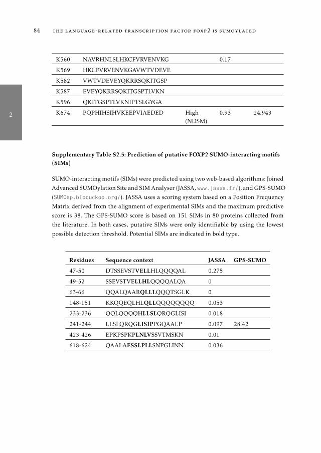
2
84 the language-related transcription factor foxp2 is sumoylated
K560 NAVRHNLSLHKCFVRVENVKG 0.17
K569 HKCFVRVENVKGAVWTVDEVE
K582 VWTVDEVEYQKRRSQKITGSP
K587 EVEYQKRRSQKITGSPTLVKN
K596 QKITGSPTLVKNIPTSLGYGA
K674 PQPHIHSIHVKEEPVIAEDED High(NDSM)
0.93 24.943
Supplementary Table S2.5: Prediction of putative FOXP2 SUMO-interacting motifs(SIMs)
SUMO-interacting motifs (SIMs) were predicted using two web-based algorithms: Joined
Advanced SUMOylation Site and SIM Analyser (JASSA, www.jassa.fr/), and GPS-SUMO
(SUMOsp.biocuckoo.org/). JASSA uses a scoring system based on a Position Frequency
Matrix derived from the alignment of experimental SIMs and the maximum predictive
score is 38. The GPS-SUMO score is based on 151 SIMs in 80 proteins collected from
the literature. In both cases, putative SIMs were only identifiable by using the lowest
possible detection threshold. Potential SIMs are indicated in bold type.
Residues Sequence context JASSA GPS-SUMO
47-50 DTSSEVSTVELLHLQQQQAL 0.275
49-52 SSEVSTVELLHLQQQQALQA 0
63-66 QQALQAARQLLLQQQTSGLK 0
148-151 KKQQEQLHLQLLQQQQQQQQ 0.053
233-236 QQLQQQQHLLSLQRQGLISI 0.018
241-244 LLSLQRQGLISIPPGQAALP 0.097 28.42
423-426 EPKPSPKPLNLVSSVTMSKN 0.01
618-624 QAALAESSLPLLSNPGLINN 0.036

3
3A S S E S S M E N T O F F OX P 2 - C A N D I DAT E P R O T E I NI N T E R AC T I O N S I D E N T I F I E D I N P R E V I O U S M A S SS P E C T R O M E T RY S C R E E N S
Abstract: Heterozygous disruptions in the FOXP2 gene cause a rare speech and lan-guage disorder characterized by difficulty in coordinating the oral motor sequencesnecessary for fluent speech. The FOXP2 gene encodes a transcription factor that isexpressed in a subset of brain structures such as the cortex, striatum and cerebel-lum, where it regulates aspects of neural development and function. The activityof transcription factors is dependent on and regulated by multiple protein-proteininteractions. Therefore, determining which co-factors FOXP2 interacts with can pro-vide insights into the molecular functions of this protein in the context of speechand language development. Although a number of studies have investigated roles ofFOXP2 in the brain and sought to identify downstream target genes, knowledge re-garding its protein-protein interactions remains scarce. Prior research in our groupsought to identify FOXP2-interacting proteins by means of immunoprecipitation fol-lowed by mass spectrometry. In this study, I re-evaluated the dataset from this earlierscreen, selecting two promising candidate FOXP2 interactors, NONO and SFPQ, fortargeted investigation. I aimed to validate these interactions using multiple comple-mentary methods: bioluminescence resonance energy transfer assay, cobalt-affinitypurification, and mammalian two-hybrid assay. The interaction between FOXP2and NONO/SFPQ failed to validate using any of these techniques, suggesting thatthese proteins are unlikely to be physiological interaction partners. Consequently,the methodology used in the previous mass spectrometry screen may have been vul-nerable to false positive results, indicating the need for new screens that search forFOXP2-interacting proteins.
85

3
86 assessment of foxp2-candidate interactors previously identified
3.1 introduction
The transcription factor FOXP2 was the first gene to be conclusively linkedwith speech and language development (Lai et al. , 2001). In particular, a het-erozygous etiological mutation in this gene was identified in a large, multi-generational family (the KE family) in which half of the members suffered fromchildhood apraxia of speech (CAS), a disorder characterized by difficulties coor-dinating the precise oral motor sequences required for speech production. Thismutation yields a substitution in the DNA-binding domain of the FOXP2 pro-tein and abolishes its ability to bind its usual target sequences and regulategene expression (Stroud et al. , 2006; Vernes et al. , 2006). Since then, a numberof other alterations affecting the gene have been found in individuals with sim-ilar speech and language deficits (Laffin et al. , 2012; MacDermot et al. , 2005;Reuter et al. , 2016; Turner et al. , 2013), firmly establishing FOXP2 as a criticalgenetic factor underlying language development and function.
FOXP2 belongs to the FOXP family of transcription factors, which also in-cludes FOXP1, FOXP3 and FOXP4. FOXP2, FOXP1 and FOXP4 are expressedin the brain where they play overlapping but distinct roles in its development(Bowers & Konopka, 2012a; Shu et al. , 2001). These three proteins share thesame domain organization: a glutamine-rich region, a zinc finger domain, aleucine zipper and a forkhead DNA-binding domain. FOXP3 is divergent fromthe other FOXPs in sequence and structure, and its expression is restricted to theimmune system (Fontenot et al. , 2003). FOXP2 DNA-binding sites and targetgenes have been characterized in several studies, which show that FOXP2 regu-lates genes implicated in neuronal processes such as synaptic plasticity and neu-rite outgrowth (Spiteri et al. , 2007; Vernes et al. , 2011, 2007). In contrast, knowl-edge of FOXP2 protein interactions remains scarce. In order to regulate geneexpression, transcription factors need to physically interact with transcription-related proteins, such as chromatin remodelling enzymes or other transcriptionfactors (Smith & Matthews, 2016). Protein-protein interactions regulate numer-ous aspects of a transcription factor function, such as DNA-binding affinityand specificity, transcriptional activation/repression activity, subcellular local-ization, and protein turnover.
FOXP2 forms homo-dimers and also hetero-dimerizes with FOXP1 and FOXP4through the leucine zipper domain (Li et al. , 2004). This dimerization is essen-tial for transcriptional regulatory activity, and heterodimerization among FOXP

3
introduction 87
proteins may generate a repertoire of dimers with different DNA-binding speci-ficities (Sin et al. , 2015).
A small number of other proteins have also been reported to interact withFOXP2, including the transcriptional co-repressors CTBP1, CTBP2 and GATAD2B(Chokas et al. , 2010; Estruch et al. , 2016a). Large-scale yeast two-hybrid (Y2H)screens have provided only a small number of candidate FOXP2 interactors(Corominas et al. , 2014; Rolland et al. , 2014; Sakai et al. , 2011). Most ofthe candidates identified in these screens have not been confirmed, but pre-vious studies from our group have successfully validated and further inves-tigated two of these proteins, namely, the transcription factor TBR1 and thepost-translational modification enzyme PIAS1 (Estruch et al. , 2016b; Derizi-otis et al. , 2014a). Rare mutations in TBR1 cause autism spectrum disorder(ASD) together with language deficits (Deriziotis et al. , 2014a). Strikingly, incellular assays the interaction between FOXP2 and TBR1 could be disrupted byknown mutations in either transcription factor, suggesting a shared molecularetiology for distinct neurodevelopmental disorders that include language im-pairment (Deriziotis et al. , 2014a). As described in Chapter 2, investigations ofthe interaction between FOXP2 and PIAS1 led to the discovery that FOXP2 isSUMOylated (Estruch et al. , 2016b). Evidence of FOXP2 SUMOylation has alsobeen found in two independent studies which additionally suggested that thispost-translational modification is involved in regulating FOXP2 activity in thecerebellum, where it may help modulate motor function and vocal communi-cation (Meredith et al. , 2015; Usui et al. , 2016). These studies represent clearexamples of how identifying and understanding FOXP2 physical interactionswith other proteins can reveal valuable knowledge about the transcription fac-tor and its roles in brain development.
Further characterizing the FOXP2 interactome is critical to fully comprehendits molecular functions. In this chapter I aimed to expand the known FOXP2interactome by validating putative binding partners suggested by mass spec-trometry analysis of immunoprecipitated FOXP2 complexes in a previous un-published investigation by our group. From this dataset I selected the transcrip-tional regulators SFPQ and NONO as promising candidates, and attempted tovalidate their interaction with FOXP2 using a range of complementary protein-protein interaction techniques. The results consistently demonstrate a lack of in-teraction between FOXP2 and NONO/SFPQ with all methods employed, whichsuggests that these proteins are not physiological interaction partners. I con-

3
88 assessment of foxp2-candidate interactors previously identified
clude that the experimental and/or analytical methods used in the previousmass spectrometry study to create a list of candidate FOXP2-interacting pro-teins were likely suboptimal and vulnerable to false positives. This chapterthus highlights the need for fresh mass spectrometry screens using improvedmethodology to deliver further insights into the FOXP2 interactome.
3.2 material and methods
DNA constructs
The cloning of human FOXP2 (NM_014491) has been described previously (Es-truch et al., 2016b). The coding sequences of NONO (NM_007363) and SFPQ(NM_005066) were amplified from human foetal brain cDNA using the primerslisted in Table 1 and then cloned into pCR2.1-TOPO (Invitrogen). For expres-sion of fusion proteins with Renilla luciferase, yellow fluorescent protein (YFP),mCherry, HisV5 and myc tags, the coding sequences of FOXP2, NONO andSFPQ were subcloned into the pLuc, pYFP, pmCherry, pHisV5 and pMyc ex-pression vectors, respectively, which have been described previously (Estruchet al. , 2016b).
Table 3.1: Cloning primers for NONO and SFPQ.∗ BclI generates sticky ends that are complementary to BamHI.
Forward primer Reverse primer Restriction sites
NONO AGGATCCAGAGTAATAAAACTTTTAACTTGG
CTCTAGATTAGTATCGGCGACGTTTGTTTGGG
BamHI + XbaI
SFPQ GATGATCAGCTCTCGGGATCGGTTCCGGAGTC
CGCTAGCCTAAAATCGGGGTTTTTTGTTTGGG
BclI* + XbaI
For the mammalian two-hybrid assay, modified versions of the vectors pBINDand pACT (Promega) were created. The pBIND and pACT vectors contain theyeast GAL4 DNA-binding domain and the herpes simplex virus VP16 activa-tion domain, respectively, upstream of a multiple cloning region. The Renilla lu-ciferase gene was removed from pBIND, together with its promoter and polyA

3
material and methods 89
signal, by excising a ClaI fragment and religating the backbone, creating themodified construct pBIND2 (Supplementary Figure S2.1). The construct pACT2was generated by inserting the functional region of the original pACT vectorinto the pBIND2 backbone using the NdeI and XbaI sites (Supplementary Fig-ure S2.2). The cDNAs of FOXP2, NONO and SFPQ were subcloned into thepBIND2 and pACT2 vectors using the BamHI and XbaI sites. A firefly luciferasereporter vector for the mammalian two-hybrid assay (pGL4-luc2-GAL4UAS-AP) was engineered by inserting a KpnI-NcoI fragment of pGL5 (Promega),containing the yeast GAL4 upstream activating sequence (GAL4UAS) directlyupstream of the adenovirus major late promoter, into the backbone of pGL4.23(Promega), replacing the minimal promoter in this vector (Supplementary Fig-ure S2.3). A negative control reporter vector, which lacks the GAL4UAS (pGL4-luc2-AP) was engineered by inserting a NheI-AgeI fragment of pGL4-luc2-GAL4UAS-AP, containing the adenovirus promoter and partial luciferase coding sequence,into pGL4.23 (Promega) (Supplementary Figure S2.4). All constructs were veri-fied by Sanger sequencing.
Cell culture and transfection
HEK293 cells were obtained from ECACC (cat. no. 85120602) and cultured inDMEM supplemented with 10% foetal bovine serum. Transfections were per-formed using GeneJuice (Merck-Millipore) according to the manufacturer?s in-structions.
BRET assay
BRET assays were performed as described (Deriziotis et al., 2014a). Briefly, HEK293cells were transfected in 96-well plates with plasmids encoding YFP- and luci-ferase-fusion proteins. After 36-48 h, Enduren live cell luciferase substrate (Promega)was added at a final concentration of 60 µ M. Cells were cultured for a further4 h, and emission readings (integrated over 10 s) were taken using a TECANF200PRO microplate reader using the Blue1 and Green1 filter sets. Expressionlevels of the YFP-fusion proteins were measured by taking fluorescence inten-sity readings using the filter set and dichroic mirror suitable for green fluores-cent protein (excitation 480 nm, emission 535 nm). The corrected BRET ratio

3
90 assessment of foxp2-candidate interactors previously identified
was calculated with the following formula: [Green1(experimental condition)/Blue1(expe-
rimental condition)]-[Green1(control condition)/Blue1(control condition)]. The control condi-tions used luciferase or YFP fused to a C-terminal nuclear localization signal.
Fluorescence microscopy
HEK293 cells were seeded on coverslips coated with poly-L-lysine. Cells werecultured for 30 h post-transfection, and then fixed with 4% paraformaldehyde.Nuclei were stained with Hoechst 33342. Fluorescence images were acquiredusing an Axiovert A-1 fluorescent microscope with ZEN Image software (Zeiss).
Pull-down assays
Cells were cultured in 6-well plates and transfected with HisV5-tagged FOXP2,NONO, or SFPQ, or empty pHisV5 vector, together with myc-tagged FOXP2,NONO, or SFPQ, or pMyc vector. After 48 h cells were lysed in 25 mM sodiumphosphate pH 8.0, 250 mM NaCl, 1% Triton X-100, 20 mM imidazole, 1% v/vPMSF and 0.5% v/v EDTA-free protease inhibitor cocktail set III (Sigma-Aldrich).An aliquot of the lysate (10% ) was retained as the input sample and the remain-der was incubated with His-tag Dynabeads (Life Technologies) overnight at 4◦
C with rotation. Beads were washed with 25 mM sodium phosphate pH 8.0, 250mM NaCl, 0.1% Triton X-100, 20 mM imidazole. His-V5-tagged proteins wereeluted in 25 mM sodium phosphate pH 8.0, 250 mM NaCl, 0,1% Triton X-100,400 mM imidazole. Proteins were resolved on 10% SDS-polyacrylamide gelsand transferred to PVDF membranes using a TransBlot Turbo Blotting appara-tus (Bio-Rad). Membranes were blocked in PBS containing 5% milk and 0.1%Tween-20, and were then incubated overnight at 4◦ C with primary antibody inPBS containing 5% milk. The following antibodies were used: anti-V5 tag (Gene-tex cat. no. GTX42525, 1:3000); anti-Myc tag (Abcam cat. no. ab9106, 1:1000).After washing, membranes were incubated with horseradish peroxidase-conju-gated goat anti-mouse or anti-rabbit IgG for 45 min at room temperature. Pro-teins were visualized using Novex ECL Chemiluminescent Substrate ReagentKit (Life Technologies) and a ChemiDoc XRS+ imaging system (Bio-Rad).

3
results 91
Mammalian two-hybrid assay
Protein interactions were assayed using the CheckMate Mammalian Two-HybridSystem (Promega). In brief, HEK293T cells were seeded in clear-bottomed white96-well plates and cultured for 24 h. Cells were transfected in triplicate with anequimolar mixture of pACT2 and pBIND2 constructs (5 mol each per well), to-gether with the firefly luciferase reporter construct pGL4-luc2-GAL4UAS-AP orthe negative control reporter pGL4-luc2-AP (5 fmol), and the pGL4.74 Renillaluciferase normalization control (2 fmol). The total mass of transfected DNAwas adjusted to 6 ng with filler plasmid. After 48 h, cells were lysed with pas-sive lysis buffer and firefly and Renilla luciferase activities were measured usingthe Dual Luciferase Reporter Assay System (Promega) with a TECAN F200PROmicroplate reader. In order to correct for variation in transfection efficiency,each firefly luciferase activity value was normalized to the respective Renillaluciferase activity value.
3.3 results
3.3.1 Evaluation of candidate FOXP2-interacting proteins
As part of previous work in our group to identify potential interacting pro-teins, FOXP2 protein complexes immunoprecipitated using a FOXP2-specificantibody were analysed by mass spectrometry (Elahi, 2008). FOXP2 proteincomplexes were purified from four different sources: (1) SH-SY5Y (human neu-roblastoma) cells stably expressing wild-type FOXP2; (2) SH-SY5Y cells stablyexpressing FOXP2.pR553H, the variant found in the KE family; (3) HEK293cells, which endogenously express FOXP2; and (4) embryonic mouse brain atday E16.5, a time point of high Foxp2 expression. The proteins identified in theimmunoprecipitated complexes are summarized in Table 3.2.
To prioritize potential FOXP2-interacting proteins, I first excluded previouslyreported FOXP2 interactors (i.e. FOXP1, FOXP2 and FOXP4) and probable non-specific interactors (histones, ribosomal subunits). I then selected proteins that(1) are localized to the cell nucleus, (2) are expressed in the brain, and (3) wereidentified in two or more experiments. This filtering process resulted in a short-

3
92 assessment of foxp2-candidate interactors previously identified
list of 6 putative FOXP2 interactors: FOXR1, NCOR1, NONO, PRKRIR, SBF1and SFPQ.
Table 3.2: Candidate FOXP2-interacting proteins previously suggested byimmunoprecipitation and mass spectrometry.
Immunoprecipitation was performed on 4 different sources: SH-SY5Y cell line stably
expressing wild-type FOXP2 (a); SH-SY5Y cell line stably expressing FOXP2.pR533H
(b); HEK293 cells (c); and mouse brain at embryonic day E16.5 (d). Proteins identified
with p<0.05 are indicated with an "X". Italicized words highlight biological information
suggesting that the interaction is not physiologically relevant.
Genename
a b c d Description Expression
Tissue Cell compart-ment
ACTB X Beta-actin Ubiquitous Cytoplasm
ACTL6B X Chromatin remodeller Brain Nucleus
ARMC3 X Beta-catenin-like protein Ubiquitous
BPTF X Nucleosome-remodelling factor Ubiquitous Cytoplasm/nucleus
CRB1 X Calcium receptor Brain Membrane/ se-creted
CTSD X Protease Brain Secreted
EEF1A2 X Protein elongation factor Brain Nucleus
EGF X Growth factor Brain Membrane
EIF4B X X Protein translation initiation factor Ubiquitous Cytoplasm
FN1 X Extracellular matrix Ubiquitous Secreted
FOXP1 X Transcription factor Brain Nucleus
FOXP2 X X Transcription factor Brain Nucleus
FOXP4 X Transcription factor Brain Nucleus
FOXR1 X X Transcription factor Brain Nucleus
GABRA3 X GABA receptor subunit Brain Membrane
GFAP X Intermediate filament Brain Cytoplasm
GRIN3A X Glutamate receptor ionotropic Brain Membrane
H2AZ X X Histone Ubiquitous Nucleus
HIST1H2AB X Histone Ubiquitous Nucleus
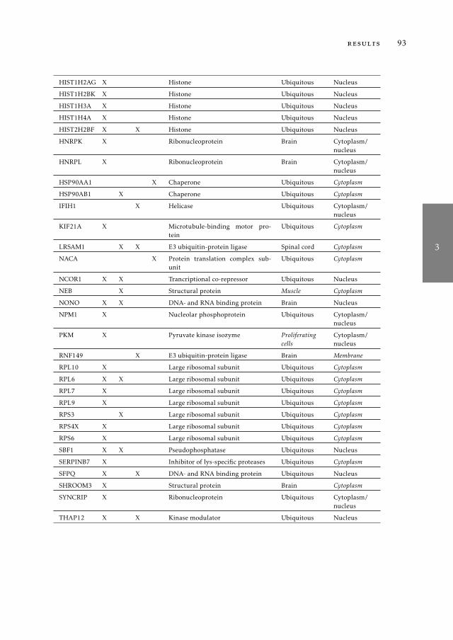
3
results 93
HIST1H2AG X Histone Ubiquitous Nucleus
HIST1H2BK X Histone Ubiquitous Nucleus
HIST1H3A X Histone Ubiquitous Nucleus
HIST1H4A X Histone Ubiquitous Nucleus
HIST2H2BF X X Histone Ubiquitous Nucleus
HNRPK X Ribonucleoprotein Brain Cytoplasm/nucleus
HNRPL X Ribonucleoprotein Brain Cytoplasm/nucleus
HSP90AA1 X Chaperone Ubiquitous Cytoplasm
HSP90AB1 X Chaperone Ubiquitous Cytoplasm
IFIH1 X Helicase Ubiquitous Cytoplasm/nucleus
KIF21A X Microtubule-binding motor pro-tein
Ubiquitous Cytoplasm
LRSAM1 X X E3 ubiquitin-protein ligase Spinal cord Cytoplasm
NACA X Protein translation complex sub-unit
Ubiquitous Cytoplasm
NCOR1 X X Trancriptional co-repressor Ubiquitous Nucleus
NEB X Structural protein Muscle Cytoplasm
NONO X X DNA- and RNA binding protein Brain Nucleus
NPM1 X Nucleolar phosphoprotein Ubiquitous Cytoplasm/nucleus
PKM X Pyruvate kinase isozyme Proliferatingcells
Cytoplasm/nucleus
RNF149 X E3 ubiquitin-protein ligase Brain Membrane
RPL10 X Large ribosomal subunit Ubiquitous Cytoplasm
RPL6 X X Large ribosomal subunit Ubiquitous Cytoplasm
RPL7 X Large ribosomal subunit Ubiquitous Cytoplasm
RPL9 X Large ribosomal subunit Ubiquitous Cytoplasm
RPS3 X Large ribosomal subunit Ubiquitous Cytoplasm
RPS4X X Large ribosomal subunit Ubiquitous Cytoplasm
RPS6 X Large ribosomal subunit Ubiquitous Cytoplasm
SBF1 X X Pseudophosphatase Ubiquitous Nucleus
SERPINB7 X Inhibitor of lys-specific proteases Ubiquitous Cytoplasm
SFPQ X X DNA- and RNA binding protein Ubiquitous Nucleus
SHROOM3 X Structural protein Brain Cytoplasm
SYNCRIP X Ribonucleoprotein Ubiquitous Cytoplasm/nucleus
THAP12 X X Kinase modulator Ubiquitous Nucleus

3
94 assessment of foxp2-candidate interactors previously identified
TMEFF1 X Egf-like domains containing pro-tein
Brain Membrane
TRIM21 X X E3 ubiquitin-protein ligase Heart andlung
Cytoplasm/nucleus
VIM X X Class-iii intermediate filaments Fibroblasts Cytoplasm
WNT7A X Frizzled receptor ligand Brain Secreted
WWP2 X E3 ubiquitin-protein ligase Brain Nucleus
From this list, NONO and SFPQ were chosen as a primary focus becausethey are transcription factors, and are also known to interact with each other(Fox et al., 2005), which is consistent with both proteins being pulled-down inthe same protein complexes. NONO and SFPQ, together with a third protein,PSPC1, form the Drosophila behaviour/human splicing (DBHS) protein family(Knott et al. , 2016). DBHS proteins form homo- and heterodimers (Fox et al. ,2005) and have roles in transcriptional activation and repression, splicing, pre-mRNA processing and RNA transport (Amelio et al. , 2007; Dong et al. , 2007;Izumi et al. , 2014; Kanai et al. , 2004; Kaneko et al. , 2007; Kim et al. , 2011;Park et al. , 2013).Although NONO and SFPQ are expressed in the brain, littleis known regarding their roles in neural processes. NONO is implicated in theregulation of the circadian clock function, acting as a transcriptional cofactorof PER proteins (Kowalska et al. , 2012, 2013) and SFPQ is involved in the ac-tivation of neural-specific alternative splicing (Kim et al. , 2011). NONO andSFPQ therefore represented particularly interesting candidates for interactionwith FOXP2.
3.3.2 Bioluminescence Resonance Energy Transfer assay
To try to confirm the interaction between FOXP2 and NONO/SFPQ, I first usedthe Bioluminescence Resonance Energy Transfer (BRET) assay, a method whichallows the monitoring of protein-protein interactions in living cells, and whichhas been used successfully in prior studies of FOXP2 (Deriziotis et al. , 2014b).To test for an interaction between two proteins using the BRET assay, the twoproteins of interest are expressed as fusion proteins in cultured cells: one pro-tein (the donor) is fused to Renilla luciferase (RLuc), and the second (the ac-ceptor) is fused to yellow fluorescent protein (YFP). An interaction between the

3
results 95
-0.10
0.00
0.10
0.20
0.30
0.40
0.50Co
ntrol
NONO
SFPQ
FOXP
2
Donor: FOXP2
BR
ET
rat
io
Acceptor
-0.20
0.00
0.20
0.40
0.60
0.80
1.00
1.20
Control
NONO
SFPQ
FOXP
2
Donor: NONO
BR
ET
rat
io
Acceptor
-0.10
0.00
0.10
0.20
0.30
0.40
0.50
Control
NONO
SFPQ
FOXP
2
Donor: SFPQ
BR
ET
rat
io
Acceptor
a b c
Figure 3.1: BRET assay for testing interaction between FOXP2 and NONO/SFPQ.(a) HEK293 cells were transfected with FOXP2 fused to Renilla luciferase, and FOXP2,NONO or SFPQ fused to YFP. (b) HEK293 cells were transfected with NONO fused toRenilla luciferase, and FOXP2, NONO and SFPQ fused to YFP. (c) HEK293 cells weretransfected with SFPQ fused to Renilla luciferase, and FOXP2, NONO and SFPQ fusedto YFP. Values are mean corrected BRET ratio± SEM (n = 3).
two proteins may bring the RLuc and YFP moieties in sufficient proximity toallow resonance energy transfer to occur from RLuc to YFP when a luciferasesubstrate is supplied, which leads to a shift in the wavelength of the emittedlight (the BRET signal).
When RLuc-FOXP2 (donor) was co-expressed with YFP-NONO or YFP-SFPQ(acceptor), there was no increase in the BRET signal compared with the control(Figure 3.1a). Similar results were obtained when the assay was performed withFOXP2 as the acceptor and NONO or SFPQ as the donor (Figure 3.1b,c). In con-trast, both the homodimerization of FOXP2 and the interaction between NONOand SFPQ were efficiently detected (Figure 3.1), confirming the suitability ofthe assay to detect protein-protein interactions involving these transcriptionfactors. The results of the BRET assay therefore do not support the existence ofan interaction between FOXP2 and NONO/SFPQ.
In parallel to the BRET experiments, the subcellular localization of the FOXP2,NONO and SFPQ proteins was examined by transfecting cells with fusions of

3
96 assessment of foxp2-candidate interactors previously identified
these proteins to the fluorescent proteins YFP and mCherry. FOXP2, NONOand SFPQ all localize to the cell nucleus (Figure 3.2a), confirming that the lackof interaction between these proteins in the BRET assay is not due to mislo-calization of one or more proteins. FOXP2 presents a diffuse distribution inthe nucleus, whereas NONO and SFPQ exhibit a speckled pattern, as reportedin previous studies (Fox et al. , 2005; Shav-Tal & Zipori, 2002) (Figure 3.2a).Co-transfections of YFP-tagged SFPQ/NONO and mCherry-tagged FOXP2 sug-gest that FOXP2 is not incorporated into the SFPQ/NONO-containing nuclearsubstructures (Figure 3.2b), consistent with a lack of interaction between theseproteins.
3.3.3 Pull-down assay
The BRET assay is a sensitive and versatile technique for detecting protein-protein interactions because it can detect weak and transient interactions, andit is applied to proteins in their native environment within living cells. Howeverfor some pairs of interacting proteins, the BRET assay may give a false negativeresult because the luciferase and YFP moieties are not sufficiently close, or notappropriately oriented, to allow resonance energy transfer to occur. Because theputative interaction between FOXP2 and NONO/SFPQ was initially detectedby immunoprecipitation, I next attempted to confirm the interaction using apull-down assay. Cells were transiently transfected with a bait protein carry-ing an N-terminal hexahistidine and V5 epitope (HisV5) tag, and a prey pro-tein carrying an N-terminal Myc epitope tag. Bait-containing complexes werepulled down from cell lysates using magnetic beads coated with cobalt, whichhas a high affinity for histidine. The bait and prey proteins were detected by im-munoblotting. The His-tag/cobalt pull-down system offers an advantage overco-immunoprecipitation strategies because the latter require an antibody withhigh affinity and specificity for the protein of interest, as well as optimizationof binding and elution conditions for each antibody-antigen pair.
In cells co-transfected with HisV5-FOXP2 and Myc-FOXP2, Myc-tagged FOXP2was detected in the affinity-purified (elution) fraction, demonstrating the ef-ficacy of the pull-down (Fig. 3a). However, HisV5-FOXP2 did not pull downMyc-tagged SFPQ, suggesting that SFPQ does not interact with FOXP2 in thesecells (Figure 3.3a). In cells transfected with HisV5-tagged FOXP2, NONO, orempty vector together with Myc-FOXP2, an enrichment of Myc-FOXP2 in the

3
results 97
NONO
SFPQ
YFP-NONO a
d
b c
merge mCherry-FOXP2 YFP
mCherry-FOXP2 YFP-SFPQ
Figure 3.2: Co-localization of FOXP2 with NONO and SFPQ in HEK293 cells. (a-c)Fluorescence micrographs of cells transfected with FOXP2 fused to mCherry (a), NONOfused to YFP (b) and SFPQ fused to YFP (c). (d) Fluorescence micrographs of cells trans-fected with FOXP2 fused to mCherry (red) and NONO or SFPQ fused to YFP (green).Nuclei were stained with Hoechst 33342 (blue). Scale bars represent 10 µm.

3
98 assessment of foxp2-candidate interactors previously identified
a b
100
150
75
100
150
75
Anti-V5 (bait)
Anti-myc (prey)
empt
y
FO
XP
2
FO
XP
2
empt
y
FO
XP
2
FO
XP
2
His-V5 (bait)
SF
PQ
SF
PQ
FO
XP
2
SF
PQ
SF
PQ
FO
XP
2
myc (prey)
Input Elution
MW (KDa)
100
150
75
100
150
75
100
150
75
50
100
150
75
50
empt
y
FO
XP
2
NO
NO
Input Elution
empt
y
FO
XP
2
NO
NO
Anti-V5 (bait)
Anti-myc (prey)
His-V5 (bait)
MW (KDa)
Anti-V5 (bait)
Anti-myc (prey) Anti-myc (prey)
100
150
75
MW (KDa)
100
150
75
Anti-V5 (bait)
myc (prey) F
OX
P2
FO
XP
2
FO
XP
2
FO
XP
2
FO
XP
2
FO
XP
2
Figure 3.3: Cobalt-affinity pull-down assay for interaction between FOXP2 andNONO/SFPQ. (a) Pull- down assay to test for interaction of FOXP2 with SFPQ. HEK293cells were transfected with HisV5- tagged FOXP2 (bait) or empty vector together withmyc-SFPQ or myc-FOXP2 (prey). His-tagged species were isolated using cobalt affinitypurification. Western blots of total lysate (input) and affinity-purified material (elution)were probed with anti-V5 tag antibody to visualize FOXP2 (top panels). Myc-SFPQ andmyc-FOXP2 were visualized using anti-myc antibody (bottom panels). (b) Pull-downassay to test for interaction of FOXP2 with NONO. HEK293 cells were transfected withHisV5-tagged FOXP2, NONO or empty vector (baits) together with myc-FOXP2 (prey).His-tagged species were isolated using cobalt affinity purification. Western blots of to-tal lysate (input) and affinity-purified material (elution) were probed with anti-V5 tagantibody to visualize NONO and FOXP2 (top panels). Myc-FOXP2 and myc-FOXP2 wasvisualized using anti-myc antibody (bottom panels).

3
results 99
eluted material was observed only when HisV5-FOXP2 was used as the bait(Figure 3.3b). No enrichment of Myc-FOXP2 compared to the control conditionwas observed when HisV5-NONO was used as the bait, suggesting that the pres-ence of FOXP2 in the affinity-purified fraction was due to non-specific binding(Figure 3.3b). Thus, the pull-down assay also fails to support the existence ofan interaction between FOXP2 and NONO/SFPQ.
3.3.4 Mammalian Two-Hybrid assay
Affinity purification can be a reliable technique for detecting protein-proteininteractions. However the appropriate experimental conditions must be identi-fied for each specific pair of proteins, because it is critical that the interaction ofinterest is preserved after the cells have been lysed. Excessively stringent wash-ing steps can result in the loss of specific interactors. Conversely, non-specificbinding of the prey protein to the affinity matrix can result in contaminationof the eluted material if binding conditions or washing steps are not strin-gent enough. In some cases it may not be possible to achieve ideal balance ofconditions that retain specific interactions while eliminating non-specific ones.Therefore, I employed a third technique, the mammalian two-hybrid (M2H) as-say, to assess the putative interaction of FOXP2 with NONO and SFPQ.In the M2H assay, bait and prey proteins are fused to a GAL4 DNA-bindingdomain (encoded by the pBIND2 vector) and a VP16 transcriptional activationdomain (encoded by the pACT2 vector), respectively. Cells are transfected withthe two vectors coding for the fusion proteins, together with a reporter vectorthat contains tandem GAL4 DNA binding sites upstream of a basic promoterand the firefly luciferase gene. An interaction between the bait and prey pro-teins physically connects the GAL4 and VP16 domains, creating a functionaltranscriptional activator that will drive transcription of the reporter firefly lu-ciferase gene (Figure 3.4a, Supplementary Figure S2.5). The sensitivity withwhich luciferase activity can be detected makes the M2H assay potentially ableto pick up weak and transient interactions. In addition, the interaction is notdetected in real time, as in the case of the BRET assay; rather it is capturedover a longer period in the expression of the luciferase protein, which may alsoincrease sensitivity. The M2H assay must however be used with caution wheninvestigating protein-protein interactions of transcription factors. For example,when fused to the GAL4 DNA-binding domain, a transcription factor may acti-

3
100 assessment of foxp2-candidate interactors previously identified
vate transcription in the absence of any interaction with the VP16 transactiva-tion domain fusion protein, leading to a high background signal that can makethe results challenging to interpret.I first verified that the M2H assay was effective, by using the known interactionbetween the proteins Id and MyoD as a positive control, and empty bait/preyvectors as a negative control (Finkel et al. , 1993). A reporter vector that lacksthe GAL4 binding sites served as an additional negative control. These exper-iments confirmed that an increase in luciferase activity occurred only whenboth the MyoD and Id fusion proteins, and the reporter construct containingthe GAL4 binding sites, were present (Figure 3.4b).
No interaction was observed between FOXP2 and NONO/SFPQ, when FOXP2was expressed as the bait protein and NONO/SFPQ as the prey (Figure 3.4c).The assay could not be performed in the reverse configuration, with FOXP2 asthe prey, because FOXP2 activated luciferase transcription when fused to theVP16 activation domain, suggesting that the reporter plasmid contains bindingsites for FOXP2 (data not shown). In contrast, the interaction between SFPQand NONO was readily detected (Figure 3.4d) (Fox et al. , 2005; Knott et al. ,2016). Thus, the mammalian two-hybrid assay also provides little support forthe existence of an interaction between FOXP2 and NONO/SFPQ.
3.4 discussion
In the work described in this chapter I sought to define novel FOXP2-interactingproteins by evaluating an existing dataset of putative interactors generated bya previous mass spectrometry screen. The most promising of these putativeFOXP2 interactors, NONO and SFPQ, were selected for in-depth follow-up.However, the interaction between FOXP2 and SFPQ/NONO consistently failedto validate, whether using BRET assays, pull-down methods or a mammaliantwo-hybrid system. The use of these three complementary methodologies, eachof which optimally detects protein-protein interactions with different character-istics, reduces the probability that the lack of interaction observed in our vali-dations is a false negative result. Moreover, these assays were able to detect theknown interactions of the proteins under investigation, such as the homodimer-ization of FOXP2 and the heterodimerization of NONO and SFPQ, indicatingthat the methods used are suitable for detecting interactions of these transcrip-

3
discussion 101
Reporter Vector
Gal4 Binding sites
GAL4 binding domain
Protein X
Protein Y
Firefly luciferase gene
VP16 activation domain
a
b
0.00
0.50
1.00
1.50
2.00
SFPQ empty SFPQ
empty NONO NONO
mea
n re
lativ
e lu
cife
rase
pBIND
pACT 0.00
0.50
1.00
1.50
2.00
empty NONO SFPQ
FoxP2 FoxP2 FoxP2pBIND
pACT mea
n re
lativ
e lu
cife
rase
d
mea
n re
lativ
e lu
cife
rase
0.00
5.00
10.00
15.00
empty MyoD empty MyoD
empty empty ID ID
CONTROL
GAL4UAS
pBIND
pACT
c
Figure 3.4: Mammalian two-hybrid assay for interaction of FOXP2 with NONO/SFPQ(a) Schematic representation of the mammalian two-hybrid assay. Cells are transfectedwith plasmids coding for Proteins “X” and “Y” fused to the GAL4 DNA binding DNAbinding domain (pBIND2 vector) and to the VP16 transcriptional domain (pACT2 vec-tor), respectively; and a reporter vector that contains GAL4 DNA binding sites upstreamof the firefly luciferase gene. An interaction between proteins “X” and “Y” physicallyconnects the GAL4 DNA-binding domain and the VP16 transactivation domain, recon-structing a functional transcriptional factor that will activate expression of the firefly lu-ciferase reporter gene.(b) Validation of the mammalian two-hybrid assay. HEK293 cellswere transfected with pBIND2-MyoD or empty pBIND2; pACT2-ID or empty pACT2;the GAL4UAS firefly reporter plasmid or a control reporter lacking the GAL4UAS bind-ing sites; and a normalizer vector that expresses Renilla luciferase. c M2H assay forFOXP2 interaction with NONO and SFPQ. HEK293 cells were transfected with pBIND2-FOXP2; pACT2-NONO, pACT2-SFPQ or empty pACT2; the GAL4UAS firefly reporterplasmid; and a normalizer vector that expresses Renilla luciferase. d M2H assay for theinteraction between NONO and SFPQ. HEK293 cells were transfected with pBIND2-NONO or empty pBIND2; pACT2-SFPQ or empty pACT2; the GAL4UAS firefly reporter;and a normalizer vector that expresses Renilla luciferase. In (b)-(d), values are mean rel-ative luciferase activity ± S.E.M. (n = 3)

3
102 assessment of foxp2-candidate interactors previously identified
tion factors. FOXP2 and SFPQ/NONO also showed different patterns of local-ization within the nucleus, which is not suggestive of a functional interaction.
It is therefore likely that NONO and SFPQ were false positive results in themass spectrometry experiments carried out previously. The immunoprecipi-tation that was performed to isolate FOXP2-containing protein complexes inthese experiments may not have been effective due to the sub-optimal affinityand specificity of the antibody used, or due to the composition of the lysis andwash buffers. Of note, the band representing FOXP2 in the Coomassie-stainedpolyacrylamide gels of precipitated proteins was not stronger following pull-down with anti-FOXP2 compared to the control condition, casting doubt on theefficiency of the pull-down (Elahi, 2008). In those prior experiments, FOXP2 it-self was not identified in the mass spectrometry analysis of FOXP2-affinity pu-rifications from HEK293 cells or mouse brain tissue, indicating inefficient pre-cipitation in these samples. In addition, the known FOXP2 interactors FOXP1and FOXP4, which would be expected to strongly co-precipitate with FOXP2in all the conditions, were only identified in one condition (SH-SY5Y cells over-expressing wild-type FOXP2) (Table 3.2). There were also insufficient replicatesincluded for each protein source to test the reproducibility of the results (Elahi,2008).
The selection of NONO and SFPQ as putative FOXP2 interactors to furtherinvestigate was based on multiple factors, including their biological function,since both proteins are neural transcription factors and they are already knownto interact with one another (Fox et al. , 2005; Knott et al. , 2016), and the relativestrength of the data, since both proteins had been detected in experiments usingtwo different cell lines (Table 3.2). Following completion of the work describedin this chapter, mutations in NONO were reported to lead to neurodevelopmen-tal disorders including intellectual disability, confirming the importance of thistranscription factor in brain development (Mircsof et al. , 2015; Reinstein et al., 2016). However, the phenotype associated with NONO mutations is not es-pecially similar to that caused by FOXP2 haploinsufficiency. Because NONOand SFPQ were among the most biologically-plausible and highest-confidencecandidates from the previous mass spectrometry effort, it appears unlikely thatother candidates identified in this screen would prove to be true FOXP2 interac-tors upon further investigation. Thus, this chapter highlights the need for freshmass spectrometry-based screens to identify further candidate FOXP2 interac-tors. Optimization of the pull-down methodology and data analysis pipeline,

3
discussion 103
together with use of adequate replicates, represent important factors for im-proving the sensitivity and specificity of such a screen.

3
104 assessment of foxp2-candidate interactors previously identified
3.5 supplementary material
Supplementary Figure S3.1. Cloning strategy to generate M2H plasmid pBIND2.
T7 promoter
CMV early enhancer/promoter
SV40 Late Polyadenylation signal
Intron
GAL4
MCS
Ampicillin resistance
T7 promoter
CMV early enhancer/promoter
SV40 early enhancer/promoter
SV40 Late Polyadenylation signal
Intron
GAL4
MCS
Renilla luciferase
Ampicillin resistance
pBIND
Synthetic Polyadenilation signal
ClaI ClaI
T7 promoter CMV early enhancer/promoter
SV40 Late Polyadenylation signal
Intron
GAL4
MCS
pBIND2
Ampicillin resistance
Digestion with ClaI
Ligation

3
supplementary material 105
Supplementary Figure S3.2. Cloning strategy to generate M2H plasmid pACT2.
T7 promoter CMV early enhancer/promoter
SV40 Late Polyadenylation signal
Intron
GAL4
MCR
pBIND2
Ampicillin resistance
NdeI
XbaI
T7 promoter
Intron
VP16
T7 promoter CMV early enhancer/promoter
SV40 Late Polyadenylation signal
Intron
VP16
MCR
pACT
Ampicillin resistance
neo
SV40 early enhancer/promoter
Synthetic Polyadenylation signal
NdeI
XbaI
CMV early enhancer/promoter
Ampicillin resistance
SV40 Late Polyadenylation signal
T7 promoter CMV early enhancer/promoter
SV40 Late Polyadenylation signal
Intron
VP16
MCR
pACT2
Ampicillin resistance
Digestion with NdeI and XbaI Digestion with NdeI and XbaI
Ligation

3
106 assessment of foxp2-candidate interactors previously identified
Supplementary Figure S3.3. Cloning strategy to generate M2H plasmidpGL4-luc2-GAL4UAS-AP.
Upstream Polyadenylation Signal and Transcription Pause site
SV40 Late Polyadenilation signal
GAL4 binding sites
luc+
pG5luc
Ampicillin resistance
KpnI
NcoI
MCR
SV40 Late Polyadenilation signal
minimal Promoter
luc2
pGL4.23
Ampicillin resistance
KpnI
NcoI
GAL4 binding sites
SV40 Late Polyadenilation signal
luc2
Ampicillin resistance
MCR
SV40 Late Polyadenilation signal
GAL4 binding sites
luc2
pGL4-luc2-GAL4UAS-AP
Ampicillin resistance
Digestion with KpnI and NcoI Digestion with KpnI and NcoI
Ligation

3
supplementary material 107
Supplementary Figure S3.4. Cloning strategy to generate M2H plasmidpGL4-luc2-AP.
MCR
SV40 Late Polyadenilation signal
minimal Promoter
luc2
pGL4.23
Ampicillin resistance
NheI
AgeI
MCR
SV40 Late Polyadenilation signal
GAL4 binding sites
luc2
pGL4-luc2-GAL4UAS-AP
Ampicillin resistance
NheI
AgeI
SV40 Late Polyadenilation signal
Ampicillin resistance
luc2
SV40 Late Polyadenilation signal
luc2
pGL4-luc2-AP Ampicillin resistance MCR
Digestion with NheI and AgeI Digestion with NheI and AgeI
Ligation

3
108 assessment of foxp2-candidate interactors previously identified
Supplementary Figure S3.5. Diagram of the plasmids used in the M2H assay.
T7 promoter CMV early enhancer/promoter
SV40 Late Polyadenylation signal
Intron
GAL4
MCR
pBIND2
Ampicillin resistance
T7 promoter CMV early enhancer/promoter
SV40 Late Polyadenylation signal
Intron
VP16
MCR
pACT2
Ampicillin resistance
MCR
SV40 Late Polyadenilation signal
GAL4 binding sites
luc2
pGL4-luc2-GAL4UAS-AP Ampicillin resistance
SV40 Late Polyadenilation signal
luc2
pGL4-luc2-AP
Ampicillin resistance
MCR

4
4P R O T E O M I C A N A LY S I S O F F OX P P R O T E I N S R E V E A L SI N T E R AC T I O N S B E T W E E N C O R T I C A L T R A N S C R I P T I O NFAC T O R S A S S O C I AT E D W I T H N E U R O D E V E L O P M E N TA LD I S O R D E R S 1
Abstract: FOXP transcription factors play important roles in neurodevelopment,but little is known about how their transcriptional activity is regulated. FOXP pro-teins cooperatively regulate gene expression by forming homo- and hetero-dimerswith each other. Physical associations with other transcription factors might alsomodulate the functions of FOXP proteins. However, few FOXP-interacting transcrip-tion factors have been identified so far. Therefore, we sought to discover additionaltranscription factors that interact with the brain-expressed FOXP proteins, FOXP1,FOXP2 and FOXP4, through affinity-purifications of protein complexes followed bymass spectrometry. We identified seven novel FOXP-interacting transcription factors(NR2F1, NR2F2, SATB1, SATB2, SOX5, YY1 and ZMYM2), five of which havewell-established roles in cortical development. Accordingly, we found that these tran-scription factors are co-expressed with FoxP2 in the deep layers of the cerebral cortexand also in the Purkinje cells of the cerebellum, suggesting that they may cooperatewith the FoxPs to regulate neural gene expression in vivo. Moreover, we demonstratedthat etiological mutations of FOXP1 and FOXP2, known to cause neurodevelopmen-tal disorders, severely disrupted the interactions with FOXP-interacting transcrip-tion factors. Additionally, we pinpointed specific regions within FOXP2 sequenceinvolved in mediating these interactions. Thus, by expanding the FOXP interactomewe have uncovered part of a broader neural transcription factor network involvedin cortical development, providing novel molecular insights into the transcriptionalarchitecture underlying brain development and neurodevelopmental disorders.
1 This chapter has been published as: Estruch S.B., Quevedo-Calero M., Graham S.A., Vino A., SollisE., Deriziotis P., Fisher S.E. (2018). Proteomic analysis of FOXP proteins reveals interactions be-tween cortical transcription factors associated with neurodevelopmental disorders. Human Molec-ular Genetics. doi:10.1093/hmg/ddy035.
109

4
110 proteomic analysis of foxp proteins
4.1 introduction
Transcription factors have emerged as a key class of genes disrupted in mono-genic forms of neurodevelopmental disorders such as intellectual disability (ID)and autism spectrum disorder (ASD), consistent with the precise temporal andspatial control of gene expression that underpins neurodevelopmental processes(Silbereis et al. , 2016). Large-scale next-generation DNA sequencing studieshave been particularly successful in identifying monogenic ID/ASD-related dis-orders caused by high-penetrance de novo mutations in transcription factorgenes (De Rubeis et al. , 2014; Kwan, 2013; Peter et al. , 2014).
Among the many families of human transcription factors, the FOXP sub-family of forkhead box proteins is notable for the neurodevelopmental pheno-types which have been associated with their disruption (Bowers & Konopka,2012b; Charng et al. , 2016). The FOXP family includes four proteins: FOXP1,FOXP2, FOXP3 and FOXP4 (Shu et al. , 2001). FOXP1, FOXP2 and FOXP4 ex-hibit 55–65% sequence identity and show overlapping expression in the devel-oping brain, as well as in other organs (Shu et al. , 2001; Teufel et al. , 2003).FOXP3 is structurally divergent and its expression is limited to T lymphocytes(Fontenot et al. , 2003). FOXP3 disruption causes an immunological disorder,IPEX (immunodysregulation polyendocrinopathy enteropathy X-linked, OMIM# 304790) syndrome, while mutations of FOXP1, FOXP2 and FOXP4 have eachbeen linked to distinct neurodevelopmental disorders. Heterozygous disrup-tions of FOXP1 cause a broad neurodevelopmental syndrome, which includesglobal developmental delay and ID, frequently accompanied by features of autismand impaired speech and language abilities (OMIM # 613670) (Hamdan et al., 2010; Lozano et al. , 2015; O’Roak et al. , 2011; Sollis et al. , 2016; Srivastavaet al. , 2014). All pathogenic variants observed to date have occurred de novo,consistent with the severe phenotype of the disorder (Sollis et al. , 2016). Het-erozygous disruptions of FOXP2 cause a rare monogenic form of speech andlanguage impairment including childhood apraxia of speech as a core feature(OMIM # 602081) (Estruch et al. , 2016a; Laffin et al. , 2012; Lai et al. , 2001; Mac-Dermot et al. , 2005; Reuter et al. , 2016; Roll et al. , 2010; Turner et al. , 2013).Unlike FOXP1-associated disorder, general cognitive functions may be withinthe normal range, and both inherited and de novo cases have been reported (Gra-ham & Fisher, 2015). Although the FOXP2-related disorder is less severe thanthat resulting from FOXP1 disruption, individuals with FOXP2 and FOXP1 mu-

4
introduction 111
tations display overlapping features such as language impairment, suggestingthat the syndromes may involve disruption of similar molecular and cellularnetworks (Bacon & Rappold, 2012).
Disorders relating to FOXP4 disruption had not been reported until recently,when Charng et al. described a homozygous frameshift variant in a child froma consanguineous family affected by developmental delay and malformationsin the larynx and the heart (Charng et al. , 2016). It is interesting that this,the only currently suspected case of FOXP4-related disorder, involves a reces-sive mutation, in contrast to the dominant disorders described for FOXP1 andFOXP2 disruptions. The potential complete absence of FOXP4 protein in thischild may also explain why the disorder appears to have broader effects in or-gans other than the brain. Indeed, all three neurally-expressed FOXP proteinsalso have roles in the development of other organs, but their neurodevelopmen-tal functions appear to show more dosage sensitivity than roles in non-neuraltissues (Li, 2004; Li et al. , 2015b; Shu et al. , 2005, 2007). Although FOXP4 is thestrongest candidate causal gene in the case identified by Charng and colleagues,additional observations of FOXP4 disruption are necessary to confirm its etio-logical role, particularly since variants in two other genes were also found in ahomozygous state in the child and heterozygous state in the parents (Charnget al. , 2016).
The precise mechanisms by which FOXP family members regulate transcrip-tion are just beginning to be explored. Several studies have investigated FOXP2and FOXP1 target genes in the brain (Araujo et al. , 2015; Spiteri et al. , 2007;Vernes et al. , 2007, 2011) but genes regulated by FOXP4 remain to be discov-ered. An important mechanism in regulation of gene expression is that tran-scription factors act in a combinatorial fashion, and thus can generate the com-plex patterns of gene expression underlying development of the brain and othersystems using a limited set of transcription factors (Silbereis et al. , 2016; Smith& Matthews, 2016). The genes regulated by the FOXP proteins in specific celltypes are therefore likely to depend on the co-expression of other transcrip-tion factors. Indeed, the transcriptional activity of FOXP1, FOXP2 and FOXP4is regulated by their ability to form homo- and hetero-dimers with each otherthrough their leucine zipper domain (Li et al. , 2004). The three proteins showpartially overlapping patterns of expression in the brain, such that differentcombinations of FOXP homo and hetero-dimers may regulate distinct target

4
112 proteomic analysis of foxp proteins
genes (Ferland et al. , 2003; Lai et al. , 2003; Sin et al. , 2015; Takahashi et al. ,2008).
In addition to dimerization between FOXP family members, a small num-ber of interactions between FOXP proteins and other transcription factors havebeen reported. FOXP1 and FOXP2 interact with the neural transcription factorTBR1, rare mutations of which cause ASD accompanied by language deficits(Deriziotis et al. , 2014a). Notably, this interaction can be disrupted by etiologi-cal mutations in either FOXP2 or TBR1 (Deriziotis et al. , 2014a). Other reportedFOXP-interacting transcription factors include GATAD2B and NKX2.1 (Chokaset al. , 2010; Zhou et al. , 2008). We hypothesized that there may be further inter-actions between FOXPs and other transcription factors with relevance to physio-logical developmental processes and neurodevelopmental disorders. We there-fore sought to identify transcription factors which may cooperate with FOXPproteins in regulating gene expression during neurodevelopment, by applying amass spectrometry approach. We identified seven novel FOXP-interacting tran-scription factors: NR2F1, NR2F2, SATB1, SATB2, SOX5, YY1 and ZMYM2. Sev-eral of these novel interactors have well-established roles in the development ofthe nervous system and/or are associated with neurodevelopmental disorders.The interactions with these binding partners involve different regions of theFOXP2 polypeptide, and are variably affected by different pathogenic variantsin FOXP1 and FOXP2. Thus our findings provide new clues to the molecularfunction of the FOXP proteins, and point to a network of transcription factorsinvolved in cortical development, disruption of which manifests as neurodevel-opmental disorder.
4.2 materials and methods
DNA constructs
The cloning of human FOXP2 (NM_ 014491), FOXP1 (NM_ 032682), FOXP4(NM_ 001012426), has been described previously (Estruch et al., 2016b). Thecoding sequences of TFDP1 (NM_ 007111), NR2F1 (NM_ 005654), NR2F2 (NM_021005), SATB1 (NM_ 002971), SATB2 (NM_ 001172509), ZBTB2 (NM_020861),TP53 (NM_000546), ZNF687 (NM_001304763), ZMYM2 (NM_003453), NFAT5(NM_ 138714), YY1 (NM_ 003403) and SOX5 (NM_ 001330785) were amplifiedusing the primers listed in Supplementary Table S4.1 and cloned into pCR2.1-

4
materials and methods 113
TOPO. Human fetal brain cDNA was used as template for PCR, except in thecases of NR2F2 and NFAT5, which were amplified from the plasmids pAAV-hNR2F2-RFP (Addgene, # 22926) and pEGFP-NFAT5 (Addgene, # 13627), re-spectively. The cloning of FOXP2 variants p.Q17L, p.M406T, p.P416T, p.R553H,p.N597H, p.R328∗ , P.Q390Vfs∗7, of synthetic truncated forms of FOXP2, andof FOXP1 variants p.A339Sfs∗4, p.V423Hfs∗37, p.Y439∗ , p.R465G, p.R514C,p.R252∗ , and p.W534R has been described previously (Estruch et al. , 2016a;Sollis et al. , 2016). For expression of FLAG-tagged protein for affinity purifi-cation, the coding regions of FOXP1, FOXP2 and FOXP4 were subcloned intothe pEF1α FLAGbio expression vector, provided by Dr. J.W. Wang (Kim et al., 2009b). For expression of fusion proteins with Renilla luciferase, yellow fluo-rescent protein (YFP) and mCherry tags, cDNAs were subcloned into the pLuc,pYFP, and pmCherry expression vectors, respectively, which have been describedpreviously (Deriziotis et al. , 2014a; Estruch et al. , 2016a). All constructs wereverified by Sanger sequencing.
Cell culture and transfection
HEK293 cells were obtained from ECACC (cat. No. 85120602) and culturedin DMEM supplemented with 10% fetal bovine serum. Transient transfectionswere performed using GeneJuice (Merck-Millipore) according to the manufac-turer’s instructions. To generate stable cell lines expressing FLAG-tagged FOXPproteins, HEK293 cells were transfected with pEF1αFLAGbio containing thecoding sequence of FOXP1, FOXP2, or FOXP4 using GeneJuice (Merck-Milli-pore) according to the manufacturer’s instructions. Single clones were isolatedfollowing selection with culture medium containing 10 µM puromycin. Expres-sion of the tagged proteins in selected clones was confirmed by western blottingof cell lysates using anti-FLAG antibody (Sigma). Selected stable cell lines weremaintained in DMEM supplemented with 10% fetal bovine serum and 5 µMpuromycin.
Affinity Purification and Mass Spectrometry (AP/MS)
HEK293 cell lines stably expressing FLAG-tagged FOXP1, FOXP2 or FOXP4, orcontrol cell lines, were expanded to twenty 15 cm dishes, washed with PBS, and

4
114 proteomic analysis of foxp proteins
harvested by scraping. The affinity purification procedure has been describedpreviously (van den Berg et al. , 2010). Briefly, nuclear extracts were dialysedinto buffer C-100 (20 mM HEPES pH 7.6, 0.2 mM EDTA, 1.5 mM MgCl2, 100mM KCl, 20% glycerol) and then incubated with anti-FLAG M2 agarose beads(Sigma) and benzonase (Novagen) for 3 h at 4◦ C. Beads were washed 5 times for5 min with buffer C-100 containing 0.02% NP-40 (C-100∗ ) and bound proteinswere eluted 4 times for 15 min at 4◦ C with buffer C-100∗ containing 0.2 mg/mlFLAG-tripeptide (Sigma). The purification of FOXP1, FOXP2 or FOXP4 waschecked by western blot using anti-FLAG antibody. The elution fractions werepooled and proteins were TCA-precipitated, resolved by SDS-polyacrylamidegel electrophoresis, stained with Colloidal Blue Staining Kit (Invitrogen) andanalyzed by mass spectrometry as described previously (Engelen et al. , 2011;Gagliardi et al. , 2013; Ninkovic et al. , 2013; van den Berg et al. , 2010).
Mass-spectrometry analyses were performed on three independent purifica-tions from FOXP1- and FOXP2-expressing cell lines, and two independent pu-rifications from FOXP4-expressing cell lines. Each purification was performedin parallel with stable cell lines transfected with empty control vector. To re-move non-specific interactors, peptide lists were filtered using peptides foundin the control conditions or in the CRAPome database, a collection of commoncontaminants in AP/MS data (Mellacheruvu et al. , 2013). Proteins with a Mas-cot score lower than 45 were also excluded.
Western blotting
Proteins from cell lysates were resolved on 10% SDS-polyacrylamide gels andtransferred to PVDF membranes using a TransBlot Turbo Blotting apparatus(Bio-Rad). Membranes were blocked in PBS containing 5% milk and 0.1% Tween-20 and incubated overnight at 4◦ C with primary antibody. After washing, mem-branes were incubated with horseradish peroxidase-conjugated goat anti-mouseor anti-rabbit IgG for 45 min at room temperature. Proteins were visualizedusing Novex ECL Chemiluminescent Substrate Reagent Kit (Invitrogen) and aChemiDoc XRS+ imaging system (Bio-Rad).

4
materials and methods 115
BRET assay
BRET assays were performed as described previously (Deriziotis et al., 2014a).Briefly, HEK293 cells were transfected in 96-well plates with plasmids encodingYFP- and luciferase-fusion proteins. After 36-48 h, Enduren live cell luciferasesubstrate (Promega) was added at a final concentration of 60 µ M. Cells werecultured for a further 4 h, and emission readings (integrated over 10 s) weretaken using a TECAN F200PRO microplate reader using the Blue1 and Green1filter sets. Expression levels of the YFP-fusion proteins were measured by takingfluorescent readings using the filter set and dichroic mirror suitable for greenfluorescent protein (excitation 480 nm, emission 535 nm). The corrected BRETratio was calculated with the following formula: [Green1 (experimental condition)
/Blue1(experimental condition)] - [Green1(control condition) /Blue1(control condition)]. Thecontrol conditions used luciferase or YFP fused to a C-terminal nuclear local-ization signal (Deriziotis et al., 2014a).
Fluorescence microscopy
HEK293 cells were seeded on coverslips coated with poly-L-lysine. Cells weretransfected with plasmids encoding proteins of interest fused to YFP or mCherry.Cells were cultured for 30 h post-transfection, and then fixed with 4 % para-formaldehyde. Nuclei were stained with Hoechst 33342. Fluorescence imageswere acquired using an Axiovert A-1 fluorescent microscope with ZEN Imagesoftware (Zeiss).
Immunofluorescence
Immunofluorescence experiments were performed on mouse brain sections atpostnatal day 3 (P3). Brains were harvested and embedded in optimal cuttingtemperature (OCT) compound on dry ice. Sagittal sections were prepared ata thickness of 4 µ m using a Leica CM1950 cryostat and then preserved at -20◦ C. Tissue sections were fixed in ice-cold acetone for 10 minutes at -20◦ C,blocked using 10% donkey serum in PBS for 1 h at room temperature, and in-cubated overnight at 4◦ C with primary antibody diluted in 2% donkey serum.The following antibodies were used: goat anti-FOXP2 N-16 antibody (sc-21069,

4
116 proteomic analysis of foxp proteins
Santa Cruz), rabbit anti-FOXP2 antibody (ab16046, Abcam), rabbit anti-SOX5(ab94396, Abcam), goat anti-SATB1 E-15 antibody (sc-5990, Santa Cruz), rabbitanti-SATB2 antibody (ab34735, Abcam), rabbit anti-NR2F1 (ab181137, Abcam)and rabbit anti-NR2F2 (ab42672, Abcam). After washing, tissue sections wereincubated with fluorescently-labeled secondary antibodies diluted in 2% don-key serum: donkey anti-rabbit IgG Alexa488 (a21206, Invitrogen) and donkeyanti-goat IgG Alexa594 (a11058, Invitrogen). Slides were mounted with Vec-taShield Antifade Mounting Medium with DAPI (Vector Labs) and then imagedusing Axiovert A-1 fluorescent microscope with ZEN Image software (Zeiss).
4.3 results
4.3.1 Identification of FOXP-interacting transcription factors
To identify transcription factors that may cooperate with FOXP proteins toregulate gene expression, we generated HEK293 cell lines stably expressingFOXP1, FOXP2 or FOXP4 fused to an N-terminal FLAG tag. HEK293 cells en-dogenously express all three FOXP proteins as well as many other neural genes(Shaw et al. , 2002; Stepanenko & Dmitrenko, 2015). FLAG affinity-purified pro-tein complexes from these cell lines were analyzed by mass spectrometry (Fig-ure 4.1a,b). Three independent experiments were performed for FOXP1 andFOXP2, and two experiments for FOXP4. After filtering out non-specific interac-tors, a total of 381 putative FOXP-interacting proteins were identified, with sub-stantial overlap between the three FOXP proteins (Figure 4.1c, SupplementaryTable S4.2). For each FOXP protein, the other two FOXPs were among the in-teracting proteins identified in each experiment, reflecting heterodimerizationbetween FOXP family members (Li et al. , 2004) (Supplementary Table S4.2).Moreover, the set of putative FOXP-interacting proteins included previously re-ported interaction partners such as CTBP1, CTBP2 and GATAD2B, confirmingthat the affinity purification procedure retrieved physiologically relevant FOXP-interacting proteins (Chokas et al. , 2010; Estruch et al. , 2016a) (SupplementaryTable S4.2).
The transcription factors among the putative FOXP-interacting proteins wereidentified, resulting in a set of 28 proteins (Figure 4.1c, Table 4.1, Supplemen-tary Table S4.2). To prioritize potential interacting transcription factors of mostinterest for further investigation, we excluded previously reported FOXP inter-

4
results 117
75 100 150 250
50
37
25
10
MW (kDa)
Control FOXP4 FOXP1 FOXP2
a b
FOXP1
FOXP2
FOXP4
29 8
44 2
23 0
6 2
235 10
21 2
23 4
c
75 100
MW (kDa)
FOXP1
75 100
75 100
10% input wash
cont
rol
FO
XP
cont
rol
FO
XP
FOXP2
FOXP4
elutions
1 2 3 4
All proteins Transcription factors
Figure 4.1: Identification of FOXP1-, FOXP2- and FOXP4-interacting proteins. (a)Affinity purifications of FOXP protein complexes using HEK293 cell lines stably express-ing FLAG- FOXP1 (top panel), -FOXP2 (middle panel) and -FOXP4 (bottom panel) pro-tein complexes. FLAG-tagged species were isolated using a FLAG affinity purificationstrategy. Western blots of total lysate (10% Input), washed proteins (wash), and affinity-purified material (elutions) were probed with an anti-FLAG antibody. (b) Coomassie-stained SDS- polyacrylamide gels of the affinity purifications of FOXP1, FOXP2, FOXP4and control purification. Arrows indicate bands corresponding to FOXP1, FOXP2 orFOXP4 for each IP. (c) Venn diagram showing the overlaps between the proteins (pur-ple) or transcription factors (light blue) identified in each FOXP purification.
actors, and then selected proteins that were present in two or more replicates,or that had a known involvement in neurodevelopmental disorder. Because theFOXP proteins have high sequence similarity and are likely to have shared in-

4
118 proteomic analysis of foxp proteins
teractors, the appearance of a protein in two experiments with different FOXPswas treated as a replicate. We also included transcription factors if two or moremembers of the same family of transcription factors were observed (Supplemen-tary Table S4.3). This selection resulted in a filtered list of 12 putative FOXPinteractors for follow up.
4.3.2 Validation of interactions between FOXPs and transcription factors
To validate the selected putative FOXP-interacting transcription factors, we useda Bioluminescence Resonance Energy Transfer (BRET) assay. The BRET assayallows protein-protein interactions to be observed in live cells, and its effec-tiveness has previously been shown for successfully confirming interactionsbetween FOXPs and other proteins (Deriziotis et al. , 2014b,a; Estruch et al. ,2016b,a; Sollis et al. , 2016). In the BRET assay, cells are transfected with a pro-tein of interest fused to Renilla luciferase, and a candidate interaction partnerfused to yellow fluorescent protein (YFP). An interaction between the two pro-teins under investigation can bring the luciferase and YFP moieties into suffi-cient proximity for resonance energy transfer to occur, causing a shift in thewavelength of the emitted light.
We generated YFP-fusion proteins for the 12 putative FOXP interactors, andtested them for interaction with luciferase-fusions of FOXP1, FOXP2 and FOXP4using the BRET assay. We detected interactions between FOXP proteins andseven of the 12 putative interactors: SOX5, SATB1, SATB2, NR2F1, NR2F2, YY1and ZMYM2 (Figure 4.2a,b). In some cases a specific FOXP-cofactor interactionwas detected in the BRET assay that was not identified in the mass spectrome-try screen. For example, NR2F1, NR2F2 and SATB1 (identified through FOXP4interactome screening) interacted with FOXP1 and FOXP2 in the BRET but hadnot been detected by mass spectrometry for those two FOXP proteins (Figure4.2b). This is likely due to the greater sensitivity of the targeted BRET inves-tigations; these proteins may not have been detected among the complex mix-ture of proteins isolated by affinity purification. Transfection of cells with YFP-fusions of the interactors validated using the BRET assay and mCherry-FOXP2showed that all seven interactors exhibited nuclear localization that completelyoverlapped with FOXP2 (Figure 4.2c). ZMYM2 and SOX5 formed nuclear speck-les when expressed as YFP-fusions, which caused partial re-distribution of co-

4
results 119
Table 4.1: List of candidate FOXP-interacting transcription factors identified in themass spectrometry screens∗ Tentative association
GeneSymbol
Function Associated Phenotype
BNC2 Regulation of skin pigmentation
CDC5L Regulation of cell cycle
EMSY DNA repair
FOXC1 Embryonic and ocular development Iris hypoplasia and glau-coma
FOXP1 Brain, heart and lung development ID with language impair-ment
FOXP2 Brain, heart and lung development Severe language impairment
FOXP4 Brain, heart and lung development Developmental delay*
FUBP1 Regulation of cell cycle
GATAD2B Brain development ID
HIC2 Unknown function
NFAT5 Regulation of inflammatory response
NR2F1 Brain and heart development ID with optic atrophy
NR2F2 Brain and heart development Congenital heart defects
RFX1 Unknown function
SATB1 Regulation of immune system andbrain development
SATB2 Brain and bone development ID with language impair-ment
SOX13 T-cell development
SOX5 Brain and bone development ID with language impair-ment
TFDP1 Regulation of cell cycle ID with language impair-ment*
TP53 Regulation of cell cycle Li-Fraumeni syndrome
YY1 Embryonic development ID
ZBTB2 Regulation of cell cycle
ZBTB39 Unknown function
ZFHX4 Brain and muscle development Ptosis
ZMYM2 Immune system regulation
ZNF148 Unknown function ID with language impair-ment
ZNF687 Bone development Paget disease of bone

4
120 proteomic analysis of foxp proteins
expressed FOXP2, consistent with the presence of a physical interaction be-tween ZMYM2/SOX5 and FOXP2 (Figure 4.2c).
Five candidate interactors (NFAT5, TFDP1, TP53, ZBTB2 and ZNF687) didnot show evidence of interaction in the BRET assay. The lack of interaction inthis assay was not due to the candidate interactors being localized outside thenucleus in live cells, as all these proteins showed total or partial nuclear localiza-tion, overlapping with that of FOXP2 (Figure 4.2c). NFAT5, ZBTB2 and TFDP1were present in the cytoplasm, as has been reported previously, but still showedoverlapping expression with FOXP2 in the nucleus (Ishida et al. , 2005; Jeon et al., 2009) (Figure 4.2c). The failure to observe an interaction with these proteinsin the BRET assay does not exclude that these proteins might be true FOXP in-teractors. For a signal to be detected in the BRET assay, the luciferase and YFPpeptides need to be in close proximity and also must be oriented correctly sothat resonance energy transfer may occur. Therefore certain pairs of interactorsmay not allow for efficiency resonance transfer (Sun et al. , 2016). In addition,the affinity purification procedure can purify large complexes of proteins, someof which may interact only indirectly with the bait protein (Budayeva & Cristea,2014; Hayes et al. , 2016). Pairs of proteins that interact indirectly are less likelyto produce a signal in the BRET assay because the interaction-mediating pro-teins are not overexpressed, and the distance between the protein pair mayalso be too great for efficient energy transfer. Therefore, NFAT5, TFDP1, TP53,ZBTB2 and ZNF687 may have been isolated by affinity purification due to indi-rect interaction with the FOXP bait protein.
4.3.3 Neural transcription factors interact with distinct sites within FOXP proteins
We next sought to determine which FOXP regions are involved in the inter-actions with each of the transcription factors validated using the BRET assay.Here, we focused on FOXP2, building on our prior experience with mappinginteraction sites within this particular protein. Specifically, we performed fur-ther BRET assays using a series of synthetic variants of FOXP2 isoform I (NM_014491.3, NP_055306.1) truncated at the N- or C-terminus, which have beenemployed previously to map the binding sites of FOXP2 interaction partners(Figure 4.3a) (Deriziotis et al. , 2014a; Estruch et al. , 2016a,b). For SOX5 andZMYM2, an N-terminal FOXP2 fragment containing residues 1-258 of isoformI retained the ability to interact, while the counterpart C-terminal fragment

4
results 121
-0.10
-0.05
0.00
0.05
0.10
0.15
0.20Co
ntrol
NFA
T5
NR2
F1
NR2
F2
SATB
1
SATB
2
SOX5
TFDP
1
TP53
YY1
ZBTB
2
ZMYM
2
ZNF687
FOXP1 FOXP2 FOXP4
a b
YFP FOXP2-mCherry merge
NR2F1
SATB2
SATB1 ZBTB2
NR2F2
TFDP1
Donor:
Acceptor
BR
ET
rat
io
YFP FOXP2-mCherry merge
ZNF687
ZMYM2
NFAT5
YY1
SOX5
TP53
c
Scale bar = 15 µm Scale bar = 15 µm
AP/MS BRET
FOXP
1FOXP
2FOXP
4FOXP
1
FOXP
2FOXP
4
NFAT5 NR2F1 NR2F2 SATB1SATB2SOX5TFDP1TP53YY1ZBTB2ZMYM2ZNF687
Figure 4.2: Validation of identified FOXP-interacting proteins. (a) Bioluminescenceresonance energy transfer (BRET) assay for interaction of FOXP1, FOXP2 and FOXP4variants with the putative FOXP-interacting transcription factors. HEK293 cells weretransfected with Renilla luciferase-FOXP1, -FOXP2 or –FOXP4 (donor) and YFP (accep-tor) fusion proteins of the putative FOXP-interacting transcription factors. The controlacceptor protein is a nuclear-targeted YFP. Values are mean corrected BRET ratios ±S.E.M (n = 3).

4
122 proteomic analysis of foxp proteins
Figure 4.2: (Cont.) (b) Heatmap summarizing the results of the affinity purification fol-lowed by mass spectrometry (AP/MS) and the BRET assays. Grey shading indicates inter-action. (c)Fluorescence micrographs of HEK293 cells transfected with YFP-fusions of theputative FOXP-interacting transcription factors, together with FOXP2 fused to mCherry.Nuclei were stained with Hoechst 33342 (Scale bar = 15 µm).
(residues 259-715) did not, indicating that the critical binding determinants forSOX5 and ZMYM2 lie within the N-terminal 258 residues of FOXP2 (Figure4.3b,c). In addition, SOX5 and ZMYM2 interacted with a naturally-occurringalternative FOXP2 isoform (isoform III) that lacks the N-terminal 92 aminoacids of other isoforms (Supplementary Figure S4.1), narrowing down the criti-cal region to residues 93 to 258.The N-terminal region of FOXP2 also mediatesinteraction with the transcription factor TBR1, and with members of the PIASfamily of proteins which are involved in FOXP2 post-translational modification,which suggests that this region may coordinate multiple protein-protein inter-actions (Deriziotis et al. , 2014a; Estruch et al. , 2016b). The region includes twopolyglutamine tracts of unknown function that are expanded in FOXP2 rela-tive to other FOXP family members. Shortening of these tracts to bring theminto line with the sequence of FOXP1 does not affect interaction with TBR1 orPIAS proteins in cellular assays (Deriziotis et al. , 2014a; Estruch et al. , 2016b).Tract shortening also did not affect interaction with SOX5 and ZMYM2 in thecurrent study, as expected given that these proteins also interact with FOXP1(Supplementary Fig. S4.2). Thus, the binding sites for these proteins may lie inthe glutamine-rich regions flanking the polyglutamine tracts
For the paralogous transcription factors SATB1 and SATB2, only C-terminallytruncated FOXP2 forms containing an intact forkhead DNA-binding domain re-tained a full ability to interact with SATB1/2, suggesting that the interactionrequires an intact forkhead domain together with additional elements fromthe N-terminal region of FOXP2 (Figure 4.3d). Interestingly, a previous massspectrometry study reported SATB1/2 in affinity purifications of FOXP1 andFOXP3 as well as of FOX proteins from several other subfamilies (Li et al. ,2015a). Given that SATBs interact with diverse FOX proteins that have substan-tial structural divergence, it is highly likely that the forkhead DNA-bindingdomain itself plays an important role in mediating the interaction.

4
results 123
FGQFLKHLFSXXLXXL
0.00
0.20
0.40
0.60
0.80
1.00
1.20
1.40
0.00
0.20
0.40
0.60
0.80
1.00
1.20
1.40
NR2F1 NR2F2
0.00
0.20
0.40
0.60
0.80
1.00
1.20
1.40
SATB1 SATB2
-0.60
-0.40
-0.20
0.00
0.20
0.40
0.60
0.80
1.00
1.20
Control
delC1
delC3
delC5
delN1
delN3
0.000.200.400.600.801.001.201.401.601.802.00
aQ-rich ZF LZ FOX
N-329
N-258
N-422
Full-length
N-669
N-613
N-487
259-C
330-C
423-C
c d
b
e
acceptor:SOX5
norm
alize
dBR
ETra
Ko
donor
Control
Full-length
N-669
N-613
N-487
N-422
N-329
N-258
259-C
330-C
423-C
norm
alize
dBR
ETra
Ko
acceptor:ZMYM2
donorControl
Full-length
N-669
N-613
N-487
N-422
N-329
N-258
259-C
330-C
423-C
norm
alize
dBR
ETra
Ko
acceptor:YY1
donorControl
Full-length
N-669
N-613
N-487
N-422
N-329
N-258
259-C
330-C
423-C
donor
norm
alize
dBR
ETra
Ko
acceptor:
Control
Full-length
N-669
N-613
N-487
N-422
N-329
N-258
259-C
330-C
423-C
donor
norm
alize
dBR
ETra
Ko
acceptor:
Control
Full-length
N-669
N-613
N-487
N-422
N-329
N-258
259-C
330-C
423-C
f g
FOXP2
Q-rich ZF LZ FOX
NR2Fbindingregion
FOXP2NR2bindingmoKf
Figure 4.3

4
124 proteomic analysis of foxp proteins
Figure 4.3: Mapping of the regions in FOXP2 that mediate protein-protein interac-tions (a) Schematic representation of synthetic truncated forms of FOXP2 isoform I(NM_014491.3, NP_055306.1). Known domains are labeled: glutamine-rich (Q-rich) re-gion (hatched shading) including long and short poly-glutamine tracts, zinc finger (ZF),leucine zipper (LZ), and forkhead domain (FOX). Nuclear localization signals are indi-cated with red bars. A synthetic nine-residue nuclear targeting sequence (orange bars)was appended to the C-terminus of variants that lack one or both of the endogenousnuclear localization signals. (b-e,g) BRET assay for interaction of synthetic truncatedFOXP2 variants with FOXP-interacting transcription factors. HEK293 cells were trans-fected with truncated FOXP2 variants fused to Renilla luciferase (donor) and SOX5 (b),ZMYM2 (c), SATB1 (d), SATB2 (d), NR2F1 (e), NR2F2 (e) or YY1 (g) fused to YFP(acceptor). The control donor protein is a nuclear-targeted luciferase. Values are meancorrected BRET ratios normalized to full-length FOXP2 ± S.E.M. (n = 3). (f) Schematicrepresentation of FOXP2 showing the putative NRF2 binding motif (light blue bar). Thesequence alignment of the region shows the putative NRF2 binding motif in FOXP2 par-alogues FOXP1 and FOXP4.
The second pair of paralogous transcription factors in our screen, NR2F1and NR2F2, displayed normal interaction with a FOXP2 form truncated be-yond residue 422, but reduced interaction with more severely truncated forms,suggesting a role for the region encompassing residues 330-422 (Figure 4.3e).Accordingly, normal interaction was observed with a C-terminal fragment ofFOXP2 beginning at residue 330, whereas a fragment beginning at residue 423showed little or no binding (Figure 4.3e). Residues 330-422 include a leucinezipper domain which mediates dimerization, and a zinc finger domain with un-known function.
FOXP2 dimerization does not appear to be essential for interaction with NR2F1/2,because we found that a synthetic dimerization-deficient FOXP2 variant re-tained the ability to interact with NR2F1 (Supplementary Figure S4.2d) (Li et al., 2004). Interestingly, the zinc finger domain in FOXP2 contains a short aminoacid sequence (residues 360-367) with high similarity to the NR2 binding motifF/YSXXLXXL/Y, which has been shown to mediate the interaction of NR2F1/2with other transcription factors and co-repressors (Chan et al. , 2013) (Figure4.3f). This motif is conserved in FOXP1 but not FOXP4, which could explainwhy NR2F1/2 interacted with FOXP1 and FOXP2, but not FOXP4, in our BRETassays (Figure 4.2a,b) – the presence of NR2F1/2 in FOXP4 complexes analyzed

4
results 125
by mass spectrometry may then be due to an indirect interaction mediated byFOXP1/2.
Finally, in the case of YY1, all of the truncated FOXP2 variants tested retainedsome degree of interaction, suggesting that the interaction between FOXP2 andYY1 may involve multiple binding sites (Figure 4.3g).
4.3.4 Disorder-related FOXP variants disrupt interactions with transcription fac-tors
We next investigated etiological variants of FOXP proteins related to neurode-velopmental disorders, assessing their effects on the transcription factor inter-actions that we identified. Heterozygous disruptions of the FOXP2 gene cause arare form of speech and language disorder (Graham & Fisher, 2015). The disor-der was first reported in a three-generation family, in which all of the affectedmembers carry the FOXP2 variant p.R553H, which is unable to bind to its typ-ical DNA target sequence (Lai et al. , 2001; Vernes et al. , 2006). Several othervariants in FOXP2 have since been reported in individuals with neurodevelop-mental disorders; functional studies indicate that some of these are etiologicalvariants whereas others are benign rare variants (Figure 4.4a) (Estruch et al. ,2016a; Laffin et al. , 2012; MacDermot et al. , 2005; Reuter et al. , 2016; Roll et al., 2006; Turner et al. , 2013). Etiological variants had been shown to severely dis-rupt core molecular aspects of FOXP2 protein function, such as transcriptionalactivity and subcellular localization (Estruch et al. , 2016a). Using the BRET as-say, we examined the effects of seven rare variants on the interaction of FOXP2with the transcription factors identified in our interactor screen (Figure 4.4b-h).Four of these seven rare variants had been shown to be benign in prior cellularassays (Estruch et al. , 2016a). All four of these benign rare variants displayednormal interaction with the novel interaction partners, whereas almost all in-teractions involving one of the three pathogenic FOXP2 variants were eitherabolished or disrupted (Figure 4.4b-i).
Heterozygous mutations of the FOXP1 gene result in a neurodevelopmen-tal syndrome characterized by mild to moderate ID with features of ASD andspeech and language impairments (Lozano et al. , 2015; Sollis et al. , 2016). Weexamined the effects of seven etiological variants of FOXP1 on interactions withthe six FOXP1-interacting transcription factors identified in our screen. The eti-ological FOXP1 variants comprised four truncated and three missense variants.

4
126 proteomic analysis of foxp proteins
WT
p.Q17
Lp.M
406T
p.P41
6T
p.N597H
p.R553H
p.R328*
p.Q390V
fs*7
SOX5SATB1SATB2NR2F1NR2F2YY1ZMYM2
0.000.200.400.600.801.001.201.40
Control
WT
Q17
LM406T
P416T
N597H
R5
53H
R328*
Q390V
fs*7
-0.200.000.200.400.600.801.001.201.401.601.80
Control
WT
Q17
LM406T
P416T
N597H
R5
53H
R328*
Q390V
fs*70.00
0.200.400.600.801.001.20
Control
WT
Q17
LM406T
P416T
N597H
R5
53H
R328*
Q390V
fs*7
0.000.200.400.600.801.001.201.40
Control
WT
Q17
LM406T
P416T
N597H
R5
53H
R328*
Q390V
fs*70.00
0.200.400.600.801.001.201.401.601.802.00
Control
WT
Q17
LM406T
P416T
N597H
R5
53H
R328*
Q390V
fs*70.00
0.200.400.600.801.001.201.401.60
Control
WT
Q17
LM406T
P416T
N597H
R5
53H
R328*
Q390V
fs*7
0.000.200.400.600.801.001.20
Control
WT
Q17
LM406T
P416T
N597H
R5
53H
R328*
Q390V
fs*7
aAcceptor:SOX5
norm
alize
dBR
ETra
Qo
Acceptor:SATB1
norm
alize
dBR
ETra
Qo
Acceptor:NR2F2
norm
alize
dBR
ETra
Qo
Acceptor:SATB2no
rmalize
dBR
ETra
Qo
Acceptor:YY1
norm
alize
dBR
ETra
Qo
Acceptor:NR2F1
norm
alize
dBR
ETra
Qo
Acceptor:ZMYM2
norm
alize
dBR
ETra
Qo
Donor Donor Donor
Donor Donor Donor
Donor
b
c d e
f g h
i
FOXZF LZQ-rich
Q17L Q390Vfs*7M406T
P416TR553H
N597HR328*
FOXP2
unaffected reduced
severelyreduced
InteracQon:
PathogenicBenignFOXP2variant:
QLQS
Figure 4.4

4
results 127
Figure 4.4: Etiological mutations in FOXP2 disrupt protein interactions (a) Schematicrepresentation of the FOXP2 protein isoform I (NM_014491.3, NP_055306.1) show-ing rare variants found in individuals with neurodevelopmental disorders. Pathogenicvariants are shown in red and benign variants in black. Known domains are la-beled: glutamine-rich (Q-rich) region (hatched shading), long (QL) and short (QS) poly-glutamine tracts, zinc finger (ZF), leucine zipper (LZ) and forkhead domain (FOX). Nu-clear localization signals are indicated with red bars. (b-h) BRET assays for interactionof FOXP2 variants with FOXP-interacting proteins. HEK293 cells were transfected withRenilla luciferase-FOXP2 wild type (WT) or variants (donor) and YFP (acceptor) fusionsof SOX5 (b), SATB1 (c), SATB2 (d), NR2F1 (e), NR2F2 (f), YY1 (g) or ZMYM2 (h).The control acceptor protein is a nuclear-targeted YFP. Values are mean corrected BRETratios normalized to wild-type FOXP2 ± S.E.M. (n = 3). (i) Heatmap summarizing the re-sults of the BRET assays between FOXP2 variants and the FOXP-interacting transcriptionfactors. White squares indicate an unaffected interaction; a hatched pattern, a reducedinteraction; and grey shading, a severely reduced interaction.
The truncated variants did not interact with any of the transcription factorstested in our assays, whereas two of the three missense variants retained somedegree of interaction with one or more interaction partner (Figure 4.5b-h). TheFOXP1 missense variants p.R465G and p.R514C retained the ability to inter-act with SOX5 (Figure 4.5b). Accordingly, co-expression of these variants withSOX5 led to mislocalization of SOX5 into the nuclear aggregates formed by theabnormal FOXP1 proteins, an effect not observed when SOX5 was co-expressedwith other FOXP1 variants (Figure 4.5i). Thus, most etiological FOXP1 andFOXP2 variants represent a loss of function with respect to interaction withother neural transcription factors. However the FOXP1 p.R465G and p.R514Cvariants may exert dominant negative effects in vivo by interfering with thefunction of interacting transcription factors such as SOX5. Such differences inthe behaviour of different protein variants has the potential to contribute tophenotypic variation observed between individuals with FOXP1-related disor-der (Sollis et al. , 2016).

4
128 proteomic analysis of foxp proteins
-0.40-0.200.000.200.400.600.801.001.20
Control
WT
A339Sfs*4
V423Hfs*37
Y439*
R465G
R514C
R525*
W534R
-0.40-0.200.000.200.400.600.801.001.20
Control
WT
A339Sfs*4
V423Hfs*37
Y439*
R465G
R514C
R525*
W534R
-0.40-0.200.000.200.400.600.801.001.20
Control
WT
A339Sfs*4
V423Hfs*37
Y439*
R465G
R514C
R525*
W534R
-0.40-0.200.000.200.400.600.801.001.20
Control
WT
A339Sfs*4
V423Hfs*37
Y439*
R465G
R514C
R525*
W534R
-0.60-0.40-0.200.000.200.400.600.801.001.20
Control
WT
A339Sfs*4
V423Hfs*37
Y439*
R465G
R514C
R525*
W534R
-0.200.000.200.400.600.801.001.20
Control
WT
A339Sfs*4
V423Hfs*37
Y439*
R465G
R514C
R525*
W534R
a
Acceptor:SOX5
norm
alize
dBR
ETra
Jo
Acceptor:SATB1
norm
alize
dBR
ETra
Jo
Acceptor:NR2F2
norm
alize
dBR
ETra
Jo
Acceptor:YY1
norm
alize
dBR
ETra
Jo
Acceptor:NR2F1
norm
alize
dBR
ETra
Jo
Acceptor:ZMYM2
norm
alize
dBR
ETra
Jo
Donor Donor
Donor Donor Donor
Donor
b c d
e f g
h i
Q-rich ZF LZ
A339Sfs*4 V423Hfs*37 R525* W534R
R514CR456GY349*
FOXP1
WT
p.A339Sfs*4
p.V423H
fs*37
p.Y439*
p.R465G
p.R514C
p.R525*
p.W
534R
SOX5SATB1NR2F1NR2F2YY1ZMYM2
unaffected
reduced
severelyreduced
Interac4on:
Scalebar=15μm
mCherrySOX5
YFPFOXP1
merge
WT
A339Sfs*4
V423Hfs*37 Y439
*R465G R514C R525* W534R
FOX
Figure 4.5

4
results 129
Figure 4.5: Etiological mutations in FOXP1 disrupt protein interactions. (a) Schematicrepresentation of the FOXP1 protein (NM_032682, NP_116071) showing rare variantsfound in individuals with neurodevelopmental disorders. Known domains are labeled:glutamine-rich (Q-rich) region (hatched shading), zinc finger (ZF), leucine zipper (LZ)and forkhead domain (FOX). Nuclear localization signals are indicated with red bars.(b-g) BRET assays for interaction of FOXP1 variants with the FOXP-interacting proteins.HEK293 cells were transfected with Renilla luciferase-FOXP1 wild type (WT) or vari-ants (donor) and YFP (acceptor) fusions of SOX5 (b), SATB1 (c), NR2F1 (d), NR2F2(e), YY1 (f) or ZMYM2 (g). The control acceptor protein is a nuclear-targeted YFP. Val-ues are mean corrected BRET ratios normalized to wild-type FOXP1 ± S.E.M. (n = 3).(h) Heatmap summarizing the results of the BRET assays between FOXP2 variants andthe FOXP-interacting transcription factors. White squares indicate an unaffected inter-action; a hatched pattern, a reduced interaction; and grey shading, a severely reducedinteraction. (i) Fluorescence micrographs of HEK293 cells transfected with YFP-fusionsof FOXP1 wild-type or etiological variants, together with SOX5 fused to mCherry. Nucleiwere stained with Hoechst 33342. (Scale bar = 15 µm).
4.3.5 FOXP2 is co-expressed with interacting transcription factors in specific neu-ronal subpopulations
We sought to identify the cell types in which the newly identified interactionsbetween FOXP2 and transcription factors may be physiologically relevant. Ofthe seven FOXP-interacting transcription factors validated using the BRET as-say, ZMYM2 and YY1 are ubiquitously expressed, while SOX5, SATB1, SATB2,NR2F1 and NR2F2 are all neural transcription factors that are expressed insubsets of neurons within the cerebral cortex (Huang et al. , 2011; Kim et al., 2009a; Lai et al. , 2008; Tripodi et al. , 2004). We therefore examined the co-localization of Foxp2 with the interacting neural transcription factors in mousebrain by immunostaining (Figure 4.6). Brains from mice at postnatal day 3 wereused because by this point in development the cortical layers have formed andlayer-specific Foxp2 expression can readily be detected (Ferland et al. , 2003).Foxp2 was observed in the deep layers of the cortex, as well as in the striatum,as previously reported (Figure 4.6) (Ferland et al. , 2003) Each of the five neu-ral transcription factors showed a different pattern of expression in the cortex.Sox5 exhibited extensive co-expression with Foxp2 in deep layer cortical neu-rons (Figure 4.6). Satb2 expression was found throughout all the six layers of

4
130 proteomic analysis of foxp proteins
the cortex and its expression coincided with Foxp2 in a subpopulation of neu-rons of layer V (Figure 4.6). Satb1 was detected in layers IV to VI, including asubpopulation of Foxp2-expressing cells (Figure 4.6). Nr2f1 was expressed inall the layers of the cortex, including the vast majority of Foxp2-positive cellsin layers V and VI (Figure 4.6). In contrast, and despite being expressed in afew scattered cells in the cortex layers I, IV and V, Nr2f2 was not co-expressedwith Foxp2 in any cortical neurons (Figure 4.6). However, Nr2f2 was found tobe extensively co-expressed with Foxp2 in the Purkinje cells of the cerebellum,another key neuronal subtype with high Foxp2 expression (Figure 4.7). Thesedata are consistent with the prevailing view that expression of different combi-nations of transcription factors is essential to determining and maintaining cellidentity and function.
4.4 discussion
In this study, through mass spectrometry-based proteomics followed by tar-geted analysis using BRET, we identified and characterized the interactions be-tween FOXP family proteins and seven neurally-expressed transcription factors.We demonstrated co-expression of the interactors with Foxp2 in functionally-relevant neuronal subtypes in the early post-natal mouse brain, confirming thatthe reported interactions are of potential physiological significance in vivo. Wefound that different interaction partners have distinct binding sites within theFOXP2 protein, and that disorder-associated variants in FOXP1 and FOXP2 gen-erally cause broad disruption of protein-protein interactions, but in some casesmay lead to a dominant-negative effect in which abnormal FOXP protein mayinterfere with the functions of a normal interactor.
The interaction partners we identified have important, yet diverse, roles inneurodevelopment. Sox5 is expressed in deep cortical layers during embryonicand early post-natal stages and controls timing of the generation of distinctcorticofugal neuron subtypes (Lai et al. , 2008). Loss of Sox5 results in aber-rant differentiation and abnormal migration of cortical projection neurons (Laiet al. , 2008). Satb2 is a key regulator of cortical development and is expressedthroughout all cortical layers (Huang et al. , 2011; Szemes et al. , 2006). Theneural functions of Satb1 have not been as extensively investigated as its rolesin the immune system (Alvarez et al. , 2000), but it is reported to regulate de-velopment of cortical interneurons and facilitate cortical neuron plasticity by

4
discussion 131
str
V-VI
II-III IV
str
V-VI
II-III IV
str
V-VI
II-III IV
str
V-VI
II-III IV
str
V-VI
II-III
IV Sox5
a Foxp2 Interactor merge
Satb2
Satb1
Nr2f1
Nr2f2
Sox5
Satb2
Satb1
Nr2f1
Nr2f2
b Foxp2
b
b
b
b
b
Scale bar = 200 µm Scale bar = 50 µm
Figure 4.6: FOXP2 is co-expressed with SOX5, SATB1, SATB2 and NR2F2 in the cere-bral cortex. (a) Immunofluorescence experiments to assess endogenous co-expressionof Sox5, Satb1, Satb2, Nr2f1 and Nr2f2 with FoxP2 in the cerebral cortex of P3 micebrains. A white line indicates the cerebral cortex, layer numbers are indicated. A dashedwhite line demarks the striatum (str) (scale bar = 200 µm). (b) Magnified views of theregion indicated with a white square in (a). White arrows indicate cells that co-expressboth FoxP2 and the FoxP2-interacting transcription factor (Scale bar = 50 µm). For(a)and (b), Foxp2 protein expression is shown in red; FOXP2-interacting proteins, in green;and nuclei stained with DAPI, in blue

4
132 proteomic analysis of foxp proteins
a merge Foxp2 Nr2f2
Scale bar = 200 µm
Figure 4.7: FOXP2 is co-expressed with NR2F2 in the Purkinje cells in the cerebellumImmunofluorescence experiments to assess endogenous co-expression of Nr2f2 withFoxP2 in the cerebellum of P3 mice brains. Foxp2 protein expression is shown in red;Nr2f2, in green; and nuclei stained with DAPI, in blue. (Scale bar = 200 µm).
modulating dendritic spine density (Balamotis et al. , 2012; Denaxa et al. , 2012).Nr2f1 regulates the differentiation of motor neurons, axonal projection and cor-tical arealization and, together with Nr2f2, plays a role in cell migration andregulation of neurogenesis (Armentano et al. , 2006, 2007; Faedo et al. , 2008;Tomassy et al. , 2010; Tripodi et al. , 2004). The interactions we demonstratedbetween these proteins and FOXP family members may therefore impact ona range of crucial processes in cortical development and maturation. Protein-protein interactions and their downstream consequences are naturally limitedto the cell populations in which the proteins are co-expressed. Our analysis ofco-expression of Foxp2 and interacting transcription factors in early post-natalmouse cortex shows that the subpopulations of co-expressing cells vary from aminor fraction of cells to the majority of deep layer projection neurons. Foxp1and Foxp4 show different patterns of expression in the developing cortex com-pared to Foxp2, leading to further combinations of protein-protein interactionswith differing functional outcomes.
In addition to roles in cortical development, our findings highlight potentialinteractions in the cerebellum. We found co-expression of Nr2f2 and Foxp2 in

4
discussion 133
Purkinje cells of the cerebellum; expression of the individual proteins in Purk-inje cells has been reported previously in separate studies (Ferland et al. , 2003;Kim et al. , 2009a; Lai et al. , 2003). Nr2f2 regulates cerebellar growth and pat-terning (Kim et al. , 2009a), while Foxp2 has been implicated in dendritic out-growth and arborization of Purkinje cells (Usui et al. , 2016) and mice lackingfunctional Foxp2 have small cerebellums (Groszer et al. , 2008). Foxp4, which issimilarly expressed in Purkinje cells, may also be involved in the maintenanceof dendritic arborization (Tam et al. , 2011).
FOXP1, FOXP2 and FOXP4 have all been implicated in human genetic dis-orders with neurodevelopmental phenotypes. Several of the FOXP-interactingtranscription factors identified in this study are known to cause neurodevelop-mental disorders through haploinsufficiency. SOX5 haploinsufficiency resultsin ID and language impairment (OMIM # 616803) (Lamb et al. , 2012; Nes-bitt et al. , 2015). De novo mutations in SATB2 cause syndromic ID, usuallywith absent or near absent speech, and often cleft palate or dental anomalies(OMIM # 612313) (Bengani et al. , 2017; Zarate & Fish, 2016; Zarate et al. , 2017).Mutations in NR2F1 cause Bosch-Boonstra-Schaaf optic atrophy syndrome, adominant disorder characterized by optic atrophy and ID, frequently accompa-nied by hypotonia, seizures, ASD and oromotor dysfunction (OMIM # 615722)(Bosch et al. , 2014; Chen et al. , 2016a). YY1 haploinsufficiency leads to asyndrome that includes ID often accompanied by motor problems and vari-ous congenital malformations (OMIM # 617557) (Gabriele et al. , 2017; Visserset al. , 2010). ZMYM2 de novo mutations have been found in large-scale ex-ome sequencing studies of cases of ASD (Neale et al. , 2012). However, sinceZMYM2 is ubiquitously expressed, further cases of disruptions are needed inorder to confidently establish its involvement in neurodevelopmental disorders.Of note, following completion of experiments for this study, recurrent de novomutations in ZNF148, another transcription factor identified in our mass spec-trometry screen, have been identified in individuals with developmental delayand ID (OMIM # 617260) (Stevens et al. , 2016). The differing levels of ID andspeech/language impairment associated with disruption of FOXP proteins andtheir interacting transcription factors suggests that reduced levels of these tran-scription factors lead to differing, but potentially related, disturbances in down-stream gene expression and cortical development.
Most of the identified FOXP-interacting transcription factors control corticaldevelopment in an interconnected fashion, cooperating with each other but also

4
134 proteomic analysis of foxp proteins
forming positive and negative loops of regulation between them (Greig et al. ,2013). For instance, to maintain the specific neuronal identities in deeper layersof the cortex, SOX5 directly represses Fezf2, a key node of the cortical transcrip-tional network, in layer VI and the subplate (Shim et al. , 2012), which at thesame time is repressed by TBR1 (Han et al. , 2011), an ASD-related transcriptionfactor also known to interact with FOXP2 and FOXP1 (Deriziotis et al. , 2014a).In contrast, in layer V, SATB2 promotes the expression of SOX5 and Fezf2, whileFezf2 represses SATB2 (McKenna et al. , 2015). Also, NR2F1, SATB2 and SOX5control the development of cortical projection neurons at least in part by re-pressing the same target gene: the transcription factor BCL11B (Alcamo et al. ,2008; Britanova et al. , 2008; Lai et al. , 2008; Tomassy et al. , 2010). Strikingly,NR2F1 and NR2F2 physically interact with transcription factors BCL11B andBCL11A (Chan et al. , 2013), which also plays roles in cortical development byrepressing TBR1 (Cánovas et al. , 2015). BCL11A haploinsufficiency has recentlybeen identified as a cause of sporadic ID (Dias et al. , 2016). Therefore, a numberof genetically-distinct neurodevelopmental disorders may have shared roots inthis complex regulatory network of transcription factors that orchestrates cor-tical development, and our results contribute to extend the characterization ofthis network. It will be interesting in the future to further determine the spe-cific roles the FOXPs play in this network and, in general, better understand thetranscriptional architecture underlying cortical development.
Clearly the proteins investigated here are only a subset of the biologically-im-portant interactors of FOXP proteins. Our mass spectrometry screens providethe largest list of candidate FOXP-interacting proteins reported to date, andrepresent a resource for further investigation of protein networks surroundingthese transcription factors. We elected to focus on transcription factors withpotential roles in neurodevelopment, and a logical next step would be to con-sider proteins mediating transcription factor activity, such as chromatin modi-fying and remodelling complexes. For example, the candidate FOXP-interactingproteins include the chromatin remodeler SETD2, haploinsufficiency of whichleads to Luscan-Lumish syndrome (OMIM # 616831), a neurodevelopmentaldisorder characterized by ID and macrocephaly accompanied by speech delay(Lumish et al. , 2015; Luscan et al. , 2014). In addition, FOXP proteins haveroles in multiple tissues and candidate interactors in our screen may be of rele-vance to the function of FOXPs in non-neuronal contexts. For example, FOXP1,FOXP2 and FOXP4 are all expressed in the embryonic heart, and mice lacking

4
supplementary material 135
Foxp1 or Foxp4 die before birth due to heart defects (Ferland et al. , 2003; Li,2004; Teufel et al. , 2003; Wang et al. , 2004). Heterozygous mutations in ourconfirmed FOXP-interaction transcription factor NR2F2 cause a variety of con-genital heart defects (Al Turki et al. , 2014), and a critical role in heart develop-ment has also been described for our confirmed interactor YY1 (Beketaev et al., 2015). Therefore the FOXPs may co-operate with NR2F2 and YY1 to regulatedownstream target genes important for cardiac development.
Our strategy employed affinity purification coupled to mass spectrometry toscreen for potential FOXP-interacting transcription factors, followed by valida-tion of selected candidate interactors using a BRET assay. These two methodshave different strengths in relation to identifying protein interaction partners.The affinity purification method gives a proteome-wide picture of protein inter-actions, but may miss weak or transient interactions because it is performed oncell lysates, and may suffer from false-positives due to non-specific and indirectinteractions. In contrast, BRET is a targeted method which monitors protein in-teractions in living cells, can detect transient interactions, is less vulnerable tofalse positives, but may give false negatives due to the constraints on energytransfer (Deriziotis et al. , 2014b). We show here that this two-stage approachis effective in identifying high-confidence protein interaction partners with po-tential biological importance.
In conclusion, our findings situate the FOXPs within a broader molecular net-work that orchestrates cortical development, and thus provide novel clues as tohow these proteins work to build a functioning brain, and why their deficiencyleads to neurodevelopmental disorders.
4.5 supplementary material

4
136 proteomic analysis of foxp proteins
Sup
ple
men
tary
Tab
leS4
.1:P
rim
ers

4
supplementary material 137
Sup
ple
men
tary
Tab
leS4
.2:L
ist
ofid
enti
fied
can
did
ate
FOX
P-in
tera
ctin
gp
rote
ins
The
com
pel
ete
sup
ple
men
tary
Tabl
e1
can
beac
cess
edvi
ath
isli
nk:
https://docs.google.com/spreadsheets/d/1XAa2H6aKAkNeB8fBDhvMAToUHWGng2BJK2OE2PlGG5Q/edit?usp=sharing

4
138 proteomic analysis of foxp proteins
Sup
ple
men
tary
Tab
leS4
.3:P
uta
tive
FOX
P-in
tera
ctin
gtr
ansc
rip
tion
fact
ors
∗Ten
tati
veas
soci
atio
n.

4
supplementary material 139

4
140 proteomic analysis of foxp proteins
0.000.200.400.600.801.001.201.40
Control
FOXP
2
IsoformIII
Isoform10+
-0.200.000.200.400.600.801.001.201.40
Control
FOXP
2
IsoformIII
Isoform10+
0.00
0.20
0.40
0.60
0.80
1.00
1.20
Control
FOXP
2
IsoformIII
Isoform10+
-0.40-0.200.000.200.400.600.801.001.201.40
Control
FOXP
2
IsoformIII
Isoform10+-0.20
0.000.200.400.600.801.001.201.401.60
Control
FOXP
2
IsoformIII
Isoform10+
0.000.200.400.600.801.001.201.40
Control
FOXP
2
IsoformIII
Isoform10+
-0.20
0.00
0.20
0.40
0.60
0.80
1.00
1.20
Control
FOXP
2
IsoformIII
Isoform10+
acceptor: SOX5
donor
norm
aliz
ed B
RE
T r
atio
acceptor: ZMYM2
norm
aliz
ed B
RE
T r
atio
donor
b c a
d
Q-rich ZF LZ FOX Wild-type
Isoform III
Isoform 10+
acceptor: SATB1
donor
norm
aliz
ed B
RE
T r
atio
acceptor: SATB2
donor
norm
aliz
ed B
RE
T r
atio
acceptor: YY1
donor
norm
aliz
ed B
RE
T r
atio
acceptor: NR2F1
donor
norm
aliz
ed B
RE
T r
atio
acceptor: NR2F2
donor
norm
aliz
ed B
RE
T r
atio
e f
g h
Supplementary Figure 4.1: FOXP2 isoforms interaction with FOXP-interacting pro-
teins. (a) Schematic representation of synthetic FOXP2 variants. Known domains are
labeled: glutamine-rich (Q-rich) region (hatched shading), long (QL) and short (QS) poly-

4
supplementary material 141
Q tracts, zinc finger (ZF), leucine zipper (LZ) and forkhead domain (FOX) (b-h) BRET
assays for for interaction of FOXP2 isoforms with FOXP-interacing proteins. HEK293
cells were transfected with Renilla luciferase-FOXP2 wild type (WT) or variants (donor)
and YFP (acceptor) fusions of SOX5 (b), ZMYM2 (c), SATB1 (d), SATB2 (e), YY1 (f),
NR2F1 (g) or NR2F2 (h). The control acceptor protein is a nuclear-targeted YFP. Values
are mean corrected BRET ratios normalized to wild-type FOXP2 ± S.E.M (n=3).
0.00
0.50
1.00
1.50
2.00
2.50
Control
FOXP
2
FOXP
2ΔS
FOXP
2ΔL
FOXP
2ΔS
+L0.00
0.200.400.600.801.001.201.40
Control
FOXP
2
FOXP
2ΔS
FOXP
2ΔL
FOXP
2ΔS
+L
0.00
0.20
0.40
0.60
0.80
1.00
1.20
Control
FOXP
2
FOXP
2ΔE
400
acceptor: SOX5
donor
norm
aliz
ed B
RE
T r
atio
acceptor: ZMYM2
norm
aliz
ed B
RE
T r
atio
donor
b c
Full-length
ΔS
ΔL
ΔS+L
QL QS FOX ZF LZ Q-rich
a
acceptor: NR2F1
donor
norm
aliz
ed B
RE
T r
atio
d
Supplementary Figure 4.2: Synthetic FOXP2 variants interaction with SOX5, ZMYM2
and YY1. (a) Schematic representation synthetic FOXP2 poly-Q deletion variants. Known
domains are labeled: glutamine-rich (Q-rich) region (hatched shading), long (QL) and
short (QS) poly-Q tracts, zinc finger (ZF), leucine zipper (LZ) and forkhead domain
(FOX) (b-c) BRET assays for interaction of FOXP2 poly-Q synthetic variants with FOXP-
interacting proteins. HEK293 cells were transfected with Renilla luciferase-FOXP2 wild

4
142 proteomic analysis of foxp proteins
type (WT) or variants (donor) and YFP (acceptor) fusions of SOX5 (b) or ZMYM2 (c).
The control acceptor protein is a nuclear-targeted YFP. Values are mean corrected BRET
ratios normalized to wild-type FOXP2 ± S.E.M. (n = 3). (d) BRET assay for the interaction
of a FOXP2 synthetic variant lacking residue E400 (key residue for homo-dimerization
through the leucine zipper) with NR2F1. HEK293 cells were transfected with Renilla
luciferase-FOXP2 wild type (WT) or variant (donor) and YFP (donor) fusion of NR2F1.
The control acceptor protein is a nuclear-targeted YFP. Values are mean corrected BRET
ratios normalized to wild-type FOXP2 ± S.E.M. (n = 3).

5
5F U N C T I O N A L C H A R AC T E R I Z AT I O N O F B C L 1 1 AM U TAT I O N S I N N E U R O D E V E L O P M E N TA L D I S O R D E R S 1
Abstract: De novo missense variants in BCL11A were identified in three unrelatedinfants with developmental delay through trio-based exome sequencing performedas part of the Deciphering Developmental Disorders study. The variants in thesepatients are notably clustered within the N-terminal region of the protein, furthersuggesting that disruption of BCL11A function is the cause of the disorder in thesepatients. BCL11A encodes a zinc finger transcription factor, expressed in both thehematopoietic system and the developing brain. Whole gene deletions affecting BCL11Ahave previously been described in individuals with neurodevelopmental disorders. Amicrodeletion only affecting BCL11A was found in a child with childhood apraxia ofspeech together with mild autistic traits, developmental delay and hypotonia. BCL11Ahaploinsufficiency has also been suggested to underlie some of the clinical features ofthe rare chromosome 2p16.1-p15 deletion syndrome, which is characterized by intel-lectual disability, dysmorphic features, microcephaly, and skeletal and organ abnor-malities, although not all individuals with this syndrome have deletions encompass-ing BCL11A. The role of BCL11A in brain development is poorly understood, but thegene is expressed in the developing cortex, hippocampus, basal ganglia and cerebel-lum, and has recently been reported to play a role in the specification of subcorticalprojection neurons by repressing expression of TBR1, a gene recurrently mutated incases of autism. The missense variants found in the new patients lie outside the zincfinger DNA-binding domains, in a region of unclear function. We therefore sought tocharacterize the effects of these variants on protein function in order to confirm theiretiological role, and to illuminate the molecular mechanism of disorder. We foundthat all three variants disrupt the localization of BCL11A within the nucleus andabolish protein dimerization and transcriptional regulatory activity.
1 Results in this chapter have been published as part of: Dias C.∗, Estruch S.B.∗, Graham S.A., McRaeJ., Sawiak S.J., Hurst J.A. , Joss S.K., Holder S.E., Morton J.E.V., Turner C., Thevenon J., Mellul K.,Sanxhez-Andrade G., Ibarra-Soria X., Deriziotis P., Santos R.F., Lee S., Faivre L., Kleefstra T., LiuP., Hurles M.E., DDD Study, Fisher S.E., Logan D.W. (2016). BCL11A haploinsufficiency causes anintellectual disability syndrome and dysregulates transcription. The American Journal of HumanGenetics, 99(2), 253-274. doi:10.1016 /j.ajhg.2016.05.030. ∗joint first authors
143

5
144 bcl11a mutations in neurodevelopmental disorders
Our results therefore strongly support a causal role for BCL11A missense variantsin the developmental delay of these patients, and contribute to the delineation of anovel monogenic intellectual disability syndrome. Furthermore the characterizationof these variants reveals a key role for the N-terminal region of BCL11A in mediatingprotein dimerization and regulation of transcription, and suggest that this regionmay be a mutational hotspot.

5
introduction 145
5.1 introduction
The Deciphering Developmental Disorders (DDD) study has contributed sig-nificantly to the understanding of neurodevelopmental disorders through theidentification of novel causative genes on a large scale (Deciphering Develop-mental Disorders Study, 2015). Large studies of this kind could prove to be apowerful tool for discovery of language-related genes, as a large proportion ofthe patient cohort exhibit speech and language deficits. The strategy of identi-fying de novo variants in patients with severe sporadic disorders by exome se-quencing can enable disorder genes to be identified based on the recurrence ofmutations in the same gene in patients with similar phenotypes. For some genesthere are insufficient patients, or the phenotype is not distinctive enough, to al-low pathogenicity to be conclusively assigned to the variants based on statisticalanalysis of the sequencing data alone. In such cases, functional characterizationof the variants can provide compelling evidence of pathogenicity.
The DDD identified de novo missense mutations in the BCL11A gene (alsoknown as CTIP1 or EVI9) in three patients with intellectual disability (ID). Thisgene is a promising candidate for involvement in neurodevelopmental disorder.Haploinsufficiency of BCL11A has been suggested to be a key contributor to theclinical features of the 2p15p16.1 microdeletion syndrome (OMIM # 612513), avery rare genetic disorder caused by a heterozygous deletions in the short armof chromosome 2 that encompasses several different genes. The first 2p15p16.1deletion was described in 2007 in two individuals who presented with pheno-types that included developmental delay, structural brain abnormalities, dys-morphic features, gross motor impairments, ID and autistic behaviors (Rajcan-Separovic et al. , 2007). Since then, several cases presenting similar phenotypicprofiles have been reported with partially overlapping deletions of this chromo-somal region that range from 0.3 to 5.7 Mb (Peter et al. , 2014). The 2p15p16.1microdeletion syndrome presents a variable neurodevelopmental phenotypebut some features are common in all the patients, such as ID, abnormal muscletone, gross motor delays, growth retardation, non verbal or low verbal statusand craniofacial and skeletal dysmorphic features (Peter et al. , 2014). In 2014,the smallest deletion, which involved only the BCL11A gene, was reported inan individual who presented with mild ID, motor delay, hypotonia and CAS(Peter et al. , 2014). The proband differed from other microdeletion cases inthat he did not have any craniofacial or skeletal anomalies and he presented a

5
146 bcl11a mutations in neurodevelopmental disorders
milder form of ID. Moreover, while other microdeletion cases exhibited mildto severe language delays, this individual presented a distinctive speech pheno-type, specifically characterized by childhood apraxia of speech (CAS), as wellas dysarthria, which was likely caused by the general hypotonia. Interestingly,the BCL11A gene is located within a dyslexia susceptibility candidate region(DYX3), previously identified in several dyslexia studies using linkage analysis,although evidence of association of common variants in the gene has not beenreported (Carrion-Castillo et al. , 2013).
BCL11A is a zinc finger transcription factor that is expressed at high levelsin the hematopoietic system and also in the fetal brain (Satterwhite et al. , 2001).BCL11A (CTIP-1, COUP-TF interacting partner 1) was first identified, togetherwith its close homologue BCL11B (CTIP-2, COUP-TF interacting partner 2), asa binding partner of NR2F2 (COUP-TFII) in a yeast two-hybrid screen (Avramet al. , 2000). BCL11A acts as a co-repressor of NR2F1 and NR2F2 but it can alsofunction as a transcription factor and bind DNA to regulate gene expression in-dependently (Avram et al. , 2002). Alternative splicing of BCL11A producesmultiple isoforms: the most abundant ones are known as the extra long (XL, ac-cession AJ404611), long (L, accession AJ404612), short (S, accession AJ404613),and extra short (XS, accession AY692278) isoforms (Figure 5.1). BCL11A-XL isthe most abundant isoform in blood cells while BCL11A-L is most abundant inthe brain (Kuo & Hsueh, 2007; Liu et al. , 2006; Satterwhite et al. , 2001). Iso-forms S and XS can act as natural antagonists of the L and XL isoforms by form-ing hetero-dimers with them and preventing their binding to DNA (Kuo et al., 2009). All four isoforms share exons 1 and 2, which encode a non-canonicalC2CH zinc finger domain (shaded green box in Figure 5.1). This type of zincfinger usually mediates protein-protein interactions and is a putative hetero-and homo-dimerization domain (Avram et al. , 2000; Liu et al. , 2006). All iso-forms except for BCL11A-XS also contain a non DNA-binding C2H2 zinc fingerdomain (shaded grey). Only BCL11A-XL and –L have the DNA-binding C2H2zinc fingers (shaded blue boxes in Figure 5.1), which allow these isoforms toregulate gene expression by direct binding to DNA. The proline-rich domain(shaded yellow in Figure 5.1), which usually mediates multiple transcriptional-related protein (Hsieh et al. , 1994); and a glutamate-rich domain (shaded pink),which is involved in regulating gene expression (Chou & Wang, 2015) are alsospecific to the L and XL isoforms. Although BCL11A is expressed in both thehematopoietic system and the developing brain (Kuo and Hsueh, 2007; Leid et

5
introduction 147
al., 2004; Saiki et al., 2000), its functions have primarily been studied in thehematopoietic system, where BCL11A is essential for B-cell development; inthe absence of this gene no B-cells are produced (Liu et al. , 2003). BCL11Ais also functionally important in the development of erythrocytes where it me-diates the switching between fetal and adult forms of hemoglobin (Sankaranet al. , 2010). Fetal hemoglobin, which has an α 2γ 2 subunit composition, isable to bind oxygen with greater affinity than the adult form, which has an α2β 2 composition, giving the developing fetus better access to oxygen from themother bloodstream (Sankaran et al. , 2010). The switch from expression of γsubunits to expression of β subunits occurs over the first few months of postna-tal life and crucially involves transcriptional repression of the γ-globin gene byBCL11A (Xu et al. , 2010). A single-nucleotide polymorphism (SNP) identifiedat the BCL11A locus decreases the expression of BCL11A in erythroblasts 3.5-fold, resulting in the continued expression of fetal γ-globin in adults (Sankaranet al. , 2010). Remarkably, reducing the levels of BCL11A in a mouse model ofsickle cell disease (which is caused by mutation of the hemoglobin β subunit) re-activates transcription of fetal hemoglobin, which is sufficient to ameliorate thesymptoms of the disease (Xu et al. , 2011). Currently, post-natal hematopoietic-specific suppression of BCL11A is being developed as a potential therapy forpatients with sickle cell disease (Bjurström et al. , 2016; Hossain & Bungert,2017).
The functions of BCL11A in the brain have been far less well studied thanin the hematopoietic system. In the developing brain, BCL11A is expressed inseveral regions including the cortex, hippocampus, striatum, cerebellum, andspinal cord (Kuo & Hsueh, 2007; Leid et al. , 2004) and it has been shown tonegatively regulate axon branching and dendrite outgrowth (Kuo et al. , 2009).Knocking down isoform L of Bcl11a in cultured neurons resulted in increaseddendrite number and total dendrite length, as well as a higher number of axonalbranches, as compared to control neurons. Down-regulation of Bcl11a-L alsoincreased the arbor complexity of both axons and dendrites (Kuo et al. , 2009).Strikingly, overexpression of a construct that encoded a truncated version ofBcl11a-L (resembling Bcl11a-S) promoted neurite arborization, mimicking theeffect of Bcl11a-L knockdown. This suggests that Bcl11a-S may function as anendogenous negative regulator for Bcl11a-L by interacting with it and prevent-ing it from binding to DNA (Kuo et al. , 2009). The negative effects of Bcl11a-Lon axon morphogenesis may be controlled in part by its physical interaction

5
148 bcl11a mutations in neurodevelopmental disorders
E1 E2 E3 E4 E XS E5
18 110 14 34 372 33 (S) 29 (L)
Exons number of amino acids
XL isoform
L isoform
S isoform
XS isoform
ZF ZF ZF ZF ZF ZF ZF Pro Glu
C2CH zinc finger
Non DNA-binding C2H2 zinc finger
DNA-binding C2H2 zinc finger
Proline-rich domain
Glutamate-rich domain
Figure 5.1: Schematic representation of BCL11A isoforms. BCL11A most abundantisoforms are the extra long (XL), long (L), short (S), and extra short (XS) isoforms. Knowndomains are shown: the glutamate-rich domain (Glu) is shaded in red; the proline-richdomain (Pro), in green; the C2CH zinc finger (ZF), in yellow; the non-DNA bindingC2H2 zinc finger, in grey; and the DNA-binding C2H2 zinc finger domain, in dark blue.The different exons (E) and the numbers of amino acids that compose each exon areindicated at the top.
with the ID-related transcription factor CASK, since disruption of the interac-tion between CASK and Bcl11a increased axon outgrowth and branching (Kuoet al. , 2010).
More recently, Bcl11a has been reported to control the migration of corticalprojection neurons by repressing the expression of Sema3c (Wiegreffe et al. ,2015) and to be involved in determining the identity of the different types ofcortical projection neurons (Woodworth et al. , 2016). In the absence of Bcl11a,corticothalamic and deep-layer callosal projection neurons show impaired dif-ferentiation, and deep-layer neurons adopt characteristics of subcerebral pro-

5
introduction 149
jection neurons. In contrast, overexpression of Bcl11a in the brain results insuppressed expression of subcerebral projection neuron specific genes, result-ing in an abnormal projection of this subtype of projection neuron; instead ofprojecting to subcerebral brain regions, they abnormally project to the thala-mus and across the corpus callosum, resembling corticothalamic and callosalprojection neurons (Woodworth et al. , 2016). This suggests that Bcl11a is cru-cial for determining and maintaining the identity of these specific subtypes ofprojection neurons.
Many known BCL11A interaction partners play important roles during neu-rodevelopment, such as NR2F1 and NR2F2 (Avram et al. , 2000, 2002; Chanet al. , 2013), which are critical determinants of cortical development; NR2E1(Chan et al. , 2013; Estruch et al. , 2012), which is a neurogenesis regulator; orCASK (Kuo et al. , 2010), which carries out multiple functions in neurons. No-tably, most of these are transcription factors, which suggests that they couldinteract with Bcl11a to cooperatively regulate downstream target genes impor-tant for brain development. Bcl11a target genes include Dcc and Map1b, whichmediate the effects of Bcl11A on neurite branching and outgrowth (Kuo et al. ,2009), and the autism-related transcription factor Tbr1, the repression of whichby Bcl11A is necessary for specification of cortical subcerebral projection neu-rons (Cánovas et al. , 2015). Interestingly, haploinsufficiency of TBR1 results inautism spectrum disorder (ASD) together with language impairment and etio-logical TBR1 variants disrupt the interaction with FOXP2, mutations of whichlead to CAS (Deriziotis et al. , 2014a). Also, Sema3c, which is required to controlmigration of cortical projection neurons, is a major downstream target gene ofBcl11a (Wiegreffe et al. , 2015).
There is therefore growing evidence that BCL11A is an important regulatorof neurodevelopment and that it is potentially implicated in neurodevelopmen-tal disorders. In the work described in this chapter, I sought to confirm the roleof BCL11A in neurodevelopmental disorder by performing functional character-ization of BCL11A variants arising from point mutations in patients with suchdisorders. Assays in cell-based models support the hypothesis that the missensemutations result in loss of function of the mutated proteins in vivo, highlightingBCL11A haploinsufficiency as a cause of the disorder in the patients. These as-says, combined with detailed clinical characterization of patients with BCL11Avariants by our collaborators, including identification of additional cases withloss-of-function variants, provide the evidence to define a novel ID syndrome.

5
150 bcl11a mutations in neurodevelopmental disorders
5.2 material and methods
DNA constructs
The coding sequences of BCL11A-S (NM_138559), BCL11A-L (NM_018014),NONO (NM_001145408) and NR2F1 (NM_005654) were amplified from hu-man fetal brain cDNA using the primers in Table S1 and cloned into pCR2.1-TOPO (Invitrogen). The patient missense mutations were introduced using theQuik-Change Lightning SDM kit (Agilent). For expression of fusion proteinswith Renilla luciferase, YFP and mCherry, cDNAs were subcloned into the pLuc,pYFP and pmCherry expression vectors, respectively, which have been describedpreviously, using the BamHI and XbaI sites (Deriziotis, Graham, Estruch, &Fisher, 2014; Deriziotis, O’Roak, et al., 2014). For the mammalian one-hybridassay, a vector for expression of BCL11A fused in frame with the yeast GAL4DNA-binding domain was created by cutting and re-ligating pBIND (Promega)at the ClaI sites to remove the Renilla luciferase expression cassette. Wild-typeand mutant forms of BCL11A-L were subcloned into the BamHI and XbaI sitesof this vector. A reporter plasmid was generated by inserting a KpnI-NcoI frag-ment of pG5luc (Promega) containing five GAL4 binding sites and a minimaladenovirus major late promoter into the vector pGL4.23 (Promega), which con-tains a codon-optimized firefly luciferase gene. A plasmid containing Renillaluciferase downstream of the herpes simplex virus thymidine kinase promoter(pGL4.74, Promega) was used for normalization.
Cell culture
HEK293 cells (ECACC, cat. no. 85120602) were cultured in DMEM supple-mented with 10% FBS. Transfections were performed using GeneJuice (Merck-Millipore) according to the manufacturer’s instructions.
Western blotting
Cells were transfected in 6-well plates and cultured for 48 h. Cells were lysedfor 10 min at 4◦ C with 100 mM Tris pH 7.5, 150 mM NaCl, 10 mM EDTA,0.2% Triton X-100, 1% PMSF, and protease inhibitor cocktail. Cell lysates were

5
material and methods 151
cleared by centrifugation at 10,000 x g for 3 min at 4◦ C. Proteins were re-solved on 10% SDS-polyacrylamide gels and transferred to PVDF membranesusing a TransBlot Turbo blotting apparatus (Bio-Rad). Membranes were blockedin phosphate buffered saline containing 5% non-fat milk powder and 0.1%Tween-20, and incubated overnight at 4◦ C with primary antibody. The fol-lowing antibodies were used: anti-GFP (Clontech cat. no. 632380, 1:8000, forYFP constructs); anti-β -actin (Sigma cat. no. A5441, 1:10,000). After washing,membranes were incubated with horseradish peroxidase-conjugated goat anti-mouse or anti-rabbit IgG for 45 min at room temperature. Proteins were visu-alized using Novex ECL Chemiluminescent Substrate Reagent Kit (Invitrogen)and a ChemiDoc XRS+ imaging system (Bio-Rad).
Cellular assay fluorescence microscopy
Cells were seeded on coverslips coated with poly-L-lysine. Cells were culturedfor 30 h post-transfection, and then fixed with methanol. Fluorescence imageswere acquired using Zeiss Axiovert A-1 or Axio Imager 2 fluorescence micro-scopes with ZEN Image software.
BRET assay
The BRET assay has been described in detail elsewhere (Deriziotis et al., 2014a).Briefly, cells were transfected in white, clear-bottomed 96-well plates, in tripli-cate, with 6 fmol Renilla luciferase fusion expression plasmid and 6 fmol YFPfusion expression plasmid (total mass of DNA was adjusted to 60 ng with fillerplasmid). Renilla luciferase or YFP with a nuclear localization signal were usedas controls. Cells were cultured for 48 h post-transfection. Enduren luciferasesubstrate (Promega) was added at a final concentration of 60 µM, and cells werecultured for a further 4 h. Luminescence was measured in a TECAN InfiniteF200PRO microplate reader using the Blue1 and Green1 filters.
Mammalian one-hybrid assay
Cells were transfected in white, clear-bottomed 96-well plates, in triplicate,with 8.5 fmol pBIND-BCL11A, 5 fmol firefly luciferase reporter plasmid, and 2

5
152 bcl11a mutations in neurodevelopmental disorders
fmol Renilla luciferase normalization plasmid (total mass of DNA was adjustedto 60 ng with filler plasmid). Cells were cultured for 48 h post-transfection. Fire-fly and Renilla luciferase activities were measured using the Dual LuciferaseReporter Assay (Promega).
5.3 results
5.3.1 Identification of mutations in BCL11A
In the DDD cohort six individuals were identified with de novo heterozygousvariants in the BCL11A gene, from a total of 4,295 affected individuals studiedusing whole exome sequencing (Figure 5.2; Table 5.1). The first three variantsto be discovered in the DDD cohort were all missense variants, which clusteredtogether in exon 2 of BCL11A (Patients 1, 2 and 3) (Deciphering Developmen-tal Disorders Study, 2015). Three additional variants discovered later in an ex-panded DDD cohort were all loss-of-function (LoF) variants (one nonsense, Pa-tient 4, and two frameshift, Patients 5 and 6) (Figure 5.2b). Searching for addi-tional BCL11A variants in other unpublished developmental disorder cohortsuncovered three additional patients with de novo LoF variants (Patients 7 to9). Finally, two patients with LoF variants were identified among cohorts of in-dividuals with ASD in published exome sequencing studies; the relevance ofthese variants to the patients’ phenotypes had been unclear at the time of orig-inal publication (Patients 10 and 11). Thus, this study included a total of 11patients, with 3 missense variants and 8 LoF variants.

5
results 153
243 aa BCL11A-S
835 aa
Dimerization and paraspeckle localization
BCL11A-XL
BCL11A-L 773 aa
T47P C48F H66Q Patient 1 Patient 2 Patient 3
Q177* Patient 4
S679Qfs*47 Patient 5
E593Gfs*9
F515Lfs*5
L442Pfs*37
L265Pfs*3
Q52*
E65*
Patient 6
Patient 7
Patient 8
Patient 9
Patient 10 (DeRubeis et al. 2014 )
Patient 11 (Iossivof et al. 2012)
Missense variants
Frameshift and nonsense variants
a
b
C2HC zinc finger
Non-DNA binding C2H2 zinc finger
DNA binding C2H2 zinc finger
Figure 5.2: Variants in BCL11A found in patients with neurodevelopmental disorders.(a) Schematic representation of BCL11A isoforms S, L and XL, and BCL11A missensemutations. Known domains are shown: the C2CH zinc finger (ZF) is shaded in green;the non-DNA binding C2H2 zinc finger, in grey; and the DNA-binding C2H2 zinc fingerdomain, in dark blue. (b) Schematic representation of frameshift and nonsense BCL11Avariants.

5
154 bcl11a mutations in neurodevelopmental disorders

5
results 155
Tab
le5.
1:G
enet
ican
dcl
inic
alch
arac
teri
stic
sof
ind
ivid
ual
sw
ithde
novo
BCL11
Am
uta
tion
s.B
CL1
1Am
uta
tion
sar
e
anno
tate
dto
tran
scri
ptE
NST
0000
0335
712,
GR
Ch3
7.B
Pati
ent6
has
anad
dit
iona
lpro
babl
ep
atho
geni
cco
pynu
mbe
rva
riat
ion
(4.3
Mb
dup
lica
tion
:du
p15
q15.
3q21
.1).
CM
uta
tion
rep
orte
dby
de
Ru
beis
et.a
l.,20
14.D
Mu
tati
onre
por
ted
byIo
ssif
ovet
al,
2012
.n.a
.:no
tav
aila
ble.
EO
FCm
easu
red
betw
een
per
cent
iles
9an
d25
.FSe
veri
tyu
nkno
wn.
Gno
wor
ds
at28
mo.
(mon
ths)
.
ASD
:au
tism
spec
tru
md
isor
der
.A
bn.:
abno
rmal
itie
s.W
M:
whi
tem
atte
r.V
M:
Ven
tric
ulo
meg
aly.
CV
:ce
rebe
llar
verm
is.
CC
:
corp
us
call
osu
m.P
atie
ntas
cert
ainm
ent:
Pati
ents
1to
6,th
eD
DD
stu
dy.
Pati
ents
7an
d8,
clin
ical
exom
ese
quen
cing
.Pat
ient
9,
PAR
I20
11st
ud
y.

5
156 bcl11a mutations in neurodevelopmental disorders
Interestingly, given the exonic structure of BCL11A, LoF variants might af-fect each isoform differently (Table 5.2). Isoforms S and XS are unaffected by 5/8patient LoF variants, suggesting that a functional S/XS isoform is insufficientfor normal neurodevelopment, consistent with the lack of DNA-binding capac-ity for these isoforms (Table 5.2). All LoF variants are predicted to affect iso-form L through nonsense-mediated RNA decay, whereas isoform XL might beaffected by either premature truncation or nonsense-mediated decay, depend-ing on the type of mutation. Given that isoform L is known to be the majorisoform in the brain, all LoF variants might be expected to have comparable ef-fects on neurodevelopment. However, it is possible that the differing effects onisoform XL may contribute to the observed variability in phenotypic profiles ofthe patients (Table 5.1).
Table 5.2: BCL11A patient variants effects on each isoform.
NMD, nonsense-mediated mRNA decay.
Patient # BCL11A Variant Predicted effect on each isoform
XL L S
1 p.T47P Substitution Substitution Substitution
2 p.C48F Substitution Substitution Substitution
3 p.H66Q Substitution Substitution Substitution
4 p.Q177* NMD NMD NMD
5 p.S679Qfs*47 Premature truncation NMD Unaffected
6 p.S515Lfs*5 Premature truncation NMD Unaffected
7 p.E593Gfs*9 Premature truncation NMD Unaffected
8 p.Q52* NMD NMD NMD
9 p.E65* NMD NMD NMD
10 p.L442Pfs*37 Premature truncation NMD Unaffected
11 p.L265Pfs*3 Premature truncation NMD Unaffected

5
results 157
5.3.2 Clinical features of patients with BCL11A mutations
Clinical characterization of the patients was carried out by C. Dias. All indi-viduals identified with BCL11A mutations presented with global delay in de-velopmental milestones, including speech and language delay. Most patientsexhibit moderate ID, though the degree of cognitive dysfunction varies (Table5.1). One of the two patients previously ascertained through an ASD study(De Rubeis et al. , 2014; Iossifov et al. , 2014) is also known to have severeID (Patient 10); intellectual capacity was not reported in the other. Patient 2has received a diagnosis of ASD, and four other patients present a spectrum ofbehaviour abnormalities including repetitive behaviour and sensory problems.Shared physical features among patients include joint laxity (87% ), strabismus(100% ), microcephaly (55% ), thin upper lip and flat midface. 62% of patientshave external ear abnormalities (Table 5.1), which are more severe in patientswith nonsense and frameshift mutations. Patients 4, 5 and 6, all of whom havenonsense or frameshift mutations, had blue sclerae in infancy. As well as theBCL11A variant, Patient 6 also carries a probably pathogenic 4.3 Mb duplicationof 15q15.3q21.1, and a contribution of this duplication to the phenotype, specif-ically to delayed skeletal maturation and short stature, cannot be excluded.
Fetal hemoglobin was significantly elevated in all patients in whom it was as-sessed, including all those with missense mutations in BCL11A (Table 5.1). Thephenotypes of patients with missense and LoF variants are therefore similar,suggesting that the effect of the missense variants is to impair normal proteinfunction. To determine if and how the missense variants affect protein function,I performed in silico analyses of the variants, followed by functional character-ization using cell-based assays.
5.3.3 Patient missense mutations cluster in a non-canonical zinc finger domain inBCL11A
All three missense mutations are located in the N-terminal region of BCL11A,which is reported to be required for homo- and hetero-dimerization of BCL11Aisoforms (Liu et al. , 2006), as well as for interaction with repressive nucleo-some remodeling complexes (Cismasiu et al. , 2005). Specifically, these muta-tions cluster in the non-canonical C2HC zinc finger domain of BCL11A (Figure5.3). A zinc finger is a small protein structure domain that mediates protein-

5
158 bcl11a mutations in neurodevelopmental disorders
protein and protein-nucleic acid interactions. The C2H2 is the canonical zincfinger domain and forms a stable secondary structure by the coordination of azinc atom by two cysteine residues and two histidine residues (Krishna et al. ,2003).
DHDLLTCGQCQMNFPLGDILIFIEHKRKQCNGSL
T47P C48F H66Q
a
b
WT
Zinc atom
Cys48 Cys51
His66
Cys71
C48F
C48F
H66Q H66Q
C2HC zinc finger
Non-DNA binding C2H2 zinc finger
DNA binding C2H2 zinc finger
BCL11A
c d
Figure 5.3: BCL11A missense substitutions cluster in the C2HC zinc finger domainof BCL11A. (a) Schematic representation of BCL11A isoform XL showing the three mis-sense variants clustering in the C2HC zinc finger domain. The C2CH zinc finger (ZF) isshaded in yellow; the non-DNA binding C2H2 zinc finger, in grey; and the DNA-bindingC2H2 zinc finger domain, in dark blue. Core cysteine and histidine residues of the do-main are shown in dark and light blue, respectively. (b-d) 3D models of C2HC zincfinger (Protein Bata Bank [PDB] ID 2JYD) visualized with YASARA. The wildtype (WT)C2HC zinc finger (b) consists of three cysteine residues, in dark blue, and one histidineresidue, in light blue, that hold in place the zinc atom, in red, to stabilize the secondarystructure. BCL11A variants p.C48F (c) and p.H66Q (d), in purple, directly disrupt corecysteine and histidine residues of the domain.

5
results 159
All of the zinc finger domains in BCL11A are of the C2H2 type, with theexception of the N-terminal C2HC domain (Avram et al. , 2000).The C2HC is anon-canonical type of zinc finger domain that utilizes 3 cysteine residues andone histidine residue to hold the zinc atom in place and stabilize the secondarystructure (Krishna et al. , 2003). Molecular modelling suggests that the missensemutation p.T47P is adjacent to a central cysteine residue of this domain whilep.C48F and p.H66Q affect core cysteine and histidine residues of the domain(Figure 5.3b-d). This suggests that all three missense mutations are likely toabolish zinc binding and disturb the folding of the C2HC zinc finger domain ofBCL11A, resulting in the loss of molecular interactions involving this domain.
5.3.4 Patient missense mutations disrupt cellular localization of BCL11A-L.
To study the impact of the missense mutations on BCL11A protein function, Iperformed a battery of cellular assays focusing on BCL11A-L and BCL11A-S,the two isoforms of BCL11A reported to be more abundant in the brain (Kuo &Hsueh, 2007; Liu et al. , 2006; Satterwhite et al. , 2001). We generated mCherryand YFP-tagged versions of these isoforms containing each of the three patientmissense mutations and expressed them in HEK293 cells (Figure 5.4).
The expression levels of wild-type and mutant BCL11A proteins were as-sessed by western blotting of lysates from transfected cells (Figure 5.4a). Wild-type BCL11A-L was detected at ∼ 120 KDa and BCL11A-S at ∼ 50 KDa. Proteinswith the three missense mutations were of identical molecular weight and ex-pressed in similar amounts to each corresponding wild-type BCL11A isoform.
Subcellular localization of BCL11A protein variants was examined in HEK293cells transfected with mCherry-tagged BCL11A-L and BCL11A-S. Consistentwith what was previously reported (Liu et al. , 2006), BCL11A-L was local-ized to the nucleus and BCL11A-S was observed only in the cytoplasm. Thesubcellular localization of the mutant forms of BCL11A-S was unaffected andindistinguishable from the wildtype form (Figure 5.4b). Wildtype BCL11A-Lwas found in nuclear paraspeckles in agreement with previous studies (Fox &Lamond, 2010). Nuclear paraspeckles are subnuclear structures that may be in-volved in the regulation of transcription and RNA processing, as inferred by thepresence of paraspeckle specific proteins (i.e. NONO, PSPC1 and SFPQ) thatare known to contribute to such processes (Fox & Lamond, 2010; Morimoto &Boerkoel, 2013). Strikingly, all three patient missense mutations disrupted the

5
160 bcl11a mutations in neurodevelopmental disorders
paraspeckle distribution of BCL11A-L (Figure 5.4c) and showed a diffuse pat-tern of localization in the nucleus.
The nuclear paraspeckle localization of wild-type BCL11A-L was demon-strated by its co-localization with the paraspeckle-specific protein NONO (Fig-ure 5.5a). Co-localization of the mutant forms of BCL11A-L with NONO wasseverely reduced but did not completely disappear. To further investigate thisphenomenon, I used a bioluminescence resonance energy transfer (BRET) as-say, which allows the monitoring of protein-protein interactions in living cells.In the BRET assay, a protein of interest is expressed as a fusion with Renillaluciferase (Luc) and a candidate interaction partner is expressed as a fusion
WT T47
P
C48F
H66Q
WT T47
P
C48F
H66Q
BCL11A-L BCL11A-S
YFP
β-actin
MW (KDa)
37
50
50
75 100 150 250 T47P
WT
C48F
H66Q 10µm
BCL11A-L Merge
T47P
C48F
H66Q
WT
10µm
BCL11A-S Merge a b c
Figure 5.4: Expression of wildtype and mutant BCL11A-L and –S isoforms in HEK293cells. (a) Western blot of mutant and wildtype BCL11A-L and BCL11A-S isoforms.HEK293 cells were transfected with mutant or wildtype (WT) BCL11A fused to YFP.Blots of whole cell lysates were probed with anti-YFP to detect BCL11A and withanti-β-actin to confirm equal loading. (b) Fluorescence micrographs of HEK293 cellstransfected with wildtype (WT) or mutant short isoform of BCL11A fused to mCherry(red). Nuclei were stained with Hoechst 33342 (blue). (c) Fluorescence micrographs ofHEK293 cells transfected with wildtype or mutant long isoform of BCL11A fused tomCherry (red). Nuclei were stained with Hoechst 33342 (blue). Scale bars represent 10µm.

5
results 161
0.000.010.020.030.040.050.060.070.080.09
Control WT T47P C48F H66Q
BRETra
=o
NONOBCL11A-L
WT
C48F
T47P
H66Q 10μm
Mergea b
donor:BCL11A-L
acceptor:NONO
Figure 5.5: BCL11A missense substitutions alter paraspeckle localization of isoformL. (a) Fluorescence micrographs of cells transfected with wild-type or mutant BCL11A-L fused to mCherry (red) and NONO fused to YFP (green). Nuclei were stained withHoechst 33342 (blue). Scale bars represent 10 µm. (b) Bioluminescence resonance energytransfer (BRET) assay for interaction of wild-type or mutant BCL11A-L with NONO.HEK293 cells were transfected with BCL11A-L fused to Renilla luciferase, and NONOfused to YFP. Values are mean corrected BRET ratios ± SEM (n = 3).
with yellow fluorescent protein (YFP). An interaction between the two proteinsbrings the Luc and YFP molecules into sufficient proximity to allow resonanceenergy transfer to occur upon addition of a luciferase substrate, shifting thewavelength of the emitted light from 480 nm to 530 nm. Using Luc-BCL11A-L and YFP-NONO fusion proteins, I confirmed that BCL11A-L interacted withNONO (Figure 5.5b), consistent with the co-localization observed in the nu-clear paraspeckles. The patient mutations reduced, but did not completely abol-ish, the interaction of BCL11A-L with NONO, which together with the reducedco-localization suggested that a fraction of the expressed BCL11A mutant pro-tein was still found in nuclear paraspeckles. Interestingly, NONO has recentlybeen implicated in ID, therefore the interaction between BCL11A and NONOmay represent a functional link between different ID syndromes (Mircsof et al., 2015).

5
162 bcl11a mutations in neurodevelopmental disorders
5.3.5 Patient missense mutations disrupt homo- and hetero-dimerization
The three patient missense mutations all lie within a non-canonical C2CH zincfinger domain and two of the mutations disrupt a core cysteine and a core his-tidine of this domain (Figure 5.3). This zinc finger domain is present in allBCL11A isoforms (Figure 5.1), and is within the region of the protein reportedto mediate BCL11A dimerization, suggesting that it could potentially have acentral role in mediating homo- and hetero-dimerization of BCL11A isoforms.I used the BRET assay coupled with fluorescence imaging of protein localiza-tion to study the effects of these variants on homo- and hetero-dimerization ofBCL11A isoforms. The hetero-dimerization of BCL11A-L and BCL11A-XL withBCL11A-S mediates the translocation of BCL11A-S from the cytoplasm into thenucleus (Liu et al. , 2006). The results confirmed that wild-type BCL11A-L andBCL11A-S form homo- and heterodimers (Figure 5.6a,b; Figure 5.7a,b) (Kuo& Hsueh, 2007; Liu et al. , 2006) and that co-expression of BCL11A-L causesBCL11A-S to translocate from the cytoplasm into nuclear paraspeckles (Fig-ures 5.6c, 5.7c) (Liu et al. , 2006). In contrast, BCL11A-L isoforms carrying mu-tations have a substantially reduced interaction with both L and S wild-typeBCL11A isoforms (Figure 5.6a,b) and an abolished capacity to translocate wild-type BCL11A-S into the nucleus (Figure 5.6c). Analogous effects were observedwhen the patient mutations were introduced into BCL11A-S (Figure 5.7). Takentogether, these results suggest that the variants could be blocking the correctfolding of the C2CH zinc finger domain, thereby disrupting the dimerizationability of BCL11A.
5.3.6 Patient missense mutations do not disrupt interaction with NR2F1
BCL11A-L interacts with members of the nuclear receptor family 2F, NR2F1and NR2F2, as well as with members of the nuclear receptor family 2E, NR2E1and NR2E3, which are transcription factors expressed in neural tissues andwith important roles in brain development (Avram et al. , 2000; Chan et al. ,2013; Estruch et al. , 2012). Moreover, mutations in NR2F1 have been reportedin patients with ID (Bosch et al. , 2014). I used the BRET assay to confirmBCL11A interaction with NR2F1 and observed that none of the missense mu-tations in BCL11A-L disrupted the interaction. This finding is in line with thefact that the domains in BCL11A-L that mediate this interaction, which encom-

5
results 163
-0.1
0.0
0.1
0.2
0.3
0.4
NLS
WTL
T47PL
C48FL
H66QL
a b
c
-0.1
0.0
0.1
0.2
0.3
0.4
NLS
WTL
T47PL
C48FL
H66QL
donor:BCL11A-L
acceptor:BCL11A-LWT
BRETra
<o
WT T47P C48F H66QControl
acceptor:BCL11A-SWT
donor:BCL11A-LWT T47PC48FH66QControl
BRETra
<o
C48F
T47P
H66Q
BCL11A-SWTBCL11A-L
WT
10μm
Merge
Figure 5.6: Missense substitutions in BCL11A-L disrupt homo and hetero-dimerization. (a) BRET assay for interaction of mutant BCL11A-L with wildtype (WT)BCL11A-L. HEK293 cells were transfected with wildtype or mutant BCL11A-L fused toRenilla luciferase (donor) and wildtype BCL11A-L fused to YFP (acceptor). The controldonor protein is a nuclear-targeted luciferase. Values are mean corrected BRET ratios ±SEM (n = 3).

5
164 bcl11a mutations in neurodevelopmental disorders
Figure 5.6: (Cont.) (b) BRET assay for interaction of mutant BCL11A-L with wild-typeBCL11A-S. HEK293 cells were transfected with wildtype or mutant BCL11A-L fusedto Renilla luciferase (donor) and wildtype BCL11A-S fused to YFP (acceptor). Valuesare mean corrected BRET ratios ± SEM (n = 3). c Fluorescence micrographs of HEK293cells transfected with wildtype or mutant BCL11A-L fused to YFP (green), together withwildtype BCL11A-S fused to mCherry (red). Nuclei were stained with Hoechst 33342(blue). Scale bar represents 10 µm.
pass residues 310-325 and 651-670, are located some distance away from thelocations of the mutations (Chan et al. , 2013; Estruch et al. , 2012) (Figure5.8a). When transfected alone, NR2F1 normally exhibits a diffuse nuclear distri-bution (Figure 5.8b) but when co-expressed with wild-type BCL11A-L, NR2F1showed a nuclear speckled pattern that coincided with BCL11A-L (Figure 5.8c).In agreement with the BRET results, NR2F1 protein was diffuse in the nucleuswhen co-expressed with the patient BCL11A patient missense variants, whichalso localized in the nucleus in a diffuse manner (Figures 5.4c,5.8c).
5.3.7 Patient missense mutations disrupt transcriptional activity
BCL11A-L acts as a transcription factor by binding to DNA in a sequence-specific manner via two C2H2 zinc finger domains, while BCL11A-S lacks theDNA-binding zinc finger domains and is unable to bind to DNA (Figure 5.1)(Avram et al. , 2002; Liu et al. , 2006). We used a mammalian one-hybrid (M1H)assay to examine the effects of the patient missense mutations on the capacityof BCL11A to regulate transcription. BCL11A-L was fused to the DNA-bindingdomain of yeast GAL4 and co-transfected with a reporter plasmid containingfive sequential GAL4 binding sites upstream of a luciferase gene (Figure 5.9a).Wild-type BCL11A-L produced a ∼ 2.5-fold activation of reporter transcription,showing that BCL11A can stimulate transcription in this system. The activationof reporter transcription was significantly reduced for all three mutant variants(p<0.05, one-way ANOVA followed by Bonferroni post-hoc test; Figure 5.9b),suggesting that these variants would have a reduced ability to regulate tran-scription of BCL11A target genes in vivo.

5
results 165
-0.1
0.0
0.1
0.2
0.3
0.4
NLS WTC48FT47PH66Q-0.05
0.00
0.05
0.10
0.15
0.20
NLS
WT
C48F
T47P
H66Q
BRETra
<o
donor:BCL11A-S
acceptor:BCL11A-LWT b
donor:BCL11A-S
BRETra
<o
acceptor:BCL11A-SWT
WT T47P C48F H66QControl WT T47P C48F H66QControl
a
cBCL11A-LWTBCL11A-S
WT
C48F
T47P
H66Q 10μm
Merge
Figure 5.7: Missense substitutions in BCL11A-S disrupt homo and hetero-dimerization. (a) BRET assay for interaction of mutant BCL11A-S with wildtype (WT)BCL11A-S. HEK293 cells were transfected with wildtype or mutant BCL11A-S fused toRenilla luciferase (donor) and wildtype BCL11A-S fused to YFP (acceptor). The controldonor protein is a nuclear-targeted luciferase. Values are mean corrected BRET ratios ±SEM (n = 3).

5
166 bcl11a mutations in neurodevelopmental disorders
Figure 5.7: (Cont.) (b) BRET assay for interaction of mutant BCL11A-S with wild-typeBCL11A-L. HEK293 cells were transfected with wildtype or mutant BCL11A-S fused toRenilla luciferase (donor) and wildtype BCL11A-L fused to YFP (acceptor). Values aremean corrected BRET ratios ± SEM (n = 3). (c) Fluorescence micrographs of HEK293cells transfected with wildtype or mutant BCL11A-S fused to YFP (green), together withwildtype BCL11A-L fused to mCherry (red). Nuclei were stained with Hoechst 33342(blue). Scale bars represent 10 µm.
5.4 discussion
The results of this study demonstrate that missense variants in BCL11A foundin children with neurodevelopmental disorders have consistent deleterious ef-fects on BCL11A localization, dimerization and transcriptional regulatory activ-ity. Together with the clinical observation that patients with such variants haveelevated fetal hemoglobin and a profile of other clinical features similar to loss-of-function mutations, the data support the hypothesis that these N-terminalmissense mutations result in a loss of BCL11A function that results in neurode-velopmental impairments. The cellular assays suggest that BCL11A missensemutations are hypomorphic alleles, where the loss of function may not be com-plete. The missense mutations severely impair BCL11A transcriptional activityand dimerization but they do not completely abolish the localization of BCL11Ain nuclear paraspeckles and do not alter its interaction with NR2F1. This couldexplain the similar but milder phenotypes seen in the patients with missensemutations, as compared to what is observed in patients with truncations, whereloss of function of the affected allele is expected to be complete.
The work described in this chapter identifies a novel syndrome in individu-als with missense and truncating BCL11A mutations that is most notably char-acterized by ID and persistence of fetal hemoglobin. While the patients do notpresent consistent recognizable dysmorphic features, the presence of mild dys-morphism with ID and persistence of fetal hemoglobin define a clinical syn-drome, with the latter providing a valuable diagnostic tool. Persistence of fetalhemoglobin has also been noted in individuals with chromosomal microdele-tions encompassing BCL11A (Basak et al. , 2015; Funnell et al. , 2015). In individ-uals with 2p15p16.1 deletions that do not disrupt the BCL11A coding sequence,testing for persistence of fetal hemoglobin could be a useful method for ascer-

5
discussion 167
baBR
ETra
(o
donor:BCL11A-L
0.0
0.1
0.2
0.3
0.4
Control WT
T47PC48F
H66Q
acceptor:NR2F1
WT T47P C48F H66QControl
donor:BCL11A-L
NR2F1
NR2F1BCL11A-L
WT
C48F
T47P
H66Q
Merge
10μm
c
Merge
10μm
Figure 5.8: Missense substitutions in BCL11A-L do not affect interaction with NR2F1.(a) BRET assay for interaction of mutant BCL11A-L with wildtype (WT) NR2F1.HEK293 cells were transfected with wildtype or mutant BCL11A-L fused to Renilla lu-ciferase (donor) and wildtype NR2F1 fused to YFP (acceptor). The control donor proteinis a nuclear-targeted luciferase. Values are mean corrected BRET ratios ± SEM (n = 3).

5
168 bcl11a mutations in neurodevelopmental disorders
Figure 5.8: (Cont.) (b) Fluorescence micrographs of HEK293 cells transfected withNR2F1 fused to YFP. (c) Fluorescence micrographs of HEK293 cells transfected withwildtype or mutant BCL11A-L fused to mCherry (red), together with NR2F1 fused toYFP (green). For (b) and (c), Nuclei were stained with Hoechst 33342 (blue). Scale barrepresents 10 µm.
taining whether critical regulatory sequences have been deleted, leading to lossof BCL11A expression. Fetal hemoglobin persistence is asymptomatic and doesnot affect the normal performance of the hematopoietic system in individualswith BCL11A disruptions.
BCL11A has previously been proposed as a susceptibility gene for ASD (De Rubeiset al. , 2014). However, despite a high frequency of behaviour abnormalitiesamong patients with BCL11A disruptions, only 30% fulfill the diagnostic crite-ria for ASD, whereas all have ID. Thus in contrast to ID, ASD is a variable fea-ture of the BCL11A syndrome. The microdeletion spanning only the BCL11Agene in a child with CAS suggested a strong involvement of this gene in speechand language development (Peter et al. , 2014). The findings here support arole of BCL11A in communication skills, but in this set of cases the languagephenotype is predominantly characterized by a considerable delay of languagedevelopment. Moreover, the clinical profiles also underscore the importance ofthis gene in other features, such as developmental delay, ID and hypotonia. Arecent review of 36 cases of 2p15p16.1 microdeletion syndrome further con-firmed that the phenotype caused by BCL11A haploinsufficiency includes dis-ruptions of speech or language functions, but the manifestation of these signsis variable and the affected patients also present other mild cognitive deficitsand weak muscular tone (Lévy et al. , 2017). Recently, a new frameshift mu-tation was identified in a child with CAS, dyspraxia, mild ID and hypotoniaacross motor systems including the oral and speech motor systems (Soblet et al., 2017). Taken together, these findings point towards a broad role of BCL11A inthe development of social cognitive abilities.
Results in this chapter have been published as part of a larger study thatincluded a mouse model of BCL11A haploinsufficiency (Dias et al. , 2016). No-tably, mice carrying only one copy of the BCL11A gene exhibit microcephalytogether with impaired social behaviour, a phenotypic profile that is consistent

5
discussion 169
0.0
0.5
1.0
1.5
2.0
2.5
3.0
emptyWTT47PC48F
H66Q
Normalize
dluciferaseac>vity
GAL4-fusionprotein
WT T47P C48F H66QControl
***
GAL4
Luciferase
GAL4
Luciferase
BCL11A
Control
BCL11A
Transcriptional machinery
***
a b
Figure 5.9: BCL11A missense substitutions disrupt transcriptional regulation. (a)Schematic representation of the mammalian one-hybrid (M1H) assay to asses transcrip-tional regulatory activity. (b) M1H assay for BCL11A-L transcriptional regulatory ac-tivity. HEK293 cells were transfected with wildtype or mutant BCL11A-L fused to theDNA-binding domain of GAL4, together with a firefly luciferase reporter plasmid con-taining GAL4 binding sites, and a Renilla luciferase normalization plasmid. Values aremean firefly luciferase activity normalized to Renilla luciferase activity ± SEM (n = 3),expressed relative to the control (∗∗∗p < 0.001, one-way ANOVA followed by Bonferronipost hoc correction).
with that observed in patients with loss-of-function mutations of the humanorthologue. This supports the hypothesis that haploinsufficiency of BCL11A issufficient to cause neurodevelopmental disorder. Additionally, large-scale tran-scriptomic analyses of this mouse model revealed differences in gene expres-sion in the cortex and the hippocampus between heterozygous and wildtypeneonatal mice. Together with the impaired transcriptional activity that I demon-strated for the BCL11A missense mutations, these findings suggest that tran-scriptional dysregulation is an underlying etiological mechanism in the associ-ated syndrome.
The functional experiments described in this chapter also add to the knowl-edge of BCL11A function, supporting the hypothesis that the zinc finger do-

5
170 bcl11a mutations in neurodevelopmental disorders
main in the N-terminus of the protein is involved in dimerization and is neces-sary for transcriptional regulatory activity. Notably, the DNA-binding domainsof BCL11A remain intact in patients with missense BCL11A variants, but regu-lation of transcription is impaired. Thus, cell-based assays and patient features(clinical and hematological) indicate the importance of protein-protein interac-tions for the role of BCL11A in regulating gene expression. This implies that theN-terminus region of BCL11A has an underappreciated significance for tran-scriptional repression of fetal hemoglobin and other target genes. This regionmay prove to be a hotspot for disorder-related missense mutations in BCL11Aas more cases come to light. The suite of assays employed here to investigate thefunctional consequences of missense mutations in BCL11A proved to be highlyeffective in detecting the deleterious effects of these variants and therefore pro-vides a framework for functional assessment of novel BCL11A variants in thefuture.

6
6G E N E R A L D I S C U S S I O N
6.1 summary
The aim of this dissertation was to shed light on the biological foundations ofhuman speech and language by investigating the molecular function of tran-scription factors related to monogenic language-related disorders. As describedin Chapter 1, the complex foundations of speech and language entail a challeng-ing search for the underlying genes. Large-scale genetic association studies haverevealed a few common genetic variants with small contributions to speechand language, but the most fruitful entry points into the biological bases havecome from investigating monogenic language-related disorders, such as casesof childhood apraxia of speech (CAS) caused by rare FOXP2 mutations. FOXP2was the first gene to be clearly linked with speech and language, and the vastmajority of current knowledge on relevant molecular mechanisms comes frominvestigating its functions in cellular and animal models. FOXP2 encodes a tran-scription factor that belongs to the FOXP subgroup of forkhead-box proteins. Inthis subgroup, the related proteins FOXP2, FOXP1 and FOXP4 are expressed inthe central nervous system and play overlapping yet distinct roles in brain de-velopment. FOXP1 is the closest homologue to FOXP2 and has been implicatedin an intellectual disability (ID) syndrome that includes language impairment.
The common aim of Chapters 2 to 4 was to identify and investigate novelFOXP interaction partners. Deciphering the physical interaction networks ofthe FOXP proteins can reveal valuable insights into the ways these transcrip-tion factors contribute to brain development and how are they regulated, aswell as pinpointing novel language-related candidate genes. Prior to the workdescribed here, this particular aspect of the molecular biology of the FOXPshad been scarcely studied. The two most commonly used protein-protein inter-action screening methods are yeast two-hybrid (Y2H) assays and affinity purifi-cation followed by mass spectrometry (AP/MS). Previous studies by our grouphad used both these techniques to search for FOXP2 interacting proteins pro-
171

6
172 general discussion
viding a list of potential candidate partners that awaited confirmation with dif-ferent methods.
In Chapter 2, I validated a putative interaction between FOXP2 and PIAS1found in prior Y2H screens and demonstrated that it mediates the post-trans-lational modification (PTM) of FOXP2 with SUMO molecules. I identified a ma-jor single sumoylation site at K674 conserved in FOXP2 orthologues and par-alogues, which suggests that sumoylation of this protein has an evolutionarilyancient role. Abolishing the major sumoylation site disrupted FOXP2 sumoyla-tion but did not produce any changes in subcellular localization, stability, tran-scriptional regulation, dimerization with wild-type FOXP2, or interaction withthe co-repressor CtBP1. Sumoylation is a fast and dynamic process, which im-plies that only a minor fraction of protein is sumoylated at a given time. There-fore, the functional consequences of FOXP2 sumoylation may be subtle andonly detectable in the context of a developing organism. Additionally, I anal-ysed sumoylation of the FOXP2 variant R553H, which disrupts the forkhead-box domain and is known to be the cause of CAS in the large multigenerationalKE family. I found that the mutated protein is sumoylated to a lesser extent thanwild-type FOXP2 and shows a reduced interaction with PIAS proteins. Theseresults suggest that the forkhead-box domain may also be required for propersumoylation of FOXP2.
In Chapter 3, I sought to confirm the putative interactions of FOXP2 withNONO and SFPQ, which were found in AP/MS screens previously performedin our group. As neurodevelopmental transcription factors that interact witheach other, NONO and SFPQ were promising candidates for interaction withFOXP2. I used a range of different complementary assays to test for FOXP2 in-teractions with SFPQ and NONO, but such interactions could not be detected inany of the experiments. In contrast, FOXP2 homo-dimerization and NONO in-teraction with SFPQ were successfully observed with the techniques employed.Based on the findings of this chapter, together with particular experimentalcaveats of the previously performed AP/MS screens, it seems likely that NONOand SFPQ were false positives in the original FOXP2 protein-protein interactionscreens. Thus, this work suggested that new mass spectrometry screens shouldbe performed in order to robustly identify novel FOXP2 interaction partners.
Consequently, in Chapter 4, I performed new AP/MS screens to search forFOXP2 and also for FOXP1 and FOXP4 protein-protein interactions, providingthe most extensive list of putative FOXP interacting-proteins reported to date.

6
summary 173
I characterized the interaction between the FOXPs and 7 neurally-expressedtranscription factors– SOX5, NR2F1, NR2F2, SATB1, SATB2, ZMYM2 and YY1.I demonstrated overlapping expression of these transcription factors with theFOXPs in regions of the developing brain, which suggests that they can cooper-ate in vivo to regulate downstream genes in neural sites of co-expression. Sev-eral of the interacting transcription factors are known to be crucial for brain de-velopment and are disrupted in individuals with neurodevelopmental and/orpsychiatric disorders. I showed that these interactions are disrupted by FOXP1and FOXP2 mutations that cause language-related neurodevelopmental disor-ders, revealing novel functional links between transcription factors underlyingsuch conditions. Finally, I identified specific regions in FOXP2 mediating the in-teractions. The findings in this chapter highlight the power of proteomic-basedtools in expanding our understanding of the role of FOXP proteins and otherneural transcription factors in brain development, and in the biology of speechand language.
In Chapter 5, I set the FOXPs aside to focus on a novel language-related can-didate gene, BCL11A. Recent advances in DNA sequencing have revolutionizedthe field of genetics making it easier, faster and cheaper to find molecular causesof neurodevelopmental disorders. This is fueling the discovery of novel geneswith putative roles in neurodevelopment, and in speech and language, one ofwhich is BCL11A. This gene encodes a transcription factor that has mostly beenstudied in relation to its functions in the hematopoietic system; few investiga-tions have been carried out in a neuronal context. A microdeletion affectingonly the BCL11A gene found in a patient with CAS and mild ID highlighted apossible role of this gene in speech and language. Deletions involving BCL11Ahad been previously found in individuals with a neurodevelopmental pheno-type that included some common features such as ID and variable degrees oflanguage impairment. In this chapter, I characterized the functional impactsof three newly identified BCL11A missense variants found in several individu-als with ID and delayed developmental milestones, including speech and lan-guage delay. The substitutions in BCL11A cluster in the zinc-finger domain inthe N-terminal region. My functional assays revealed that BCL11A missensemutations negatively affect several aspects of protein function including sub-cellular localization, dimerization and transcriptional activity. Overall, theseexperiments confirmed the pathogenic role of the missense mutations, and un-covered roles of the N-terminal zinc-finger domain of BCL11A in protein dimer-

6
174 general discussion
ization, subcellular localization and transcriptional activity. By combining thefunctional data with detailed clinical descriptions of the patient phenotypesobtained by our collaborators, we were able to define a novel monogenic neu-rodevelopmental disorder characterized by ID, persistence of fetal hemoglobin,and other variable features (OMIM 617101).
6.2 fine-tuning foxp2 with post-translational modifications
Transcription factors are often regulated by post-translational modifications(PTMs) (Filtz et al. , 2014; Skelly et al. , 2016). PTMs are reversible protein chem-ical modifications that can transiently modulate protein function in a myriad ofways (Seo & Lee, 2004). Typically, transcription factors are subject to a consider-able number of PTMs, which are often interconnected with each other formingcomplex regulatory systems (Filtz et al. , 2014; Skelly et al. , 2016). For instance,it is known that FOXP3 can be acetylated, ubiquitinylated and phosphorylatedand that these PTMs affect many core aspects of FOXP3 function, such as tran-scriptional activity and protein stability (van Loosdregt & Coffer, 2014). In con-trast, the regulation of the other FOXP proteins (FOXP1/2/4) by PTMs has beenclearly underexplored.
In chapter 2, I demonstrated that FOXP2 is subject to sumoylation, the firstPTM reported for the major isoform of this protein. This finding was also re-ported by two independent studies within the same period of time when wepublished the results of chapter 2 (Meredith et al. , 2015; Usui et al. , 2016).The three investigations agree on the basic molecular mechanisms of FOXP2sumoylation: it occurs at lysine K674 and is promoted by members of the PIASfamily. However, there are some discrepancies regarding the effects that thisPTM has on FOXP2 function (Table 6.1). The consequences of sumoylation arevaried but the underlying principle is that it alters inter- and/or intramolecularinteractions to change protein localization, stability, and/or activity. My workin HEK293 cells showed that the K674R substitution in FOXP2, which abolishesthe sumoylation site, behaves in a similar way to the wild-type protein in all theassays that I performed (Table 6.1).
Conflicting with the work in this dissertation and with the investigations byMeredith et al., Usui and colleagues reported that inhibition of sumoylation re-sulted in an increased cytoplasmic localization of FOXP2, and that the K674Rmutant was found both in the nucleus and in the cytoplasm in human HEK293

6
fine-tuning foxp2 with post-translational modifications 175
Table 6.1: Comparison of the effects of FOXP2 sumoylation on protein function ob-served in three independent studies. The effects of sumoylation on the functions ofFOXP2 have been investigated in three independent studies (Chapter 2, (Meredith et al., 2015; Usui et al. , 2016)) The different investigations found that some FOXP2 proteinfunctions were altered by sumoylation (Affected) and others were not (Unaffected).
Protein sta-bility
Protein-Proteininterac-tions
Subcellularlocaliza-tion
Transcriptional activity
HEK293 hNP
Meredithet al.
Unaffected Not tested Unaffected Affected Not tested
Chapter 2 Unaffected Unaffected Unaffected Unaffected Not tested
Usui et al. Unaffected Unaffected Affected Unaffected Affected
cells, and in the mouse brain (cortical layer 6 neurons and Purkinje cells in thecerebellum) (Usui et al. , 2016). Thus, Usui et al. suggested that sumoylation isrequired for nuclear localization of FOXP2 and that this PTM affects its tran-scriptional activity, as cytoplasmic protein is not able to bind to DNA. However,they did not find any substantial differences between wild-type and K674RFOXP2 in the transcriptional repression of a canonical FOXP2 motif, in linewith the findings in Chapter 2. Conversely, Meredith et al. found that K674RFOXP2 exhibited a reduced repression of DISC1, SRPX2 and MIR200c promot-ers compared to wild-type protein (Meredith et al. , 2015). However, thoughthey were statistically significant, the differences between K674R and wild-typeFOXP2 were relatively small. The modulation of FOXP2 transcriptional activityby sumoylation might be affecting an aspect of protein function that has notbeen assessed in any of the studies, such as a specific DNA or protein inter-action. Alternatively, it could be the case that the functional consequences ofsumoylation are subtle, and/or cell-type and promoter dependent. Usui et al.found that CNTNAP2 mRNA levels were higher in human neural progenitorcells expressing FOXP2 K674R than in those expressing the wild-type protein,possibly suggesting that in human neuronal progenitor cells, FOXP2 sumoyla-tion is necessary to repress CNTNAP2 expression (Usui et al. , 2016). However,

6
176 general discussion
none of the putative functional consequences of FOXP2 sumoylation has beenreplicated in (at least) two independent studies and additional confirmatorystudies are needed.
In any case, the effects of sumoylation may precisely fine-tune FOXP2 activ-ity in such a way that they may only be evident in the context of a developingbrain. It has been shown that in the brain, sumoylation has an impact on diverseaspects of neuronal morphology and function, and that dysregulation of theSUMO pathway often leads to neurological and neurodegenerative disorders(Henley et al. , 2014). Strikingly, the work by Usui and colleagues suggests thatFOXP2 sumoylation in mice may be important for motor control and ultrasonicvocalizations through the regulation of Purkinje cell growth in the developingcerebellum (Usui et al. , 2016). In the study, they detected a sumoylated formof FOXP2 protein in the neonatal cerebellum. According to their data, almostall FOXP2 is sumoylated at specific neonatal stages (P7, P10, P14), which arecritical time points of cerebellar development (Usui et al. , 2016). This is anunusual finding since other studies suggest that the fast dynamics of sumoyla-tion means that only a minor fraction of a protein is sumoylated at a certaintime point (Wilson, 2017). FOXP2 knockdown in the neonatal cerebellum re-sulted in a reduction of neuron outgrowth, a phenotype that could be rescuedby overexpressing the wild-type protein but not with a K674R mutant (Usuiet al. , 2016). Moreover, FOXP2 knockdown also altered motor function, as wellas the number and length of ultrasonic vocalizations, phenotypes that againcould only be rescued with wild-type FOXP2 but not with the K674R variant(Usui et al. , 2016). In the future it will be crucial to confirm these findings with,for instance, the generation of transgenic mice that express FOXP2 K674R onlyin the cerebellum.
The sumoylation site in FOXP2 is conserved among its paralogues and or-thologues, suggesting an ancestral role for this modification in the FOXP family.In Chapter 2, I showed that FOXP1, like FOXP2, interacts with members of thePIAS family and with SUMO molecules, which indicates that FOXP1 is proba-bly sumoylated. Indeed, a subsequent study confirmed that FOXP1 is sumoy-lated at the same conserved site as FOXP2 (K670 in FOXP1, K674 in FOXP2)(Rocca et al. , 2017). That study demonstrated that FOXP1 sumoylation reg-ulates transcription cooperating with the CtBP1 co-repressor complex to pro-mote dendritic morphogenesis in rat neurons (Rocca et al. , 2017). In HEK293cells, FOXP1 mutant K670R did not repress transcription in a luciferase assay
6
fine-tuning foxp2 with post-translational modifications 177
using the SV40 promoter, a common binding promoter of FOXP1 and FOXP2.Conversely, in chapter 2 I show, in the same cell line, that FOXP2 K674R re-pressed the SV40 promoter with the same strength as wild-type FOXP2. In neu-ronal cell lines, Rocca and colleagues reported that FOXP1 sumoylation is re-quired for the regulation of CNTNAP2 expression, similarly to the findings thatUsui and colleagues reported for FOXP2 (Usui et al. , 2016; Rocca et al. , 2017).This suggests that sumoylation affects the specific transcriptional activities ofFOXP1 and FOXP2 in different but overlapping ways to regulate neuronal de-velopment.
The rapid and dynamic nature of sumoylation makes it a well-suited mecha-nism to modify the activity of proteins such as FOXP2 and FOXP1 in response toactivity within neural circuits. Remarkably, activity-dependent calcium signal-ing regulates the ability of FOXP1 sumoylation to recruit the CtBP1 co-repressorcomplex, as neuronal activity results in the loss of HDAC2 from this complex(Rocca et al. , 2017). In the future, it will be interesting to determine how FOXP2sumoylation may be regulated in the brain in response to environmental cues,and/or whether there are other mechanisms that regulate neural FOXP1 sumoy-lation.
FOXP1/2 sumoylation is probably not the only PTM regulating transcrip-tion factor function; hence, additional FOXP1/2 PTMs remain to be identified.Various PTMs are known to fine-tune FOXP3 function, some of which lie onresidues that are conserved in the other FOXPs (van Loosdregt & Coffer, 2014).For instance, FOXP3 lysine residues 268 and 393 are modified by ubiquitin toregulate FOXP3 degradation (van Loosdregt et al. , 2013). These sites are con-served in the other FOXPs and lie in the leucine zipper and forkhead domain,respectively. Additionally, lysine 268 can be acetylated, which has been shownto regulate both FOXP3 protein stability and transcriptional activity (Kwon et al., 2012). These findings pinpoint sites in FOXP1 and FOXP2 that may be post-translationally modified, uncovering additional PTMs that potentially regulateFOXP1 and FOXP2 other than sumoylation.
PTMs such as acetylation, ubiquitinylation or phosphorylation in FOXP1and FOXP2 could be identified utilizing proteomic mass-spectrometry approaches.Additionally, it would be interesting to analyze missense variants in patientswith neurodevelopmental disorders to see if these affect PTM sites.

6
178 general discussion
FOXP2
FKD LZ ZF Q-rich QL QS
SOX5 TBR1
ZMYM2
SATB1 SATB2
PIAS1
NR2F1
NR2F2
FOXPs
SUMO1/2/3
SATB1 SATB2
Figure 6.1: Protein interaction regions of FOXP2. Schematic representation of FOXP2protein. Known domains are shown in grey: the glutamine-rich region (Q-rich), thepolyglutamine tracts shaded in a dashed pattern, the zinc finger (ZF), the leucine zip-per (LZ), the forkhead domain (FOX) and the nuclear localization signals (NLS). Theregions in FOXP2 that mediate specific protein-protein interactions are indicated withhorizontal lines, which are colour-coded for each interaction partner. The sumoylationsite is indicated with an arrow.
6.3 novel functional regions in foxp proteins
When I started this thesis, the roles of the FOXP forkhead-box domain in DNAbinding and of the leucine zipper in homo- and hetero-dimerization were wellknown, but the functional significance of other protein regions remained to bediscovered. The protein-protein interaction work of this dissertation has un-covered new functions for uncharacterized regions of FOXP2, such as the N-and C-terminal parts, and for known domains, such as the zinc finger and theforkhead-box domains (Figure 6.1).
The N-terminal region of FOXP2 seems to coordinate several different protein-protein interactions. This part of FOXP2 is essential for the interaction with thetranscription factors TBR1, SOX5, ZMYM2 and also the PIAS enzymes (Chap-ters 2 and 4) (Deriziotis et al. , 2014a),and may additionally be important for theinteraction with SATB1 and SATB2 (Chapter 4). The N-terminal part of FOXP2

6
novel functional regions in foxp proteins 179
is an intrinsically disordered region (IDR) (Viscardi et al. , 2017). This kind ofregion is often found in transcription factors, in which it can also mediate differ-ent protein interactions (Shammas, 2017; Wang et al. , 2015). IDRs lack stablethree-dimensional structures, which allows for a wide range of conformations(Dyson & Wright, 2005). Their versatility in adopting different conformationsenables interactions with multiple proteins (Hsu et al. , 2012).
The conformational plasticity of IDRs also confers accessibility to PTM en-zymes, such as PIAS proteins, and many PTMs occur preferentially in IDRs(Mittag et al. , 2010). In line with this, FOXP2 sumoylation takes place at residueK674 in the C-terminal region, which is also intrinsically disordered. Manyother PTMs may be occurring in the N-terminal and C-terminal regions ofFOXP2. As discussed in the previous section, it would be very interesting toidentify more PTMs regulating FOXP2 and investigate how they crosstalk witheach other to precisely control FOXP2 function.
Results in chapter 4 suggest that both the N-terminal and the forkhead-boxdomain in FOXP2 are required for the interaction with SATB1 and SATB2 (Fig-ure 6.1). This means that either the SATB proteins physically interact with bothdomains or that FOXP2 has to be bound to DNA in order for the interaction tooccur.
Notably, the FOXP2 substitution p.R533H, which is located in the forkhead-box domain and prevents the mutant protein from binding to DNA, disruptsmost of the FOXP2 protein-protein interactions and also its ability to be sumoy-lated (Chapters 2 and 4), despite the fact that most of the mapped FOXP2binding-regions for these interactions do not include this domain. A hypoth-esis to explain this paradox would be that some FOXP2 protein-protein inter-actions are DNA-dependent and can only occur if FOXP2 is bound to DNA.Alternatively, it could be the case that the substitution p.R553H destabilizesthe forkhead-box domain, causing other protein regions to adopt an aberrantconformation that renders them inaccessible to other proteins.
Finally, the BRET results in chapter 4 suggest the zinc-finger domain ofFOXP2 probably mediates the interaction with the transcription factors NR2F1and NR2F2. Accordingly, this domain contains a site that partially overlaps witha known NR2F binding motif (Chan et al. , 2013). This would be the first indica-tion of the functional role of the zinc-finger domain in FOXP2. However, furtherexperiments involving the mutation of the core residues of the NR2F binding-

6
180 general discussion
TBR1 FEZF2 BC111A BCL11B FOXP1 FOXP2 FOXP4 SOX5 SATB1 SATB2 NR2F1 NR2F2
VI
V
IV
III
II
I
Figure 6.2: Cortical expression patterns in the developing brain of transcription fac-tors studied or mentioned in this thesis. Schematic vertical representation of the differ-ent layers of the cortex. Dashed lines separate the different cortical layers and shaded arethe layers in which each transcription factor is expressed. Dashed shading indicates lowlevels of expression (Alcamo et al. , 2008; Cánovas et al. , 2015; Denaxa et al. , 2012; Fer-land et al. , 2003; Lai et al. , 2008; McKenna et al. , 2015; Szemes et al. , 2006; Takahashiet al. , 2008; Tripodi et al. , 2004; Woodworth et al. , 2016)
motif in FOXP2 would need to be done in order to confirm that this region doesindeed mediate these interactions.
6.4 a transcription factor network underlying cortical develop-
ment
The cerebral cortex processes higher order cognitive functions, such as learn-ing, planning and social behaviours, and also motor skills, which are criticalneuronal mechanisms underpinning speech and language (Donovan & Basson,2016; Krishnan et al. , 2016). Cortical development is tightly controlled bytranscription factors that cooperate with each other to regulate gene expres-sion forming complex molecular networks. Intriguingly, several of the FOXP-interacting transcription factors characterized in chapter 4, namely SOX5, SATB1,SATB2, NR2F1 and NR2F2, as well as the FOXPs, are expressed in the devel-oping cortex. TBR1, which is able to interact with FOXP1 and FOXP2, is alsoexpressed in this part of the brain (Deriziotis et al. , 2014a). These transcriptionfactors display distinct yet overlapping patterns of expression throughout thedifferent layers of the cortex (Figure 6.2), indicating that interactions could beoccurring in vivo in specific layers of the cortex.

6
a transcription factor network in cortical development 181
Most of these transcription factors are known to play important roles in thedeveloping cortex. Among other functions, FOXP2 regulates neurite outgrowthand synaptic plasticity in cortical neurons, as well as in other brain areas (Enardet al. , 2009; Groszer et al. , 2008; Reimers-Kipping et al. , 2011; Vernes et al. ,2011). A role for FOXP2 and also FOXP1 in controlling migration of corticaland striatal neurons has also been reported in a few studies (Li et al. , 2015b).Itis well known that SOX5, TBR1 and SATB2 are essential transcription factorsfor the development of a normal cortex. SOX5 controls the timing of the gener-ation of the distinct corticofugal neuron subtypes and TBR1 is crucial for thedifferentiation and identity determination of deep-layer projection neurons(Laiet al. , 2008; Cánovas et al. , 2015). SATB2 is an important regulator of the dif-ferentiation and axonal path finding of cortical projection neurons (Britanovaet al. , 2008).In contrast, its close homologue SATB1 is mostly known for its rolein the immune system, although there is a study indicating that in the brainSATB1 helps regulate cortical interneuron development (Denaxa et al. , 2012).The nuclear receptors NR2F1 and NR2F2 are also involved in cortical devel-opment. NR2F1 regulates the differentiation of cortical motor neurons, axonalprojection and cortical arealization and, together with NR2F2, plays roles in cellmigration and regulation of neurogenesis (Tomassy et al. , 2010; Tripodi et al. ,2004).
The functions of the protein-protein interactions identified here remains un-known but there are different possible mechanisms (Figure 6.3). One plausiblescenario is that two transcription factors bind overlapping or adjacent bindingsites and cooperate with each other to regulate common target genes (Figure6.3a). For example, a transcription factor may require an interacting partner tofirst contact DNA so it can be brought in close proximity to bind a specific low-affinity binding site (Deplancke et al. , 2016). Two interacting transcription fac-tors can also be mutually dependent if the binding affinity of the pair togetheris greater than the sum of the individual affinities(Deplancke et al. , 2016). An-other possibility is that two interacting transcription factors contact distal DNAsites creating a DNA loop that can alter the chromatin state and conformation(Deplancke et al. , 2016) (Figure 6.3b). Alternatively, one transcription factorcould prevent the other from binding DNA by interacting with its binding do-main or blocking its accessibility (Figure 6.3c), as has been suggested for theinteraction of Foxp2 and Nxk2.1 in the lung (Zhou et al. , 2008).

6
182 general discussion
a b c
FOXP TF
FOXP TF
FOXP
TF
DNA DNA DNA
Figure 6.3: Hypothetical different types of interplay between the FOXPs and theirinteracting-transcription factors. (a) Cooperative binding to overlapping or adjacentbinding sites. (b) Binding to distant cis-elements forming a DNA loop. (c) Interactionthat blocks the binding of one ore both transcription factors to their DNA-binding sites.TF, Transcription factor.
Interestingly, many of the cortical FOXP-interacting transcription factorsregulate the expression of each other and are connected through a networkof both transcriptional regulation and protein-protein interactions, which alsoinvolves other language- and neurodevelopment-related transcription factors(Figure 6.4). In layer VI and in the developing subplate, SOX5 and TBR1 di-rectly repress the transcription of FEZF2, which is essential for maintaining thespecific neuronal identities of deep cortical neurons (Shim et al. , 2012; Hanet al. , 2011).
Besides interacting with FOXP2, SOX5 and TBR1 have also been hypoth-esized to regulate FOXP2 expression in the developing cortex (Becker, 2016).In layer V, FEZF2 represses SATB2, while SATB2 promotes the expression ofFEZF2 and also SOX5 (McKenna et al. , 2015). The repression of the transcrip-tion factor BCL11B by NR2F1, SATB2 and SOX5 is important to control the de-velopment of cortical projection neurons (Alcamo et al. , 2008; Britanova et al. ,2008; Lai et al. , 2008; Tomassy et al. , 2010). BCL11B physically interacts withBCL11A, and also NR2F1 and NR2F2 interact with both BCL11B and BCL11A(Chan et al. , 2013; Estruch et al. , 2012). Moreover, in the developing cortex,BCL11A represses TBR1 and also regulates SEMA3C, a signaling molecule that

6
a transcription factor network in cortical development 183
FOXP-interac-ngprotein
Transcriptional repression
Transcriptional activation
Protein-protein interaction
SOX5
FEZF2
TBR1
SATB2
BCL11ABCL11B
NR2F1 NR2F2
SATB1
FOXP2
FOXP1
Figure 6.4: Potential molecular network of cortical transcription factors. Schematicrepresentation of the molecular interactions between FOXP- or language-related corti-cal transcription factors. Red lines indicate transcriptional repression; green, transcrip-tional activation and black, protein-protein interactions. Dashed lines indicate unpub-lished findings from our group. FOXP-interacting transcription factors are dark-shaded.
provides guidance cues to migrating neurons (Cánovas et al. , 2015). It is im-portant, though, to bear in mind that all of these regulatory and physical in-teractions do not occur in the same neuronal subtypes and at the same timeduring development, which adds an extra level of complexity to this intricatetranscription factor network.
Given their important roles in cortical development, it is perhaps not sur-prising that the disruption of the genes encoding some of these transcriptionfactors result in neurodevelopmental disorders. Haploinsufficiency of SOX5,SATB2, NR2F1 or YY1 results in ID syndromes, which partially overlap withthe phenotypes caused by mutations of FOXP1 and FOXP2 (Bacon & Rappold,2012; Bosch et al. , 2014; Gabriele et al. , 2017; Nesbitt et al. , 2015; Zarate &Fish, 2016). SOX5- and SATB2-related syndromes are particularly interestingbecause subjects with heterozygous mutations in these genes present severelyaltered language functions (Lamb et al. , 2012; Nesbitt et al. , 2015; Zarate& Fish, 2016). BCL11A haploinsufficiency leads to a neurodevelopmental syn-drome that includes motor and language delays (Chapter 5) and, intriguingly,

6
184 general discussion
work in our group recently identified two rare intronic SEMA3C variants inmultiple affected members of a family with developmental dyslexia Carrion-Castillo (2016).
De novo mutations in TBR1 found in ASD patients have been shown to dis-rupt its interaction with FOXP2 or the ID-linked protein CASK mutations dis-rupting FOXP2 also negatively altered the FOXP2-TBR1 interaction (Deriziotiset al. , 2014a). In chapter 4, I have shown that FOXP1 and FOXP2 pathogenicvariants disrupt many of the interactions with these transcription factors, whichpoints towards a convergent molecular etiology for distinct yet overlapping neu-rodevelopmental disorders. In the future, it would be interesting to investigatethe other side of the coin, looking at how pathogenic mutations in each of theFOXP-interacting transcription factors affect their association with the FOXPs,and how this network of cortical transcription factors may be disrupted.Overall, this shows that neurodevelopmental disorders linked to these tran-scription factors are connected at the molecular level. The data presented heresupport the hypothesis that shared molecular networks underlying cortical de-velopment are involved in distinct neurodevelopmental disorders such as ASD,ID and speech and language disorders (Kwan, 2013; Voineagu et al. , 2011).Because these are transcription factors, the connections between them are notonly physical interactions between proteins but also negative or positive regula-tory links. This implies that disrupting one gene can affect both the expressionand function of many other genes. Depending on which gene is affected, thenetwork can be destabilized in diverse ways leading to distinct neurodevelop-mental phenotypes with variable degrees of severity. The disruption of one geneof this network can also potentially lead to normal cortical development sincethe network might be readjusted to compensate for the dysfunction of the geneduring development.
FOXP-interacting transcription factors are also co-expressed with the FOXPsin further brain regions other than the cortex, and hence, some of these interac-tions may also have other functions in the brain beyond cortical development.In chapter 4, I reported that FOXP2 and NR2F2 physically interact and showedthat they are specifically co-expressed in the Purkinje cells of the cerebellum.Other FOXP2 interacting partners, namely FOXP4, CTBP1, and CTBP2, are alsoexpressed in Purkinje cells. The cerebellum is classically known for its role inmotor functions but it is also involved in non-motor cognitive skills such asspeech and language (Mariën et al. , 2014). Among other structural alterations

6
a transcription factor network in cortical development 185
of the brain, individuals with CAS carrying FOXP2 mutations exhibit reducedgray matter density in parts of the cerebellum, which could be relevant for theirspeech and language impairment (Vargha-Khadem et al. , 2005). Disruptions ofFoxp2 in mice result in a reduction of cerebellar size (Groszer et al. , 2008).Similarly, NR2F2 conditional knockout in the cerebellum results in decreasedcerebellar growth and, reduced differentiation of cerebellar neurons (Kim et al., 2009a)
Because disruptions of NR2F2 and FOXP2 lead to similar cerebellar phe-notypes it is reasonable to hypothesize that the physical interaction betweenFOXP2 and NR2F2 may be involved in cerebellar growth. To date, the molecu-lar behaviour of FOXP2 and NR2F2 in cerebellar development has been scarcelyinvestigated. One study suggests that the decreased cerebellar growth uponknocking out NR2F2 may be in part mediated by a reduced expression of IGF-1(Kim et al. , 2009a). Also, another study reported that CNTNAP2 mRNA levelsare increased in the cerebellum of mice carrying a Foxp2 p.R552H mutation(corresponding to p.R553H in its human orthologue), but did not demonstratethat these increased CNTNAP2 mRNA levels were causing a reduced cerebellarsize (Fujita-Jimbo & Momoi, 2014). In any case, additional work encompassingall possible downstream target genes will be needed to unravel how FOXP2 andNR2F2 take part in the development of the cerebellum and what behaviouralconsequences that may have.
This work highlights the importance of investigating the molecular linksbetween genes identified in complex neurodevelopmental disorders. The com-plex heterogeneity of language-related disorders and the substantial comorbid-ity between neurodevelopmental disorders suggests that complex shared molec-ular networks underlie their etiologies. Therefore, in order to comprehend themolecular neurobiological foundations of language-related disorders, we notonly have to keep on searching for associated genes in patient cohorts but alsowe need to decipher whether these genes are functionally linked to each other.In this dissertation I have only considered rare language-related gene variants,but common variation should also be taken into account when investigatinglanguage-related networks, as it is likely that common and rare gene variantsconverge on the same molecular pathways.
The transcription factors studied in this dissertation might represent justthe tip of the iceberg of a broader molecular network. A logical next step wouldbe to further characterize and expand this network, first, by assessing whether

6
186 general discussion
there are other protein-protein interactions involving these transcription fac-tors, and, second, by identifying more interaction partners. This could be donein a relatively efficient way with methods like the BRET assay and AP/MS. Whatwould be more challenging but particularly valuable is to investigate in whichspecific neuron subtypes and developmental time points these interactions oc-cur.
Additionally, identifying the downstream target genes of these transcriptionfactors would also be extremely informative. Research on FOXP2 neural targetgenes revealed that these participate in key pathways involved in brain devel-opment (Spiteri et al. , 2007; Vernes et al. , 2007, 2011). Likewise, ChIP-seqexperiments for TBR1 and FOXP1 highlighted that their downstream pathwaysare enriched for genes involved in ASD (Araujo et al. , 2015; Notwell et al. ,2016). Finally, it would also be interesting to search for shared target genes ofdifferent transcription factors and see whether they are also involved in braindevelopment and/or mutated in neurodevelopmental disorders.
Systems biology methodologies combined with the systematic characteriza-tion of neural gene function make it possible to fully and deeply characterizelanguage-related molecular networks. This type of approach combines experi-mental data with statistical methods to model large sets of gene networks.
6.5 the expanding foxp interactome
A major part of this thesis has involved the identification and characterizationof FOXP-interacting proteins. In Chapter 2 I characterized the interaction be-tween FOXP2 and PIAS proteins and discovered that this mediated the sumoy-lation of FOXP2, and in Chapter 4 I identified and characterized seven FOXP-interacting transcription factors, thereby expanding the FOXP interactome (Fig-ure 6.5).
In chapter 4, I searched for FOXP-interacting proteins using affinity purifi-cation coupled by mass spectrometry (AP/MS). The fact that my screens iden-tified several known FOXP interactors (i.e. FOXP1, FOXP2, FOXP4, GATAD2B,CTBP1, CTBP2) indicated that the technical approach was effective (Chapter 4,Supplementary Table S4.2). The AP/MS screens identified more than 100 puta-tive FOXP-interacting proteins (Chapter 4, Supplementary Table S4.2), of whichI chose to focus on 12 transcription factors for validation. Therefore, within theidentified proteins there could be more interesting candidate FOXP-interactors

6
the expanding foxp interactome 187
S
S
S Transcriptional-related proteins
Enzymes
Structural proteins
FOXP2
NKX2.1
TBR1
GATAD2B
POT1
FOXP1
NFAT1
CTBP1/2
HDAC1/2
MTA1
FOXP4
NR2F1
NR2F2
SATB1
SATB2 YY1
ZMYM2 PIAS1
SOX5
Associated with neurodevelopmental disorders
Interaction Putative interaction
S Sumoylation
SMRT
Identified in this dissertation
Figure 6.5: The FOXP interactome. Network diagram of FOXP protein-protein interac-tions and post-translational modifications at least confirmed with two different experi-mental approaches. Lines indicate protein-protein interactions. Protein-protein interac-tions identified in this dissertation are shaded in dark blue purple font. A pink borderindicates that the genes encoding these proteins have been associated with neurodevel-opmental disorders. Shaded in light blue are the members of the FOXP family. A dashedline indicates an unclear interaction. Different shapes represent different types of pro-teins: ovals are transcription factors; triangles, enzymes; and pentagons, structural pro-teins. A small circle represents sumoylation.

6
188 general discussion
to confirm and further characterize in the future. Examples include the tran-scription factor ZNF148, recurrent de novo mutations of which have been iden-tified in individuals with developmental delay and ID (Stevens et al. , 2016),and the chromatin remodeler SETD2, haploinsufficiency of which leads to aneurodevelopmental syndrome characterized by ID and macrocephaly accom-panied by speech delay (Lumish et al. , 2015; Luscan et al. , 2014).
One of the major limitations of AP/MS lies in the difficulty to detect weakor transient protein-protein interactions (such as interactions with enzymes),which, presumably, might have not been captured in our screens. Protein-proteininteraction screening methods that allow the detection of weak and transientinteractions, such as Y2H, represent a reasonable complementary approach toAP/MS screens. In chapter 2, I characterized the interaction of FOXP2 withPIAS1, which was first identified in Y2H screens previously performed in ourgroup. These screens also identified additional putative FOXP-interacting pro-teins (Chapter 2, Supplementary Table S2.3), but these need to be further con-firmed. Additionally, it could be worthwhile to perform new Y2H screens usingdifferent batches of brain cDNA prey libraries. Because the composition andrelative abundances of cDNAs in each batch is variable (Sainz, 1993), the cD-NAs encoding some FOXP-interacting proteins may have not been present orcould have been too little expressed in the cDNA batch used to perform theY2H screens.
Similarly, in the AP/MS screens, FOXP-interacting proteins (such as TBR1)might have not been identified because they are not expressed in the cell lineemployed (i.e. HEK293 cells). Also, this cell line may lack specific biologicalcues necessary for the FOXPs to bind some protein partners. Therefore, a log-ical next step would be to carry out the experiments using different cell lines,such as neural cell lines or tissue that endogenously expresses FOXP proteins.However, this approach hits upon a challenge because FOXP2-expressing celltypes are post-mitotic neurons that no longer divide and are therefore chal-lenging to grow and manipulate. Therefore, obtaining a sufficient amount ofFOXP2-expressing neurons to perform AP/MS screens is not clearly feasible.
Alternatively, recently emerged genome-editing techniques such as CRISPR/-Cas9 could be used to genetically engineer a mouse line that expresses endoge-nous FOXPs fused to an epitope tag, therefore enabling a more efficient and spe-cific affinity purification of FOXP complexes. Future advances in the proteomicsfield, such as continued improvements in the sensitivity of mass spectrometry

6
the value of functional characterization studies in language-related disorders 189
methods and increasingly sophisticated data analysis to discriminate betweenspurious results and biologically relevant interactions will lead to more effec-tive ways of detecting protein-protein interactions, which will help expand theneural FOXP interactome.
6.6 the value of functional characterization studies in language-
related disorders
Advanced DNA sequencing techniques are accelerating the discovery of genesinvolved in complex neurodevelopmental conditions such as speech and lan-guage impairment. Therefore, it is becoming more and more important to dis-cern which genetic variants are causal and which ones are incidental to the phe-notype of interest. Predictive algorithms are commonly used to rank sequencevariants according to their predicted degree of pathogenicity. In silico modelingof protein-coding mutations in the three-dimensional structure of the protein isalso a valuable tool that can help predict the impact of a mutation on the struc-ture of the protein. However, data from computational methods need to be inter-preted with caution, as they are predictions and can seldom provide sufficientproof of causality. Functional characterization in living model systems is there-fore crucial to clarify a pathogenic role for a suspected causal variant. Moreover,this kind of functional work also sheds light on the molecular functions of theprotein being disrupted and therefore on the biology underlying the disorder.
The relevance of functional characterization of variants in speech and lan-guage impairment has been principally proven by studies of FOXP2 variants incases of CAS. p.R553H, which was found to co-segregate with CAS in a largemultigenerational pedigree, was the first FOXP2 variant to be functionally char-acterized (Vernes et al. , 2006). This substitution is located in the forkhead-boxdomain of the encoded protein and functional assays in cellular models re-vealed that it severely disrupts FOXP2’s binding to DNA and transcriptionalactivity as well as its subcellular localization, confirming a causal role in thedisorder. Variants p.R328∗ and p.Q390Vfs∗ , which result in truncated FOXP2proteins that lack the forkhead-box domain, also impair the ability of FOXP2 tobind to DNA (Estruch et al. , 2016a).
In Chapter 4, I found that disorder-related FOXP1 and FOXP2 variants showedaltered interactions with the newly discovered FOXP-interacting transcriptionfactors. The protein interaction assays revealed that FOXP2 variants p.Q17L,

6
190 general discussion
p.M406T, p.P416T and p.N597H exhibited similar behaviour to the wild-typeprotein, further supporting the view that they are incidental to the phenotype,as observed in a previous study (Estruch et al. , 2016a). In contrast, the etio-logical FOXP2 variants p.R553H, p.R328∗ and p.Q390Vfs∗ 7, as well as all ofthe FOXP1 variants that I assessed, had an abolished or reduced ability to in-teract with most of the FOXP-interacting transcription factors. These findingsfurther support the pathogenic roles of these variants, and add novel insightsinto the molecular mechanisms by which they can lead to neurodevelopmentaldisorders (Estruch et al. , 2016a; Sollis et al. , 2016).
Intriguingly, the interaction between FOXP2 p.R553H variant and SATB2was slightly less reduced than for the other transcription factors, and co-expressionof both proteins led to the mislocalization of SATB2 in the cytoplasm. In a sim-ilar way, FOXP1 variants p.R465G and p.R514C retained the ability to interactwith SOX5, which was abnormally redistributed into nuclear aggregates uponco-expression with these variants. The same effects were observed for the inter-action between FOXP2 p.R465G variant and ZMYM2. These findings suggestthat the relevant variants might additionally interfere with the normal functionof a subset of interacting transcription factors, which may also contribute topathology. This is in line with previous studies that propose a partial dominant-negative effect for these variants by interfering with the function of wild-typeFOXPs (Estruch et al. , 2016a; Sollis et al. , 2016).
In Chapter 5, I functionally characterized three de novo missense BCL11Avariants identified in patients with neurodevelopmental disorders that includedlanguage impairment. The three substitutions lie within a non-canonical zincfinger domain with previously unknown function and affected many aspects ofprotein function such as subcellular localization, homo-dimerization and tran-scriptional activity, suggesting that the loss of function of BCL11A caused thedisorder. Moreover, this implies that the non-canonical zinc finger domain and,more concretely, the specific residues mutated are involved in the aforemen-tioned BCL11A protein functions. This work highlights the power of functionalcharacterization not only in ascertaining the contribution of particular variantsto disorder, but also in the discovery of novel molecular aspects of the functionof the protein. Identifying new functional protein domains can help interpretfurther variants in that region identified in patients in the future.
Results in chapter 5 were published as a part of a study that included thecharacterization of a mouse model of BCL11A haploinsufficiency (Dias et al.

6
the value of functional characterization studies 191
, 2016). Mice carrying only one copy of the gene showed abnormal social be-haviour and microcephaly, in line with the phenotype observed in human pa-tients. Furthermore, comparative transcriptomic analyses between heterozygousand wild-type neonatal brains revealed transcriptional dysregulation in the cor-tex and the hippocampus of the haploinsufficient mice, consistent with the re-sults from my luciferase assays. Interestingly, in the differentially expressedgenes there is a considerable enrichment for genes belonging to pathways in-volved in neurodevelopment, such as semaphorin-plexin and slit-robo signalingpathways. Notably, members of the Semaphorin and the Robo families of genesbelonging to these pathways have been associated with dyslexia and SLI, respec-tively (Carrion-Castillo, 2016; Chen et al. , 2017; Hannula-Jouppi et al. , 2005;St Pourcain et al. , 2014a). This work demonstrates how coupling functional ex-periments in cellular and animal models allows for a thorough delineation ofthe molecular etiology underlying neurodevelopmental disorders that includespeech and language impairment.
The rapid pace of discovery of language-related genes, pathways and net-works will require the development of efficient, systematic methods to interro-gate gene function. Functional interpretation of genetic variants is nowadaysone of the major bottlenecks in the field, as experiments using cellular andanimal models are laborious, expensive and time-consuming and it is not yetfeasible to perform them in a systematic way to filter or rank variants from se-quencing studies. Existing methods that perturb the expression or inhibit thefunction of a gene product involve systematic mutagenesis using RNA interfer-ence or overexpression approaches using cDNA, ORF and miRNA expressionlibraries. Although these are useful methods, they do not allow the modeling ofsingle nucleotide changes.
The recently emerged genome editing technique CRISPR/Cas9 is beginningto be used for the systematic functional analysis of whole genomes (Rauscheret al. , 2017). Similarly, massively parallel single-amino acid mutagenesis tech-niques, which enable missense mutational scans, can also be extremely useful tointerpret single nucleotide variants identified by clinical sequencing (Kitzmanet al. , 2015; Shendure & Fields, 2016). Approaches like this allow for the char-acterization of both coding and non-coding DNA variation, but they have beenmore extensively used to assess regulatory DNA regions, such as enhancers(Patwardhan et al. , 2012). This is perhaps not surprising, since the impact ofmutations in regulatory regions can be clearly assessed by measuring gene ex-

6
192 the big picture of functional genomics
pression. For instance, Cas9-mediated in situ saturating mutagenesis has beenused to characterize an erythroid enhancer of human BCL11A, which is sub-ject to common genetic variation associated with the fetal hemoglobin levels(Canver et al. , 2015). Conversely, the effects of protein-coding substitutions canbe multiple and unpredictable, therefore it is more challenging to find simplescalable tests that can be coupled to high-throughput methodologies. However,this has been successfully done to functionally analyze the impact of nearly2000 missense substitutions of uncertain significance in the tumor suppressorgene BRCA1 on its core functions (Starita et al. , 2015).
High-throughput methods like the above-mentioned represent highly promis-ing approaches to systematically characterize the impact of missense variantsin language-related disorders.
6.7 the big picture of functional genomics in language-related
disorders.
Developmental disorders of speech and language affect approximately 10% ofchildren at school entry and have major impacts on educational success, so-cial relationships and job opportunities (Norbury et al. , 2016). Speech therapycan significantly ameliorate certain speech and language deficits in childrenwith these disorders (Ullrich et al. , 2009). According to the American speech-language-hearing association, it is important to start these therapies as early aspossible to guarantee an optimal outcome, since the earlier the intervention, themore effective it is in restoring speech and language abilities for a given subject.
However, probands with language-related disorders may present with com-plex phenotypes in which often some cognitive deficits are overlooked. Func-tional genomics studies in language-related disorders can offer the possibilityof a genotype first-approach in the diagnosis of neurodevelopmental disorders(Stessman et al. , 2014). A comprehensive characterization of the genetic causesof monogenic neurodevelopmental syndromes will allow for earlier and clearerdiagnoses by genotyping the proband prior to fully characterizing the pheno-type in sporadic cases.
The molecular characterization of monogenic language-related disorders canalso provide valuable biomarkers to facilitate reliable low-cost diagnosis of aspecific syndrome. This is the case for the BCL11A-related syndrome describedin chapter 5. Because of the non-neural role of BCL11A in repressing fetal

6
the big picture of functional genomics 193
hemoglobin, haploinsufficiency leads to abnormally elevated fetal hemoglobinlevels. This hematopoietic phenotype does not represent a health risk for thepatient and can be successfully used as a physiological biomarker, enablingdetection of this syndrome with a simple blood test coupled to genetic analy-sis. It is therefore important to also understand the non-neuronal functions oflanguage-related genes and use this knowledge to comprehend the completephenotype.
In this dissertation, I take monogenic language-related disorders as entrypoints to the molecular basis of speech and language abilities. Ideally, the bestcases to do so would be the ones that solely present language impairment. How-ever, patients with language impairment in isolation are not normally referredfor genetic analysis because the phenotype is not usually considered in a clinicalcontext. Genetic data on this type of subjects usually come from research stud-ies and are not as abundant as those for other neurodevelopmental conditions(i.e. ASD, schizophrenia). Hence, it has been challenging to recruit sufficientlylarge cohorts to guarantee the necessary power to carry out genome-wide asso-ciation studies.
So far, the most convincing monogenic cases of clearly disproportionate ef-fects on the speech and language domain are those arising from FOXP2 muta-tions. Undoubtedly, the discovery of FOXP2 disruption in the KE family wasa groundbreaking finding and this has provided an extremely valuable molec-ular window into the neurobiological basis of speech and language. However,speech and language skills are certainly influenced by numerous other geneticfactors, most of which remain to be discovered.
Exploiting FOXP2 molecular networks has successfully led to the discoveryof novel language-related genes that are implicated in speech and language-related disorders, such as FOXP2-interacting transcription factors FOXP1, TBR1,SOX5 and SATB2 or target genes like CNTNAP2. The phenotypes caused by mu-tations in these genes are broader than the one typically caused by FOXP2 hap-loinsufficiency, including additional cognitive impairments such as ID or ASD.However, these genes also offer molecular gateways into speech and languagedisorders. Language is deeply intermingled with other cognitive abilities at ge-netic, molecular, anatomical, and phenotypic levels. Therefore it is necessary tocontemplate other behavioural traits when investigating aspects of speech andlanguage impairment.

6
194 the big picture of functional genomics
Intriguingly, BCL11A is indirectly connected with FOXP1 and FOXP2, sincethey share NR2F1 and NR2F2 as interaction partners (Figure 6.4). But, mostlikely, not all the genetic factors influencing speech and language will neces-sary revolve around FOXP2 and it will be necessary to study different geneticpathways in order to fully comprehend the genetic foundations of speech andlanguage. For instance, probands with GRIN2A mutations present a wide rangeof epilepsy syndromes together with severe speech impairment that can includedysarthria, speech dyspraxia, and both receptive and expressive language delay(Turner et al. , 2015). GRIN2A encodes a subunit of NMDA receptors, ionotropicglutamate receptors that regulate mediate calcium transport through the mem-branes of excitatory synapses. Another NMDA receptor subunit is encoded byGRIN2B, mutations of which lead to a wide range of neurodevelopmental dis-orders including motor and language disorders, ASD, attention deficit hyperac-tivity disorder (ADHD), developmental delay, epilepsy and schizophrenia (Huet al. , 2016). The study of the molecular dynamics of GRIN2A and GRIN2Bgene function in the excitatory synapses, or other language-related molecularpathways yet to be discovered, can also tell us something about speech and lan-guage.
However, as discussed earlier, the monogenic cases that may be of most di-rect relevance for understanding the neurobiology of speech and language arethose in which language impairments occur against a background of normalnon-verbal cognitive abilities. Chen and colleagues recently reported whole-exome sequencing in a cohort of 43 probands with specific language impair-ment (SLI) and found several potentially pathogenic genetic variants. Some ofthese were in genes that were already related to language, such as GRIN2A,ROBO1 or ERC1, but a few others may point to new candidate language-relatedgenes, such as OXR1, SCN9A and KMT2D (Chen et al. , 2017).
In the future, intensive analyses of genotype-phenotype correlations of arange of neurodevelopmental disorders will be crucial to decipher how specificgenetic factors contribute to speech and language. Advances in computationalmethodologies will eventually allow generation of comprehensive networks tomodel the complicated molecular links underlying speech and language. Incor-porating data from experimental studies into large in silico molecular networkswill be of vital importance to enhance the veracity of such models and supporttheir biological relevance.

6
the big picture of functional genomics 195
Computational systems biology methods allow the effective integration ofmultiple large data sets that come from different sources. These will be essentialto keep up with the rapid pace at which genetic data is being generated usingnext-generation sequencing techniques and the potentially large amount of datathat will arise from systematic functional characterizations. Moreover, to fullyunderstand the biology of speech and language, findings from other disciplinessuch as neuroscience, evolutionary anthropology and linguistics will also needto be taken into account and integrated with molecular genetic data.

6

B I B L I O G R A P H Y
Adegbola, Abidemi A, Cox, Gerald F, Bradshaw, Elizabeth M, Hafler, David A, Gimel-brant, Alexander, & Chess, Andrew. 2015. Monoallelic expression of the humanFOXP2 speech gene. Proceedings of the National Academy of Sciences of the United Statesof America, 112(22), 6848–6854.
Al Turki, Saeed, Manickaraj, Ashok K, Mercer, Catherine L, Gerety, Sebastian S, Hitz,Marc-Phillip, Lindsay, Sarah, D’Alessandro, Lisa C A, Swaminathan, G Jawahar, Ben-tham, Jamie, Arndt, Anne-Karin, Louw, Jacoba, Low, Jacoba, Breckpot, Jeroen, Gewil-lig, Marc, Thienpont, Bernard, Abdul-Khaliq, Hashim, Harnack, Christine, Hoff,Kirstin, Kramer, Hans-Heiner, Schubert, Stephan, Siebert, Reiner, Toka, Okan, Cos-grove, Catherine, Watkins, Hugh, Lucassen, Anneke M, O’Kelly, Ita M, Salmon,Anthony P, Bu’lock, Frances A, Granados-Riveron, Javier, Setchfield, Kerry, Thorn-borough, Chris, Brook, J David, Mulder, Barbara, Klaassen, Sabine, Bhattacharya,Shoumo, Devriendt, Koen, Fitzpatrick, David F, UK10K Consortium, Wilson, David I,Mital, Seema, & Hurles, Matthew E. 2014. Rare variants in NR2F2 cause congenitalheart defects in humans. American journal of human genetics, 94(4), 574–585.
Alcamo, Elizabeth A, Chirivella, Laura, Dautzenberg, Marcel, Dobreva, Gergana, Fariñas,Isabel, Grosschedl, Rudolf, & McConnell, Susan K. 2008. Satb2 regulates callosalprojection neuron identity in the developing cerebral cortex. Neuron, 57(3), 364–377.
Alcock, K J, Passingham, R E, Watkins, K, & Vargha-Khadem, F. 2000. Pitch and timingabilities in inherited speech and language impairment. Brain and Language, 75(1),34–46.
Alvarez, J D, Yasui, D H, Niida, H, Joh, T, Loh, D Y, & Kohwi-Shigematsu, T. 2000. TheMAR-binding protein SATB1 orchestrates temporal and spatial expression of multiplegenes during T-cell development. Genes & development, 14(5), 521–535.
Amelio, Antonio L, Miraglia, Loren J, Conkright, Juliana J, Mercer, Becky A, Bat-alov, Serge, Cavett, Valerie, Orth, Anthony P, Busby, Jennifer, Hogenesch, John B, &Conkright, Michael D. 2007. A coactivator trap identifies NONO (p54nrb) as a com-ponent of the cAMP-signaling pathway. Proceedings of the National Academy of Sciencesof the United States of America, 104(51), 20314–20319.
Anderson, Garret R, Galfin, Timothy, Xu, Wei, Aoto, Jason, Malenka, Robert C, & Süd-hof, Thomas C. 2012. Candidate autism gene screen identifies critical role for cell-
197

198 bibliography
adhesion molecule CASPR2 in dendritic arborization and spine development. Pro-ceedings of the National Academy of Sciences of the United States of America, 109(44),18120–18125.
Anthoni, Heidi, Zucchelli, Marco, Matsson, Hans, Müller-Myhsok, Bertram, Fransson,Ingegerd, Schumacher, Johannes, Massinen, Satu, Onkamo, Päivi, Warnke, Andreas,Griesemann, Heide, Hoffmann, Per, Nopola-Hemmi, Jaana, Lyytinen, Heikki, Schulte-Körne, Gerd, Kere, Juha, Nöthen, Markus M, & Peyrard-Janvid, Myriam. 2007. Alocus on 2p12 containing the co-regulated MRPL19 and C2ORF3 genes is associatedto dyslexia. Human molecular genetics, 16(6), 667–677.
Araujo, Daniel J, Anderson, Ashley G, Berto, Stefano, Runnels, Wesley, Harper, Matthew,Ammanuel, Simon, Rieger, Michael A, Huang, Hung-Chung, Rajkovich, Kacey, Loer-wald, Kristofer W, Dekker, Joseph D, Tucker, Haley O, Dougherty, Joseph D, Gibson,Jay R, & Konopka, Genevieve. 2015. FoxP1 orchestration of ASD-relevant signalingpathways in the striatum. Genes & development, 29(20), 2081–2096.
Arber, Silvia. 2008. FoxP1: conducting the Hox symphony in spinal motor neurons. Na-ture neuroscience, 11(10), 1122–1124.
Armentano, Maria, Filosa, Alessandro, Andolfi, Gennaro, & Studer, Michèle. 2006.COUP-TFI is required for the formation of commissural projections in the forebrainby regulating axonal growth. Development (Cambridge, England), 133(21), 4151–4162.
Armentano, Maria, Chou, Shen-Ju, Tomassy, Giulio Srubek, Leingärtner, Axel, O’Leary,Dennis D M, & Studer, Michèle. 2007. COUP-TFI regulates the balance of cortical pat-terning between frontal/motor and sensory areas. Nature neuroscience, 10(10), 1277–1286.
Avram, D, Fields, A, Pretty On Top, K, Nevrivy, D J, Ishmael, J E, & Leid, M. 2000.Isolation of a novel family of C(2)H(2) zinc finger proteins implicated in transcrip-tional repression mediated by chicken ovalbumin upstream promoter transcriptionfactor (COUP-TF) orphan nuclear receptors. The Journal of biological chemistry, 275(14),10315–10322.
Avram, Dorina, Fields, Andrew, Senawong, Thanaset, Topark-Ngarm, Acharawan, &Leid, Mark. 2002. COUP-TF (chicken ovalbumin upstream promoter transcriptionfactor)-interacting protein 1 (CTIP1) is a sequence-specific DNA binding protein. TheBiochemical journal, 368(Pt 2), 555–563.
Bacon, C, Schneider, M, Le Magueresse, C, Froehlich, H, Sticht, C, Gluch, C, Monyer, H,& Rappold, G A. 2015. Brain-specific Foxp1 deletion impairs neuronal developmentand causes autistic-like behaviour. Molecular psychiatry, 20(5), 632–639.

bibliography 199
Bacon, Claire, & Rappold, Gudrun A. 2012. The distinct and overlapping phenotypicspectra of FOXP1 and FOXP2 in cognitive disorders. Human genetics, 131(11), 1687–1698.
Balamotis, Michael A, Tamberg, Nele, Woo, Young Jae, Li, Jingchuan, Davy, Brian, Kohwi-Shigematsu, Terumi, & Kohwi, Yoshinori. 2012. Satb1 ablation alters temporal expres-sion of immediate early genes and reduces dendritic spine density during postnatalbrain development. Molecular and cellular biology, 32(2), 333–347.
Balci, Tugce B, Sawyer, Sarah L, Davila, Jorge, Humphreys, Peter, & Dyment, David A.2015. Brain malformations in a patient with deletion 2p16.1: A refinement of thephenotype to BCL11A. European journal of medical genetics, 58(6-7), 351–354.
Barry, J G, Yasin, I, & Bishop, D V M. 2007. Heritable risk factors associated with lan-guage impairments. Genes, brain, and behavior, 6(1), 66–76.
Basak, Anindita, Hancarova, Miroslava, Ulirsch, Jacob C, Balci, Tugce B, Trkova, Marie,Pelisek, Michal, Vlckova, Marketa, Muzikova, Katerina, Cermak, Jaroslav, Trka, Jan,Dyment, David A, Orkin, Stuart H, Daly, Mark J, Sedlacek, Zdenek, & Sankaran, Vi-jay G. 2015. BCL11A deletions result in fetal hemoglobin persistence and neurodevel-opmental alterations. The Journal of clinical investigation, 125(6), 2363–2368.
Beauclair, Guillaume, Bridier-Nahmias, Antoine, Zagury, Jean-François, Saïb, Ali, &Zamborlini, Alessia. 2015. JASSA: a comprehensive tool for prediction of SUMOy-lation sites and SIMs. Bioinformatics (Oxford, England), 31(21), 3483–3491.
Becker, Martin. 2016 (Nov.). On the identification of FOXP2 gene enhancers and their rolein brain development. Ph.D. thesis, Radboud University of Nijmegen.
Beketaev, Ilimbek, Zhang, Yi, Kim, Eun Young, Yu, Wei, Qian, Ling, & Wang, Jun. 2015.Critical role of YY1 in cardiac morphogenesis. Developmental Dynamics, 244(5), 669–680.
Belaguli, Narasimhaswamy S, Zhang, Mao, Brunicardi, F Charles, & Berger, David H.2012. Forkhead box protein A2 (FOXA2) protein stability and activity are regulatedby sumoylation. PloS one, 7(10), e48019.
Belton, Emma, Salmond, Claire H, Watkins, Kate E, Vargha-Khadem, Faraneh, & Gadian,David G. 2003. Bilateral brain abnormalities associated with dominantly inheritedverbal and orofacial dyspraxia. Human brain mapping, 18(3), 194–200.
Benayoun, Bérénice A, Caburet, Sandrine, & Veitia, Reiner A. 2011. Forkhead transcrip-tion factors: key players in health and disease. Trends in genetics : TIG, 27(6), 224–232.

200 bibliography
Bengani, Hemant, Handley, Mark, Alvi, Mohsan, Ibitoye, Rita, Lees, Melissa, Lynch,Sally Ann, Lam, Wayne, Fannemel, Madeleine, Nordgren, Ann, Malmgren, H, Kvar-nung, M, Mehta, Sarju, McKee, Shane, Whiteford, Margo, Stewart, Fiona, Connell,Fiona, Clayton-Smith, Jill, Mansour, Sahar, Mohammed, Shehla, Fryer, Alan, Morton,Jenny, UK10K Consortium, Grozeva, Detelina, Asam, Tara, Moore, David, Sifrim, Ale-jandro, McRae, Jeremy, Hurles, Matthew E, Firth, Helen V, Raymond, F Lucy, Kini,Usha, Nellåker, Christoffer, DDD Study, & FitzPatrick, David R. 2017. Clinical andmolecular consequences of disease-associated de novo mutations in SATB2. Geneticsin medicine : official journal of the American College of Medical Genetics, Feb.
Bishop, D V, North, T, & Donlan, C. 1995. Genetic basis of specific language impairment:evidence from a twin study. Developmental medicine and child neurology, 37(1), 56–71.
Bishop, D V M. 2010. Overlaps between autism and language impairment: phe-nomimicry or shared etiology? Behavior genetics, 40(5), 618–629.
Bishop, D V M, & Hayiou-Thomas, M E. 2008. Heritability of specific language impair-ment depends on diagnostic criteria. Genes, brain, and behavior, 7(3), 365–372.
Bjurström, Carmen F, Mojadidi, Michelle, Phillips, John, Kuo, Caroline, Lai, Stephen,Lill, Georgia R, Cooper, Aaron, Kaufman, Michael, Urbinati, Fabrizia, Wang, Xiaoyan,Hollis, Roger P, & Kohn, Donald B. 2016. Reactivating Fetal Hemoglobin Expressionin Human Adult Erythroblasts Through BCL11A Knockdown Using Targeted Endonu-cleases. Molecular therapy. Nucleic acids, 5, e351.
Bonkowsky, Joshua L, Wang, Xu, Fujimoto, Esther, Lee, Ji Eun, Chien, Chi-Bin, & Dorsky,Richard I. 2008. Domain-specific regulation of foxP2 CNS expression by lef1. BMCdevelopmental biology, 8(1), 103.
Bosch, Daniëlle G M, Boonstra, F Nienke, Gonzaga-Jauregui, Claudia, Xu, Mafei, de Ligt,Joep, Jhangiani, Shalini, Wiszniewski, Wojciech, Muzny, Donna M, Yntema, Helger G,Pfundt, Rolph, Vissers, Lisenka E L M, Spruijt, Liesbeth, Blokland, Ellen A W, Chen,Chun-An, Baylor-Hopkins Center for Mendelian Genomics, Lewis, Richard A, Tsai,Sophia Y, Gibbs, Richard A, Tsai, Ming-Jer, Lupski, James R, Zoghbi, Huda Y, Cremers,Frans P M, de Vries, Bert B A, & Schaaf, Christian P. 2014. NR2F1 mutations causeoptic atrophy with intellectual disability. American journal of human genetics, 94(2),303–309.
Bowers, J Michael, & Konopka, Genevieve. 2012a. ASD-relevant Animal Models of theFoxp Family of Transcription Factors. Autism-open access, Suppl 1(10).
Bowers, J Michael, & Konopka, Genevieve. 2012b. The role of the FOXP family of tran-scription factors in ASD. Disease markers, 33(5), 251–260.

bibliography 201
Britanova, Olga, de Juan Romero, Camino, Cheung, Amanda, Kwan, Kenneth Y,Schwark, Manuela, Gyorgy, Andrea, Vogel, Tanja, Akopov, Sergey, Mitkovski, Miso,Agoston, Denes, Sestan, Nenad, Molnár, Zoltán, & Tarabykin, Victor. 2008. Satb2 is apostmitotic determinant for upper-layer neuron specification in the neocortex. Neu-ron, 57(3), 378–392.
Bruce, Heather A, & Margolis, Russell L. 2002. FOXP2: novel exons, splice variants, andCAG repeat length stability. Human genetics, 111(2), 136–144.
Budayeva, Hanna G, & Cristea, Ileana M. 2014. A mass spectrometry view of stableand transient protein interactions. Advances in experimental medicine and biology,806(Chapter 11), 263–282.
Butland, Stefanie L, Devon, Rebecca S, Huang, Yong, Mead, Carri-Lyn, Meynert, Ali-son M, Neal, Scott J, Lee, Soo Sen, Wilkinson, Anna, Yang, George S, Yuen, MacaireM S, Hayden, Michael R, Holt, Robert A, Leavitt, Blair R, & Ouellette, B F Fran-cis. 2007. CAG-encoded polyglutamine length polymorphism in the human genome.BMC genomics, 8(1), 126.
Calvete, Oriol, Martinez, Paula, Garcia-Pavia, Pablo, Benitez-Buelga, Carlos, Paumard-Hernández, Beatriz, Fernandez, Victoria, Dominguez, Fernando, Salas, Clara, Romero-Laorden, Nuria, Garcia-Donas, Jesus, Carrillo, Jaime, Perona, Rosario, Triviño,Juan Carlos, Andrés, Raquel, Cano, Juana María, Rivera, Bárbara, Alonso-Pulpon, Luis,Setien, Fernando, Esteller, Manel, Rodriguez-Perales, Sandra, Bougeard, Gaelle, Fre-bourg, Tierry, Urioste, Miguel, Blasco, Maria A, & Benítez, Javier. 2015. A mutation inthe POT1 gene is responsible for cardiac angiosarcoma in TP53-negative Li-Fraumeni-like families. Nature communications, 6(Sept.), 8383.
Cánovas, José, Berndt, F Andrés, Sepúlveda, Hugo, Aguilar, Rodrigo, Veloso, Felipe A,Montecino, Martín, Oliva, Carlos, Maass, Juan C, Sierralta, Jimena, & Kukuljan,Manuel. 2015. The Specification of Cortical Subcerebral Projection Neurons Dependson the Direct Repression of TBR1 by CTIP1/BCL11a. The Journal of neuroscience : theofficial journal of the Society for Neuroscience, 35(19), 7552–7564.
Canver, Matthew C, Smith, Elenoe C, Sher, Falak, Pinello, Luca, Sanjana, Neville E,Shalem, Ophir, Chen, Diane D, Schupp, Patrick G, Vinjamur, Divya S, Garcia, Sara P,Luc, Sidinh, Kurita, Ryo, Nakamura, Yukio, Fujiwara, Yuko, Maeda, Takahiro, Yuan,Guo-Cheng, Zhang, Feng, Orkin, Stuart H, & Bauer, Daniel E. 2015. BCL11A enhancerdissection by Cas9-mediated in situ saturating mutagenesis. Nature, 527(7577), 192–197.

202 bibliography
Carr, Christopher W, Moreno-De-Luca, Daniel, Parker, Colette, Zimmerman, Holly H,Ledbetter, Nikki, Martin, Christa Lese, Dobyns, William B, & Abdul-Rahman, Omar A.2010. Chiari I malformation, delayed gross motor skills, severe speech delay, andepileptiform discharges in a child with FOXP1 haploinsufficiency. European journal ofhuman genetics : EJHG, 18(11), 1216–1220.
Carrion-Castillo, Amaia. 2016 (Nov.). Deciphering common and rare genetic effects on read-ing ability. Ph.D. thesis, Radboud University of Nijmegen.
Carrion-Castillo, Amaia, Franke, Barbara, & Fisher, Simon E. 2013. Molecular Geneticsof Dyslexia: An Overview. Dyslexia (Chichester, England), 19(4), 214–240.
Chabchoub, E, Vermeesch, J R, de Ravel, T, de Cock, P, & Fryns, J-P. 2008. The facialdysmorphy in the newly recognised microdeletion 2p15-p16.1 refined to a 570 kbregion in 2p15. Journal of medical genetics, 45(3), 189–192.
Chae, Wook-Jin, Henegariu, Octavian, Lee, Sang-Kyou, & Bothwell, Alfred L M. 2006.The mutant leucine-zipper domain impairs both dimerization and suppressive func-tion of Foxp3 in T cells. Proceedings of the National Academy of Sciences of the UnitedStates of America, 103(25), 9631–9636.
Chan, Chun Ming, Fulton, Joel, Montiel-Duarte, Cristina, Collins, Hilary M, Bharti,Neetu, Wadelin, Frances R, Moran, Paula M, Mongan, Nigel P, & Heery, David M. 2013.A signature motif mediating selective interactions of BCL11A with the NR2E/F sub-family of orphan nuclear receptors. Nucleic acids research, 41(21), 9663–9679.
Charng, Wu-Lin, Karaca, Ender, Coban Akdemir, Zeynep, Gambin, Tomasz, Atik,Mehmed M, Gu, Shen, Posey, Jennifer E, Jhangiani, Shalini N, Muzny, Donna M, Dod-dapaneni, Harsha, Hu, Jianhong, Boerwinkle, Eric, Gibbs, Richard A, Rosenfeld, Jill A,Cui, Hong, Xia, Fan, Manickam, Kandamurugu, Yang, Yaping, Faqeih, Eissa A, Al As-mari, Ali, Saleh, Mohammed A M, El-Hattab, Ayman W, & Lupski, James R. 2016.Exome sequencing in mostly consanguineous Arab families with neurologic diseaseprovides a high potential molecular diagnosis rate. BMC medical genomics, 9(1), 42.
Chatila, T A, Blaeser, F, Ho, N, Lederman, H M, Voulgaropoulos, C, Helms, C, & Bow-cock, A M. 2000. JM2, encoding a fork head-related protein, is mutated in X-linkedautoimmunity-allergic disregulation syndrome. The Journal of clinical investigation,106(12), R75–81.
Chen, Chun-An, Bosch, Daniëlle G M, Cho, Megan T, Rosenfeld, Jill A, Shinawi, Marwan,Lewis, Richard Alan, Mann, John, Jayakar, Parul, Payne, Katelyn, Walsh, Laurence,Moss, Timothy, Schreiber, Allison, Schoonveld, Cheri, Monaghan, Kristin G, Elm-slie, Frances, Douglas, Ganka, Boonstra, F Nienke, Millan, Francisca, Cremers, Frans

bibliography 203
P M, McKnight, Dianalee, Richard, Gabriele, Juusola, Jane, Kendall, Fran, Ramsey,Keri, Anyane-Yeboa, Kwame, Malkin, Elfrida, Chung, Wendy K, Niyazov, Dmitriy, Pas-cual, Juan M, Walkiewicz, Magdalena, Veluchamy, Vivekanand, Li, Chumei, Hisama,Fuki M, de Vries, Bert B A, & Schaaf, Christian. 2016a. The expanding clinical phe-notype of Bosch-Boonstra-Schaaf optic atrophy syndrome: 20 new cases and possiblegenotype-phenotype correlations. Genetics in medicine : official journal of the AmericanCollege of Medical Genetics, 18(11), 1143–1150.
Chen, Xiaowei Sylvia, Reader, Rose H, Hoischen, Alexander, Veltman, Joris A, Simp-son, Nuala H, Francks, Clyde, Newbury, Dianne F, & Fisher, Simon E. 2017. Next-generation DNA sequencing identifies novel gene variants and pathways involved inspecific language impairment. Scientific reports, 7(Apr.), 46105.
Chen, Yi-Chuan, Kuo, Hsiao-Ying, Bornschein, Ulrich, Takahashi, Hiroshi, Chen, Shih-Yun, Lu, Kuan-Ming, Yang, Hao-Yu, Chen, Gui-May, Lin, Jing-Ruei, Lee, Yi-Hsin, Chou,Yun-Chia, Cheng, Sin-Jhong, Chien, Cheng-Ting, Enard, Wolfgang, Hevers, Wulf,Pääbo, Svante, Graybiel, Ann M, & Liu, Fu-Chin. 2016b. Foxp2 controls synapticwiring of corticostriatal circuits and vocal communication by opposing Mef2c. Natureneuroscience, 19(11), 1513–1522.
Chiu, Yi-Chi, Li, Ming-Yang, Liu, Yuan-Hsuan, Ding, Jing-Ya, Yu, Jenn-Yah, & Wang, Tsu-Wei. 2014. Foxp2 regulates neuronal differentiation and neuronal subtype specifica-tion. Developmental neurobiology, 74(7), 723–738.
Chokas, Ann L, Trivedi, Chinmay M, Lu, Min Min, Tucker, Philip W, Li, Shanru, Epstein,Jonathan A, & Morrisey, Edward E. 2010. Foxp1/2/4-NuRD interactions regulate geneexpression and epithelial injury response in the lung via regulation of interleukin-6.The Journal of biological chemistry, 285(17), 13304–13313.
Chou, Chia-Cheng, & Wang, Andrew H-J. 2015. Structural D/E-rich repeats play multi-ple roles especially in gene regulation through DNA/RNA mimicry. Molecular bioSys-tems, 11(8), 2144–2151.
Cismasiu, Valeriu B, Adamo, Karen, Gecewicz, Jennifer, Duque, Javier, Lin, Qishan, &Avram, Dorina. 2005. BCL11B functionally associates with the NuRD complex in Tlymphocytes to repress targeted promoter. Oncogene, 24(45), 6753–6764.
Clovis, Yoanne M, Enard, Wolfgang, Marinaro, Federica, Huttner, Wieland B, &De Pietri Tonelli, Davide. 2012. Convergent repression of Foxp2 3’UTR by miR-9 andmiR-132 in embryonic mouse neocortex: implications for radial migration of neurons.Development (Cambridge, England), 139(18), 3332–3342.

204 bibliography
Corominas, Roser, Yang, Xinping, Lin, Guan Ning, Kang, Shuli, Shen, Yun, Ghamsari,Lila, Broly, Martin, Rodriguez, Maria, Tam, Stanley, Trigg, Shelly A, Fan, Changyu, Yi,Song, Tasan, Murat, Lemmens, Irma, Kuang, Xingyan, Zhao, Nan, Malhotra, Dheeraj,Michaelson, Jacob J, Vacic, Vladimir, Calderwood, Michael A, Roth, Frederick P, Tav-ernier, Jan, Horvath, Steve, Salehi-Ashtiani, Kourosh, Korkin, Dmitry, Sebat, Jonathan,Hill, David E, Hao, Tong, Vidal, Marc, & Iakoucheva, Lilia M. 2014. Protein interac-tion network of alternatively spliced isoforms from brain links genetic risk factors forautism. Nature communications, 5(Apr.), 3650.
Coutinho, Pedro, Pavlou, Sofia, Bhatia, Shipra, Chalmers, Kevin J, Kleinjan, Dirk A, &van Heyningen, Veronica. 2011. Discovery and assessment of conserved Pax6 targetgenes and enhancers. Genome research, 21(8), 1349–1359.
Dasen, Jeremy S, De Camilli, Alessandro, Wang, Bin, Tucker, Philip W, & Jessell,Thomas M. 2008. Hox Repertoires for Motor Neuron Diversity and ConnectivityGated by a Single Accessory Factor, FoxP1. Cell, 134(2), 304–316.
de Leeuw, N, Pfundt, R, Koolen, D A, Neefs, I, Scheltinga, I, Mieloo, H, Sistermans, E A,Nillesen, W, Smeets, D F, de Vries, B B A, & Knoers, N V A M. 2008. A newly recog-nised microdeletion syndrome involving 2p15p16.1: narrowing down the critical re-gion by adding another patient detected by genome wide tiling path array compara-tive genomic hybridisation analysis. Journal of medical genetics, 45(2), 122–124.
De Rubeis, Silvia, He, Xin, Goldberg, Arthur P, Poultney, Christopher S, Samocha,Kaitlin, Cicek, A Erucment, Kou, Yan, Liu, Li, Fromer, Menachem, Walker, Susan,Singh, Tarinder, Klei, Lambertus, Kosmicki, Jack, Shih-Chen, Fu, Aleksic, Branko,Biscaldi, Monica, Bolton, Patrick F, Brownfeld, Jessica M, Cai, Jinlu, Campbell,Nicholas G, Carracedo, Angel, Chahrour, Maria H, Chiocchetti, Andreas G, Coon,Hilary, Crawford, Emily L, Curran, Sarah R, Dawson, Geraldine, Duketis, Eftichia,Fernandez, Bridget A, Gallagher, Louise, Geller, Evan, Guter, Stephen J, Hill, R Sean,Ionita-Laza, Juliana, Jimenz Gonzalez, Patricia, Kilpinen, Helena, Klauck, Sabine M,Kolevzon, Alexander, Lee, Irene, Lei, Irene, Lei, Jing, Lehtimäki, Terho, Lin, Chiao-Feng, Ma’ayan, Avi, Marshall, Christian R, McInnes, Alison L, Neale, Benjamin, Owen,Michael J, Ozaki, Noriio, Parellada, Mara, Parr, Jeremy R, Purcell, Shaun, Puura,Kaija, Rajagopalan, Deepthi, Rehnström, Karola, Reichenberg, Abraham, Sabo, Aniko,Sachse, Michael, Sanders, Stephan J, Schafer, Chad, Schulte-Rüther, Martin, Skuse,David, Stevens, Christine, Szatmari, Peter, Tammimies, Kristiina, Valladares, Otto,Voran, Annette, Li-San, Wang, Weiss, Lauren A, Willsey, A Jeremy, Yu, Timothy W,Yuen, Ryan K C, DDD Study, Homozygosity Mapping Collaborative for Autism,UK10K Consortium, Cook, Edwin H, Freitag, Christine M, Gill, Michael, Hultman,Christina M, Lehner, Thomas, Palotie, Aaarno, Schellenberg, Gerard D, Sklar, Pamela,

bibliography 205
State, Matthew W, Sutcliffe, James S, Walsh, Christiopher A, Scherer, Stephen W,Zwick, Michael E, Barett, Jeffrey C, Cutler, David J, Roeder, Kathryn, Devlin, Bernie,Daly, Mark J, & Buxbaum, Joseph D. 2014. Synaptic, transcriptional and chromatingenes disrupted in autism. Nature, 515(7526), 209–215.
Deciphering Developmental Disorders Study. 2015. Large-scale discovery of novel ge-netic causes of developmental disorders. Nature, 519(7542), 223–228.
Denaxa, Myrto, Kalaitzidou, Melanie, Garefalaki, Anna, Achimastou, Angeliki, Lasrado,Reena, Maes, Tamara, & Pachnis, Vassilis. 2012. Maturation-promoting activity ofSATB1 in MGE-derived cortical interneurons. Cell reports, 2(5), 1351–1362.
Deplancke, Bart, Alpern, Daniel, & Gardeux, Vincent. 2016. The Genetics of Transcrip-tion Factor DNA Binding Variation. Cell, 166(3), 538–554.
Deriziotis, Pelagia, O’Roak, Brian J, Graham, Sarah A, Estruch, Sara B, Dimitropoulou,Danai, Bernier, Raphael A, Gerdts, Jennifer, Shendure, Jay, Eichler, Evan E, & Fisher,Simon E. 2014a. De novo TBR1 mutations in sporadic autism disrupt protein func-tions. Nature communications, 5(Sept.), 4954.
Deriziotis, Pelagia, Graham, Sarah A, Estruch, Sara B, & Fisher, Simon E. 2014b. In-vestigating protein-protein interactions in live cells using bioluminescence resonanceenergy transfer. Journal of visualized experiments : JoVE, May.
Devanna, Paolo, Middelbeek, Jeroen, & Vernes, Sonja C. 2014. FOXP2 drives neuronaldifferentiation by interacting with retinoic acid signaling pathways. Frontiers in cellu-lar neuroscience, 8, 305.
Dias, Cristina, Estruch, Sara B, Graham, Sarah A, McRae, Jeremy, Sawiak, Stephen J,Hurst, Jane A, Joss, Shelagh K, Holder, Susan E, Morton, Jenny E V, Turner, Claire,Thevenon, Julien, Mellul, Kelly, Sánchez-Andrade, Gabriela, Ibarra-Soria, Ximena, De-riziotis, Pelagia, Santos, Rui F, Lee, Song-Choon, Faivre, Laurence, Kleefstra, Tjitske,Liu, Pentao, Hurles, Mathew E, DDD Study, Fisher, Simon E, & Logan, Darren W.2016. BCL11A Haploinsufficiency Causes an Intellectual Disability Syndrome andDysregulates Transcription. American journal of human genetics, 99(2), 253–274.
Diella, Francesca, Haslam, Niall, Chica, Claudia, Budd, Aidan, Michael, Sushama,Brown, Nigel P, Trave, Gilles, & Gibson, Toby J. 2008. Understanding eukaryotic lin-ear motifs and their role in cell signaling and regulation. Frontiers in bioscience : ajournal and virtual library, 13(May), 6580–6603.

206 bibliography
Diella, Francesca, Chabanis, Sophie, Luck, Katja, Chica, Claudia, Ramu, Chenna, Nerlov,Claus, & Gibson, Toby J. 2009. KEPE–a motif frequently superimposed on sumoyla-tion sites in metazoan chromatin proteins and transcription factors. Bioinformatics(Oxford, England), 25(1), 1–5.
Dong, Xuesen, Sweet, Joan, Challis, John R G, Brown, Theodore, & Lye, Stephen J. 2007.Transcriptional activity of androgen receptor is modulated by two RNA splicing fac-tors, PSF and p54nrb. Molecular and cellular biology, 27(13), 4863–4875.
Donovan, Alex P A, & Basson, M Albert. 2016. The neuroanatomy of autism - a develop-mental perspective. Journal of anatomy, Sept.
Dyson, H Jane, & Wright, Peter E. 2005. Intrinsically unstructured proteins and theirfunctions. Nature reviews. Molecular cell biology, 6(3), 197–208.
Elahi, Fanny M. 2008 (Aug.). Functional studies of FOXP2— a gene involved in brain devel-opment and language acquisition . Ph.D. thesis, Oxford University, Oxford University.
Enard, Wolfgang, Gehre, Sabine, Hammerschmidt, Kurt, Hölter, Sabine M, Blass, Torsten,Somel, Mehmet, Brückner, Martina K, Schreiweis, Christiane, Winter, Christine, Sohr,Reinhard, Becker, Lore, Wiebe, Victor, Nickel, Birgit, Giger, Thomas, Müller, Uwe,Groszer, Matthias, Adler, Thure, Aguilar, Antonio, Bolle, Ines, Calzada-Wack, Ju-lia, Dalke, Claudia, Ehrhardt, Nicole, Favor, Jack, Fuchs, Helmut, Gailus-Durner,Valérie, Hans, Wolfgang, Hölzlwimmer, Gabriele, Javaheri, Anahita, Kalaydjiev, Sve-toslav, Kallnik, Magdalena, Kling, Eva, Kunder, Sandra, Mossbrugger, Ilona, Naton,Beatrix, Racz, Ildikó, Rathkolb, Birgit, Rozman, Jan, Schrewe, Anja, Busch, Dirk H,Graw, Jochen, Ivandic, Boris, Klingenspor, Martin, Klopstock, Thomas, Ollert, Markus,Quintanilla-Martinez, Leticia, Schulz, Holger, Wolf, Eckhard, Wurst, Wolfgang, Zim-mer, Andreas, Fisher, Simon E, Morgenstern, Rudolf, Arendt, Thomas, de Angelis,Martin Hrabé, Fischer, Julia, Schwarz, Johannes, & Pääbo, Svante. 2009. A humanizedversion of Foxp2 affects cortico-basal ganglia circuits in mice. Cell, 137(5), 961–971.
Engelen, Erik, Akinci, Umut, Bryne, Jan Christian, Hou, Jun, Gontan, Cristina, Moen,Maaike, Szumska, Dorota, Kockx, Christel, van Ijcken, Wilfred, Dekkers, Dick H W,Demmers, Jeroen, Rijkers, Erik-Jan, Bhattacharya, Shoumo, Philipsen, Sjaak, Pevny,Larysa H, Grosveld, Frank G, Rottier, Robbert J, Lenhard, Boris, & Poot, Raymond A.2011. Sox2 cooperates with Chd7 to regulate genes that are mutated in human syn-dromes. Nature genetics, 43(6), 607–611.
Estruch, Sara B, Buzón, Víctor, Carbó, Laia R, Schorova, Lenka, Lüders, Jens, &Estébanez-Perpiñá, Eva. 2012. The oncoprotein BCL11A binds to orphan nuclear re-ceptor TLX and potentiates its transrepressive function. PloS one, 7(6), e37963.

bibliography 207
Estruch, Sara B, Graham, Sarah A, Chinnappa, Swathi M, Deriziotis, Pelagia, & Fisher,Simon E. 2016a. Functional characterization of rare FOXP2 variants in neurodevelop-mental disorder. Journal of Neurodevelopmental Disorders, 8(1), 354.
Estruch, Sara B, Graham, Sarah A, Deriziotis, Pelagia, & Fisher, Simon E. 2016b. Thelanguage-related transcription factor FOXP2 is post-translationally modified withsmall ubiquitin-like modifiers. Scientific reports, 6(Feb.), 20911.
Faedo, Andrea, Tomassy, Giulio Srubek, Ruan, Youlin, Teichmann, Hannah, Krauss, Ste-fan, Pleasure, Samuel J, Tsai, Sophia Y, Tsai, Ming-Jer, Studer, Michèle, & Rubenstein,John L R. 2008. COUP-TFI coordinates cortical patterning, neurogenesis, and lami-nar fate and modulates MAPK/ERK, AKT, and beta-catenin signaling. Cerebral cortex(New York, N.Y. : 1991), 18(9), 2117–2131.
Fannemel, Madeleine, Barøy, Tuva, Holmgren, Asbjørn, Rødningen, Olaug K, Haugsand,Trine M, Hansen, Børre, Frengen, Eirik, & Misceo, Doriana. 2014. Haploinsufficiencyof XPO1 and USP34 by a de novo 230 kb deletion in 2p15, in a patient with mildintellectual disability and cranio-facial dysmorphisms. European journal of medicalgenetics, 57(9), 513–519.
Félix, Têmis Maria, Petrin, Aline Lourenço, Sanseverino, Maria Teresa Vieira, & Murray,Jeffrey C. 2010. Further characterization of microdeletion syndrome involving 2p15-p16.1. American journal of medical genetics. Part A, 152A(10), 2604–2608.
Ferland, Russell J, Cherry, Timothy J, Preware, Patricia O, Morrisey, Edward E, & Walsh,Christopher A. 2003. Characterization of Foxp2 and Foxp1 mRNA and protein in thedeveloping and mature brain. The Journal of comparative neurology, 460(2), 266–279.
Feuk, Lars, Kalervo, Aino, Lipsanen-Nyman, Marita, Skaug, Jennifer, Nakabayashi,Kazuhiko, Finucane, Brenda, Hartung, Danielle, Innes, Micheil, Kerem, Batsheva,Nowaczyk, Malgorzata J, Rivlin, Joseph, Roberts, Wendy, Senman, Lili, Summers,Anne, Szatmari, Peter, Wong, Virginia, Vincent, John B, Zeesman, Susan, Osborne,Lucy R, Cardy, Janis Oram, Kere, Juha, Scherer, Stephen W, & Hannula-Jouppi, Katari-ina. 2006. Absence of a paternally inherited FOXP2 gene in developmental verbaldyspraxia. American journal of human genetics, 79(5), 965–972.
Filtz, Theresa M, Vogel, Walter K, & Leid, Mark. 2014. Regulation of transcription factoractivity by interconnected post-translational modifications. Trends in pharmacologicalsciences, 35(2), 76–85.
Finkel, T, Duc, J, Fearon, E R, Dang, C V, & Tomaselli, G F. 1993. Detection and modu-lation in vivo of helix-loop-helix protein-protein interactions. The Journal of biologicalchemistry, 268(1), 5–8.

208 bibliography
Fisher, S E, Vargha-Khadem, F, Watkins, K E, Monaco, A P, & Pembrey, M E. 1998. Local-isation of a gene implicated in a severe speech and language disorder. Nature genetics,18(2), 168–170.
Fisher, Simon E, & Scharff, Constance. 2009. FOXP2 as a molecular window into speechand language. Trends in genetics : TIG, 25(4), 166–177.
Fisher, Simon E, & Vernes, Sonja C. 2015. Genetics and the Language Sciences. AnnualReview of Linguistics, 1(1), 289–310.
Fisher, Simon E, Lai, Cecilia S L, & Monaco, Anthony P. 2003. Deciphering the geneticbasis of speech and language disorders. Annual review of neuroscience, 26(1), 57–80.
Florisson, Joyce M G, Mathijssen, Irene M J, Dumee, Belinda, Hoogeboom, Jeannette A M,Poddighe, Pino J, Oostra, Ben A, Frijns, Jean Pierre, Koster, Linda, de Klein, Annelies,Eussen, Bert, de Vries, Bert B A, Swagemakers, Sigrid, van der Spek, Peter J, & Verkerk,Annemieke J M H. 2013. Complex craniosynostosis is associated with the 2p15p16.1microdeletion syndrome. American journal of medical genetics. Part A, 161A(2), 244–253.
Fontenot, Jason D, Gavin, Marc A, & Rudensky, Alexander Y. 2003. Foxp3 programsthe development and function of CD4+CD25+ regulatory T cells. Nature immunology,4(4), 330–336.
Fox, Archa H, & Lamond, Angus I. 2010. Paraspeckles. Cold Spring Harbor perspectivesin biology, 2(7), a000687–a000687.
Fox, Archa H, Bond, Charles S, & Lamond, Angus I. 2005. P54nrb forms a heterodimerwith PSP1 that localizes to paraspeckles in an RNA-dependent manner. Molecularbiology of the cell, 16(11), 5304–5315.
Francks, Clyde, Paracchini, Silvia, Smith, Shelley D, Richardson, Alex J, Scerri, Tom S,Cardon, Lon R, Marlow, Angela J, MacPhie, I Laurence, Walter, Janet, Pennington,Bruce F, Fisher, Simon E, Olson, Richard K, DeFries, John C, Stein, John F, & Monaco,Anthony P. 2004. A 77-kilobase region of chromosome 6p22.2 is associated withdyslexia in families from the United Kingdom and from the United States. Ameri-can journal of human genetics, 75(6), 1046–1058.
French, C A, Jin, X, Campbell, T G, Gerfen, E, Groszer, M, Fisher, S E, & Costa, R M. 2012.An aetiological Foxp2 mutation causes aberrant striatal activity and alters plasticityduring skill learning. Molecular psychiatry, 17(11), 1077–1085.
French, Catherine A, & Fisher, Simon E. 2014. What can mice tell us about Foxp2 func-tion? Current opinion in neurobiology, 28(Oct.), 72–79.

bibliography 209
Fu, Lijuan, Shi, Zhimin, Luo, Guanzheng, Tu, Weihong, Wang, XiuJie, Fang, Zhide, & Li,XiaoChing. 2014. Multiple microRNAs regulate human FOXP2 gene expression bytargeting sequences in its 3’ untranslated region. Molecular brain, 7(1), 71.
Fujita-Jimbo, Eriko, & Momoi, Takashi. 2014. Specific expression of FOXP2 in cerebel-lum improves ultrasonic vocalization in heterozygous but not in homozygous Foxp2(R552H) knock-in pups. Neuroscience letters, 566(Apr.), 162–166.
Funnell, Alister P W, Prontera, Paolo, Ottaviani, Valentina, Piccione, Maria, Giambona,Antonino, Maggio, Aurelio, Ciaffoni, Fiorella, Stehling-Sun, Sandra, Marra, Manuela,Masiello, Francesca, Varricchio, Lilian, Stamatoyannopoulos, John A, Migliaccio,Anna R, & Papayannopoulou, Thalia. 2015. 2p15-p16.1 microdeletions encompassingand proximal to BCL11A are associated with elevated HbF in addition to neurologicimpairment. Blood, 126(1), 89–93.
Gabriele, Michele, Vulto-van Silfhout, Anneke T, Germain, Pierre-Luc, Vitriolo, Alessan-dro, Kumar, Raman, Douglas, Evelyn, Haan, Eric, Kosaki, Kenjiro, Takenouchi,Toshiki, Rauch, Anita, Steindl, Katharina, Frengen, Eirik, Misceo, Doriana, Pedurupil-lay, Christeen Ramane J, Stromme, Petter, Rosenfeld, Jill A, Shao, Yunru, Craigen,William J, Schaaf, Christian P, Rodriguez-Buritica, David, Farach, Laura, Friedman,Jennifer, Thulin, Perla, McLean, Scott D, Nugent, Kimberly M, Morton, Jenny, Nicholl,Jillian, Andrieux, Joris, Stray-Pedersen, Asbjørg, Chambon, Pascal, Patrier, Sophie,Lynch, Sally A, Kjaergaard, Susanne, Tørring, Pernille M, Brasch-Andersen, Charlotte,Ronan, Anne, van Haeringen, Arie, Anderson, Peter J, Powis, Zöe, Brunner, Han G,Pfundt, Rolph, Schuurs-Hoeijmakers, Janneke H M, van Bon, Bregje W M, Lelieveld,Stefan, Gilissen, Christian, Nillesen, Willy M, Vissers, Lisenka E L M, Gecz, Jozef,Koolen, David A, Testa, Giuseppe, & de Vries, Bert B A. 2017. YY1 HaploinsufficiencyCauses an Intellectual Disability Syndrome Featuring Transcriptional and ChromatinDysfunction. American journal of human genetics, 100(6), 907–925.
Gagliardi, Alessia, Mullin, Nicholas P, Ying Tan, Zi, Colby, Douglas, Kousa, Anastasia I,Halbritter, Florian, Weiss, Jason T, Felker, Anastasia, Bezstarosti, Karel, Favaro, Re-becca, Demmers, Jeroen, Nicolis, Silvia K, Tomlinson, Simon R, Poot, Raymond A, &Chambers, Ian. 2013. A direct physical interaction between Nanog and Sox2 regulatesembryonic stem cell self-renewal. The EMBO journal, 32(16), 2231–2247.
Garcia-Calero, Elena, Botella-Lopez, Arancha, Bahamonde, Olga, Perez-Balaguer, Ari-adna, & Martinez, Salvador. 2016. FoxP2 protein levels regulate cell morphologychanges and migration patterns in the vertebrate developing telencephalon. BrainStructure and Function, 221(6), 2905–2917.

210 bibliography
Geiss-Friedlander, Ruth, & Melchior, Frauke. 2007. Concepts in sumoylation: a decadeon. Nature reviews. Molecular cell biology, 8(12), 947–956.
Golebiowski, Filip, Matic, Ivan, Tatham, Michael H, Cole, Christian, Yin, Yili, Naka-mura, Akihiro, Cox, Jürgen, Barton, Geoffrey J, Mann, Matthias, & Hay, Ronald T.2009. System-wide changes to SUMO modifications in response to heat shock. Sci-ence signaling, 2(72), ra24–ra24.
Golson, Maria L, & Kaestner, Klaus H. 2016. Fox transcription factors: from developmentto disease. Development (Cambridge, England), 143(24), 4558–4570.
Graham, Sarah A, & Fisher, Simon E. 2015. Understanding Language from a GenomicPerspective. Annual review of genetics, 49(1), 131–160.
Graham, Sarah A, Deriziotis, Pelagia, & Fisher, Simon E. 2015. Insights into the geneticfoundations of human communication. Neuropsychology review, 25(1), 3–26.
Greig, Luciano C, Woodworth, Mollie B, Greppi, Chloé, & Macklis, Jeffrey D. 2016. Ctip1Controls Acquisition of Sensory Area Identity and Establishment of Sensory InputFields in the Developing Neocortex. Neuron, 90(2), 261–277.
Greig, Luciano Custo, Woodworth, Mollie B, Galazo, Maria J, Padmanabhan, Hari, &Macklis, Jeffrey D. 2013. Molecular logic of neocortical projection neuron specifica-tion, development and diversity. Nature reviews. Neuroscience, 14(11), 755–769.
Groszer, Matthias, Keays, David A, Deacon, Robert M J, de Bono, Joseph P, Prasad-Mulcare, Shweta, Gaub, Simone, Baum, Muriel G, French, Catherine A, Nicod, Jérôme,Coventry, Julie A, Enard, Wolfgang, Fray, Martin, Brown, Steve D M, Nolan, Patrick M,Pääbo, Svante, Channon, Keith M, Costa, Rui M, Eilers, Jens, Ehret, Günter, Rawlins, JNicholas P, & Fisher, Simon E. 2008. Impaired synaptic plasticity and motor learningin mice with a point mutation implicated in human speech deficits. Current biology :CB, 18(5), 354–362.
Gwizdek, Carole, Cassé, Frédéric, & Martin, Stéphane. 2013. Protein sumoylation inbrain development, neuronal morphology and spinogenesis. Neuromolecular medicine,15(4), 677–691.
Haesler, Sebastian, Rochefort, Christelle, Georgi, Benjamin, Licznerski, Pawel, Osten,Pavel, & Scharff, Constance. 2007. Incomplete and inaccurate vocal imitation afterknockdown of FoxP2 in songbird basal ganglia nucleus Area X. PLoS biology, 5(12),e321.

bibliography 211
Hamdan, Fadi F, Daoud, Hussein, Rochefort, Daniel, Piton, Amélie, Gauthier, Julie,Langlois, Mathieu, Foomani, Gila, Dobrzeniecka, Sylvia, Krebs, Marie-Odile, Joober,Ridha, Lafrenière, Ronald G, Lacaille, Jean-Claude, Mottron, Laurent, Drapeau, Pierre,Beauchamp, Miriam H, Phillips, Michael S, Fombonne, Eric, Rouleau, Guy A, &Michaud, Jacques L. 2010. De novo mutations in FOXP1 in cases with intellectual dis-ability, autism, and language impairment. American journal of human genetics, 87(5),671–678.
Hammarström, Harald. 2016. Linguistic diversity and language evolution. Journal ofLanguage Evolution, 1(1), 19–29.
Han, Wenqi, Kwan, Kenneth Y, Shim, Sungbo, Lam, Mandy M S, Shin, Yurae, Xu, Xum-ing, Zhu, Ying, Li, Mingfeng, & Sestan, Nenad. 2011. TBR1 directly represses Fezf2to control the laminar origin and development of the corticospinal tract. Proceedingsof the National Academy of Sciences of the United States of America, 108(7), 3041–3046.
Hancarova, Miroslava, Simandlova, Martina, Drabova, Jana, Mannik, Katrin, Kurg, Ants,& Sedlacek, Zdenek. 2013. A patient with de novo 0.45 Mb deletion of 2p16.1: the roleof BCL11A, PAPOLG, REL, and FLJ16341 in the 2p15-p16.1 microdeletion syndrome.American journal of medical genetics. Part A, 161A(4), 865–870.
Hannenhalli, Sridhar, & Kaestner, Klaus H. 2009. The evolution of Fox genes and theirrole in development and disease. Nature reviews. Genetics, 10(4), 233–240.
Hannula-Jouppi, Katariina, Kaminen-Ahola, Nina, Taipale, Mikko, Eklund, Ranja,Nopola-Hemmi, Jaana, Kääriäinen, Helena, & Kere, Juha. 2005. The axon guidancereceptor gene ROBO1 is a candidate gene for developmental dyslexia. PLoS genetics,1(4), e50.
Hasegawa, Yuta, Yoshida, Daisuke, Nakamura, Yuki, & Sakakibara, Shin-ichi. 2014. Spa-tiotemporal distribution of SUMOylation components during mouse brain develop-ment. The Journal of comparative neurology, 522(13), 3020–3036.
Hayes, Sheri, Malacrida, Beatrice, Kiely, Maeve, & Kiely, Patrick A. 2016. Studyingprotein-protein interactions: progress, pitfalls and solutions. Biochemical Society trans-actions, 44(4), 994–1004.
Henley, Jeremy M, Craig, Tim J, & Wilkinson, Kevin A. 2014. Neuronal SUMOylation:mechanisms, physiology, and roles in neuronal dysfunction. Physiological reviews,94(4), 1249–1285.
Heston, Jonathan B, & White, Stephanie A. 2015. Behavior-linked FoxP2 regulation en-ables zebra finch vocal learning. The Journal of neuroscience : the official journal of theSociety for Neuroscience, 35(7), 2885–2894.

212 bibliography
Hoischen, Alexander, Krumm, Niklas, & Eichler, Evan E. 2014. Prioritization of neurode-velopmental disease genes by discovery of new mutations. Nature neuroscience, 17(6),764–772.
Horn, Denise, Kapeller, Johannes, Rivera-Brugués, Núria, Moog, Ute, Lorenz-Depiereux,Bettina, Eck, Sebastian, Hempel, Maja, Wagenstaller, Janine, Gawthrope, Alex,Monaco, Anthony P, Bonin, Michael, Riess, Olaf, Wohlleber, Eva, Illig, Thomas,Bezzina, Connie R, Franke, Andre, Spranger, Stephanie, Villavicencio-Lorini, Pablo,Seifert, Wenke, Rosenfeld, Jochen, Klopocki, Eva, Rappold, Gudrun A, & Strom,Tim M. 2010. Identification of FOXP1 deletions in three unrelated patients with men-tal retardation and significant speech and language deficits. Human mutation, 31(11),E1851–60.
Hossain, Mir A, & Bungert, Jörg. 2017. Genome Editing for Sickle Cell Disease: A LittleBCL11A Goes a Long Way. Molecular therapy : the journal of the American Society ofGene Therapy, 25(3), 561–562.
Hsieh, M, Tintut, Y, & Gralla, J D. 1994. Functional roles for the glutamines within theglutamine-rich region of the transcription factor sigma 54. The Journal of biologicalchemistry, 269(1), 373–378.
Hsu, Wei-Lun, Oldfield, Christopher, Meng, Jingwei, Huang, Fei, Xue, Bin, Uversky,Vladimir N, Romero, Pedro, & Dunker, A Keith. 2012. Intrinsic protein disorder andprotein-protein interactions. Pacific Symposium on Biocomputing. Pacific Symposium onBiocomputing, 116–127.
Hu, Chun, Chen, Wenjuan, Myers, Scott J, Yuan, Hongjie, & Traynelis, Stephen F. 2016.Human GRIN2B variants in neurodevelopmental disorders. Journal of pharmacologicalsciences, 132(2), 115–121.
Hu, Hui, Wang, Bin, Borde, Madhuri, Nardone, Julie, Maika, Shan, Allred, Laura, Tucker,Philip W, & Rao, Anjana. 2006. Foxp1 is an essential transcriptional regulator of B celldevelopment. Nature immunology, 7(8), 819–826.
Huang, Ying, Zhang, Lei, Song, Ning-Ning, Hu, Ze-Lan, Chen, Jia-Yin, & Ding, Yu-Qiang.2011. Distribution of Satb1 in the central nervous system of adult mice. Neuroscienceresearch, 71(1), 12–21.
Hucthagowder, Vishwanathan, Liu, Ta-Chiang, Paciorkowski, Alex R, Thio, Liu Lin,Keller, Martin S, Anderson, Christopher D, Herman, Thomas, Dehner, Louis P, Grange,Dorothy K, & Kulkarni, Shashikant. 2012. Chromosome 2p15p16.1 microdeletion syn-drome: 2.5 Mb deletion in a patient with renal anomalies, intractable seizures and acholedochal cyst. European journal of medical genetics, 55(8-9), 485–489.

bibliography 213
Iossifov, Ivan, O’Roak, Brian J, Sanders, Stephan J, Ronemus, Michael, Krumm,Niklas, Levy, Dan, Stessman, Holly A, Witherspoon, Kali T, Vives, Laura, Patterson,Karynne E, Smith, Joshua D, Paeper, Bryan, Nickerson, Deborah A, Dea, Jeanselle,Dong, Shan, Gonzalez, Luis E, Mandell, Jeffrey D, Mane, Shrikant M, Murtha,Michael T, Sullivan, Catherine A, Walker, Michael F, Waqar, Zainulabedin, Wei,Liping, Willsey, A Jeremy, Yamrom, Boris, Lee, Yoon-ha, Grabowska, Ewa, Dalkic,Ertugrul, Wang, Zihua, Marks, Steven, Andrews, Peter, Leotta, Anthony, Kendall,Jude, Hakker, Inessa, Rosenbaum, Julie, Ma, Beicong, Rodgers, Linda, Troge, Jen-nifer, Narzisi, Giuseppe, Yoon, Seungtai, Schatz, Michael C, Ye, Kenny, McCombie,W Richard, Shendure, Jay, Eichler, Evan E, State, Matthew W, & Wigler, Michael. 2014.The contribution of de novo coding mutations to autism spectrum disorder. Nature,515(7526), 216–221.
Ishida, Hironori, Masuhiro, Yoshikazu, Fukushima, Akie, Argueta, Jose Guillermo Mar-tinez, Yamaguchi, Noboru, Shiota, Susumu, & Hanazawa, Shigemasa. 2005. Identi-fication and characterization of novel isoforms of human DP-1: DP-1alpha regulatesthe transcriptional activity of E2F1 as well as cell cycle progression in a dominant-negative manner. The Journal of biological chemistry, 280(26), 24642–24648.
Izumi, Hiroto, McCloskey, Asako, Shinmyozu, Kaori, & Ohno, Mutsuhito. 2014.p54nrb/NonO and PSF promote U snRNA nuclear export by accelerating its exportcomplex assembly. Nucleic acids research, 42(6), 3998–4007.
Jackson, Brian C, Carpenter, Christopher, Nebert, Daniel W, & Vasiliou, Vasilis. 2010.Update of human and mouse forkhead box (FOX) gene families. Human genomics,4(5), 345–352.
Jakobs, Astrid, Koehnke, Jesko, Himstedt, Fabian, Funk, Martin, Korn, Bernhard, Gaestel,Matthias, & Niedenthal, Rainer. 2007. Ubc9 fusion-directed SUMOylation (UFDS): amethod to analyze function of protein SUMOylation. Nature methods, 4(3), 245–250.
Jeon, Bu-Nam, Choi, Won-Il, Yu, Mi-Young, Yoon, A-Rum, Kim, Myung-Hwa, Yun, Chae-Ok, & Hur, Man-Wook. 2009. ZBTB2, a novel master regulator of the p53 pathway.The Journal of biological chemistry, 284(27), 17935–17946.
Jepsen, Kristen, Gleiberman, Anatoli S, Shi, Can, Simon, Daniel I, & Rosenfeld,Michael G. 2008. Cooperative regulation in development by SMRT and FOXP1. Genes& development, 22(6), 740–745.
Kahyo, T, Nishida, T, & Yasuda, H. 2001. Involvement of PIAS1 in the sumoylation oftumor suppressor p53. Molecular cell, 8(3), 713–718.

214 bibliography
Kanai, Yoshimitsu, Dohmae, Naoshi, & Hirokawa, Nobutaka. 2004. Kinesin transportsRNA: isolation and characterization of an RNA-transporting granule. Neuron, 43(4),513–525.
Kaneko, Syuzo, Rozenblatt-Rosen, Orit, Meyerson, Matthew, & Manley, James L. 2007.The multifunctional protein p54nrb/PSF recruits the exonuclease XRN2 to facilitatepre-mRNA 3’ processing and transcription termination. Genes & development, 21(14),1779–1789.
Kang, Changsoo, & Drayna, Dennis. 2011. Genetics of speech and language disorders.Annual review of genomics and human genetics, 12(1), 145–164.
Kang, Changsoo, Riazuddin, Sheikh, Mundorff, Jennifer, Krasnewich, Donna, Friedman,Penelope, Mullikin, James C, & Drayna, Dennis. 2010. Mutations in the lysosomalenzyme-targeting pathway and persistent stuttering. The New England journal ofmedicine, 362(8), 677–685.
Kim, Bum Jun, Takamoto, Norio, Yan, Jun, Tsai, Sophia Y, & Tsai, Ming-Jer. 2009a.Chicken Ovalbumin Upstream Promoter-Transcription Factor II (COUP-TFII) regu-lates growth and patterning of the postnatal mouse cerebellum. Developmental biology,326(2), 378–391.
Kim, Jonghwan, Cantor, Alan B, Orkin, Stuart H, & Wang, Jianlong. 2009b. Use of invivo biotinylation to study protein-protein and protein-DNA interactions in mouseembryonic stem cells. Nature protocols, 4(4), 506–517.
Kim, Kee K, Kim, Yong C, Adelstein, Robert S, & Kawamoto, Sachiyo. 2011. Fox-3 andPSF interact to activate neural cell-specific alternative splicing. Nucleic acids research,39(8), 3064–3078.
Kimura, Shioko. 2003. [Homeodomain transcription factor T/EBP/NKX2.1 in develop-ment and differentiation of the thyroid and lung]. Seikagaku. The Journal of JapaneseBiochemical Society, 75(12), 1493–1504.
Kitzman, Jacob O, Starita, Lea M, Lo, Russell S, Fields, Stanley, & Shendure, Jay. 2015.Massively parallel single-amino-acid mutagenesis. Nature methods, 12(3), 203–6– 4 pfollowing 206.
Knott, Gavin J, Bond, Charles S, & Fox, Archa H. 2016. The DBHS proteins SFPQ, NONOand PSPC1: a multipurpose molecular scaffold. Nucleic acids research, 44(9), 3989–4004.

bibliography 215
Konstantoulas, Constantine James, Parmar, Malin, & Li, Meng. 2010. FoxP1 promotesmidbrain identity in embryonic stem cell-derived dopamine neurons by regulatingPitx3. Journal of neurochemistry, 113(4), 836–847.
Kotaja, Noora, Karvonen, Ulla, Janne, Olli A, & Palvimo, Jorma J. 2002. PIAS proteinsmodulate transcription factors by functioning as SUMO-1 ligases. Molecular and cel-lular biology, 22(14), 5222–5234.
Kowalska, Elzbieta, Ripperger, Jürgen A, Muheim, Christine, Maier, Bert, Kurihara, Ya-suyuki, Fox, Archa H, Kramer, Achim, & Brown, Steven A. 2012. Distinct roles ofDBHS family members in the circadian transcriptional feedback loop. Molecular andcellular biology, 32(22), 4585–4594.
Kowalska, Elzbieta, Ripperger, Juergen A, Hoegger, Dominik C, Bruegger, Pascal, Buch,Thorsten, Birchler, Thomas, Mueller, Anke, Albrecht, Urs, Contaldo, Claudio, &Brown, Steven A. 2013. NONO couples the circadian clock to the cell cycle. Proceed-ings of the National Academy of Sciences of the United States of America, 110(5), 1592–1599.
Krishna, S Sri, Majumdar, Indraneel, & Grishin, Nick V. 2003. Structural classificationof zinc fingers: survey and summary. Nucleic acids research, 31(2), 532–550.
Krishnan, Saloni, Watkins, Kate E, & Bishop, Dorothy V M. 2016. Neurobiological Basisof Language Learning Difficulties. Trends in cognitive sciences, 20(9), 701–714.
Kuhl, Patricia K. 2004. Early language acquisition: cracking the speech code. Naturereviews. Neuroscience, 5(11), 831–843.
Kuo, Ting-Yu, & Hsueh, Yi-Ping. 2007. Expression of zinc finger transcription factorBcl11A/Evi9/CTIP1 in rat brain. Journal of Neuroscience Research, 85(8), 1628–1636.
Kuo, Ting-Yu, Hong, Chen-Jei, & Hsueh, Yi-Ping. 2009. Bcl11A/CTIP1 regulates ex-pression of DCC and MAP1b in control of axon branching and dendrite outgrowth.Molecular and cellular neurosciences, 42(3), 195–207.
Kuo, Ting-Yu, Hong, Chen-Jei, Chien, Hsu-Ling, & Hsueh, Yi-Ping. 2010. X-linked men-tal retardation gene CASK interacts with Bcl11A/CTIP1 and regulates axon branchingand outgrowth. Journal of Neuroscience Research, 88(11), 2364–2373.
Kwan, Kenneth Y. 2013. Transcriptional dysregulation of neocortical circuit assembly inASD. International review of neurobiology, 113, 167–205.

216 bibliography
Kwon, Hye-Sook, Lim, Hyung W, Wu, Jessica, Schnölzer, Martina, Verdin, Eric, & Ott,Melanie. 2012. Three novel acetylation sites in the Foxp3 transcription factor regulatethe suppressive activity of regulatory T cells. Journal of immunology (Baltimore, Md. :1950), 188(6), 2712–2721.
Laffin, Jennifer J S, Raca, Gordana, Jackson, Craig A, Strand, Edythe A, Jakielski, Kathy J,& Shriberg, Lawrence D. 2012. Novel candidate genes and regions for childhoodapraxia of speech identified by array comparative genomic hybridization. Genetics inmedicine : official journal of the American College of Medical Genetics, 14(11), 928–936.
Lai, C S, Fisher, S E, Hurst, J A, Levy, E R, Hodgson, S, Fox, M, Jeremiah, S, Povey, S,Jamison, D C, Green, E D, Vargha-Khadem, F, & Monaco, A P. 2000. The SPCH1 re-gion on human 7q31: genomic characterization of the critical interval and localizationof translocations associated with speech and language disorder. American journal ofhuman genetics, 67(2), 357–368.
Lai, C S, Fisher, S E, Hurst, J A, Vargha-Khadem, F, & Monaco, A P. 2001. A forkhead-domain gene is mutated in a severe speech and language disorder. Nature, 413(6855),519–523.
Lai, Cecilia S L, Gerrelli, Dianne, Monaco, Anthony P, Fisher, Simon E, & Copp, An-drew J. 2003. FOXP2 expression during brain development coincides with adult sitesof pathology in a severe speech and language disorder. Brain : a journal of neurology,126(Pt 11), 2455–2462.
Lai, Tina, Jabaudon, Denis, Molyneaux, Bradley J, Azim, Eiman, Arlotta, Paola, Menezes,Joao R L, & Macklis, Jeffrey D. 2008. SOX5 controls the sequential generation of dis-tinct corticofugal neuron subtypes. Neuron, 57(2), 232–247.
Lamb, Allen N, Rosenfeld, Jill A, Neill, Nicholas J, Talkowski, Michael E, Blumenthal,Ian, Girirajan, Santhosh, Keelean-Fuller, Debra, Fan, Zheng, Pouncey, Jill, Stevens,Cathy, Mackay-Loder, Loren, Terespolsky, Deborah, Bader, Patricia I, Rosenbaum,Kenneth, Vallee, Stephanie E, Moeschler, John B, Ladda, Roger, Sell, Susan, Martin, Ju-dith, Ryan, Shawnia, Jones, Marilyn C, Moran, Rocio, Shealy, Amy, Madan-Khetarpal,Suneeta, McConnell, Juliann, Surti, Urvashi, Delahaye, Andrée, Heron-Longe, Béné-dicte, Pipiras, Eva, Benzacken, Brigitte, Passemard, Sandrine, Verloes, Alain, Isidor,Bertrand, Le Caignec, Cedric, Glew, Gwen M, Opheim, Kent E, Descartes, Maria, Eich-ler, Evan E, Morton, Cynthia C, Gusella, James F, Schultz, Roger A, Ballif, Blake C,& Shaffer, Lisa G. 2012. Haploinsufficiency of SOX5 at 12p12.1 is associated withdevelopmental delays with prominent language delay, behavior problems, and milddysmorphic features. Human mutation, 33(4), 728–740.

bibliography 217
Lambert, Nelle, Wermenbol, Vanessa, Pichon, Bruno, Acosta, Sandra, van den Ameele,Jelle, Perazzolo, Camille, Messina, Diana, Musumeci, Maria-Franca, Dessars, Barbara,De Leener, Anne, Abramowicz, Marc, & Vilain, Catheline. 2014. A familial heterozy-gous null mutation of MET in autism spectrum disorder. Autism research : officialjournal of the International Society for Autism Research, 7(5), 617–622.
Le Fevre, Anna K, Taylor, Sharelle, Malek, Neva H, Horn, Denise, Carr, Christopher W,Abdul-Rahman, Omar A, O’Donnell, Sherindan, Burgess, Trent, Shaw, Marie, Gecz,Jozef, Bain, Nicole, Fagan, Kerry, & Hunter, Matthew F. 2013. FOXP1 mutations causeintellectual disability and a recognizable phenotype. American journal of medical ge-netics. Part A, 161A(12), 3166–3175.
Lee, Florence Y, Faivre, Emily J, Suzawa, Miyuki, Lontok, Erik, Ebert, Daniel, Cai, Fang,Belsham, Denise D, & Ingraham, Holly A. 2011. Eliminating SF-1 (NR5A1) sumoyla-tion in vivo results in ectopic hedgehog signaling and disruption of endocrine devel-opment. Developmental cell, 21(2), 315–327.
Leid, Mark, Ishmael, Jane E, Avram, Dorina, Shepherd, David, Fraulob, Valérie, & Dollé,Pascal. 2004. CTIP1 and CTIP2 are differentially expressed during mouse embryoge-nesis. Gene expression patterns : GEP, 4(6), 733–739.
Lennertz, Leonhard, Rujescu, Dan, Wagner, Michael, Frommann, Ingo, Schulze-Rauschenbach, Svenja, Schuhmacher, Anna, Landsberg, Martin W, Franke, Petra,Möller, Hans-Jürgen, Wölwer, Wolfgang, Gaebel, Wolfgang, Häfner, Heinz, Maier,Wolfgang, & Mössner, Rainald. 2011. Novel schizophrenia risk gene TCF4 influencesverbal learning and memory functioning in schizophrenia patients. Neuropsychobiol-ogy, 63(3), 131–136.
Lennon, P A, Cooper, M L, Peiffer, D A, Gunderson, K L, Patel, A, Peters, Sarika, Cheung,S W, & Bacino, C A. 2007. Deletion of 7q31.1 supports involvement of FOXP2 inlanguage impairment: clinical report and review. American journal of medical genetics.Part A, 143A(8), 791–798.
Levinson, Stephen C, & Holler, Judith. 2014. The origin of human multi-modal commu-nication. Philosophical transactions of the Royal Society of London. Series B, Biologicalsciences, 369(1651), 20130302.
Lévy, Jonathan, Coussement, Aurélie, Dupont, Céline, Guimiot, Fabien, Baumann,Clarisse, Viot, Géraldine, Passemard, Sandrine, Capri, Yline, Drunat, Séverine, Ver-loes, Alain, Pipiras, Eva, Benzacken, Brigitte, Dupont, Jean-Michel, & Tabet, Anne-Claude. 2017. Molecular and clinical delineation of 2p15p16.1 microdeletion syn-drome. American journal of medical genetics. Part A, 173(8), 2081–2087.

218 bibliography
Lewis, B A, & Thompson, L A. 1992. A study of developmental speech and languagedisorders in twins. Journal of speech and hearing research, 35(5), 1086–1094.
Lewis, Barbara A, Freebairn, Lisa A, Hansen, Amy J, Miscimarra, Lara, Iyengar, Sudha K,& Taylor, H Gerry. 2007. Speech and language skills of parents of children with speechsound disorders. American journal of speech-language pathology, 16(2), 108–118.
Li, S. 2004. Advanced Cardiac Morphogenesis Does Not Require Heart Tube Fusion.Science, 305(5690), 1619–1622.
Li, Shanru, Weidenfeld, Joel, & Morrisey, Edward E. 2004. Transcriptional and DNAbinding activity of the Foxp1/2/4 family is modulated by heterotypic and homotypicprotein interactions. Molecular and cellular biology, 24(2), 809–822.
Li, X, Wang, W, Wang, J, Malovannaya, A, Xi, Y, Li, W, Guerra, R, Hawke, D H, Qin, J, &Chen, J. 2015a. Proteomic analyses reveal distinct chromatin-associated and solubletranscription factor complexes. Molecular Systems Biology, 11(1), 775–775.
Li, Xue, Xiao, Jian, Fröhlich, Henning, Tu, Xiaomeng, Li, Lianlian, Xu, Yue, Cao, Huateng,Qu, Jia, Rappold, Gudrun A, & Chen, Jie-Guang. 2015b. Foxp1 regulates corticalradial migration and neuronal morphogenesis in developing cerebral cortex. PloS one,10(5), e0127671.
Liang, J-S, Shimojima, K, Sugiura, C, Une, Y, Ohno, K, & Yamamoto, T. 2009. A newlyrecognised microdeletion syndrome of 2p15-16.1 manifesting moderate developmen-tal delay, autistic behaviour, short stature, microcephaly, and dysmorphic features: anew patient with 3.2 Mb deletion. Journal of medical genetics, 46(9), 645–647.
Liégeois, F, Morgan, A T, Connelly, A, & Vargha-Khadem, F. 2011. Endophenotypesof FOXP2: dysfunction within the human articulatory network. European journal ofpaediatric neurology : EJPN : official journal of the European Paediatric Neurology Society,15(4), 283–288.
Liégeois, Frédérique, Baldeweg, Torsten, Connelly, Alan, Gadian, David G, Mishkin, Mor-timer, & Vargha-Khadem, Faraneh. 2003. Language fMRI abnormalities associatedwith FOXP2 gene mutation. Nature neuroscience, 6(11), 1230–1237.
Liu, Bin, Mink, Sheldon, Wong, Kelly A, Stein, Natalie, Getman, Crescent, Dempsey,Paul W, Wu, Hong, & Shuai, Ke. 2004. PIAS1 selectively inhibits interferon-induciblegenes and is important in innate immunity. Nature immunology, 5(9), 891–898.
Liu, Hui, Ippolito, Gregory C, Wall, Jason K, Niu, Teresa, Probst, Loren, Lee, Baeck-Seung, Pulford, Karen, Banham, Alison H, Stockwin, Luke, Shaffer, Arthur L, Staudt,

bibliography 219
Louis M, Das, Chhaya, Dyer, Martin J S, & Tucker, Philip W. 2006. Functional studiesof BCL11A: characterization of the conserved BCL11A-XL splice variant and its inter-action with BCL6 in nuclear paraspeckles of germinal center B cells. Molecular cancer,5(1), 18.
Liu, Pentao, Keller, Jonathan R, Ortiz, Mariaestela, Tessarollo, Lino, Rachel, Rivka A,Nakamura, Takuro, Jenkins, Nancy A, & Copeland, Neal G. 2003. Bcl11a is essentialfor normal lymphoid development. Nature immunology, 4(6), 525–532.
Loriol, Céline, Parisot, Joséphine, Poupon, Gwénola, Gwizdek, Carole, & Martin,Stéphane. 2012. Developmental regulation and spatiotemporal redistribution of thesumoylation machinery in the rat central nervous system. PloS one, 7(3), e33757.
Lozano, Reymundo, Vino, Arianna, Lozano, Cristina, Fisher, Simon E, & Deriziotis, Pela-gia. 2015. A de novo FOXP1 variant in a patient with autism, intellectual disabilityand severe speech and language impairment. European journal of human genetics :EJHG, 23(12), 1702–1707.
Lu, Min Min, Li, Shanru, Yang, Honghua, & Morrisey, Edward E. 2002. Foxp4: a novelmember of the Foxp subfamily of winged-helix genes co-expressed with Foxp1 andFoxp2 in pulmonary and gut tissues. Mechanisms of development, 119 Suppl 1(Dec.),S197–202.
Lumish, Heidi S, Wynn, Julia, Devinsky, Orrin, & Chung, Wendy K. 2015. Brief Report:SETD2 Mutation in a Child with Autism, Intellectual Disabilities and Epilepsy. Jour-nal of autism and developmental disorders, 45(11), 3764–3770.
Luscan, Armelle, Laurendeau, Ingrid, Malan, Valérie, Francannet, Christine, Odent,Sylvie, Giuliano, Fabienne, Lacombe, Didier, Touraine, Renaud, Vidaud, Michel, Pas-mant, Eric, & Cormier-Daire, Valérie. 2014. Mutations in SETD2 cause a novel over-growth condition. Journal of medical genetics, 51(8), 512–517.
Ma, Mandy, Adams, Heather R, Seltzer, Laurie E, Dobyns, William B, & Paciorkowski,Alex R. 2016. Phenotype Differentiation of FOXG1 and MECP2 Disorders: A NewMethod for Characterization of Developmental Encephalopathies. The Journal of pedi-atrics, 178(Nov.), 233–240.e10.
MacDermot, Kay D, Bonora, Elena, Sykes, Nuala, Coupe, Anne-Marie, Lai, Cecilia S L,Vernes, Sonja C, Vargha-Khadem, Faraneh, McKenzie, Fiona, Smith, Robert L, Monaco,Anthony P, & Fisher, Simon E. 2005. Identification of FOXP2 truncation as a novelcause of developmental speech and language deficits. American journal of human ge-netics, 76(6), 1074–1080.

220 bibliography
Maricic, Tomislav, Günther, Viola, Georgiev, Oleg, Gehre, Sabine, Curlin, Marija, Schrei-weis, Christiane, Naumann, Ronald, Burbano, Hernán A, Meyer, Matthias, Lalueza-Fox, Carles, de la Rasilla, Marco, Rosas, Antonio, Gajovic, Srecko, Kelso, Janet, Enard,Wolfgang, Schaffner, Walter, & Pääbo, Svante. 2013. A recent evolutionary change af-fects a regulatory element in the human FOXP2 gene. Molecular biology and evolution,30(4), 844–852.
Mariën, Peter, Ackermann, Herman, Adamaszek, Michael, Barwood, Caroline H S,Beaton, Alan, Desmond, John, De Witte, Elke, Fawcett, Angela J, Hertrich, Ingo, Küper,Michael, Leggio, Maria, Marvel, Cherie, Molinari, Marco, Murdoch, Bruce E, Nicolson,Roderick I, Schmahmann, Jeremy D, Stoodley, Catherine J, Thürling, Markus, Tim-mann, Dagmar, Wouters, Ellen, & Ziegler, Wolfram. 2014 (June). Consensus paper:Language and the cerebellum: an ongoing enigma. Pages 386–410 of: Cerebellum (Lon-don, England). Department of Clinical and Experimental Neurolinguistics, CLIN, VrijeUniversiteit Brussel, Brussels, Belgium, [email protected].
Marongiu, Mara, Deiana, Manila, Meloni, Alessandra, Marcia, Loredana, Puddu,Alessandro, Cao, Antonio, Schlessinger, David, & Crisponi, Laura. 2010. The fork-head transcription factor Foxl2 is sumoylated in both human and mouse: sumoylationaffects its stability, localization, and activity. PloS one, 5(3), e9477.
McKenna, William L, Ortiz-Londono, Christian F, Mathew, Thomas K, Hoang, Kendy,Katzman, Sol, & Chen, Bin. 2015. Mutual regulation between Satb2 and Fezf2 pro-motes subcerebral projection neuron identity in the developing cerebral cortex. Pro-ceedings of the National Academy of Sciences of the United States of America, 112(37),11702–11707.
Meerschaut, Ilse, Rochefort, Daniel, Revencu, Nicole, Petre, Justine, Corsello, Christina,Rouleau, Guy A, Hamdan, Fadi F, Michaud, Jacques L, Morton, Jenny, Radley, Jes-sica, Ragge, Nicola, Garcia-Minaur, Sixto, Lapunzina, Pablo, Bralo, Maria Palomares,Mori, Maria Angeles, Moortgat, Stephanie, Benoit, Valerie, Mary, Sandrine, Bockaert,Nele, Oostra, Ann, Vanakker, Olivier, Velinov, Milen, de Ravel, Thomy Jl, Mekahli,Djalila, Sebat, Jonathan, Vaux, Keith K, DiDonato, Nataliya, Hanson-Kahn, Andrea K,Hudgins, Louanne, Dallapiccola, Bruno, Novelli, Antonio, Tarani, Luigi, Andrieux,Joris, Parker, Michael J, Neas, Katherine, Ceulemans, Berten, Schoonjans, An-Sofie,Prchalova, Darina, Havlovicova, Marketa, Hancarova, Miroslava, Budişteanu, Mag-dalena, Dheedene, Annelies, Menten, Bjorn, Dion, Patrick A, Lederer, Damien, &Callewaert, Bert. 2017. FOXP1-related intellectual disability syndrome: a recognis-able entity. Journal of medical genetics, 54(9), 613–623.
Mellacheruvu, Dattatreya, Wright, Zachary, Couzens, Amber L, Lambert, Jean-Philippe,St-Denis, Nicole A, Li, Tuo, Miteva, Yana V, Hauri, Simon, Sardiu, Mihaela E, Low,

bibliography 221
Teck Yew, Halim, Vincentius A, Bagshaw, Richard D, Hubner, Nina C, Al-Hakim,Abdallah, Bouchard, Annie, Faubert, Denis, Fermin, Damian, Dunham, Wade H,Goudreault, Marilyn, Lin, Zhen-Yuan, Badillo, Beatriz Gonzalez, Pawson, Tony,Durocher, Daniel, Coulombe, Benoit, Aebersold, Ruedi, Superti-Furga, Giulio, Col-inge, Jacques, Heck, Albert J R, Choi, Hyungwon, Gstaiger, Matthias, Mohammed,Shabaz, Cristea, Ileana M, Bennett, Keiryn L, Washburn, Mike P, Raught, Brian, Ewing,Rob M, Gingras, Anne-Claude, & Nesvizhskii, Alexey I. 2013. The CRAPome: a con-taminant repository for affinity purification-mass spectrometry data. Nature methods,10(8), 730–736.
Mendoza, Ezequiel, Tokarev, Kirill, Düring, Daniel N, Retamosa, Eva Camarillo, Weiss,Michael, Arpenik, Nshdejan, & Scharff, Constance. 2015. Differential coexpression ofFoxP1, FoxP2, and FoxP4 in the Zebra Finch ( Taeniopygia guttata) song system. TheJournal of comparative neurology, 523(9), 1318–1340.
Meng, Haiying, Smith, Shelley D, Hager, Karl, Held, Matthew, Liu, Jonathan, Olson,Richard K, Pennington, Bruce F, DeFries, John C, Gelernter, Joel, O’Reilly-Pol, Thomas,Somlo, Stefan, Skudlarski, Pawel, Shaywitz, Sally E, Shaywitz, Bennett A, Marchione,Karen, Wang, Yu, Paramasivam, Murugan, LoTurco, Joseph J, Page, Grier P, & Gruen,Jeffrey R. 2005. DCDC2 is associated with reading disability and modulates neuronaldevelopment in the brain. Proceedings of the National Academy of Sciences of the UnitedStates of America, 102(47), 17053–17058.
Meredith, Leslie J, Wang, Chiung-Min, Nascimento, Leticia, Liu, Runhua, Wang,Lizhong, & Yang, Wei-Hsiung. 2015. The Key Regulator for Language and SpeechDevelopment, FOXP2, is a Novel Substrate for SUMOylation. Journal of Cellular Bio-chemistry, 117(2), 426–438.
Meulmeester, Erik, & Melchior, Frauke. 2008. Cell biology: SUMO. Nature, 452(7188),709–711.
Miller, Julie E, Spiteri, Elizabeth, Condro, Michael C, Dosumu-Johnson, Ryan T,Geschwind, Daniel H, & White, Stephanie A. 2008. Birdsong decreases protein levelsof FoxP2, a molecule required for human speech. Journal of neurophysiology, 100(4),2015–2025.
Mircsof, Dennis, Langouët, Maéva, Rio, Marlène, Moutton, Sébastien, Siquier-Pernet,Karine, Bole-Feysot, Christine, Cagnard, Nicolas, Nitschke, Patrick, Gaspar, Lud-mila, Žnidarič, Matej, Alibeu, Olivier, Fritz, Ann-Kristina, Wolfer, David P, Schröter,Aileen, Bosshard, Giovanna, Rudin, Markus, Koester, Christina, Crestani, Florence,Seebeck, Petra, Boddaert, Nathalie, Prescott, Katrina, DDD Study, Hines, Rochelle,Moss, Steven J, Fritschy, Jean-Marc, Munnich, Arnold, Amiel, Jeanne, Brown, Steven A,

222 bibliography
Tyagarajan, Shiva K, & Colleaux, Laurence. 2015. Mutations in NONO lead to syn-dromic intellectual disability and inhibitory synaptic defects. Nature neuroscience,18(12), 1731–1736.
Mittag, Tanja, Kay, Lewis E, & Forman-Kay, Julie D. 2010. Protein dynamics and con-formational disorder in molecular recognition. Journal of molecular recognition : JMR,23(2), 105–116.
Mizutani, Akifumi, Matsuzaki, Ayumi, Momoi, Mariko Y, Fujita, Eriko, Tanabe, Yuko,& Momoi, Takashi. 2007. Intracellular distribution of a speech/language disorderassociated FOXP2 mutant. Biochemical and biophysical research communications, 353(4),869–874.
Morgan, Angela, Fisher, Simon E, Scheffer, Ingrid, & Hildebrand, Michael. 2016. FOXP2-Related Speech and Language Disorders. Gene Reviews.
Morikawa, Yoshihiro, Komori, Tadasuke, Hisaoka, Tomoko, & Senba, Emiko. 2009. De-tailed Expression Pattern of Foxp1 and Its Possible Roles in Neurons of the SpinalCord during Embryogenesis. Developmental Neuroscience, 31(6), 511–522.
Morimoto, Marie, & Boerkoel, Cornelius F. 2013. The role of nuclear bodies in geneexpression and disease. Biology, 2(3), 976–1033.
Mukamel, Zohar, Konopka, Genevieve, Wexler, Eric, Osborn, Gregory E, Dong, Hongmei,Bergman, Mica Y, Levitt, Pat, & Geschwind, Daniel H. 2011. Regulation of MET byFOXP2, genes implicated in higher cognitive dysfunction and autism risk. The Journalof neuroscience : the official journal of the Society for Neuroscience, 31(32), 11437–11442.
Murugan, Malavika, Harward, Stephen, Scharff, Constance, & Mooney, Richard. 2013.Diminished FoxP2 levels affect dopaminergic modulation of corticostriatal signalingimportant to song variability. Neuron, 80(6), 1464–1476.
Nacerddine, Karim, Lehembre, François, Bhaumik, Mantu, Artus, Jérôme, Cohen-Tannoudji, Michel, Babinet, Charles, Pandolfi, Pier Paolo, & Dejean, Anne. 2005. TheSUMO pathway is essential for nuclear integrity and chromosome segregation in mice.Developmental cell, 9(6), 769–779.
Nazaryan, Lusine, Stefanou, Eunice G, Hansen, Claus, Kosyakova, Nadezda, Bak, Mads,Sharkey, Freddie H, Mantziou, Theodora, Papanastasiou, Anastasios D, Velissariou,Voula, Liehr, Thomas, Syrrou, Maria, & Tommerup, Niels. 2014. The strength ofcombined cytogenetic and mate-pair sequencing techniques illustrated by a germlinechromothripsis rearrangement involving FOXP2. European journal of human genetics :EJHG, 22(3), 338–343.

bibliography 223
Neale, Benjamin M, Kou, Yan, Liu, Li, Ma’ayan, Avi, Samocha, Kaitlin E, Sabo, Aniko,Lin, Chiao-Feng, Stevens, Christine, Wang, Li-San, Makarov, Vladimir, Polak, Paz,Yoon, Seungtai, Maguire, Jared, Crawford, Emily L, Campbell, Nicholas G, Geller,Evan T, Valladares, Otto, Schafer, Chad, Liu, Han, Zhao, Tuo, Cai, Guiqing, Lihm,Jayon, Dannenfelser, Ruth, Jabado, Omar, Peralta, Zuleyma, Nagaswamy, Uma, Muzny,Donna, Reid, Jeffrey G, Newsham, Irene, Wu, Yuanqing, Lewis, Lora, Han, Yi, Voight,Benjamin F, Lim, Elaine, Rossin, Elizabeth, Kirby, Andrew, Flannick, Jason, Fromer,Menachem, Shakir, Khalid, Fennell, Tim, Garimella, Kiran, Banks, Eric, Poplin, Ryan,Gabriel, Stacey, DePristo, Mark, Wimbish, Jack R, Boone, Braden E, Levy, Shawn E,Betancur, Catalina, Sunyaev, Shamil, Boerwinkle, Eric, Buxbaum, Joseph D, Cook, Ed-win H, Devlin, Bernie, Gibbs, Richard A, Roeder, Kathryn, Schellenberg, Gerard D,Sutcliffe, James S, & Daly, Mark J. 2012. Patterns and rates of exonic de novo muta-tions in autism spectrum disorders. Nature, 485(7397), 242–245.
Neils, J, & Aram, D M. 1986. Family history of children with developmental languagedisorders. Perceptual and motor skills, 63(2 Pt 1), 655–658.
Nelson, Christopher S, Fuller, Chris K, Fordyce, Polly M, Greninger, Alexander L, Li,Hao, & DeRisi, Joseph L. 2013. Microfluidic affinity and ChIP-seq analyses convergeon a conserved FOXP2-binding motif in chimp and human, which enables the detec-tion of evolutionarily novel targets. Nucleic acids research, 41(12), 5991–6004.
Nesbitt, Addie, Bhoj, Elizabeth J, McDonald Gibson, Kristin, Yu, Zhenming, Denen-berg, Elizabeth, Sarmady, Mahdi, Tischler, Tanya, Cao, Kajia, Dubbs, Holly, Zackai,Elaine H, & Santani, Avni. 2015. Exome sequencing expands the mechanism of SOX5-associated intellectual disability: A case presentation with review of sox-related disor-ders. American journal of medical genetics. Part A, 167A(11), 2548–2554.
Newbury, D F, & Monaco, A P. 2010. Genetic advances in the study of speech and lan-guage disorders. Neuron, 68(2), 309–320.
Newbury, Dianne F, & Monaco, Anthony P. 2002. Molecular genetics of speech andlanguage disorders. Current opinion in pediatrics, 14(6), 696–701.
Newbury, Dianne F, Winchester, Laura, Addis, Laura, Paracchini, Silvia, Bucking-ham, Lyn-Louise, Clark, Ann, Cohen, Wendy, Cowie, Hilary, Dworzynski, Katha-rina, Everitt, Andrea, Goodyer, Ian M, Hennessy, Elizabeth, Kindley, A David,Miller, Laura L, Nasir, Jamal, O’Hare, Anne, Shaw, Duncan, Simkin, Zoe, Simonoff,Emily, Slonims, Vicky, Watson, Jocelynne, Ragoussis, Jiannis, Fisher, Simon E, Seckl,Jonathon R, Helms, Peter J, Bolton, Patrick F, Pickles, Andrew, Conti-Ramsden, Gina,Baird, Gillian, Bishop, Dorothy V M, & Monaco, Anthony P. 2009. CMIP and ATP2C2

224 bibliography
modulate phonological short-term memory in language impairment. American journalof human genetics, 85(2), 264–272.
Ninkovic, Jovica, Steiner-Mezzadri, Andrea, Jawerka, Melanie, Akinci, Umut, Masser-dotti, Giacomo, Petricca, Stefania, Fischer, Judith, von Holst, Alexander, Beckers, Jo-hanes, Lie, Chichung D, Petrik, David, Miller, Erik, Tang, Jiong, Wu, Jiang, Lefebvre,Véronique, Demmers, Jeroen, Eisch, Amelia, Metzger, Daniel, Crabtree, Gerald, Irm-ler, Martin, Poot, Raymond, & Götz, Magdalena. 2013. The BAF complex interactswith Pax6 in adult neural progenitors to establish a neurogenic cross-regulatory tran-scriptional network. Cell stem cell, 13(4), 403–418.
Norbury, Courtenay Frazier, Gooch, Debbie, Wray, Charlotte, Baird, Gillian, Charman,Tony, Simonoff, Emily, Vamvakas, George, & Pickles, Andrew. 2016. The impact ofnonverbal ability on prevalence and clinical presentation of language disorder: evi-dence from a population study. Journal of child psychology and psychiatry, and allieddisciplines, 57(11), 1247–1257.
Notwell, James H, Heavner, Whitney E, Darbandi, Siavash Fazel, Katzman, Sol,McKenna, William L, Ortiz-Londono, Christian F, Tastad, David, Eckler, Matthew J,Rubenstein, John L R, McConnell, Susan K, Chen, Bin, & Bejerano, Gill. 2016. TBR1regulates autism risk genes in the developing neocortex. Genome research, 26(8), 1013–1022.
O’Roak, Brian J, Deriziotis, Pelagia, Lee, Choli, Vives, Laura, Schwartz, Jerrod J, Giri-rajan, Santhosh, Karakoc, Emre, Mackenzie, Alexandra P, Ng, Sarah B, Baker, Carl,Rieder, Mark J, Nickerson, Deborah A, Bernier, Raphael, Fisher, Simon E, Shendure,Jay, & Eichler, Evan E. 2011. Exome sequencing in sporadic autism spectrum disordersidentifies severe de novo mutations. Nature genetics, 43(6), 585–589.
Pal, Deb K. 2011. Epilepsy and neurodevelopmental disorders of language. Currentopinion in neurology, 24(2), 126–131.
Palka, Chiara, Alfonsi, Melissa, Mohn, Angelika, Cerbo, Renato, Guanciali Franchi,Paolo, Fantasia, Donatella, Morizio, Elisena, Stuppia, Liborio, Calabrese, Giuseppe,Zori, Roberto, Chiarelli, Francesco, & Palka, Giandomenico. 2012. Mosaic 7q31 dele-tion involving FOXP2 gene associated with language impairment. Pediatrics, 129(1),e183–8.
Palmesino, Elena, Rousso, David L, Kao, Tzu-Jen, Klar, Avihu, Laufer, Ed, Uemura, Os-amu, Okamoto, Hitoshi, Novitch, Bennett G, & Kania, Artur. 2010. Foxp1 and lhx1coordinate motor neuron migration with axon trajectory choice by gating Reelin sig-nalling. PLoS biology, 8(8), e1000446.

bibliography 225
Pariani, Mitchel J, Spencer, Andrew, Graham, John M, & Rimoin, David L. 2009. A 785kbdeletion of 3p14.1p13, including the FOXP1 gene, associated with speech delay, con-tractures, hypertonia and blepharophimosis. European journal of medical genetics, 52(2-3), 123–127.
Park, Yoojin, Lee, Ja-Myong, Hwang, Min-Young, Son, Gi-hoon, & Geum, Dongho. 2013.NonO binds to the CpG island of oct4 promoter and functions as a transcriptionalactivator of oct4 gene expression. Molecules and cells, 35(1), 61–69.
Patwardhan, Rupali P, Hiatt, Joseph B, Witten, Daniela M, Kim, Mee J, Smith, Robin P,May, Dalit, Lee, Choli, Andrie, Jennifer M, Lee, Su-In, Cooper, Gregory M, Ahituv,Nadav, Pennacchio, Len A, & Shendure, Jay. 2012. Massively parallel functional dis-section of mammalian enhancers in vivo. Nature biotechnology, 30(3), 265–270.
Peñagarikano, Olga, Abrahams, Brett S, Herman, Edward I, Winden, Kellen D, Gdalyahu,Amos, Dong, Hongmei, Sonnenblick, Lisa I, Gruver, Robin, Almajano, Joel, Bra-gin, Anatol, Golshani, Peyman, Trachtenberg, Joshua T, Peles, Elior, & Geschwind,Daniel H. 2011. Absence of CNTNAP2 leads to epilepsy, neuronal migration abnor-malities, and core autism-related deficits. Cell, 147(1), 235–246.
Peng, Yun, Huentelman, Matthew, Smith, Christopher, & Qiu, Shenfeng. 2013. METreceptor tyrosine kinase as an autism genetic risk factor. International review of neuro-biology, 113, 135–165.
Petek, Erwin, Windpassinger, Christian, Simma, Burkhard, Mueller, Thomas, Wagner,Klaus, & Kroisel, Peter M. 2003. Molecular characterisation of a 15 Mb constitutionalde novo interstitial deletion of chromosome 3p in a boy with developmental delay andcongenital anomalies. Journal of human genetics, 48(6), 283–287.
Peter, Beate, Matsushita, Mark, Oda, Kaori, & Raskind, Wendy. 2014. De novo microdele-tion of BCL11A is associated with severe speech sound disorder. American journal ofmedical genetics. Part A, 164A(8), 2091–2096.
Piccione, Maria, Piro, Ettore, Serraino, Francesca, Cavani, Simona, Ciccone, Roberto,Malacarne, Michela, Pierluigi, Mauro, Vitaloni, Marianna, Zuffardi, Orsetta, &Corsello, Giovanni. 2012. Interstitial deletion of chromosome 2p15-16.1: report oftwo patients and critical review of current genotype-phenotype correlation. Europeanjournal of medical genetics, 55(4), 238–244.
Poot, Martin. 2015. Connecting the CNTNAP2 Networks with Neurodevelopmental Dis-orders. Molecular syndromology, 6(1), 7–22.

226 bibliography
Precious, S V, Kelly, C M, Reddington, A E, Vinh, N N, Stickland, R C, Pekarik, V, Scherf,C, Jeyasingham, R, Glasbey, J, Holeiter, M, Jones, L, Taylor, M V, & Rosser, A E. 2016.FoxP1 marks medium spiny neurons from precursors to maturity and is required fortheir differentiation. Experimental neurology, 282(Aug.), 9–18.
Prontera, Paolo, Bernardini, Laura, Stangoni, Gabriela, Capalbo, Anna, Rogaia, Daniela,Romani, Rita, Ardisia, Carmela, Dallapiccola, Bruno, & Donti, Emilio. 2011. Deletion2p15-16.1 syndrome: case report and review. American journal of medical genetics. PartA, 155A(10), 2473–2478.
Rajcan-Separovic, E, Harvard, C, Liu, X, McGillivray, B, Hall, J G, Qiao, Y, Hurlburt, J,Hildebrand, J, Mickelson, E C R, Holden, J J A, & Lewis, M E S. 2007. Clinical andmolecular cytogenetic characterisation of a newly recognised microdeletion syndromeinvolving 2p15-16.1. Journal of medical genetics, 44(4), 269–276.
Ramsay, Andrew J, Quesada, Víctor, Foronda, Miguel, Conde, Laura, Martínez-Trillos,Alejandra, Villamor, Neus, Rodríguez, David, Kwarciak, Agnieszka, Garabaya, Cecilia,Gallardo, Mercedes, López-Guerra, Mónica, López-Guillermo, Armando, Puente,Xose S, Blasco, Maria A, Campo, Elías, & López-Otín, Carlos. 2013. POT1 mutationscause telomere dysfunction in chronic lymphocytic leukemia. Nature genetics, 45(5),526–530.
Rauscher, Benedikt, Heigwer, Florian, Breinig, Marco, Winter, Jan, & Boutros, Michael.2017. GenomeCRISPR - a database for high-throughput CRISPR/Cas9 screens. Nu-cleic acids research, 45(D1), D679–D686.
Ravasi, Timothy, Suzuki, Harukazu, Cannistraci, Carlo Vittorio, Katayama, Shintaro, Ba-jic, Vladimir B, Tan, Kai, Akalin, Altuna, Schmeier, Sebastian, Kanamori-Katayama,Mutsumi, Bertin, Nicolas, Carninci, Piero, Daub, Carsten O, Forrest, Alistair R R,Gough, Julian, Grimmond, Sean, Han, Jung-Hoon, Hashimoto, Takehiro, Hide, Win-ston, Hofmann, Oliver, Kamburov, Atanas, Kaur, Mandeep, Kawaji, Hideya, Kubosaki,Atsutaka, Lassmann, Timo, van Nimwegen, Erik, MacPherson, Cameron Ross, Ogawa,Chihiro, Radovanovic, Aleksandar, Schwartz, Ariel, Teasdale, Rohan D, Tegnér, Jes-per, Lenhard, Boris, Teichmann, Sarah A, Arakawa, Takahiro, Ninomiya, Noriko, Mu-rakami, Kayoko, Tagami, Michihira, Fukuda, Shiro, Imamura, Kengo, Kai, Chikatoshi,Ishihara, Ryoko, Kitazume, Yayoi, Kawai, Jun, Hume, David A, Ideker, Trey, &Hayashizaki, Yoshihide. 2010. An atlas of combinatorial transcriptional regulationin mouse and man. Cell, 140(5), 744–752.
Reader, Rose H, Covill, Laura E, Nudel, Ron, & Newbury, Dianne F. 2014. Genome-WideStudies of Specific Language Impairment. Current behavioral neuroscience reports, 1(4),242–250.

bibliography 227
Reimers-Kipping, S, Hevers, W, Pääbo, S, & Enard, W. 2011. Humanized Foxp2 specifi-cally affects cortico-basal ganglia circuits. Neuroscience, 175(Feb.), 75–84.
Reinstein, Eyal, Tzur, Shay, Cohen, Rony, Bormans, Concetta, & Behar, Doron M. 2016.Intellectual disability and non-compaction cardiomyopathy with a de novo NONOmutation identified by exome sequencing. European journal of human genetics : EJHG,24(11), 1635–1638.
Reuter, Miriam S, Riess, Angelika, Moog, Ute, Briggs, Tracy A, Chandler, Kate E, Rauch,Anita, Stampfer, Miriam, Steindl, Katharina, Gläser, Dieter, Joset, Pascal, DDD Study,Krumbiegel, Mandy, Rabe, Harald, Schulte-Mattler, Uta, Bauer, Peter, Beck-Wödl, Ste-fanie, Kohlhase, Jürgen, Reis, André, & Zweier, Christiane. 2016. FOXP2 variantsin 14 individuals with developmental speech and language disorders broaden themutational and clinical spectrum. Journal of medical genetics, Aug., jmedgenet–2016–104094.
Rice, Gregory M, Raca, Gordana, Jakielski, Kathy J, Laffin, Jennifer J, Iyama-Kurtycz,Christina M, Hartley, Sigan L, Sprague, Rae E, Heintzelman, Anne T, & Shriberg,Lawrence D. 2012. Phenotype of FOXP2 haploinsufficiency in a mother and son. Amer-ican journal of medical genetics. Part A, 158A(1), 174–181.
Rocca, Daniel L, Wilkinson, Kevin A, & Henley, Jeremy M. 2017. SUMOylation of FOXP1regulates transcriptional repression via CtBP1 to drive dendritic morphogenesis. Sci-entific reports, 7(1), 877.
Rochefort, Christelle, He, Xiaolu, Scotto-Lomassese, Sophie, & Scharff, Constance. 2007.Recruitment of FoxP2-expressing neurons to area X varies during song development.Developmental neurobiology, 67(6), 809–817.
Rodenas-Cuadrado, Pedro, Ho, Joses, & Vernes, Sonja C. 2014. Shining a light on CNT-NAP2: complex functions to complex disorders. European journal of human genetics :EJHG, 22(2), 171–178.
Rodenas-Cuadrado, Pedro, Pietrafusa, Nicola, Francavilla, Teresa, La Neve, Angela, Stri-ano, Pasquale, & Vernes, Sonja C. 2016. Characterisation of CASPR2 deficiencydisorder–a syndrome involving autism, epilepsy and language impairment. BMC med-ical genetics, 17(1), 8.
Roll, Patrice, Rudolf, Gabrielle, Pereira, Sandrine, Royer, Barbara, Scheffer, Ingrid E, Mas-sacrier, Annick, Valenti, Maria-Paola, Roeckel-Trevisiol, Nathalie, Jamali, Sarah, Be-clin, Christophe, Seegmuller, Caroline, Metz-Lutz, Marie-Noëlle, Lemainque, Arnaud,Delepine, Marc, Caloustian, Christophe, de Saint Martin, Anne, Bruneau, Nadine,

228 bibliography
Depétris, Danièle, Mattéi, Marie-Geneviève, Flori, Elisabeth, Robaglia-Schlupp, An-drée, Lévy, Nicolas, Neubauer, Bernd A, Ravid, Rivka, Marescaux, Christian, Berkovic,Samuel F, Hirsch, Edouard, Lathrop, Mark, Cau, Pierre, & Szepetowski, Pierre. 2006.SRPX2 mutations in disorders of language cortex and cognition. Human moleculargenetics, 15(7), 1195–1207.
Roll, Patrice, Vernes, Sonja C, Bruneau, Nadine, Cillario, Jennifer, Ponsole-Lenfant, Mag-ali, Massacrier, Annick, Rudolf, Gabrielle, Khalife, Manal, Hirsch, Edouard, Fisher,Simon E, & Szepetowski, Pierre. 2010. Molecular networks implicated in speech-related disorders: FOXP2 regulates the SRPX2/uPAR complex. Human molecular ge-netics, 19(24), 4848–4860.
Rolland, Thomas, Tasan, Murat, Charloteaux, Benoit, Pevzner, Samuel J, Zhong, Quan,Sahni, Nidhi, Yi, Song, Lemmens, Irma, Fontanillo, Celia, Mosca, Roberto, Kam-burov, Atanas, Ghiassian, Susan D, Yang, Xinping, Ghamsari, Lila, Balcha, Dawit,Begg, Bridget E, Braun, Pascal, Brehme, Marc, Broly, Martin P, Carvunis, Anne-Ruxandra, Convery-Zupan, Dan, Corominas, Roser, Coulombe-Huntington, Jasmin,Dann, Elizabeth, Dreze, Matija, Dricot, Amélie, Fan, Changyu, Franzosa, Eric, Ge-breab, Fana, Gutierrez, Bryan J, Hardy, Madeleine F, Jin, Mike, Kang, Shuli, Kiros,Ruth, Lin, Guan Ning, Luck, Katja, MacWilliams, Andrew, Menche, Jörg, Murray,Ryan R, Palagi, Alexandre, Poulin, Matthew M, Rambout, Xavier, Rasla, John, Reichert,Patrick, Romero, Viviana, Ruyssinck, Elien, Sahalie, Julie M, Scholz, Annemarie, Shah,Akash A, Sharma, Amitabh, Shen, Yun, Spirohn, Kerstin, Tam, Stanley, Tejeda, Alexan-der O, Trigg, Shelly A, Twizere, Jean-Claude, Vega, Kerwin, Walsh, Jennifer, Cu-sick, Michael E, Xia, Yu, Barabási, Albert-László, Iakoucheva, Lilia M, Aloy, Patrick,De Las Rivas, Javier, Tavernier, Jan, Calderwood, Michael A, Hill, David E, Hao, Tong,Roth, Frederick P, & Vidal, Marc. 2014. A proteome-scale map of the human interac-tome network. Cell, 159(5), 1212–1226.
Rousso, David L, Pearson, Caroline Alayne, Gaber, Zachary B, Miquelajauregui, Amaya,Li, Shanru, Portera-Cailliau, Carlos, Morrisey, Edward E, & Novitch, Bennett G. 2012.Foxp-mediated suppression of N-cadherin regulates neuroepithelial character andprogenitor maintenance in the CNS. Neuron, 74(2), 314–330.
Rytinki, Miia M, Kaikkonen, Sanna, Pehkonen, Petri, Jaaskelainen, Tiina, & Palvimo,Jorma J. 2009. PIAS proteins: pleiotropic interactors associated with SUMO. Cellu-lar and molecular life sciences : CMLS, 66(18), 3029–3041.
Sainz, J. 1993. Human cDNA libraries and brain banks. Journal of neural transmission.Supplementum, 39, 135–141.

bibliography 229
Sakai, Yasunari, Shaw, Chad A, Dawson, Brian C, Dugas, Diana V, Al-Mohtaseb, Zaina,Hill, David E, & Zoghbi, Huda Y. 2011. Protein interactome reveals converging molec-ular pathways among autism disorders. Science translational medicine, 3(86), 86ra49–86ra49.
Sankaran, Vijay G, Xu, Jian, & Orkin, Stuart H. 2010. Transcriptional silencing of fetalhemoglobin by BCL11A. Annals of the New York Academy of Sciences, 1202(1), 64–68.
Santti, H, Mikkonen, L, Anand, A, Hirvonen-Santti, S, Toppari, J, Panhuysen, M, Vauti,F, Perera, M, Corte, G, Wurst, W, Jänne, O A, & Palvimo, J J. 2005. Disruption of themurine PIASx gene results in reduced testis weight. Journal of molecular endocrinology,34(3), 645–654.
Satterwhite, E, Sonoki, T, Willis, T G, Harder, L, Nowak, R, Arriola, E L, Liu, H, Price,H P, Gesk, S, Steinemann, D, Schlegelberger, B, Oscier, D G, Siebert, R, Tucker, P W,& Dyer, M J. 2001. The BCL11 gene family: involvement of BCL11A in lymphoidmalignancies. Blood, 98(12), 3413–3420.
Seo, Jawon, & Lee, Kong-Joo. 2004. Post-translational modifications and their biologicalfunctions: proteomic analysis and systematic approaches. Journal of biochemistry andmolecular biology, 37(1), 35–44.
Shammas, Sarah L. 2017. Mechanistic roles of protein disorder within transcription.Current opinion in structural biology, 42(Feb.), 155–161.
Shav-Tal, Yaron, & Zipori, Dov. 2002. PSF and p54(nrb)/NonO–multi-functional nuclearproteins. FEBS letters, 531(2), 109–114.
Shaw, Gerry, Morse, Silas, Ararat, Miguel, & Graham, Frank L. 2002. Preferential trans-formation of human neuronal cells by human adenoviruses and the origin of HEK293 cells. FASEB journal : official publication of the Federation of American Societies forExperimental Biology, 16(8), 869–871.
Shendure, Jay, & Fields, Stanley. 2016. Massively Parallel Genetics. Genetics, 203(2),617–619.
Shi, Can, Zhang, Xiaobin, Chen, Zhiping, Sulaiman, Karina, Feinberg, Mark W, Ballan-tyne, Christie M, Jain, Mukesh K, & Simon, Daniel I. 2004. Integrin engagement reg-ulates monocyte differentiation through the forkhead transcription factor Foxp1. TheJournal of clinical investigation, 114(3), 408–418.
Shi, Can, Sakuma, Masashi, Mooroka, Toshifumi, Liscoe, Alison, Gao, Huiyun, Croce,Kevin J, Sharma, Arjun, Kaplan, David, Greaves, David R, Wang, Yunmei, & Simon,

230 bibliography
Daniel I. 2008. Down-regulation of the forkhead transcription factor Foxp1 is re-quired for monocyte differentiation and macrophage function. Blood, 112(12), 4699–4711.
Shim, Sungbo, Kwan, Kenneth Y, Li, Mingfeng, Lefebvre, Véronique, & Sestan, Nenad.2012. Cis-regulatory control of corticospinal system development and evolution. Na-ture, 486(7401), 74–79.
Shriberg, Lawrence D, Ballard, Kirrie J, Tomblin, J Bruce, Duffy, Joseph R, Odell,Katharine H, & Williams, Charles A. 2006. Speech, prosody, and voice characteris-tics of a mother and daughter with a 7;13 translocation affecting FOXP2. Journal ofspeech, language, and hearing research : JSLHR, 49(3), 500–525.
Shu, W, Yang, H, Zhang, L, Lu, M M, & Morrisey, E E. 2001. Characterization of a newsubfamily of winged-helix/forkhead (Fox) genes that are expressed in the lung andact as transcriptional repressors. The Journal of biological chemistry, 276(29), 27488–27497.
Shu, Weiguo, Cho, Julie Y, Jiang, Yuhui, Zhang, Minhua, Weisz, Donald, Elder, Gre-gory A, Schmeidler, James, De Gasperi, Rita, Sosa, Miguel A Gama, Rabidou, Donald,Santucci, Anthony C, Perl, Daniel, Morrisey, Edward, & Buxbaum, Joseph D. 2005. Al-tered ultrasonic vocalization in mice with a disruption in the Foxp2 gene. Proceedingsof the National Academy of Sciences of the United States of America, 102(27), 9643–9648.
Shu, Weiguo, Lu, Min Min, Zhang, Yuzhen, Tucker, Philip W, Zhou, Deying, & Morrisey,Edward E. 2007. Foxp2 and Foxp1 cooperatively regulate lung and esophagus devel-opment. Development (Cambridge, England), 134(10), 1991–2000.
Silbereis, John C, Pochareddy, Sirisha, Zhu, Ying, Li, Mingfeng, & Sestan, Nenad. 2016.The Cellular and Molecular Landscapes of the Developing Human Central NervousSystem. Neuron, 89(2), 248–268.
Sin, Cora, Li, Hongyan, & Crawford, Dorota A. 2015. Transcriptional regulation byFOXP1, FOXP2, and FOXP4 dimerization. Journal of molecular neuroscience : MN, 55(2),437–448.
Siper, Paige M, De Rubeis, Silvia, Trelles, Maria Del Pilar, Durkin, Allison, Di Marino,Daniele, Muratet, Francois, Frank, Yitzchak, Lozano, Reymundo, Eichler, Evan E,Kelly, Morgan, Beighley, Jennifer, Gerdts, Jennifer, Wallace, Arianne S, Mefford,Heather C, Bernier, Raphael A, Kolevzon, Alexander, & Buxbaum, Joseph D. 2017.Prospective investigation of FOXP1 syndrome. Molecular autism, 8, 57.

bibliography 231
Skelly, Michael J, Frungillo, Lucas, & Spoel, Steven H. 2016. Transcriptional regulationby complex interplay between post-translational modifications. Current opinion inplant biology, 33(Oct.), 126–132.
Smith, Amena W, Holden, Kenton R, Dwivedi, Alka, Dupont, Barbara R, & Lyons,Michael J. 2015. Deletion of 16q24.1 supports a role for the ATP2C2 gene in specificlanguage impairment. Journal of child neurology, 30(4), 517–521.
Smith, Kenny, & Kirby, Simon. 2008. Cultural evolution: implications for understandingthe human language faculty and its evolution. Philosophical transactions of the RoyalSociety of London. Series B, Biological sciences, 363(1509), 3591–3603.
Smith, Ngaio C, & Matthews, Jacqueline M. 2016. Mechanisms of DNA-binding speci-ficity and functional gene regulation by transcription factors. Current opinion in struc-tural biology, 38(June), 68–74.
Soblet, Julie, Dimov, Ivan, Graf von Kalckreuth, Clemens, Cano-Chervel, Julie, Baijot, Si-mon, Pelc, Karin, Sottiaux, Martine, Vilain, Catheline, Smits, Guillaume, & Deconinck,Nicolas. 2017. BCL11A frameshift mutation associated with dyspraxia and hypotoniaaffecting the fine, gross, oral, and speech motor systems. American journal of medicalgenetics. Part A, Sept.
Sollis, Elliot, Graham, Sarah A, Vino, Arianna, Froehlich, Henning, Vreeburg, Maaike,Dimitropoulou, Danai, Gilissen, Christian, Pfundt, Rolph, Rappold, Gudrun A, Brun-ner, Han G, Deriziotis, Pelagia, & Fisher, Simon E. 2016. Identification and functionalcharacterization of de novo FOXP1 variants provides novel insights into the etiologyof neurodevelopmental disorder. Human molecular genetics, 25(3), 546–557.
Song, Wenjia, Li, Wenyan, Feng, Jinong, Heston, Leonard L, Scaringe, William A, & Som-mer, Steve S. 2008. Identification of high risk DISC1 structural variants with a 2at-tributable risk for schizophrenia. Biochemical and biophysical research communications,367(3), 700–706.
Spiteri, Elizabeth, Konopka, Genevieve, Coppola, Giovanni, Bomar, Jamee, Oldham,Michael, Ou, Jing, Vernes, Sonja C, Fisher, Simon E, Ren, Bing, & Geschwind, Daniel H.2007. Identification of the transcriptional targets of FOXP2, a gene linked to speechand language, in developing human brain. American journal of human genetics, 81(6),1144–1157.
Srivastava, Siddharth, Cohen, Julie S, Vernon, Hilary, Barañano, Kristin, McClellan, Re-becca, Jamal, Leila, Naidu, SakkuBai, & Fatemi, Ali. 2014. Clinical whole exome se-quencing in child neurology practice. Annals of neurology, 76(4), 473–483.

232 bibliography
St Pourcain, Beate, Cents, Rolieke A M, Whitehouse, Andrew J O, Haworth, ClaireM A, Davis, Oliver S P, O’Reilly, Paul F, Roulstone, Susan, Wren, Yvonne, Ang, Qi W,Velders, Fleur P, Evans, David M, Kemp, John P, Warrington, Nicole M, Miller, Laura,Timpson, Nicholas J, Ring, Susan M, Verhulst, Frank C, Hofman, Albert, Rivadeneira,Fernando, Meaburn, Emma L, Price, Thomas S, Dale, Philip S, Pillas, Demetris, Yli-herva, Anneli, Rodriguez, Alina, Golding, Jean, Jaddoe, Vincent W V, Jarvelin, Marjo-Riitta, Plomin, Robert, Pennell, Craig E, Tiemeier, Henning, & Davey Smith, George.2014a. Common variation near ROBO2 is associated with expressive vocabulary ininfancy. Nature communications, 5(Sept.), 4831.
St Pourcain, Beate, Skuse, David H, Mandy, William P, Wang, Kai, Hakonarson, Hakon,Timpson, Nicholas J, Evans, David M, Kemp, John P, Ring, Susan M, McArdle,Wendy L, Golding, Jean, & Smith, George Davey. 2014b. Variability in the commongenetic architecture of social-communication spectrum phenotypes during childhoodand adolescence. Molecular autism, 5(1), 18.
Starita, Lea M, Young, David L, Islam, Muhtadi, Kitzman, Jacob O, Gullingsrud, Justin,Hause, Ronald J, Fowler, Douglas M, Parvin, Jeffrey D, Shendure, Jay, & Fields, Stan-ley. 2015. Massively Parallel Functional Analysis of BRCA1 RING Domain Variants.Genetics, 200(2), 413–422.
Stepanenko, A A, & Dmitrenko, V V. 2015. HEK293 in cell biology and cancer research:phenotype, karyotype, tumorigenicity, and stress-induced genome-phenotype evolu-tion. Gene, 569(2), 182–190.
Stessman, Holly A, Bernier, Raphael, & Eichler, Evan E. 2014. A genotype-first approachto defining the subtypes of a complex disease. Cell, 156(5), 872–877.
Stevens, Servi J C, van Essen, Anthonie J, van Ravenswaaij, Conny M A, Elias, Abdal-lah F, Haven, Jaclyn A, Lelieveld, Stefan H, Pfundt, Rolph, Nillesen, Willy M, Yntema,Helger G, van Roozendaal, Kees, Stegmann, Alexander P, Gilissen, Christian, & Brun-ner, Han G. 2016. Truncating de novo mutations in the Krüppel-type zinc-finger geneZNF148 in patients with corpus callosum defects, developmental delay, short stature,and dysmorphisms. Genome medicine, 8(1), 131.
Stranneheim, H, & Wedell, A. 2016. Exome and genome sequencing: a revolution for thediscovery and diagnosis of monogenic disorders. Journal of internal medicine, 279(1),3–15.
Stroud, James C, Wu, Yongqing, Bates, Darren L, Han, Aidong, Nowick, Katja, Pääbo,Svante, Tong, Harry, & Chen, Lin. 2006. Structure of the forkhead domain of FOXP2bound to DNA. Structure (London, England : 1993), 14(1), 159–166.

bibliography 233
Sun, Sihuai, Yang, Xiaobing, Wang, Yao, & Shen, Xihui. 2016. In Vivo Analysisof Protein-Protein Interactions with Bioluminescence Resonance Energy Transfer(BRET): Progress and Prospects. International journal of molecular sciences, 17(10),1704.
Sussel, L, Marin, O, Kimura, S, & Rubenstein, J L. 1999. Loss of Nkx2.1 homeoboxgene function results in a ventral to dorsal molecular respecification within the basaltelencephalon: evidence for a transformation of the pallidum into the striatum. De-velopment (Cambridge, England), 126(15), 3359–3370.
Szemes, Marianna, Gyorgy, Andrea, Paweletz, Cloud, Dobi, Albert, & Agoston, Denes V.2006. Isolation and characterization of SATB2, a novel AT-rich DNA binding proteinexpressed in development- and cell-specific manner in the rat brain. Neurochemicalresearch, 31(2), 237–246.
Taipale, Mikko, Kaminen, Nina, Nopola-Hemmi, Jaana, Haltia, Tuomas, Myllyluoma,Birgitta, Lyytinen, Heikki, Muller, Kurt, Kaaranen, Minna, Lindsberg, Perttu J,Hannula-Jouppi, Katariina, & Kere, Juha. 2003. A candidate gene for developmen-tal dyslexia encodes a nuclear tetratricopeptide repeat domain protein dynamicallyregulated in brain. Proceedings of the National Academy of Sciences of the United Statesof America, 100(20), 11553–11558.
Takahashi, Kaoru, Liu, Fu-Chin, Hirokawa, Katsuiku, & Takahashi, Hiroshi. 2003. Ex-pression of Foxp2, a gene involved in speech and language, in the developing andadult striatum. Journal of Neuroscience Research, 73(1), 61–72.
Takahashi, Kaoru, Liu, Fu-Chin, Hirokawa, Katsuiku, & Takahashi, Hiroshi. 2008. Ex-pression of Foxp4 in the developing and adult rat forebrain. Journal of NeuroscienceResearch, 86(14), 3106–3116.
Tam, W Y, Leung, C K Y, Tong, K K, & Kwan, K M. 2011. Foxp4 is essential in mainte-nance of Purkinje cell dendritic arborization in the mouse cerebellum. Neuroscience,172(Jan.), 562–571.
Tamura, Shinobu, Morikawa, Yoshihiro, Iwanishi, Hiroki, Hisaoka, Tomoko, & Senba,Emiko. 2003. Expression pattern of the winged-helix/forkhead transcription factorFoxp1 in the developing central nervous system. Gene Expression Patterns, 3(2), 193–197.
Tanabe, Yuko, Fujita, Eriko, & Momoi, Takashi. 2011. FOXP2 promotes the nucleartranslocation of POT1, but FOXP2(R553H), mutation related to speech-languagedisorder, partially prevents it. Biochemical and biophysical research communications,410(3), 593–596.

234 bibliography
Tatham, Michael H, Rodriguez, Manuel S, Xirodimas, Dimitris P, & Hay, Ronald T. 2009.Detection of protein SUMOylation in vivo. Nature protocols, 4(9), 1363–1371.
Tatham, Michael H, Matic, Ivan, Mann, Matthias, & Hay, Ronald T. 2011. Comparativeproteomic analysis identifies a role for SUMO in protein quality control. Science sig-naling, 4(178), rs4–rs4.
Teramitsu, Ikuko, & White, Stephanie A. 2006. FoxP2 regulation during undirectedsinging in adult songbirds. The Journal of neuroscience : the official journal of the Societyfor Neuroscience, 26(28), 7390–7394.
Teramitsu, Ikuko, Kudo, Lili C, London, Sarah E, Geschwind, Daniel H, & White,Stephanie A. 2004. Parallel FoxP1 and FoxP2 expression in songbird and human brainpredicts functional interaction. The Journal of neuroscience : the official journal of theSociety for Neuroscience, 24(13), 3152–3163.
Teufel, Andreas, Wong, Eric A, Mukhopadhyay, Mahua, Malik, Nasir, & Westphal,Heiner. 2003. FoxP4, a novel forkhead transcription factor. Biochimica et BiophysicaActa (BBA) - Gene Structure and Expression, 1627(2-3), 147–152.
Thevenon, Julien, Callier, Patrick, Andrieux, Joris, Delobel, Bruno, David, Albert, Su-kno, Sylvie, Minot, Delphine, Mosca Anne, Laure, Marle, Nathalie, Sanlaville, Damien,Bonnet, Marlène, Masurel-Paulet, Alice, Levy, Fabienne, Gaunt, Lorraine, Farrell, San-dra, Le Caignec, Cedric, Toutain, Annick, Carmignac, Virginie, Mugneret, Francine,Clayton-Smith, Jill, Thauvin-Robinet, Christel, & Faivre, Laurence. 2013. 12p13.33microdeletion including ELKS/ERC1, a new locus associated with childhood apraxiaof speech. European journal of human genetics : EJHG, 21(1), 82–88.
Thompson, Christopher K, Schwabe, Fabian, Schoof, Alexander, Mendoza, Ezequiel,Gampe, Jutta, Rochefort, Christelle, & Scharff, Constance. 2013. Young and intense:FoxP2 immunoreactivity in Area X varies with age, song stereotypy, and singing inmale zebra finches. Frontiers in neural circuits, 7, 24.
Tomassy, Giulio Srubek, De Leonibus, Elvira, Jabaudon, Denis, Lodato, Simona, Alfano,Christian, Mele, Andrea, Macklis, Jeffrey D, & Studer, Michèle. 2010. Area-specifictemporal control of corticospinal motor neuron differentiation by COUP-TFI. Proceed-ings of the National Academy of Sciences of the United States of America, 107(8), 3576–3581.
Tomblin, J B. 1989. Familial concentration of developmental language impairment. TheJournal of speech and hearing disorders, 54(2), 287–295.

bibliography 235
Tomblin, J Bruce, O’Brien, Marlea, Shriberg, Lawrence D, Williams, Charles, Murray,Jeff, Patil, Shivanand, Bjork, Jonathan, Anderson, Steve, & Ballard, Kirrie. 2009. Lan-guage features in a mother and daughter of a chromosome 7;13 translocation involv-ing FOXP2. Journal of speech, language, and hearing research : JSLHR, 52(5), 1157–1174.
Trimarchi, Jeffrey M, Cho, Seo-Hee, & Cepko, Constance L. 2009. Identification of genesexpressed preferentially in the developing peripheral margin of the optic cup. Devel-opmental Dynamics, 238(9), 2327–2329.
Tripodi, Marco, Filosa, Alessandro, Armentano, Maria, & Studer, Michèle. 2004. TheCOUP-TF nuclear receptors regulate cell migration in the mammalian basal forebrain.Development (Cambridge, England), 131(24), 6119–6129.
Tsui, David, Vessey, John P, Tomita, Hideaki, Kaplan, David R, & Miller, Freda D. 2013.FoxP2 regulates neurogenesis during embryonic cortical development. The Journal ofneuroscience : the official journal of the Society for Neuroscience, 33(1), 244–258.
Turner, Samantha J, Hildebrand, Michael S, Block, Susan, Damiano, John, Fahey, Michael,Reilly, Sheena, Bahlo, Melanie, Scheffer, Ingrid E, & Morgan, Angela T. 2013. Smallintragenic deletion in FOXP2 associated with childhood apraxia of speech anddysarthria. American journal of medical genetics. Part A, 161A(9), 2321–2326.
Turner, Samantha J, Mayes, Angela K, Verhoeven, Andrea, Mandelstam, Simone A, Mor-gan, Angela T, & Scheffer, Ingrid E. 2015. GRIN2A: an aptly named gene for speechdysfunction. Neurology, 84(6), 586–593.
Ullrich, D, Ullrich, K, & Marten, M. 2009. [Long-term school-development of childrenwith impaired language skills: 15-years follow up after dismissal from child care cen-tres with speech therapy]. Laryngo- rhino- otologie, 88(10), 647–652.
Usui, Noriyoshi, Co, Marissa, Harper, Matthew, Rieger, Michael A, Dougherty, Joseph D,& Konopka, Genevieve. 2016. Sumoylation of FOXP2 Regulates Motor Function andVocal Communication Through Purkinje Cell Development. Biological psychiatry, 0(0).
Utine, G Eda, Haliloglu, Goknur, Volkan-Salanci, Bilge, Cetinkaya, Arda, Kiper, Pelin O,Alanay, Yasemin, Aktas, Dilek, Anlar, Banu, Topcu, Meral, Boduroglu, Koray, &Alikasifoglu, Mehmet. 2014. Etiological yield of SNP microarrays in idiopathic in-tellectual disability. European journal of paediatric neurology : EJPN : official journal ofthe European Paediatric Neurology Society, 18(3), 327–337.
van den Berg, Debbie L C, Snoek, Tim, Mullin, Nick P, Yates, Adam, Bezstarosti, Karel,Demmers, Jeroen, Chambers, Ian, & Poot, Raymond A. 2010. An Oct4-centered pro-tein interaction network in embryonic stem cells. Cell stem cell, 6(4), 369–381.

236 bibliography
van Loosdregt, Jorg, & Coffer, Paul J. 2014. Post-translational modification networksregulating FOXP3 function. Trends in immunology, 35(8), 368–378.
van Loosdregt, Jorg, Fleskens, Veerle, Fu, Juan, Brenkman, Arjan B, Bekker, Cornelis P J,Pals, Cornelieke E G M, Meerding, Jenny, Berkers, Celia R, Barbi, Joseph, Gröne, An-drea, Sijts, Alice J A M, Maurice, Madelon M, Kalkhoven, Eric, Prakken, Berent J, Ovaa,Huib, Pan, Fan, Zaiss, Dietmar M W, & Coffer, Paul J. 2013. Stabilization of the tran-scription factor Foxp3 by the deubiquitinase USP7 increases Treg-cell-suppressive ca-pacity. Immunity, 39(2), 259–271.
Vargha-Khadem, F, Watkins, K E, Price, C J, Ashburner, J, Alcock, K J, Connelly, A, Frack-owiak, R S, Friston, K J, Pembrey, M E, Mishkin, M, Gadian, D G, & Passingham, R E.1998. Neural basis of an inherited speech and language disorder. Proceedings of theNational Academy of Sciences of the United States of America, 95(21), 12695–12700.
Vargha-Khadem, Faraneh, Gadian, David G, Copp, Andrew, & Mishkin, Mortimer. 2005.FOXP2 and the neuroanatomy of speech and language. Nature reviews. Neuroscience,6(2), 131–138.
Veltman, Joris A, & Brunner, Han G. 2012. De novo mutations in human genetic disease.Nature reviews. Genetics, 13(8), 565–575.
Vernes, Sonja C, Nicod, Jérôme, Elahi, Fanny M, Coventry, Julie A, Kenny, Niamh, Coupe,Anne-Marie, Bird, Louise E, Davies, Kay E, & Fisher, Simon E. 2006. Functional ge-netic analysis of mutations implicated in a human speech and language disorder. Hu-man molecular genetics, 15(21), 3154–3167.
Vernes, Sonja C, Spiteri, Elizabeth, Nicod, Jérôme, Groszer, Matthias, Taylor, Jennifer M,Davies, Kay E, Geschwind, Daniel H, & Fisher, Simon E. 2007. High-throughput anal-ysis of promoter occupancy reveals direct neural targets of FOXP2, a gene mutated inspeech and language disorders. American journal of human genetics, 81(6), 1232–1250.
Vernes, Sonja C, MacDermot, Kay D, Monaco, Anthony P, & Fisher, Simon E. 2009. As-sessing the impact of FOXP1 mutations on developmental verbal dyspraxia. Europeanjournal of human genetics : EJHG, 17(10), 1354–1358.
Vernes, Sonja C, Oliver, Peter L, Spiteri, Elizabeth, Lockstone, Helen E, Puliyadi, Rathi,Taylor, Jennifer M, Ho, Joses, Mombereau, Cedric, Brewer, Ariel, Lowy, Ernesto, Nicod,Jérôme, Groszer, Matthias, Baban, Dilair, Sahgal, Natasha, Cazier, Jean-Baptiste,Ragoussis, Jiannis, Davies, Kay E, Geschwind, Daniel H, & Fisher, Simon E. 2011.Foxp2 regulates gene networks implicated in neurite outgrowth in the developingbrain. PLoS genetics, 7(7), e1002145.

bibliography 237
Vihma, Hanna, Luhakooder, Mirjam, Pruunsild, Priit, & Timmusk, Tonis. 2016. Regula-tion of different human NFAT isoforms by neuronal activity. Journal of neurochemistry,137(3), 394–408.
Villanueva, Pía, Nudel, Ron, Hoischen, Alexander, Fernández, María Angélica, Simp-son, Nuala H, Gilissen, Christian, Reader, Rose H, Jara, Lillian, Echeverry, María Mag-dalena, Francks, Clyde, Baird, Gillian, Conti-Ramsden, Gina, O’Hare, Anne, Bolton,Patrick F, Hennessy, Elizabeth R, SLI Consortium, Palomino, Hernán, Carvajal-Carmona, Luis, Veltman, Joris A, Cazier, Jean-Baptiste, De Barbieri, Zulema, Fisher,Simon E, & Newbury, Dianne F. 2015. Exome sequencing in an admixed isolated pop-ulation indicates NFXL1 variants confer a risk for specific language impairment. PLoSgenetics, 11(3), e1004925.
Viscardi, Lucas Henriques, Tovo-Rodrigues, Luciana, Pare, Pamela, Fagundes, Nelson Ju-randi Rosa, Salzano, Francisco Mauro, Paixao-Cortes, Vanessa Rodrigues, Bau, ClaitonHenrique Dotto, & Bortolini, Maria Catira. 2017. FOXP in Tetrapoda: Intrinsically Dis-ordered Regions, Short Linear Motifs and their evolutionary significance. Genetics andmolecular biology, 40(1), 181–190.
Vissers, Lisenka E L M, de Ligt, Joep, Gilissen, Christian, Janssen, Irene, Steehouwer, Mar-loes, de Vries, Petra, van Lier, Bart, Arts, Peer, Wieskamp, Nienke, del Rosario, Marisol,van Bon, Bregje W M, Hoischen, Alexander, de Vries, Bert B A, Brunner, Han G, &Veltman, Joris A. 2010. A de novo paradigm for mental retardation. Nature genetics,42(12), 1109–1112.
Voineagu, Irina, Wang, Xinchen, Johnston, Patrick, Lowe, Jennifer K, Tian, Yuan, Hor-vath, Steve, Mill, Jonathan, Cantor, Rita M, Blencowe, Benjamin J, & Geschwind,Daniel H. 2011. Transcriptomic analysis of autistic brain reveals convergent molec-ular pathology. Nature, 474(7351), 380–384.
Vorstman, Jacob A S, Parr, Jeremy R, Moreno-De-Luca, Daniel, Anney, Richard J L, Nurn-berger, John I Jr, & Hallmayer, Joachim F. 2017. Autism genetics: opportunities andchallenges for clinical translation. Nature reviews. Genetics, 18(6), 362–376.
Walker, Rosie M, Hill, Alison E, Newman, Alice C, Hamilton, Gillian, Torrance, He-len S, Anderson, Susan M, Ogawa, Fumiaki, Derizioti, Pelagia, Nicod, Jérôme, Vernes,Sonja C, Fisher, Simon E, Thomson, Pippa A, Porteous, David J, & Evans, Kathryn L.2012. The DISC1 promoter: characterization and regulation by FOXP2. Human molec-ular genetics, 21(13), 2862–2872.
Wang, Bin, Lin, Danjuan, Li, Chuan, & Tucker, Philip. 2003. Multiple domains definethe expression and regulatory properties of Foxp1 forkhead transcriptional repressors.The Journal of biological chemistry, 278(27), 24259–24268.

238 bibliography
Wang, Bin, Weidenfeld, Joel, Lu, Min Min, Maika, Shanna, Kuziel, William A, Morrisey,Edward E, & Tucker, Philip W. 2004. Foxp1 regulates cardiac outflow tract, endocar-dial cushion morphogenesis and myocyte proliferation and maturation. Development(Cambridge, England), 131(18), 4477–4487.
Wang, Feng, Marshall, Christopher B, & Ikura, Mitsuhiko. 2015. Forkhead followed bydisordered tail: The intrinsically disordered regions of FOXO3a. Intrinsically disor-dered proteins, 3(1), e1056906.
Watkins, K E, Vargha-Khadem, F, Ashburner, J, Passingham, R E, Connelly, A, Friston,K J, Frackowiak, R S J, Mishkin, M, & Gadian, D G. 2002. MRI analysis of an inher-ited speech and language disorder: structural brain abnormalities. Brain : a journal ofneurology, 125(Pt 3), 465–478.
Wen, Donghua, Xu, Zhijian, Xia, Li, Liu, Xinyi, Tu, Yaoyao, Lei, Hu, Wang, Weiwei, Wang,Tongdan, Song, Lili, Ma, Chunmin, Xu, Hanzhang, Zhu, Weiliang, Chen, Guoqiang, &Wu, Yingli. 2014. Important role of SUMOylation of Spliceosome factors in prostatecancer cells. Journal of proteome research, 13(8), 3571–3582.
Wiegreffe, Christoph, Simon, Ruth, Peschkes, Katharina, Kling, Carolin, Strehle,Michael, Cheng, Jin, Srivatsa, Swathi, Liu, Pentao, Jenkins, Nancy A, Copeland,Neal G, Tarabykin, Victor, & Britsch, Stefan. 2015. Bcl11a (Ctip1) Controls Migra-tion of Cortical Projection Neurons through Regulation of Sema3c. Neuron, 87(2),311–325.
Willemsen, Marjolein H, Nijhof, Bonnie, Fenckova, Michaela, Nillesen, Willy M, Bongers,Ernie M H F, Castells-Nobau, Anna, Asztalos, Lenke, Viragh, Erika, van Bon, BregjeW M, Tezel, Emre, Veltman, Joris A, Brunner, Han G, de Vries, Bert B A, de Ligt, Joep,Yntema, Helger G, van Bokhoven, Hans, Isidor, Bertrand, Le Caignec, Cedric, Lorino,Elsa, Asztalos, Zoltan, Koolen, David A, Vissers, Lisenka E L M, Schenck, Annette, &Kleefstra, Tjitske. 2013. GATAD2B loss-of-function mutations cause a recognisablesyndrome with intellectual disability and are associated with learning deficits andsynaptic undergrowth in Drosophila. Journal of medical genetics, 50(8), 507–514.
Wilson, Van G. 2017. Introduction to Sumoylation. Advances in experimental medicineand biology, 963(Chapter 1), 1–12.
Wolfe, Scot A, Grant, Robert A, & Pabo, Carl O. 2003. Structure of a designed dimericzinc finger protein bound to DNA. Biochemistry, 42(46), 13401–13409.
Wong, Kelly A, Kim, Rachel, Christofk, Heather, Gao, Jing, Lawson, Gregory, & Wu,Hong. 2004. Protein inhibitor of activated STAT Y (PIASy) and a splice variant lack-

bibliography 239
ing exon 6 enhance sumoylation but are not essential for embryogenesis and adult life.Molecular and cellular biology, 24(12), 5577–5586.
Woodworth, Mollie B, Greig, Luciano C, Liu, Kevin X, Ippolito, Gregory C, Tucker, Ha-ley O, & Macklis, Jeffrey D. 2016. Ctip1 Regulates the Balance between Specificationof Distinct Projection Neuron Subtypes in Deep Cortical Layers. Cell reports, 15(5),999–1012.
Wu, Yongqing, Borde, Madhuri, Heissmeyer, Vigo, Feuerer, Markus, Lapan, Ariya D,Stroud, James C, Bates, Darren L, Guo, Liang, Han, Aidong, Ziegler, Steven F, Mathis,Diane, Benoist, Christophe, Chen, Lin, & Rao, Anjana. 2006. FOXP3 controls regula-tory T cell function through cooperation with NFAT. Cell, 126(2), 375–387.
Xu, Jian, Sankaran, Vijay G, Ni, Min, Menne, Tobias F, Puram, Rishi V, Kim, Woojin,& Orkin, Stuart H. 2010. Transcriptional silencing of gamma-globin by BCL11Ainvolves long-range interactions and cooperation with SOX6. Genes & development,24(8), 783–798.
Xu, Jian, Peng, Cong, Sankaran, Vijay G, Shao, Zhen, Esrick, Erica B, Chong, Bryan G,Ippolito, Gregory C, Fujiwara, Yuko, Ebert, Benjamin L, Tucker, Philip W, & Orkin,Stuart H. 2011. Correction of sickle cell disease in adult mice by interference withfetal hemoglobin silencing. Science, 334(6058), 993–996.
Zarate, Yuri A, & Fish, Jennifer L. 2016. SATB2-associated syndrome: Mechanisms, phe-notype, and practical recommendations. American journal of medical genetics. Part A,Oct.
Zarate, Yuri A, Kalsner, Louisa, Basinger, Alice, Jones, Julie R, Li, Chumei, Szybowska,Marta, Xu, Zhuo Luan, Vergano, Samantha, Caffrey, Aisling R, Gonzalez, Cruz Velasco,Dubbs, Holly, Zackai, Elaine, Millan, Francisca, Telegrafi, Aida, Baskin, Berivan, Per-son, Richard, Fish, Jennifer L, & Everman, David B. 2017. Genotype and Phenotypein 12 additional individuals with SATB2-Associated Syndrome. Clinical genetics, Jan.
Zeesman, Susan, Nowaczyk, Małgorzata J M, Teshima, Ikuko, Roberts, Wendy, Cardy,Janis Oram, Brian, Jessica, Senman, Lili, Feuk, Lars, Osborne, Lucy R, & Scherer,Stephen W. 2006. Speech and language impairment and oromotor dyspraxia due todeletion of 7q31 that involves FOXP2. American journal of medical genetics. Part A,140(5), 509–514.
Zhang, Yuzhen, Li, Shanru, Yuan, Lijun, Tian, Ying, Weidenfeld, Joel, Yang, Jifu, Liu,Feiyan, Chokas, Ann L, & Morrisey, Edward E. 2010. Foxp1 coordinates cardiomy-ocyte proliferation through both cell-autonomous and nonautonomous mechanisms.Genes & development, 24(16), 1746–1757.

240 bibliography
Zhao, Qi, Xie, Yubin, Zheng, Yueyuan, Jiang, Shuai, Liu, Wenzhong, Mu, Weiping, Liu,Zexian, Zhao, Yong, Xue, Yu, & Ren, Jian. 2014. GPS-SUMO: a tool for the predictionof sumoylation sites and SUMO-interaction motifs. Nucleic acids research, 42(WebServer issue), W325–30.
Zhou, Beiyun, Zhong, Qian, Minoo, Parviz, Li, Changgong, Ann, David K, Frenkel,Baruch, Morrisey, Edward E, Crandall, Edward D, & Borok, Zea. 2008. Foxp2 in-hibits Nkx2.1-mediated transcription of SP-C via interactions with the Nkx2.1 home-odomain. American journal of respiratory cell and molecular biology, 38(6), 750–758.
Zilina, O, Reimand, T, Zjablovskaja, P, Männik, K, Männamaa, M, Traat, A, Puusepp-Benazzouz, H, Kurg, A, & Ounap, K. 2012. Maternally and paternally inherited dele-tion of 7q31 involving the FOXP2 gene in two families. American journal of medicalgenetics. Part A, 158A(1), 254–256.

A P P E N D I X I : F U N C T I O N A L C H A R AC T E R I Z AT I O N O FR A R E F OX P 2 VA R I A N T S I N N E U R O D E V E L O P M E N TA LD I S O R D E R
241

RESEARCH Open Access
Functional characterization of rare FOXP2variants in neurodevelopmental disorderSara B. Estruch1†, Sarah A. Graham1†, Swathi M. Chinnappa1, Pelagia Deriziotis1 and Simon E. Fisher1,2*
Abstract
Background: Heterozygous disruption of FOXP2 causes a rare form of speech and language impairment. Screens ofthe FOXP2 sequence in individuals with speech/language-related disorders have identified several rare protein-alteringvariants, but their phenotypic relevance is often unclear. FOXP2 encodes a transcription factor with a forkhead boxDNA-binding domain, but little is known about the functions of protein regions outside this domain.
Methods: We performed detailed functional analyses of seven rare FOXP2 variants found in affected cases, includingthree which have not been previously characterized, testing intracellular localization, transcriptional regulation,dimerization, and interaction with other proteins. To shed further light on molecular functions of FOXP2, wecharacterized the interaction between this transcription factor and co-repressor proteins of the C-terminal bindingprotein (CTBP) family. Finally, we analysed the functional significance of the polyglutamine tracts in FOXP2, since tractlength variations have been reported in cases of neurodevelopmental disorder.
Results: We confirmed etiological roles of multiple FOXP2 variants. Of three variants that have been suggested to causespeech/language disorder, but never before been characterized, only one showed functional effects. For the other two,we found no effects on protein function in any assays, suggesting that they are incidental to the phenotype. Weidentified a CTBP-binding region within the N-terminal portion of FOXP2. This region includes two amino acidsubstitutions that occurred on the human lineage following the split from chimpanzees. However, we did notobserve any effects of these amino acid changes on CTBP binding or other core aspects of FOXP2 function.Finally, we found that FOXP2 variants with reduced polyglutamine tracts did not exhibit altered behaviour incellular assays, indicating that such tracts are non-essential for core aspects of FOXP2 function, and that tractvariation is unlikely to be a highly penetrant cause of speech/language disorder.
Conclusions: Our findings highlight the importance of functional characterization of novel rare variants in FOXP2in assessing the contribution of such variants to speech/language disorder and provide further insights into themolecular function of the FOXP2 protein.
Keywords: Transcription factor, Speech, Language, Functional genetics, Neuroscience
BackgroundFOXP2 is a member of the forkhead box (FOX) familyof transcription factors and has crucial roles in thedevelopment of the brain and other organs [1, 2]. Het-erozygous disruptions of the FOXP2 gene cause a rareand severe speech and language disorder (OMIM 602081)[3]. This disorder was first reported in a three-generation
pedigree (the KE family), in which approximately half ofthe individuals have difficulties with learning to make theco-ordinated orofacial movements required for speech(childhood apraxia of speech, CAS), together with wide-ranging impairments in comprehension and production ofspoken and written language, but without major deficits inother aspects of cognitive functioning [4]. All affectedmembers of the family were found to carry a missensevariant in FOXP2 that alters a critical residue within theDNA-recognition helix of the FOX domain and thus pre-vents DNA binding and regulation of transcription [4–6].A number of individuals have since been reported to
* Correspondence: [email protected]†Equal contributors1Language and Genetics Department, Max Planck Institute forPsycholinguistics, Wundtlaan 1, 6525 XD Nijmegen, the Netherlands2Donders Institute for Brain, Cognition and Behaviour, Radboud University,6525 EN Nijmegen, the Netherlands
© The Author(s). 2016 Open Access This article is distributed under the terms of the Creative Commons Attribution 4.0International License (http://creativecommons.org/licenses/by/4.0/), which permits unrestricted use, distribution, andreproduction in any medium, provided you give appropriate credit to the original author(s) and the source, provide a link tothe Creative Commons license, and indicate if changes were made. The Creative Commons Public Domain Dedication waiver(http://creativecommons.org/publicdomain/zero/1.0/) applies to the data made available in this article, unless otherwise stated.
Estruch et al. Journal of Neurodevelopmental Disorders (2016) 8:44 DOI 10.1186/s11689-016-9177-2

present with severe speech/language difficulties togetherwith heterozygous whole gene deletions or chromosomaltranslocations disrupting FOXP2, confirming the necessityof two functional copies of this gene for typical speechand language development [3].Screening of the FOXP2 coding region for protein-
altering variants has been performed in a few smallcohorts of children with speech articulation disorderssimilar to those reported in the KE family [7–9]. Inaddition, to address the possibility that FOXP2 disruptionmight also be a factor in other disorders characterized byspeech/language problems, similar screens have beenperformed in individuals with specific language impair-ment, speech sound disorder, autism, schizophrenia, andepilepsy of the speech cortex [10–18]. There are no com-mon non-synonymous variants in FOXP2 in the generalpopulation, and relatively little coding sequence variationhas been observed in individuals with speech/language-re-lated disorders, indicating that FOXP2 disruptions are arare cause of such disorders, which likely have a highlyheterogeneous genetic basis. Nonetheless, screening forFOXP2 variants in individuals with neurodevelopmentalphenotypes has identified a small number of rare protein-altering variants, including five missense variants, onestop-gain variant, one 2-bp deletion resulting in a frame-shift, and several in-frame insertions or deletions of glu-tamine residues within polyglutamine tracts. However, thecontribution of individual rare variants to disorder oftenremains unclear because the genetic evidence in isolationis insufficient to confirm a causal or contributory role, andthe effect of the variant on protein function is unknown.To clarify the etiological contribution of the rare FOXP2
variants reported to date in individuals with neurodevelop-mental disorders, we performed functional characterizationof these variants by assaying their effects on a range ofmolecular properties. In addition, we characterize theinteraction between FOXP2 and the co-repressors ofthe C-terminal binding protein (CTBP) family, whichmay have a central role in FOXP2-mediated transcrip-tional repression. Finally, we provide the first detailedexamination of the role of the polyglutamine tract inFOXP2 function, in order to shed light on the contribu-tion of tract length variation to disorder.
MethodsDNA constructsThe cloning of human FOXP2 (NM_014491), FOXP1(NM_032682), and CTBP1 (NM_001328) and mouseFoxp2 (NM_053242) has been described previously [6, 19].The coding sequence of CTBP2 (NM_001329) was ampli-fied from human foetal brain cDNA using the primers listedin Additional file 1. The FOXP2 p.Q17L, p.M406T,p.P416T, p.R553H, p.N597H, p.N303T, p.S325N, andp.N303T/p.S325N variants were generated by site-
directed mutagenesis using the Quick-Change Site-Directed Mutagenesis kit (Stratagene) following themanufacturer’s protocol. (Note that the numbering ofFOXP2 variants throughout this manuscript is givenwith respect to the coding sequence of the predomin-ant isoform, NM_014491, which normally encodes a715 amino-acid protein.) Primers used in site-directedmutagenesis are listed in Additional file 2. The FOXP2p.R328* variant and synthetic truncated forms of FOXP2were generated using the primers listed in Additional file1. Synthetic FOXP2 variants with reduced polyglutaminetracts were generated using a PCR-based strategy [20]. AllFOXP2 variants were initially generated in an intermedi-ary plasmid, pCR2.1-TOPO (Life Technologies), and theentire coding sequence of FOXP2 was verified by Sangersequencing before being subcloned into the final expres-sion vector. For expression of fusion proteins with Renillaluciferase, yellow fluorescent protein (YFP), mCherry, andthree tandem N-terminal Myc tags, cDNAs were sub-cloned into the pLuc, pYFP, pmCherry, and pMyc expres-sion vectors, respectively, which have been describedpreviously [19–21]. The SRPX2 luciferase reporter plas-mid was generated by subcloning a 1146-bp region of theSRPX2 promoter into the promoterless firefly luciferasevector pGL4.23 (Promega), as described previously [21].All constructs were verified by Sanger sequencing.Plasmid sequences are available upon request.
Cell culture and transfectionHEK293 were obtained from ECACC (cat. no. 85120602)and cultured in DMEM supplemented with 10% foetalbovine serum. Transfections were performed usingGeneJuice (Merck-Millipore) according to the manufac-turer’s instructions.
Western blottingHEK293 cells were transfected in 6-well plates and cul-tured for 48 h. Cells were lysed for 10 min at 4 °C with100 mM Tris pH 7.5, 150 mM NaCl, 10 mM EDTA,0.2% Triton X-100, 1% PMSF, and protease inhibitorcocktail. Cell lysates were cleared by centrifugation at10,000×g for 3 min at 4 °C. Proteins were resolved on10% SDS-polyacrylamide gels and transferred to PVDFmembranes using a TransBlot Turbo Blotting appar-atus (Bio-Rad). Membranes were blocked in PBS con-taining 5% milk and 0.1% Tween-20 and incubatedovernight at 4 °C with primary antibody. The followingantibodies were used: anti-GFP (Clontech cat. no.632380, 1:8000, for YFP constructs); anti-Myc tag(Abcam cat. no. ab9106, 1:1000); and anti-β-actin(Sigma cat. no. A5441, 1:10,000). After washing, mem-branes were incubated with horseradish peroxidase-conjugated goat anti-mouse or anti-rabbit IgG for45 min at room temperature. Proteins were visualized
Estruch et al. Journal of Neurodevelopmental Disorders (2016) 8:44 Page 2 of 19

using Novex ECL Chemiluminescent Substrate Re-agent Kit (Invitrogen) and a ChemiDoc XRS+ imagingsystem (Bio-Rad).
BRET assayBioluminescence resonance energy transfer (BRET) assayswere performed as described [19]. Briefly, HEK293 cellswere transfected in 96-well plates with DNA plasmidsencoding YFP- and luciferase-fusion proteins. After 36–48 h, Enduren Live Cell Luciferase Substrate (Promega)was added at a final concentration of 60 μM. Cells werecultured for a further 4 h, and emission readings (inte-grated over 10 s) were taken using a TECAN F200PROmicroplate reader using the Blue1 and Green1 filtersets. Expression levels of the YFP-fusion proteins weremeasured by taking fluorescent readings using the filterset and dichroic mirror suitable for green fluorescentprotein (excitation 480 nm, emission 535 nm). The cor-rected BRET ratio was calculated with the followingformula: [Green1(experimental condition)/Blue1(experimental
condition)] − [Green1(control condition)/Blue1(control condition)].The control conditions used luciferase or YFP fused toa C-terminal nuclear localization signal.
Fluorescence microscopyHEK293 cells were seeded on coverslips coated with poly-L-lysine. Cells were cultured for 30 h post-transfectionand then fixed with methanol. Nuclei were stained withHoechst 33342. Fluorescence images were acquired usingan Axiovert A-1 fluorescent microscope with ZEN Imagesoftware (Zeiss).
Fluorescence-based quantitation of protein expressionlevelsHEK293 cells were transfected in triplicate with YFP-taggedFOXP2 variants and mCherry, in clear-bottomed black 96-well plates. Cells were cultured at 37 °C with 5% CO2 in aTECAN M200PRO microplate reader equipped with a GasControl Module for live-cell kinetic assays. Fluorescence in-tensity was measured 48 h post-transfection. For each welland time point, the background-subtracted YFP intensitywas divided by the background-subtracted mCherry inten-sity. Triplicate conditions were averaged.
Luciferase reporter assaysHEK293 cells were seeded in clear-bottomed white 96-well plates and transfected in triplicate. For the SV40assay, cells were transfected with 12 ng of pGL3-promoterfirefly luciferase reporter construct containing the SV40promoter (Promega), 5 ng of pRL-TK Renilla luciferasenormalization control (Promega), and 16 ng of YFP-FOXP2 (wild-type or variant) or YFP control construct.For the SRPX2 assay, cells were transfected with 4.3 ng ofSRPX2 luciferase reporter construct, 5 ng of pGL4.74
Renilla luciferase normalization control (Promega), and45 ng of YFP-FOXP2 (wild-type or variant) or YFP controlconstruct. After 48 h, luciferase activity was measured in aTECAN F200PRO microplate reader using the Dual-Luciferase Reporter Assay system (Promega).
Statistical analysisThe statistical significance of the luciferase reporterassays and BRET assays was analysed using a one-wayanalysis of variance (ANOVA) followed by Bonferroni’spost hoc correction.
ResultsRare FOXP2 variants implicated in neurodevelopmentaldisorderWe examined seven rare FOXP2 variants that have beenobserved in individuals with neurodevelopmental disorders,including five missense variants, one stop-gain variant, andone frameshift variant (Table 1, Fig. 1a). For three of thevariants examined (p.N597H, p.P416T, p.Q390Vfs*7), this isthe first report of any functional characterization. Theremaining four variants have been studied previously tovarying extents and are included here for comparison withthe uncharacterized variants. All of the variants were add-itionally characterized using novel assays that have not beenused to study FOXP2 in prior literature. Two of the vari-ants are of particular interest because of their uncertain sig-nificance with regard to the phenotype of the affectedcases. The p.N597H variant was found by targeted sequen-cing in a proband with CAS and was described as a likelypathogenic variant, but it was not ascertained if this variantoccurred de novo, and no functional characterization wasperformed [7]. The p.M406T variant was identified in aproband with an epilepsy-aphasia spectrum disorder (focalepilepsy with continuous spike-and-waves during sleep),cognitive and language deficits, and polymicrogyria of theleft rolandic operculum [15]. The variant was also carriedby two siblings who were not known to have any neuro-logical abnormality and was inherited from the father, whodid not display any neurological or MRI abnormalities. Itwas suggested that this variant plays an etiological role inthe epilepsy-aphasia spectrum disorder observed in theproband, acting as a risk factor with incomplete penetrance[15]. However, the contribution of this variant to thephenotype is tentative because of the lack of segregationwith the disorder, the atypical phenotypic presentation incomparison to other cases of FOXP2 disruption, and thelimited functional characterization performed to date.The seven FOXP2 variants were expressed as fusion
polypeptides with YFP in HEK293 cells. The expressionof these YFP-tagged variants (and of all other YFP-taggedforms of FOXP2 used in this study) was verified by westernblotting, as shown in Additional file 3. Direct measurementof fluorescence intensity in live cells indicated that the
Estruch et al. Journal of Neurodevelopmental Disorders (2016) 8:44 Page 3 of 19

Table 1 Rare FOXP2 variants in individuals with neurodevelopmental disorders
Variant descriptiona Frequency in ExACb Phenotype and inheritance pattern Role in disorderc Reference
p.Q17L (missense) c.50A > T chr7:114426561A >T rs201649896
56/120570 (1 homozygote) Found in a proband with CAS but not in an affected sibling.Parental genotypes not determined
Probably incidental MacDermot et al. [8]
p.M406T (missense) c.1217 T > C chr7:114653960T > C no rs ID
Not observed Found in a proband with rolandic epilepsy and polymicrogyria,in two unaffected siblings and in unaffected father
Uncertain significance Roll et al. [15]
p.P416T (missense) c.1246C > A chr7:114653989C >A rs369313543
1/121328 Present in two siblings with severe stuttering. Absent inaffected father. Inherited from mother, who does not stutterbut has oral motor impairments
Probably incidental Turner et al. [9]
p.R553H (missense) c.1658G > A chr7:114662075G >A rs121908377
Not observed Segregates with CAS in three generations of the KE family Causal Lai et al. [4]
p.N597H (missense) c.1789A > C chr7:114663469A >C no rs ID
1/120986 Found in a proband with CAS. Parental genotypes notdetermined
Uncertain significance Laffin et al. [7]
p.R328* (stop-gain) c.982C > T chr7:114642616C >T rs121908378
Not observed Present in a proband with CAS and in affected sibling.Inherited from affected mother
Causal MacDermot et al. [8]
p.Q390Vfs*7 (frameshift) c.1168_1169del chr7:114652276_ 114652277del no rs ID
Not observed De novo variant in a proband with sporadic CAS, dysarthria,and fine motor apraxia
Causal Turner et al. [9]
aVariants are described in accordance with Human Genome Variation Society recommendations (www.hgvs.org/mutnomen, accessed June 2016) with reference to the major transcript NM_014491.3(ENST00000350908). Genomic coordinates refer to the hg38 assembly. The rs ID number is provided for variants that are present in dbSNPbVariant allele frequency in the Exome Aggregation Consortium (ExAC) dataset (http://exac.broadinstitute.org, accessed June 2016)cVariants are described as causal if they segregate perfectly with childhood apraxia of speech (CAS) affection status, or if they occurred de novo in sporadic cases, and if they additionally have been demonstrated tocause loss of protein function, or are very likely to do so because of protein truncation. Variants are described as probably incidental if they do not segregate with CAS and are observed in the Exome AggregationConsortium (ExAC) dataset. Other variants are described as of uncertain significanceNote: This table does not include newly described variants that were reported by Reuter et al. [58] after the completion of the present study
Estruchet
al.JournalofNeurodevelopm
entalDisorders
(2016) 8:44 Page
4of
19

missense variants and the truncated p.Q390Vfs*7 variantare expressed at broadly similar levels to the wild-type pro-tein, whereas the p.R553H and p.R328* variants may haveslightly increased expression (Fig. 1b). In cells of people car-rying the p.R328* and p.Q390Vfs*7 variants, the expressionlevels of these protein variants are likely to be very low dueto nonsense-mediated decay of the aberrant transcripts, al-though this has not been formally tested due to the lack ofFOXP2 expression in accessible tissue [8, 9]. The inclusionof these truncated variants in functional assays provides auseful comparison with missense variants because the
truncated variants lack the DNA-binding domain andhence cannot function normally in transcriptionalregulation.
Effects of variants on nuclear localization andtranscriptional regulationThe effect of the variants on FOXP2 localization was ex-amined by direct imaging of the YFP-fusion proteins intransfected cells (Fig. 1c). Wild-type FOXP2 exhibitednuclear localization, with exclusion from nucleoli, as re-ported previously (Fig. 1c) [6, 22]. The two previously
a
c
d
R553HQ17L R328*M406T N597H Q390Vfs*7P416T
Rel
ativ
e ex
pres
sion
0.00.51.01.52.02.53.0
WT
Q17
L
M40
6T
P41
6T
R55
3H
N59
7H
R32
8*
Q39
0Vfs
*7
b
e
Con
trol
WT
Q17
L
M40
6T
P41
6T
R55
3H
N59
7H
R32
8*
Q39
0Vfs
*7Rel
ativ
e lu
cife
rase
ac
tivity *
* * *
SV40 promoter
NS
NS
NS
NS
Con
trol
WT
Q17
L
M40
6T
P41
6T
R55
3H
N59
7H
R32
8*
Q39
0Vfs
*7Rel
ativ
e lu
cife
rase
ac
tivity
SRPX2 promoter
**
*
*N
S
NS
NS
NS
WT
20 µm
FOXZF LZQ-rich
Q17L Q390Vfs*7M406T
P416TR553H
N597HR328*
Mer
geY
FP
FO
XP
2
Fig. 1 Functional characterization of rare FOXP2 variants. a Schematic representation of the FOXP2 protein showing rare variants found inindividuals with neurodevelopmental disorders. Stop-gain and frameshift variants are shown in black and missense variants in red. Knowndomains are labelled: glutamine-rich (Q-rich) region (hatched shading) including polyglutamine tracts (solid shading), zinc finger (ZF), leucinezipper (LZ) and forkhead domain (FOX). Nuclear localization signals are indicated with red bars. b Fluorescence-based measurement of FOXP2expression level. HEK293 cells were transfected with YFP-FOXP2, together with mCherry for normalization. Fluorescence intensity was measured48 h post-transfection. Values are mean YFP/mCherry fluorescence ratios ± S.D. (n = 3), expressed relative to the value for wild-type (WT) FOXP2.c Fluorescence micrographs of HEK293 cells transfected with YFP-FOXP2. Nuclei were stained with Hoechst 33342. d, e Luciferase reporter assaysfor transcriptional regulatory activity of FOXP2. HEK293 cells were transfected with a firefly luciferase reporter vector containing the SV40 promoter(d) or the human SRPX2 promoter (e), together with a Renilla luciferase normalization plasmid and YFP-FOXP2, or YFP alone (control). Values aremean relative luciferase activity ± S.D. (n = 3), expressed relative to the control. Asterisks indicate significant differences compared to wild-type(WT) FOXP2 (p < 0.05, one-way ANOVA followed by Bonferroni post hoc correction). NS not significant. Exact p values for both the SV40 and theSRPX2 assays are <0.0001 for the control and the R553H, R328*, and Q390Vfs*7 variants, and >0.9999 for all other variants
Estruch et al. Journal of Neurodevelopmental Disorders (2016) 8:44 Page 5 of 19

characterized etiological variants, p.R553H and p.R328*,showed disrupted nuclear localization and formation ofaggregates, also consistent with earlier reports (Fig. 1c)[6, 22]. The uncharacterized Q390Vfs*7 variant formedcytoplasmic aggregates similar to those observed for thep.R328* variant, consistent with the loss of endogenousnuclear localization signals in these truncated variants(Fig. 1a,c) [22]. The p.Q390Vfs*7 variant therefore has asimilarly deleterious effect on protein localization as thetwo known etiological variants. None of the other mis-sense variants showed any sign of abnormal localization,including the putatively pathogenic p.N597H and p.M406Tvariants (Fig. 1c). Altered localization has been reportedpreviously for the p.M406T variant, but in multiple in-dependent experiments, we did not observe any loss ofnuclear localization for this variant [15]. Thus, with theexception of the p.R553H variant, all the missense variantsretain the nuclear localization necessary for transcriptionalregulatory activity.To assess the ability of the FOXP2 variants to regulate
transcription, we performed luciferase reporter assaysusing the SV40 promoter, a viral promoter which is re-pressed by FOXP2 [6, 23]. The etiological p.R553H andp.R328* variants exhibited the expected loss of repressionactivity in this assay [6] (Fig. 1d). The uncharacterizedp.Q390Vfs*7 variant showed a comparable loss of repres-sion to the other etiological variants (Fig. 1f). In contrast,the remaining four missense variants showed similaractivity to the wild-type protein (Fig. 1d). To verify theseresults using an endogenous human FOXP2 target gene,we performed luciferase reporter assays using a region ofthe SRPX2 promoter [15]. As observed for the SV40 pro-moter, the truncated variants and the p.R553H variantshowed loss of repressive activity, whereas the other mis-sense variants did not differ significantly in activity fromthe wild-type protein (Fig. 1e). The p.M406T variant haspreviously been reported to show reduced transcriptionalrepression activity in relation to the SRPX2 promoter [15].Small differences between the promoter regions and vec-tor backbones used in this and the previous study mightaccount for these conflicting results. Nonetheless, our dataindicate that the p.M406T variant does not exhibit a gen-eralized reduction in transcriptional regulatory activity.
Effects of variants on protein dimerizationFOXP2 and other proteins of the FOXP subfamily formdimers via their leucine zipper domains, a propertywhich appears to be essential for transcriptional regulatoryactivity [24] (Fig. 1a). In FOXP3, loss of dimerization cap-acity as a result of an in-frame single amino acid deletionin the leucine zipper domain results in an immunologicaldisorder known as IPEX syndrome (MIM 304790), withdisease severity comparable to that resulting from loss ofDNA-binding activity in FOXP3 [25, 26]. Variants
affecting the dimerization of FOXP2 might therefore be acause of speech/language disorder. Additionally, FOXP2variants that have lost DNA-binding capacity but retaindimerization ability might interfere with the function ofwild-type protein in cells of affected individuals, and thusexert a dominant-negative effect. Notably, FOXP2 not onlyforms homodimers but can also heterodimerize withFOXP1 and FOXP4, proteins which have partially over-lapping expression patterns in the developing brain [24,27–29]. (Note that FOXP3 is expressed only in haemato-poietic cells.) Heterodimerization among certain FOXPproteins may therefore be an important mechanism fordifferential regulation of target genes in different neuronalsubtypes. Of note, heterozygous disruption of FOXP1results in a severe neurodevelopmental phenotype that in-cludes language deficits, potentially reflecting dysregula-tion of some of the same target genes impacted by FOXP2disruption [30, 31].Two of the five FOXP2 missense variants reported in
individuals with neurodevelopmental disorders lie withinor near the leucine zipper domain, which spans residuesp.V388-L409 (Fig. 1a). These variants (p.M406T andp.P416T) could therefore affect FOXP2 dimerization.The interaction between FOXP2 variants and wild-typeFOXP2 was assayed using a BRET assay, which enablesprotein-protein interactions to be monitored in live cells[19]. The interaction of the FOXP2 variants with wild-type FOXP1 and FOXP4 was also examined. As expected,wild-type FOXP2 was able to homodimerize and also toheterodimerize with FOXP1 and FOXP4 (Fig. 2a–c) [24].The truncated p.R328* and p.Q390fs*7 variants were notable to dimerize with the wild-type proteins, consistentwith the loss of the leucine zipper domain in these vari-ants (Fig. 2a–c). Thus, even if these abnormal proteins arepresent in cells of affected individuals despite the acti-vation of nonsense-mediated decay mechanisms, theycould not interfere with the function of wild-type pro-teins via dimerization. In contrast, the p.R553H variantfound in the KE family has a normal leucine zipper domainand showed only a modest reduction in interaction withwild-type FOXP2, FOXP1, and FOXP4 (Fig. 2a–c). Fur-thermore, co-transfection of wild-type FOXP2 with thep.R553H variant led to some wild-type protein beingmislocalized to the cytoplasm (Fig. 2d). Mislocalizationof wild-type protein was not observed upon co-transfectionwith the non-interacting variants p.R328* and p.Q390Vfs*7(Fig. 2d). The p.R553H variant might therefore interferewith the function of wild-type FOXP2 in cells of peoplecarrying this variant, and this effect could contributeto the phenotype observed in the affected members ofthe KE family. All four of the other missense variantsgenerally demonstrated normal dimerization capacity(Fig. 2a–c). The p.P416T variant showed a statisticallysignificant difference in interaction with FOXP1 (Fig. 2b),
Estruch et al. Journal of Neurodevelopmental Disorders (2016) 8:44 Page 6 of 19

but this difference was not consistently observed acrossindependent experiments. Therefore, despite being locatedin or near the leucine zipper domain, the p.M406Tand p.P416T variants do not appear to affect FOXP2dimerization and are unlikely to exert a pathogenic ef-fect via this mechanism.
Effects of variants on interaction with the co-repressorsCTBP1 and CTBP2A further mechanism by which variants in FOXP2 mightlead to speech/language disorder is through disruptionof protein-protein interactions between FOXP2 and crucialmediators of transcriptional regulation. Relatively, little is
known about the interaction partners of FOXP2; however,proteins of the CTBP family have been identified as candi-date FOXP2 interactors in multiple independent yeast two-hybrid screens [24, 32–34]. The CTBP family consists oftwo proteins, CTBP1 and CTBP2, which function asco-repressors for multiple transcription factors [35, 36].The interaction between FOXP2 and CTBP1 has previ-ously been validated by us using the BRET assay, andby others via co-immunoprecipitation, but the inter-action with CTBP2 has not yet been confirmed [19, 24].Using the BRET assay, we validated the interactionbetween CTBP2 and FOXP2 and found that this inter-action produces a notably higher BRET signal than the
Fig. 2 Interaction of rare FOXP2 variants with wild-type FOXP proteins. a–c Bioluminescence resonance energy transfer (BRET) assays for interaction ofFOXP2 variants with wild-type (WT) FOXP proteins. HEK293 cells were transfected with YFP-FOXP2 variants (acceptor) and Renilla luciferase (donor) fusionsof FOXP2 (a), FOXP1 (b) or FOXP4 (c). The control acceptor protein is a nuclear-targeted YFP. Values are mean corrected BRET ratios ± S.D. (n= 3). Asterisksindicate significant differences compared to wild-type (WT) FOXP2 (p < 0.05, one-way ANOVA followed by Bonferroni post hoc correction). NSnot significant. Exact p values for a are <0.0001 for the control and the R553H, R328* and Q390Vfs*7 variants and >0.9999 for all other variants.Exact p values for b are <0.0001 for the control and the R553H, R328* and Q390Vfs*7 variants, 0.024 for the P416T variant, and >0.9999 for allother variants. Exact p values for c are <0.0001 for the control and the R553H, R328* and Q390Vfs*7 variants, 0.088 for the N597H variant, and >0.9999for all other variants. d Fluorescence micrographs of HEK293 cells transfected with YFP-fusions of the FOXP2 variants p.R553H, p.R328* and p.Q390Vfs*7,together with wild-type FOXP2 fused to mCherry. Nuclei were stained with Hoechst 33342
Estruch et al. Journal of Neurodevelopmental Disorders (2016) 8:44 Page 7 of 19

interaction between CTBP1 and FOXP2 (Fig. 3a,b). Thedifference in the magnitude of the BRET signal may reflectsubcellular localization differences between the CTBPs:CTBP2 is wholly nuclear and therefore shows a large de-gree of co-localization with wild-type FOXP2, whereasCTBP1 is found in both the cytoplasm and nucleus, andtherefore has an overall lower degree of co-localization withFOXP2 (Fig. 3c) [37, 38]. Strikingly, we found that CTBP1and CTBP2 are also able to interact with FOXP1, but notwith FOXP4, in line with previously reported findings forCTBP1 (Fig. 3a,b) [24]. The lack of interaction betweenFOXP4 and CTBPs suggests that the FOXP proteins havepartially divergent mechanisms for transcriptional regula-tion, which is particularly interesting given the ability ofFOXP2 to dimerize with FOXP4 (Fig. 2c).We next evaluated the effects of rare variants in
FOXP2 on the interaction with CTBP1/2. The p.R553Hvariant found in the KE family showed slightly reducedinteraction with CTBP2, but increased interaction withCTBP1, when compared with wild-type FOXP2 (Fig. 3d,e).Interestingly, the p.R553H variant displayed a mixednuclear and cytoplasmic localization when transfectedalone or with CTBP2, but became predominantly cyto-plasmic in the presence of CTBP1 (Figs. 1e and 3f,g). Incontrast, the nuclear localization of wild-type FOXP2was not affected by co-transfection with CTBP1 (Fig. 3c).The cytoplasmic retention of the p.R553H variant, but notof wild-type FOXP2, upon co-transfection with CTBP1may be a consequence of the loss of DNA-bindingcapacity in the p.R553H variant. These co-transfectionexperiments indicate that the p.R553H variant showsgreater co-localization with CTBP1 but reduced co-localization with CTBP2, compared with wild-type FOXP2,consistent with the BRET data on interactions betweenthese proteins (Fig. 3d,e). Although altered interaction withCTBP1/2 probably does not play a substantive role in thespeech/language pathology in the KE family, the retainedinteraction between the p.R553H variant and CTBP1/2 isnotable because it indicates that DNA-binding activity isnot a prerequisite for interaction of FOXP2 with co-repressors of the CTBP family. The four other missensevariants did not differ substantially from the wild-typeprotein in their interaction with CTBP1/2 in the BRETassay (Fig. 3d,e). These four variants thus have comparableproperties to wild-type FOXP2 in assays of subcellularlocalization, transcriptional repression, protein dimerization,and interaction with co-repressor proteins, suggesting thatall of these variants are benign rare polymorphisms (Table 2).In particular, we do not find any support for the etio-logical roles suggested previously for the p.M406T andp.N597H variants [7, 15].Interestingly, the p.R328* and p.Q390Vfs*7 variants
partially retained the ability to interact with CTBP1/2,despite lacking a large proportion of the normal FOXP2
polypeptide (Fig. 3d,e). The interaction with CTBP1 inparticular was only mildly affected (Fig. 3d). Furthermore,co-transfection of CTBP1 with the p.R328* and p.Q390Vfs*7variants resulted in clear co-localization of CTBP1 with thetruncated variants within cytoplasmic aggregates (Fig. 3d,f).The interaction with CTBP2 was more severely affected, butco-localization of CTBP2 and the p.R328* variantwithin aggregates was still readily apparent (Fig. 3e,g).The interaction between these severely truncated FOXP2variants and CTBP1/2 suggests that some key determi-nants of CTBP binding lie near the N-terminus of FOXP2.
Characterization of the interaction between FOXPproteins and CTBPsThe retention of CTBP binding by the severely truncatedp.R328* and p.Q390Vfs*7 variants (Fig. 3d,e) prompted usto investigate further the interaction between FOXP2 andCTBPs. The CTBP binding site in FOXP2 was previouslysuggested to be a PLNLV motif at residues 422–426, dueto the similarity of this motif to the consensus CTBP-binding motif PXDLS (Fig. 4a) [24, 36]. The PLNLV motifis conserved in FOXP1 but not FOXP4, consistent withthe lack of interaction between CTBP1 and FOXP4 [24](Fig. 3a,b). However, the residual interaction of CTBPswith the p.R328* and p.Q390Vfs*7 variants indicates thatthe PLNLV motif is not essential for the interaction andthat the CTBP binding site is at least partially localized toa more N-terminal region of FOXP2 (Fig. 3d,e). Further-more, one of the yeast two-hybrid screens that identifiedCTBP1 and CTBP2 as FOXP2 interactors employed afragment of FOXP2 encompassing residues 122–382, indi-cating that this region, which lacks the PLNLV motif, issufficient for the interaction [34].To map the region of FOXP2 involved in binding to
CTBP2, we performed a BRET assay using a series ofsynthetic truncated versions of FOXP2 (Fig. 4a) [21].Truncated protein variants lacking the endogenousnuclear localization signals were targeted to the nu-cleus by addition of an artificial localization signal tothe protein C-terminus (Fig. 4a,b). The efficacy of thistruncation series in mapping interaction domains wasdemonstrated using a BRET assay for FOXP2 homodi-merization (Fig. 4c). Truncation of FOXP2 after resi-due 423 did not interfere with homodimerization,whereas truncation after residue 329 dramatically re-duced the interaction, reflecting the loss of the criticalleucine zipper domain (Fig. 4c) [24].In a BRET assay for interaction with CTBP2, a FOXP2
variant truncated after residue 487 showed a similar level ofinteraction to the full-length protein (Fig. 4d), indicatingthat the C-terminal region of FOXP2 including the FOXdomain is not involved in the interaction with CTBPs.More severely truncated forms of FOXP2 showed a pro-gressive reduction in interaction with CTBP2. The
Estruch et al. Journal of Neurodevelopmental Disorders (2016) 8:44 Page 8 of 19

Fig. 3 Interaction of rare FOXP2 variants with CTBP1/2. a, b BRET assays for interaction of FOXP proteins with CTBP1 and CTBP2. HEK293 cellswere transfected with FOXP1, FOXP2, or FOXP4 fused to Renilla luciferase (donor) and CTBP1 (a) or CTBP2 (b) fused to YFP (acceptor). The controldonor protein is a nuclear-targeted luciferase. Values are mean corrected BRET ratios ± S.D. (n= 3). Asterisks indicate significant differences compared tocontrol (p< 0.05, one-way ANOVA followed by Bonferroni post hoc correction). NS not significant. Exact p values for both a and b are <0.0001 for FOXP2and FOXP1 and >0.9999 for FOXP4. c Fluorescence micrographs of HEK293 cells transfected with mCherry-CTBP1 or mCherry-CTBP2, togetherwith YFP-FOXP2. Nuclei were stained with Hoechst 33342. d, e BRET assays for interaction of rare FOXP2 variants with CTBP1 and CTBP2.HEK293 cells were transfected with FOXP2 variants fused to Renilla luciferase (donor) and CTBP1 (d) or CTBP2 (e) fused to YFP (acceptor). Thecontrol donor protein is a nuclear-targeted luciferase. Values are mean corrected BRET ratios ± S.D. (n= 3). Asterisks indicate significant differences comparedto wild-type (WT) FOXP2 (p< 0.05, one-way ANOVA followed by Bonferroni post hoc correction). NS not significant. Exact p values for d are <0.0001 for thecontrol and the R553H, R328* and Q390Vfs*7 variants, 0.221 for the M406T variant, and >0.9999 for all other variants. Exact p values for e are <0.0001 forthe control and the R553H, R328* and Q390Vfs*7 variants and >0.9999 for all other variants. f, g Fluorescence micrographs of HEK293 cells transfected withYFP-FOXP2 and mCherry-CTBP1 (f) or mCherry-CTBP2 (g). Nuclei were stained with Hoechst 33342
Estruch et al. Journal of Neurodevelopmental Disorders (2016) 8:44 Page 9 of 19

Table 2 Summary of functional characterization of rare FOXP2 variants in individuals with neurodevelopmental disorders
Varianta Localization Transcriptional repression Dimerization with FOXPs Interaction with CTBPs Role in disorderb
SV40 SRPX2 FOXP2 FOXP1 FOXP4 CTBP1 CTBP2
p.Q17L + + + + + + + + Incidental
p.M406T + + + + + + + + Incidental
p.P416T + + + + + + + + Incidental
p.R553H − − − − − − − − Causal
p.N597H + + + + + + + + Incidental
p.R328* − − − − − − − − Causal
P.Q390Vfs*7 − − − − − − − − Causal
+ Behaviour of the variant is comparable to that of the wild-type protein in this assay, − Behaviour of the variant differs from that of the wild-type protein inthis assayaVariants are described in accordance with Human Genome Variation Society recommendations (www.hgvs.org/mutnomen, accessed June 2016) with reference tothe major transcript NM_014491.3 (ENST00000350908)bProbable role of the variant in the disorder in the affected individual, based on the results of functional characterization
Fig. 4 Mapping of the CTBP binding site in FOXP2. a Schematic representation of synthetic truncated forms of FOXP2. The R328* and Q390Vfs*7variants identified in patients are included for comparison. Known domains are labelled: glutamine-rich (Q-rich) region (hatched shading) includingpolyglutamine tracts (solid shading), zinc finger (ZF), leucine zipper (LZ), and forkhead domain (FOX). The PLNLV motif is indicated with a greenbar. Nuclear localization signals are indicated with red bars. A synthetic nine-residue nuclear targeting sequence (hatched red bars) was appendedto the C-terminus of variants which lack one or both of the endogenous nuclear localization signals. b Fluorescence micrographs of HEK293 cellstransfected with synthetic truncated FOXP2 variants. Nuclei were stained with Hoechst 33342. c, d BRET assay for interaction of synthetic truncatedFOXP2 variants with full-length FOXP2 and CTBP2. HEK293 cells were transfected with truncated FOXP2 variants fused to Renilla luciferase (donor) andFOXP2 (c) or CTBP2 (d) fused to YFP (acceptor). The control donor protein is a nuclear-targeted luciferase. Values are mean corrected BRET ratios ± S.D.(n = 3). Asterisks indicate significant differences compared to control (p < 0.05, one-way ANOVA followed by Bonferroni post hoc correction). NSnot significant. Exact p values for c are 0.002 for FOXP2.330*, 0.025 for FOXP2.259* and <0.0001 for all other variants. Exact p values for d are0.001 for FOXP2.423*, 0.012 for FOXP2.330*, 0.97 for FOXP2.259*, and <0.0001 for all other variants
Estruch et al. Journal of Neurodevelopmental Disorders (2016) 8:44 Page 10 of 19

substantial retention of interaction between CTBP2 andFOXP2 forms truncated after residue 422 or residue 329 in-dicates that the PLNLV motif, zinc finger, and leucine zip-per domains are not essential for interaction, although theymay enhance it, and furthermore, FOXP2 dimerization isnot necessary for CTBP2 binding (Fig. 4d). Only the short-est FOXP2 variant, which is truncated after residue 258,showed a complete or near-complete loss of interaction(Fig. 4d). The protein region including residues 259–329may therefore be sufficient for interaction with CTBP2.The region of FOXP2 encompassing residues 259–329
corresponds to a single exon in which non-synonymousnucleotide substitutions between different species arerelatively common in comparison to the exons encodinghighly conserved elements such as the FOX and leucinezipper domains [39–41]. In particular, this exon includestwo non-synonymous nucleotide substitutions that oc-curred after the split between the human and chimpanzeelineages, but before the split between modern humans andNeanderthals (Fig. 5a) [39, 42]. Mice carrying a ‘human-ized’ version of Foxp2, containing these two recent aminoacid changes, exhibit altered neuronal morphology andelectrophysiology and subtle behavioural changes [43–45].Strikingly, the morphological and electrophysiologicalchanges have a direction of effect opposite to that ob-served in mice with only one functional copy of Foxp2,suggesting that the changes might have been selected fordue to an enhancement of protein function [1, 46].The molecular mechanisms underlying the neurobio-
logical changes observed in mice carrying humanizedFoxp2 are unknown, but might entail an alteration inthe strength of the interaction between Foxp2 and oneor more proteins involved in Foxp2-mediated transcrip-tional regulation. Therefore, to test if the recent aminoacid changes affect interaction with CTBPs, we introducedthe ancestral amino acid residues, individually and incombination, into human FOXP2 (Fig. 5a). Versions ofhuman FOXP2 carrying the ancestral amino acids werecompared with the natural human and mouse proteins infunctional assays. Introduction of the ancestral aminoacids did not result in any change in protein localizationor expression, and furthermore, the human and mouseversions of the protein were indistinguishable in theseassays (Fig. 5b,c). Notably, the versions of FOXP2 car-rying the ancestral amino acids also did not differ fromthe natural human protein in assays of transcriptionalrepression using the SV40 and SRPX2 promoters, andthere was no difference between the human and mouseorthologues in these assays (Fig. 5d,e). Homodimerizationwas also comparable for all variants tested (Fig. 5f). Finally,the versions of FOXP2 carrying the ancestral amino acidsdid not differ from the natural human protein in their inter-action with CTBP1 and CTBP2 (Fig. 5g,h). Therefore, des-pite the CTBP binding site in FOXP2 encompassing the
protein region that includes the recent amino acid changes,it is unlikely that an alteration in the strength of the inter-action between FOXP2 and CTBP1/2 is responsible for theneurobiological differences observed in mice carrying a par-tially humanized version of Foxp2.
The FOXP2 polyglutamine tract and neurodevelopmentaldisorderFOXP2 contains a large polyglutamine tract of 40 residues(p.Q152-Q191) and a small tract of 10 residues (p.Q200-Q209), separated by 8 residues of intervening polypeptide(Fig. 6a). The tracts are located in the N-terminal portionof the protein, within a larger glutamine-rich region(Fig. 6a). Of note, the large tract in FOXP2 is the longestpolyglutamine tract in any human protein in healthy indi-viduals [47]. Abnormal expansions of polyglutamine tractsin at least nine different proteins result in neurodegenera-tive diseases [48]. However, FOXP2 is not regarded as astrong candidate for involvement in such disorders becauseits large polyglutamine tract shows relatively little lengthvariation in the human population [47]. The mixture ofCAG and CAA codons without long pure CAG repeatsprobably makes the tract less prone to expansion, and noexpansions were detected in a sample of 142 individ-uals with progressive movement disorders [47, 49].However, some rare variations in tract length havebeen observed in individuals with neurodevelopmentaldisorders (Additional file 4) [8, 11, 13, 14, 17, 18].Most of the observed variants are deletions of 1–6 resi-dues from the large tract (Additional file 4). Such deletionshave also been observed in the general population (Ex-ome Aggregation Consortium (ExAC), Cambridge, MA(http://exac.broadinstitute.org)), suggesting that theyare probably not high-penetrance causal variants incases of severe disorder, but might still represent riskfactors, or produce a milder phenotype.The significance of the polyglutamine tracts to the
functions of the FOXP2 protein remains unknown.Polyglutamine tracts may be critical to the function ofsome proteins [50], and the absence of equivalent tractsin human FOXP1 and FOXP4 (despite presence ofglutamine-rich regions) raises the possibility that tractexpansion in FOXP2 may have contributed to divergencein FOXP protein function. To investigate the roles of thepolyglutamine tracts in FOXP2, we engineered versions ofthe protein with reduced tract lengths reflecting the pre-sumed ancestral protein sequence. The long tract inFOXP2 underwent expansion early in tetrapod evolutionand has subsequently remained relatively stable: ray-finnedfish species have a cluster of only 3 glutamine residues inplace of the long tract in human FOXP2 [51, 52], whereasthe coelacanth, an evolutionary intermediate betweenray-finned fish and tetrapods, has a 25-residue tract,
Estruch et al. Journal of Neurodevelopmental Disorders (2016) 8:44 Page 11 of 19

Fig. 5 (See legend on next page.)
Estruch et al. Journal of Neurodevelopmental Disorders (2016) 8:44 Page 12 of 19

and tetrapod species typically have tracts of 35–41 resi-dues (Additional file 5). We therefore reduced the lengthof the long tract in human FOXP2 to 3 residues. Similarly,the shorter tract was also reduced to 3 residues.The two tract length reductions were engineered both
individually and together (Fig. 6a). The resulting syntheticFOXP2 variants were assayed for a range of molecularproperties including subcellular localization (Fig. 6b),expression level (Fig. 6c), transcriptional repression ofthe SV40 and SRPX2 promoters (Fig. 6d,e), homodimeri-zation (Fig. 6f), and interaction with CTBPs (Fig. 6g,h).Across these assays, there were no large differencesbetween wild-type FOXP2 and versions of the proteinwith reduced polyglutamine tracts. Versions of FOXP2lacking either the large tract or both tracts showed asmall but statistically significant reduction in repressiveability in the SV40 luciferase reporter assay and also asmall but statistically significant increase in homodimeri-zation and CTBP1 interaction (Fig. 6e,f,g). However, theseeffects were not consistently observed across independentexperiments and therefore may not represent genuinebiological differences. The lack of any substantial differ-ences between the wild-type protein and variants with re-duced polyglutamine tracts in our assays suggests that thepresence of the tracts may not have a marked impact onfundamental aspects of FOXP2 biology.
DiscussionOur detailed functional characterization of rare FOXP2variants reported in individuals with neurodevelopmentaldisorders confirms the etiological role of the Q390Vfs*7variant, which has similarly deleterious effects on protein
function to the two previously characterized pathogenicvariants, p.R553H and p.R328*, in our assays. The datapresented here point to potential diversity in molecularmechanisms in FOXP2-related speech/language disorder.Variants which abolish both transcriptional regulatory ac-tivity and protein dimerization, such as the p.R328* andp.Q390Vfs*7 variants, may act as null alleles, resulting inhaploinsufficiency of FOXP2. In contrast, variants whichshow a loss of transcriptional regulatory activity but retainthe ability to dimerize, like the p.R553H variant, mayadditionally interfere with the functioning of wild-typeprotein, as has been suggested for comparable variantsin FOXP1 [31]. Variants producing these dominant-negative effects might result in a more severe pheno-type. However, it will be necessary to identify furthercases of FOXP2-related speech/language disorder result-ing from missense variants in order to assess differencesin phenotypic outcome.In our assays, the p.Q17L, p.M406T, p.P416T, or
p.N597H variants do not display any substantial effectson the core functions of the FOXP2 protein, suggestingthat they may be incidental variants. Notably, the p.N597Hvariant was previously described as a likely causal variant ina child with CAS [7]. While we cannot rule out that thep.N597H variant disrupts an aspect of FOXP2 function nottested here, subtle effects on protein function are un-likely to lead to a speech/language disorder of compar-able severity to complete loss-of-function variants. Thiscase illustrates that, given the high level of rare codingvariation in the general population (The 1000 GenomesProject Consortium, 2015), novel rare variants in knowndisorder-related genes cannot automatically be regarded
(See figure on previous page.)Fig. 5 Characterization of FOXP2 variants with ancestral amino acid substitutions. a Schematic representation of FOXP2 variants with ancestral amino acidsubstitutions. The N303T, S325N and N303T/S325N constructs are synthetic variants of human FOXP2 carrying ancestral amino acid substitutions. Thechimpanzee and mouse orthologues of FOXP2 are included for comparison. Amino acid differences relative to human FOXP2 are indicated byarrowheads: N303T (red), S325N (blue), D80E (green), glutamine insertion (+Q, black), and glutamine deletion (−Q, white). Known domains arelabelled: glutamine-rich (Q-rich) region (hatched shading) including polyglutamine tracts (solid shading), zinc finger (ZF), leucine zipper (LZ),and forkhead domain (FOX). The minimal CTBP interaction region determined using the BRET assay is indicated by the grey-shaded box. bFluorescence micrographs of HEK293 cells transfected with FOXP2 variants with ancestral amino acid substitutions. Nuclei were stained withHoechst 33342. c Fluorescence-based measurement of the expression level of FOXP2 variants with ancestral amino acid substitutions. HEK293cells were transfected with YFP-FOXP2, together with mCherry for normalization. Fluorescence intensity was measured 48 h post-transfection.Values are mean YFP/mCherry fluorescence ratios ± S.D. (n= 3), relative to the value for human FOXP2. d, e Luciferase reporter assays for transcriptionalregulatory activity of FOXP2 variants with ancestral amino acid substitutions. HEK293 cells were transfected with a luciferase reporter vector containing theSV40 promoter (d) or the human SRPX2 promoter (e), together with a Renilla luciferase normalization plasmid, and YFP-FOXP2 or YFP alone (control). Valuesare mean relative luciferase activity ± S.D. (n = 3), expressed relative to the control. Asterisks indicate significant differences compared to humanFOXP2 (*p< 0.05, one-way ANOVA followed by Bonferroni post hoc correction). NS not significant. Exact p values for d are <0.0001 for the control, >0.9999for the N303T, S325N, and N303T/S325N variants and 0.2730 for the mouse protein. Exact p values for e are <0.0001 for the control and >0.9999 for theN303T, S325N, N303T/S325N and mouse variants. f–h BRET assays for protein-protein interactions of FOXP2 variants with ancestral amino acid substitutions.HEK293 cells were transfected with FOXP2 variants with ancestral amino acid substitutions fused to Renilla luciferase (donor), together with YFP (acceptor)fusions of the same FOXP2 variants (f), CTBP1 (g), or CTBP2 (h). The control donor protein is a nuclear-targeted luciferase and the control acceptor proteinis a nuclear-targeted YFP. Values are mean corrected BRET ratios ± S.D (n= 3). Asterisks indicate significant differences compared to wild-type (WT) FOXP2(p< 0.05, one-way ANOVA followed by Bonferroni post hoc correction). NS not significant. Exact p values for f are <0.0001 for the control, >0.9999 for theN303T, S325N, and N303T/S325N variants and 0.434 for the mouse protein. Exact p values for g are <0.0001 for the control and >0.9999 for the N303T,S325N, N303T/S325N and mouse variants. Exact p values for h are <0.0001 for the control, >0.9999 for the N303T, S325N, and mouse variants and 0.10 forthe N303T/S325N variant
Estruch et al. Journal of Neurodevelopmental Disorders (2016) 8:44 Page 13 of 19

Fig. 6 Characterization of synthetic FOXP2 variants with reduced polyglutamine tracts. a Schematic representation of synthetic FOXP2 variantswith reduced polyglutamine tracts. Known domains are labelled: glutamine-rich (Q-rich) region (hatched shading) including the long polyglutaminetract (QL) and short polyglutamine tract (QS), zinc finger (ZF), leucine zipper (LZ), and forkhead domain (FOX). The ΔS variant has a short polyglutaminetract reduced from 10 to 3 residues. The ΔL variant has a long polyglutamine tract reduced from 40 to 3 residues. The ΔS + L variant has shortenedversions of both tracts. b Fluorescence micrographs of HEK293 cells transfected with FOXP2 variants with reduced polyglutamine tracts. Nuclei werestained with Hoechst 33342. c Fluorescence-based measurement of expression level for FOXP2 variants with reduced polyglutamine tracts. HEK293cells were transfected with YFP-FOXP2 variants together with mCherry for normalization. Fluorescence intensity was measured 48 h post-transfection.Values are mean YFP/mCherry fluorescence ratios ± S.D. (n = 3), expressed relative to the value for full-length FOXP2. d, e Luciferase reporter assays fortranscriptional regulatory activity of FOXP2 variants with reduced polyglutamine tracts. Cells were transfected with a luciferase reporter vectorcontaining the SV40 promoter (d) or the human SRPX2 promoter (e), together with a Renilla luciferase normalization plasmid, and YFP-FOXP2or YFP alone (control). Values are mean relative luciferase activity ± S.D. (n = 3), expressed relative to the control. Asterisks indicate significantdifferences compared to full-length FOXP2 (p < 0.05, one-way ANOVA followed by Bonferroni post hoc correction). NS not significant. Exact pvalues for d are <0.0001 for the control, 0.4892 for ΔS, 0.0003 for ΔL, and 0.0010 for ΔS + L. Exact p values for e are <0.0001 for the control,0.0768 for ΔS, 0.0633 for ΔL, and 0.1506 for ΔS+ L. f–h BRET assays for protein-protein interactions of FOXP2 variants with reduced polyglutamine tracts.HEK293 cells were transfected with FOXP2 variants with reduced polyglutamine tracts fused to Renilla luciferase (donor) and YFP (acceptor) fusions of thesame variants (f), CTBP1 (g) or CTBP2 (h). The control donor protein is a nuclear-targeted luciferase and the control acceptor protein is a nuclear-targetedYFP. Values are mean corrected BRET ratios ± S.D. (n= 3). Asterisks indicate significant differences compared to wild-type (WT) FOXP2 (p< 0.05, one-wayANOVA followed by Bonferroni post hoc correction). NS not significant. Exact p values for f are <0.0001 for the control, ΔL and ΔS+ L, and >0.9999 for ΔS.Exact p values for g are <0.0001 for the control, >0.9999 for ΔS, 0.038 for ΔL, and 0.024 for ΔS+ L. Exact p values for h are <0.0001 for the control, 0.4 forΔS, and >0.9999 for ΔL and ΔS+ L
Estruch et al. Journal of Neurodevelopmental Disorders (2016) 8:44 Page 14 of 19

as causal. This is particularly true if it is not possible toestablish segregation of the variant with disorder (or denovo occurrence of a variant in sporadic disorder) or ifthe disorder is genetically heterogeneous and many gen-etic risk factors remain unknown, as is the situation forCAS. Functional characterization of novel rare FOXP2variants is therefore essential to provide a concrete diag-nosis of FOXP2-related disorder and to shed light on theaspects of protein function that are disrupted. Our experi-ments provide a framework for the characterization ofnovel FOXP2 variants uncovered through future geneticanalyses of individuals with speech/language disorder.Concurrent with the present study, two further cases
of probable FOXP2-related speech/language disorder wereadded to the DECIPHER database (patient IDs 271859 and271246) [53]. One DECIPHER case presented with delayedspeech and language development, dysarthria, and pul-monic stenosis and carries a de novo p.R553H variantidentical to that observed in the KE family. The probabilityof recurrence for this particular variant is elevated becauseit involves mutation of a CpG site. Indeed, pathologicalmutation events have been observed at the equivalent CpGsite in the FOX proteins FOXC2, FOXE1, FOXF1, FOXL2,and FOXP1 [31, 54–57]. The second probable new case ofFOXP2-related speech/language disorder in DECIPHERpresented with delayed speech and language development,delayed fine motor development, strabismus and tall statureand carries a novel de novo stop-gain variant, p.R564*,truncating the FOXP2 protein within the FOX domain.These two new DECIPHER cases underline the charac-teristic manifestation of heterozygous FOXP2 disrup-tion as a motor speech disorder. In addition, fine motordelays have now been reported in connection with twoFOXP2 variants (p.R564* and p.Q390Vfs*7), suggestingthat the effects of FOXP2 disruption on motor coordinationmay in some cases extend beyond the orofacial movementsrequired for speech, consistent with the broader motor skilllearning deficits observed in mice with heterozygousFOXP2 disruption [1, 9].Very recently, while the current manuscript was under
review, eight further families with potential FOXP2-relatedspeech/language disorder have been newly described, basedon sequencing data [58]. Although functional characteriza-tions were not carried out in that study, the new cases sug-gest that there is some variation in the nature and severityof the speech and language deficits resulting from FOXP2disruption and that there may also be variable cognitiveimpairments and behavioural anomalies in affected individ-uals. Of particular note, the newly described cohort in-cluded two novel missense variants in the FOX domain ofFOXP2, but unlike the p.R553H variant found in the KEfamily, these missense variants do not occur in the DNA-binding helix of the domain. Future functional analyses ofthese newly identified variants would therefore be valuable
to reveal how they might interfere with protein functionand to confirm that these variants play a causal role in thedisorder. The description of these eight new familiesrepresents a step towards a ‘genotype-first’ approach tothe characterization of the FOXP2-related phenotype[59]. However, sequencing of FOXP2 in much larger co-horts of language-impaired individuals, preferably coupledto functional analyses, will be necessary to obtain acomplete picture of the clinical spectrum associatedwith FOXP2-related disorder and to gain further in-sights into the role of FOXP2 in neurodevelopment.In addition to assessing the effects of rare variants on
FOXP2 protein function, we performed the first detailedcharacterization of the interactions between FOXP2 andthe transcriptional co-repressors CTBP1 and CTBP2, find-ing that these interactions are independent of FOXP2homodimerization and DNA binding. Our results point toan extended CTBP binding site in FOXP2 within residues258–487, possibly encompassing multiple subsites. Previousyeast two-hybrid screens identified residues 122–382 inFOXP2 as sufficient for interaction with CTBPs [34]. Wepropose a narrower critical region of 71 residues, fromresidue 259 to 329. This minimal region contains bothof the amino acid substitutions that have occurredsince the divergence of the human and chimpanzee lin-eages. However, we did not find any effect of these sub-stitutions on CTBP interaction, and the human andmouse versions of FOXP2 exhibit comparable levels ofinteraction with CTBPs. Changes in CTBP interactiondo not therefore appear to underlie the neurobiologicalchanges observed in mice carrying a partially humanizedversion of Foxp2. The amino acid changes on the humanlineage may instead result in altered interaction with asyet unidentified protein interaction partners or in changesto post-translational modifications. In addition, we con-firmed that the FOXP2 paralog FOXP1 also interactswith CTBPs, whereas FOXP4 does not, pointing to a di-vergence in mechanisms of transcriptional regulationwithin the FOXP subfamily.The CTBPs are conserved vertebrate proteins that
interact with several different transcription factors andmediate transcriptional repression primarily through re-cruitment of the histone deacetylases HDAC1/2 [36].The CTBPs are widely expressed during embryonic de-velopment and have essential and partially overlappingroles in the development of multiple organs [35, 36].They are expressed in the brain from a very early stage,although their specific roles in neurodevelopment havenot been extensively investigated [35, 36]. CTBPs maytherefore be key players in FOXP2- and FOXP1-mediatedtranscriptional repression across multiple organs anddevelopmental stages, including in neurodevelopment.In addition, CTBPs may play a role in the post-translationalmodification of FOXP2. We and other groups have recently
Estruch et al. Journal of Neurodevelopmental Disorders (2016) 8:44 Page 15 of 19

demonstrated that FOXP2 is post-translationally modifiedby SUMOylation [21, 60, 61]. The essential SUMOylationenzyme UBC9 is a core component of the protein complexcontaining CTBP1, and CTBP1 has been suggested to func-tion as a platform for protein SUMOylation [62]. Further-more, the SUMO E3 ligase PIAS1, which promotes FOXP2SUMOylation, is an auxilliary component of the CTBP1complex [21, 62]. If CTBPs are among the key mediators ofFOXP-mediated transcriptional repression, variants inFOXP2 or FOXP1 which disrupt the interaction withCTBPs might have a similar impact on neurodevelopmentto variants which disrupt DNA binding, and an assessmentof the effects on CTBP binding should therefore be in-cluded when characterizing novel, putatively pathogenicvariants in FOXP proteins.Finally, we reported the first detailed analysis of the
role of the polyglutamine tracts in FOXP2 function. Inour experiments, shortening the polyglutamine tracts inFOXP2 did not have a substantial impact on proteinexpression, nuclear localization, homodimerization,interaction with CTBPs, or transcriptional repressionactivity. The tracts do not therefore appear to be essentialfor core aspects of protein function in cellular assays. How-ever, the tracts may have critical roles in vivo that are notapparent in cell models, and the generation of mice carry-ing a version of Foxp2 lacking the polyglutamine tractscould therefore be informative. Deletion of the polygluta-mine tracts in a small number of other proteins has beenfound to affect behaviour in mouse models. For example,deletion of the polyglutamine tract in mouse huntingtinresults in subtle behavioural changes relating to learn-ing and memory and motor coordination [63]. Replace-ment of mouse POU3F2 with a Xenopus orthologuethat lacks polyglutamine tracts and other homopolymeramino acid repeats resulted in neurochemical changesand a dramatic deterioration in pup retrieval behaviour[64]. The polyglutamine tract in mouse SRY has importantroles in protein stabilization and transcriptional activationthat are essential for sex determination [65]. It is also pos-sible that the polyglutamine tracts in FOXP2 do not havecritical roles in normal protein function. The tracts liewithin a glutamine-rich protein region that is conservedin FOXP1 and FOXP4 and may be prone to developingexpanded stretches of glutamine residues. In FOXP1, adifferent cluster of glutamine residues has undergone ex-pansion in the rodent lineage to produce tracts of 37 resi-dues in the mouse and 39 in rat, compared to 7 residuesin primates. Expansions of up to ~40 glutamine residueswithin the glutamine-rich region of FOXP proteins maytherefore have negligible effect on protein function.The failure to observe any substantial effects of poly-
glutamine tract reduction on core aspects of FOXP2function makes it less likely that tract length reductionscontribute to risk for neurodevelopmental disorder.
However, one recent study suggested that a FOXP2 poly-glutamine tract length variant tentatively associated withspeech sound disorder (p.Q172del) exhibits altered tran-scriptional repression activity in a cell model [18, 66]. Thep.Q172del variant was reported to increase transcript levelsof the FOXP2 target gene CNTNAP2 in transfected cells,whereas transcript levels were reduced in cells transfectedwith wild-type protein [66]. Nonetheless, both the variantand wild-type forms of FOXP2 produced increases in pro-tein levels of CNTNAP2, with the variant having a strongereffect [66]. It is therefore unclear what effect the p.Q172delvariant might have on transcriptional regulation in vivo, butour experiments suggest that the effect of a single glutam-ine deletion is likely to be extremely small. The p.Q172delvariant is observed in the general population with a minorallele frequency of 0.6 % in the ExAC database (714 of115484 chromosomes), and larger tract length reductionsare also present in the ExAC database at very low fre-quency. Studies of larger numbers of individuals withFOXP2 proteins with standard-length and reduced-lengthtracts are therefore necessary to determine if tract lengthreduction might confer an increased risk for neurodevelop-mental disorder or a reduction in linguistic abilities.
ConclusionsBy performing detailed functional characterization ofrare variants in FOXP2 found in individuals with neuro-developmental disorders, we confirm the causal role ofone recently reported uncharacterized variant and providefurther characterization of two further causal variants. Inaddition, we highlight two variants which had been sug-gested to contribute to the neurodevelopmental disordersin the affected individuals, but which do not have detri-mental effects on protein function, suggesting that they arein fact incidental. These findings underline the importanceof performing functional characterization of novel vari-ants identified in individuals with neurodevelopmentaldisorders, even when the affected gene has previouslybeen implicated in disorder. The provision of accuratemolecular diagnoses in cases of neurodevelopmentaldisorder will require consideration of protein functiondata together with detailed clinical observations andgenetic characterization of affected individuals andtheir parents on a genome-wide scale.Our research also provides important new insights
into the biology of the FOXP2 protein, expanding ourknowledge of the molecular functions of this key neuraltranscription factor. Our characterization of the inter-action of FOXP2 with co-repressor proteins of the CTBPfamily provides additional mechanistic insight intoFOXP2-mediated repression of transcription. We alsoreport the first detailed examination of the role of thepolyglutamine tracts in FOXP2. We find that these tractsare not essential to the core molecular functions of the
Estruch et al. Journal of Neurodevelopmental Disorders (2016) 8:44 Page 16 of 19

FOXP2 protein, suggesting that variations in tract lengthare unlikely to be a highly penetrant cause of neurodeve-lopmental disorder.
Additional files
Additional file 1: DNA sequences of primers used for molecular cloning.(PDF 239 kb)
Additional file 2: DNA sequences of primers used for site-directedmutagenesis. (PDF 234 kb)
Additional file 3: Western blots of YFP-tagged FOXP2 variants. (PDF 521 kb)
Additional file 4: Summary of studies examining FOXP2 polyglutaminetract length variation in individuals with neurodevelopmental andneuropsychiatric disorders. (PDF 251 kb)
Additional file 5: FOXP2 polyglutamine tract lengths in selectedvertebrates. (PDF 325 kb)
AbbreviationsBRET: Bioluminescence resonance energy transfer; CAS: Childhood apraxia ofspeech; CTBP: C-terminal binding protein; FOX: Forkhead Box; MRI: Magneticresonance imaging; YFP: Yellow fluorescent protein
AcknowledgementsWe wish to thank Elliot Sollis for help generating DNA constructs.
FundingThis work was supported by the Max Planck Society.
Availability of data and materialsThe data supporting the conclusions of this article are included within thearticle and its additional files.
Authors’ contributionsSEF and SAG designed and supervised the study. SBE, SAG, SMC and PD performedthe experiments. SAG, SBE and SEF wrote the manuscript. All authors were involvedin data analysis and interpretation, and approved the final manuscript.
Competing interestsThe authors declare that they have no competing interests.
Consent for publicationNot applicable.
Ethics approval and consent to participateNot applicable.
Received: 29 June 2016 Accepted: 8 November 2016
References1. Groszer M, Keays DA, Deacon RM, de Bono JP, Prasad-Mulcare S, Gaub S,
Baum MG, French CA, Nicod J, Coventry JA, Enard W, Fray M, Brown SD,Nolan PM, Paabo S, Channon KM, Costa RM, Eilers J, Ehret G, Rawlins JN,Fisher SE. Impaired synaptic plasticity and motor learning in mice with apoint mutation implicated in human speech deficits. Curr Biol. 2008;18:354–62.doi:10.1016/j.cub.2008.01.060.
2. Shu W, Cho JY, Jiang Y, Zhang M, Weisz D, Elder GA, Schmeidler J, De GasperiR, Sosa MA, Rabidou D, Santucci AC, Perl D, Morrisey E, Buxbaum JD. Alteredultrasonic vocalization in mice with a disruption in the Foxp2 gene. Proc NatlAcad Sci U S A. 2005;102:9643–8. doi:10.1073/pnas.0503739102.
3. Graham SA, Fisher SE. Understanding language from a genomic perspective.Annu Rev Genet. 2015;49:131–60. doi:10.1146/annurev-genet-120213-092236.
4. Lai CS, Fisher SE, Hurst JA, Vargha-Khadem F, Monaco AP. A forkhead-domaingene is mutated in a severe speech and language disorder. Nature. 2001;413:519–23. doi:10.1038/35097076.
5. Stroud JC, Wu Y, Bates DL, Han A, Nowick K, Paabo S, Tong H, Chen L.Structure of the forkhead domain of FOXP2 bound to DNA. Structure. 2006;14:159–66. doi:10.1016/j.str.2005.10.005.
6. Vernes SC, Nicod J, Elahi FM, Coventry JA, Kenny N, Coupe AM, Bird LE,Davies KE, Fisher SE. Functional genetic analysis of mutations implicated ina human speech and language disorder. Hum Mol Genet. 2006;15:3154–67.doi:10.1093/hmg/ddl392.
7. Laffin JJ, Raca G, Jackson CA, Strand EA, Jakielski KJ, Shriberg LD. Novelcandidate genes and regions for childhood apraxia of speech identified byarray comparative genomic hybridization. Genet Med. 2012;14:928–36. doi:10.1038/gim.2012.72.
8. MacDermot KD, Bonora E, Sykes N, Coupe AM, Lai CS, Vernes SC, Vargha-Khadem F, McKenzie F, Smith RL, Monaco AP, Fisher SE. Identification ofFOXP2 truncation as a novel cause of developmental speech and languagedeficits. Am J Hum Genet. 2005;76:1074–80. doi:10.1086/430841.
9. Turner SJ, Hildebrand MS, Block S, Damiano J, Fahey M, Reilly S, Bahlo M,Scheffer IE, Morgan AT. Small intragenic deletion in FOXP2 associated withchildhood apraxia of speech and dysarthria. Am J Med Genet A. 2013;161A:2321–6. doi:10.1002/ajmg.a.36055.
10. Gauthier J, Joober R, Mottron L, Laurent S, Fuchs M, De Kimpe V, RouleauGA. Mutation screening of FOXP2 in individuals diagnosed with autisticdisorder. Am J Med Genet A. 2003;118A:172–5. doi:10.1002/ajmg.a.10105.
11. Laroche F, Ramoz N, Leroy S, Fortin C, Rousselot-Paillet B, Philippe A, ColleauxL, Bresson JL, Mogenet A, Golse B, Mouren-Simeoni MC, Gorwood P, Galli T,Simonneau M, Krebs MO, Robel L. Polymorphisms of coding trinucleotiderepeats of homeogenes in neurodevelopmental psychiatric disorders. PsychiatrGenet. 2008;18:295–301. doi:10.1097/YPG.0b013e3283060fa5.
12. Levchenko A, Davtian S, Petrova N, Malashichev Y. Sequencing of five left-rightcerebral asymmetry genes in a cohort of schizophrenia and schizotypal disorderpatients from Russia. Psychiatr Genet. 2014;24:75–80. doi:10.1097/YPG.0000000000000021.
13. Li H, Yamagata T, Mori M, Momoi MY. Absence of causative mutations andpresence of autism-related allele in FOXP2 in Japanese autistic patients.Brain Dev. 2005;27:207–10. doi:10.1016/j.braindev.2004.06.002.
14. Newbury DF, Bonora E, Lamb JA, Fisher SE, Lai CS, Baird G, Jannoun L,Slonims V, Stott CM, Merricks MJ, Bolton PF, Bailey AJ, Monaco AP,International Molecular Genetic Study of Autism C. FOXP2 is not a majorsusceptibility gene for autism or specific language impairment. Am J HumGenet. 2002;70:1318–27. doi:10.1086/339931.
15. Roll P, Vernes SC, Bruneau N, Cillario J, Ponsole-Lenfant M, Massacrier A,Rudolf G, Khalife M, Hirsch E, Fisher SE, Szepetowski P. Molecular networksimplicated in speech-related disorders: FOXP2 regulates the SRPX2/uPARcomplex. Hum Mol Genet. 2010;19:4848–60. doi:10.1093/hmg/ddq415.
16. Tolosa A, Sanjuan J, Dagnall AM, Molto MD, Herrero N, de Frutos R. FOXP2gene and language impairment in schizophrenia: association and epigeneticstudies. BMC Med Genet. 2010;11:114. doi:10.1186/1471-2350-11-114.
17. Wassink TH, Piven J, Vieland VJ, Pietila J, Goedken RJ, Folstein SE, SheffieldVC. Evaluation of FOXP2 as an autism susceptibility gene. Am J Med Genet.2002;114:566–9. doi:10.1002/ajmg.10415.
18. Zhao Y, Ma H, Wang Y, Gao H, Xi C, Hua T, Zhao Y, Qiu G. Associationbetween FOXP2 gene and speech sound disorder in Chinese population.Psychiatry Clin Neurosci. 2010;64:565–73. doi:10.1111/j.1440-1819.2010.02123.x.
19. Deriziotis P, Graham SA, Estruch SB, Fisher SE. Investigating protein-proteininteractions in live cells using bioluminescence resonance energy transfer.J Vis Exp. 2014. doi:10.3791/51438.
20. Deriziotis P, O’Roak BJ, Graham SA, Estruch SB, Dimitropoulou D, BernierRA, Gerdts J, Shendure J, Eichler EE, Fisher SE. De novo TBR1 mutations insporadic autism disrupt protein functions. Nat Commun. 2014;5:4954.doi:10.1038/ncomms5954.
21. Estruch SB, Graham SA, Deriziotis P, Fisher SE. The language-relatedtranscription factor FOXP2 is post-translationally modified with smallubiquitin-like modifiers. Sci Rep. 2016;6:20911. doi:10.1038/srep20911.
22. Mizutani A, Matsuzaki A, Momoi MY, Fujita E, Tanabe Y, Momoi T. Intracellulardistribution of a speech/language disorder associated FOXP2 mutant. BiochemBiophys Res Commun. 2007;353:869–74. doi:10.1016/j.bbrc.2006.12.130.
23. Wang B, Lin D, Li C, Tucker P. Multiple domains define the expression andregulatory properties of Foxp1 forkhead transcriptional repressors. J BiolChem. 2003;278:24259–68. doi:10.1074/jbc.M207174200.
24. Li S, Weidenfeld J, Morrisey EE. Transcriptional and DNA binding activity ofthe Foxp1/2/4 family is modulated by heterotypic and homotypic proteininteractions. Mol Cell Biol. 2004;24:809–22.
Estruch et al. Journal of Neurodevelopmental Disorders (2016) 8:44 Page 17 of 19

25. Chatila TA, Blaeser F, Ho N, Lederman HM, Voulgaropoulos C, Helms C,Bowcock AM. JM2, encoding a fork head-related protein, is mutated in X-linked autoimmunity-allergic dysregulation syndrome. J Clin Invest. 2000;106:R75–81. doi:10.1172/JCI11679.
26. d’Hennezel E, Bin Dhuban K, Torgerson T, Piccirillo CA. The immunogenetics ofimmune dysregulation, polyendocrinopathy, enteropathy, X linked (IPEX)syndrome. J Med Genet. 2012;49:291–302. doi:10.1136/jmedgenet-2012-100759.
27. Ferland RJ, Cherry TJ, Preware PO, Morrisey EE, Walsh CA. Characterization ofFoxp2 and Foxp1 mRNA and protein in the developing and mature brain.J Comp Neurol. 2003;460:266–79. doi:10.1002/cne.10654.
28. Li X, Wang W, Wang J, Malovannaya A, Xi Y, Li W, Guerra R, Hawke DH,Qin J, Chen J. Proteomic analyses reveal distinct chromatin-associatedand soluble transcription factor complexes. Mol Syst Biol. 2015;11:775.doi:10.15252/msb.20145504.
29. Takahashi K, Liu FC, Hirokawa K, Takahashi H. Expression of Foxp4 in thedeveloping and adult rat forebrain. J Neurosci Res. 2008;86:3106–16.doi:10.1002/jnr.21770.
30. Bacon C, Rappold GA. The distinct and overlapping phenotypic spectra ofFOXP1 and FOXP2 in cognitive disorders. Hum Genet. 2012;131:1687–98.doi:10.1007/s00439-012-1193-z.
31. Sollis E, Graham SA, Vino A, Froehlich H, Vreeburg M, Dimitropoulou D,Gilissen C, Pfundt R, Rappold GA, Brunner HG, Deriziotis P, Fisher SE.Identification and functional characterization of de novo FOXP1 variantsprovides novel insights into the etiology of neurodevelopmental disorder.Hum Mol Genet. 2015. doi:10.1093/hmg/ddv495.
32. Corominas R, Yang X, Lin GN, Kang S, Shen Y, Ghamsari L, Broly M, Rodriguez M,Tam S, Trigg SA, Fan C, Yi S, Tasan M, Lemmens I, Kuang X, Zhao N, Malhotra D,Michaelson JJ, Vacic V, Calderwood MA, Roth FP, Tavernier J, Horvath S, Salehi-Ashtiani K, Korkin D, Sebat J, Hill DE, Hao T, Vidal M, Iakoucheva LM. Proteininteraction network of alternatively spliced isoforms from brain links genetic riskfactors for autism. Nat Commun. 2014;5:3650. doi:10.1038/ncomms4650.
33. Rolland T, Tasan M, Charloteaux B, Pevzner SJ, Zhong Q, Sahni N, Yi S,Lemmens I, Fontanillo C, Mosca R, Kamburov A, Ghiassian SD, Yang X,Ghamsari L, Balcha D, Begg BE, Braun P, Brehme M, Broly MP, Carvunis AR,Convery-Zupan D, Corominas R, Coulombe-Huntington J, Dann E, Dreze M,Dricot A, Fan C, Franzosa E, Gebreab F, Gutierrez BJ, Hardy MF, Jin M, KangS, Kiros R, Lin GN, Luck K, MacWilliams A, Menche J, Murray RR, Palagi A,Poulin MM, Rambout X, Rasla J, Reichert P, Romero V, Ruyssinck E, SahalieJM, Scholz A, Shah AA, Sharma A, Shen Y, Spirohn K, Tam S, Tejeda AO,Trigg SA, Twizere JC, Vega K, Walsh J, Cusick ME, Xia Y, Barabasi AL,Iakoucheva LM, Aloy P, De Las RJ, Tavernier J, Calderwood MA, Hill DE, HaoT, Roth FP, Vidal M. A proteome-scale map of the human interactomenetwork. Cell. 2014;159:1212–26. doi:10.1016/j.cell.2014.10.050.
34. Sakai Y, Shaw CA, Dawson BC, Dugas DV, Al-Mohtaseb Z, Hill DE, Zoghbi HY.Protein interactome reveals converging molecular pathways among autismdisorders. Sci Transl Med. 2011;3:86ra49. doi:10.1126/scitranslmed.3002166.
35. Hildebrand JD, Soriano P. Overlapping and unique roles for C-terminalbinding protein 1 (CtBP1) and CtBP2 during mouse development. Mol CellBiol. 2002;22:5296–307.
36. Stankiewicz TR, Gray JJ, Winter AN, Linseman DA. C-terminal bindingproteins: central players in development and disease. Biomol Concepts.2014;5:489–511. doi:10.1515/bmc-2014-0027.
37. Bergman LM, Morris L, Darley M, Mirnezami AH, Gunatilake SC, Blaydes JP. Role ofthe unique N-terminal domain of CtBP2 in determining the subcellular localisationof CtBP family proteins. BMC Cell Biol. 2006;7:35. doi:10.1186/1471-2121-7-35.
38. Verger A, Quinlan KG, Crofts LA, Spano S, Corda D, Kable EP, Braet F,Crossley M. Mechanisms directing the nuclear localization of the CtBP familyproteins. Mol Cell Biol. 2006;26:4882–94. doi:10.1128/MCB.02402-05.
39. Enard W, Przeworski M, Fisher SE, Lai CS, Wiebe V, Kitano T, Monaco AP,Paabo S. Molecular evolution of FOXP2, a gene involved in speech andlanguage. Nature. 2002;418:869–72. doi:10.1038/nature01025.
40. Li G, Wang J, Rossiter SJ, Jones G, Zhang S. Accelerated FoxP2 evolution inecholocating bats. PLoS One. 2007;2:e900. doi:10.1371/journal.pone.0000900.
41. Webb DM, Zhang J. FoxP2 in song-learning birds and vocal-learningmammals. J Hered. 2005;96:212–6. doi:10.1093/jhered/esi025.
42. Krause J, Lalueza-Fox C, Orlando L, Enard W, Green RE, Burbano HA, HublinJJ, Hanni C, Fortea J, de la Rasilla M, Bertranpetit J, Rosas A, Paabo S. Thederived FOXP2 variant of modern humans was shared with Neandertals.Curr Biol. 2007;17:1908–12. doi:10.1016/j.cub.2007.10.008.
43. Enard W, Gehre S, Hammerschmidt K, Holter SM, Blass T, Somel M, BrucknerMK, Schreiweis C, Winter C, Sohr R, Becker L, Wiebe V, Nickel B, Giger T,
Muller U, Groszer M, Adler T, Aguilar A, Bolle I, Calzada-Wack J, Dalke C,Ehrhardt N, Favor J, Fuchs H, Gailus-Durner V, Hans W, Holzlwimmer G,Javaheri A, Kalaydjiev S, Kallnik M, Kling E, Kunder S, Mossbrugger I, Naton B,Racz I, Rathkolb B, Rozman J, Schrewe A, Busch DH, Graw J, Ivandic B,Klingenspor M, Klopstock T, Ollert M, Quintanilla-Martinez L, Schulz H, WolfE, Wurst W, Zimmer A, Fisher SE, Morgenstern R, Arendt T, de Angelis MH,Fischer J, Schwarz J, Paabo S. A humanized version of Foxp2 affects cortico-basalganglia circuits in mice. Cell. 2009;137:961–71. doi:10.1016/j.cell.2009.03.041.
44. Reimers-Kipping S, Hevers W, Paabo S, Enard W. Humanized Foxp2specifically affects cortico-basal ganglia circuits. Neuroscience. 2011;175:75–84.doi:10.1016/j.neuroscience.2010.11.042.
45. Schreiweis C, Bornschein U, Burguiere E, Kerimoglu C, Schreiter S,Dannemann M, Goyal S, Rea E, French CA, Puliyadi R, Groszer M, Fisher SE,Mundry R, Winter C, Hevers W, Paabo S, Enard W, Graybiel AM. HumanizedFoxp2 accelerates learning by enhancing transitions from declarative toprocedural performance. Proc Natl Acad Sci U S A. 2014;111:14253–8. doi:10.1073/pnas.1414542111.
46. Enard W. FOXP2 and the role of cortico-basal ganglia circuits inspeech and language evolution. Curr Opin Neurobiol. 2011;21:415–24.doi:10.1016/j.conb.2011.04.008.
47. Butland SL, Devon RS, Huang Y, Mead CL, Meynert AM, Neal SJ, Lee SS,Wilkinson A, Yang GS, Yuen MM, Hayden MR, Holt RA, Leavitt BR, OuelletteBF. CAG-encoded polyglutamine length polymorphism in the humangenome. BMC Genomics. 2007;8:126. doi:10.1186/1471-2164-8-126.
48. Riley BE, Orr HT. Polyglutamine neurodegenerative diseases and regulationof transcription: assembling the puzzle. Genes Dev. 2006;20:2183–92. doi:10.1101/gad.1436506.
49. Bruce HA, Margolis RL. FOXP2: novel exons, splice variants, and CAG repeatlength stability. Hum Genet. 2002;111:136–44. doi:10.1007/s00439-002-0768-5.
50. Faux N. Single amino acid and trinucleotide repeats: function and evolution.Adv Exp Med Biol. 2012;769:26–40.
51. Bonkowsky JL, Chien CB. Molecular cloning and developmental expressionof foxP2 in zebrafish. Dev Dyn. 2005;234:740–6. doi:10.1002/dvdy.20504.
52. Itakura T, Chandra A, Yang Z, Xue X, Wang B, Kimura W, Hikosaka K, InohayaK, Kudo A, Uezato T, Miura N. The medaka FoxP2, a homologue of humanlanguage gene FOXP2, has a diverged structure and function. J Biochem.2008;143:407–16. doi:10.1093/jb/mvm235.
53. Firth HV, Richards SM, Bevan AP, Clayton S, Corpas M, Rajan D, Van VoorenS, Moreau Y, Pettett RM, Carter NP. DECIPHER: Database of ChromosomalImbalance and Phenotype in Humans Using Ensembl Resources. Am J HumGenet. 2009;84:524–33. doi:10.1016/j.ajhg.2009.03.010.
54. Baris I, Arisoy AE, Smith A, Agostini M, Mitchell CS, Park SM, Halefoglu AM,Zengin E, Chatterjee VK, Battaloglu E. A novel missense mutation in humanTTF-2 (FKHL15) gene associated with congenital hypothyroidism but notathyreosis. J Clin Endocrinol Metab. 2006;91:4183–7. doi:10.1210/jc.2006-0405.
55. Beysen D, Moumne L, Veitia R, Peters H, Leroy BP, De Paepe A, De Baere E.Missense mutations in the forkhead domain of FOXL2 lead to subcellularmislocalization, protein aggregation and impaired transactivation. Hum MolGenet. 2008;17:2030–8. doi:10.1093/hmg/ddn100.
56. Brice G, Mansour S, Bell R, Collin JR, Child AH, Brady AF, Sarfarazi M, BurnandKG, Jeffery S, Mortimer P, Murday VA. Analysis of the phenotypicabnormalities in lymphoedema-distichiasis syndrome in 74 patients withFOXC2 mutations or linkage to 16q24. J Med Genet. 2002;39:478–83.
57. Sen P, Yang Y, Navarro C, Silva I, Szafranski P, Kolodziejska KE, DharmadhikariAV, Mostafa H, Kozakewich H, Kearney D, Cahill JB, Whitt M, Bilic M, MargrafL, Charles A, Goldblatt J, Gibson K, Lantz PE, Garvin AJ, Petty J, Kiblawi Z,Zuppan C, McConkie-Rosell A, McDonald MT, Peterson-Carmichael SL,Gaede JT, Shivanna B, Schady D, Friedlich PS, Hays SR, Palafoll IV, Siebers-Renelt U, Bohring A, Finn LS, Siebert JR, Galambos C, Nguyen L, Riley M,Chassaing N, Vigouroux A, Rocha G, Fernandes S, Brumbaugh J, Roberts K,Ho-Ming L, Lo IF, Lam S, Gerychova R, Jezova M, Valaskova I, Fellmann F,Afshar K, Giannoni E, Muhlethaler V, Liang J, Beckmann JS, Lioy J, DeshmukhH, Srinivasan L, Swarr DT, Sloman M, Shaw-Smith C, van Loon RL, HagmanC, Sznajer Y, Barrea C, Galant C, Detaille T, Wambach JA, Cole FS, Hamvas A,Prince LS, Diderich KE, Brooks AS, Verdijk RM, Ravindranathan H, Sugo E,Mowat D, Baker ML, Langston C, Welty S, Stankiewicz P. Novel FOXF1mutations in sporadic and familial cases of alveolar capillary dysplasia withmisaligned pulmonary veins imply a role for its DNA binding domain. HumMutat. 2013;34:801–11. doi:10.1002/humu.22313.
58. Reuter MS, Riess A, Moog U, Briggs TA, Chandler KE, Rauch A, Stampfer M,Steindl K, Glaser D, Joset P, Study DDD, Krumbiegel M, Rabe H, Schulte-
Estruch et al. Journal of Neurodevelopmental Disorders (2016) 8:44 Page 18 of 19

Mattler U, Bauer P, Beck-Wodl S, Kohlhase J, Reis A, Zweier C. FOXP2variants in 14 individuals with developmental speech and languagedisorders broaden the mutational and clinical spectrum. J Med Genet. 2016.doi:10.1136/jmedgenet-2016-104094.
59. Stessman HA, Bernier R, Eichler EE. A genotype-first approach to defining thesubtypes of a complex disease. Cell. 2014;156:872–7. doi:10.1016/j.cell.2014.02.002.
60. Meredith LJ, Wang CM, Nascimento L, Liu R, Wang L, Yang WH. The keyregulator for language and speech development, FOXP2, is a novelsubstrate for SUMOylation. J Cell Biochem. 2015. doi:10.1002/jcb.25288.
61. Usui N, Co M, Harper M, Rieger MA, Dougherty JD, Konopka G. Sumoylation ofFOXP2 regulates motor function and vocal communication through Purkinjecell development. Biol Psychiatry. 2016. doi:10.1016/j.biopsych.2016.02.008.
62. Kuppuswamy M, Vijayalingam S, Zhao LJ, Zhou Y, Subramanian T, Ryerse J,Chinnadurai G. Role of the PLDLS-binding cleft region of CtBP1 inrecruitment of core and auxiliary components of the corepressor complex.Mol Cell Biol. 2008;28:269–81. doi:10.1128/MCB.01077-07.
63. Clabough EB, Zeitlin SO. Deletion of the triplet repeat encoding polyglutaminewithin the mouse Huntington’s disease gene results in subtle behavioral/motor phenotypes in vivo and elevated levels of ATP with cellular senescencein vitro. Hum Mol Genet. 2006;15:607–23. doi:10.1093/hmg/ddi477.
64. Nasu M, Yada S, Igarashi A, Sutoo D, Akiyama K, Ito M, Yoshida N, Ueda S.Mammalian-specific sequences in pou3f2 contribute to maternal behavior.Genome Biol Evol. 2014;6:1145–56. doi:10.1093/gbe/evu072.
65. Zhao L, Ng ET, Davidson TL, Longmuss E, Urschitz J, Elston M, Moisyadi S,Bowles J, Koopman P. Structure-function analysis of mouse Sry reveals dualessential roles of the C-terminal polyglutamine tract in sex determination.Proc Natl Acad Sci U S A. 2014;111:11768–73. doi:10.1073/pnas.1400666111.
66. Zhao Y, Liu X, Sun H, Wang Y, Yang W, Ma H. Contactinassociated proteinlike 2expression in SHSY5Y cells is upregulated by a FOXP2 mutant with a shortenedpolyglutamine tract. Mol Med Rep. 2015;12:8162–8. doi:10.3892/mmr.2015.4483.
• We accept pre-submission inquiries
• Our selector tool helps you to find the most relevant journal
• We provide round the clock customer support
• Convenient online submission
• Thorough peer review
• Inclusion in PubMed and all major indexing services
• Maximum visibility for your research
Submit your manuscript atwww.biomedcentral.com/submit
Submit your next manuscript to BioMed Central and we will help you at every step:
Estruch et al. Journal of Neurodevelopmental Disorders (2016) 8:44 Page 19 of 19

N E D E R L A N D S E SA M E N VAT T I N G
Taal is een onderscheidende eigenschap van de mens. Taal gaat verder danenkel het overbrengen van simpele en praktische informatie zoals andere dier-soorten doen, het geeft ons de mogelijkheid om gedachten uit te wisselen, gevoe-lens uit te drukken en te praten over de meest abstracte zaken die we kunnenbedenken. Ondanks de complexiteit van taal, is het iets dat ons van naturegemakkelijk af gaat. Ons genoom bevat alle instructies voor het vormen vanhersenen die voorbereid zijn voor taal, waarin veel verschillende genen aan enuit gezet worden om de hersencircuits te ontwikkelen die ervoor zorgen datwe gesproken taal kunnen opnemen uit onze omgeving. Genen bevatten de in-formatie om moleculen te vormen die bijna elke eigenschap in elke cel in onslichaam bepalen.
Om te ontrafelen welke genen bijdragen aan onze taalvaardigheden, kun-nen onderzoekers kijken naar mutaties in het genoom van personen met eentaalstoornis. Op deze manier is FOXP2 geïdentificeerd als het eerste gen dat inverband gebracht kon worden met een spraak- en taalstoornis. In een grote fam-ilie van meerdere generaties (de KE familie) was de helft van de familieledengediagnostiseerd met een spraak- en taalprobleem. Alleen in de familieledenmet het spraak- en taalprobleem werd een mutatie in FOXP2 gevonden. On-derzoek wees uit dat deze mutatie de functie van het FOXP2 eiwit aantastte.Inmiddels zijn meer mutaties in FOXP2 ontdekt in mensen met een spraak- entaalstoornis, hoewel in de meeste gevallen de oorzaak van een spraak- en taal-stoornis nog niet gevonden kan worden. Studies naar de functie van FOXP2 incel- en diermodellen hebben tot op heden de meeste informatie verschaft overde moleculaire mechanismen van taal.
Het FOXP2 gen komt tot expressie in specifieke hersengebieden en codeertvoor een transcriptiefactor, een type eiwit dat de activiteit van veel anderegenen reguleert. Als de functie van FOXP2 verstoord is, kan het niet langergenexpressie reguleren. FOXP2 is onderdeel van de FOXP familie van eiwit-ten, samen met FOXP1 en FOXP4, die ook in het centrale zenuwstelsel tot ex-pressie komen en overlappende, maar ook afzonderlijke functies hebben tijdens
261

262 Nederlandse samenvatting
de hersenontwikkeling. FOXP1 lijkt het meest op FOXP2 en is betrokken bij eensyndroom met verstandelijke beperking en taalproblemen
Om de biologische mechanismen van spraak en taal verder op te helderen,is het grootste deel van dit proefschrift gewijd aan onderzoek naar de molecu-laire functies van de FOXP familieleden. In hoofdstuk 2 tot en met 4 heb ik mijgericht op de identificatie en karakterisatie van eiwitten die interacteren metFOXPs. Het FOXP interactoom was nauwelijks in kaart gebracht en informatiehierover zou waardevolle inzichten kunnen geven over hoe deze eiwitten dehersen- en taalontwikkeling reguleren, en helpen om nieuwe taalgerelateerdekandidaatgenen te identificeren.
In hoofdstuk 2 heb ik de interactie tussen FOXP2 en PIAS eiwitten onder-zocht. PIAS eiwitten zijn enzymen die SUMOylatie van andere eiwitten reg-uleren. SUMOylatie is een dynamische en omkeerbare vorm van posttransla-tionele modificatie, waarin SUMO moleculen aan een eiwit bevestigd worden,wat invloed kan hebben op de functie van het eiwit. Ik heb ontdekt dat PIAS1met FOXP2 interacteert om de SUMOylatie van FOXP2 te reguleren, de eersteen enige posttranslationele modificatie van FOXP2 die tot nu toe bekend is.FOXP2 SUMOylatie vindt plaats op een specifieke plek in het eiwit die doorde evolutie heen niet veranderd is, wat suggereert dat deze posttranslationelemodificatie al vroeg in de evolutie een rol heeft gespeeld. FOXP1 en FOXP4hebben eenzelfde SUMOylatie positie, en FOXP1 en FOXP4 interacteren ookmet SUMO moleculen en PIAS eiwitten, wat er op duidt dat FOXP1 en FOXP4ook geSUMOyleerd worden. De etiologische mutatie in FOXP2, ontdekt in deKE familie, zorgt voor een duidelijk verminderde SUMOylatie van het FOXP2eiwit. In verschillende experimenten met celmodellen heb ik geen aanwijzingenkunnen vinden dat SUMOylatie aspecten van de functie van het FOXP2 eiwit ve-randert. Deze effecten zijn echter zo subtiel, dat ze wellicht alleen waargenomenkunnen worden in de context van een ontwikkelend organisme.
In hoofdstuk 3 heb ik de gegevens van eerdere experimenten van onze on-derzoeksgroep over potentiële FOXP2 eiwitinteracties opnieuw geëvalueerd, enheb ik twee kandidaat FOXP2 interactoren, NONO en SFPQ, verder onderzocht.NONO en SFPQ zijn twee transcriptiefactoren die tot expressie komen in hetcentrale zenuwstelsel en met elkaar interacteren. De interactie tussen FOXP2 enNONO/SFPQ kon echter niet worden aangetoond in verschillende eiwitinteractie-experimenten. Deze resultaten suggereren dat deze eiwitten hoogstwaarschi-jnlijk geen fysiologische FOXP2 interactiepartners zijn en dat de methodolo-

Nederlandse samenvatting 263
gie van de eerdere experimenten gevoelig geweest zou kunnen zijn voor vals-positieve resultaten.
Daarom heb ik in hoofdstuk 4 een nieuwe screening opgezet om eiwitinter-acties van FOXP2, FOXP1 en FOXP4 te identificeren. Ik heb zeven nieuwe FOXP-interacterende transcriptiefactoren ontdekt (NR2F1, NR2F2, SATB1, SATB2, SOX5,YY1 en ZMYM2), waarvan van vijf bekend is dat ze een rol spelen in hersenon-twikkeling. Deze transcriptiefactoren komen samen tot expressie met FoxP2 inde hersenschors en het cerebellum, wat betekent dat ze daar mogelijk genex-pressie reguleren in vivo. Een aantal van deze interacties worden ernstig ver-stoord door etiologische mutaties in FOXP1 en FOXP2 die ontwikkelingsstoor-nissen met spraak- en taalproblemen veroorzaken. Dit zouden nieuwe poten-tiële moleculaire mechanismen kunnen zijn die deze ontwikkelingsstoornissenveroorzaken.
De FOXPs zijn niet de enige genen die gelinkt zijn aan spraak en taal. Degenetische achtergrond van zulke eigenschappen is complex en veel meer an-dere genen – de meeste nog onbekend – dragen ook bij aan spraak en taal. Inhoofdstuk 5 focus ik op een nieuw taal-gerelateerd gen, BCL11A. Mutaties indit gen veroorzaken een syndroom met een verstandelijke beperking en taal-problemen. BCL11A is ook een transcriptiefactor en tot voor kort was de func-tie van dit gen enkel onderzocht in het bloed. De functies van BCL11A in dehersenen waren nog nauwelijks bestudeerd. De identificatie van BCL11A mu-taties in kinderen met een ontwikkelingsstoornis belichtte de rol van dit gen inde hersenontwikkeling. In hoofdstuk 5 heb ik de effecten van drie mutaties inBCL11A op de functies van BCL11A in de cel onderzocht. Ik heb ontdekt datdeze drie mutaties de lokalisatie van BCL11A in de cel ernstig verstoren, eenaantal BCL11A eiwitinteracties aantasten en de transcriptionele regulerendeactiviteit beïnvloeden. Deze resultaten bevestigen de etiologische rol van dezemutaties in ontwikkelingsstoornissen.
Het werk dat ik beschrijf in mijn proefschrift heeft de kennis vergroot overhoe bepaalde genen de taal- en hersenontwikkeling reguleren. De karakterisatievan nieuwe FOXP interactiepartners geeft nieuwe inzichten in de transcrip-tieprocessen in de ontwikkelende hersenschors, en geeft een nieuwe kijk op demoleculaire mechanismen die de FOXP functies reguleren in spraak- en taalon-twikkeling. De functionele karakterisatie van de BCL11A mutaties bevestigt datdit gen betrokken is in een nieuwe monogene ontwikkelingsstoornis en wijst opeen mogelijke bijdrage van dit gen in de ontwikkeling van spraak en taal.


S U M M A RY
Language is a defining feature of the human condition. Human language goesbeyond transmitting simple and practical information like other species do, italso enables us to share our thoughts, express our feelings and talk about eventhe most abstract matters we can imagine. Given its high complexity, it is quiteextraordinary that language comes so naturally to us. The instructions to builda language-ready brain are encoded in our genome, where a myriad of differ-ent genes will be switched on and off in order to develop the brain circuits thatallow us to soak up language from our environment. Genes contain the informa-tion to create the molecules that determine almost every single feature of eachcell in our body.
To decipher which genes contribute to our language abilities scientist canlook for mutations in the genomes of individuals with language impairment.This is how FOXP2 was identified as the first gene implicated in human speechand language. A mutation in FOXP2 was found only in the affected individu-als of a large multigenerational family (the KE family) in which half of theirmembers presented speech and language difficulties. Following investigationsrevealed that the mutation disrupted FOXP2 protein function. Since then multi-ple other cases of FOXP2 mutations leading to speech and language impairmenthave been discovered, although most cases of speech and language problems re-mains unexplained. The study of FOXP2 function in cellular and animal modelshas provided to date the vast majority of knowledge on the molecular mecha-nisms underlying language function.
The FOXP2 gene is expressed in specific areas of the brain and encodes fora transcription factor, a type of protein which regulates the switching on andoff of many genes. When disrupted, FOXP2 can no longer regulate gene expres-sion. FOXP2 belongs to the FOXP family of proteins together with FOXP1 andFOXP4, which are also expressed in the central nervous system and play over-lapping yet distinct roles in brain development. FOXP1 is the most similar pro-tein to FOXP2 and has been implicated in an intellectual disability syndromethat includes language impairment.
265

266 Summary
To shed light on the biological foundations of speech and language, the ma-jor part of this dissertation further investigates the molecular function of theFOXP family of proteins. In Chapters 2 to 4 I sought to identify and character-ize which proteins interact with the FOXPs. The FOXP interactome had beenscarcely explored and promised to reveal valuable insights into how these pro-teins function to regulate brain and language development, as well as to pin-point novel language-related candidate genes.
In Chapter 2, I studied the interaction between FOXP2 and the PIAS familyof proteins, which are enzymes that mediate the SUMOylation of other proteins.SUMOylation is a dynamic and reversible post-translational modification thatconsists in the physical attachment of SUMO molecules to proteins in order tofinely modulate protein function. I discovered that PIAS1 interacts with FOXP2to mediate its SUMOylation which is the first and only post-translational modi-fication ever reported for FOXP2. I found that FOXP2 SUMOylation takes placesat a specific site of the protein that is evolutionarily conserved, suggesting anancient role for this post-translational modification. The SUMOylation site isalso present in FOXP1 and FOXP4, for which I found that they also interactwith SUMO molecules and PIAS proteins, suggesting that they are also sub-ject to SUMOylation. Finally I observed that the etiological mutation in FOXP2found in the KE family leads to substantially reduced SUMOylation of the en-coded protein. Using a range of diverse cellular assays, I did not observe thatSUMOylation altered any aspect of FOXP2 protein function, however the effectsof SUMOylation are often so subtle that they may only detectable in the contextof a developing organism.
In Chapter 3, I re-evaluated a dataset of FOXP2 putative protein interactorsfrom prior experiments performed in our group and selected two promising-candidate FOXP2 interactors, NONO and SFPQ for targeted investigation. NONOand SFPQ are two neural transcription factors that interact with each other. Theinteraction between FOXP2 and NONO/SFPQ was consistently not detected inany of the several protein-protein interactions assays I employed. This suggeststhat these proteins are unlikely to be true physiological FOXP2 interaction part-ners and that the methodology used in the previous experiments may have beenvulnerable to false positive results.
Therefore, in Chapter 4, I set up fresh screens to identify protein-protein in-teractions of FOXP2, FOXP1 and FOXP4. I discovered and characterized sevennovel FOXP-interacting transcription factors (NR2F1, NR2F2, SATB1, SATB2,

Summary 267
SOX5, YY1 and ZMYM2), five of which have well-established roles in braindevelopment. I found that these transcription factors are co-expressed withFoxP2 in the brain cortex and in the cerebellum, suggesting that they may reg-ulate gene expression in vivo in these neural sites of co-expression. Several ofthese interactions were severely disrupted by etiological mutations in FOXP1and FOXP2 known to cause neurodevelopmental disorders that include speechand language, which reveals novel putative molecular mechanisms underlyingthese disorders.
The FOXPs are not the only genes implicated in speech and language. Thegenetic architecture underlying these traits is complex and many other genes –although most of them unkown – also contribute to this trait. In Chapter 5, Ifocus on a novel candidate language-related gene, BCL11A, mutations in whichcause an intellectual disability syndrome that includes language impairment.BCL11A is also a transcription factor and, until recently, it had been primarilystudied by its role in the blood. Its function in the brain had been rarely ex-plored. The discovery of mutations in BCL11A in children with neurodevelop-mental disorders highlighted a role for this gene in brain development. In Chap-ter 5, I investigated how three mutations in BCL11A affect its cellular functions.I discovered that the three mutations severely disrupt BCL11A localization in-side the cell, some of its protein interactions and also its transcriptional regu-latory activity, therefore confirming its etiological role in neurodevelopmentaldisorder.
The work in this dissertation has expanded a bit more the knowledge on howcertain genes regulate language and brain development. The characterizationof novel FOXP-interactors revealed novel insights into the transcriptional archi-tecture underlying cortical development and provided fresh understanding ofthe molecular mechanisms that regulate FOXP function in language and speechdevelopment. The functional characterization study of BCL11A mutations con-firmed its involvement in a novel monogenic neurodevelopmental disorder andhighlighted its putative contributions to speech and language.


AC K N OW L E D G E M E N T S
Just as transcription factors wouldn’t be able to function without interactingwith other proteins, the work presented in this dissertation would not have beenpossible without all the humans I’ve interacted with during these past years.
I’ve had the luxury of having two great thesis supervisors. Simon, thank youfor giving me the opportunity to pursue my PhD project in your incredibledepartment, and for all your valuable help during these years, your feedbackalways hit the nail on the head. Sarah, I’ve learnt so much from you I can’tthank you enough. I will never be able again to do an experiment that doesn’tinclude a hundred control conditions, at least, and I will forever be fearful of"scary capitals" and messy labs with no tags. Thank you for everything.
My collaborators during these years have been also crucial to complete thisdissertation. Christina and Darren made it very easy and efficient to collaboratein the BCL11A project. Thanks for the synergy. Martí and Raymond welcomedme in their lab and taught me how to purify transcription factor complexes,identify its components and helped interpreting the results from mass spec-trometry, a key piece of work in this thesis. Bedankt, Gràcies!
I’ve also been lucky to work in the Protein Group, which has been like afamily to me. Pela, ευχαριστώ πολύ for always having my back, for being mycareer and life coach, for having the best sense of humor in the lab and for allthe raspberry brownies and calzones. Ary, Grazie mile for always making melaugh with your stories, for all the baking and for all your help in the lab. Elliot,we’ve walked the same path side by side and I couldn’t be happier about that.Thank you, for being a wonderful companion inside and outside the lab. Fabian,Lot, Joery, Swathi and Moritz, Bedankt, Danke, Thanks "a Lot a Lot" to you toofor adding even more good vibes to the Protein Group and for all the insightfulscientific discussions!
I want also to give a huge Thanks to the other people in the lab, Pedro, Paolo,Past, Present and Future Kai, Midas, Martin, Jasper, Jurgen, Janine, Laura, andJoses. Thanks, Grazie, Gracias, Bedankt and Danke for all the coffee, surreal con-versations and all the never-ending fun! And, sí, Paolo, Grazie mile for always
269

270 acknowledgements
making me think about the bigger picture, but even more for introducing me toBattista and Pocket Coffee, they’ve fueled the work in this thesis.
Thanks to the rest of the Language & Genetics department too! I started myPhD knowing little about bioinformatics, I am happy that after four years ofbiweekly department meetings I understand 20% of your talks and that, afterAmaia has fixed all the errors for me, I can even press the "run" button on R!Speaking of which, Amaia, it wasn’t friendship at first sight but you’ve becomeone of my closest friends and my best scientific colleague. Coffee breaks witha hint of science should be mandatory for any researcher that wants to keephimself mentally sane. Eskerrik asko for all the priceless xerrameca. A specialDanke to Martina for taking care of all of us in the department, coordinatingeveryone’s agendas and making sure all the myriad of non-scientific tasks weredone in time so I could complete this dissertation.
It’s been a pleasure to work in such an interdisciplinary institute. Most ofthe times I felt out of my comfort zone, but this has enriched me as a scientist,it’s opened my mind and made me think outside the box. Thanks, also to allthe friends I’ve made in the MPI. Suz & Lisa, Dank je wel and Merci beacoup forbeing my go-to duo when I needed a cocktail of fun, sarcasm and dark humor(perhaps in a rooftop). Ewelina, I just want to say one thing to you Jesteś z zelaza.Sara, Grazie for filling my last year with your positive energy. But also Gaby,Valeria, Amie, Harald, Francisco, Nietzsche, Ashley and many others, Thanksfor all the amusing moments we’ve shared. Nijmegen have surpassed my initialexpectations thanks to these guys but, specially, thanks to Varun and Amaia.Eskerrik asko for all the food, movies, walks in the woods, trips and your warmcompany. It has been orixi bera!
I won the lottery with my friends from Barcelona. They’re all crazy in theirown particular way and that’s why I love them. They are now spread around theworld, I feel the long distances but we keep the closest friendships. And I wantto thank them all because they’ve shaped my personality and always boost myconfidence. Laia was there when I was 4, she still is and forever will be, Gràcies,my person. Martí, you were my first scientific collaborator in our high schoolresearch project on polyploid strawberries, who would have thought we’d becollaborating in our PhDs too! It’s a dream come true. Marta, we met one day,we started talking and we haven’t stop since, Gràcies for always listening tome. Núria, spending time with you just feels naturally good, Gràcies for alwaysbeing honest. Albert, it’s all about feeling and we have an incredible one, Grà-

acknowledgements 271
cies. Xavi, it may not look like it when we’re talking but after our usually longconversations I always have the clearest thoughts, Gràcies. Gràcies també to theneosímies Cris and Alexita for your fresh friendship and to Victor, my mentor,supervisor and friend during my Master’s thesis. You gave me the final push tomove to the Netherlands to pursue this PhD. Estel, Mireia i Lluis, Gràcies forall the evening walks in Irvine, I really needed them after long days of writingwhile taking care of a baby.
Moltes Gràcies to my family back home. My parents set no limits for me,they are the most easygoing persons I know and I am here because of them,Mil gràcies. Anna, Toni, Cristina, Miquel, Mar, Pol, Ramon, Marian iaia Maria,avi Jordi, iaia Marta, avi Xavier i els incontables Estruchs, no podia haver crescutrodejada de millor gent. Familia Lobo, Gràcies for always making me feel like athome and for all your support and encouragement.
Finally, my best friend, "my team", my partner in life, Gràcies amb majúscules,Joan. And Gràcies també to little Ona, my most successful experiment, pure per-fection.
To everyone who made this work possible, Thank You!
Sara Busquets EstruchIrvine, California, April 2018


B I O G R A P H Y
Sara Busquets Estruch was born on the 16th March of 1988 in Barcelona (Spain).She studied Biology at the University of Barcelona where she also received herMsc. degree in Biomedical Sciences in 2012. She carried out her master thesisin the Institute of Biomedicine of the University of Barcelona (IBUB) whereshe characterized protein-protein interactions of the neural transcription factorNR2E1.
After graduating in 2012 she decided to move to the Netherlands to carryout a PhD at the Language and Genetics department in the Max Planck Insti-tute for Psycholinguistics in Nijmegen (the Netherlands) under the supervisionof Prof. Simon Fisher and Dr. Sarah Graham. There she continued performingproteomic studies involving neural transcription factors, this time focusing inthe FOXP family of proteins and also specialized in functional genetics by in-vestigating the effect of genetic mutations that lead to neurodevelopmental dis-orders on protein function. The research during her PhD has led to this disserta-tion entitled "Characterization of transcription factors in monogenic disordersof speech and language".
273


L I S T O F P U B L I C AT I O N S
Estruch S.B., Quevedo-Calero M., Graham S.A., Vino A., Sollis E., DeriziotisP., Fisher S.E. (2018). Proteomic analysis of FOXP proteins reveals interactionsbetween cortical transcription factors associated with neurodevelopmental dis-orders. Human Molecular Genetics. doi:10.1093/hmg/ddy035.
Estruch S.B.§, Graham S.A.§, Chinnappa S.M., Deriziotis P., Fisher S.E. (2016).Functional characterization of rare FOXP2 variants in neurodevelopmental dis-order. Journal of Neurodevelopmental Disorders, 8: 44. doi:10.1186/s11689-016-9177-2.
Dias C.§, Estruch S.B.§, Graham S.A., McRae J., Sawiak S.J., Hurst J.A. , Joss S.K.,Holder S.E., Morton J.E.V., Turner C., Thevenon J., Mellul K., Sanxhez-AndradeG., Ibarra-Soria X., Deriziotis P., Santos R.F., Lee S., Faivre L., Kleefstra T., Liu P.,Hurles M.E., DDD Study, Fisher S.E., Logan D.W. (2016). BCL11A haploinsuf-ficiency causes an intellectual disability syndrome and dysregulates transcrip-tion. The American Journal of Human Genetics, 99(2), 253-274. doi:10.1016/j.ajhg.2016.05.030.
Estruch S.B., Graham S.A., Deriziotis P., Fisher S.E. (2016). The language-relatedtranscription factor FOXP2 is post-translationally modified with small ubiquitin-like modifiers. Scientific reports, 6:20911. 10.1038/srep20911.
Deriziotis P., O’Roak B. J., Graham S. A., Estruch S. B., Dimitropoulou D., BernierR. A., Gerdts J., Shendure J., Eichler E. E., Fisher S. E. (2014). De novo TBR1 mu-tations in sporadic autism disrupt protein functions. Nature Communications,5: 4954. doi:10.1038/ncomms5954.
Deriziotis P.§, Graham S. A.§, Estruch S. B., Fisher S. E. (2014). Investigatingprotein-protein interactions in live cells using Bioluminescence Resonance En-
§ indicates equal contribution
275

276 publications
ergy Transfer. Journal of visualized experiments, 87: e51438. doi:10.3791/51438.
Estruch S.B., Buzón V., Carbó L. R., Schorova L., Lüders J., Estébanez-Perpiñá E.(2012). The Oncoprotein BCL11A Binds to Orphan Nuclear Receptor TLX andPotentiates its Transrepressive Function. PLoS one, 7(6), e37963. doi:10.1371/journal.pone.0037963.
Carbó L.R., Buzón-Redorta V., Estruch S.B., Fletterick R.J., Estébanez-PerpiñáE. (2012). A Surface on the Androgen Receptor that Allosterically RegulatesCoactivator Binding. Molecular and Cellular Endocrinology, 348(2), 394-402.doi:10.1016/j.mce.2011.08.012.

M P I S E R I E S I N P S YC H O L I N G U I S T I C S
1. The electrophysiology of speaking: Investigations on the time course ofsemantic, syntactic, and phonological processing. Miranda van Turennout
2. The role of the syllable in speech production: Evidence from lexical statis-tics, metalinguistics, masked priming, and electromagnetic midsagittal ar-ticulography. Niels O. Schiller
3. Lexical access in the production of ellipsis and pronouns. Bernadette M.Schmitt
4. The open-/closed-class distinction in spoken-word recognition. Alette Have-man
5. The acquisition of phonetic categories in young infants: A self-organisingartificial neural network approach. Kay Behnke
6. Gesture and speech production. Jan-Peter de Ruiter
7. Comparative intonational phonology: English and German. Esther Grabe
8. Finiteness in adult and child German. Ingeborg Lasser
9. Language input for word discovery. Joost van de Weijer
10. Inherent complement verbs revisited: Towards an understanding of argu-ment structure in Ewe. James Essegbey
11. Producing past and plural inflections. Dirk Janssen
12. Valence and transitivity in Saliba: An Oceanic language of Papua NewGuinea. Anna Margetts
13. From speech to words. Arie van der Lugt
14. Simple and complex verbs in Jaminjung: A study of event categorisationin an Australian language. Eva Schultze-Berndt
277

278 MPI Series in Psycholinguistics
15. Interpreting indefinites: An experimental study of children’s languagecomprehension. Irene Krämer
16. Language-specific listening: The case of phonetic sequences. Andrea Weber
17. Moving eyes and naming objects. Femke van der Meulen
18. Analogy in morphology: The selection of linking elements in Dutch com-pounds. Andrea Krott
19. Morphology in speech comprehension. Kerstin Mauth
20. Morphological families in the mental lexicon. Nivja H. de Jong
21. Fixed expressions and the production of idioms. Simone A. Sprenger
22. The grammatical coding of postural semantics in Goemai (a West Chadiclanguage of Nigeria). Birgit Hellwig
23. Paradigmatic structures in morphological processing: Computational andcross-linguistic experimental studies. Fermín Moscoso del Prado Martín
24. Contextual influences on spoken-word processing: An electrophysiologi-cal approach. Daniëlle van den Brink
25. Perceptual relevance of prevoicing in Dutch. Petra M. van Alphen
26. Syllables in speech production: Effects of syllable preparation and syllablefrequency. Joana Cholin
27. Producing complex spoken numerals for time and space. Marjolein Meeuwis-sen
28. Morphology in auditory lexical processing: Sensitivity to fine phonetic de-tail and insensitivity to suffix reduction. Rachèl J. J. K. Kemps
29. At the same time...: The expression of simultaneity in learner varieties.Barbara Schmiedtová
30. A grammar of Jalonke argument structure. Friederike Lüpke
31. Agrammatic comprehension: An electrophysiological approach. MarliesWassenaar

279
32. The structure and use of shape-based noun classes in Miraña (North WestAmazon). Frank Seifart
33. Prosodically-conditioned detail in the recognition of spoken words. AnnePier Salverda
34. Phonetic and lexical processing in a second language. Mirjam Broersma
35. Retrieving semantic and syntactic word properties. Oliver Müller
36. Lexically-guided perceptual learning in speech processing. Frank Eisner
37. Sensitivity to detailed acoustic information in word recognition. Keren B.Shatzman
38. The relationship between spoken word production and comprehension.Rebecca Özdemir
39. Disfluency: Interrupting speech and gesture. Mandana Seyfeddinipur
40. The acquisition of phonological structure: Distinguishing contrastive fromnon-contrastive variation. Christiane Dietrich
41. Cognitive cladistics and the relativity of spatial cognition. Daniel B.M.Haun
42. The acquisition of auditory categories. Martijn Goudbeek
43. Affix reduction in spoken Dutch. Mark Pluymaekers
44. Continuous-speech segmentation at the beginning of language acquisi-tion: Electrophysiological evidence. Valesca Kooijman
45. Space and iconicity in German Sign Language (DGS).Pamela Perniss
46. On the production of morphologically complex words with special atten-tion to effects of frequency. Heidrun Bien
47. Crosslinguistic influence in first and second languages: Convergence inspeech and gesture. Amanda Brown
48. The acquisition of verb compounding in Mandarin Chinese. Jidong Chen

280 MPI Series in Psycholinguistics
49. Phoneme inventories and patterns of speech sound perception. Anita Wag-ner
50. Lexical processing of morphologically complex words: An information-theore-tical perspective. Victor Kuperman
51. A grammar of Savosavo, a Papuan language of the Solomon Islands. Clau-dia Wegener
52. Prosodic structure in speech production and perception. Claudia Kuzla
53. The acquisition of finiteness by Turkish learners of German and Turkishlearners of French: Investigating knowledge of forms and functions in pro-duction and comprehension. Sarah Schimke
54. Studies on intonation and information structure in child and adult Ger-man. Laura de Ruiter
55. Processing the fine temporal structure of spoken words. Eva Reinisch
56. Semantics and (ir)regular inflection in morphological processing. WiekeTabak
57. Processing strongly reduced forms in casual speech. Susanne Brouwer
58. Ambiguous pronoun resolution in L1 and L2 German and Dutch. MiriamEllert
59. Lexical interactions in non-native speech comprehension: Evidence fromelec-tro-encephalography, eye-tracking, and functional magnetic resonanceimaging. Ian FitzPatrick
60. Processing casual speech in native and non-native language. Annelie Tu-inman
61. Split intransitivity in Rotokas, a Papuan language of Bougainville. StuartRobinson
62. Evidentiality and intersubjectivity in Yurakaré: An interactional account.Sonja Gipper
63. The influence of information structure on language comprehension: Aneurocognitive perspective. Lin Wang

281
64. The meaning and use of ideophones in Siwu. Mark Dingemanse
65. The role of acoustic detail and context in the comprehension of reducedpronunciation variants. Marco van de Ven
66. Speech reduction in spontaneous French and Spanish. Francisco Torreira
67. The relevance of early word recognition: Insights from the infant brain.Caroline Junge
68. Adjusting to different speakers: Extrinsic normalization in vowel percep-tion. Matthias J. Sjerps
69. Structuring language. Contributions to the neurocognition of syntax. Ka-trien R. Segaert
70. Infants’ appreciation of others’ mental states in prelinguistic communica-tion: A second person approach to mindreading. Birgit Knudsen
71. Gaze behavior in face-to-face interaction. Federico Rossano
72. Sign-spatiality in Kata Kolok: how a village sign language of Bali inscribesits signing space. Conny de Vos
73. Who is talking? Behavioural and neural evidence for norm-based codingin voice identity learning. Attila Andics
74. Lexical processing of foreign-accented speech: Rapid and flexible adapta-tion. Marijt Witteman
75. The use of deictic versus representational gestures in infancy. Daniel Puc-cini
76. Territories of knowledge in Japanese conversation. Kaoru Hayano
77. Family and neighbourhood relations in the mental lexicon: A cross-languageperspective. Kimberley Mulder
78. Contributions of executive control to individual differences in word pro-duction. Zeshu Shao
79. Hearing speech and seeing speech: Perceptual adjustments in auditory-visual processing. Patrick van der Zande

282 MPI Series in Psycholinguistics
80. High pitches and thick voices: The role of language in space-pitch associ-ations. Sarah Dolscheid
81. Seeing what’s next: Processing and anticipating language referring to ob-jects. Joost Rommers
82. Mental representation and processing of reduced words in casual speech.Iris Hanique
83. The many ways listeners adapt to reductions in casual speech. Katja Poell-mann
84. Contrasting opposite polarity in Germanic and Romance languages: VerumFocus and affirmative particles in native speakers and advanced L2 learn-ers. Giuseppina Turco
85. Morphological processing in younger and older people: Evidence for flex-ible dual-route access. Jana Reifegerste
86. Semantic and syntactic constraints on the production of subject-verb agree-ment. Alma Veenstra
87. The acquisition of morphophonological alternations across languages. He-len Buckler
88. The evolutionary dynamics of motion event encoding. Annemarie Verkerk
89. Rediscovering a forgotten language. Jiyoun Choi
90. The road to native listening: Language-general perception, language-specificinput. Sho Tsuji
91. Infants’understanding of communication as participants and observers.Gudmundur Bjarki Thorgrímsson
92. Information structure in Avatime. Saskia van Putten
93. Switch reference in Whitesands. Jeremy Hammond
94. Machine learning for gesture recognition from videos. Binyam GebrekidanGebre

283
95. Acquisition of spatial language by signing and speaking children: a com-parison of Turkish sign language (TID) and Turkish. Beyza Sümer
96. An ear for pitch: on the effects of experience and aptitude in processingpitch in language and music. Salomi Savvatia Asaridou
97. lncrementality and Flexibility in Sentence Production. Maartje van de Velde
98. Social learning dynamics in chimpanzees: Reflections on (nonhuman) an-imal culture. Edwin van Leeuwen
99. The request system in Italian interaction. Giovanni Rossi
100. Timing turns in conversation: A temporal preparation account. Lilla Mag-yari
101. Assessing birth language memory in young adoptees. Wencui Zhou
102. A social and neurobiological approach to pointing in speech and gesture.David Peeters
103. Investigating the genetic basis of reading and language skills. AlessandroGialluisi
104. Conversation Electrified: The Electrophysiology of Spoken Speech Act Recog-nition. Rósa Signý Gisladottir
105. Modelling Multimodal Language Processing. Alastair Smith
106. Predicting language in different contexts: The nature and limits of mech-anisms in anticipatory language processing. Florian Hintz
107. Situational variation in non-native communication. Huib Kouwenhoven
108. Sustained attention in language production. Suzanne Jongman
109. Acoustic reduction in spoken-word processing: Distributional, syntactic,morphosyntatic, and orthographic effects. Malte Viebahn
110. Nativeness, dominance, and the flexibility of listening to spoken language.Laurence Bruggeman
111. Semantic specificity of perception verbs in Maniq. Ewelina Wnuk

284 MPI Series in Psycholinguistics
112. On the identification of FOXP2 gene enhancers and their role in braindevelopment. Martin Becker
113. Events in language and thought: The case of serial verb constructions inAvatime. Rebecca Defina
114. Deciphering common and rare genetic effects on reading ability. AmaiaCarrión Castillo
115. Music and language comprehension in the brain Richard Kunert
116. Comprehending comprehension: Insights from neuronal oscillations onthe neuronal basis of language. Nietzsche Lam
117. The biology of variation in anatomical brain asymmetries. Tulio Guadalupe
118. Language processing in a conversation context Lotte Schoot
119. Achieving mutual understanding in Argentine Sign Language (LSA). Elis-abeth Manrique
120. Talking Sense: the behavioural and neural correlates of sound symbolismGwylm Lockwood
121. Getting under your skin: The role of perspective and simulation of expe-rience in narrative comprehension. Franziska Hartung
122. Sensorimotor experience in speech perception. William Schuerman
123. Explorations of beta-band neural oscillations during language compre-hension: Sentence processing and beyond. Ashley Lewis
124. Influences on the magnitude of syntactic priming Evelien Heyselaar
125. Lapse organization in interaction. Elliott Hoey
126. The processing of reduced word pronunciation variants by natives andlearners: Evidence from French casual speech. Sophie Brand
127. The neighbors will tell you what to expect: Effects of aging and predictabil-ity on language processing. Cornelia Moers

285
128. The role of voice and word order in incremental sentence processing: Stud-ies on sentence production and comprehension in Tagalog and German.Sebastian Sauppe
129. Learning from the (un)expected: Age and individual differences in statis-tical learning and perceptual learning in speech. Thordis Neger
130. Mental representations of Dutch regular morphologically complex neolo-gisms. Laura de Vaan
131. Speech production, perception, and input of simultaneous bilingual preschool-ers: Evidence from voice onset time. Antje Stoehr
132. A holistic approach to understanding pre-history. Vishnupriya Kolipakam
133. Characterization of transcription factors in monogenic disorders of speechand language. Sara Busquets Estruch


![arXiv:1507.05738v2 [cs.CV] 31 Jul 2015 - MPG.PuRe](https://static.fdokumen.com/doc/165x107/630be2756bf81e60e60b1225/arxiv150705738v2-cscv-31-jul-2015-mpgpure.jpg)