Validation of a Pseudomonas aeruginosa porcine model of septic shock
Upload
hms-harvardCategory
view
3download
0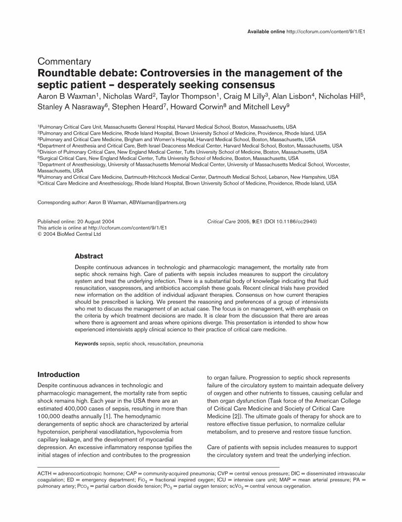
1
ACTH = adrenocorticotropic hormone; CAP = community-acquired pneumonia; CVP = central venous pressure; DIC = disseminated intravascularcoagulation; ED = emergency department; FiO2 = fractional inspired oxygen; ICU = intensive care unit; MAP = mean arterial pressure; PA =pulmonary artery; PCO2 = partial carbon dioxide tension; PO2 = partial oxygen tension; scVO2 = central venous oxygenation.
Available online http://ccforum.com/content/9/1/E1
IntroductionDespite continuous advances in technologic andpharmacologic management, the mortality rate from septicshock remains high. Each year in the USA there are anestimated 400,000 cases of sepsis, resulting in more than100,000 deaths annually [1]. The hemodynamicderangements of septic shock are characterized by arterialhypotension, peripheral vasodilatation, hypovolemia fromcapillary leakage, and the development of myocardialdepression. An excessive inflammatory response typifies theinitial stages of infection and contributes to the progression
to organ failure. Progression to septic shock representsfailure of the circulatory system to maintain adequate deliveryof oxygen and other nutrients to tissues, causing cellular andthen organ dysfunction (Task force of the American Collegeof Critical Care Medicine and Society of Critical CareMedicine [2]). The ultimate goals of therapy for shock are torestore effective tissue perfusion, to normalize cellularmetabolism, and to preserve and restore tissue function.
Care of patients with sepsis includes measures to supportthe circulatory system and treat the underlying infection.
CommentaryRoundtable debate: Controversies in the management of theseptic patient – desperately seeking consensusAaron B Waxman1, Nicholas Ward2, Taylor Thompson1, Craig M Lilly3, Alan Lisbon4, Nicholas Hill5,Stanley A Nasraway6, Stephen Heard7, Howard Corwin8 and Mitchell Levy9
1Pulmonary Critical Care Unit, Massachusetts General Hospital, Harvard Medical School, Boston, Massachusetts, USA2Pulmonary and Critical Care Medicine, Rhode Island Hospital, Brown University School of Medicine, Providence, Rhode Island, USA3Pulmonary and Critical Care Medicine, Brigham and Women’s Hospital, Harvard Medical School, Boston, Massachusetts, USA4Department of Anesthesia and Critical Care, Beth Israel Deaconess Medical Center, Harvard Medical School, Boston, Massachusetts, USA5Division of Pulmonary Critical Care, New England Medical Center, Tufts University School of Medicine, Boston, Massachusetts, USA6Surgical Critical Care, New England Medical Center, Tufts University School of Medicine, Boston, Massachusetts, USA7Department of Anesthesiology, University of Massachusetts Memorial Medical Center, University of Massachusetts Medical School, Worcester,Massachusetts, USA8Pulmonary and Critical Care Medicine, Dartmouth-Hitchcock Medical Center, Dartmouth Medical School, Lebanon, New Hampshire, USA9Critical Care Medicine and Anesthesiology, Rhode Island Hospital, Brown University School of Medicine, Providence, Rhode Island, USA
Corresponding author: Aaron B Waxman, [email protected]
Published online: 20 August 2004 Critical Care 2005, 9:E1 (DOI 10.1186/cc2940)This article is online at http://ccforum.com/content/9/1/E1© 2004 BioMed Central Ltd
Abstract
Despite continuous advances in technologic and pharmacologic management, the mortality rate fromseptic shock remains high. Care of patients with sepsis includes measures to support the circulatorysystem and treat the underlying infection. There is a substantial body of knowledge indicating that fluidresuscitation, vasopressors, and antibiotics accomplish these goals. Recent clinical trials have providednew information on the addition of individual adjuvant therapies. Consensus on how current therapiesshould be prescribed is lacking. We present the reasoning and preferences of a group of intensivistswho met to discuss the management of an actual case. The focus is on management, with emphasis onthe criteria by which treatment decisions are made. It is clear from the discussion that there are areaswhere there is agreement and areas where opinions diverge. This presentation is intended to show howexperienced intensivists apply clinical science to their practice of critical care medicine.
Keywords sepsis, septic shock, resuscitation, pneumonia

2
Critical Care February 2005 Vol 9 No 1 Waxman et al.
Although there is a substantial body of knowledge indicatingthat fluid resuscitation, vasopressors, and antibioticsaccomplish these goals, consensus on how they should beprescribed is lacking. Recent clinical trials have provided newinformation on the addition of individual adjuvant therapies,including adrenal supplementation therapy, tight glucosecontrol, and drotrecogin alpha (activated) to standardtherapies. These therapies are effective with statisticalcertainty in their respective study populations, but they donot provide insights into potential synergistic or antagonisticinteractions, making it challenging to determine whichcombination of treatments, if any, is best for a given patient. Itis the reasoning that clinicians use to process thisinformation and synthesize individual care plans that is thefocus of this commentary.
We present the reasoning and preferences of a group ofintensivists who met to discuss the management of an actualcase. Throughout this presentation, the focus is onmanagement, with emphasis on the points of discussion ofthe criteria by which treatment decision are made. It is clearfrom the discussion that there are areas where there isagreement and areas where opinions diverge. Participantssupport their opinions with literature citations and provide aperspective on how clinical practice can be distinct fromparticipation in a clinical trial.
Case presentation part 1The patient is a 56-year-old male who awoke the morning ofadmission with nausea, shortness of breath, and diaphoresis.Over the previous 2 days he had noted a productive cough,associated with midline chest pain, shaking chills, and threeto four episodes of watery diarrhea. He had no abdominalpain and no swelling or pain in the legs. Over the past3 months he had lost 30 lb (approximately 13.6 kg) in weightand he had recently sought medical attention for left shoulderpain. A bone scan was reportedly negative. He had no knowndrug allergies and took no medications. Family history wasunremarkable. He was a former smoker, of less than 20 pack-years, having stopped about 3 years previously, and hedenied alcohol or illicit drug use. He had worked as a drycleaner for the past 30 years.
Physical examination in the emergency department (ED)revealed a temperature of 97°F (36.1°C), a heart rate of180 beats/min, and blood pressure of 120/60 mmHg. Hisrespirations were labored at 26 breaths/min, and oxygensaturation was 89% on 4 l nasal cannulae oxygen. During theexamination he had an episode of coffee ground emesis. Hewas put on a nonrebreather mask, and his oxygen saturationincreased to 98%. Breath sounds were diminished in theright upper lung zones. His stool was hemoccult positive.Initial laboratory study findings are summarized in Table 1.
Dr A As we stand at the bedside, reviewing the admissionlaboratory findings and waiting impatiently for a chest
radiograph, let us try and put these findings together. First ofall, this patient presents with an acute illness with fever,chills, a cough that is productive of purulent sputum, and3 days of watery diarrhea. At first thought this sounds likecommunity-acquired pneumonia (CAP), possibly with anatypical pathogen. Perhaps the diarrhea is a red herring.Weight loss and left shoulder pain in a 53-year-old formersmoker raises concern for lung cancer with possiblepostobstructive pneumonia, and we shall look for signs ofvolume loss or chest wall metastasis on the chest radiograph.This patient also has evidence of severe sepsis, with a leftshift, and tachycardia. He has signs of impending organfailures with renal dysfunction, a creatinine level of 1.9 mg/dl,metabolic acidosis, a coagulopathy with an elevatedprothrombin time and D-dimer, and low platelets that couldsignal early disseminated intravascular coagulation (DIC). Hehas respiratory failure, with an A–a gradient over 500. He isprobably hemoconcentrated, and clinically we would expecthim to be hypovolemic from diarrhea, high insensible losses,and poor oral intake. My immediate therapeutic concern isthe tachycardia, the nature of the cardiac rhythm, and thepossibility of myocardial ischemia.
Dr D I might disagree with you that this patient has severesepsis. What if, after 2 or 3 l saline, his blood pressure, heart
Table 1
Initial laboratory values
Parameter Value
Potassium 3.5 mmol/l
Chloride 99 mmol/l
Carbon dioxide 19.9 mmol/l
BUN 26 mg/dl
Creatinine 1.9 mg/dl
Glucose 89 mg/dl
Hematocrit 43.7%
Platelet count 137,000 mm3
Bands 18%
PT/INR 14.8 s/1.4
PTT 48.9 s
D-dimer >1000 ng/ml
PH 7.32
PO2 101 mmHg
PCO2 27 mmHg
ECG Narrow complex tachycardia with 2–3 mm ST-segment depressions in
leads V3–V5
BUN, blood urea nitrogen; INR, international normalized ratio; PCO2,partial carbon dioxide tension; PO2, partial oxygen tension; PT,prothrombin time; PTT, partial thromboplastin time.
3
rate, and creatinine normalize? I think this illustrates one of thegreatest triage challenges in this area, and that isdifferentiating infection with sepsis from another very commonscenario – infection with dehydration and hypovolemia. I thinkthe most important point is not to confuse sepsis withhypovolemia from any cause, including hemorrhage.
Dr B The firsts steps in the care of patients like this shouldbe very systematic. Once the tachycardia was assessed andassuming the chest readiograph confirms pneumonia, I wouldinitiate prompt antibiotic therapy based on the most likelytype(s) of infection; ensure adequate hemodynamic andrespiratory support; try to identify the source of infection;identify and ascertain the extent of his organ dysfunction;and, based on that, develop an overall treatment plan.
Dr C I agree – we need to treat this suspected infection.Although a chest radiograph is not initially available, there is astrong clinical suspicion for pneumonia, as a source of majorinfection, and empiric antibiotic coverage should be started.When the differential diagnosis includes a central nervoussystem infection, there is never a question in regard to earlyantibiotic coverage, and in fact antibiotics are started within awindow of 6–8 hours. Data for most infections, especiallypneumonia, have the same window of opportunity in regardto mortality. Animal models of septic shock also indicate thatmortality can directly relate to time delay in antibiotics. If thereis any delay in the work up, including a delay in obtaining achest radiograph, then empiric treatment for CAP withceftriaxone and azithromycin should be initiated.
Case presentation part 2The patient was given 2 l of normal saline and a chestradiograph was obtained. He was given a total of 18 mgadenosine with no response; and then 2.5 mg intravenousverapamil in three doses, with a decrease in heart rate to110 beats/min. Atrial fibrillation was diagnosed. His bloodpressure fell with these interventions. The chest radiographshowed a dense opacity in the right-upper lobe with patchyopacities in the right-lower lobe, the lingula, and left-upperlobe. There were no clear air bronchograms or lateral shift tosuggest lobar collapse (Fig. 1).
Dr B There are now additional diagnostic data available, andclinically important interventions have been done that raiseimportant considerations. The additional radiographicinformation addresses the concerns raised about the 30 lbweight loss and any chronic process that may be acutelyinfected. There is no clear evidence of an endobronchiallesion or airway obstruction. This, along with the history of arecent negative bone scan, makes cancer less likely. Activetuberculosis is possible, and sputum should be sent forappropriate studies. With the complaint of midsternalpleuritic chest pain, there is concern of pericardialinvolvement. However, there are no suggestive changes onthe ECG, and the heart size looks normal, somewhat narrow,
and probably under-filled, which is consistent withhypovolemia.
Dr A Do we need to consider any other diagnostic tests forCAP in a patient with severe sepsis?
Dr C It can be very helpful in cases like this to have bloodand sputum cultures. It is usually possible to get a set ofblood cultures in patients like this, prior to starting antibiotics.Although they do not have a high yield, they can play animportant role in the treatment of the infection by identifyingpossible sources and unusual or resistant organisms. Theycan also help to identify high-risk patients.
Dr B The purist’s goal is to try to establish a bacteriologicdiagnosis, but the realization is that one must initially treatbroadly because no single test has the sensitivity andspecificity to allow one to treat narrowly. Regardless, thecoverage we use includes coverage for almost every possiblebacteriologic pathogen. Knowing that this patient is from thecommunity, we do not need to consider double Gram-negative coverage, even in a septic patient. If a patient isfrom the community but has recently been hospitalized, isimmunosuppressed, is being treated with steroids, or has
Available online http://ccforum.com/content/9/1/E1
Figure 1
Initial radiograph showing a right-upper lobe opacity and opacities inthe right-lower, lingula, and left-upper lobes.

4
structural lung disease, such as bronchiectasis, then wewould consider alternative or additional antibiotic coverage tocover for Pseudomonas spp. or other resistant Gram-negative rods. For the time being there is no indication tochange either the azithromycin or ceftriaxone. I agree that totry to obtain a bacteriologic diagnosis, we use blood, urineand sputum cultures, if available, and might performbronchoscopy once the patient is hemodynamically stable.
Dr C Getting back to the ECG, the patient has a narrowcomplex tachycardia with a rate of 190 beats/min with2–3 mm ST-segment depressions across the precordium.Should the heart rate be directly controlled? Could thepatient be having a myocardial infarction or is he simplyvolume depleted? I am concerned about using adenosinebefore adequately addressing the patient’s volume status.The initial approach should be to volume replete himaggressively and see whether the hemodynamics improve,and then address any persistent tachycardia only after he isvolume replete. In this case, rate controlling agents may notbe the first line of therapy, given a strong suspicion forsepsis. One question that comes up is, what if he is ischemicand you start volume resuscitating him? Is he going to gointo heart failure? Does that matter? This should not be anissue. If the need arises then support him with invasive ornoninvasive ventilation and get him through this. Regardlessof the cause of the tachycardia, fluid load this patient.
Dr A Would there be any indication to treat him for an acutecoronary syndrome with aspirin or heparin at this point?Would there be a problem with starting heparin in this man ifone felt he truly had a coronary basis of ischemia versusdemand ischemia? It is likely that this represents demandrelated ischemia, as opposed to an unstable coronarysyndrome. This is a 55-year-old man. The ECG shows anarrow complex tachycardia at 190 beats/min with ST-segment depressions in V3–V5. Creatine phosphokinase andtroponin levels were all initially normal. Is there a down sideto giving aspirin? Aspirin is not going to be of much value, ifyou think this is demand-related ischemia. It does complicatethe decision making for drotrecogin alpha (activated) shouldhe develop severe sepsis. There is also the concern that heis having gastrointestinal bleeding. There is reluctance to goafter his ischemia in a big way until we get his rate controlled.If ECG changes persist after control of heart rate, then wemight consider aspirin. If heparin is started then it can easilybe discontinued.
Dr B I agree that aspirin may complicate the decision making,should you want to give a new agent like drotrecogin alpha(activated). Patients receiving aspirin or other antiplateletagents were excluded from the PROWESS (RecombinantHuman Activated Protein C Worldwide Evaluation in SevereSepsis) trial, and so its safety in the presence of antiplateletagents is unclear. If you think the patient is more at risk fordeath from severe sepsis than from demand ischemia, then
your attention should be directed toward the interventionsthat are most likely to give the patient benefit. Because thispatient had no prior history of coronary disease, and becausedemand related ischemia is more likely than an acutecoronary syndrome that may benefit from aspirin, therisk/benefit moves one step toward sepsis interventionsrather than acute myocardial infarction interventions.Coronary flow is upregulated in sepsis [3–5]. It is actuallyunusual for patients to develop myocardial ischemia.
Case presentation part 3The patient was admitted to the general medical floor. Hewas still tachycardic at 110 beats/min in sinus rhythm, andST-segment changes had resolved. He was described asbeing cold and clammy. His systolic blood pressure was nowin the 90s. He was given an additional 3 l normal saline,resulting in a blood pressure of 110/60 mmHg. He wasincreasingly tired, and his breathing became more labored.He received ceftriaxone and azithromycin. The patientsubsequently required orotracheal intubation with a #8endotracheal tube. He was given fentanyl, propofol, andmidazolam for intubation. Eight hours after presenting in theED, he was transferred to the intensive care unit (ICU). Achest radiograph following intubation showed progression ofthe right-upper lobe process. The heart rate was106 beats/min, and blood pressure was 94/47 mmHg with amean arterial pressure (MAP) of 60 mmHg. Oxygensaturations were 96% on 100% oxygen. An additional 3 lnormal saline was given, for a total of about 6.5 l (Fig. 2).
Dr A What is interesting in this case is that, in the ED, whenyou have someone who already has single organ failure andsome borderline findings that make you suspect he is on hisway to multiorgan failure, should this patient automatically bean ICU admission? We argue this frequently because, if youvolume resuscitate this patient in the ED and he turns aroundin the ED, the tachycardia goes away, the ST segmentsnormalize, his respiratory distress gets better, and he eithergoes to an ICU or a regular floor bed. This becomes a veryimportant triage question. This patient is a great example ofsomeone who has the potential to crash very hard, and if youintervene right away with early therapy you may be able toreverse at least what we see as early organ dysfunction andprevent multiorgan failure. This man could have gone straightto the ICU, and we could have figured all this out there,rather than try to get him floor ready. The other thing is that ifyou send him to the ICU, he could spend 24 hours there andthen be ready to go to the floor; instead he ended up comingto the ICU for a week.
Case presentation part 4Two hours later, the patient was agitated on the ventilatorand had frequent high peak airway pressure alarms; theoxygen saturations were stable. The blood pressure hadbeen trending downward, with systolic in the high 70s, MAPin the 50s, and urine output under 20 cc/hour. His sodium
Critical Care February 2005 Vol 9 No 1 Waxman et al.

5
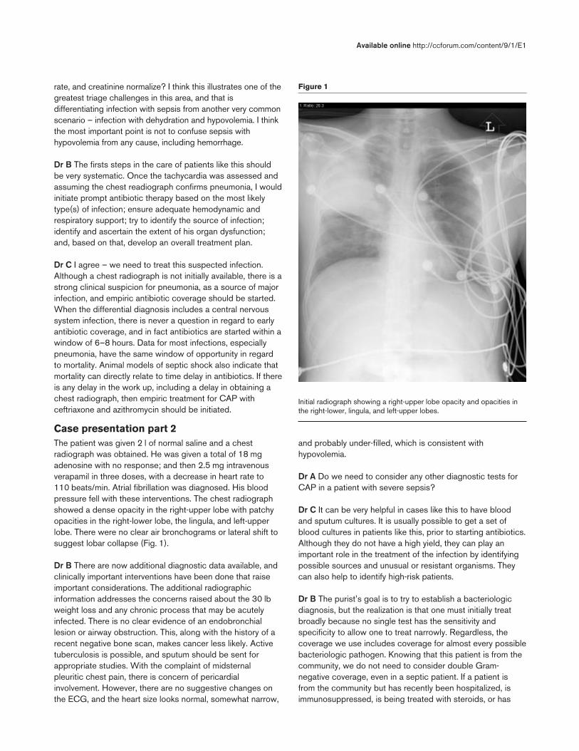
was 137 mmol/l, potassium 3.2 mmol/l, chloride 118 mmol/l,carbon dioxide 17.8 mmol/l, blood urea nitrogen 18 mg/dl,and creatinine 1.1 mg/dl, the latter representing animprovement. The glucose was 170 mg/dl, phosphate wasdown to 1.4 mg/dl, and albumin was 1.3 g/dl. The white cellcount was 6500/mm3 with 15% bands; platelets were56,000/mm3. The hematocrit was down to 28.4%. Cardiacenzymes were within normal limits. The arterial blood gaseson 50% inspired fractional oxygen (FiO2) were as follows:pH 7.46, partial carbon dioxide tension (PCO2) 24 mmHg,and partial oxygen tension (PO2) 99 mmHg. Central venouspressure (CVP) was 14 cmH2O. Central venous PO2 was39 mmHg. Lactate was 5.6 mg/dl. Norepinephrine(noradrenaline) was being started (Fig. 3).
Dr D Resuscitation is clearly ongoing, and appropriate end-points have not yet been reached. The patient is hypotensive,in spite of 8–10 l crystalloid. He has not received any blood,and pressors are being started. He was just intubated andheavily sedated. The chest radiograph needs to be checkedfor anything that could have a negative hemodynamic impact,such as a pneumothorax or other mechanical reasons forshock.
Dr A How do we judge the completeness of his volumeresuscitation?
Dr B Clinical indices should be the first guide to theappropriateness of volume resuscitation. Clinical markers thatwere originally deranged can be followed, looking for
improvement. Regardless, many of these patients may go onto develop organ failure. A threshold is often reached atwhich volume is given to the point that the presumption isthat the patient is intravascularly replete. How is that pointdefined in the patient with organ failure? It depends, in part,on the age of the patient and cardiac function. If the patient isyoung or left ventricular function is known from a previousechocardiogram, then a central venous line with CVP andcentral venous oxygenation (scVO2) monitoring may beadequate. Alternatively, if cardiac function is not known orthere is a suspicion of compromise, especially in an elderlypatient, or if a large volume of fluid has been given withoutimprovement, then invasive monitoring with a pulmonaryartery (PA) line would be a consideration. When do youdecide that enough fluid has been given? When is it time tohang a pressor or an inotrope? Initial volume resuscitationshould be between 8 and 10 l, but after that how do youdecide if that is enough or if are they leaking into the thirdspace?
Dr A Adequacy of volume resuscitation is a big picture issue,and I never rely on a single variable. Multiple parameters areconsidered, including the hemodynamic measurements ofheart rate, blood pressure, urine output, skin perfusion, andmentation. If these measures do not improve with aggressivevolume expansion, then a PA line is considered. Thissuggests that it takes an experienced intensivist to judge fluidrequirements. How does the resident or nonintensivist do it?As part of the protocol for early goal-directed therapy,pressors are used to keep the MAP about 60 or the systolic
Available online http://ccforum.com/content/9/1/E1
Figure 2
Postinstubation radiograph showing progression of the right-upperlobe opacity.
Figure 3
Radiograph showing progressive involvement of both lungs.
6
pressure above 90. Fluids are given to raise the CVP tobetween 10 and 12. The central venous oxygen saturation ispushed up with dobutamine and/or red cells. If these goalsare not achieved within a reasonable time frame, thenconsider what additional information can be obtained fromthe PA catheter. The reason why the PA catheter trials havebeen so inconclusive is because there have been suchvarying goals or protocols associated with their use. If wecould figure out the best protocol to resuscitate people, thenwe might be able to use the PA line as a guide and perhapsshow benefit over non-PA-guided protocols.
Dr D This raises the question as to when pressors should bestarted. The preference is not to hang vasopressors early inthe resuscitation. If the patient is not adequately volumeresuscitated, then splanchnic blood flow could becompromised by pressors. Conversely, we cannot allow apatient to remain hypotensive while striving to achieve volumeresuscitation goals. Although the preference is to refrain fromgiving vasopressors before adequate volume resuscitation,sometimes we have to maintain an adequate blood pressure.In patients who are on pressors, should target goals beadjusted? It is likely that a higher central venous pressure isrequired in patients who are on pressors. A patient who has aCVP of 12 or 15, but who requires significant doses ofvasopressors to maintain adequate MAP, might really need aCVP of 25.
Dr B When resuscitating a septic patient, there is a need, ifwe have not achieved our therapeutic goal, to double checkthe status of the left ventricle. We should not make anassumption in the septic patient that left ventricular functionis normal. Many of these patients, perhaps even this one,have pulmonary vascular alterations in which there is adisconnect between the left and right side. They can haveelevated CVPs in the presence of a low wedge. In thissetting the PA catheter may give us additional usefulinformation that CVP does not. An important point is not tofall into the trap of searching for the one best number. Thereis no one best number. Generally, there is a lot of informationbeing generated for each patient, and it is important to lookat all this information, especially the stroke volume or cardiacoutput for given CVP or wedge values, and how all thesevariables change, or do not change, after fluid boluses.Noninvasive estimates of preload and cardiac function canalso be obtained. We all want to be reassured that we havegiven enough, but not too much, volume. If more fluid isgiven, and whatever index of cardiac function is beingfollowed does not change, then we’re probably close toadequate fluid resuscitation. That said, we have no ideawhether this is the right construct, even though we all seemto agree on it. It may be that a strategy that favorsnorepinephrine, vasopressin, and/or inotropes allowing forless fluid resuscitation produces better outcomes than onethat favors aggressive fluid resuscitation aimed at optimizingcardiac output and minimizing vasopressors. One recent
study of elective surgical patients [6] showed superioroutcomes with a severely restricted perioperative fluidmanagement strategy. We need much better studies in thisarea.
Case presentation part 5He received over 10 l isotonic crystalloid, and his initial CVPwas around 6 mmHg. He received additional volume as acombination of crystalloid and colloid; the resulting CVP was15 mmHg.
Dr A Is it possible to be more efficient in the resuscitation?Do colloids make a difference in resuscitation?
Dr D Colloids make a difference in the sense that we canvolume resuscitate much more quickly. We also add oncoticpressure, which hopefully keeps more crystalloidintravascularly, and the total volume of fluid is less. If theissue is one of timing, then the more aggressively youachieve your end-diastolic volume goals, the better.
Dr C Inadequate preload in sepsis and septic shock ismultifactorial and can be related to venodilation, fever andfluid losses, and diffuse capillary leak. Vascular dysfunctionpresumably results from damage to underlying endotheliumand likely results in compromised endothelial barrier function.Larger colloids are more likely to stay in the intravascularspace. This, in combination with widespread inflammatoryactivation, explains one of the challenges of fluid therapy –capillary leak and formation of edema. This also plays a role inthe requirement for large volume resuscitation. Restoration ofadequate circulatory volume is necessary to permit adequatetissue perfusion, but it may not be sufficient on its own tocorrect microvascular abnormalities associated with sepsis.
Dr A The literature does not support a benefit of one colloidover the other, or colloids over crystalloids. So the questionis, you know you can give 10 l crystalloid or you can give 2.5 lcolloid, so why wouldn’t you just give 2.5 l? It is faster, withthe same gauge intravenous line. Many intensivists wouldgenerally use 100% crystalloid, and many of us would lookfor reasons to give blood. If the hemoglobin is below 10, thenpacked red blood cells would be reasonable.
Dr B For the most part I would use crystalloid as a first-lineagent. Some of us would consider using both starch andblood, depending on the hemoglobin. As far as a specificapproach, once I have reached a point where the patient hasreceived over 6–10 l crystalloid, there is a desire to givesomething that is going to stay intravascular and providemore volume expansion with less total volume. At this point, Iwould start using colloid and, especially knowing hishemoglobin is where it is, I would probably give him starch.The bottom line is that most of us agree that the quantity andtiming of whatever you pick is much more important than thefluid you pick.
Critical Care February 2005 Vol 9 No 1 Waxman et al.
7
Dr A Would anybody transfuse this patient with a hematocritof 28.4?
Dr C Knowing he has a hematocrit that was 43 when hecame in is important. He has had close to 10 l of crystalloid.Based on the protocol of Rivers and coworkers [7], initialvolume resuscitation was given to get the CVP to 8–12.scVO2 is then examined. If the scVO2 is under 70, then eitherdobutamine or red cells (to get to a hematocrit of 30) aregiven to achieve a scVO2 above 70%. It is not entirely clearwhy the patients assigned to early goal-directed therapy inthe Rivers study did better. The data suggest that it was notnecessarily because they got blood but rather the timing ofhow much fluid they received. At the end of 72 hours, bothgroups received the same amount of fluid but it wasfrontloaded in the early goal-directed therapy group,potentially having only to do with the aggressiveness ofresuscitation. However, the protocol group did receive moreblood (64% versus 16% transfused) during the first 6 hours.
Dr B There are several studies that show that packed redblood cells may not increase oxygen utilization in sepsis. It isbelieved their main benefit in a situation like this is as acolloid — one that is very expensive, is of limited supply, andmay have a variety of infection risks.
Dr A Should we treat this patient with empiric steroids?
Dr D The best current evidence with which to answer thisquestion comes from the randomized controlled trialconducted by Annane and coworkers [8]. A prior trialsuggested that failure to augment serum cortisol by 9 µg/dlafter high-dose adrenocorticotropic hormone (ACTH) was abad prognostic sign and was independent of the pre-ACTHstimulated cortisol value [9]. The subsequent randomizedcontrolled trial by Annane and coworkers showed benefitfrom steroids in the ACTH nonresponder patients only.Therefore, a random cortisol without cosyntropin may nothelp you decide whether to treat relative adrenalinsufficiency. On the practical side, in many institutionscortisol results are not available for 2 days. If that were thecase, then I would draw a pre- and post-ACTH cortisol andbegin hydrocortisone 50 mg every 6 hours until the cortisolresults return. I think we all would withdraw steroids if theserum cortisol increased by more than 9 µg/dl followingACTH.
Dr A According to the study by Annane and coworkers [8], ifthe baseline cortisol goes from 55 to 61, then the patientmust be treated because they have relative adrenalinsufficiency. An argument could be made that if the baselinecortisol is 55, then the patient may not benefit from additionalsteroids. Of the 299 patients who were studied and the 229who were said to be adrenally insufficient, we do not knowwhat fraction of those patients truly had a baseline cortisol of55. In the patients who had an elevated cortisol to start with,
it is unclear whether there is really any benefit in steroidreplacement.
Dr D A cosyntropin stimulation test is easy and safe and isthe only clear way, at the present time, to distinguish patientswho appear to have an insufficient adrenal response.Because the random cortisol is not a reliable index, the bestapproach would be to treat the patient with steroidreplacement therapy, pending the results of the cosyntropinstimulation test, and withdraw steroids, once those resultsshow a normal response. This assumes that patients whorespond are not harmed by 2 days of steroids.
Case presentation part 6Two hours later the sodium was 135 mmol/l, potassium4.5 mmol/l, chloride 94 mmol/l, carbon dioxide 17 mmol/l,blood urea nitrogen 21 mg/dl, creatinine 1.8 mg/dl, andglucose 153 mg/dl. An arterial blood gas on 60% FiO2revealed pH 7.33, PCO2 34 mmHg, and PO2 70 mmHg, witha central venous oxygen saturation of 66%. The white bloodcell count was 32,000/mm3 with 32% bands. The plateletshad dropped to 28,000/mm3, and the internationalnormalized ratio was 1.8, with a prothrombin time of 19.5 sand a partial thromboplastin time of 50 s. Urine output hadincreased to 40 cc/hour.
Dr A Should this patient be treated with drotrecogin alpha(activated)?
Dr B This patient has septic shock, organ injury,coagulopathy, and signs of DIC. His platelets are 28,000,which is below the 30,000 cutoff used in the PROWESStrial [10], but I think this may be exactly the kind of patientwho should receive Drotrecogin alpha (activated). In thePROWESS trial DIC was part of the enrollment criteria.Actually, in the subgroup analysis, the DIC patients didbetter. If there is concern with the platelet count, we caneither give platelets if this man had a lot of incisions orpreviously showed bleeding or forget about the plateletcount, give the drotrecogin alpha (activated), and watch himexpectantly.
Dr D My understanding of the mechanisms by which thisdrug works tells me that drotrecogin alpha (activated) worksby reversing the underlying pathophysiology in sepsis, and ifyou can improve this patient rapidly in the first 24 hours withstandard therapies, then he is likely to have a better mortality.If you can’t, then an intervention with a drug like drotrecoginalpha (activated) makes sense [11]. The data make a casefor early treatment in all respects. Early intervention of almostanything is better than later intervention. Given what we knowof the sepsis cascade, the further it rolls on the worse it gets.For most of us who have been sepsis investigators for a longtime, the generalized expectation is that the earlier weintervene the better the chances that one can interrupt thesepsis cascade, and so on.
Available online http://ccforum.com/content/9/1/E1

8
Case presentation part 7
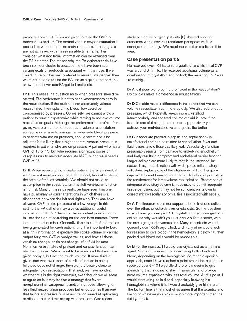
Over the next 48 hours the findings on chest radiographyimproved. A chest computed tomography scan was doneand did not reveal an endobronchial lesion. By day 3 thepatient was off pressors. Urine output was up and the FiO2 isdown. The patient was awake, alert, and responsive. On thethird hospital day he was extubated. The white count was14,000/mm3 without bands. The platelets and prothrombintime normalized, and the creatinine was normalizing (Fig. 4).
Dr A The next question is whether he is clearly getting better.Do we keep the patient in the ICU to complete the full courseof drotrecogin alpha (activated) or send him to the floor?Those are two different questions: do we have to complete thefull 96-hour infusion and do we keep a patient who no longerrequires ICU level care in the unit to complete the treatment?
Dr D Here is a man who is off the vent; renal failure andhypotension are resolved. If someone meets every othercriterion for leaving the ICU – except for the fact they arereceiving drotrecogin alpha (activated) – do you send him outof the ICU on drotrecogin alpha (activated) or stop theinfusion early to send him out to the medical floor? If he isthis stable, then I would stop the infusion and send him out. Ithink most would agree that you wouldn’t keep the patient inthe ICU solely to keep him on drotrecogin alfa (activated) ifhe is no longer critically ill. However, the much more commonquestion is if he were still intubated, off pressors, lookingmuch better, and one was thinking about extubating,72 hours into the course, would one continue drotrecoginalpha (activated)? In that case I feel compelled to continue
the drug. We don’t raise that same question with antibiotics,and in that scenario he would continue to receive antibioticsfor the next 7–10 days.
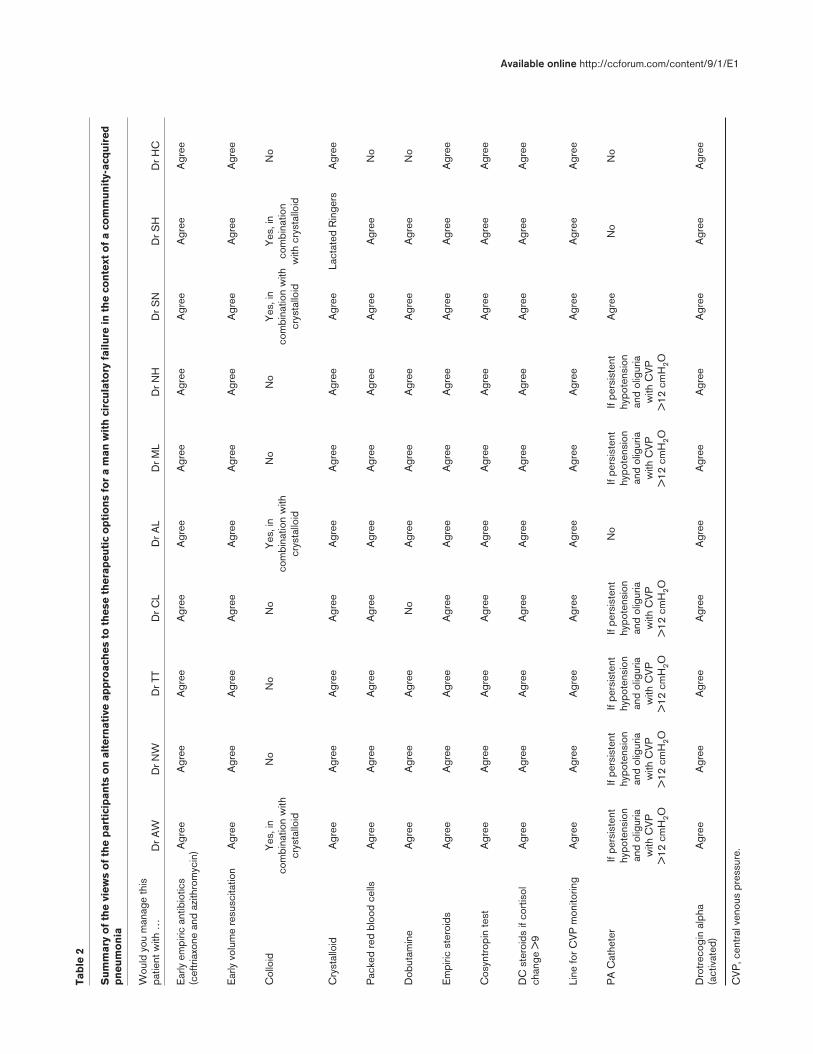
SummaryThe analysis and commentary of the care provided to thispatient with severe sepsis and septic shock highlight thechallenges in assessing and managing critically ill patients.The participants provided their perspectives on therecognition of physiological instability and the definition ofsevere sepsis and septic shock; at what point a patientshould be admitted to the ICU; the importance of early goal-directed resuscitation therapy and the choice of resuscitationfluid; when to consider intravascular monitoring; the use oflow-dose corticosteroids and drotrecogin-alpha; and at whatpoint an improving patient should leave the ICU (Table 2).
In this case, the assessment of the patient begins withrecognition of the signs and symptoms of a serious infection.The participants agreed not only that antibiotic treatmentshould be given within 8 hours of presentation but also thatcombination therapy for CAP was appropriate. It was agreedthat cultures of blood, urine, and sputum are indicated butshould not delay the initiation of antibiotic therapy. Diagnosticbronchoscopy was not recommended while the patient washemodynamically unstable.
Patients often present with tachycardia andelectrocardiographic abnormalities that may reflect sepsis,infection with vasodilation and hypovolemia (so-calledineffective arterial circulation), or cardiac ischemia.Distinguishing sepsis and an ineffective circulation from anacute coronary syndrome can be challenging. All of theparticipants agreed that, regardless, the most important effortshould be directed toward restoring adequate intravascularvolume. Resolution of tachycardia and increased urine flowwith fluid resuscitation would suggest that hypovolemia andsepsis were the problems. Although there were differencesof opinion regarding the timing of CVP or PA catheterplacement, all were in agreement that a CVP or pulmonarycapillary wedge pressure in the single digits wasinappropriate for this patient. Furthermore, all agreed that theadequacy of volume resuscitation was best determined fromcumulative data, including serial hemodynamicmeasurements, measurements of arterial and central venousoxygenation, and the response to volume infusion.
Both the availability and potential risks (although small) oftransfusion of red blood cells made their use more restricted.The group agreed that patients with a hematocrit of less than30 and a mixed or central venous oxygen saturation of lessthan 70% might benefit from red cell transfusion.Additionally, everyone agreed that patients who are not likelyto be harmed by 2 days of steroid replacement withhydrocortisone and fludrocortisone, but continued treatmentwith steroids should be guided by the results of a
Critical Care February 2005 Vol 9 No 1 Waxman et al.
Figure 4
Radiograph showing generalized improvement.
9
Available online http://ccforum.com/content/9/1/E1Ta
ble
2
Su
mm
ary
of
the
view
s o
f th
e p
arti
cip
ants
on
alt
ern
ativ
e ap
pro
ach
es t
o t
hes
e th
erap
euti
c o
pti
on
s fo
r a
man
wit
h c
ircu
lato
ry f
ailu
re in
th
e co
nte
xt o
f a
com
mu
nit
y-ac
qu
ired
pn
eum
on
ia
Wou
ld y
ou m
anag
e th
is
patie
nt w
ith …
Dr A
WD
r NW
Dr T
TD
r CL
Dr A
LD
r ML
Dr N
HD
r SN
Dr S
HD
r HC
Ear
ly e
mpi
ric a
ntib
iotic
s A
gree
Agr
eeA
gree
Agr
eeA
gree
Agr
eeA
gree
Agr
eeA
gree
Agr
ee(c
eftr
iaxo
ne a
nd a
zith
rom
ycin
)
Ear
ly v
olum
e re
susc
itatio
nA
gree
Agr
eeA
gree
Agr
eeA
gree
Agr
eeA
gree
Agr
eeA
gree
Agr
ee
Col
loid
Yes
, in
No
No
No
Yes
, in
No
No
Yes
, in
Yes
, in
No
com
bina
tion
with
com
bina
tion
with
com
bina
tion
with
com
bina
tion
crys
tallo
idcr
ysta
lloid
crys
tallo
idw
ith c
ryst
allo
id
Cry
stal
loid
Agr
eeA
gree
Agr
eeA
gree
Agr
eeA
gree
Agr
eeA
gree
Lact
ated
Rin
gers
Agr
ee
Pac
ked
red
bloo
d ce
llsA
gree
Agr
eeA
gree
Agr
eeA
gree
Agr
eeA
gree
Agr
eeA
gree
No
Dob
utam
ine
Agr
eeA
gree
Agr
eeN
oA
gree
Agr
eeA
gree
Agr
eeA
gree
No
Em
piric
ste
roid
sA
gree
Agr
eeA
gree
Agr
eeA
gree
Agr
eeA
gree
Agr
eeA
gree
Agr
ee
Cos
yntr
opin
test
Agr
eeA
gree
Agr
eeA
gree
Agr
eeA
gree
Agr
eeA
gree
Agr
eeA
gree
DC
ste
roid
s if
cort
isol
A
gree
Agr
eeA
gree
Agr
eeA
gree
Agr
eeA
gree
Agr
eeA
gree
Agr
eech
ange
>9
Line
for C
VP
mon
itorin
gA
gree
Agr
eeA
gree
Agr
eeA
gree
Agr
eeA
gree
Agr
eeA
gree
Agr
ee
PA
Cat
hete
rIf
pers
iste
nt
If pe
rsis
tent
If pe
rsis
tent
If
pers
iste
nt
No
If pe
rsis
tent
If
pers
iste
nt
Agr
eeN
oN
ohy
pote
nsio
n hy
pote
nsio
n hy
pote
nsio
n hy
pote
nsio
n hy
pote
nsio
n hy
pote
nsio
n an
d ol
igur
ia
and
olig
uria
an
d ol
igur
ia
and
olig
uria
an
d ol
igur
ia
and
olig
uria
w
ith C
VP
w
ith C
VP
w
ith C
VP
w
ith C
VP
w
ith C
VP
w
ith C
VP
>
12 c
mH
2O>
12 c
mH
2O>
12 c
mH
2O>
12 c
mH
2O>
12 c
mH
2O>
12 c
mH
2O
Dro
trec
ogin
alp
ha
Agr
eeA
gree
Agr
eeA
gree
Agr
eeA
gree
Agr
eeA
gree
Agr
eeA
gree
(act
ivat
ed)
CV
P, c
entr
al v
enou
s pr
essu
re.
10
cosyntropin stimulation test. All of the participants agreedthat this patient with severe sepsis and no contraindicationsshould be treated with drotrecogin-alpha (activated).
ConclusionThe Blue Ginger Group has prepared this synthesis ofopinion on the optimal treatment of a specific case of severesepsis in order to share how experienced critical careproviders treat their patients. This presentation in notintended to advocate particular treatment algorithms butrather to show how experienced intensivists apply clinicalscience to their practice of critical care medicine. Applyinginformation provided by clinical trials to practice requiressynthetic reasoning, judgment, and knowledge about thepatient as well as those patients included in the studies. Evenmore important than the specific details of this analysis is thatwe need more high-quality clinical trial data to clarify whichtreatments or combinations of treatments best help individualpatients with severe sepsis.
Competing interestsThe author(s) declare that they have no competing interests.
AcknowledgmentsWe want to extend our appreciation to Chef Ming Tsai, who hasallowed us to meet on a regular basis at his restaurant, The BlueGinger, in Wellesley, Massachusetts. This not only has stimulated ourongoing efforts at improving critical care delivery but it also hasenhanced our individual gastronomic development.
We also acknowledge the administrative support provided by BarbaraShott at The Rhode Island Hospital, without which none of this wouldhave ever happened.
The Blue Ginger Group includes Howard Corwin, MD; Stephen Heard,MD; Nicholas Hill, MD; Mitchell Levy, MD; Craig M Lilly, MD; AlanLisbon, MD; Stanley A Nasraway, MD; Taylor Thompson, MD; NicholasWard, MD; and Aaron B Waxman, MD, PhD.
References1. Angus DC, Linde-Zwirble WT, Lidicker J, Clermont G, Carcillo J,
Pinsky MR: Epidemiology of severe sepsis in the UnitedStates: analysis of incidence, outcome, and associated costsof care. Crit Care Med 2001, 29:1303-1310.
2. Hollenberg SM, Aherns TS, Astiz ME, Chalfin DB, Dasta JF, HeardSO, Martin C, Sulsa GM, Vincent JL Practice parameters forhemodynamic support of sepsis in adult patients. Crit CareMed 1999, 27:639-660.
3. Cunnion RE, Schaer GL, Parker MM, Natanson C, Parrillo JE: Thecoronary circulation in human septic shock. Circulation 1986,73:637-644.
4. Ellrodt AG, Riedinger MS, Kimchi A: Left ventricular perfor-mance in septic shock: reversible segmental and globalabnormalities. Am Heart J 1985, 110:402-409.
5. Raper RF, Sibbald WJ, Hobson J, Neal A, Cheung H: Changes inmyocardial blood flow rates during hyperdynamic sepsis withinduced changes in arterial perfusing pressures and meta-bolic need. Crit Care Med 1993, 21:1192-1199.
6. Brandstrup B, Tonnesen H, Beier-Holgersen R, Hjortso E, Ording H,Lindorff-Larsen K, Rasmussen MS, Lanng C, Wallin L, The DanishStudy Group on Perioperative Fluid Therapy: Effects of intravenousfluid restriction on postoperative complications: comparison oftwo perioperative fluid regimens: a randomized assessor-blinded multicenter trial. Ann Surg 2003, 238:641-648.
7. Rivers E, Nguyen B, Havstad S, Ressler J, Muzzin A, Knoblich B,Peterson E, Tomlanovich M: Early goal-directed therapy in thetreatment of severe sepsis and septic shock. N Engl J Med2001, 345:1368-1377.
8. Annane D, Sebille V, Charpentier C, Bollaert P-E, Francois B,Korach J-M, Capellier G, Cohen Y, Azoulay E, Troche G, et al.:Effect of treatment with low doses of hydrocortisone and flu-drocortisone on mortality in patients with septic shock. JAMA2002, 288:862-871.
9. Annane D, Sebille V, Troche G, Raphael JC, Gajdos P, BellissantE: A 3-level prognostic classification in septic shock based oncortisol levels and cortisol response to corticotropin. JAMA2000, 283:1038-1045.
10. Bernard GR, Vincent JL, Laterre PF, LaRosa SP, Dhainaut J-F,Lopez-Rodriguez A, Steingrub JS, Garber GE, Helterbrand JD, ElyEW, et al.: Efficacy and safety of recombinant human activatedprotein C for severe sepsis. N Engl J Med 2001, 344:699-709.
11. Levy MM, Macias WL, Russell JA, Williams MD, Trzaskoma BL,Silva E, Vincent J-L. Failure to improve during first day oftherapy is predictive of 28-day mortality in severe sepsis.Chest 2003, 124:120S-123S.
Critical Care February 2005 Vol 9 No 1 Waxman et al.
Copyright © 2022 FDOKUMEN