Role of Vitamin D in Preventing and Treating Selected ... - MDPI
36
nutrients Review Role of Vitamin D in Preventing and Treating Selected Extraskeletal Diseases—An Umbrella Review Friederike Maretzke 1 , Angela Bechthold 1 , Sarah Egert 2 , Jana B. Ernst 1 , Debora Melo van Lent 3 , Stefan Pilz 4 , Jörg Reichrath 5 , Gabriele I. Stangl 6 , Peter Stehle 7 , Dorothee Volkert 8 , Michael Wagner 9 , Julia Waizenegger 1, *, Armin Zittermann 10 and Jakob Linseisen 1,11 on behalf of the German Nutrition Society (DGE) 1 German Nutrition Society, 53175 Bonn, Germany; [email protected] (F.M.); [email protected] (A.B.); [email protected] (J.B.E.); [email protected] (J.L.) 2 Institute of Nutritional Medicine, University of Hohenheim, 70599 Stuttgart, Germany; [email protected] 3 Glenn Biggs Institute for Alzheimer’s and Neurodegenerative Diseases, University of Texas Health Sciences Center, San Antonio, TX 78229, USA; [email protected] 4 Division of Endocrinology and Diabetology, Department of Internal Medicine, Medical University of Graz, 8036 Graz, Austria; [email protected] 5 Department of Adult and Pediatric Dermatology, Venereology, Allergology, University Hospital Saarland, 66424 Homburg, Germany; [email protected] 6 Institute for Agricultural and Nutritional Sciences, Martin Luther University Halle-Wittenberg, 06120 Halle (Saale), Germany; [email protected] 7 Department of Nutrition and Food Sciences, University of Bonn, 53115 Bonn, Germany; [email protected] 8 Institute for Biomedicine of Aging, Friedrich-Alexander-Universität Erlangen-Nürnberg, 90408 Nuremberg, Germany; [email protected] 9 Department for Neurodegenerative Diseases and Geriatric Psychiatry, University Hospital Bonn, 53127 Bonn, Germany; [email protected] 10 Clinic for Thoracic and Cardiovascular Surgery, Heart and Diabetes Center North Rhine-Westphalia, 32545 Bad Oeynhausen, Germany; [email protected] 11 University Center of Health Sciences at Klinikum Augsburg (UNIKA-T), Ludwig Maximilian University of Munich, 86156 Augsburg, Germany * Correspondence: [email protected]; Tel.: +49-228-3776-628 Received: 21 February 2020; Accepted: 25 March 2020; Published: 31 March 2020 Abstract: Evidence is accumulating that vitamin D may have beneficial effects on respiratory tract, autoimmune, neuro-degenerative, and mental diseases. The present umbrella review of systematic reviews (SRs) of cohort studies and randomised controlled trials (RCTs), plus single Mendelian randomisation studies aims to update current knowledge on the potential role of vitamin D in preventing and treating these extraskeletal diseases. Altogether, 73 SRs were identified. Observational data on primary prevention suggest an inverse association between vitamin D status and the risk of acute respiratory tract infections (ARI), dementia and cognitive decline, and depression, whereas studies regarding asthma, multiple sclerosis (MS), and type 1 diabetes mellitus (T1DM) are scarce. SRs of RCTs support observational data only for the risk of ARI. No respective RCTs are available for the prevention of chronic obstructive pulmonary disease (COPD), MS, and T1DM. SRs of RCTs indicate beneficial therapeutic effects in vitamin D-deficient patients with asthma and COPD, while effects on major depression and T1DM need to be further elucidated. Mendelian randomisation studies do not consistently support the results of SRs. Since several limitations of the included SRs and existing RCTs do not permit definitive conclusions regarding vitamin D and the selected diseases, further high-quality RCTs are warranted. Keywords: vitamin D; 25-hydroxyvitamin D (25(OH)D), asthma; COPD; ARI; dementia and cognitive decline; depression; multiple sclerosis; T1DM; umbrella review Nutrients 2020, 12, 969; doi:10.3390/nu12040969 www.mdpi.com/journal/nutrients
-
Upload
khangminh22 -
Category
Documents
-
view
4 -
download
0
Transcript of Role of Vitamin D in Preventing and Treating Selected ... - MDPI
nutrients
Review
Role of Vitamin D in Preventing and Treating SelectedExtraskeletal Diseases—An Umbrella Review
Friederike Maretzke 1, Angela Bechthold 1, Sarah Egert 2, Jana B. Ernst 1, Debora Melo van Lent 3,Stefan Pilz 4, Jörg Reichrath 5, Gabriele I. Stangl 6, Peter Stehle 7 , Dorothee Volkert 8,Michael Wagner 9, Julia Waizenegger 1,*, Armin Zittermann 10 and Jakob Linseisen 1,11 onbehalf of the German Nutrition Society (DGE)
1 German Nutrition Society, 53175 Bonn, Germany; [email protected] (F.M.);[email protected] (A.B.); [email protected] (J.B.E.); [email protected] (J.L.)
2 Institute of Nutritional Medicine, University of Hohenheim, 70599 Stuttgart, Germany;[email protected]
3 Glenn Biggs Institute for Alzheimer’s and Neurodegenerative Diseases, University of Texas Health SciencesCenter, San Antonio, TX 78229, USA; [email protected]
4 Division of Endocrinology and Diabetology, Department of Internal Medicine, Medical University of Graz,8036 Graz, Austria; [email protected]
5 Department of Adult and Pediatric Dermatology, Venereology, Allergology, University Hospital Saarland,66424 Homburg, Germany; [email protected]
6 Institute for Agricultural and Nutritional Sciences, Martin Luther University Halle-Wittenberg,06120 Halle (Saale), Germany; [email protected]
7 Department of Nutrition and Food Sciences, University of Bonn, 53115 Bonn, Germany; [email protected] Institute for Biomedicine of Aging, Friedrich-Alexander-Universität Erlangen-Nürnberg,
90408 Nuremberg, Germany; [email protected] Department for Neurodegenerative Diseases and Geriatric Psychiatry, University Hospital Bonn,
53127 Bonn, Germany; [email protected] Clinic for Thoracic and Cardiovascular Surgery, Heart and Diabetes Center North Rhine-Westphalia,
32545 Bad Oeynhausen, Germany; [email protected] University Center of Health Sciences at Klinikum Augsburg (UNIKA-T), Ludwig Maximilian University of
Munich, 86156 Augsburg, Germany* Correspondence: [email protected]; Tel.: +49-228-3776-628
Received: 21 February 2020; Accepted: 25 March 2020; Published: 31 March 2020�����������������
Abstract: Evidence is accumulating that vitamin D may have beneficial effects on respiratory tract,autoimmune, neuro-degenerative, and mental diseases. The present umbrella review of systematicreviews (SRs) of cohort studies and randomised controlled trials (RCTs), plus single Mendelianrandomisation studies aims to update current knowledge on the potential role of vitamin D inpreventing and treating these extraskeletal diseases. Altogether, 73 SRs were identified. Observationaldata on primary prevention suggest an inverse association between vitamin D status and therisk of acute respiratory tract infections (ARI), dementia and cognitive decline, and depression,whereas studies regarding asthma, multiple sclerosis (MS), and type 1 diabetes mellitus (T1DM) arescarce. SRs of RCTs support observational data only for the risk of ARI. No respective RCTs areavailable for the prevention of chronic obstructive pulmonary disease (COPD), MS, and T1DM. SRsof RCTs indicate beneficial therapeutic effects in vitamin D-deficient patients with asthma and COPD,while effects on major depression and T1DM need to be further elucidated. Mendelian randomisationstudies do not consistently support the results of SRs. Since several limitations of the included SRsand existing RCTs do not permit definitive conclusions regarding vitamin D and the selected diseases,further high-quality RCTs are warranted.
Keywords: vitamin D; 25-hydroxyvitamin D (25(OH)D), asthma; COPD; ARI; dementia and cognitivedecline; depression; multiple sclerosis; T1DM; umbrella review
Nutrients 2020, 12, 969; doi:10.3390/nu12040969 www.mdpi.com/journal/nutrients

Nutrients 2020, 12, 969 2 of 36
1. Introduction
Vitamin D occupies a unique position among vitamins as humans can meet their requirementsvia dual sources: through the consumption of vitamin D (supplements or vitamin D-fortified foods),as well as through formation in the human body via exposure of the skin to solar or artificial ultravioletB (UVB) radiation. According to current guidelines, vitamin D is a conditionally indispensablenutrient: in situations where endogenous synthesis is strongly impaired (vulnerable person groupsinclude infants, veiled or dark-skinned people, and home-bound older people such as nursing homeresidents [1]), age-specific requirements can be met with a daily vitamin D intake (supplement) of10–20 µg (400–800 international units (IU)) [2–5]. The cutaneous vitamin D synthesis can provideapproximately 25 µg (1000 IU) vitamin D per day being formed within only one minute of whole-bodyexposure at cloudless midlatitudes and solar noon (end of June at northern latitudes) [6]. Thus,under optimal physiological conditions, the endogenous synthesis is generally sufficient to maintainan adequate vitamin D status during the summer months. Regarding cutaneous vitamin D synthesis,it should be noted that in countries with high UV exposure, individual behaviour with avoidance of sunexposure (e.g., in Saudi Arabia) can lead to a relatively high prevalence of vitamin D deficiency, whereasabove 37◦N latitude there are marked decreases (80–100%, depending on latitude) in the number ofUVB photons reaching the earth’s surface during the months of November through February. Hence,during winter the endogenous synthesis of vitamin D is limited or even negligible, thus, individualsare at a higher risk to develop vitamin D insufficiency [7]. Due to varying geographical latitudesand outdoor activities, the general population in Central Europe and North America synthesisesmerely 12.5–15 µg (500–600 IU) per day in summer and virtually no vitamin D in winter [1]. Naturally,only few foods contain vitamin D, including fatty fish (e.g., herring, mackerel), liver and fat fromaquatic mammals (e.g., seals and polar bears), and egg yolks. The daily dietary intake in adults livingin Central Europe is only about 2–3 µg (80–120 IU) [4,5].
Circulating 25-hydroxyvitamin D (25(OH)D) reflects both endogenous synthesis and dietaryintake of vitamin D and is thus the internationally accepted marker for assessing vitamin D status [8,9].While deficient vitamin D supply is predominantly defined as 25(OH)D serum concentrations below25–30 nmol/l (10–12 ng/mL), there is no clear agreement on the optimal range [10,11]. The NorthAmerican Institute of Medicine (IOM), the D-A-CH nutrition societies (D-A-CH: Germany, Austria,Switzerland), the Scandinavian nutrition societies, the German Osteology governing body (DVO)and the European Society for Clinic and Economic Aspects of Osteoporosis and Osteoarthritis allspecify a serum 25(OH)D level of ≥50 nmol/l (20 ng/mL) as the lower target value for an adequatevitamin D supply. In contrast, the Endocrine Society and the International Osteoporosis Foundationconsider an adequate vitamin D supply to be guaranteed at levels of at least 75 nmol/l (30 ng/mL).Despite inconsistent target values, there is a broad consensus that blood levels of 25(OH)D shouldnot fall below 50 nmol/l [1,10,11]. Globally, a high prevalence of vitamin D deficiency has beenreported [12–14]. Results of an analysis of 14 population studies throughout Europe showed that 13%of 55,844 European individuals had serum 25(OH)D concentrations of <30 nmol/l (12 ng/mL) and40.4% concentrations below <50 nmol/l (20 ng/mL), irrespective of age group, ethnic mix, and latitudeof study populations [12].
It has long been proven that the biologically most active vitamin D metabolite, 1,25(OH)2D(calcitriol), plays a pivotal role in regulating calcium and phosphate homoeostasis and is thus importantfor bone health [10,15]. Since calcitriol is a well-known ligand of the vitamin D receptor (VDR),which is a transcription factor that influences the expression of thousands of genes [16], it canthus be assumed that bioactive vitamin D has multiple other functions besides its role in mineralmetabolism and skeletal health. The VDR and the enzyme 1-α-hydroxylase, of which the latter isnecessary for the hydroxylation of 25(OH)D into calcitriol, are expressed in various types of body
Nutrients 2020, 12, 969 3 of 36
cells (e.g., renal proximal tubule cells, intestinal cells, keratinocytes, monocytes, T lymphocytes,and dendritic cells) [17]. In fact, abundant evidence exists concerning a potential association ofvitamin D status or supplementation on cardiovascular diseases [18–20], cancer [21–23], type 2 diabetesmellitus [24,25], and hypertension [26,27]. Concerning cancer, a recently published MA of RCTsshowed that vitamin D supplementation significantly reduced total cancer mortality but did notreduce total cancer incidence [28]. In addition, vitamin D may also be relevant for other autoimmunediseases such as rheumatoid arthritis [29]. Nevertheless, in a literature review of the German NutritionSociety on vitamin D and prevention of selected chronic diseases in 2011, the evidence for a causalrelationship between vitamin D status and the reviewed diseases (cancer, type 2 diabetes mellitus,hypertension, cardiovascular diseases) was considered only ‘possible’ or ‘insufficient’ [10]. In line withthis conclusion, two previously published umbrella reviews of meta-analyses (MAs) and systematicreviews (SRs) reported predominantly no clearly established effect of vitamin D supplementation onthe risk of these diseases [30,31].
However, evidence is accumulating that vitamin D may have beneficial effects regarding the risk ofneurodegenerative and mental diseases [18], as well as autoimmune [32] and respiratory diseases [33].In this context, several RCTs and cohort studies, as well as SRs, have been published over the lastfew years, which were not considered in earlier umbrella reviews [30,31]. Therefore, the aim of thisumbrella review is to provide a comprehensive update on the potential relationship between vitamin Dsupply and the prevention and therapy of the following specific extraskeletal diseases, which have notyet been comprehensively covered in previous reviews: asthma, COPD, ARI, dementia and cognitivedecline, depression, MS, and T1DM. The rationale for such a review is further supported by the factthat there is an increase in uncritical self-supplementation of vitamin D in the general populationthat may be driven by unproven health claims of vitamin D in terms of certain extraskeletal healthoutcomes [34].
2. Methods
2.1. Protocol, Registration, and Study Design
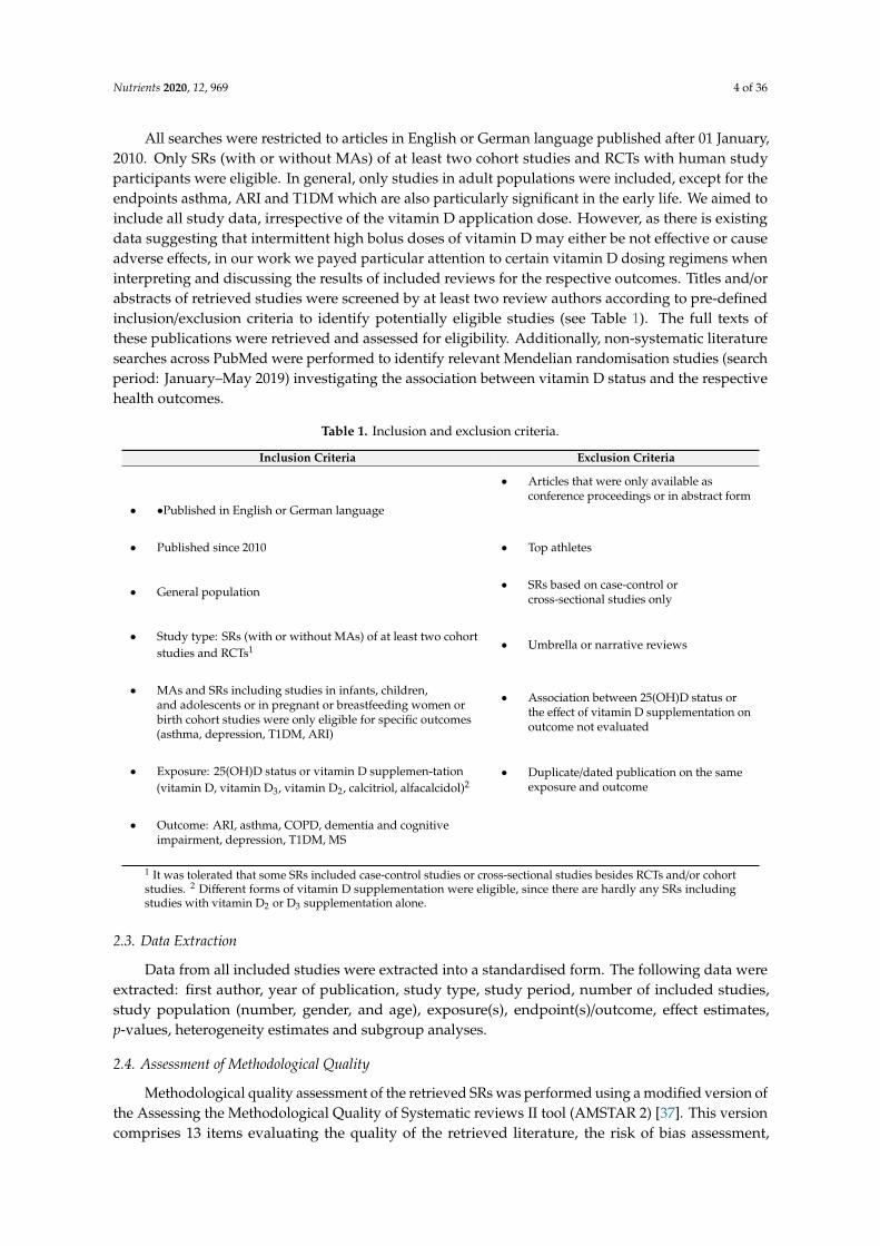
A prospectively developed methodological approach for this umbrella review was registeredon PROSPERO (CRD42019103670). The present umbrella review comprises SRs (with or withoutMAs) of cohort studies and RCTs investigating the association between 25(OH)D status or vitamin Dsupplementation and the following extraskeletal health diseases (prevention and therapy): respiratorytract diseases (asthma, chronic obstructive pulmonary disease (COPD), and acute respiratory tractinfections (ARI)), neurodegenerative and mental diseases (depression, dementia, and cognitive decline)and autoimmune diseases (multiple sclerosis (MS) and type 1 diabetes mellitus (T1DM)). In addition,relevant Mendelian randomisation studies were identified to complement the results of SRs. Mendelianrandomisation uses genetic variants to study putative causal effects of modifiable exposures on anoutcome. The principle behind is that if a biomarker, such as 25(OH)D, is involved in disease etiology,then the genetic factors influencing the biomarker will in turn influence disease risk. This methodavoids some of the limitations of classical epidemiology (e.g., less prone to confounding, free ofreverse causation) but has also its restrictions (see “Overall Discussion and Conclusion”, page 23, line1071) [35,36].
2.2. Search Strategy and Eligibility Criteria
Systematic literature searches for SRs and MAs across the databases PubMed and CochraneReviews library, using comprehensive search strategies, were performed for each extraskeletal healthoutcome separately in January 2019 (dementia and cognitive decline, depression, COPD), March 2019(asthma) and April 2019 (MS, T1DM, ARI). Search terms for vitamin D, study type (SR or MA) and therespective disease were combined. The search strategy is presented in Supplemental Table S1.
Nutrients 2020, 12, 969 4 of 36
All searches were restricted to articles in English or German language published after 01 January,2010. Only SRs (with or without MAs) of at least two cohort studies and RCTs with human studyparticipants were eligible. In general, only studies in adult populations were included, except for theendpoints asthma, ARI and T1DM which are also particularly significant in the early life. We aimed toinclude all study data, irrespective of the vitamin D application dose. However, as there is existingdata suggesting that intermittent high bolus doses of vitamin D may either be not effective or causeadverse effects, in our work we payed particular attention to certain vitamin D dosing regimens wheninterpreting and discussing the results of included reviews for the respective outcomes. Titles and/orabstracts of retrieved studies were screened by at least two review authors according to pre-definedinclusion/exclusion criteria to identify potentially eligible studies (see Table 1). The full texts ofthese publications were retrieved and assessed for eligibility. Additionally, non-systematic literaturesearches across PubMed were performed to identify relevant Mendelian randomisation studies (searchperiod: January–May 2019) investigating the association between vitamin D status and the respectivehealth outcomes.
Table 1. Inclusion and exclusion criteria.
Inclusion Criteria Exclusion Criteria
• •Published in English or German language
• Articles that were only available asconference proceedings or in abstract form
• Published since 2010 • Top athletes
• General population • SRs based on case-control orcross-sectional studies only
• Study type: SRs (with or without MAs) of at least two cohortstudies and RCTs1 • Umbrella or narrative reviews
• MAs and SRs including studies in infants, children,and adolescents or in pregnant or breastfeeding women orbirth cohort studies were only eligible for specific outcomes(asthma, depression, T1DM, ARI)
• Association between 25(OH)D status orthe effect of vitamin D supplementation onoutcome not evaluated
• Exposure: 25(OH)D status or vitamin D supplemen-tation(vitamin D, vitamin D3, vitamin D2, calcitriol, alfacalcidol)2
• Duplicate/dated publication on the sameexposure and outcome
• Outcome: ARI, asthma, COPD, dementia and cognitiveimpairment, depression, T1DM, MS
1 It was tolerated that some SRs included case-control studies or cross-sectional studies besides RCTs and/or cohortstudies. 2 Different forms of vitamin D supplementation were eligible, since there are hardly any SRs includingstudies with vitamin D2 or D3 supplementation alone.
2.3. Data Extraction
Data from all included studies were extracted into a standardised form. The following data wereextracted: first author, year of publication, study type, study period, number of included studies,study population (number, gender, and age), exposure(s), endpoint(s)/outcome, effect estimates,p-values, heterogeneity estimates and subgroup analyses.
2.4. Assessment of Methodological Quality
Methodological quality assessment of the retrieved SRs was performed using a modified version ofthe Assessing the Methodological Quality of Systematic reviews II tool (AMSTAR 2) [37]. This versioncomprises 13 items evaluating the quality of the retrieved literature, the risk of bias assessment,

Nutrients 2020, 12, 969 5 of 36
the quality of statistical analyses and reporting of results and transparency of potential sources ofconflict of each study. Studies were rated on a scale from high quality to very low quality, based on theexistence of critical and non-critical methodological weaknesses.
2.5. Summarisation of Data Results and Conclusion Drawing
Based on the systematic description and on the quality assessment of the selected reviews includedin synoptical tables (see Supplemental Tables S2–S26), at least two authors summarised the results anddrew a conclusion for each disease.
3. Results
A total of 349 articles were identified for the seven diseases (Figure 1). After removal of14 duplicates, 335 articles were screened by title and abstract. Of the 335 articles, 203 did not match ourinclusion criteria and were thus excluded. For assessment 132 full-text articles were obtained and finally73 SRs (with or without MAs) of prospective cohort studies and RCTs were included in qualitativesynthesis (see Supplemental Tables S2–S26). Main reasons for the exclusion of 59 assessed full-textarticles were an irrelevant study type (e.g., umbrella/narrative review, only/mainly cross-sectionaland/or case-control studies) or an irrelevant outcome (e.g., prevention of falls in elderly, quality oflife, poststroke depression). More detailed reasons for exclusion are shown in the SupplementalFigures S1–S7.
Nutrients 2020, 11, x FOR PEER REVIEW 5 of 35
2.5. Summarisation of Data Results and Conclusion Drawing
Based on the systematic description and on the quality assessment of the selected reviews included in synoptical tables (see Supplemental Tables S2–S26), at least two authors summarised the results and drew a conclusion for each disease.
3. Results
A total of 349 articles were identified for the seven diseases (Figure 1). After removal of 14 duplicates, 335 articles were screened by title and abstract. Of the 335 articles, 203 did not match our inclusion criteria and were thus excluded. For assessment 132 full-text articles were obtained and finally 73 SRs (with or without MAs) of prospective cohort studies and RCTs were included in qualitative synthesis (see Supplemental Tables S2–26). Main reasons for the exclusion of 59 assessed full-text articles were an irrelevant study type (e.g., umbrella/narrative review, only/mainly cross-sectional and/or case-control studies) or an irrelevant outcome (e.g., prevention of falls in elderly, quality of life, poststroke depression). More detailed reasons for exclusion are shown in the Supplemental Figures S1–S7.
Figure 1. PRISMA flow diagram—all outcomes.
Figure 1. PRISMA flow diagram—all outcomes.

Nutrients 2020, 12, 969 6 of 36
3.1. Respiratory Tract Diseases
3.1.1. Asthma
BackgroundAsthma is a chronic inflammatory disorder of the airways that is characterised by recurring
symptoms such as shortness of breath, wheeze, chest tightness, cough (varying over time and inintensity), as well as variable expiratory airflow limitation and often attended by increased serumimmunoglobulin E (IgE) levels. A variety of factors may cause asthma exacerbations, including allergens,pollutants, and poor adherence to prescribed medications, while viral upper respiratory infectionis the most predominant reason [38,39]. Virus-induced asthma exacerbations are associated withincreased production of pro-inflammatory cytokines such as interleukin (IL)-17, which exacerbateallergic airway responses [40]. These cytokines are also associated with the severity of asthma andsteroid responsiveness [41]. Acute exacerbations are major causes of morbidity and mortality [42,43].
Asthma affects more than 300 million people of all ages and all ethnic backgrounds, and it isestimated to cause 400,000 deaths annually [39,42,44]. Over the past few decades, the prevalence ofasthma has increased steadily worldwide, especially in children and young adults of high-incomecountries [45–47]. Asthma is the most common chronic disease of childhood, affecting approximately10% of children, with prevalence varying by definition and country of origin [48]. Until now,no convincing preventive strategies have been identified, and evidence concerning modifiablerisk factors is inconsistent [49]. To improve clinical outcomes, it is important to prevent asthmaexacerbations [38].
Vitamin D deficiency has been associated with increased incidence [50] and severity [51] ofchildhood asthma. Regarding the potential role of vitamin D and asthma, it should be consideredthat some data suggest an association between passive smoking and vitamin D deficiency in childrenwith asthma [52]. Vitamin D has immunoregulatory properties [53,54] and plays an importantrole in inflammation [55]. Airway epithelial cells express the activating enzyme 1α-hydroxylase,which catalyses the formation of calcitriol from 25(OH)D [56–58]. VDRs, for which calcitriol is a knownligand, are present in cells of the immune system such as macrophages, dendritic cells and activated T-and B-cells [59]. Calcitriol shifts the balance of T lymphocyte response from T helper (Th)1 phenotypeto Th2 phenotype [60,61]. In addition, calcitriol can suppress the proinflammatory cytokine IL-17and the IL-4-mediated expression of IL-13 [62,63]. Calcitriol may also act directly on CD4+ T cells topromote T-regulatory cells (Tregs) that secrete the anti-inflammatory cytokine IL-10 [62–66]. Here,we synthesise results of SRs regarding vitamin D and asthma outcomes.
ResultsIn our search for SRs on vitamin D and asthma risk, we identified 18 articles [38,67–83] of which
10 reported data on cohort studies [67–76] and eight reported data on RCTs [38,77–83] (SupplementalTable S2–S5 and Figure S1). Out of the 18 SRs, 14 were restricted to children [67,69–81], three includedboth, studies in children and adults [38,68,82], and one was based on individual participant data(IPD) with individuals aged 1.6–85.0 years [83]. Overall, the majority of the SRs were of high quality,assessed by AMSTAR 2 tool. We also identified a large Mendelian randomisation study on 146,761study participants [35].
Primary PreventionThe aforementioned 10 SRs on cohort studies [67–76] were all primary prevention studies,
performed in early childhood. Three out of the 10 SRs did not perform MAs [67,72,76]. Of theremaining seven MAs, six compared high versus low 25(OH)D levels [68–71,74,75]. No significantassociation between vitamin D status and asthma risk was reported in four of the six studies [70,71,74,75],whereas two studies [68,69] reported a significantly lower risk at higher 25(OH)D levels. AnotherSR [73] analysed nonlinear dose–response relationship by restricted cubic spline model. Data indicate asignificant U-shaped association between maternal 25(OH)D levels and offspring asthma risk with the

Nutrients 2020, 12, 969 7 of 36
lowest risk at approximately 60–70 nmol/l (24–28 ng/mL) of 25(OH)D. Regarding study quality, sevenwere of high, one of moderate, and two of low or very low quality (Supplemental Tables S3 and S5).
One out of the eight two-step MAs on RCTs focused on primary prevention [81] and used as anunspecific surrogate of asthma risk data on recurrent wheezing in offspring whose mothers participatedin vitamin D supplementation studies. It was suggested that vitamin D may have a beneficial effecton recurrent asthma in children. This MA included three RCTs that used daily vitamin D3 doses of60 µg and 100 µg (2400 and 4000 IU), respectively, or a single vitamin D3 bolus of 5000 µg (200,000 IU).The MA was of moderate quality (Supplemental Table S2).
The aforementioned large Mendelian randomisation study [35] investigated four singlenucleotide polymorphisms (SNPs) influencing 25(OH)D transport (GC globulin (vitamin D-bindingprotein)), synthesis (7-dehydrocholesterol reductase), hepatic hydroxylation (CYP2R1), and catabolism(CYP24A1), to estimate the association of genetically determined 25(OH)D and the risk of asthma.None of the four 25(OH)D-lowering alleles was significantly associated with asthma or elevated IgElevels, and there was also no significant association between genetically determined 25(OH)D and riskof asthma or IgE levels.
Therapeutic or Adjuvant Vitamin D Supplementation in Asthma PatientsSix SRs performed a two-step MA on vitamin D supplementation in asthma patients [38,77–80,82].
Four out of these six MAs reported beneficial [77–79,82] and two non-significant effects [38,80]. FiveMAs used asthma exacerbations as outcome [38,77–79,82], and one [80] relied on the lung functionparameter FEV (forced expiratory volume). None of the six MAs performed subgroup analysisaccording to baseline 25(OH)D levels. Study quality was considered as moderate in two SRs and highin four SRs (Supplemental Table S2). In total, the six MAs were based on 15 RCTs. The vitamin D3
doses in the included RCTs were very heterogeneous and ranged between daily doses of 10 µg to100 µg (400–4000 IU) or bolus doses of 1500 µg to 3000 µg (60,000–120,000 IU).
The identified IPD MA [83] was based on seven RCTs (five RCTs enrolled children, two RCTsenrolled adults), including 978 asthma patients, of whom 955 had outcome data (Supplemental TableS2). This analysis showed that vitamin D3 supplementation reduced the adjusted incidence rate ratio(aIRR) of asthma exacerbations requiring treatment with corticosteroids to 0.74 (95% CI: 0.56–0.97;p = 0.03) and the results were considered as high-quality evidence. Subgroup analyses suggested thepossibility that protective vitamin D effects may only be seen in participants with baseline 25(OH)Dof less than 25 nmol/l (10 ng/mL) (aIRR = 0.33; 95% CI: 0.11–0.98; p = 0.046) and not in participantswith higher baseline 25(OH)D levels (aIRR: 0.77; 95% CI: 0.58–1.03; p = 0.08). However, the p-value forinteraction of 0.25 did not provide definitive evidence for an interaction of baseline 25(OH)D with thevitamin D effect on asthma exacerbations. Study quality could not be assessed, because the AMSTAR 2tool was not developed for use in IPD MAs [37].
Discussion and ConclusionThe majority of cohort studies were primary prevention studies in early childhood, whereas the
majority of RCTs were performed in patients with already existing asthma. Results of MAs on theassociation between asthma risk and vitamin D status are inconclusive. This may be due to a potentialU-shaped relationship between 25(OH)D and asthma risk [73], which remains uncovered in MAs thatcompared high versus low 25(OH)D levels to estimate the vitamin D effect on asthma risk [68–72,74,75].The only MA of RCTs on primary prevention [81] supports the assumption that vitamin D may reduceasthma risk in early childhood, but this MA is limited by the use of an unspecific surrogate endpoint(wheezing). A Mendelian randomisation study [35], however, is not in line with a beneficial vitaminD effect on asthma risk. Nevertheless, it is noteworthy that the four SNPs could only explain 0.13%,0.12%, 0.09%, and 0.02% of the variance in 25(OH)D [35] and are therefore subject to weak instrumentbias [84]. Thus, the null effect reported in that study does not necessarily exclude a causal associationbetween vitamin D status and asthma risk.
Regarding RCTs in asthma patients, the majority of MAs support the assumption of a beneficialvitamin D effect. Importantly, subgroup analyses of an IPD MA raise the hypothesis that protective

Nutrients 2020, 12, 969 8 of 36
vitamin D effects might be restricted to individuals with baseline 25(OH)D of less than 25 nmol/l(10 ng/mL). Besides heterogeneity in vitamin D dosing, baseline 25(OH)D level is an issue thatneeds consideration and long-term future RCTs should only be performed in populations with ahigh prevalence of vitamin D deficiency to draw conclusions concerning the impact of vitamin Dsupplementation for asthma risk. Another limitation is that most results are based on MAs in children.Therefore, caution is necessary when translating the results to the adult population. Two RCTs(not yet published) regarding the prevention of asthma exacerbations in children were identified atclinicaltrials.gov (identifier: NCT02687815 and NCT03365687) and may provide additional informationon the effect of vitamin D in asthma patients. Overall, the majority of published SRs were of high quality.
In conclusion, adequate vitamin D status in childhood may reduce the risk of asthma exacerbations.Regarding vitamin D and asthma in the adult population, available data are insufficient to drawreliable conclusions.
3.1.2. Chronic Obstructive Pulmonary Disease
BackgroundChronic obstructive pulmonary disease (COPD) is a progressive, systematic inflammatory
illness characterised by chronic airflow limitation. COPD patients suffer from reduction of lungfunction, loss of exercise capacity, frequent disease exacerbations, and development of extra-pulmonarycomorbidities—such as osteoporosis, infection, and cardiovascular disease [85]. COPD exacerbationsare commonly triggered by respiratory viruses and bacteria, which increase airway inflammation [86].Episodes of acute worsening of symptoms are associated with increased mortality [87].
COPD affects more than 170 million people worldwide and caused an estimated 3.2 million deathsin 2015 [88]. COPD is the fourth leading cause of mortality globally [89] and is expected to become thethird leading cause of death by 2020 [87]. Tobacco smoking is considered to be a major risk factor ofCOPD [90], but only 10–15% of long-term smokers develop symptomatic airflow obstruction [91].
Besides genetic susceptibility for COPD, poor vitamin D status has also been discussed as playinga role in the development of the disease [92]. Vitamin D has immunomodulatory properties [93],and calcitriol plays a role in modulating functions of the innate and adaptive immune systems [94].Briefly, vitamin D signalling may stimulate innate immunity by upregulating antimicrobial peptideproduction and may suppress adaptive immunity by decreasing pro-inflammatory and increasinganti-inflammatory cytokine expression [59,95]. Cohort studies reported a positive association betweencirculating 25(OH)D and pulmonary function [96,97]. However, when interpreting observationalstudies on vitamin D and COPD, it should be considered that some investigations indicate that smokingis associated with lower 25(OH)D concentrations [98]. Nevertheless, additional evidence for a potentiallink between vitamin D and COPD is provided by a MA of RCTs on vitamin D supplementationand acute respiratory tract infections [99]: vitamin D supplements reduced infections significantlyand subgroup analysis indicated that this effect may be highest in individuals with deficient vitaminD status at study inclusion, i.e., circulating 25(OH)D concentrations < 25 nmol/l (10 ng/mL). Thus,vitamin D may indirectly influence COPD exacerbations by reducing the risk of airway infections.
Results of SRs regarding vitamin D and COPD outcomes are summarised below.ResultsIn our search for SRs on vitamin D and COPD risk, we identified six SRs [31,100–104] (Supplemental
Tables S6–S9 and Figure S2) of which three reported MAs on pooled data regarding vitamin D status orvitamin D supplementation and COPD risk [101,102,104]. The study quality assessed by the AMSTAR2 tool was very heterogeneous (Supplemental Tables S6–S9). No Mendelian randomisation studieswere identified.
Primary PreventionOur literature search did not identify SRs of prospective cohort studies or RCTs on the primary
prevention of COPD by vitamin D.
Nutrients 2020, 12, 969 9 of 36
Therapeutic or Adjuvant Vitamin D Supplementation in COPD PatientsOf the three SRs reporting MAs, the analysis by Zhu et al. [101] included 18 studies, of which
five were case-control studies, eight were cohort studies, and five were RCTs. Both, the case-controland cohort studies compared 25(OH)D concentrations between COPD patients and controls. Sincethese studies used a cross-sectional approach, they are of limited relevance for our review. The fiveRCTs of that MA were not evaluated by the authors using the statistical approach of a MA, but bynarrative description of study results only. They concluded that four RCTs showed beneficial vitamin Deffects in COPD patients, at least in those with circulating 25(OH)D concentrations less than 50 nmol/l(20 ng/mL). Another MA by Zhu et al. [102] included 21 observational studies. The vast majority ofstudies were case-control studies that compared vitamin D status of COPD patients with controls orvitamin D status with COPD risk/severity. Zhu et al. performed a MA of seven studies (two cohortstudies, five case-control studies) showing that patients with severe to very severe COPD had lowerserum 25(OH)D levels compared with patients with mild to moderate COPD (SMD: −0.87, 95% CI:−1.51, −0.22). A MA of five studies (two cohort studies, three case-control studies) indicated thatacute exacerbation COPD (AECOPD) patients had lower levels of serum 25(OH)D compared to stableCOPD patients (SMD: −0.43, 95% CI: −0.70, −0.15). Three studies (two case-control studies, one cohortstudy; thus, failing to meet our inclusion criteria) were included in an evaluation of the associationbetween vitamin D deficiency and COPD exacerbations, revealing no significant association (oddsratio: 1.17, 95% CI: 0.86–1.59) [102]. However, since these studies included by Zhu et al. mostly used across-sectional approach, they are of limited relevance to our review. Of the three SRs not reportingMAs [31,100,103], the SR by Autier et al. [100], which was based on two prospective studies, statedan inverse association between circulating 25(OH)D and COPD exacerbations. On the contrary, ina SR by Ferrari et al. [103], based on five prospective cohort studies, no association could be shownbetween exacerbations frequency and circulating 25(OH)D. Nevertheless, from the RCTs included inthat SR, it was concluded that especially a group of patients with low 25(OH)D level, i.e., < 50 nmol/l(20 ng/mL), may benefit from vitamin D supplementation. Similarly, another SR [31] reported significantreductions in pulmonary exacerbations of COPD patients by vitamin D supplementation only at low25(OH)D concentrations.
An IPD MA by Jolliffe et al. [104] identified four RCTs, of which three (472 randomised patients)were included in their analysis. The MA concluded that vitamin D supplementation did not influenceoverall rate of moderate/severe COPD exacerbations (adjusted incidence rate ratio 0.94 (95% CI: 0.78 to1.13)). However, subgroup analysis indicated protective vitamin D effects in patients with baseline25(OH)D concentrations < 25 nmol/l (10 ng/mL) (adjusted incidence rate ratio 0.55 (95% CI: 0.36to 0.84)).
The study quality assessed by the AMSTAR 2 tool was very heterogeneous for the included SRsand MAs (Supplemental Tables S6–S9). One SR [104] was an IPD MA, for which AMSTAR 2 wasnot developed. Overall, RCTs were included in four SRs. All RCTs included in the SRs [105–109]investigated effects of vitamin D supplementation as primary exposure. Study duration was sixweeks in one study [105], six months in two studies [106,107], and one year in two RCTs [108,109].In four RCTs [105,107–109], the vitamin supplement was D3 and in one study it was not specifiedwhether vitamin D2 or D3 was used [106]. Daily and bolus vitamin D administration was performedin two [105,107] and three RCTs [106,108,109], respectively. The vitamin D dose ranged between 20 µgper day (800 IU) [105] and 2500 µg (100,000 IU) per 4 weeks [106,108]. The calculated mean dailydose ranged between 20 µg (800 IU) [105] and 89 µg (3560 IU) [106,108]. Mean baseline 25(OH)Dconcentrations were reported in four RCTs [105,107–109] and were below 50 nmol/l (20 ng/mL) inall these four studies. In-study 25(OH)D concentrations were also presented in these four RCTS, ofwhich three RCTs exceeded a mean in-study 25(OH)D concentration of 75 nmol/l (30 ng/mL) [107–109],whereas the in-study 25(OH)D concentration remained on average below 75 nmol/l (30 ng/mL) in the6-week study with a daily dose of 20 µg (800 IU) vitamin D [105].
Nutrients 2020, 12, 969 10 of 36
Discussion and ConclusionData on primary prevention of COPD by vitamin D are scarce. Results of SRs on prospective
cohort studies regarding the association of vitamin D status on COPD exacerbations are inconclusiveand no reliable MAs are currently available on this topic. With respect to RCTs in COPD patients,only the SR by Jolliffe et al. [104] analysed the data according to the approach of a MA. Results indicatea potentially beneficial vitamin D effect on COPD exacerbations solely in patients with deficient25(OH)D concentrations—i.e., concentrations below 25 nmol/l (10 ng/mL)—but not in patients withhigher baseline 25(OH)D concentrations. Although results are based on a pre-specified subgroupanalysis [104], caution is necessary in interpreting these results. The medical literature is replete withexciting secondary end points that have failed when they were subsequently formally tested as primaryend points in adequately powered RCTs [110]. Moreover, two out of the three studies included in thisIPD MA [108,109] used high dose bolus administration of vitamin D. Formally, these studies have tobe considered as phase 2 clinical trials, which are usually performed for drug approval. Nevertheless,it is also noteworthy that the recommended daily intake for adequacy by nutrition societies of 10 µgto 20 µg (400–800 IU) vitamin D (by supplements) [2–5] are able to increase circulating 25(OH)Dconcentrations of 25 nmol/l to 50 nmol/l (10–20 ng/mL) [111] and thus into the range, which manynutrition societies consider adequate [2–5].
In conclusion, the quality of SRs on COPD patients is very heterogeneous. At present, the effectof vitamin D on the primary prevention of COPD is unclear. Based on a relatively small number ofRCTs and participants, vitamin D-deficient patients with already existing COPD probably benefit fromvitamin D supplementation. As identified at clinicaltrials.gov, two RCTs on the effects of vitamin Dsupplementation in COPD patients are expected to report additional data on this topic (identifier:NCT02122627 and NCT03781895).
3.1.3. Acute Respiratory Tract Infections
BackgroundAcute respiratory tract infections (ARI) comprise a group of infections that can occur in the upper
and lower respiratory tract and are leading causes of global morbidity and mortality in children andadults [112]. The upper respiratory tract infection is one of the most common acute illnesses in theoutpatient setting and is often characterised by a mild and self-limited disease course. Symptomsassociated with upper respiratory tract infections are irritations and swelling of the nose, sinuses,pharynx, larynx, and the large airways. In contrast, lower respiratory tract infections affect the bronchialtubes and the lungs and can cause bronchitis, pneumonia, and pulmonary tuberculosis.
Lower respiratory tract infections are the fifth leading cause of death and the leading infectiouscause of death worldwide [113]. Lower respiratory tract infections mainly affect children andindividuals older than 65 years. In 2015, pneumonia accounted for 15% of the deaths in children under5 years worldwide [114]. Approximately 45% of all community-acquired pneumonia occur in patientsaged >65 years [115–117]. Pneumonia is an inflammation of the lung that compromises gas exchangein the lungs and leads to symptoms such as cough, fever, and breathing difficulties [118]. ARIs are themost common reason for antibiotic therapy in adults [119].
The most common etiological factors that are responsible for ARIs are bacteria and viruses.Risk factors of pneumonia are crowded living conditions, malnutrition, HIV infection, lack ofbreastfeeding in infants, lack of immunisation, chronic health conditions, and exposure to tobaccosmoke or indoor air pollutants [112]. Vitamin D exerts multiple effects in the immune systemwhich includes the synthesis of antimicrobial peptides and modulation of T cell system [120,121];more importantly, activation of toll-like receptors upregulates the VDR and vitamin D hydroxylases inhuman macrophages and the production of the antimicrobial cathelicidin which can kill intracellularMycobacterium tuberculosis [122]. These data support a link between vitamin D and the innate immunesystem and justify the assumption that vitamin D can impact the prevalence and course of ARIs.Observational studies have reported an association between vitamin D status and ARIs [123–125].
Nutrients 2020, 12, 969 11 of 36
However, the benefits of vitamin D for prevention and treatment of ARI are ambiguous. Thus, we aimedto summarise data of SRs regarding vitamin D and ARI outcomes.
ResultsIn our search for SRs on vitamin D and ARI, we identified 14 records [71,74,77,100,126–135];
of which five reported data on cohort studies; [71,74,100,126,128] and 11 included MAs fromRCTs [77,100,126,127,129–135] (Supplemental Tables S10–S13 and Figure S3). Using AMSTAR 2as an instrument for assessing methodological quality, 8 of the 14 SRs that examined the associationbetween vitamin D and the prevention or treatment of ARI can be categorised as high quality; threestudies were categorised as medium quality; and the other two studies as low and very low quality,whereas AMSTAR 2 was not developed to assess the quality of the only IPD MA. We could not identifyMendelian randomisation studies on vitamin D and ARI.
Primary PreventionRegarding the association of vitamin D status and risk of ARI, we identified five SRs of cohort
studies with very heterogeneous study quality according to AMSTAR 2 assessment [71,74,100,126,128].In adults, Autier et al. found two studies reporting on an inverse association between serum 25(OH)Dand respiratory infections and one study showing an inverse association between serum 25(OH)Dand days of absence due to respiratory infections [100]. Similar results in adults were reported byJolliffe et al. [126]. Some SRs on prenatal vitamin D status (assessed by maternal or cord blood 25(OH)Dmainly derived from birth cohort studies) and ARI reported mixed results [71,74,126,128]. In the MAby Pacheco-Gonzalez et al., the pooled odds ratio for ARI in the offspring was 0.64 (95% CI 0.47 to 0.87)when comparing the highest with the lowest 25(OH)D category [74], whereas there was no significantresult for such an association in the MA by Feng et al. [71].
Regarding data on RCTs reporting effects of vitamin D supplementation on risk of ARI,we identified nine SRs with heterogeneous, yet largely high study quality [77,100,126,130–135].Among these studies, the largest MA, and the only one following an IPD approach, was publishedby Martineau et al. [99,135]. They included 25 RCTs with a total of 11,321 participants aged 0 to 95years including 10,933 participants with available IPD data. The main outcome was that vitamin Dsupplementation reduced the risk of at least one ARI with an adjusted odds ratio of 0.88 (95% CI: 0.81to 0.96; p = 0.003). In subgroup analyses, it was shown that this protective effect was only significantin those individuals receiving daily (7.5 to 100 µg (300–4000 IU)) or weekly vitamin D doses (35 to500 µg (1400–20,000 IU)), but not in those receiving any bolus doses (i.e., one bolus of at least 750 µg(30,000 IU) vitamin D; range 750 to 10,000 µg (30,000–400,000 IU)). Moreover, the protective effect wasstronger in individuals with baseline 25(OH)D below 25 nmol/l (10 ng/mL) as compared to those with25(OH)D concentrations ≥25 nmol/l (10 ng/mL), whereas no other effect modifiers were identified.Regarding the clinical effect size, the overall number needed to treat was 33 (95% CI: 20 to 101) anddropped to 8 (95% CI: 5 to 21) in individuals with baseline 25(OH)D concentrations below 25 nmol/l(10 ng/mL). Martineau et al. concluded that the evidence contributing to the findings of their MA wasof high quality. It should, however, be stressed that there was a very high heterogeneity across thestudies and funnel plot analysis showed a degree of asymmetry suggesting that small RCTs showingadverse effects might not have been included or published. Findings from the other MAs that includedsignificantly fewer participants compared to the work by Martineau et al. were inconsistent, showingeither a protective effect of vitamin D supplementation [126,130,131] or no effect [77,100,126,132–134].Importantly, the vast majority of the included original studies of these MAs were also included in thework by Martineau et al. [99,135].
Therapeutic or Adjuvant Vitamin D Supplementation in Patients Suffering from ARIRegarding data on RCTs reporting effects of vitamin D supplementation on the outcome of
pneumonia in children, we identified two SRs with moderate to high study quality [127,129]. In theonly MA on this topic, there was no significant effect of vitamin D3 supplementation, neither on timeto resolution of acute pneumonia nor on duration of hospitalisation [129]. Thus, data from RCTs onvitamin D supplementation and ARI document no significant safety concerns regarding vitamin D.
Nutrients 2020, 12, 969 12 of 36
Discussion and ConclusionData from cohort studies found an inverse association between serum 25(OH)D and respiratory
infections in adults. The main finding of our literature search is that the largest and well conducted IPDMA of RCTs reported a significant role of vitamin D supplementation to reduce the risk of ARI [135].In subgroup analyses, this significant effect was, however, restricted to individuals who did not receivebolus doses of vitamin D and was particularly strong in individuals with 25(OH)D concentrations below25 nmol/l (10 ng/mL). Effect modification by baseline 25(OH)D is biologically sound and consistentwith the concept that vitamin D supplementation is most effective in individuals with severe vitaminD deficiency. Why bolus doses of vitamin D (in ranges between 750 to 10,000 µg (30,000–400,000 IU))are ineffective for prevention of ARI remains speculative, but it has been hypothesised that widefluctuations in vitamin D metabolites might cause adverse effects, including dysregulation of vitaminD metabolising enzymes [99]. Funnel plot analyses suggest that RCTs showing adverse vitamin Deffects may not have been included in the MA. Another limitation is that the definitions of ARI wereheterogeneous and hardly supported by virological, microbiological, or radiological confirmation.Some caution is therefore warranted with the final claim on vitamin D and prevention of ARI. In general,the preventive effect of vitamin D on ARI is, however, biologically plausible because VDR activationexerts numerous immunological effects that may translate to protection against ARI. Furthermore,epidemiological studies have also largely—although not consistently—supported the concept thatvitamin D may protect against ARI. Camargo et al. recently (after the search period) published datafrom a pre-specified analysis of the ViDA (Vitamin D Assessment) study that aimed to investigate theeffects of monthly high-dose vitamin D3 supplementation (initial oral dose of 5000 µg (200,000 IU)vitamin D3 followed by 2500 µg (100,000 IU) monthly) on ARI prevention in more than 5000 olderadults [136]. The results of this RCT failed to show a preventive effect of vitamin D supplementationon ARI. However, it should be noted that these findings were obtained from a study collective with alow prevalence of vitamin D deficiency.
Regarding treatment of ARI, the current literature including RCTs in children indicates that thereis no significant effect of vitamin D.
Several RCTs on the effects of vitamin D supplementation on the prevention and treatment ofARI are expected to report additional data on this topic in the future (e.g., clinicaltrials.gov identifierNCT02185196; NCT02054182; NCT02046577; NCT03799406). However, most of these RCTs do notexclusively target vitamin D deficient individuals and some use bolus doses. Therefore, these RCTs havea high probability of documenting neutral effects of vitamin D supplementation on ARI in view of effectmodification by baseline 25(OH)D and the use of bolus doses as reported by Martineau et al. [99,135].
The majority of SRs that examined the association between vitamin D and the prevention ortreatment of ARI had a moderate or high methodological quality. Current evidence from MAs of RCTssuggests that vitamin D supplementation may prevent ARI. This effect was only evident in individualswho did not receive bolus doses of vitamin D and was particularly strong in those with baseline serum25(OH)D concentrations below 25 nmol/l (10 ng/mL). Complementary to this finding, SRs of cohortstudies indicated an inverse association between vitamin D status and ARI. Current data from RCTssuggest that vitamin D supplementation has no significant effect on the treatment of ARI.
3.2. Neurodegenerative and Mental Diseases
3.2.1. Dementia and Cognitive Decline
BackgroundDementia is a clinical syndrome characterised by global cognitive impairment with a decline
in memory and at least one other cognitive domain, such as language, visuospatial, or executivefunction. It is a chronic, malignant, and continuously progressing disease, associated with impairmentin functional abilities and in many cases behavioural and psychiatric disorders, leading invariablyto dependence on others [137]. Development of dementia from normal cognition is a continuous,
Nutrients 2020, 12, 969 13 of 36
slow process of cognitive decline over many years which is difficult to distinguish from normal ageing.Diagnosis of dementia is based on comprehensive assessment of symptoms which need to be persistentfor at least six months. The two most common causes of dementia are Alzheimer’s disease andcerebrovascular disorders, which often overlap. Worldwide, currently around 50 million people areaffected with nearly 10 million new cases expected every year [138]. The prevalence of dementia ismounting with increasing age. In Germany, only 1.5% of currently 1.7 million people with dementiaare younger than 65 years; 16% of those aged 80–84 years and more than 40% of those aged 90 years orolder are affected [139]. Since no effective treatment is available at present, maintenance of cognitiveabilities into old age and the prevention of cognitive decline are a major public health concern.
In recent years, the association between vitamin D and dementia or neurocognitive declinehas attracted growing interest, and evidence is accumulating for potential neuroprotective effects ofvitamin D. Data from cross-sectional analyses suggest that low serum concentrations of 25(OH)Dare associated with increased risk of Alzheimer’s disease and other forms of dementia and cognitiveimpairment [140,141]. In addition, there is evidence from animal models and in vitro studies thatvitamin D may influence the development of neurodegenerative disorders. Exact mechanismsare unclear, but evidence suggests that it may protect against cognitive dysfunction through itseffect on synaptic plasticity, immune modulation, neuronal calcium regulation, and enhanced nerveconduction [141–143]. In addition, vitamin D may affect vascular brain disease by mediatingharmful effects of inflammation, calcium dysregulation and increased oxidative stress and alsoby modulation of vascular disease risk factors such as elevated blood pressure [141]. Moreover,some data suggest that vitamin D might exert some beneficial effects with relevance for cardiometabolichealth, including antiatherogenic effects, improvement of endothelial function, arterial elasticity andmetabolic profile and inhibition of the renin-angiotensin-aldosterone system, which could be anothermechanism for the possible protective effects of vitamin D against dementia and cognitive decline [144].
The relationship between serum 25(OH)D status and dementia and/or cognition has beenexamined in many cross-sectional studies, in an increasing number of longitudinal studies and in afew interventional studies with conflicting results. Several SRs and MAs have been performed in thelast 10 years. Results of SRs regarding vitamin D and dementia and cognitive decline outcomes aresummarised below.
Results13 SRs (including eight MAs) [100,145–156] on vitamin D and risk of dementia and/or cognitive
decline were included in this analysis (Supplemental Tables S14–S17 and Figure S4). In addition, fourMendelian randomisation studies were considered [157–160]. The quality of the publications accordingto AMSTAR 2 is summarised in Supplemental Tables S14–S17. All articles addressed preventive effectsof vitamin D status or supplementation in adults without diagnosed dementia. No reviews or relevantstudies were identified regarding therapeutic effects of vitamin D in patients with dementia.
Results from SRs of Cohort StudiesAll identified 13 SRs examined cohort studies, six with the outcome dementia [148–150,152,153,155]
(including three with the outcome Alzheimer’s disease) [148,152,153] and eight with the outcomescognition, cognitive impairment or cognitive decline [100,145–147,149,151,154,156] using single orcombinations of several neuropsychological tests.
The most recent and also largest high-quality MA regarding dementia risk included data from10 cohort studies with 28,640 participants (mean/median age of 56 to 85 years) and follow-up periodsof 2–21 years [153]. A significant inverse association was found between 25(OH)D concentrations andthe risk of dementia (RR 0.72, 95% CI: 0.59–0.88; I2 = 33%, comparison highest vs. lowest 25(OH)Dcategories) and the risk of Alzheimer’s disease (RR 0.78, 95% CI: 0.60–1.00; I2 = 57%). In addition, adose-response analysis revealed that the risk of dementia decreased by 5% (RR 0.95, 95% CI: 0.93–0.98)and the risk of Alzheimer’s disease by 7% (RR 0.93, 95% CI: 0.89–0.97) for every 10 nmol/l (2.5 ng/mL)increase in 25(OH)D concentrations [153]. Jayedi et al. [152] meta-analysed eight of these cohortstudies and reported an increased risk of dementia in vitamin D-deficient (defined as <25 nmol/l
Nutrients 2020, 12, 969 14 of 36
(10 ng/mL); n = 5; HR 1.33) but not in vitamin D-insufficient participants (25–50 nmol/l (10–20 ng/mL);n = 6). In addition, a 17% reduced risk of dementia (n = 7) and of Alzheimer’s disease (n = 6) per25 nmol/l (10 ng/mL) increase in serum 25(OH)D concentrations was found, however with a largeheterogeneity between studies (I2 = 81 and 82%, respectively). Another high-quality MA of five ofthese cohort studies reported a 1.54 times increased risk of dementia in adults with serious deficiency(<25 nmol/l (10 ng/mL)) compared to sufficient supply (≥50 nmol/l (20 ng/mL)) [150]. Two MAs andone SR published in 2015 and 2016 of low or very low quality with only two or three cohort studies,respectively, also reported an increased risk of dementia in adults with low 25(OH)D concentrations atbaseline [148,149,155].
The most recent and largest high-quality MA addressing cognition in middle-aged and olderadults without a diagnosis of dementia included 14 prospective cohort studies [151]. The probabilityof cognitive decline was higher in participants with low 25(OH)D concentrations than in those havinghigher 25(OH)D levels (OR 1.26; CI 1.09–1.23). Heterogeneity between the study effect sizes was againlarge (I2 = 75%) and the possibility of publication bias was rated as high [151]. Other available MAs areolder (publication dates 2012 and 2013), of lower quality and included only three [147] and two [145] ofthese prospective cohort studies. They focused on executive functions and also found a higher risk ofcognitive decline over four to seven years in older adults with low 25(OH)D concentrations at baseline(heterogeneity 0% and 10%) [145,147]. A SR (low quality) on the association between vitamin D statusand cognition including six prospective cohort studies reported a significant decline in one or morecognitive function tests in participants aged 65 years or older with lower 25(OH)D concentrationscompared to participants with higher 25(OH)D levels in four of these six studies [154]. The SR (lowquality) of Autier et al. [100] included four cohort studies and concluded that frequency of cognitivedecline is increased in participants with low 25(OH)D concentrations. Balion et al. [146] included onlytwo cohort studies in their review (high quality) and reported conflicting results on cognitive outcomes.
Results from SRs of RCTsAmong the SRs with focus on cognition, six [100,145–147,151,156] (two high, one medium,
three low quality) included up to three intervention studies in their analyses. In total, nine studieswere evaluated, reporting conflicting results: seven RCTs (two using multi-nutrient supplements) andtwo pre-post studies in very heterogeneous populations (adolescents, young adults, older ambulatorypeople with history of falling, healthy older people, nursing home residents). Supplementation modes(e.g., daily, weekly or monthly oral supplementation, single injection, vitamin D2 or vitamin D3,and vitamin D3 plus calcium), and dosages differed widely between the studies. The latter rangedfrom 10 µg per day (400 IU) vitamin D3 [100] to 125 µg per day (5000 IU) vitamin D3 and to a bolusdose of 15,000 µg (600,000 IU) vitamin D2 [147,151]. In addition, sample sizes were mostly smalland supplementation periods were short. Only one larger placebo-controlled intervention study isdescribed in one SR [100], which investigated the effects of 10 µg per day (400 IU) of vitamin D3 and1000 mg per day of calcium carbonate supplementation on cognitive outcomes in 4143 women aged65 and older without probable dementia at baseline. Mean follow-up was 7.8 years. There were nosignificant differences in incident dementia or mild cognitive impairment or in global or domain-specificcognitive function between verum and placebo groups [161].
Two of the abovementioned reviews performed a MA. Goodwill et al. [151] (high quality) includedtwo studies (I2 = 35%) and Annweiler et al. [147] (moderate quality) three intervention studies (I2 = 49%).Both analyses found no effect of vitamin D supplementation on cognitive parameters.
Results from Mendelian Randomisation StudiesWe identified four publications that applied a Mendelian randomisation approach to address
causal inference [157–160]. Kuzma et al. [160] investigated the association between any risk factor andglobal cognitive function, all-cause dementia or dementia subtypes. Genetic evidence supported acausal association between telomere length and Alzheimer’s disease, whereas limited evidence forother risk factors including vitamin D status was found. Mokry et al. [158] provided evidence thatgenetically decreased 25(OH)D concentrations are associated with increased risk of Alzheimer’s disease.
Nutrients 2020, 12, 969 15 of 36
Here four SNPs were analysed, that combined described 2.44% of the variance in circulating 25(OH)Dlevels in the SUNLIGHT (Study of Underlying Genetic Determinants of Vitamin D and Highly RelatedTraits) study [158]. Alfred et al. [157] investigated whether genetic variants influencing 25(OH)Dconcentrations are associated with cognitive capability in middle-aged and older adults. They observeda negative association between the allele of rs2282679 (GC globulin), which is associated with higher25(OH)D concentrations, and word recall. A Mendelian randomisation study of Maddock et al. [159]investigated the causal nature of the association between serum 25(OH)D concentrations and cognitivefunction in mid- to later life but found no evidence for such an association.
Discussion and ConclusionIn summary, results from SRs of cohort studies report an increased risk of dementia including
Alzheimer’s disease and for cognitive decline in mainly older persons (>65 years) with low 25(OH)Dconcentrations; however, with a large heterogeneity in study effect sizes. In addition, some of theMendelian randomisation studies suggest a relationship between 25(OH)D concentration and therisk of Alzheimer’s disease and cognitive function. Only few very heterogeneous RCTs are available,which have not convincingly demonstrated a positive effect of vitamin D supplementation on cognitiveoutcomes. Except for one, these studies are small and of short duration; the only larger RCT found nodifference between the verum and placebo group.
Potential explanations for the differences between evidence from epidemiological observationalstudies, Mendelian randomisation studies and intervention studies may result from using varyingmethods (e.g., analytical and statistical approaches, vitamin D reference ranges), different baselineconcentrations of 25(OH)D, as well as cognitive function, or the possibility that low 25(OH)Dconcentration in persons with dementia or cognitive impairment is only an epiphenomenon.In addition, the intervention studies conducted so far are very heterogeneous regarding studyendpoints (e.g., cognitive function, cognitive impairment, dementia, Alzheimer’s disease) andparticipants (e.g., adolescents, healthy older adults, nursing home residents), are mostly of short-termduration and poor quality. Furthermore, a variety of neuropsychological tests were used for differentaspects of cognitive function (e.g., general cognition, reasoning and language, figural creations,visuospatial abilities, mental speed/attention).
Regarding dosage, a recent RCT [162] tested whether 50 µg per day (2000 IU) is more effectivethan 20 µg (800 IU) vitamin D3 for improving cognitive performance among relatively healthyadults aged ≥60 years (31% 25(OH)D < 50 nmol/l (20 ng/mL)), but found comparable results inboth groups over a 24-month treatment period and no significant improvement in either group.Similarly, another recent RCT found that supplementing older adults with a history of falling (58%25(OH)D < 50 nmol/l (20 ng/mL)) with 600 µg (24,000 IU) vitamin D3, 1500 µg (60,000 IU) vitamin D3
or a combination of 600 µg (24,000 IU) vitamin D3 with calcifediol once per month also led neither todifferent effects in mental health within one year, nor to any significant improvement [163]. Participantsachieving the highest serum 25(OH)D levels (112.5–247.5 nmol/l (45–99 ng/mL)), however, had a “small,clinically uncertain but statistically significant improvement in mental health scores”, irrespective ofthe supplement dose.
Unfortunately, many questions regarding the role of vitamin D in the development of dementiaand cognitive decline are currently unanswered—e.g., what, if any, is the optimal blood concentrationrequired to support neuroprotection? Is the association seen in observational studies mediated byother lifestyle factors, such as physical function or fitness? Thus, well-designed RCTs are required indifferent population groups (e.g., with adequate sample size and statistical power, vitamin D dosageand formulation, valid comprehensive cognitive assessments) to determine if and/or in whom vitaminD supplementation and increased 25(OH)D concentrations affect the varied aspects of cognitive health,and to what extent. This is particularly challenging with respect to dementia because of a very longprodromal stage and latency period.
In conclusion, available SRs—as well as included primary studies—are of heterogeneous quality(Supplemental Tables S14–S17), and available scientific evidence from RCTs does not support a clear
Nutrients 2020, 12, 969 16 of 36
benefit of vitamin D supplementation in the prevention of dementia and cognitive decline. Nevertheless,based on the associations described in prospective cohort studies, it seems prudent to aim to preventvitamin D deficiency in older adults as a viable component of brain health strategy. The resultsof several ongoing intervention studies, especially of the VIOLET-BUD Study (clinicaltrials.govidentifier: NCT03733418) may be helpful to better understand the role of vitamin D in the courseof neurodegeneration.
3.2.2. Depression
BackgroundDepression is a leading cause of disability worldwide and an important contributor to the overall
burden of disease. Worldwide more than 264 million people are affected [164]. Major depression isdefined as a period of at least two weeks when a person experiences a depressed mood or loss of interestor pleasure in daily activities, plus several other symptoms, like problems with sleep, concentration, orself-worth. Dysthymia is a less severe, but persistent, form of depression.
The biological plausibility for an association of vitamin D with depression comes from severalobservations. Within the brain, vitamin D is involved in regulating cytokines in the inflammationpathways and contributing to the release of neurotransmitters [150,165,166]. The VDR, which functionsas a gene regulator, has been found in several brain regions implicated in depression [167–170]. In thehippocampus, an interaction between VDRs and glucocorticoid receptors, which are implicated in thestress response, has been described [171]. We therefore aimed to summarise results of SRs regardingvitamin D status or supplementation and prevention or therapy of depression.
ResultsWe identified 12 SRs [31,100,156,172–180] covering 31 original publications between 1998 and
2017, plus five SRs [181–185] on depression in women before and after childbirth (SupplementalTables S18–S21 and Figure S5). Most studies relied on dimensional self-ratings of depressive symptomsas outcome measures. The AMSTAR 2 study quality was moderate or high for 11 SRs, but low or verylow for seven other SRs. Furthermore, one large Mendelian randomisation study was identified [186].
Primary PreventionTwo SRs performed a MA of observational cohort studies [172,173], four summarised data of
RCTs [31,174,175,178], while two summarised the results of RCTs and cohort studies [100,156] and fiveSRs included a MA of RCTs [176–180]. Anglin et al. (2013) performed a moderate-quality MA of threecohort studies with a total of 8815 elderly subjects, followed up for 1–6 years, and found a two-foldincreased risk of depression in those with the lowest compared to the highest vitamin D status [172].Ju et al. included one additional large cohort study in their moderate quality SR, and found an inverseassociation between 25(OH)D concentrations and incident depression [173].
Some RCTs were done in specific groups at risk of depression (e.g., elderly subjects with establishedvitamin D deficiency, or with obesity). Shaffer et al. (2014) found no effect of vitamin D3 supplementationwith 10 µg per day (400 IU) to 7500 µg once (300,000 IU) on the development of depressive symptomsin a high-quality MA of seven RCTs with 3191 at-risk subjects [177], and neither did Li et al. (2014) intheir high-quality MA when analysing six RCTs in clinically depressed subjects [176]. Including theseand three additional RCTs, comprising 4923 subjects with rather low depression levels at baseline,the high-quality MA of Gowda et al. (2015) also found no impact of vitamin D supplementation(10 µg per day—1250 µg per week (400 IU per day—50,000 IU per week)) on reducing depressivesymptoms [179]. The authors of these SRs noted the significant heterogeneity of design and outcomesof these “preventive” RCTs. Spedding (2014) sorted 15 RCTs with 42,258 participants (mostly fromat-risk groups) according to whether or not they had “biological flaws” (e.g., lack of 25(OH)Dmeasurements, or lack of 25(OH)D change during vitamin D3 supplementation, or a sufficient 25(OH)Dlevel in participants at baseline) [178]. RCTs without such validity issues, administering 25 to 375 µg(1000–15,000 IU) vitamin D3 per day, tended to show an improvement in depressive symptoms.However, this SR itself was judged to be of low quality applying the AMSTAR 2 criteria.
Nutrients 2020, 12, 969 17 of 36
One large two-sample Mendelian randomisation study [186] examined whether common genevariants, together explaining 7.5% of the variance in serum 25(OH)D concentrations in the SUNLIGHTstudy [187], would be associated with a diagnosis of major depression (i.e., whether these SNPs wouldbe less common in patients with this diagnosis than in psychiatrically healthy controls). Analysis of59,851 cases with major depression and 113,154 controls from the Psychiatric Genetics Consortiumdatabase revealed no such association, suggesting that life-long (genetically determined) decreasesin serum 25(OH)D concentrations do not increase the risk of major depression in generally healthypopulations. However, the authors noted that their MR study had limited power to examine effects ofextremes on either side of the serum 25(OH)D spectrum.
Postpartum and Antepartum Depression (Primary Prevention)Pregnancy, the postpartum period, and in particular the perinatal period are associated with an
increased risk of depression in women. In five SRs of mainly high quality, results have been summarisedcovering largely overlapping subsets of 11 original studies between 2010 and 2017 [181–185]. One SRsummarised observational cohort studies [184], two SRs summarised the results of secondary analysesof a RCT and cohort studies [181,183], and one SR summarised the results of a RCT, secondary analysesof two RCTs and cohort studies [182]. In addition, one high-quality SR by Wang et al. (2018) includedseven cohort studies, one case-control study and one RCT, which investigated the relationships betweencirculating levels or supplementation of vitamin D with antepartum or postpartum depression [185].MAs were performed with data from the seven cohort studies. The first main MA including three studieswhich investigated the association between low 25(OH)D concentrations (<30 nmol/l (12 ng/mL))and antepartum depression showed no significant association. However, the second main MA(including four studies), which studied the relationship between lower circulating levels of 25(OH)D(<50 nmol/l (20 ng/mL)) and postpartum depression, showed that lower circulating levels of 25(OH)Dwere associated with an increased odds ratio for postpartum depression (OR 3.67; 95% CI 1.72–7.85).In addition, relationships were observed in two subgroup analyses stratified by study region (Oceania:OR 2.00; 95% CI 1.31–3.06, Asia: OR 7.17; 95% CI 3.89–13.21). Evidence of statistical heterogeneity wasfound in all MAs. In addition, one RCT showed that participants given vitamin D supplements (50 µg(2000 IU)) in late pregnancy, as compared to the placebo group, had a greater reduction in depressionscores at 38 to 40 weeks of pregnancy [185].
Therapeutic or Adjuvant Vitamin D Supplementation in Depressed PatientsIn contrast to the largely negative intervention results in subjects with no or few depressive
symptoms (see Primary Prevention above), a moderate-quality MA including four RCTs performedon 948 patients with major depression [180] found a consistent and medium-sized positive effect ofvitamin D (oral 37.5 µg per day (1500 IU), or 1250 µg per week (50,000 IU), or up to 7500 µg (300,000 IU)single dose injection) versus placebo on depressive symptoms (SMD = 0.58, 95% CI: 0.45–0.72, p < 0.01,I2 = 0%). All these patient studies were conducted in Iran or China.
Discussion and ConclusionSRs of longitudinal cohort studies suggest that low levels of 25(OH)D are associated with higher
subsequent depressive symptoms in elderly subjects. A recently (after the search period) publisheddose–response MA [188] of six cohort studies (16,287 elderly subjects, 1157 cases with depression)confirmed this inverse association, and calculated that every 25 nmol/L (10 ng/mL) increase in serum25(OH)D was associated with a 12% decrease in the risk of depression in older adults (I2 = 79.0%,p < 0.001). In addition, low circulating levels of 25(OH)D are associated with postpartum depression.
In contrast, Mendelian randomisation studies do not support a causal role of (genetically) lowerlevels of serum 25(OH)D on the development of depressive symptoms or major depressive disorders.Recently, two additional Mendelian randomisation studies concerning 25(OH)D and depression werepublished (after the search period) [189,190]. A Mendelian randomisation study analysing depressivesymptoms and broadly defined depression in two large population studies (> 480,000 participants) didnot find an association of the alleles linked to serum 25(OH)D with these depression phenotypes [189].The Mendelian randomisation study with 339,256 UK biobank participants (including 23,294 cases

Nutrients 2020, 12, 969 18 of 36
with depression) by Meng et al. (2019) did also not support a causal role of life-long serum 25(OH)Dreduction on depression [190].
Several RCTs in Asian patients with major depression suggest that vitamin D may play a role inthe adjuvant treatment of depression. A recent RCT (published after the search period) with 78 elderlydepressed patients [191], which was not included in the MA by Vellekatt [180], found a significantbeneficial effect of vitamin D supplementation on depressive symptoms. However, all RCTs withdepressed patients were conducted either in Iran or in China, and as 25(OH)D levels are influenced bysunlight exposure, replication in other populations is needed.
In sum, the evidence reviewed does not support primary prevention of depression by vitamin Dsupplementation. Limited evidence suggests that vitamin D supplementation may be an efficaciousadjunctive treatment for major depression, and a single RCT indicates that postpartum depression mayalso be alleviated. The quality of the reviewed SRs on vitamin D and depression is very heterogeneous.Several RCTs, including a Finnish study with 3000 unipolar depressive patients (clinicaltrials.govidentifier: NCT02521012) are underway to allow for a more definitive assessment of the possibleantidepressant effects of vitamin D.
3.3. Autoimmune Diseases
3.3.1. Multiple Sclerosis
BackgroundMultiple sclerosis (MS) is a demyelinating disease of the central nervous system and manifests as
acute relapses and progressive disability. Autoreactive T-cells and B-cells are able to migrate throughthe blood–brain barrier and enter the brain, where they cause inflammation [192]. This persistentinflammation leads to loss of neuronal synaptic functions. Domains such as mobility, sensory perception,hand function, vision, fatigue, etc. are commonly affected [193]. Methods to quantify the progressionof MS are, for instance, the expanded disability status scale (EDSS) [194], the annual relapse rate (ARR)or measurement of brain lesions by magnetic resonance imaging (MRI), using two different methodswith distinct settings (T1 or T2–weighted sequences) [195].
MS mostly appears in adults in their late 20s or early 30s. Worldwide, an increase in the prevalenceof MS has been reported over the past decades. However, to a large extent this could be explained bybetter diagnostic accuracy [196]. According to data from 2013, prevalence varies between countriesand is highest in North America (140 per 100,000) and Europe (108 per 100,000) and lowest in Africa(2.1 per 100,000) and East Asia (2.2 per 100,000) [197]. Furthermore, mortality rate among MS patientsis higher compared to the general population [198].
The etiology of MS is still unknown, but it is suggested that multiple factors, such as genetics,low vitamin D status, Epstein–Barr virus infection, obesity in childhood or adolescence, or smokingplay a role in the development of MS [199].
Numerous studies have shown a latitude-dependent variation in MS incidence [200]. Hence,the duration and intensity of UV radiation and the resulting endogenous synthesis of vitamin Dmight influence the development of MS during life. Furthermore, several studies have examinedthe association between genetic factors and MS. As for other auto-immune diseases, genetic variantswithin the HLA-complex are among the strongest genetic risk factors. Also, non-HLA single nucleotidepolymorphisms, influencing the immune system or the serum 25(OH)D level, have been discussed aspotential risk factors [201,202].
Thus, an adequate vitamin D status reveals a possible approach for primary prevention of MSand also for modifying the natural course of the disease [203]. However, whether or not a low serum25(OH)D level is causally linked to the onset and course of MS is still a matter of scientific debate.In this review, we therefore aimed to summarise data of SRs regarding a possible role of vitamin D inthe prevention and therapy of MS.
Nutrients 2020, 12, 969 19 of 36
ResultsOverall, 11 records were included in this review [100,204–213] (Supplemental Figure S6).
We focused on publications that examined the association between vitamin D and the development ofMS or clinical activity of MS. Supplemental Tables S22–S24 provide information about the includedstudies and the assessed quality based on AMSTAR 2. Furthermore, three Mendelian randomisationstudies were identified [214–216].
Primary PreventionRegarding primary prevention, three SRs included data from prospective cohort studies [100,204,205].
According to AMSTAR 2, study quality was low for two SRs and high for one [204]. However, most ofthe studies were looking at MS patients and very few at non-diseased population groups. The mostrecent SR [205] included results from the Nurses’ Health Study as the first large prospective studythat examined the risk of developing MS in a group of non-diseased women. The authors reporteda 40% lower risk of developing MS in U.S. women who supplemented vitamin D, but failed to findan association for vitamin D intake by food consumption [217]. A prospective study examining theassociation between serum 25(OH)D concentrations and risk of MS in a cohort of US military personnelfound an inverse association in whites, but not in blacks or Hispanics [218]. This paper was includedin the SR provided by Autier et al. [100].
Observational studies remain limited by residual confounding and potential for reverse causation,particularly in MS where time of onset of pathophysiological processes remains known. Recent studiesusing Mendelian randomisation have provided some support for a causal role of low vitamin Dstatus in the development of MS [214–216]. Mokry et al. were the first to explore the idea of a causalrelationship between vitamin D status and risk of MS by defining an instrumental variable using serum25(OH)D predicting SNPs (four SNPs explaining an important proportion of the population-levelvariance in 25(OH)D level) and applying the Mendelian randomisation approach [214]. Rhead et al.conducted a combined MA including two studies and using an instrumental variable consisting ofthree SNPs associated with serum 25(OH)D concentrations [215]. The authors found strong evidencethat low serum 25(OH)D concentration is causally related to MS. The same working group was alsoable to confirm this suggested causal association for paediatric-onset MS by analysing three SNPswhich together explain about 2.8% of the variance in 25(OH)D levels [216].
Therapeutic or Adjuvant Vitamin D Supplementation in MS PatientsRegarding clinical outcomes, eight publications that conducted SRs of mainly RCTs [204,205,207,209–213]
focused on changes in the expanded disability status scale (EDSS). In summary, the studies found nosignificant therapeutic effect of vitamin D on EDSS and/or concluded that vitamin D supplementationhad no effects on EDSS. The study quality according to AMSTAR 2 ranged from low to high.
Considering annualised relapse rate (ARR), researchers of four more recent studies [206,208,210,211,213]found that vitamin D did not reduce the number of relapses.
Data on MRI outcomes in RCTs regarding new T1 or T2 brain lesions is inconsistent. Some RCTshint at a potential clinical treatment effect in favour of vitamin D, while others do not report a changein T1 or T2 brain lesions compared to the placebo group [204,207,210,211,213].
The authors of the most recent publication of the Cochrane Collaboration concluded that availablevery low-quality evidence does not suggest benefits of vitamin D for important health outcomes inpatients with a diagnosis of MS [210].
Discussion and ConclusionThe prevalence of MS varies geographically, namely that in areas increasingly distant from the
equatorial region—and thus with less UV exposure to sunlight—higher numbers occur. However,such ecological data are often misleading and other study designs are necessary to clarify a causalrole of vitamin D supply for the development of MS. Observational studies should be prospective innature and capture the right time window when the disease starts to develop. With respect to thepeak age of MS diagnoses in the 20s or early 30s, many available cohorts are not suited to address thisquestion because most of the adult cohort studies start at a higher age. Thus, available studies are

Nutrients 2020, 12, 969 20 of 36
few and probably do not address the vulnerable phase important for predisposition of this disease.An exception may be cohort studies starting very early in life that also recorded paediatric-onset MS.
The study quality of the included SRs as assessed by the AMSTAR 2 tool was heterogeneous(Supplemental Tables S22–S24). Based on the few prospective studies included in the identified SRs,no firm conclusion on the role of vitamin D status and the risk of MS in adults can be drawn. This is inagreement with the results of a recently (after the search period) published SR (including MA) focusingon vitamin D in early life and later risk of MS; three studies found inverse relationships while theremaining three found no significant associations [219].
The emerging use of Mendelian randomisation studies as a new statistical tool to better approachcausality helps to overcome some limitations of observational studies [84]. Several preconditionsneed to be fulfilled in order to draw valid estimates from them, and violations of these conditionscannot be fully excluded. Interpretation of MR study results has to take such risks into account.Nonetheless, all three available MR studies on vitamin D status and the development of MS assume acausal association. These findings are judged as important since interventional studies for the primaryprevention of MS are difficult to perform.
The currently available evidence does not support a beneficial effect of vitamin D supplementation(and thus a better vitamin D status) on the natural course of diagnosed MS. However, the underlyingindividual studies are of low quality as judged, e.g., by a Cochrane Collaboration group [210].Most studies administered vitamin D3 or D2 orally in high/pharmacological doses. In the SOLARstudy, for example, 232 MS patients received a daily dose of vitamin D3 (167 µg (6680 IU) for thefirst four weeks, 350 µg (14,000 IU) for the following 44 weeks) over a period of 48 weeks, and nosignificant change was observed for the primary endpoint, which was the percentage of patients withdisease activity-free status [210]. Several ongoing studies (clinicaltrials.gov identifier: NCT03385356,NCT01490502, NCT01817166, and NCT01440062) with different study designs and characteristics ofthe MS patients (including baseline vitamin D status) will provide additional information to guideclinicians and MS patients.
At the moment, however, MS patients seek guidance on the question of vitamin D supplementationand optimal vitamin D status. For example, the Multiple Sclerosis Society of Canada has developedrecommendations that take into account both the hopes of patients for possible favourable effects on MSsymptoms and development and possible negative effects through very high doses of supplementaryvitamin D. All statements are based on the available scientific literature [220].
In conclusion, with respect to the results of few prospective cohort studies in early life andin adulthood, there is insufficient evidence to conclude an inverse association between vitamin Dstatus and the incidence of MS. Recent Mendelian randomisation studies, however, indicate a causalrelationship between vitamin D status, characterised by serum 25(OH)D concentrations, and the riskof developing MS.
There are several SRs examining suggested effects of oral vitamin D administration on thenatural course of MS. According to the most recent high-quality report, i.e., the 2018 Cochrane Report,the current conclusions are based on low-quality evidence and overall do not support a clear benefit ofvitamin D supplementation on MS patients’ outcomes.
3.3.2. Type 1 Diabetes Mellitus
BackgroundT1DM is an autoimmune disorder characterised by an almost complete defect in insulin secretion.
Short-term symptoms are hyperglycaemia or hypoglycaemia, the latter being the result of insulintreatment. Well-known long-term consequences are micro- and macrovascular changes, retinopathy,nephropathy, or neuropathy [221]. T1DM is generally diagnosed early in childhood or adolescence;in many countries, its prevalence is annually increasing by estimated 3% with broad geographicaldifferences. The highest prevalence rates are observed in Europe and North America/Caribbean [222].Overall, T1DM accounts for about 5–10% of all diabetes cases [223].

Nutrients 2020, 12, 969 21 of 36
The causes of T1DM are still unclear; presently, several environmental and genetic factors are beingdiscussed. There is no doubt that polygenetic disorders, specifically based on variants in HLA-complexgenes coding for immunoactive proteins, increase the risk of T1DM [224]. Potential environmental riskfactors are, for instance, viral infections, or intoxications.
Accumulating evidence has emerged for an association between vitamin D status and T1DMrisk [225]. Modulated by activation of the VDR, vitamin D influences several metabolic pathways andaffects the immune system. Interestingly, the VDR has been identified in the membrane of pancreaticislet cells, suggesting a potential role of vitamin D for glucose homoeostasis [30]. Consequently,vitamin D (supplementation) might retard the progression of T1DM, probably by stimulating insulinsecretion in the pancreatic ß-cells, even after the onset of the disease, or by immunomodulatory effects.We therefore aimed to summarise recent knowledge with respect to the role of vitamin D in theprevention and therapy of T1DM.
ResultsOverall, three records were included in this paper with heterogeneous study quality [226–228]
(Supplemental Figure S7). Supplemental Tables S25–S26 provide information about the included studiesand the assessed quality based on AMSTAR 2. No Mendelian randomisation studies were identified.
Primary PreventionA MA including six case-control studies and two cohort studies (low quality according to AMSTAR
2) reported an inverse association between vitamin D supplementation in early life and risk of T1DMwith an odds ratio of 0.71 (95% CI: 0.51–0.98) [227]. Subgroup analyses by study design showed thatthe pooled risk estimate for the case-control studies was similar to the overall one (OR = 0.68; 95%CI, 0.49–0.94); however, for the two cohort studies it was non-significant with a wide CI (relativerisk = 0.62; 95% CI, 0.11–3.45). A further MA [226] reported that supplementation of vitamin D ininfants is significantly associated with the risk of developing T1DM (four case-control studies; resultsof MA supported by one cohort study), though the quality according to AMSTAR 2 was low [226,229].
MAs of observational data indicate that maternal vitamin D intake during pregnancy possiblyhas no effect on the risk of T1DM in the offspring [226,227].
There were no SRs of RCTs available regarding vitamin D and the risk of T1DM.Therapeutic or Adjuvant Vitamin D SupplementationGregoriou et al. conducted a SR of high quality (AMSTAR 2) including seven RCTs. They evaluated
the effect of vitamin D3, calcitriol and its active analogues (i.e., alfacalcidol) on the daily insulindose (DID) and the glycaemic indices [228]. While significant beneficial effects on DID, fastingC-peptide levels and stimulated C-peptide levels were observed after supplementation with alfacalcidol(0.25–0.5 µg per day) and vitamin D3 (50 µg per day (2000 IU) or 1.75 µg/kg body weight per day(70 IU/kg body weight)), no effects were seen after supplementation with calcitriol. Also, Antico et al.concluded that application of alfacalcidol significantly reduced the insulin requirement and protectedthe β-cell function compared to controls [226].
Discussion and ConclusionRegarding primary prevention of T1DM, there are no RCT data on vitamin D supplementation
available. MAs of observational data derived from case-control and a few cohort studies indicate thatvitamin D supplementation in infancy significantly reduces the risk to T1DM later in life [226,227].By contrast, maternal vitamin D intake during pregnancy is not associated with risk of T1DM in theoffspring [226,227], and the few data on maternal or infant 25(OH)D concentrations and risk of T1DM inthe offspring are inconsistent, showing either a protective effect or no association [230]. Epidemiologicaldata on an increased risk of T1DM with increasing distance from the equator and the seasonal patternwith the highest risk of onset of T1DM in winter have been argued to support a protective roleof vitamin D against T1DM [231], although ecological studies cannot provide information aboutcausality. The study quality as assessed by the AMSTAR 2 tool [37] was heterogeneous (high and lowquality) for the included SRs (Supplemental Tables S25 and S26). There have been numerous geneticstudies [232,233] mainly on SNPs of the VDR and T1DM, but these investigations produced inconsistent
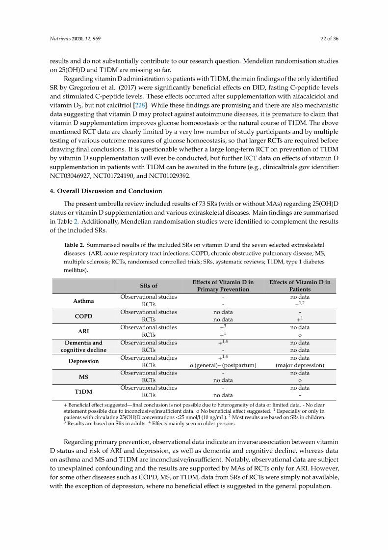
Nutrients 2020, 12, 969 22 of 36
results and do not substantially contribute to our research question. Mendelian randomisation studieson 25(OH)D and T1DM are missing so far.
Regarding vitamin D administration to patients with T1DM, the main findings of the only identifiedSR by Gregoriou et al. (2017) were significantly beneficial effects on DID, fasting C-peptide levelsand stimulated C-peptide levels. These effects occurred after supplementation with alfacalcidol andvitamin D3, but not calcitriol [228]. While these findings are promising and there are also mechanisticdata suggesting that vitamin D may protect against autoimmune diseases, it is premature to claim thatvitamin D supplementation improves glucose homoeostasis or the natural course of T1DM. The abovementioned RCT data are clearly limited by a very low number of study participants and by multipletesting of various outcome measures of glucose homoeostasis, so that larger RCTs are required beforedrawing final conclusions. It is questionable whether a large long-term RCT on prevention of T1DMby vitamin D supplementation will ever be conducted, but further RCT data on effects of vitamin Dsupplementation in patients with T1DM can be awaited in the future (e.g., clinicaltrials.gov identifier:NCT03046927, NCT01724190, and NCT01029392.
4. Overall Discussion and Conclusion
The present umbrella review included results of 73 SRs (with or without MAs) regarding 25(OH)Dstatus or vitamin D supplementation and various extraskeletal diseases. Main findings are summarisedin Table 2. Additionally, Mendelian randomisation studies were identified to complement the resultsof the included SRs.
Table 2. Summarised results of the included SRs on vitamin D and the seven selected extraskeletaldiseases. (ARI, acute respiratory tract infections; COPD, chronic obstructive pulmonary disease; MS,multiple sclerosis; RCTs, randomised controlled trials; SRs, systematic reviews; T1DM, type 1 diabetesmellitus).
SRs of Effects of Vitamin D inPrimary Prevention
Effects of Vitamin D inPatients
AsthmaObservational studies - no data
RCTs - +1,2
Observational studies no data -COPD RCTs no data +1
ARIObservational studies +3 no data
RCTs +1 oObservational studies +1,4 no dataDementia and
cognitive decline RCTs - no data
Depression Observational studies +1,4 no dataRCTs o (general)– (postpartum) (major depression)
Observational studies - no dataMS RCTs no data o
T1DMObservational studies - no data
RCTs no data -
+ Beneficial effect suggested—final conclusion is not possible due to heterogeneity of data or limited data. - No clearstatement possible due to inconclusive/insufficient data. o No beneficial effect suggested. 1 Especially or only inpatients with circulating 25(OH)D concentrations <25 nmol/l (10 ng/mL). 2 Most results are based on SRs in children.3 Results are based on SRs in adults. 4 Effects mainly seen in older persons.
Regarding primary prevention, observational data indicate an inverse association between vitaminD status and risk of ARI and depression, as well as dementia and cognitive decline, whereas dataon asthma and MS and T1DM are inconclusive/insufficient. Notably, observational data are subjectto unexplained confounding and the results are supported by MAs of RCTs only for ARI. However,for some other diseases such as COPD, MS, or T1DM, data from SRs of RCTs were simply not available,with the exception of depression, where no beneficial effect is suggested in the general population.
Nutrients 2020, 12, 969 23 of 36
With respect to possible effects on the course of the disease, SRs of RCTs suggest a beneficialimpact of vitamin D in deficient patients with asthma and COPD, whereas no favourable effects havebeen reported in patients with ARI and MS. In patients with major depression and T1DM, respectivestudies are too scarce to draw a final conclusion.
Compared with the conclusions reported in previously published umbrella reviews [30,31],our evaluation considering more current studies leads to similar results. Consistent with ourconclusion, both previous reviews (only focusing on MAs of RCTs) reported beneficial effects of vitaminD regarding the risk of respiratory tract infections and no effects of vitamin D supplementation ondepression scores. Also, in accordance with our evaluation on the impact of vitamin D in patients withMS, Autier et al. (2017) reported no favourable effects [31]. A very recently published narrative reviewindicated a possible health benefit of vitamin D on COPD, ARI, as well as Alzheimer’s disease andother dementia, albeit based only on one or two recently published individual studies [234].
There are several limitations that have to be considered while interpreting the results of theincluded SRs in the present umbrella review. First, the issue of initial 25(OH)D status was not alwaysadequately addressed. It is rather unlikely that individuals with an adequate vitamin D status willbenefit from vitamin D supplementation. For several of the selected extraskeletal diseases (asthma,COPD, ARI) SRs of RCTs reported on a beneficial effect of vitamin D supplementation especially inpersons with low baseline 25(OH)D concentrations (<25 nmol/l (10 ng/mL)), whereas in persons withadequate baseline 25(OH)D levels (>50 nmol/l (20 ng/mL)) often no effects were observed. In line withthis, SRs of observational studies on several of the reviewed extraskeletal diseases (e.g., depression,dementia, and cognitive decline) support this finding concerning 25(OH)D status. Thus, it seemslogical that large vitamin D RCTs with inclusion of individuals with adequate serum 25(OH)D levelsfailed to show significant effects [235].
Second, the mode, dose and frequency of supplementation varied widely, including low (10 µg(400 IU)) and high (100µg (4000 IU)) daily doses and high intermittent bolus doses (3000µg (120,000 IU)).In addition, different forms of vitamin D were administered (e.g., vitamin D3, or vitamin D2), and insome studies a multivitamin supplement was used or vitamin D was applied together with calcium.Ideally, vitamin D3 supplements at low daily doses and frequent intervals should be performed sincethis leads to a continuous elevation of 25(OH)D levels into the physiological range [126,236]. Third,the inconsistent definitions of endpoints (especially heterogeneous for the outcomes ARI and dementia)are an issue which might have influenced results. Fourth, duration of the individual studies variedwidely (e.g., for COPD from six weeks to one year) as well as the studied population (from new-bornsto elderly). Fifth, for some diseases such as COPD, dementia, depression, MS, and T1DM, the assessedquality of the SRs was very heterogeneous.
Apart from these limitations, the additionally identified Mendelian randomisation studies onlypartially supported the results of the SRs. However, Mendelian randomisation studies are not withoutweaknesses because they are based on assumptions which are often only partly fulfilled: (i) singlenucleotide polymorphisms (SNPs) are directly associated with the risk factor; (ii) SNPs are not thecause of known or unknown bias; and (iii) SNPs do not influence the risk of the disease through otherfactors. Moreover, it must be kept in mind that genetic variants explain only a small fraction of the25(OH)D variance. Consequently, effects in subgroups at increased risk of either 25(OH)D deficiencyor for the outcome may be overlooked.
To conclude, with the exception of beneficial vitamin D effects on the prevention of ARI and on thetreatment of vitamin D deficient patients with asthma and COPD, available data do not clearly supportbeneficial vitamin D effects in preventing the reviewed extraskeletal diseases or affecting the courseof the disease. Therefore, further high-quality RCTs are warranted. Ideally, these RCTs should beperformed in patients with insufficient vitamin D status, and moderate daily vitamin D doses should bepreferred. Also, geographical aspects and seasonal variations contributing to an altered sun-exposuremust be considered. In addition, the trials should be designed with well-defined diseases or endpoints.
Nutrients 2020, 12, 969 24 of 36
Furthermore, they should be adequately powered to assess whether or not vitamin D is able to influenceclinically relevant endpoints. Both efficacy and safety should be adequately considered.
The main strength of the present umbrella review is the comprehensive overview of systematicallyidentified SRs (with or without MAs) of RCTs and cohort studies on vitamin D status andsupplementation and the seven selected diseases, thereby updating the knowledge concerningthe possible role of vitamin D for the development and course of these diseases. More importantly,we assessed the methodological quality of all included SRs with the AMSTAR 2 tool so that ourconclusions are also based on the quality of the included studies. A limitation of this review is thatwe did not evaluate the quality of evidence of the studies and that limitations inherent to the originalindividual studies cannot be further reduced by our umbrella review.
As identified at clinicaltrial.gov, several RCTs are already underway that may provide additionalinformation on the effect of vitamin D on the seven extraskeletal diseases investigated in this review.Besides, for diseases which develop over a longer time span, such as dementia, or a certain periodof life (e.g., MS diagnosis in early adulthood), vitamin D effects may be difficult to assess in RCTs.Therefore, well-designed longitudinal observational studies could be extremely helpful to providefurther information.
Generally speaking, ingested or endogenously produced vitamin D doses of 20 µg (800 IU) areconsidered safe, and can significantly contribute to attain circulating 25(OH)D concentrations of atleast 50 nmol/l (20 ng/mL). However, if supplementation is necessary (e.g., due to lack of endogenoussynthesis), continuous daily doses (in the amount of the recommended daily intake level of 10–20 µg(400–800 IU) should be applied instead of high bolus doses [2–5,237].
Supplementary Materials: The following Supplementary Materials for this article are available online athttp://www.mdpi.com/2072-6643/12/4/969/s1, Table S1: Search strategy in PubMed, Tables S2–S26: Synopticaltables of included SRs/MAs of RCTs or prospective cohort studies for the individual diseases, Figures S1–S7:PRISMA flow diagrams for the individual diseases.
Author Contributions: J.L., S.E., A.B., and F.M. designed the study and all authors contributed to the conceptionof the manuscript. Databases were searched by at least two or three authors for each disease (all diseases: F.M. andJ.B.E.; asthma and COPD: A.Z.; ARI: S.P. and G.I.S.; dementia and cognitive decline: S.E. and D.V.; Depression:M.W. and D.M.v.L.; MS: J.L. and G.I.S.; T1DM: S.P. and J.L.). F.M. and J.B.E. performed the selection and extractionof studies and assessed the study quality via the modified AMSTAR 2 tool. All authors were involved in draftingthe paper and revised the draft critically. F.M., J.W., and J.L. organised and coordinated the revision and editing ofthe draft. All authors read and approved the final manuscript.
Funding: This research was funded by the Federal Ministry of Food, Agriculture, and Consumer protection.
Conflicts of Interest: S.P. received consultant and speakers honoraria from Merck Selbstmedikation GmbH,DiaSorin S.p.A. and SYNLAB Holding Deutschland GmbH and A.Z. from DiaSorin S.p.A., Fresenius SE & Co.KGaA and DSM Nutritional Products GmbH. These institutions had no role in the design, execution, interpretation,or writing of this review. The other authors declare no potential conflict of interest.
References
1. Zittermann, A.; Pilz, S. Vitamin D in Klinik und Praxis. Dtsch. Med. Wochenschr. 2017, 142, 601–616.2. Institute of Medicine. Dietary Reference Intakes: Calcium and Vitamin D; The National Academic Press:
Washington, DC, USA, 2011.3. Nordic Council of Ministers. Nordic Nutrition Recommendations 2012. Integrating Nutrition and Physical Activity,
5th ed.; Nordic Council of Ministers: Copenhagen, Denmark, 2014; pp. 349–384.4. German Nutrition Society; Austrian Nutrition Society; Swiss Nutrition Society. Referenzwerte für die
Nährstoffzufuhr, 2nd ed.; DGE: Bonn, Germany, 2015.5. EFSA (European Food Safety Authority). Dietary reference values for vitamin D: EFSA Panel on Dietetic
Products, Nutrition and Allergies (NDA). EFSA J. 2016, 14, e04547. [CrossRef]6. Seckmeyer, G.; Schrempf, M.; Wieczorek, A.; Riechelmann, S.; Graw, K.; Seckmeyer, S.; Zankl, M. A novel
method to calculate solar UV exposure relevant to vitamin D production in humans. Photochem. Photobiol.2013, 89, 974–983. [CrossRef] [PubMed]
Nutrients 2020, 12, 969 25 of 36
7. Holick, M.F. Sunlight and vitamin D for bone health and prevention of autoimmune diseases, cancers,and cardiovascular disease. Am. J. Clin. Nutr. 2004, 80, 1678–1688. [CrossRef]
8. Dawson-Hughes, B.; Heaney, R.P.; Holick, M.F.; Lips, P.; Meunier, P.J.; Vieth, R. Estimates of optimal vitaminD status. Osteoporos. Int. 2005, 16, 713–716. [CrossRef] [PubMed]
9. Holick, M.F. Vitamin D status: Measurement, interpretation, and clinical application. Ann. Epidemiol. 2009,19, 73–78. [CrossRef]
10. Linseisen, J.; Bechthold, A.; Bischoff-Ferrari, H.A.; Hintzpeter, B.; Leschik-Bonnet, E.; Reichrath, J.; Stehle, P.;Volkert, D.; Wolfram, G.; Zittermann, A. DGE-Stellungnahme: Vitamin D und Prävention AusgewählterChronischer Krankheiten. Available online: http://www.dge.de/pdf/ws/DGE-Stellungnahme-VitD-111220.pdf (accessed on 26 June 2013).
11. Pilz, S.; Zittermann, A.; Trummer, C.; Theiler-Schwetz, V.; Lerchbaum, E.; Keppel, M.H.; Grübler, M.R.;März, W.; Pandis, M. Vitamin D testing and treatment: A narrative review of current evidence. Endocr. Connect.2019, 8, R27–R43. [CrossRef] [PubMed]
12. Cashman, K.D.; Dowling, K.G.; Škrabáková, Z.; Gonzalez-Gross, M.; Valtueña, J.; de Henauw, S.; Moreno, L.;Damsgaard, C.T.; Michaelsen, K.F.; Mølgaard, C.; et al. Vitamin D deficiency in Europe: Pandemic? Am. J.Clin. Nutr. 2016, 103, 1033–1044. [CrossRef]
13. Van Schoor, N.M.; Lips, P. Worldwide vitamin D status. Best Pract. Res. Clin. Endocrinol. Metab. 2011, 25,671–680. [CrossRef]
14. Lips, P. Vitamin D status and nutrition in Europe and Asia. J. Steroid. Biochem. Mol. Biol. 2007, 103, 620–625.[CrossRef]
15. Muscogiuri, G.; Altieri, B.; Annweiler, C.; Balercia, G.; Pal, H.B.; Boucher, B.J.; Cannell, J.J.; Foresta, C.;Grübler, M.R.; Kotsa, K.; et al. Vitamin D and chronic diseases: The current state of the art. Arch. Toxicol.2017, 91, 97–107. [CrossRef] [PubMed]
16. Nagpal, S.; Na, S.; Rathnachalam, R. Noncalcemic actions of vitamin D receptor ligands. Endocr. Rev. 2005,26, 662–687. [CrossRef] [PubMed]
17. Adams, J.S.; Hewison, M. Extrarenal expression of the 25-hydroxyvitamin D-1-hydroxylase. Arch. Biochem.Biophys. 2012, 523, 95–102. [CrossRef]
18. Barnard, K.; Colón-Emeric, C. Extraskeletal effects of vitamin D in older adults: Cardiovascular disease,mortality, mood, and cognition. Am. J. Geriatr. Pharmacother. 2010, 8, 4–33. [CrossRef]
19. Elamin, M.B.; Abu Elnour, N.O.; Elamin, K.B.; Fatourechi, M.M.; Alkatib, A.A.; Almandoz, J.P.; Liu, H.;Lane, M.A.; Mullan, R.J.; Hazem, A.; et al. Vitamin D and cardiovascular outcomes: A systematic review andmeta-analysis. J. Clin. Endocrinol. Metab. 2011, 96, 1931–1942. [CrossRef]
20. Martins, D.; Wolf, M.; Pan, D.; Zadshir, A.; Tareen, N.; Thadhani, R.; Felsenfeld, A.; Levine, B.; Mehrotra, R.;Norris, K. Prevalence of cardiovascular risk factors and the serum levels of 25-hydroxyvitamin D in theUnited States: Data from the Third National Health and Nutrition Examination Survey. Arch. Intern. Med.2007, 167, 1159–1165. [CrossRef]
21. Gorham, E.D.; Garland, C.F.; Garland, F.C.; Grant, W.B.; Mohr, S.B.; Lipkin, M.; Newmark, H.L.;Giovannucci, E.; Wei, M.; Holick, M.F. Optimal vitamin D status for colorectal cancer prevention:A quantitative meta analysis. Am. J. Prev. Med. 2007, 32, 210–216. [CrossRef] [PubMed]
22. Yin, L.; Grandi, N.; Raum, E.; Haug, U.; Arndt, V.; Brenner, H. Meta-analysis: Longitudinal studies of serumvitamin D and colorectal cancer risk. Aliment. Pharmacol. Ther. 2009, 30, 113–125. [CrossRef] [PubMed]
23. Gandini, S.; Boniol, M.; Haukka, J.; Byrnes, G.; Cox, B.; Sneyd, M.J.; Mullie, P.; Autier, P. Meta-analysis ofobservational studies of serum 25-hydroxyvitamin D levels and colorectal, breast and prostate cancer andcolorectal adenoma. Int. J. Cancer 2011, 128, 1414–1424. [CrossRef] [PubMed]
24. Pittas, A.G.; Lau, J.; Hu, F.B.; Dawson-Hughes, B. The role of vitamin D and calcium in type 2 diabetes.A systematic review and meta-analysis. J. Clin. Endocrinol. Metab. 2007, 92, 2017–2029. [CrossRef]
25. Mitri, J.; Muraru, M.D.; Pittas, A.G. Vitamin D and type 2 diabetes: A systematic review. Eur. J. Clin. Nutr.2011, 65, 1005–1015. [CrossRef] [PubMed]
26. Witham, M.D.; Nadir, M.A.; Struthers, A.D. Effect of vitamin D on blood pressure: A systematic review andmeta-analysis. J. Hypertens. 2009, 27, 1948–1954. [CrossRef] [PubMed]
27. Golzarand, M.; Shab-Bidar, S.; Koochakpoor, G.; Speakman J, R.; Djafarian, K. Effect of vitamin D3
supplementation on blood pressure in adults: An updated meta-analysis. Nutr. Metab. Cardiovasc. Dis. 2016,26, 663–673. [CrossRef] [PubMed]

Nutrients 2020, 12, 969 26 of 36
28. Keum, N.; Lee, D.H.; Greenwood, D.C.; Manson, J.E.; Giovannucci, E. Vitamin D supplementation and totalcancer incidence and mortality: A meta-analysis of randomized controlled trials. Ann. Oncol. 2019, 30,733–743. [CrossRef] [PubMed]
29. Caraba, A.; Crisan, V.; Romosan, I.; Mozos, I.; Murariu, M. Vitamin D status, disease activity, and endothelialdysfunction in early rheumatoid arthritis patients. Dis. Markers 2017, 2017, 5241012. [CrossRef]
30. Rejnmark, L.; Bislev, L.S.; Cashman, K.D.; Eiríksdottir, G.; Gaksch, M.; Grübler, M.; Grimnes, G.; Gudnason, V.;Lips, P.; Pilz, S.; et al. Non-skeletal health effects of vitamin D supplementation: A systematic review onfindings from meta-analyses summarizing trial data. PLoS ONE 2017, 12, e0180512. [CrossRef]
31. Autier, P.; Mullie, P.; Macacu, A.; Dragomir, M.; Boniol, M.; Coppens, K.; Pizot, C.; Boniol, M. Effect of vitaminD supplementation on non-skeletal disorders: A systematic review of meta-analyses and randomised trials.Lancet Diabetes Endocrinol. 2017, 5, 986–1004. [CrossRef]
32. Murdaca, G.; Tonacci, A.; Negrini, S.; Greco, M.; Borro, M.; Puppo, F.; Gangemi, S. Emerging role of vitaminD in autoimmune diseases: An update on evidence and therapeutic implications. Autoimmun. Rev. 2019, 18,102350. [CrossRef]
33. Hejazi, M.E.; Modarresi-Ghazani, F.; Entezari-Maleki, T. A review of Vitamin D effects on common respiratorydiseases: Asthma, chronic obstructive pulmonary disease, and tuberculosis. J. Res. Pharm. Pract. 2016, 5,7–15.
34. Rooney, M.R.; Harnack, L.; Michos, E.D.; Ogilvie, R.P.; Sempos, C.T.; Lutsey, P.L. Trends in use of high-dosevitamin D supplements exceeding 1000 or 4000 international units daily, 1999-2014. JAMA 2017, 317,2448–2450. [CrossRef]
35. Manousaki, D.; Paternoster, L.; Standl, M.; Moffatt, M.F.; Farrall, M.; Bouzigon, E.; Strachan, D.P.;Demenais, F.; Lathrop, M.; Cookson, W.O.C.M.; et al. Vitamin D levels and susceptibility to asthma,elevated immunoglobulin E levels, and atopic dermatitis: A Mendelian randomization study. PLoS Med.2017, 14, e1002294. [CrossRef] [PubMed]
36. Vimaleswaran, K.S.; Berry, D.J.; Lu, C.; Tikkanen, E.; Pilz, S.; Hiraki, L.T.; Cooper, J.D.; Dastani, Z.; Li, R.;Houston, D.K.; et al. Causal relationship between obesity and vitamin D status: Bi-directional mendelianrandomization analysis of multiple cohorts. PLoS Med. 2013, 10, e1001383. [CrossRef] [PubMed]
37. Shea, B.J.; Reeves, B.C.; Wells, G.; Thuku, M.; Hamel, C.; Moran, J.; Moher, D.; Tugwell, P.; Welch, V.;Kristjansson, E.; et al. AMSTAR 2: A critical appraisal tool for systematic reviews that include randomisedor non-randomised studies of healthcare interventions, or both. BMJ 2017, 358, j4008. [CrossRef] [PubMed]
38. Luo, J.; Liu, D.; Liu, C.-T. Can vitamin D supplementation in addition to asthma controllers improve clinicaloutcomes in patients with asthma? A meta-analysis. Medicine (Baltimore) 2015, 94, e2185. [CrossRef][PubMed]
39. Global Asthma Network. The Global Asthma Report 2018. Available online: http://www.globalasthmareport.org/Global%20Asthma%20Report%202018.pdf (accessed on 26 June 2019).
40. Mukherjee, S.; Lindell, D.M.; Berlin, A.A.; Morris, S.B.; Shanley, T.P.; Hershenson, M.B.; Lukacs, N.W.IL-17-induced pulmonary pathogenesis during respiratory viral infection and exacerbation of allergic disease.Am. J. Pathol. 2011, 179, 248–258. [CrossRef]
41. Nanzer, A.M.; Chambers, E.S.; Ryanna, K.; Richards, D.F.; Black, C.; Timms, P.M.; Martineau, A.R.;Griffiths, C.J.; Corrigan, C.J.; Hawrylowicz, C.M. Enhanced production of IL-17A in patients with severeasthma is inhibited by 1α,25-dihydroxyvitamin D3 in a glucocorticoid-independent fashion. J. AllergyClin. Immunol. 2013, 133, 297–304. [CrossRef]
42. Masoli, M.; Fabian, D.; Holt, S.; Beasley, R. The global burden of asthma: Executive summary of the GINADissemination Committee report. Allergy 2004, 59, 469–478. [CrossRef]
43. Jackson, D.J.; Johnston, S.L. The role of viruses in acute exacerbations of asthma. J. Allergy Clin. Immunol.2010, 125, 1178–1187. [CrossRef]
44. Wang, H.; Naghavi, M.; Allen, C.; Barber, R.M.; Bhutta, Z.A.; Carter, A.; Casey, D.C.; Charlson, F.J.; Chen, A.Z.;Coates, M.M.; et al. Global, regional, and national life expectancy, all-cause mortality, and cause-specificmortality for 249 causes of death, 1980–2015: A systematic analysis for the Study 2015. Lancet 2016, 388,1459–1544. [CrossRef]
45. Eder, W.; Ege, M.J.; von Mutius, E. The asthma epidemic. N. Engl. J. Med. 2006, 355, 2226–2235. [CrossRef]

Nutrients 2020, 12, 969 27 of 36
46. Björkstén, B.; Clayton, T.; Ellwood, P.; Stewart, A.; Strachan, D. Worldwide time trends for symptoms ofrhinitis and conjunctivitis: Phase III of the International Study of Asthma and Allergies in Childhood. Pediatr.Allergy Immunol. 2008, 19, 110–124. [CrossRef] [PubMed]
47. Flohr, C. Recent perspectives on the global epidemiology of childhood eczema. Allergol. Immunopathol. 2011,39, 174–182. [CrossRef] [PubMed]
48. Centers for Disease Control and Prevention (CDC). Asthma’s Effect on the Nation. Available online:https://www.cdc.gov/asthma/asthmadata.htm (accessed on 26 June 2019).
49. Global Initiative for Asthma (GINA). The Global Asthma Report; GINA: Paris, France, 2011.50. Zasloff, M. Fighting infections with vitamin D. Nat. Med. 2006, 12, 388–390. [CrossRef] [PubMed]51. Van Oeffelen, A.A.M.; Bekkers, M.B.M.; Smit, H.A.; Kerkhof, M.; Koppelman, G.H.; Haveman-Nies, A.;
van der A, D.L.; Jansen, E.H.J.M.; Wijga, A.H. Serum micronutrient concentrations and childhood asthma:The PIAMA birth cohort study. Pediatr. Allergy Immunol. 2011, 22, 784–793. [CrossRef]
52. Chinellato, I.; Piazza, M.; Sandri, M.; Paiola, G.; Tezza, G.; Boner, A.L. Correlation between vitamin D serumlevels and passive smoking exposure in children with asthma. Allergy Asthma Proc. 2018, 39, 8–14. [CrossRef]
53. Cantorna, M.T.; Mahon, B.D. D-hormone and the immune system. J. Rheumatol. Suppl. 2005, 76, 11–20.54. To, T.; Stanojevic, S.; Moores, G.; Gershon, A.S.; Bateman, E.D.; Cruz, A.A.; Boulet, L.-P. Global asthma
prevalence in adults: Findings from the cross-sectional world health survey. BMC Public Health 2012, 12, 204.[CrossRef]
55. Hewison, M. Vitamin D and immune function: Autocrine, paracrine or endocrine? Scand. J. Clin. Lab.Investig. Suppl. 2012, 243, 92–102.
56. Hansdottir, S.; Monick, M.M.; Hinde, S.L.; Lovan, N.; Look, D.C.; Hunninghake, G.W. Respiratory epithelialcells convert inactive vitamin D to its active form: Potential effects on host defense. J. Immunol. 2008, 181,7090–7099. [CrossRef]
57. Brehm, J.M.; Schuemann, B.; Fuhlbrigge, A.L.; Hollis, B.W.; Strunk, R.C.; Zeiger, R.S.; Weiss, S.T.; Litonjua, A.A.Serum vitamin D levels and severe asthma exacerbations in the Childhood Asthma Management Programstudy. J. Allergy Clin. Immunol. 2010, 126, 52–58. [CrossRef]
58. Hansdottir, S.; Monick, M.M. Vitamin D effects on lung immunity and respiratory diseases. Vitam. Horm.2011, 86, 217–237. [PubMed]
59. Hansdottir, S.; Monick, M.M.; Lovan, N.; Powers, L.; Gerke, A.; Hunninghake, G.W. Vitamin D decreasesrespiratory syncytial virus induction of NF-kappaB-linked chemokines and cytokines in airway epitheliumwhile maintaining the antiviral state. J. Immunol. 2010, 184, 965–974. [CrossRef] [PubMed]
60. Litonjua, A.A.; Weiss, S.T. Is vitamin D deficiency to blame for the asthma epidemic? J. Allergy Clin. Immunol.2007, 120, 1031–1035. [CrossRef]
61. Adams, J.S.; Hewison, M. Unexpected actions of vitamin D: New perspectives on the regulation of innateand adaptive immunity. Nat. Clin. Pract. Endocrinol. Metab. 2008, 4, 80–90. [CrossRef] [PubMed]
62. Camargo, C.A.J.; Ingham, T.; Wickens, K.; Thadhani, R.; Silvers, K.M.; Epton, M.J.; Town, G.I.; Pattemore, P.K.;Espinola, J.A.; Crane, J. Cord-blood 25-hydroxyvitamin D levels and risk of respiratory infection, wheezing,and asthma. Pediatrics 2011, 127, e180–e187. [CrossRef]
63. Brown, S.D.; Calvert, H.H.; Fitzpatrick, A.M. Vitamin D and asthma. Dermatoendocrinology 2012, 4, 137–145.[CrossRef]
64. Finklea, J.D.; Grossmann, R.E.; Tangpricha, V. Vitamin D and chronic lung disease: A review of molecularmechanisms and clinical studies. Adv. Nutr. 2011, 2, 244–253. [CrossRef]
65. Herr, C.; Greulich, T.; Koczulla, R.A.; Meyer, S.; Zakharkina, T.; Branscheidt, M.; Eschmann, R.; Bals, R.The role of vitamin D in pulmonary disease: COPD, asthma, infection, and cancer. Respir. Res. 2011, 12, 31.[CrossRef]
66. Paul, G.; Brehm, J.M.; Alcorn, J.F.; Holguín, F.; Aujla, S.J.; Celedón, J.C. Vitamin D and asthma. Am. J. Respir.Crit. Care Med. 2012, 185, 124–132. [CrossRef]
67. Rajabbik, M.H.; Lotfi, T.; Alkhaled, L.; Fares, M.; El-Hajj Fuleihan, G.; Mroueh, S.; Akl, E.A. Associationbetween low vitamin D levels and the diagnosis of asthma in children: A systematic review of cohort studies.Allergy Asthma Clin. Immunol. 2014, 10, 31. [CrossRef]
68. Cassim, R.; Russell, M.A.; Lodge, C.J.; Lowe, A.J.; Koplin, J.J.; Dharmage, S.C. The role of circulating 25hydroxyvitamin D in asthma: A systematic review. Allergy 2015, 70, 339–354. [CrossRef] [PubMed]
Nutrients 2020, 12, 969 28 of 36
69. Man, L.; Zhang, Z.; Zhang, M.; Zhang, Y.; Li, J.; Zheng, N.; Cao, Y.; Chi, M.; Chao, Y.; Huang, Q.; et al.Association between vitamin D deficiency and insufficiency and the risk of childhood asthma: Evidencefrom a meta-analysis. Int. J. Clin. Exp. Med. 2015, 8, 5699–5706. [PubMed]
70. Wei, Z.; Zhang, J.; Yu, X. Maternal vitamin D status and childhood asthma, wheeze, and eczema: A systematicreview and meta-analysis. Pediatr. Allergy Immunol. 2016, 27, 612–619. [CrossRef] [PubMed]
71. Feng, H.; Xun, P.; Pike, K.; Wills, A.K.; Chawes, B.L.; Bisgaard, H.; Cai, W.; Wan, Y.; He, K. In utero exposure to25-hydroxyvitamin D and risk of childhood asthma, wheeze, and respiratory tract infections: A meta-analysisof birth cohort studies. J. Allergy Clin. Immunol. 2017, 139, 1508–1517. [CrossRef]
72. Jat, K.R.; Khairwa, A. Vitamin D and asthma in children: A systematic review and meta-analysis ofobservational studies. Lung India 2017, 34, 355–363. [CrossRef] [PubMed]
73. Song, H.; Yang, L.; Jia, C. Maternal vitamin D status during pregnancy and risk of childhood asthma:A meta-analysis of prospective studies. Mol. Nutr. Food Res. 2017, 61, 1600657. [CrossRef] [PubMed]
74. Pacheco-González, R.M.; García-Marcos, L.; Morales, E. Prenatal vitamin D status and respiratory andallergic outcomes in childhood: A meta-analysis of observational studies. Pediatr. Allergy Immunol. 2018, 29,243–253. [CrossRef]
75. Shen, S.-Y.; Xiao, W.-Q.; Lu, J.-H.; Yuan, M.-Y.; He, J.-R.; Xia, H.-M.; Qiu, X.; Cheng, K.K.; Lam, K.B.H. Earlylife vitamin D status and asthma and wheeze: A systematic review and meta-analysis. BMC Pulm. Med.2018, 18, 120. [CrossRef]
76. Harvey, N.C.; Holroyd, C.; Ntani, G.; Javaid, K.; Cooper, P.; Moon, R.; Cole, Z.; Tinati, T.; Godfrey, K.;Dennison, E.; et al. Vitamin D supplementation in pregnancy: A systematic review. Health Technol. Assess.2014, 18, 1–190. [CrossRef]
77. Xiao, L.; Xing, C.; Yang, Z.; Xu, S.; Wang, M.; Du, H.; Liu, K.; Huang, Z. Vitamin D supplementation for theprevention of childhood acute respiratory infections: A systematic review of randomised controlled trials.Br. J. Nutr. 2015, 114, 1026–1034. [CrossRef]
78. Pojsupap, S.; Iliriani, K.; Sampaio, T.Z.A.L.; O’Hearn, K.; Kovesi, T.; Menon, K.; McNally, J.D. Efficacy ofhigh-dose vitamin D in pediatric asthma: A systematic review and meta-analysis. J. Asthma 2015, 52, 382–390.[CrossRef]
79. Riverin, B.D.; Maguire, J.L.; Li, P. Vitamin D supplementation for childhood asthma: A systematic reviewand meta-analysis. PLoS ONE 2015, 10, e0136841. [CrossRef]
80. Fares, M.M.; Alkhaled, L.H.; Mroueh, S.M.; Akl, E.A. Vitamin D supplementation in children with asthma:A systematic review and meta-analysis. BMC Res. Notes 2015, 8, 23. [CrossRef]
81. Vahdaninia, M.; Mackenzie, H.; Helps, S.; Dean, T. Prenatal intake of vitamins and allergic outcomes inthe offspring: A systematic review and meta-analysis. J. Allergy Clin. Immunol. Pract. 2017, 5, 771–778.[CrossRef] [PubMed]
82. Martineau, A.R.; Cates, C.J.; Urashima, M.; Jensen, M.; Griffiths, A.P.; Nurmatov, U.; Sheikh, A.; Griffiths, C.J.Vitamin D for the management of asthma. Cochrane Database Syst. Rev. 2016, 9, CD011511. [CrossRef][PubMed]
83. Jolliffe, D.A.; Greenberg, L.; Hooper, R.L.; Griffiths, C.J.; Camargo, C.A.; Kerley, C.P.; Jensen, M.E.; Mauger, D.;Stelmach, I.; Urashima, M.; et al. Vitamin D supplementation to prevent asthma exacerbations: A systematicreview and meta-analysis of individual participant data. Lancet Respir. Med. 2017, 5, 881–890. [CrossRef]
84. Davies, N.M.; Holmes, M.V.; Davey Smith, G. Reading Mendelian randomization studies: A guide, glossary,and checklist for clinicians. BMJ 2018, 362, k601. [CrossRef] [PubMed]
85. Barnes, P.J.; Celli, B.R. Systemic manifestations and comorbidities of COPD. Eur. Respir. J. 2009, 33, 1165–1185.[CrossRef]
86. Wedzicha, J.A.; Seemungal, T.A.R. COPD exacerbations: Defining their cause and prevention. Lancet 2007,370, 786–796. [CrossRef]
87. Global Initiative for Chronic Obstructive Lung Disease (GOLD). Global Strategy for the Diagnosis,Management and Prevention of COPD. Available online: https://goldcopd.org/gold-2017-global-strategy-diagnosis-management-prevention-copd/ (accessed on 27 March 2019).
88. GBD 2015 Chronic Respiratory Disease Collaborators. Global, regional, and national deaths, prevalence,disability-adjusted life years, and years lived with disability for chronic obstructive pulmonary disease andasthma, 1990–2015. Lancet Respir. Med. 2017, 5, 691–706. [CrossRef]
Nutrients 2020, 12, 969 29 of 36
89. Lopez, A.D.; Shibuya, K.; Rao, C.; Mathers, C.D.; Hansell, A.L.; Held, L.S.; Schmid, V.; Buist, S. Chronicobstructive pulmonary disease: Current burden and future projections. Eur. Respir. J. 2006, 27, 397–412.[CrossRef] [PubMed]
90. Faner, R.; Gonzalez, N.; Cruz, T.; Kalko, S.G.; Agustí, A. Systemic inflammatory response to smoking inchronic obstructive pulmonary disease: Evidence of a gender effect. PLoS ONE 2014, 9, e97491. [CrossRef][PubMed]
91. Smolonska, J.; Wijmenga, C.; Postma, D.S.; Boezen, H.M. Meta-analyses on suspected chronic obstructivepulmonary disease genes: A summary of 20 years’ research. Am. J. Respir. Crit. Care Med. 2009, 180, 618–631.[CrossRef] [PubMed]
92. Kokturk, N.; Baha, A.; Oh, Y.-M.; Young Ju, J.; Jones, P.W. Vitamin D deficiency: What does it mean forchronic obstructive pulmonary disease (COPD)? a compherensive review for pulmonologists. Clin. Respir. J.2018, 12, 382–397. [CrossRef] [PubMed]
93. Zittermann, A.; Tenderich, G.; Koerfer, R. Vitamin D and the adaptive immune system with special emphasisto allergic reactions and allograft rejection. Inflamm. Allergy Drug Targets 2009, 8, 161–168. [CrossRef]
94. Hewison, M. Vitamin D and the immune system: New perspectives on an old theme. Endocrinol. Metab.Clin. North Am. 2010, 39, 365–379. [CrossRef]
95. Szymczak, I.; Pawliczak, R. The active metabolite of vitamin D3 as a potential immunomodulator. Scand. J.Immunol. 2016, 83, 83–91. [CrossRef]
96. Black, P.N.; Scragg, R. Relationship between serum 25-hydroxyvitamin d and pulmonary function in thethird national health and nutrition examination survey. Chest 2005, 128, 3792–3798. [CrossRef]
97. Choi, C.-J.; Seo, M.; Choi, W.-S.; Kim, K.-S.; Youn, S.-A.; Lindsey, T.; Choi, Y.-J.; Kim, C.-M. Relationshipbetween serum 25-hydroxyvitamin D and lung function among Korean adults in Korea National Healthand Nutrition Examination Survey (KNHANES), 2008-2010. J. Clin. Endocrinol. Metab. 2013, 98, 1703–1710.[CrossRef]
98. Manavi, K.R.; Alston-Mills, B.P.; Thompson, M.P. History of tobacco, vitamin D and women. Int J Vitam.Nutr. Res. 2020. [CrossRef]
99. Martineau, A.R.; Jolliffe, D.A.; Hooper, R.L.; Greenberg, L.; Aloia, J.F.; Bergman, P.; Dubnov-Raz, G.;Esposito, S.; Ganmaa, D.; Ginde, A.A.; et al. Vitamin D supplementation to prevent acute respiratorytract infections: Systematic review and meta-analysis of individual participant data. BMJ 2017, 356, i6583.[CrossRef] [PubMed]
100. Autier, P.; Boniol, M.; Pizot, C.; Mullie, P. Vitamin D status and ill health: A systematicS review. LancetDiabetes Endocrinol. 2014, 2, 76–89. [CrossRef]
101. Zhu, B.; Zhu, B.; Xiao, C.; Zheng, Z. Vitamin D deficiency is associated with the severity of COPD: A systematicreview and meta-analysis. Int. J. Chron. Obstruct. Pulmon. Dis. 2015, 10, 1907–1916. [PubMed]
102. Zhu, M.; Wang, T.; Wang, C.; Ji, Y. The association between vitamin D and COPD risk, severity,and exacerbation: An updated systematic review and meta-analysis. Int. J. Chron. Obstruct. Pulmon. Dis.2016, 11, 2597–2607. [CrossRef]
103. Ferrari, R.; Caram, L.M.O.; Tanni, S.E.; Godoy, I.; Rupp de Paiva, S.A. The relationship between Vitamin Dstatus and exacerbation in COPD patients- a literature review. Respir. Med. 2018, 139, 34–38. [CrossRef][PubMed]
104. Jolliffe, D.A.; Greenberg, L.; Hooper, R.L.; Mathyssen, C.; Rafiq, R.; de Jongh, R.T.; Camargo, C.A.;Griffiths, C.J.; Janssens, W.; Martineau, A.R. Vitamin D to prevent exacerbations of COPD: Systematic reviewand meta-analysis of individual participant data from randomised controlled trials. Thorax 2019, 74, 337–345.[CrossRef]
105. Bjerk, S.M.; Edgington, B.D.; Rector, T.S.; Kunisaki, K.M. Supplemental vitamin D and physical performancein COPD: A pilot randomized trial. Int. J. Chron. Obstruct. Pulmon. Dis. 2013, 8, 97–104.
106. Zendedel, A.; Gholami, M.; Anbari, K.; Ghanadi, K.; Bachari, E.C.; Azargon, A. Effects of vitamin D intakeon FEV1 and COPD exacerbation: A randomized clinical trial study. Glob. J. Health Sci. 2015, 7, 243–248.[CrossRef]
107. Rafiq, R.; Prins, H.J.; Boersma, W.G.; Daniels, J.M.; den Heijer, M.; Lips, P.; de Jongh, R.T. Effects of dailyvitamin D supplementation on respiratory muscle strength and physical performance in vitamin D-deficientCOPD patients: A pilot trial. Int. J. Chron. Obstruct. Pulmon. Dis. 2017, 12, 2583–2592. [CrossRef]

Nutrients 2020, 12, 969 30 of 36
108. Lehouck, A.; Mathieu, C.; Carremans, C.; Baeke, F.; Verhaegen, J.; van Eldere, J.; Decallonne, B.; Bouillon, R.;Decramer, M.; Janssens, W. High doses of vitamin D to reduce exacerbations in chronic obstructive pulmonarydisease: A randomized trial. Ann. Intern. Med. 2012, 156, 105–114. [CrossRef]
109. Martineau, A.R.; James, W.Y.; Hooper, R.L.; Barnes, N.C.; Jolliffe, D.A.; Greiller, C.L.; Islam, K.; McLaughlin, D.;Bhowmik, A.; Timms, P.M.; et al. Vitamin D3 supplementation in patients with chronic obstructive pulmonarydisease (ViDiCO): A multicentre, double-blind, randomised controlled trial. Lancet Respir. Med. 2015, 3,120–130. [CrossRef]
110. Keaney, J.F.; Rosen, C.J. VITAL signs for dietary supplementation to prevent cancer and heart disease. N. Engl.J. Med. 2019, 380, 91–93. [CrossRef] [PubMed]
111. Zittermann, A.; Ernst, J.B.; Gummert, J.F.; Börgermann, J. Vitamin D supplementation, body weight andhuman serum 25-hydroxyvitamin D response: A systematic review. Eur. J. Nutr. 2014, 53, 367–374. [CrossRef][PubMed]
112. Forum of International Respiratory Societies. The Global Impact of Respiratory Disease, 2nd ed.; EuropeanRespiratory Society: Sheffield, UK, 2017.
113. GBD 2015 LRI Collaborators. Estimates of the global, regional, and national morbidity, mortality,and etiologies of lower respiratory tract infections in 195 countries: A systematic analysis for the GlobalBurden of Disease Study 2015. Lancet Infect. Dis. 2017, 17, 1133–1161. [CrossRef]
114. Cilloniz, C.; Martin-Loeches, I.; Garcia-Vidal, C.; San Jose, A.; Torres, A. Microbial etiology of pneumonia:Epidemiology, diagnosis and resistance patterns. Int. J. Mol. Sci. 2016, 17, 2120. [CrossRef]
115. Ochoa-Gondar, O.; Vila-Córcoles, A.; de Diego, C.; Arija, V.; Maxenchs, M.; Grive, M.; Martin, E.; Pinyol, J.L.The burden of community-acquired pneumonia in the elderly: The Spanish EVAN-65 study. BMC PublicHealth 2008, 8, 222. [CrossRef]
116. Vila-Corcoles, A.; Ochoa-Gondar, O.; Rodriguez-Blanco, T.; Raga-Luria, X.; Gomez-Bertomeu, F. Epidemiologyof community-acquired pneumonia in older adults: A population-based study. Respir. Med. 2009, 103,309–316. [CrossRef]
117. Ewig, S.; Birkner, N.; Strauss, R.; Schaefer, E.; Pauletzki, J.; Bischoff, H.; Schraeder, P.; Welte, T.; Hoeffken, G.New perspectives on community-acquired pneumonia in 388 406 patients. Results from a nationwidemandatory performance measurement programme in healthcare quality. Thorax 2009, 64, 1062–1069.[CrossRef]
118. Scott, J.A.G.; Wonodi, C.; Moïsi, J.C.; Deloria-Knoll, M.; DeLuca, A.N.; Karron, R.A.; Bhat, N.; Murdoch, D.R.;Crawley, J.; Levine, O.S.; et al. The definition of pneumonia, the assessment of severity, and clinicalstandardization in the Pneumonia Etiology Research for Child Health study. Clin. Infect. Dis. 2012, 54,S109–S116. [CrossRef]
119. Harris, A.M.; Hicks, L.A.; Qaseem, A. Appropriate antibiotic use for acute respiratory tract infection inadults: Advice for high-value care from the American College of Physicians and the Centers for DiseaseControl and Prevention. Ann. Intern. Med. 2016, 164, 425–434. [CrossRef]
120. Baeke, F.; Takiishi, T.; Korf, H.; Gysemans, C.; Mathieu, C. Vitamin D: Modulator of the immune system.Curr. Opin. Pharmacol. 2010, 10, 482–496. [CrossRef] [PubMed]
121. Tamblyn, J.A.; Hewison, M.; Wagner, C.L.; Bulmer, J.N.; Kilby, M.D. Immunological role of vitamin D at thematernal-fetal interface. J. Endocrinol. 2015, 224, R107–R121. [CrossRef] [PubMed]
122. Liu, P.T.; Stenger, S.; Li, H.; Wenzel, L.; Tan, B.H.; Krutzik, S.R.; Ochoa, M.T.; Schauber, J.; Wu, K.; Meinken, C.;et al. Toll-like receptor triggering of a vitamin D-mediated human antimicrobial response. Science 2006, 311,1770–1773. [CrossRef] [PubMed]
123. Wayse, V.; Yousafzai, A.; Mogale, K.; Filteau, S. Association of subclinical vitamin D deficiency with severeacute lower respiratory infection in Indian children under 5 y. Eur. J. Clin. Nutr. 2004, 58, 563–567. [CrossRef][PubMed]
124. Najada, A.S.; Habashneh, M.S.; Khader, M. The frequency of nutritional rickets among hospitalized infantsand its relation to respiratory diseases. J. Trop. Pediatr. 2004, 50, 364–368. [CrossRef]
125. Williams, B.; Williams, A.J.; Anderson, S.T. Vitamin D deficiency and insufficiency in children withtuberculosis. Pediatr. Infect. Dis. J. 2008, 27, 941–942. [CrossRef]
126. Jolliffe, D.A.; Griffiths, C.J.; Martineau, A.R. Vitamin D in the prevention of acute respiratory infection:Systematic review of clinical studies. J. Steroid. Biochem. Mol. Biol. 2013, 136, 321–329. [CrossRef]
Nutrients 2020, 12, 969 31 of 36
127. Das, R.R.; Singh, M.; Panigrahi, I.; Naik, S.S. Vitamin D supplementation for the treatment of acute childhoodpneumonia: A systematic review. ISRN Pediatr 2013, 19, 459160. [CrossRef]
128. Fried, D.A.; Rhyu, J.; Odato, K.; Blunt, H.; Karagas, M.R.; Gilbert-Diamond, D. Maternal and cord bloodvitamin D status and childhood infection and allergic disease: A systematic review. Nutr. Rev. 2016, 74,387–410. [CrossRef]
129. Das, R.R.; Singh, M.; Naik, S.S. Vitamin D as an adjunct to antibiotics for the treatment of acute childhoodpneumonia. Cochrane Database Syst. Rev. 2018, 7, CD011597. [CrossRef]
130. Charan, J.; Goyal, J.P.; Saxena, D.; Yadav, P. Vitamin D for prevention of respiratory tract infections:A systematic review and meta-analysis. J. Pharmacol. Pharmacother. 2012, 3, 300–303. [CrossRef] [PubMed]
131. Bergman, P.; Lindh, A.U.; Björkhem-Bergman, L.; Lindh, J.D. Vitamin D and respiratory tract infections:A systematic review and meta-analysis of randomized controlled trials. PLoS ONE 2013, 8, e65835. [CrossRef][PubMed]
132. Mao, S.; Huang, S. Vitamin D supplementation and risk of respiratory tract infections: A meta-analysis ofrandomized controlled trials. Scand. J. Infect. Dis. 2013, 45, 696–702. [CrossRef]
133. Vuichard Gysin, D.; Dao, D.; Gysin, C.M.; Lytvyn, L.; Loeb, M. Effect of vitamin D3 supplementation onrespiratory tract infections in healthy individuals: A systematic review and meta-analysis of randomizedcontrolled trials. PLoS ONE 2016, 11, e0162996. [CrossRef]
134. Yakoob, M.Y.; Salam, R.A.; Khan, F.R.; Bhutta, Z.A. Vitamin D supplementation for preventing infections inchildren under five years of age. Cochrane Database Syst. Rev. 2016, 11, CD008824. [CrossRef] [PubMed]
135. Martineau, A.R.; Jolliffe, D.A.; Greenberg, L.; Aloia, J.F.; Bergman, P.; Dubnov-Raz, G.; Esposito, S.;Ganmaa, D.; Ginde, A.A.; Goodall, E.C.; et al. Vitamin D supplementation to prevent acute respiratoryinfections: Individual participant data meta-analysis. Health Technol. Assess 2019, 23, 1–44. [CrossRef]
136. Camargo, C.A.; Sluyter, J.; Stewart, A.W.; Khaw, K.-T.; Lawes, C.M.M.; Toop, L.; Waayer, D.; Scragg, R.Effect of monthly high-dose vitamin D supplementation on acute respiratory infections in older adults:A randomized controlled trial. Clin Infect Dis 2019. [CrossRef]
137. Feldman, H.H.; Jacova, C.; Robillard, A.; Garcia, A.; Chow, T.; Borrie, M.; Schipper, H.M.; Blair, M.; Kertesz, A.;Chertkow, H. Diagnosis and treatment of dementia: 2. Diagnosis. CMAJ 2008, 178, 825–836. [CrossRef]
138. WHO. Facts in Pictures: Dementia. Available online: http://www.who.int/news-room/facts-in-pictures/detail/dementia (accessed on 6 January 2020).
139. Deutsche Alzheimer Gesellschaft e.V. Informationsblatt 1. Die Häufigkeit von Demenzerkrankungen.Available online: https://www.deutsche-alzheimer.de/fileadmin/alz/pdf/factsheets/infoblatt1_haeufigkeit_demenzerkrankungen_dalzg.pdf (accessed on 6 January 2020).
140. Annweiler, C.; Llewellyn, D.J.; Beauchet, O. Low serum vitamin D concentrations in Alzheimer’s disease:A systematic review and meta-analysis. J. Alzheimers. Dis. 2013, 33, 659–674. [CrossRef]
141. Aspell, N.; Lawlor, B.; O’Sullivan, M. Is there a role for vitamin D in supporting cognitive function as weage? Proc. Nutr. Soc. 2018, 77, 124–134. [CrossRef]
142. Buell, J.S.; Dawson-Hughes, B. Vitamin D and neurocognitive dysfunction: Preventing “D”ecline? Mol. AspectsMed. 2008, 29, 415–422. [CrossRef] [PubMed]
143. Kesby, J.P.; Eyles, D.W.; Burne, T.H.J.; McGrath, J.J. The effects of vitamin D on brain development and adultbrain function. Mol Cell Endocrinol. 2011, 347, 121–127. [CrossRef] [PubMed]
144. Mozos, I.; Stoian, D.; Luca, C.T. Crosstalk between Vitamins A, B12, D, K, C, and E status and arterial stiffness.Dis. Markers 2017, 2017, 8784971. [CrossRef] [PubMed]
145. Etgen, T.; Sander, D.; Huntgeburth, U.; Poppert, H.; Forstl, H.; Bickel, H. Physical activity and incidentcognitive impairment in elderly persons: The INVADE study. Arch. Intern. Med. 2010, 170, 186–193.[CrossRef] [PubMed]
146. Balion, C.; Griffith, L.E.; Strifler, L.; Henderson, M.; Patterson, C.; Heckman, G.; Llewellyn, D.J.; Raina, P.Vitamin D, cognition, and dementia: A systematic review and meta-analysis. Neurology 2012, 79, 1397–1405.[CrossRef]
147. Annweiler, C.; Montero-Odasso, M.; Llewellyn, D.J.; Richard-Devantoy, S.; Duque, G.; Beauchet, O.Meta-analysis of memory and executive dysfunctions in relation to vitamin D. J. Alzheimers. Dis. 2013, 37,147–171. [CrossRef]
148. Shen, L.; Ji, H.-F. Vitamin D deficiency is associated with increased risk of Alzheimer’s disease and dementia:Evidence from meta-analysis. Nutr. J. 2015, 14, 76. [CrossRef]
Nutrients 2020, 12, 969 32 of 36
149. Cao, L.; Tan, L.; Wang, H.-F.; Jiang, T.; Zhu, X.-C.; Lu, H.; Tan, M.-S.; Yu, J.-T. Dietary patterns and risk ofdementia: A systematic review and meta-analysis of cohort studies. Mol. Neurobiol. 2016, 53, 6144–6154.[CrossRef]
150. Sommer, I.; Griebler, U.; Kien, C.; Auer, S.; Klerings, I.; Hammer, R.; Holzer, P.; Gartlehner, G. Vitamin Ddeficiency as a risk factor for dementia: A systematic review and meta-analysis. BMC Geriatr. 2017, 17, 16.[CrossRef] [PubMed]
151. Goodwill, A.M.; Szoeke, C. A systematic review and meta-analysis of the effect of low vitamin D on cognition.J. Am. Geriatr. Soc. 2017, 65, 2161–2168. [CrossRef] [PubMed]
152. Jayedi, A.; Rashidy-Pour, A.; Shab-Bidar, S. Vitamin D status and risk of dementia and Alzheimer’s disease:A meta-analysis of dose-response. Nutr. Neurosci. 2018, 15, 1–10. [CrossRef] [PubMed]
153. Chen, H.; Xue, W.; Li, J.; Fu, K.; Shi, H.; Zhang, B.; Teng, W.; Tian, L. 25-hydroxyvitamin D levels and the riskof dementia and Alzheimer’s disease: A dose-response meta-analysis. Front. Aging Neurosci. 2018, 10, 368.[CrossRef]
154. Van der Schaft, J.; Koek, H.L.; Dijkstra, E.; Verhaar, H.J.J.; van der Schouw, Y.T.; Emmelot-Vonk, M.H.The association between vitamin D and cognition: A systematic review. Aging Res. Rev. 2013, 12, 1013–1023.[CrossRef] [PubMed]
155. Killin, L.O.J.; Starr, J.M.; Shiue, I.J.; Russ, T.C. Environmental risk factors for dementia: A systematic review.BMC Geriatr. 2016, 16, 1–28. [CrossRef] [PubMed]
156. Lerner, P.P.; Sharony, L.; Miodownik, C. Association between mental disorders, cognitive disturbances andvitamin D serum level: Current state. Clin. Nutr. ESPEN 2018, 23, 89–102. [CrossRef] [PubMed]
157. Alfred, T.; Ben-Shlomo, Y.; Cooper, R.; Hardy, R.; Deary, I.J.; Elliott, J.; Harris, S.E.; Hyppönen, E.; Kivimaki, M.;Kumari, M.; et al. Genetic variants influencing biomarkers of nutrition are not associated with cognitivecapability in middle-aged and older adults. J. Nutr. 2013, 143, 606–612.
158. Mokry, L.E.; Ross, S.; Morris, J.A.; Manousaki, D.; Forgetta, V.; Richards, J.B. Genetically decreased vitamin Dand risk of Alzheimer disease. Neurology 2016, 87, 2567–2574. [CrossRef]
159. Maddock, J.; Zhou, A.; Cavadino, A.; Kuzma, E.; Bao, Y.; Smart, M.C.; Saum, K.-U.; Schöttker, B.; Engmann, J.;Kjærgaard, M.; et al. Vitamin D and cognitive function: A Medelian randomization study. Sci. Rep. 2017,7, 13230. [CrossRef]
160. Kuzma, E.; Hannon, E.; Zhou, A.; Lourida, I.; Bethel, A.; Levine, D.A.; Lunnon, K.; Thompson-Coon, J.;Hyppönen, E.; Llewellyn, D.J. Which risk factors causally influence dementia? A systematic review ofmendelian randomization studies. J. Alzheimers. Dis. 2018, 64, 181–193. [CrossRef]
161. Rossom, R.C.; Espeland, M.A.; Manson, J.E.; Dysken, M.W.; Johnson, K.C.; Lane, D.S.; Leblanc, E.S.;Lederle, F.A.; Masaki, K.H.; Margolis, K.L. Calcium and vitamin D supplementation and cognitive impairmentin the women’s health initiative. J. Am. Geriatr. Soc. 2012, 60, 2197–2205. [CrossRef]
162. Schietzel, S.; Fischer, K.; Brugger, P.; Orav, E.J.; Renerts, K.; Gagesch, M.; Freystaetter, G.; Stähelin, H.B.;Egli, A.; Bischoff-Ferrari, H.A. Effect of 2000 IU compared with 800 IU vitamin D on cognitive performanceamong adults age 60 years and older: A randomized controlled trial. Am. J. Clin. Nutr. 2019, 110, 246–253.[CrossRef] [PubMed]
163. Gugger, A.; Marzel, A.; Orav, E.J.; Willett, W.C.; Dawson-Hughes, B.; Theiler, R.; Freystätter, G.; Egli, A.;Bischoff-Ferrari, H.A. Effect of monthly high-dose vitamin D on mental health in older adults: Secondaryanalysis of a RCT. J. Am. Geriatr. Soc. 2019, 67, 1211–1217. [CrossRef] [PubMed]
164. WHO (World Health Organization). WHO Fact Sheet on Depression. Available online: https://www.who.int/news-room/fact-sheets/detail/depression (accessed on 10 June 2019).
165. Gezen-Ak, D.; Yılmazer, S.; Dursun, E. Why vitamin D in Alzheimer’s disease? The hypothesis. J. Alzheimers.Dis. 2014, 40, 257–269. [CrossRef] [PubMed]
166. Landel, V.; Annweiler, C.; Millet, P.; Morello, M.; Féron, F. Vitamin D, cognition and Alzheimer’s disease:The therapeutic benefit is in the D-Tails. J. Alzheimers. Dis. 2016, 53, 419–444. [CrossRef] [PubMed]
167. McCann, J.C.; Ames, B.N. Is there convincing biological or behavioral evidence linking vitamin D deficiencyto brain dysfunction? FASEB J. 2008, 22, 982–1001. [CrossRef] [PubMed]
168. Cui, X.; Pelekanos, M.; Liu, P.-Y.; Burne, T.H.J.; McGrath, J.J.; Eyles, D.W. The vitamin D receptor in dopamineneurons; its presence in human substantia nigra and its ontogenesis in rat midbrain. Neuroscience 2013, 236,77–87. [CrossRef]
Nutrients 2020, 12, 969 33 of 36
169. Eyles, D.W.; Liu, P.Y.; Josh, P.; Cui, X. Intracellular distribution of the vitamin D receptor in the brain:Comparison with classic target tissues and redistribution with development. Neuroscience 2014, 268, 1–9.[CrossRef]
170. Brouwer-Brolsma, E.M.; de Groot, L.C.P.G.M. Vitamin D and cognition in older adults: An update of recentfindings. Curr. Opin. Clin. Nutr. Metab. Care 2015, 18, 11–16. [CrossRef]
171. Obradovic, D.; Gronemeyer, H.; Lutz, B.; Rein, T. Cross-talk of vitamin D and glucocorticoids in hippocampalcells. J. Neurochem. 2006, 96, 500–509. [CrossRef]
172. Anglin, R.E.S.; Samaan, Z.; Walter, S.D.; McDonald, S.D. Vitamin D deficiency and depression in adults:Systematic review and meta-analysis. Br. J. Psychiatry 2013, 202, 100–107. [CrossRef]
173. Ju, S.-Y.; Lee, Y.-J.; Jeong, S.-N. Serum 25-hydroxyvitamin D levels and the risk of depression: A systematicreview and meta-analysis. J. Nutr. Health Aging 2013, 17, 447–455. [CrossRef] [PubMed]
174. Sarris, J.; Murphy, J.; Mischoulon, D.; Papakostas, G.I.; Fava, M.; Berk, M.; Ng, C.H. Adjunctive nutraceuticalsfor depression: A systematic review and meta-analyses. Am. J. Psychiatry 2016, 173, 575–587. [CrossRef][PubMed]
175. Föcker, M.; Antel, J.; Ring, S.; Hahn, D.; Kanal, Ö.; Öztürk, D.; Hebebrand, J.; Libuda, L. Vitamin D andmental health in children and adolescents. Eur. Child Adolesc. Psychiatry 2017, 26, 1043–1066. [CrossRef][PubMed]
176. Li, G.; Mbuagbaw, L.; Samaan, Z.; Falavigna, M.; Zhang, S.; Adachi, J.D.; Cheng, J.; Papaioannou, A.;Thabane, L. Efficacy of vitamin D supplementation in depression in adults: A systematic review. J. Clin.Endocrinol. Metab. 2014, 99, 757–767. [CrossRef] [PubMed]
177. Shaffer, J.A.; Edmondson, D.; Wasson, L.T.; Falzon, L.; Homma, K.; Ezeokoli, N.; Li, P.; Davidson, K.W.Vitamin D supplementation for depressive symptoms: A systematic review and meta-analysis of randomizedcontrolled trials. Psychosom. Med. 2014, 76, 190–196. [CrossRef]
178. Spedding, S. Vitamin D and depression: A systematic review and meta-analysis comparing studies with andwithout biological flaws. Nutrients 2014, 6, 1501–1518. [CrossRef]
179. Gowda, U.; Mutowo, M.P.; Smith, B.J.; Wluka, A.E.; Renzaho, A.M.N. Vitamin D supplementation to reducedepression in adults: Meta-analysis of randomized controlled trials. Nutrition 2015, 31, 421–429. [CrossRef]
180. Vellekkatt, F.; Menon, V. Efficacy of vitamin D supplementation in major depression: A meta-analysis ofrandomized controlled trials. J. Postgrad. Med. 2018, 65, 74.
181. Sparling, T.M.; Nesbitt, R.C.; Henschke, N.; Gabrysch, S. Nutrients and perinatal depression: A systematicreview. J. Nutr. Sci. 2017, 6, e61. [CrossRef]
182. Aghajafari, F.; Letourneau, N.; Mahinpey, N.; Cosic, N.; Giesbrecht, G. Vitamin D deficiency and antenataland postpartum depression: A systematic review. Nutrients 2018, 10, 478. [CrossRef]
183. Amini, S.; Jafarirad, S.; Amani, R. Postpartum depression and vitamin D: A systematic review. Crit. Rev.Food Sci. Nutr. 2018, 59, 1514–1520. [CrossRef] [PubMed]
184. Trujillo, J.; Vieira, M.C.; Lepsch, J.; Rebelo, F.; Poston, L.; Pasupathy, D.; Kac, G. A systematic review of theassociations between maternal nutritional biomarkers and depression and/or anxiety during pregnancy andpostpartum. J. Affect. Disord. 2018, 232, 185–203. [CrossRef] [PubMed]
185. Wang, J.; Liu, N.; Sun, W.; Chen, D.; Zhao, J.; Zhang, W. Association between vitamin D deficiency andantepartum and postpartum depression: A systematic review and meta-analysis of longitudinal studies.Arch. Gynecol. Obstet. 2018, 298, 1045–1059. [CrossRef] [PubMed]
186. Michaëlsson, K.; Melhus, H.; Larsson, S.C. Serum 25-hydroxyvitamin D concentration and major depression:A Mendelian randomization study. Nutrients 2018, 10, 1987. [CrossRef]
187. Jiang, X.; O’Reilly, P.F.; Aschard, H.; Hsu, Y.-H.; Richards, J.B.; Dupuis, J.; Ingelsson, E.; Karasik, D.; Pilz, S.;Berry, D.; et al. Genome-wide association study in 79,366 European-ancestry individuals informs the geneticarchitecture of 25-hydroxyvitamin D levels. Nat. Commun. 2018, 9, 1–12. [CrossRef]
188. Li, H.; Sun, D.; Wang, A.; Pan, H.; Feng, W.; Ng, C.H.; Ungvari, G.S.; Tao, L.; Li, X.; Wang, W.; et al. Serum25-hydroxyvitamin D levels and depression in older adults: A dose-response meta-analysis of prospectivecohort studies. Am. J. Geriatr. Psychiatry 2019, 27, 1192–1202. [CrossRef]
189. Libuda, L.; Laabs, B.-H.; Ludwig, C.; Bühlmeier, J.; Antel, J.; Hinney, A.; Naaresh, R.; Föcker, M.; Hebebrand, J.;König, I.R.; et al. Vitamin D and the risk of depression: A causal relationship? Findings from a Mendelianrandomization study. Nutrients 2019, 11, 1085. [CrossRef]

Nutrients 2020, 12, 969 34 of 36
190. Meng, X.; Li, X.; Timofeeva, M.N.; He, Y.; Spiliopoulou, A.; Wei, W.-Q.; Gifford, A.; Wu, H.; Varley, T.; Joshi, P.;et al. Phenome-wide Mendelian-randomization study of genetically determined vitamin D on multiplehealth outcomes using the UK Biobank study. Int. J. Epidemiol. 2019, 48, 1425–1434. [CrossRef]
191. Alavi, N.M.; Khademalhoseini, S.; Vakili, Z.; Assarian, F. Effect of vitamin D supplementation on depressionin elderly patients: A randomized clinical trial. Clin. Nutr. 2019, 38, 2065–2070. [CrossRef]
192. Vaughn, C.B.; Jakimovski, D.; Kavak, K.S.; Ramanathan, M.; Benedict, R.H.B.; Zivadinov, R.;Weinstock-Guttman, B. Epidemiology and treatment of multiple sclerosis in elderly populations. Nat. Rev.Neurol. 2019, 15, 329–342. [CrossRef]
193. Kister, I.; Bacon, T.E.; Chamot, E.; Salter, A.R.; Cutter, G.R.; Kalina, J.T.; Herbert, J. Natural history of multiplesclerosis symptoms. Int. J. MS Care 2013, 15, 146–158. [CrossRef] [PubMed]
194. Kurtzke, J.F. Rating neurologic impairment in multiple sclerosis: A expanded disability status scale (EDSS).Neurology 1983, 33, 1444–1452. [CrossRef] [PubMed]
195. Kaunzner, U.W.; Gauthier, S.A. MRI in the assessment and monitoring of multiple sclerosis: An update onbest practice. Ther. Adv. Neurol. Disord. 2017, 10, 247–261. [CrossRef] [PubMed]
196. Milo, R.; Kahana, E. Multiple sclerosis: Geoepidemiology, genetics and the environment. Autoimmun. Rev.2010, 9, A387–A394. [CrossRef]
197. Thompson, A.J.; Baneke, P. Atlas of MS. Mapping Multiple Sclerosis around the World. Available online:https://www.msif.org/wp-content/uploads/2014/09/Atlas-of-MS.pdf (accessed on 25 August 2019).
198. Scalfari, A.; Knappertz, V.; Cutter, G.; Goodin, D.S.; Ashton, R.; Ebers, G.C. Mortality in patients withmultiple sclerosis. Neurology 2013, 81, 184–192. [CrossRef]
199. Olsson, T.; Barcellos, L.F.; Alfredsson, L. Interactions between genetic, lifestyle and environmental risk factorsfor multiple sclerosis. Nat. Rev. Neurol. 2017, 13, 25–36. [CrossRef]
200. Simpson, S.; Blizzard, L.; Otahal, P.; van der Mei, I.; Taylor, B. Latitude is significantly associated with theprevalence of multiple sclerosis: A meta-analysis. J. Neurol. Neurosurg. Psychiatr. 2011, 82, 1132–1141.[CrossRef]
201. Agliardi, C.; Guerini, F.R.; Saresella, M.; Caputo, D.; Leone, M.A.; Zanzottera, M.; Bolognesi, E.; Marventano, I.;Barizzone, N.; Fasano, M.E.; et al. Vitamin D receptor (VDR) gene SNPs influence VDR expression andmodulate protection from multiple sclerosis in HLA-DRB1*15-positive individuals. Brain Behav. Immun.2011, 25, 1460–1467. [CrossRef]
202. Sawcer, S.; Hellenthal, G.; Pirinen, M.; Spencer, C.C.A.; Patsopoulos, N.A.; Moutsianas, L.; Dilthey, A.; Su, Z.;Freeman, C.; Hunt, S.E.; et al. Genetic risk and a primary role for cell-mediated immune mechanisms inmultiple sclerosis. Nature 2011, 476, 214–219.
203. Ascherio, A.; Munger, K.L.; Simon, K.C. Vitamin D and multiple sclerosis. Lancet Neurol. 2010, 9, 599–612.[CrossRef]
204. Ganesh, A.; Apel, S.; Metz, L.; Patten, S. The case for vitamin D supplementation in multiple sclerosis.Mult. Scler. Relat. Disord. 2013, 2, 281–306. [CrossRef] [PubMed]
205. Iacopetta, K.; Collins-Praino, L.E.; Buisman-Pijlman, F.T.A.; Liu, J.; Hutchinson, A.D.; Hutchinson, M.R. Arethe protective benefits of vitamin D in neurodegenerative disease dependent on route of administration?A systematic review. Nutr. Neurosci. 2018, 23, 251–280. [CrossRef] [PubMed]
206. James, E.; Dobson, R.; Kuhle, J.; Baker, D.; Giovannoni, G.; Ramagopalan, S.V. The effect of vitamin D-relatedinterventions on multiple sclerosis relapses: A meta-analysis. Mult. Scler. 2013, 19, 1571–1579. [CrossRef]
207. Pozuelo-Moyano, B.; Benito-León, J.; Mitchell, A.J.; Hernández-Gallego, J. A systematic review of randomized,double-blind, placebo-controlled trials examining the clinical efficacy of vitamin D in multiple sclerosis.Neuroepidemiology 2013, 40, 147–153. [CrossRef] [PubMed]
208. Bagur, M.J.; Murcia, M.A.; Jiménez-Monreal, A.M.; Tur, J.A.; Bibiloni, M.M.; Alonso, G.L.; Martínez-Tomé, M.Influence of diet in multiple sclerosis: A systematic review. Adv. Nutr. 2017, 8, 463–472. [CrossRef]
209. Hempel, S.; Graham, G.D.; Fu, N.; Estrada, E.; Chen, A.Y.; Miake-Lye, I.; Miles, J.N.V.; Shanman, R.;Shekelle, P.G.; Beroes, J.M.; et al. A systematic review of modifiable risk factors in the progression of multiplesclerosis. Mult. Scler. 2017, 23, 525–533. [CrossRef]
210. Jagannath, V.A.; Filippini, G.; Di Pietrantonj, C.; Asokan, G.V.; Robak, E.W.; Whamond, L.; Robinson, S.A.Vitamin D for the management of multiple sclerosis. Cochrane Database Syst. Rev. 2018, 9, CD008422.[CrossRef]
Nutrients 2020, 12, 969 35 of 36
211. McLaughlin, L.; Clarke, L.; Khalilidehkordi, E.; Butzkueven, H.; Taylor, B.; Broadley, S.A. Vitamin D for thetreatment of multiple sclerosis: A meta-analysis. J. Neurol. 2018, 265, 2893–2905. [CrossRef]
212. Zheng, C.; He, L.; Liu, L.; Zhu, J.; Jin, T. The efficacy of vitamin D in multiple sclerosis: A meta-analysis.Mult. Scler. Relat. Disord. 2018, 23, 56–61. [CrossRef]
213. Berezowska, M.; Coe, S.; Dawes, H. Effectiveness of vitamin D supplementation in the management ofmultiple sclerosis: A systematic review. Int. J. Mol. Sci. 2019, 20, 1301. [CrossRef]
214. Mokry, L.E.; Ross, S.; Ahmad, O.S.; Forgetta, V.; Smith, G.D.; Goltzman, D.; Leong, A.; Greenwood, C.M.T.;Thanassoulis, G.; Richards, J.B. Vitamin D and risk of multiple sclerosis: A Mendelian randomization study.PLoS Med. 2015, 12, e1001866. [CrossRef] [PubMed]
215. Rhead, B.; Bäärnhielm, M.; Gianfrancesco, M.; Mok, A.; Shao, X.; Quach, H.; Shen, L.; Schaefer, C.; Link, J.;Gyllenberg, A.; et al. Mendelian randomization shows a causal effect of low vitamin D on multiple sclerosisrisk. Neurol. Genet. 2016, 2, e97. [CrossRef] [PubMed]
216. Gianfrancesco, M.A.; Stridh, P.; Rhead, B.; Shao, X.; Xu, E.; Graves, J.S.; Chitnis, T.; Waldman, A.; Lotze, T.;Schreiner, T.; et al. Evidence for a causal relationship between low vitamin D, high BMI, and pediatric-onsetMS. Neurology 2017, 88, 1623–1629. [CrossRef] [PubMed]
217. Munger, K.L.; Zhang, S.M.; O’Reilly, E.; Hernán, M.A.; Olek, M.J.; Willett, W.C.; Ascherio, A. Vitamin Dintake and incidence of multiple sclerosis. Neurology 2004, 62, 60–65. [CrossRef] [PubMed]
218. Munger, K.L.; Levin, L.I.; Hollis, B.W.; Howard, N.S.; Ascherio, A. Serum 25-hydroxyvitamin D levels andrisk of multiple sclerosis. JAMA 2006, 296, 2832–2838. [CrossRef] [PubMed]
219. Ismailova, K.; Poudel, P.; Parlesak, A.; Frederiksen, P.; Heitmann, B.L. Vitamin D in early life and later risk ofmultiple sclerosis-A systematic review, meta-analysis. PLoS ONE 2019, 14, e0221645. [CrossRef]
220. Multiple Sclerosis Society of Canada. MS Society of Canada Launches Vitamin D Recommendations forMS. Available online: https://mssociety.ca/research-news/article/ms-society-of-canada-launches-vitamin-d-recommendations-for-ms (accessed on 25 August 2019).
221. Atkinson, M.A.; Eisenbarth, G.S.; Michels, A.W. Type 1 diabetes. Lancet 2014, 383, 69–82. [CrossRef]222. International Diabetes Federation (IDF). IDF Diabetes Atlas. Available online: https://www.idf.org/e-library/
epidemiology-research/diabetes-atlas/134-idf-diabetes-atlas-8th-edition.html (accessed on 3 June 2019).223. Fullerton, B.; Jeitler, K.; Seitz, M.; Horvath, K.; Berghold, A.; Siebenhofer, A. Intensive glucose control versus
conventional glucose control for type 1 diabetes mellitus. Cochrane Database Syst. Rev. 2014, CD009122.[CrossRef]
224. Daneman, D. Type 1 diabetes. Lancet 2006, 367, 847–858. [CrossRef]225. Rak, K.; Bronkowska, M. Immunomodulatory effect of vitamin D and its potential role in the prevention and
treatment of type 1 diabetes mellitus—A narrative review. Molecules 2018, 24, 53. [CrossRef]226. Antico, A.; Tampoia, M.; Tozzoli, R.; Bizzaro, N. Can supplementation with vitamin D reduce the risk or
modify the course of autoimmune diseases? A systematic review of the literature. Autoimmun. Rev. 2012, 12,127–136. [CrossRef] [PubMed]
227. Dong, J.-Y.; Zhang, W.-G.; Chen, J.J.; Zhang, Z.-L.; Han, S.-F.; Qin, L.-Q. Vitamin D intake and risk of type 1diabetes: A meta-analysis of observational studies. Nutrients 2013, 5, 3551–3562. [CrossRef] [PubMed]
228. Gregoriou, E.; Mamais, I.; Tzanetakou, I.; Lavranos, G.; Chrysostomou, S. The effects of vitamin Dsupplementation in newly diagnosed type 1 diabetes patients: Systematic review of randomized controlledtrials. Rev. Diabet. Stud. 2017, 14, 260–268. [CrossRef]
229. Zipitis, C.S.; Akobeng, A.K. Vitamin D supplementation in early childhood and risk of type 1 diabetes:A systematic review and meta-analysis. Arch. Dis. Child 2008, 93, 512–517. [CrossRef]
230. Rewers, M.; Ludvigsson, J. Environmental risk factors for type 1 diabetes. Lancet 2016, 387, 2340–2348.[CrossRef]
231. Hyppönen, E. Vitamin D and increasing incidence of type 1 diabetes-evidence for an association? Diabetes Obes.Metab. 2010, 12, 737–743. [CrossRef]
232. Guo, S.-W.; Magnuson, V.L.; Schiller, J.J.; Wang, X.; Wu, Y.; Ghosh, S. Meta-analysis of vitamin D receptorpolymorphisms and type 1 diabetes: A HuGE review of genetic association studies. Am. J. Epidemiol. 2006,164, 711–724. [CrossRef] [PubMed]
233. Sahin, O.A.; Goksen, D.; Ozpinar, A.; Serdar, M.; Onay, H. Association of vitamin D receptor polymorphismsand type 1 diabetes susceptibility in children: A meta-analysis. Endocr. Connect. 2017, 6, 159–171. [CrossRef][PubMed]
Nutrients 2020, 12, 969 36 of 36
234. Grant, W.B.; Al Anouti, F.; Moukayed, M. Targeted 25-hydroxyvitamin D concentration measurements andvitamin D3 supplementation can have important patient and public health benefits. Eur. J. Clin. Nutr. 2020,74, 366–376. [CrossRef]
235. Manson, J.E.; Cook, N.R.; Lee, I.-M.; Christen, W.; Bassuk, S.S.; Mora, S.; Gibson, H.; Gordon, D.; Copeland, T.;D’Agostino, D.; et al. Vitamin D Supplements and Prevention of Cancer and Cardiovascular Disease. N. Engl.J. Med. 2018, 380, 33–44. [CrossRef]
236. Jorde, R.; Grimnes, G. Serum cholecalciferol may be a better marker of vitamin D status than25-hydroxyvitamin D. Med. Hypotheses 2018, 111, 61–65. [CrossRef] [PubMed]
237. Pilz, S.; Hahn, A.; Schön, C.; Wilhelm, M.; Obeid, R. Effect of two different multimicronutrient supplementson vitamin D status in women of childbearing age: A randomized trial. Nutrients 2017, 9, 30. [CrossRef][PubMed]
© 2020 by the authors. Licensee MDPI, Basel, Switzerland. This article is an open accessarticle distributed under the terms and conditions of the Creative Commons Attribution(CC BY) license (http://creativecommons.org/licenses/by/4.0/).