Increased plasma levels of BDNF and inflammatory markers in Alzheimer's disease

Fax +41 61 306 12 34E-Mail [email protected]
Original Report: Patient-Oriented, Translational Research
Am J Nephrol 2008;28:677–683 DOI: 10.1159/000121478
Role of Prohepcidin, Inflammatory Markers and Iron Status in Resistance to rhEPO Therapy in Hemodialysis Patients
Elísio Costa a, d, e Brian J.G. Pereira i Petronila Rocha-Pereira b Susana Rocha d, e
Flávio Reis c Elisabeth Castro d, e Frederico Teixeira c Vasco Miranda h
Maria do Sameiro Faria h Alfredo Loureiro g Alexandre Quintanilha e, f
Luís Belo d, e Alice Santos-Silva d, e
a Escola Superior de Saúde, Instituto Politécnico de Bragança, Bragança , b Universidade da Beira Interior, Covilhã , c Instituto de Farmacologia e Terapêutica Experimental, Universidade de Coimbra, Coimbra , d Faculdade de Farmácia, Serviço de Bioquímica, e Instituto de Biologia Molecular e Celular e f Instituto de Ciências Biomédicas Abel Salazar, Universidade do Porto, g Uninefro – Sociedade Prestadora de Cuidados Médicos e de Diálise, SA, e h FMC, Dinefro – Diálises e Nefrologia, SA, Porto , Portugal; i AMAG Pharmaceuticals, Inc., Cambridge, Mass. , USA
Prohepcidin levels correlated negatively with s-TfR and re-ticulocyte count. The weekly rhEPO/kg dose was found to be positively correlated with CRP, hemoglobin and s-TfR. In con-clusion, our data show that a close interaction exists be-tween inflammation, iron status and prohepcidin serum lev-els that ultimately regulate intracellular iron availability. Prohepcidin and s-TfR, together with CRP, may prove to be good markers of resistance to rhEPO therapy in HD pa-tients. Copyright © 2008 S. Karger AG, Basel
Introduction
Despite significant advances in dialysis technology and the development of innovative therapies to prevent or attenuate the complications of hemodialysis (HD), mortality and morbidity of patients on dialysis remain high, about 10–20 times higher than that found in the general population [1] . Anemia is among the most fre-quent complications associated with HD, and can lead to a reduction in tissue oxygenation, increase in cardiac out-
Key Words
Erythropoietin � Hepcidin � Iron � Iron metabolism � Prohepcidin � Resistance � rhEPO
Abstract
The aim of our study was to assess possible relations be-tween prohepcidin, iron status and inflammatory markers in hemodialysis (HD) patients, as well as its association with re-sistance to recombinant human erythropoietin (rhEPO) ther-apy. Fifty HD patients and 25 healthy controls were enrolled in the study. Among HD patients, 25 were non-responders and 25 were responders to rhEPO therapy. Complete blood cell count, reticulocyte count, and circulating levels of ferri-tin, iron, transferrin saturation, C-reactive protein (CRP), sol-uble interleukin (IL)-2 receptor (s-IL2R), soluble transferrin receptor (s-TfR), IL-6 and prohepcidin were measured in all patients and controls. HD patients showed higher circulat-ing levels of ferritin, s-TfR, CRP, IL-6, s-IL2R and prohepcidin, and lower levels of transferrin compared to healthy controls. Higher levels of s-TfR, CRP and lower levels prohepcidin were observed among non-responders compared to responders.
Received: October 17, 2007 Accepted: February 14, 2008 Published online: March 20, 2008
NephrologyAmerican Journal of
Elísio Costa Serviço de Bioquímica, Faculdade de Farmácia Universidade do Porto, Rua Aníbal Cunha, 164 PT–4099-030 Porto (Portugal) Tel. +35 1222 003 977, Fax +35 1918 653 244, E-Mail [email protected]
© 2008 S. Karger AG, Basel0250–8095/08/0284–0677$24.50/0
Accessible online at:www.karger.com/ajn
Costa et al.
Am J Nephrol 2008;28:677–683678
put, left ventricular hypertrophy, congestive heart dis-ease, fatigue, reduction in exercise capacity and immuno-deficiency [2, 3] .
The introduction of recombinant human erythropoi-etin (rhEPO) therapy in the early 1990s [4–6] has led to a significant reduction in anemia and improved patients’ quality of life. However, there is marked variability in the sensitivity to rhEPO, with up to 10-fold variability in dose requirements to achieve correction of anemia [4–10] . Ap-proximately 5–10% of the patients show a marked resis-tance to rhEPO therapy [4–6, 9–17] . This variability how-ever remains relatively unexplained. rhEPO resistance is associated with several conditions including iron defi-ciency, oxidative stress, infection, inflammation, blood loss, hyperparathyroidism, aluminum toxicity and vita-min B 12 and folate deficiencies [9–17] . However, exclusion of these factors does not eliminate the marked variability in the sensitivity to rhEPO therapy [9–11] .
Alteration in iron hemostasis is one of the most stud-ied causes of resistance to rhEPO therapy. Iron is an es-sential trace element that is required for growth and de-velopment of living organisms, but excess of free iron is toxic for the cell [18–20] . Mammals lack a regulatory pathway for iron excretion, and iron balance is main-tained by the tight regulation of iron absorption from the intestine [20, 21] . Intestinal iron absorption is regulated by the level of body iron stores and to the amount of iron needed for erythropoiesis [18–22] . In HD patients, iron absorption is similar to that found in healthy individuals, however, during rhEPO therapy the absorption of iron increases as much as 5 times [23, 24] . This increased iron absorption is not sufficient to compensate for the iron loss associated with the HD procedure, and frequent blood sampling and procedures performed on these pa-tients. For this reason, intravenous iron administration has become the standard therapy for most patients re-ceiving rhEPO therapy. To avoid iron overload, with po-tentially harmful consequences, there is a need to moni-tor iron therapy by performing regular blood tests that reflect the body’s iron stores.
Recently, a complex regulatory network that governs iron traffic has emerged, pointing to hepcidin as a major evolutionary conserved regulator of iron distribution [18–22, 25–30] . This small hormone produced by the mammalian liver has been proposed as a central media-tor of dietary iron absorption. Hepcidin was found to be associated with decreases in both iron uptake from the small intestine and release of iron from macrophages, as well as decreased placental iron transport [28, 29] . The synthesis of hepcidin is stimulated by inflammation and
iron overload. It is synthesized as a preprohepcidin of 84 amino acids. The signal peptide is cleaved, leading to the 60-amino acid prohepcidin, which is further processed giving rise to the 25-amino acid hepcidin [30, 31] .
Hepcidin synthesis is regulated by inflammation, a common finding in HD patients, being enhanced in non-responders compared with responders. Development or worsening of anemia is also common in rhEPO resis-tance due to higher doses of rhEPO and intravenous iron supplementation. Moreover, increased levels of prohep-cidin have been reported in HD patients [27–34] , and the relationship between prohepcidin serum levels and rhEPO resistance in HD patients has not been adequate-ly defined.
We sought, therefore, to study the relationship be-tween prohepcidin (a hepcidin precursor molecule), iron status and inflammatory markers in HD patients, as well as its association with resistance to rhEPO therapy.
Patients and Methods
Subjects Fifty HD patients and 25 healthy controls were enrolled in the
study. The HD patients included 25 rhEPO responders (16 males and 9 females; mean age 63.32 8 17.05 years) and 25 non-re-sponders (16 males and 9 females; mean age 62.90 8 15.08 years). Classification of HD patients as responders or non-responders was performed in accordance with the European Best Practice Guidelines [34] , which defines resistance to rhEPO as a failure to achieve target hemoglobin (Hb) levels with doses of rhEPO 1 300 IU/kg/week of epoetin or 1.5 � g/kg/week of darbepoetin- � . Healthy volunteers were selected based on normal hematological and biochemical values, and no history of kidney or inflamma-tory disease. They were also matched (as far as possible) for age and gender with HD patients (8 males and 17 females; mean age 47.81 8 14.69 years). The two groups of patients were also matched for age, gender, weight, body mass index, mean time on HD, urea reduction ratio, urea Ktv and serum levels of parathyroid hor-mone. Patients with autoimmune disease, malignancy, hemato-logical disorders and acute or chronic infection were excluded from the study. All subjects gave their informed consent to par-ticipate in this study. Intravenous iron supplementation was based on the European Best Practice Guidelines for the Management of Anemia in patients with HD [34] .
Assays Blood samples were collected with and without anticoagulant
ethylenediamine tetraacetic acid, in order to obtain whole blood, plasma and serum. Blood sample collection was performed in all patients immediately before the start of HD. Red blood cell (RBC) count, hematocrit, Hb concentration, hematological indices [mean cell volume (MCV), mean cell Hb (MCH) and mean cell Hb concentration (MCHC)] and red cell distribution width (RDW) were measured using an automatic blood cell counter (Sysmex K1000; Sysmex, Hamburg, Germany). Reticulocyte

Prohepcidin and Resistance to rhEPO Therapy
Am J Nephrol 2008;28:677–683 679
count was measured by microscopic counting on blood smears after vital staining with new methylene blue (reticulocyte stain; Sigma, St. Louis, Mo., USA). The reticulocyte production index was calculated to measure the effective RBC production [35] .
Serum iron concentration was determined using a colorimet-ric method (iron, Randox Laboratories, Crumlin, UK), whereas serum ferritin and serum transferrin were measured by immu-noturbidimetry (ferritin and transferrin, Randox Laborato-ries). Transferrin saturation (TS) was calculated by the formula: TS (%) = 70.9 ! serum iron concentration (in � g/dl)/serum transferrin concentration (in mg/dl). Enzyme-linked immuno-sorbent assays were used to measure plasma soluble transferrin receptor (s-TfR; human sTfR immunoassay, R&D Systems, Min-neapolis, Minn., USA), soluble interleukin (IL)-2 receptor (s-IL2R; human IL-2 SR � , R&D Systems) and serum prohepcidin concentrations (Hepcidin Prohormone ELISA, IBL, Hamburg, Germany). Serum C-reactive protein (CRP) was determined by immunoturbidimetry (CRP latex HS Roche kit, Roche Diagnos-tics, Basel, Switzerland). Serum IL-6 levels were quantified using the BD Cytometric Bead Array (CBA) Human Th1/Th2 Cytokine Kit II (BD Biosciences, San Diego, Calif., USA,) and analyzed us-ing the BD CBA Software.
Statistical Analysis For statistical analysis, the Statistical Package for Social Sci-
ences, version 14.0, was used. Kolmogorov-Smirnov statistics were used to evaluate normal sample distribution. Measurements are presented as means 8 SD for parametric values or as medians (interquartile range) for non-parametric values. Multiple com-parisons between groups were performed by one-way ANOVA supplemented with Tukey’s HSD post hoc test. Whenever the pa-rameters presented a non-Gaussian distribution, the Kruskal-Wallis and the Mann-Whitney U test were performed. Spear-man’s rank correlation coefficient was used to evaluate relation-ships between sets of data. Significance was accepted at p ! 0.05. For correlations, 1 � g of darbepoetin- � was considered equiva-lent to 200 IU of epoetin.
Results
HD patients were dialyzed three times per week for 3–5 h, for a median of 36 months. All patients used the high-flux polysulfone FX-class dialyzers (Fresenius Med-ical Care, Bad Homburg, Germany). The causes of renal failure were diabetic nephropathy in 16, chronic glomer-ulonephritis in 6, polycystic kidney disease in 5, hyper-tensive nephrosclerosis in 3, obstructive nephropathy in 3, pyelonephritis associated with neurogenic bladder, nephrolithiasis, chronic interstitial nephritis, Alport’s syndrome and polyarteritis in one each, and uncertain in 12 patients.
Tables 1 and 2 show hematological and biochemical data, iron status and inflammatory markers for controls and HD patients. Data are broken down based on the patient’s status as responders or non-responders. The rhEPO maintenance dose was 7.51 8 6.52 U/g/week/Hb (84.10 8 57.72 U/kg/week) for responders and 59.40 8 23.31 U/kg/week/Hb (590.92 8 189.63 U/kg/week) for non-responders. No statistically significant difference was found in total iron supplementation given during the last year between responder and non-responder patients (data not shown).
Compared to controls, HD patients presented with a significantly lower RBC count, hemoglobin and hemato-crit, and no difference in RBC indices. HD patients also had a significantly higher RDW, reticulocyte count and reticulocyte production index ( table 1 ). Among HD pa-tients, non-responders had lower Hb, hematocrit, MCH and MCHC, and a higher RDW, despite higher doses of rhEPO.
Table 1. Hematological indices among healthy controls and HD patients
Healthy controls(n = 25)
All HD patients(n = 50)
rhEPO responders(n = 25)
rhEPO non-responders(n = 25)
Hb, g/dl 14.1281.27 11.0681.77b 11.8581.50b 10.2681.69b, c
Hematocrit, % 43.5084.28 33.9385.01b 35.8284.10b 32.0485.18b, c
RBC, !1012/l 4.7280.59 3.6980.56b 3.8180.45b 3.5880.65b
MCV, fl 92.00 (90.00–94.00) 93.75 (89.00–99.28) 95.50 (92.55–98.40)a 92.30 (85.2–100.8)MCH, pg 29.80 (28.90–30.50) 30.35 (27.65–32.22) 31.50 (29.90–32.50)a 28.90 (25.70–32.10)c
MCHC, g/dl 32.4780.58 31.7582.55 33.0981.97 30.4182.38b, d
RDW, % 12.70 (12.40–13.20) 16.05 (14.53–17.58)b 14.60 (13.65–15.30)b 17.40 (16.40–18.40)b, d
Reticulocytes, !109/l 33.57822.78 60.85831.44b 54.94830.40a 66.75831.95b
RPI 0.4680.31 1.0480.55b 1.1580.62a 0.9380.47b
a p < 0.05, b p < 0.0001, vs. controls; c p < 0.05, d p < 0.0001, vs. responders.RPI = Reticulocyte production index. Results are presented as means 8 SD and as medians (interquartile ranges).
Costa et al.
Am J Nephrol 2008;28:677–683680
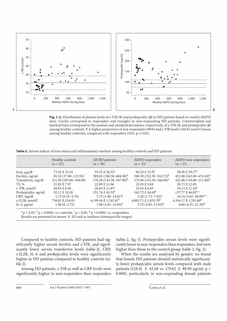
Compared to healthy controls, HD patients had sig-nificantly higher serum ferritin and s-TfR, and signif-icantly lower serum transferrin levels ( table 2 ). CRP, s-IL2R, IL-6 and prohepcidin levels were significantly higher in HD patients compared to healthy controls ( ta-ble 2 ).
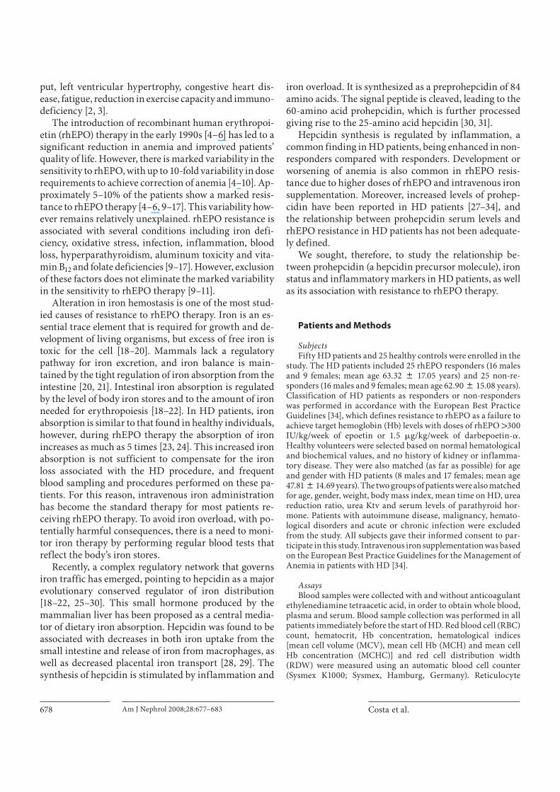
Among HD patients, s-TfR as well as CRP levels were significantly higher in non-responders than responders
( table 2 ; fig. 1 ). Prohepcidin serum levels were signifi-cantly lower in non-responders than responders, but were higher than those in the control group ( table 2 ; fig. 2 ).
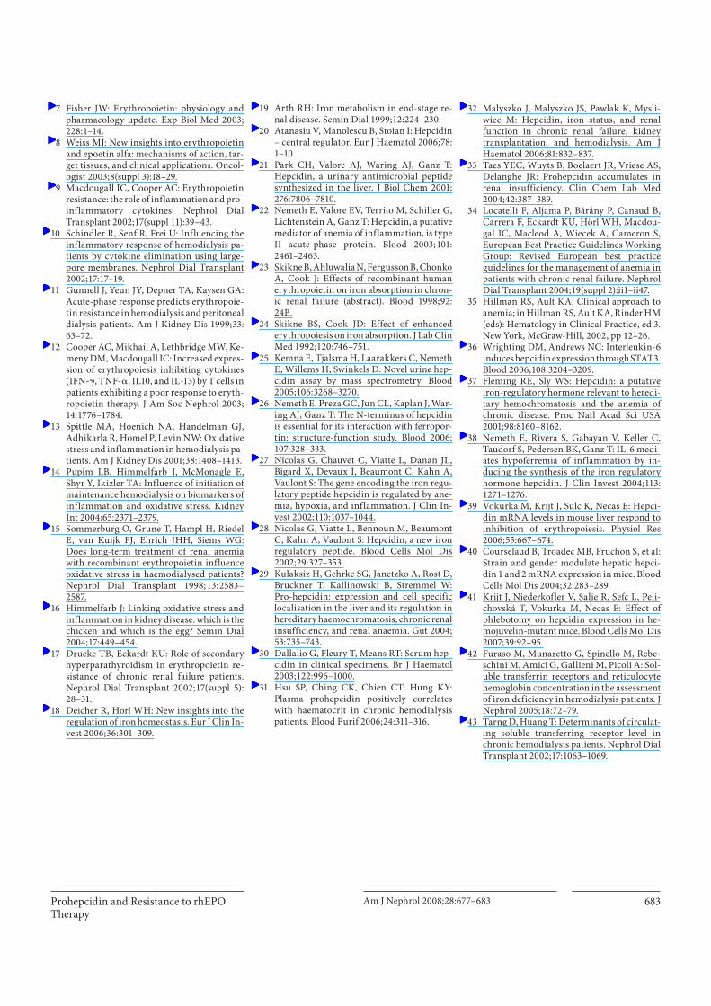
When the results are analyzed by gender, we found that female HD patients showed statistically significant-ly lower prohepcidin serum levels compared with male patients (128.41 8 42.68 vs. 179.62 8 88.89 ng/ml, p = 0.009), particularly in non-responding female patients
Table 2. Serum indices of iron status and inflammatory markers among healthy controls and HD patients
Healthy controls(n = 25)
All HD patients(n = 50)
rhEPO responders(n = 25)
rhEPO non-responders(n = 25)
Iron, �g/dl 73.42825.24 55.32826.52a 60.24822.97 50.40829.27a
Ferritin, ng/ml 85.10 (37.88–123.95) 389.65 (186.58–604.90)b 380.30 (252.30–543.75)b 452.00 (163.00–674.50)b
Transferrin, mg/dl 231.50 (205.00–268.00) 170.50 (143.50–187.00)b 173.00 (152.50–186.00)b 161.00 (139.00–211.00)b
TS, % 21.8387.97 22.89811.06 25.0589.69 20.73812.09s-TfR, nmol/l 20.8588.56 26.84811.87a 19.5686.83 34.13811.42d
Prohepcidin, ng/ml 92.11818.28 151.74843.52b 165.72836.69b 137.77846.03b, c
CRP, mg/dl 1.75 (0.76–4.70) 5.75 (1.90–14.01)b 3.20 (1.73–7.23)a 10.14 (3.82–38.99)b, c
s-IL2R, nmol/l 758.838234.95 4,199.9481,762.81b 4,005.7181,835.70b 4,394.1781,701.80b
IL-6, pg/ml 1.90 (0–3.75) 7.80 (3.85–15.05)b 5.75 (3.83–13.95)b 8.80 (4.55–21.30)b
a p < 0.05, b p < 0.0001, vs. controls; c p < 0.05, d p < 0.0001, vs. responders.Results are presented as means 8 SD and as medians (interquartile ranges).
10
20
30
40
50
60
70
0 200 400 600 800 1,000 1,200
Weekly rhEPO (IU)/kg dose
s-T
fR(n
mo
l/l)
Fig. 1, 2. Distribution of plasma levels of s-TfR ( 1 ) and prohepcidin ( 2 ) in HD patients based on weekly rhEPO dose. Circles correspond to responders and triangles to non-responding HD patients. Uninterrupted and hatched lines correspond to the median and standard deviations, respectively, of s-TfR ( 1 ) and prohepcidin ( 2 ) among healthy controls. 1 A higher proportion of non-responders (96%) had s-TfR levels 1 20.85 nmol/l (mean among healthy controls), compared with responders (32%, p ! 0.05).
50
100
150
200
250
300
0 200 400 600 800 1,000 1,200
Weekly rhEPO (IU)/kg dose
Pro
he
pci
din
(ng
/ml)
1 2

Prohepcidin and Resistance to rhEPO Therapy
Am J Nephrol 2008;28:677–683 681
( fig. 3 ). No statistically significant differences in iron sta-tus and inflammatory markers were found between male and female HD patients (data not shown).
Prohepcidin correlated negatively with reticulocyte count (r = –0.324, p = 0.023) and s-TfR (r = –0.359, p = 0.010). However, no statistically significant correlation was found between weekly rhEPO/kg dose and prohep-cidin. In HD patients, statistically significant correla-tions were noted between weekly rhEPO/kg dose with CRP (r = 0.400, p = 0.004) and s-TfR (r = 0.588, p ! 0.0001), and between CRP with MCV (r = –0.322, p = 0.023), MCH (r = –0.393, p = 0.005), iron (r = –0.582, p ! 0.0001) and TS (r = –0.456, p = 0.001).
Discussion
Hepcidin, a liver-derived peptide stimulated by in-flammatory cytokines, acts as a negative regulator of in-testinal iron absorption and its release from macrophages; it is believed to represent the molecular link between chronic inflammation, the iron status of the body and anemia. Excessive hepcidin production occurs in patients with inflammatory and infectious diseases, resulting in anemia of inflammation [18–23] . Furthermore, in vitro stimulation of fresh human hepatocytes by proinflam-matory IL-6 showed strong induction of hepcidin mRNA,
indicating that this cytokine may be the mediator of hep-cidin induction in inflammation [36–38] . This increased hepcidin expression during inflammation and infection explains sequestration of iron in the macrophages and inhibition of intestinal iron absorption, the two hall-marks of the anemia of inflammation, which is a normo-cytic or microcytic iron-refractory anemia [27–32] . This decreased availability of iron may be a host defense mech-anism against invading microorganisms.
The biological importance of the precursor molecule of hepcidin, named prohepcidin, in regulating iron me-tabolism has so far remained undetermined. However, there is some evidence that prohepcidin levels are a reli-able indicator of hepcidin levels and activity [30–33] . Due to the lack of available laboratory methods for quantify-ing circulating hepcidin in clinical samples due to the small peptide size, serum prohepcidin levels are used as an indicator of hepcidin levels, which are easily measured with the widely available ELISA kits.
We found that HD patients had higher serum levels of prohepcidin as well as higher levels of markers of chron-ic inflammation, e.g. ferritin, CRP, IL-6 and s-IL2R, and markers of reduced iron mobilization [36–38] , e.g. lower transferrin levels. These results suggest that the high lev-els of prohepcidin found in HD patients could be related to an underlying chronic inflammation. However, no correlations between prohepcidin and these inflamma-tory markers were found, suggesting that other factors than just inflammation may regulate serum prohepcidin levels.
Among HD patients, non-responders had higher lev-els of CRP than responders, but prohepcidin serum levels were lower. A recent report [39] showed that erythropoi-etin downregulates liver hepcidin expression, acting, therefore, as a hepcidin-inhibitory hormone [22, 28] . Since non-responders were treated with much higher doses of rhEPO compared with responders, the lower prohepcidin levels among non-responders could be ex-plained by this mechanism. On a broader note, despite the treatment with rhEPO, HD patients showed higher levels of prohepcidin. This could be explained by the fact that the stimulus of inflammation for prohepcidin syn-thesis in HD patients is stronger than the inhibitory effect of rhEPO. Moreover, the use of large amounts of rhEPO may result in increased iron utilization by the bone mar-row, resulting in depletion of iron stores and ultimately decreased intracellular iron availability, which could also decrease prohepcidin levels. Therefore, an additional ex-planation for the lower prohepcidin levels in rhEPO non-responding patients could be lower intracellular iron
50
100
150
200
250
300
Controls Responders Non-responders
Pro
he
pci
din
(ng
/ml)
MaleFemale
p < 0.05
Fig. 3. Serum prohepcidin levels among healthy controls and HD patients according to gender and response to rhEPO therapy.

Costa et al.
Am J Nephrol 2008;28:677–683682
availability (as reflected by the increased s-TfR observed in the non-responders).
Female HD patients showed a lower serum level of prohepcidin compared with male patients. This sex-spe-cific level of prohepcidin in HD patients has not previ-ously been described in the literature. However, in wild-type mice a slight reduction in Hamp1 mRNA levels was documented [40] and also in hemojuvelin-mutant female mice [41] , in which a more accentuated reduction was found. The reason for these sex differences is presently unknown, but may be attributed to hormonal and/or iron status differences between the two patient groups. No differences in iron status were found between male and female HD patients (data not shown), suggesting that the hormonal gender differences could be the reason for low-er prohepcidin serum levels found in female patients.
The negative correlation between prohepcidin serum levels and reticulocyte count could be an indicator that higher prohepcidin levels have an inhibitory effect on erythropoiesis. Among HD patients, the non-responders to rhEPO therapy presented with more severe anemia, as shown by the significantly lower Hb levels compared to responders ( table 1 ). However, no statistically significant differences were found in serum iron status markers be-tween the patient groups, except for s-TfR, which was higher among non-responders. Moreover, in responders s-TfR levels were similar to those in healthy controls. The levels of this soluble receptor could be increased in two clinical settings: increased erythropoietic activity or iron deficiency [42, 43] . In our patients, the positive correla-tion between s-TfR and weekly rhEPO/kg doses suggest-ed that s-TfR was an indicator of the erythropoietic effect of the rhEPO administered and not an indicator of iron body deficiency. However, considering that responders and non-responders presented similar iron stores, the significantly higher levels of s-TfR observed in non-re-sponders also indicate a disturbance in mobilizing the
iron transferrin linked into precursor erythroid cells, ex-plaining also the lower levels of MCH and MCHC ob-served in non-responders. Altogether, these data support a ‘functional iron deficiency’ in non-responding patients. The higher proportion of non-responding HD patients presenting with s-TfR levels higher than the mean of the healthy control group ( fig. 1 ) suggest that this parameter could be used as an indicator of resistance to rhEPO ther-apy in HD patients.
As previously described [9–14] , CRP is higher in HD patients who are non-responders to rhEPO therapy, indi-cating that inflammation is related to resistance to this therapy. Additionally, CRP serum levels were positively correlated with the weekly rhEPO/kg dose, suggesting that CRP is a good predictor of resistance to rhEPO ther-apy in HD patients. The inverse correlations found be-tween CRP and MCV, MCH, serum iron and TS are in agreement with the observations that chronic inflamma-tion leads to iron trapping in macrophages and reduced serum iron levels. This interaction between iron metabo-lism and inflammatory markers leads to a difficulty in distinguishing iron deficiency, which usually responds to iron therapy, from inflammatory iron block.
In conclusion, our data show that a close interaction exists between inflammation, iron status and prohep-cidin serum levels that ultimately regulate intracellu-lar iron availability. Prohepcidin and s-TfR, together with CRP, may prove to be good markers of resistance to rhEPO therapy in HD patients.
Acknowledgments
We are very grateful to the nurses of FMC, Dinefro – Diálises e Nefrologia, SA, and Uninefro – Sociedade Prestadora de Cuida-dos Médicos e de Diálise, SA, for technical support. This study was supported by a PhD grant (SFRH/BD/27688/2006) attributed to E. Costa by FCT and FSE.
References
1 Foley RN, Parfrey PS, Sarnak MJ: Clinical epidemiology of cardiovascular disease in chronic renal failure. Am J Kidney Dis 1998; 32(suppl 3):S112–S119.
2 Foley RN, Parfrey PS, Harnett JD, Kent GM, Murray DC, Barre PE: The impact of anemia on cardiomyopathy morbidity and mortality in end-stage renal disease. Am J Kidney Dis 1996; 28: 53–61.
3 Locatelli F, Conte F, Marcelli D: The impact of hematocrit levels and erythropoietin treatment on overall and cardiovascular mortality and morbidity – the experience of the Lombardy dialysis registry. Nephrol Dial Transplant 1998; 13: 1642–1644.
4 Smrzova J, Balla J, Bárány P: Inflammation and resistance to erythropoiesis-stimulating agents – what do we know and what needs to be clarified? Nephrol Dial Transpant 2005; 20(suppl 8):viii2–viii7.
5 Bárány P: Inflammation, serum C-reactive protein, and erythropoietin resistance. Ne-phrol Dial Transplant 2001; 16: 224–227.
6 Locatelli F, Pisoni RL, Combe C, Bommer J, Andreucci VE, Piera L, Greenwood R, Feld-man HI, Port FK, Held PJ: Anemia in haemo-dialysis patients of five European countries: association with morbidity and mortality in the Dialysis Outcomes and Practice Patterns Study (DOPPS). Nephrol Dial Transplant 2004; 19: 121–132.
Prohepcidin and Resistance to rhEPO Therapy
Am J Nephrol 2008;28:677–683 683
7 Fisher JW: Erythropoietin: physiology and pharmacology update. Exp Biol Med 2003; 228: 1–14.
8 Weiss MJ: New insights into erythropoietin and epoetin alfa: mechanisms of action, tar-get tissues, and clinical applications. Oncol-ogist 2003; 8(suppl 3):18–29.
9 Macdougall IC, Cooper AC: Erythropoietin resistance: the role of inflammation and pro-inflammatory cytokines. Nephrol Dial Transplant 2002; 17(suppl 11):39–43.
10 Schindler R, Senf R, Frei U: Influencing the inflammatory response of hemodialysis pa-tients by cytokine elimination using large-pore membranes. Nephrol Dial Transplant 2002; 17: 17–19.
11 Gunnell J, Yeun JY, Depner TA, Kaysen GA: Acute-phase response predicts erythropoie-tin resistance in hemodialysis and peritoneal dialysis patients. Am J Kidney Dis 1999; 33: 63–72.
12 Cooper AC, Mikhail A, Lethbridge MW, Ke-meny DM, Macdougall IC: Increased expres-sion of erythropoiesis inhibiting cytokines (IFN- � , TNF- � , IL10, and IL-13) by T cells in patients exhibiting a poor response to eryth-ropoietin therapy. J Am Soc Nephrol 2003; 14: 1776–1784.
13 Spittle MA, Hoenich NA, Handelman GJ, Adhikarla R, Homel P, Levin NW: Oxidative stress and inflammation in hemodialysis pa-tients. Am J Kidney Dis 2001; 38: 1408–1413.
14 Pupim LB, Himmelfarb J, McMonagle E, Shyr Y, Ikizler TA: Influence of initiation of maintenance hemodialysis on biomarkers of inflammation and oxidative stress. Kidney Int 2004; 65: 2371–2379.
15 Sommerburg O, Grune T, Hampl H, Riedel E, van Kuijk FJ, Ehrich JHH, Siems WG: Does long-term treatment of renal anemia with recombinant erythropoietin influence oxidative stress in haemodialysed patients? Nephrol Dial Transplant 1998; 13: 2583–2587.
16 Himmelfarb J: Linking oxidative stress and inflammation in kidney disease: which is the chicken and which is the egg? Semin Dial 2004; 17: 449–454.
17 Drueke TB, Eckardt KU: Role of secondary hyperparathyroidism in erythropoietin re-sistance of chronic renal failure patients. Nephrol Dial Transplant 2002; 17(suppl 5): 28–31.
18 Deicher R, Horl WH: New insights into the regulation of iron homeostasis. Eur J Clin In-vest 2006; 36: 301–309.
19 Arth RH: Iron metabolism in end-stage re-nal disease. Semin Dial 1999; 12: 224–230.
20 Atanasiu V, Manolescu B, Stoian I: Hepcidin – central regulator. Eur J Haematol 2006; 78: 1–10.
21 Park CH, Valore AJ, Waring AJ, Ganz T: Hepcidin, a urinary antimicrobial peptide synthesized in the liver. J Biol Chem 2001; 276: 7806–7810.
22 Nemeth E, Valore EV, Territo M, Schiller G, Lichtenstein A, Ganz T: Hepcidin, a putative mediator of anemia of inflammation, is type II acute-phase protein. Blood 2003; 101: 2461–2463.
23 Skikne B, Ahluwalia N, Fergusson B, Chonko A, Cook J: Effects of recombinant human erythropoietin on iron absorption in chron-ic renal failure (abstract). Blood 1998; 92: 24B.
24 Skikne BS, Cook JD: Effect of enhanced erythropoiesis on iron absorption. J Lab Clin Med 1992; 120: 746–751.
25 Kemna E, Tjalsma H, Laarakkers C, Nemeth E, Willems H, Swinkels D: Novel urine hep-cidin assay by mass spectrometry. Blood 2005; 106: 3268–3270.
26 Nemeth E, Preza GC, Jun CL, Kaplan J, War-ing AJ, Ganz T: The N-terminus of hepcidin is essential for its interaction with ferropor-tin: structure-function study. Blood 2006; 107: 328–333.
27 Nicolas G, Chauvet C, Viatte L, Danan JL, Bigard X, Devaux I, Beaumont C, Kahn A, Vaulont S: The gene encoding the iron regu-latory peptide hepcidin is regulated by ane-mia, hypoxia, and inflammation. J Clin In-vest 2002; 110: 1037–1044.
28 Nicolas G, Viatte L, Bennoun M, Beaumont C, Kahn A, Vaulont S: Hepcidin, a new iron regulatory peptide. Blood Cells Mol Dis 2002; 29: 327–353.
29 Kulaksiz H, Gehrke SG, Janetzko A, Rost D, Bruckner T, Kallinowski B, Stremmel W: Pro-hepcidin: expression and cell specific localisation in the liver and its regulation in hereditary haemochromatosis, chronic renal insufficiency, and renal anaemia. Gut 2004; 53: 735–743.
30 Dallalio G, Fleury T, Means RT: Serum hep-cidin in clinical specimens. Br J Haematol 2003; 122: 996–1000.
31 Hsu SP, Ching CK, Chien CT, Hung KY: Plasma prohepcidin positively correlates with haematocrit in chronic hemodialysis patients. Blood Purif 2006; 24: 311–316.
32 Malyszko J, Malyszko JS, Pawlak K, Mysli-wiec M: Hepcidin, iron status, and renal function in chronic renal failure, kidney transplantation, and hemodialysis. Am J Haematol 2006; 81: 832–837.
33 Taes YEC, Wuyts B, Boelaert JR, Vriese AS, Delanghe JR: Prohepcidin accumulates in renal insufficiency. Clin Chem Lab Med 2004; 42: 387–389.
34 Locatelli F, Aljama P, Bárány P, Canaud B, Carrera F, Eckardt KU, Hörl WH, Macdou-gal IC, Macleod A, Wiecek A, Cameron S, European Best Practice Guidelines Working Group: Revised European best practice guidelines for the management of anemia in patients with chronic renal failure. Nephrol Dial Transplant 2004; 19(suppl 2):ii1–ii47.
35 Hillman RS, Ault KA: Clinical approach to anemia; in Hillman RS, Ault KA, Rinder HM (eds): Hematology in Clinical Practice, ed 3. New York, McGraw-Hill, 2002, pp 12–26.
36 Wrighting DM, Andrews NC: Interleukin-6 induces hepcidin expression through STAT3. Blood 2006; 108: 3204–3209.
37 Fleming RE, Sly WS: Hepcidin: a putative iron-regulatory hormone relevant to heredi-tary hemochromatosis and the anemia of chronic disease. Proc Natl Acad Sci USA 2001; 98: 8160–8162.
38 Nemeth E, Rivera S, Gabayan V, Keller C, Taudorf S, Pedersen BK, Ganz T: IL-6 medi-ates hypoferremia of inflammation by in-ducing the synthesis of the iron regulatory hormone hepcidin. J Clin Invest 2004; 113: 1271–1276.
39 Vokurka M, Krijt J, Sulc K, Necas E: Hepci-din mRNA levels in mouse liver respond to inhibition of erythropoiesis. Physiol Res 2006; 55: 667–674.
40 Courselaud B, Troadec MB, Fruchon S, et al: Strain and gender modulate hepatic hepci-din 1 and 2 mRNA expression in mice. Blood Cells Mol Dis 2004; 32: 283–289.
41 Krijt J, Niederkofler V, Salie R, Sefc L, Peli-chovská T, Vokurka M, Necas E: Effect of phlebotomy on hepcidin expression in he-mojuvelin-mutant mice. Blood Cells Mol Dis 2007; 39: 92–95.
42 Furaso M, Munaretto G, Spinello M, Rebe-schini M, Amici G, Gallieni M, Picoli A: Sol-uble transferrin receptors and reticulocyte hemoglobin concentration in the assessment of iron deficiency in hemodialysis patients. J Nephrol 2005; 18: 72–79.
43 Tarng D, Huang T: Determinants of circulat-ing soluble transferring receptor level in chronic hemodialysis patients. Nephrol Dial Transplant 2002; 17: 1063–1069.
Copyright © 2022 FDOKUMEN