Structural characterization of SmMn 2 GeO 7 single microcrystals by electron microscopy
Upload
independentCategory
view
0download
0
Role of Electron Microscopy in the Diagnosis of CadasilSyndrome: A Study of 32 PatientsManrico Morroni1,2*, Daniela Marzioni1*, Michele Ragno3, Paolo Di Bella4, Elisabetta Cartechini5,
Luigi Pianese6, Teresa Lorenzi1, Mario Castellucci1, Marina Scarpelli7
1 Department of Experimental and Clinical Medicine, Section of Anatomy, School of Medicine, Universita Politecnica delle Marche, Ancona, Italy, 2 Electron Microscopy
Unit, United Hospitals, Ancona, Italy, 3 Division of Neurology, Mazzoni Hospital, Azienda Sanitaria Unica Regionale, Zona Territoriale 13, Ascoli Piceno, Italy, 4 Department
of Neurosciences, School of Medicine, Universita Politecnica delle Marche, United Hospitals, Ancona, Italy, 5 Division of Neurology, Macerata Hospital, Azienda Sanitaria
Unica Regionale, Zona Territoriale 9, Macerata, Italy, 6 Molecular Medicine Laboratory, Mazzoni Hospital, Azienda Sanitaria Unica Regionale, Zona Territoriale 13, Ascoli
Piceno, Italy, 7 Department of Biomedical Sciences and Public Health, Section of Pathological Anatomy, School of Medicine, Universita Politecnica delle Marche, United
Hospitals, Ancona, Italy
Abstract
Background and Purpose: Cerebral autosomal dominant arteriopathy with subcortical infarcts and leukoencephalopathy(CADASIL) is caused by NOTCH3 gene mutations that result in vascular smooth muscle cell (VSMC) degeneration. Itsdistinctive feature by electron microscopy (EM) is granular osmiophilic material (GOM) detected in VSMC indentations and/or the extracellular space close to VSMCs. Reports of the sensitivity of EM in detecting GOM in biopsies from CADASILpatients are contradictory. We present data from 32 patients clinically suspected to have CADASIL and discuss the role of EMin its diagnosis in this retrospective study.
Methods: Skin, skeletal muscle, kidney and pericardial biopsies were examined by EM; the NOTCH3 gene was screened formutations. Skin and muscle biopsies from 12 patients without neurological symptoms served as controls.
Results and Discussion: All GOM-positive patients exhibited NOTCH3 mutations and vice versa. This study i) confirms thatEM is highly specific and sensitive for CADASIL diagnosis; ii) extends our knowledge of GOM distribution in tissues where ithas never been described, e.g. pericardium; iii) documents a novel NOTCH3 mutation in exon 3; and iv) shows that EManalysis is critical to highlight the need for comprehensive NOTCH3 analysis. Our findings also confirm the geneticheterogeneity of CADASIL in a small Italian subpopulation and emphasize the difficulties in designing algorithms formolecular diagnosis.
Citation: Morroni M, Marzioni D, Ragno M, Di Bella P, Cartechini E, et al. (2013) Role of Electron Microscopy in the Diagnosis of Cadasil Syndrome: A Study of 32Patients. PLoS ONE 8(6): e65482. doi:10.1371/journal.pone.0065482
Editor: Jean-Claude Baron, University of Cambridge, United Kingdom
Received January 19, 2013; Accepted April 26, 2013; Published June 17, 2013
Copyright: � 2013 Morroni et al. This is an open-access article distributed under the terms of the Creative Commons Attribution License, which permitsunrestricted use, distribution, and reproduction in any medium, provided the original author and source are credited.
Funding: The study was supported by a grant from Universita Politecnica delle Marche (2011 FAR, formerly 60%) to M.M. The funder had no role in study design,data collection and analysis, decision to publish, or preparation of the manuscript.
Competing Interests: The authors have declared that no competing interests exist.
* E-mail: [email protected] (MM); [email protected] (DM)
Introduction
Cerebral autosomal dominant arteriopathy with subcortical
infarcts and leukoencephalopathy (CADASIL) is a genetic disorder
caused by mutations in the NOTCH3 gene, which maps to
chromosome 19 and encodes the transmembrane receptor
NOTCH3 [1,2]. The postnatal expression of the gene is mostly
restricted to vascular smooth muscle cells (VSMCs) of middle and
small arteries and pericytes [1,3]. The mutations are associated
with accumulation of the NOTCH3 extracellular domain in the
wall of small cerebral arteries, resulting in VSMC degeneration
[4,5,6].
The disease is characterized by migraine, transient ischemic
attacks (TIAs) and/or strokes, cognitive decline and psychiatric
symptoms [7]. Typical magnetic resonance imaging (MRI)
features are severe leukoencephalopathy frequently involving the
temporal pole and the external capsule, lacunar lesions, and
microbleeds [8,9,10]. Microscopic features are arterial wall fibrosis
and thickening and alterations of smooth muscle cells, which
eventually disappear. Electron microscopy (EM) shows deposits of
granular osmiophilic material (GOM) in VSMC indentations or in
the extracellular space in close vicinity to VSMCs [11,12,13].
Even though GOM was originally described in the CNS, it is now
accepted that CADASIL is a systemic disease [13,14,15,16,17];
accordingly, GOM has also been identified in skin and muscle
biopsies [4,18,19]. Over the past 12 years we had the opportunity
to examine skin and muscle biopsies from several patients
suspected to have CADASIL. In one patient GOM was found
in the pericardium, a tissue where it had never been described
before. This study presents the findings obtained in a series of 32
patients and discusses the role of EM in CADASIL diagnosis. Our
findings highlight the diagnostic value of EM in patients with
clinical and MRI findings strongly suggestive of CADASIL but not
showing the more common NOTCH3 mutations in earlier
analyses and provide additional information on the syndrome.
PLOS ONE | www.plosone.org 1 June 2013 | Volume 8 | Issue 6 | e65482

Materials and Methods
Ethics statementThis study was approved by the Universita Politecnica delle
Marche Research Ethics Committee. Written informed consent
was obtained from all participants and is recorded on file. The
procedures followed were in accordance with institutional
guidelines.
PatientsThe study includes 32 individuals (14 men and 18 women) with
a suspected clinical diagnosis of CADASIL; their age ranged from
29 to 74 years (mean 51 years).
The suspicion was based on a family history compatible with an
autosomal dominant inheritance, MRI findings suggestive for
multiple cerebral infarcts and leukoencephalopathy (involving in
particular the temporal pole and/or the external capsule), and
clinical findings such as recurrent TIA or stroke, cognitive defects,
epilepsy, migraine and psychiatric symptoms.
Samples were collected over a period of 12 years (2001–2012).
Given the existence in the area of a strong Electron Microscopy
Unit and the difficulty to obtain a genetic analysis, especially in the
earlier years of the study, ultrastructural examination of skin and
muscle biopsies was the first approach to the diagnosis. In 27
patients molecular analysis of the NOTCH3 gene was done soon
after the EM report; in 4 patients (#1, #2 and #3 belonging to
the same family, and #6; see Table 1) it was performed many
years after the EM study; and in the last patient (#10; Table 1) it
actually preceded it.
A punch skin (n = 23) or a muscle biopsy (n = 4) was provided in
27 cases; a pericardial tissue fragment obtained during open heart
surgery for valve replacement was available for one patient (#10;
Table 1); both skin and muscle biopsies were available for 3 more
patients, and a skin and a kidney biopsy were provided for the last
(#4; Table 1). Muscle (#1 and #3) or skin (#2) was examined in
the 3 siblings (Table 1). Skin (n = 9) and muscle biopsies (n = 3)
from 12 patients (6 men and 6 women, age range: 35–75 years,
mean 55.3) without neurological symptoms were used as negative
controls for EM. They suffered from mycosis fungoides (n = 4),
autoimmune disease (n = 4), Stevens-Johnson syndrome, pseudox-
anthoma elasticum, leishmaniasis, and Milker’s nodule (one case
each).
Electron microscopyAll samples were fixed in 2% glutaraldeyde/2% paraformalde-
hyde in 0.1 M phosphate buffer, postfixed in buffered osmium
tetroxide, dehydrated in graded alcohols, and embedded in an
Epon-Araldite mixture. Semithin sections were cut and stained
with toluidine blue to select arteries of appropriate size for thin
sectioning. Thin sections were stained with lead citrate and
examined with a CM10 transmission electron microscope (Philips,
Eindhoven, The Netherlands). The sample was considered
adequate when it contained at least five arteries with multiple
VSMC layers and the inner elastic lamina [20]. More than one
thin section per patient often had to be examined to achieve a
sufficient number of vessels.
NOTCH3 gene analysisMolecular analysis of the NOTCH3 gene was performed
according to a previously described protocol [21]. Briefly, genomic
DNA was isolated from peripheral blood leukocytes with the
MagNA Pure LC DNA isolation kit by an automated nucleic acid
extractor (MagNA Pure extractor, Roche, Basel, Switzerland).
DNA was amplified by PCR to screen the exons containing EGF-
like repeats (exons 2–23) and exons 24 and 25. In the earlier years
of the study the diagnostic protocol envisaged the search for
mutations only in exons 3 and 4, where most CADASIL mutations
have been reported; in 2005 screening was extended to all exons
containing EGF-like repeats and to exons 24 and 25. The PCR
products were directly sequenced for both sense and antisense
strands using an automated fluorescent sequencing method (Big
DyeH Terminator v3.1 Cycle Sequencing Kit) (Applied Biosys-
tems, Foster City, CA, USA) on an ABI Prism 3130 Genetic
Analyzer (Applied Biosystems) after treatment with exonuclease I
and shrimp alkaline phosphatase (ExoSap-IT, USB Corporation,
Staufen, Germany). The sequences of the PCR products were
aligned with the published human NOTCH3 DNA sequence to
identify any sequence changes using the SeqScape software
(Applied Biosystems).
Results
GOM was found in 13 patients, 8 men and 5 women (including
the 3 siblings) aged 45 to 64 years (mean 52.5), who were strongly
suspected to have CADASIL. GOM was identified in 9 skin, 3
muscle, 1 kidney and 1 pericardial tissue biopsy (Table 1). There
were no differences in GOM location or distribution between the
analysed tissues. GOM was found at extracellular sites, most often
near VSMC indentations or close to VSMCs of small and
medium-sized arteries (Fig. 1). In patient #12 the VSMC
indentations were very irregular and formed cytoplasmic pseu-
doinclusions containing GOM (Fig. 2). Occasionally, GOM was
also detected in capillaries, where it was predominantly located
very close to pericytes (Fig. 3), and in vein walls (not shown). The
indented cytoplasmic membrane of smooth muscle cells and
pericytes showed numerous pinocytotic vesicles in proximity of the
GOM deposits (Fig. 4). Besides GOM, vessel walls frequently
exhibited abnormalities such as smooth muscle cell degeneration
and cell loss, thickening of the basal membrane, and abnormal
endothelium due to the presence of vacuoles, which have all been
Table 1. Genetic findings detected in GOM-positive patientssuspected to have CADASIL.
Pt # Gender Age/years Sample GOMMutationin
Amino acidchange
*1 M 51 skeletal muscle + exon 3 C108Sa
*2 M 57 skin + exon 3 C108Sa
*3 F 50 skeletal muscle + exon 3 C108Sa
4 M 45 skin and kidney + exon 19 R1006C
5 F 64 skin + exon 2 R54C
6 M 47 skin + exon 10 G528C
7 M 53 skin + exon 10 G528C
8 M 48 skeletal muscle + exon 10 G528C
9 F 46 skin + exon 3 R110C
10 F 56 pericardium + exon 10 G528C
11 M 59 skin + exon 4 R141C
12 F 54 skin + exon 10 G528C
13 M 54 skin + exon 6 R332C
Pt: patient.*Patients from the same family (siblings).+ presence of GOM.aUnpublished mutation.doi:10.1371/journal.pone.0065482.t001
Electron Microscopy in the Diagnosis of Cadasil
PLOS ONE | www.plosone.org 2 June 2013 | Volume 8 | Issue 6 | e65482

described in CADASIL patients. Very few arteries containing
GOM exhibited mild wall changes. No distinctive ultrastructural
abnormalities were shared by the three siblings (#1, #2 and #3)
or set them apart from the other patients, demonstrating that
qualitative or semi-quantitative changes in GOM do not correlate
with age. In patient #4 (skin+kidney biopsy) the skin sample
showed abundant GOM, and molecular analysis disclosed the
R1006C CADASIL mutation in exon 19 of the NOTCH3 gene.
In the kidney biopsy, obtained at a later time due to renal
symptoms, light microscopy showed slight, non-specific glomerular
and tubulointerstitial injury and homogeneous thickening of the
wall of small arteries. Immunofluorescence microscopy for
immune complexes was negative. Moreover EM disclosed GOM
in interlobular and juxtaglomerular arteries. This case has already
been reported [22].
A NOTCH3 gene mutation was found in all 13 GOM-positive
patients (Table 1). However, no genotype-phenotype correlations
were found in this group. Genetic analysis of the samples from the
three siblings became possible 12 years after the EM study. All
three showed a novel mutation in exon 3 of the NOTCH3 gene, a
nucleotide substitution c.322T.A involving a pathogenic substi-
tution of a cysteine with a serine at codon 108 (p.C108S). In
patient #6 genetic analysis, carried out soon after the skin biopsy,
was directed only at exon 3 and 4 mutations and was negative.
The EM data prompted further NOTCH3 gene screening (exons
2–24), which documented a mutation in exon 10.
Granular debris, not GOM, was found in the other 19 patients,
6 men and 13 women aged 29 to 74 years (mean 50.2 years).
Frequently detected in the inner elastic lamina or near VSMCs in
arterial walls, it was organized as small clumps and may
conceivably derive from degenerated cells. Granular debris found
in a biopsy submitted to confirm the clinical suspicion of
CADASIL is a potential source of error. Comprehensive testing
for NOTCH3 gene mutations was negative in all these 19 patients;
their clinical follow-up was consistent with the EM and genetic
findings, supporting the exclusion of CADASIL.
The 12 patients without neurological symptoms, included as
controls, were negative for GOM as well as for NOTCH3
mutations.
Discussion
In all 32 patients biopsies were collected as part of the initial
diagnostic work-up due to the difficulty in obtaining genetic
analysis, especially in the earlier years of the study. At our
integrated University-Hospital institution EM examination has
been performed for more than 35 years to support a variety of
diagnoses including skin, kidney and muscle diseases, an approach
that has helped to create a remarkable expertise in these fields of
pathology.
GOM was found in all tissue samples from our patients, i.e. skin,
muscle, kidney and pericardium, confirming that CADASIL is a
systemic vascular syndrome and that it may exceptionally be
associated with renal disease [13,22,23,24,25].
Identification of GOM in three siblings led to extensive genetic
testing and identification of a previously unreported mutation,
which adds to the complexity of the NOTCH3 gene variation
database [26].
Figure 1. Transmission electron microscopic image of a muscle biopsy from a GOM-positive patient (#1). Smooth muscle cells in thetunica media of a small artery. The cytoplasm, which in one cell includes the nucleus, contains the characteristic organelles of smooth muscle cells:filaments (f) and dense bodies (db). Cells are surrounded by the basal membrane (bm). Several GOM deposits can be seen outside the cells, often incell membrane invaginations. The small clumps of electron dense material (arrows) between cells and included in collagen (C) are not GOM butdebris, probably deriving from cell degeneration. Scale bar = 0.6 mm.doi:10.1371/journal.pone.0065482.g001
Electron Microscopy in the Diagnosis of Cadasil
PLOS ONE | www.plosone.org 3 June 2013 | Volume 8 | Issue 6 | e65482

Figure 2. Transmission electron microscopic image of a skin biopsy from a GOM-positive patient (#12). Two GOM deposits locatedoutside the smooth muscle cell membrane (arrowheads). Due to the plane of section and to the irregular indentations, the cytoplasm of a VSMCcontains two GOM pseudoinclusions (arrows). Scale bar = 0.5 mm.doi:10.1371/journal.pone.0065482.g002
Figure 3. Transmission electron microscopic image of a skin biopsy from a GOM-positive case (#2). This dermal capillary shows athickened basal membrane (bm) and is surrounded by pericytes (P). Numerous GOM deposits can be seen in the basal membrane and very often inclose vicinity to pericytes. C, collagen; EC, endothelial cell; L, capillary lumen. Scale bar = 0.6 mm.doi:10.1371/journal.pone.0065482.g003
Electron Microscopy in the Diagnosis of Cadasil
PLOS ONE | www.plosone.org 4 June 2013 | Volume 8 | Issue 6 | e65482
Our data confirm the high sensitivity of EM for CADASIL
diagnosis described in previous studies [26,27,28,29,30]. In
particular, they agree with the results reported by Tikka et al.
[26], who identified GOM in all their 131 mutation-positive cases.
Even though the present study involved a much smaller sample, it
is worth noting that all patients but one were from a small area in
central Italy (population about 700,000), whereas the 131 patients
of Tikka et al. [26] came from an area encompassing Finland
(n = 38), Sweden (n = 13) and France (n = 80).
Confirmation of the view of Mayer et al. [28] that skin biopsies
are as reliable as muscle biopsies for CADASIL diagnosis was an
additional finding. Moreover, we detected GOM in the pericar-
dium; to the best of our knowledge this is the first report in this
tissue, since GOM has only been described in myocardium [31].
Not all researchers consider EM as a sensitive method for
CADASIL diagnosis [20,32]. In particular, Malandrini et al. [32]
reported a sensitivity as low as 57%, which they attributed to the
difficulty of examining a sufficient number of arteries in skin
biopsies and to the variable involvement of extracerebral vessels.
Based on our experience, the sensitivity of the technique is
influenced by EM operator skills in identifying GOM and in
distinguishing it from similar but unrelated deposits. To avoid
error it is essential to adhere strictly to current guidelines for GOM
identification [11,13]. Furthermore, examination of an adequate
number of medium and small arteries is mandatory. Even though
GOM was detected in the first or the first two arteries from all 13
GOM-positive patients, we agree with Markus et al. [20] that a
minimum of five arteries should be examined in each patient.
Based on this criterion patients with NOTCH3 mutations
overlapped with GOM-positive ones.
Immunohistochemistry has been proposed by Joutel et al. [33]
to be a reliable and readily available tool for CADASIL diagnosis,
and to have a sensitivity of 96% and a specificity of 100%; in
contrast Lesnik Oberstein et al. [34] reported a large number of
false-negative and even false-positive results in a study of 62
patients.
After EM detection of GOM and negative testing for the more
common NOTCH3 mutations comprehensive NOTCH3 screen-
ing should be performed. Testing only exons 3 and 4, as suggested
by Kalimo et al. [35], would have identified the mutations in only
30% of our patients. These data agree with previous reports
showing that mutations in exons 3 and 4 account for only 36% of
Italian CADASIL patients [36] and disclosing the genetic
heterogeneity of CADASIL in a small Italian subpopulation
[37]. Our GOM-positive patient #6 is a case in point, because
complete sequencing of the gene, 11 years after biopsy examina-
tion and negative testing for exon 3 and 4 mutations, identified a
mutation in exon 10.
Conclusions
In conclusion, our study confirms previous observations and
extends our knowledge of GOM distribution in tissues where it has
never been described before, such as pericardium. Moreover, we
report a novel NOTCH3 mutation in exon 3, detected in three
siblings. Our findings also confirm the genetic heterogeneity of
CADASIL in a small Italian subpopulation, emphasizing the
difficulties in designing algorithms for molecular diagnosis. Finally,
our data support the notion that EM analysis is critical to highlight
the need for screening NOTCH3 exons 2–24.
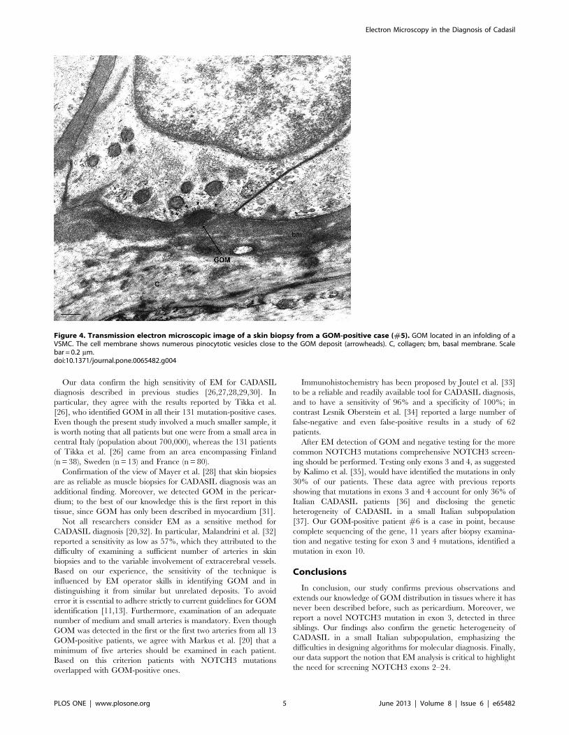
Figure 4. Transmission electron microscopic image of a skin biopsy from a GOM-positive case (#5). GOM located in an infolding of aVSMC. The cell membrane shows numerous pinocytotic vesicles close to the GOM deposit (arrowheads). C, collagen; bm, basal membrane. Scalebar = 0.2 mm.doi:10.1371/journal.pone.0065482.g004
Electron Microscopy in the Diagnosis of Cadasil
PLOS ONE | www.plosone.org 5 June 2013 | Volume 8 | Issue 6 | e65482

Acknowledgments
We are grateful to Maria Cristina Zingaretti, Plinio Ferrara, and Michela
Cardinali for their technical assistance and to Dr S. Modena (www.
silviamodena.com) for the language revision.
Author Contributions
Conceived and designed the experiments: MM. Performed the experi-
ments: MM PDB EC LP. Analyzed the data: MM. Contributed reagents/
materials/analysis tools: MM LP. Wrote the paper: MM MR TL MS.
Made critical revision of the manuscript for important intellectual content:
MM DM MR TL MC MS.
References
1. Joutel A, Corpechot C, Ducros A, Vahedi K, Chabriat H, et al. (1996) Notch3mutations in CADASIL, a hereditary adult-onset condition causing stroke and
dementia. Nature 383: 707–710.
2. Tournier-Lasserve E, Joutel A, Melki J, Weissenbach J, Lathrop GM, et al.(1993) Cerebral autosomal dominant arteriopathy with subcortical infarcts and
leukoencephalopathy maps to chromosome 19q12. Nat Genet 3: 256–259.3. Joutel A, Vahedi K, Corpechot C, Troesch A, Chabriat H, et al. (1997) Strong
clustering and stereotyped nature of Notch3 mutations in CADASIL patients.Lancet 350: 1511–1515.
4. Brulin P, Godfraind C, Leteurtre E, Ruchoux MM (2002) Morphometric
analysis of ultrastructural vascular changes in CADASIL: analysis of 50 skinbiopsy specimens and pathogenic implications. Acta Neuropathol 104: 241–248.
5. Joutel A, Andreux F, Gaulis S, Domenga V, Cecillon M, et al. (2000) Theectodomain of the Notch3 receptor accumulates within the cerebrovasculature
of CADASIL patients. J Clin Invest 105: 597–605.
6. Joutel A (2011) Pathogenesis of CADASIL: transgenic and knock-out mice toprobe function and dysfunction of the mutated gene, Notch3, in the
cerebrovasculature. Bioessays 33: 73–80.7. Chabriat H, Vahedi K, Iba-Zizen MT, Joutel A, Nibbio A, et al. (1995) Clinical
spectrum of CADASIL: a study of 7 families. Lancet 346: 934–939.8. Chabriat H, Levy C, Taillia H, Iba-Zizen MT, Vahedi K, et al. (1998) Patterns
of MRI lesions in CADASIL. Neurology 51: 452–457.
9. Dichgans M, Holtmannspotter M, Herzog J, Peters N, Bergmann M, et al.(2002) Cerebral microbleeds in CADASIL: a gradient-echo magnetic resonance
imaging and autopsy study. Stroke 33: 67–71.10. van Den Boom R, Lesnik Oberstein SA, van Duinen SG, Bornebroek M, Ferrari
MD, et al. (2002) Subcortical lacunar lesions: an MR imaging finding in patients
with cerebral autosomal dominant arteriopathy with subcortical infarcts andleukoencephalopathy. Radiology 224: 791–796.
11. Baudrimont M, Dubas F, Joutel A, Tournier-Lasserve E, Bousser MG (1993)Autosomal dominant leukoencephalopathy and subcortical ischemic stroke. A
clinicopathological study. Stroke 24: 122–125.
12. Miao Q, Paloneva T, Tuominen S, Poyhonen M, Tuisku S, et al. (2004) Fibrosisand stenosis of the long penetrating cerebral arteries: the cause of the white
matter pathology in cerebral autosomal dominant arteriopathy with subcorticalinfarcts and leukoencephalopathy. Brain Pathol 14: 358–364.
13. Ruchoux MM, Guerouaou D, Vandenhaute B, Pruvo JP, Vermersch P, et al.(1995) Systemic vascular smooth muscle cell impairment in cerebral autosomal
dominant arteriopathy with subcortical infarcts and leukoencephalopathy. Acta
Neuropathol 89: 500–512.14. Goebel HH, Meyermann R, Rosin R, Schlote W (1997) Characteristic
morphologic manifestation of CADASIL, cerebral autosomal-dominant arteri-opathy with subcortical infarcts and leukoencephalopathy, in skeletal muscle and
skin. Muscle Nerve 20: 625–627.
15. Haritoglou C, Hoops JP, Stefani FH, Mehraein P, Kampik A, et al. (2004)Histopathological abnormalities in ocular blood vessels of CADASIL patients.
Am J Ophthalmol 138: 302–305.16. Rubio A, Rifkin D, Powers JM, Patel U, Stewart J, et al. (1997) Phenotypic
variability of CADASIL and novel morphologic findings. Acta Neuropathol 94:247–254.
17. Schroder JM, Sellhaus B, Jorg J (1995) Identification of the characteristic
vascular changes in a sural nerve biopsy of a case with cerebral autosomaldominant arteriopathy with subcortical infarcts and leukoencephalopathy
(CADASIL). Acta Neuropathol 89: 116–121.18. Ruchoux MM, Chabriat H, Bousser MG, Baudrimont M, Tournier-Lasserve E
(1994) Presence of ultrastructural arterial lesions in muscle and skin vessels of
patients with CADASIL. Stroke 25: 2291–2292.19. Ruchoux MM, Maurage CA (1998) Endothelial changes in muscle and skin
biopsies in patients with CADASIL. Neuropathol Appl Neurobiol 24: 60–65.
20. Markus HS, Martin RJ, Simpson MA, Dong YB, Ali N, et al. (2002) Diagnosticstrategies in CADASIL. Neurology 59: 1134–1138.
21. Cappelli A, Ragno M, Cacchio G, Scarcella M, Staffolani P, et al. (2009) High
recurrence of the R1006C NOTCH3 mutation in central Italian patients withcerebral autosomal dominant arteriopathy with subcortical infarcts and
leukoencephalopathy (CADASIL). Neurosci Lett 462: 176–178.22. Ragno M, Trojano L, Pianese L, Boni MV, Silvestri S, et al. (2012) Renal
involvement in cerebral autosomal dominant arteriopathy with subcorticalinfarcts and leukoencephalopathy (CADASIL): report of a case with a six-year
follow-up. Histol Histopathol 27: 1307–1314.
23. Bergmann M, Ebke M, Yuan Y, Bruck W, Mugler M, et al. (1996) Cerebralautosomal dominant arteriopathy with subcortical infarcts and leukoencepha-
lopathy (CADASIL): a morphological study of a German family. ActaNeuropathol 92: 341–350.
24. Guerrot D, Francois A, Boffa JJ, Boulos N, Hanoy M, et al. (2008)
Nephroangiosclerosis in cerebral autosomal dominant arteriopathy withsubcortical infarcts and leukoencephalopathy: is NOTCH3 mutation the
common culprit? Am J Kidney Dis 52: 340–345.25. Kusaba T, Hatta T, Kimura T, Sonomura K, Tanda S, et al. (2007) Renal
involvement in cerebral autosomal dominant arteriopathy with subcorticalinfarcts and leukoencephalopathy (CADASIL). Clin Nephrol 67: 182–187.
26. Tikka S, Mykkanen K, Ruchoux MM, Bergholm R, Junna M, et al. (2009)
Congruence between NOTCH3 mutations and GOM in 131 CADASILpatients. Brain 132: 933–939.
27. Ebke M, Dichgans M, Bergmann M, Voelter HU, Rieger P, et al. (1997)CADASIL: skin biopsy allows diagnosis in early stages. Acta Neurol Scand 95:
351–357.
28. Mayer M, Straube A, Bruening R, Uttner I, Pongratz D, et al. (1999) Muscleand skin biopsies are a sensitive diagnostic tool in the diagnosis of CADASIL.
J Neurol 246: 526–532.29. Rein Gustavsen W, Reinholt FP, Schlosser A (2006) Skin biopsy findings and
results of neuropsychological testing in the first confirmed cases of CADASIL in
Norway. Eur J Neurol 13: 359–362.30. Wang Z, Yuan Y, Zhang W, Lv H, Hong D, et al. (2011) NOTCH3 mutations
and clinical features in 33 mainland Chinese families with CADASIL. J NeurolNeurosurg Psychiatry 82: 534–539.
31. Lesnik Oberstein SA, Jukema JW, van Duinen SG, Macfarlane PW, vanHouwelingen HC, et al. (2003) Myocardial infarction in cerebral autosomal
dominant arteriopathy with subcortical infarcts and leukoencephalopathy
(CADASIL). Medicine (Baltimore) 82: 251–256.32. Malandrini A, Gaudiano C, Gambelli S, Berti G, Serni G, et al. (2007)
Diagnostic value of ultrastructural skin biopsy studies in CADASIL. Neurology68: 1430–1432.
33. Joutel A, Favrole P, Labauge P, Chabriat H, Lescoat C, et al. (2001) Skin biopsy
immunostaining with a Notch3 monoclonal antibody for CADASIL diagnosis.Lancet 358: 2049–2051.
34. Lesnik Oberstein SA, van Duinen SG, van den Boom R, Maat-Schieman ML,van Buchem MA, et al. (2003) Evaluation of diagnostic NOTCH3 immuno-
staining in CADASIL. Acta Neuropathol 106: 107–111.35. Kalimo H, Ruchoux MM, Viitanen M, Kalaria RN (2002) CADASIL: a
common form of hereditary arteriopathy causing brain infarcts and dementia.
Brain Pathol 12: 371–384.36. Dotti MT, Federico A, Mazzei R, Bianchi S, Scali O, et al. (2005) The spectrum
of Notch3 mutations in 28 Italian CADASIL families. J Neurol NeurosurgPsychiatry 76: 736–738.
37. Testi S, Malerba G, Ferrarini M, Ragno M, Pradotto L, et al. (2012) Mutational
and haplotype map of NOTCH3 in a cohort of Italian patients with cerebralautosomal dominant arteriopathy with subcortical infarcts and leukoencepha-
lopathy (CADASIL). J Neurol Sci 319: 37–41.
Electron Microscopy in the Diagnosis of Cadasil
PLOS ONE | www.plosone.org 6 June 2013 | Volume 8 | Issue 6 | e65482
Copyright © 2022 FDOKUMEN


![Transmission electron microscopy and scanning transmission electron microscopy study on B-site cation ordered structures in a (1−x)Pb(Mg[sub 1/3]Nb[sub 2/3])O[sub 3]–xPbTiO[sub](https://static.fdokumen.com/doc/165x107/634364aaa94d09afc00d5037/transmission-electron-microscopy-and-scanning-transmission-electron-microscopy-study.jpg)