
UAB's Reflex on Cities and Local Support Centers to Distance Education

Role of Cerebellum in Motion Perception and Vestibulo-ocularReflex—Similarities and Disparities
Aasef G. Shaikh,Department of Neurology, Case Western Reserve University, Cleveland, OH 44106-5040, USA
Antonella Palla,D. Straumann Department of Neurology, Zurich University Hospital, Zurich, Switzerland
Sarah Marti,D. Straumann Department of Neurology, Zurich University Hospital, Zurich, Switzerland
Itsaso Olasagasti,D. Straumann Department of Neurology, Zurich University Hospital, Zurich, Switzerland
Lance M. Optican,Laboratory of Sensorimotor Research, National Eye Institute, NIH, DHHS, Bethesda, MD, USA
David S. Zee, and Dominik StraumannDepartment of Neurology, The Johns Hopkins University School of Medicine, Baltimore, MD, USA
AbstractVestibular velocity storage enhances the efficacy of the angular vestibulo-ocular reflex (VOR)during relatively low-frequency head rotations. This function is modulated by GABA-mediatedinhibitory cerebellar projections. Velocity storage also exists in perceptual pathway and hassimilar functional principles as VOR. However, it is not known whether the neural substrate forperception and VOR overlap. We propose two possibilities. First, there is the same velocitystorage for both VOR and perception; second, there are nonoverlapping neural networks: onemight be involved in perception and the other for the VOR. We investigated these possibilities bymeasuring VOR and perceptual responses in healthy human subjects during whole-body, constant-velocity rotation steps about all three dimensions (yaw, pitch, and roll) before and after 10 mg of4-aminopyridine (4-AP). 4-AP, a selective blocker of inward rectifier potassium conductance, canlead to increased synchronization and precision of Purkinje neuron discharge and possiblyenhance the GABAergic action. Hence 4-AP could reduce the decay time constant of theperceived angular velocity and VOR. We found that 4-AP reduced the decay time constant, but theamount of reduction in the two processes, perception and VOR, was not the same, suggesting thepossibility of nonoverlapping or partially overlapping neural substrates for VOR and perception.We also noted that, unlike the VOR, the perceived angular velocity gradually built up and plateauprior to decay. Hence, the perception pathway may have additional mechanism that changes thedynamics of perceived angular velocity beyond the velocity storage. 4-AP had no effects on theduration of build-up of perceived angular velocity, suggesting that the higher order processing ofperception, beyond the velocity storage, might not occur under the influence of mechanism thatcould be influenced by 4-AP.
© Springer Science+Business Media, LLC 2012
A. G. Shaikh [email protected].
Conflict of Interest Authors do not have any conflict of interest.
NIH Public AccessAuthor ManuscriptCerebellum. Author manuscript; available in PMC 2013 February 01.
Published in final edited form as:Cerebellum. 2013 February ; 12(1): 97–107. doi:10.1007/s12311-012-0401-7.
$waterm
ark-text$w
atermark-text
$waterm
ark-text

KeywordsVestibular; Eye movements; GABA; Brainstem; Velocity storage; Cerebellum
IntroductionStabilization of images on the fovea is the fundamental requirement for clear vision. In orderto stabilize gaze during natural behavior, the brain computes an estimate of head movementand ensures an appropriate reflexive eye movement [the vestibulo-ocular reflex (VOR)].Perception of self-motion and spatial orientation is also critical to assure appropriatereflexive and volitional behavior. It is possible that motion perception and VOR might sharefundamental neural strategies. The neural mechanisms for the VOR are relatively wellknown. Angular motion sensors (the semicircular canals) respond to head acceleration, butthe eyes must rotate to a new orbital position. Mathematical integration of the angularvelocity from the labyrinthine sensors is therefore required for the eye movement to be equaland opposite to the head movement [1–4]. The first integration is performed in the labyrinthby the mechanical properties of the endolymph and cupula. As a result, the afferents fromthe semicircular canals discharge in proportion to head velocity. The premotor neuronswithin the brainstem or deep cerebellar nuclei further integrate the velocity signal into aposition signal that is encoded in the activity of ocular motor neurons [5]. This network iscalled the common oculomotor integrator as it provides necessary position signal for theconjugate eye movements of all types.
Upstream from the common oculomotor integrator is the velocity storage, the integratormore specific to the vestibular system. The velocity storage extends the bandwidth overwhich the angular VOR is compensatory for head movement and improves the VORresponse during relatively low-frequency head rotations. In response to a head-velocity step,the velocity storage prolongs the decay of compensatory eye movements beyond that ofactivity of the vestibular afferents. The velocity storage is not perfect, but its output decaysexponentially [1]. The time constant of this decay, the velocity-storage time constant, isaffected by GABA-mediated inhibitory projections from the cerebellar cortex (nodulus andventral uvula) [1, 5–10]. The GABAB receptor agonist, baclofen, reduces the velocity-storage time constant in human subjects, while focal lesions of the macaque nodulus andventral uvula prolong it [6, 8].
The perception of self-motion also outlasts the semicircular canal output, suggesting avelocity-storage mechanism in motion perception [11, 12]. The dynamics of motionperception and of the VOR during angular velocity steps are same in the sense that bothprocesses feature relatively prolonged exponential decays. The key difference between thetwo processes is that motion perception gradually rises to its peak followed by a plateau,suggesting a transient, but steady percept of angular motion before the perceived angularvelocity begins to decay [13, 14]. In contrast, slow-phase eye velocity of rotational VORabruptly increases and instantaneously begins to decay [13, 14]. It was recently proposedthat differences between the dynamics of motion perception and those of the VOR duringangular velocity steps can be described by the same principle of velocity storage processing,but with minimal contribution from the direct pathway in case of perception [14]. The latterstudy, however, was not equipped to assess whether the neural substrate for perception andVOR is anatomically the same [14]. Some indirect evidence comes from macaqueexperiments [15]. Single neuron responses of macaque dorsolateral thalamic neurons hadsimilar dynamic properties as recorded from the deep cerebellar and vestibular neurons [2,15–17]. Retrograde tracer injections in dorsolateral thalamic neurons revealed theiranatomical origin in rostral contralateral anterior interposed and fastigial nuclei, and
Shaikh et al. Page 2
Cerebellum. Author manuscript; available in PMC 2013 February 01.
$waterm
ark-text$w
atermark-text
$waterm
ark-text

anterograde tracer injections in deep cerebellar and vestibular nuclei revealed theirprojections within the thalamic terminal zones [15]. Therefore, it was speculated thatvestibular signal that was recorded from the thalamus might be received from the vestibularand deep cerebellar nuclei, and transmitted to the cerebral cortex [15]. Other studies inhumans showed that the outcomes of the vestibular processing for VOR and perception areseparate [18–21]. For example, in a patient with transient ophthalmoplegia, a disease modelof visuo-vestibular adaptation, the perceptual measures of vestibular function had a differentamount of attenuation compared to the eye movements [18]. The amount of habituation tothe repeated vestibular stimuli in healthy subjects was different for perceived motion andVOR [19].
Taken together, these studies showed different VOR and perceptual responses to the samerotational stimulus, thus suggested two possibilities. One, there is identical low ordervestibular pathway (putatively in the brainstem or deep cerebellar nuclei) with additionalhigher order processing exclusively in case of perception (hypothesis A, Fig. 1). The secondpossibility is that there are nonoverlapping or partially overlapping networks; one might beprimarily involved in perception and the other for the VOR (hypothesis B, Fig. 1). Wepredict that the same manipulation of these networks would have different amounts ofeffects on the VOR and perception if the perception and VOR velocity-storage have non-overlapping or partially overlapping elements.
We assessed these hypotheses by measuring VOR and perception of angular motion inhumans during whole-body, constant-velocity rotations about an earth-vertical axis, beforeand after 10 mg of 4-aminopyridine (4-AP). 4-AP blocks inward rectifier potassiumconductances (IA and ID) that regulate the intrinsic membrane properties of secondaryvestibular neurons [22–24]. 4-AP improves synchronization and precision of the Purkinjeneurons and possibly increases the synaptic concentration of GABA [25–27]. 4-AP shouldreduce the decay time constant of perceived angular velocity if indeed perceptual velocitystorage is under the influence of GABAergic cerebellar projections. We predict that if thehypothesis A is true, but further processing of perceptual signal beyond velocity storage isnot dependent upon cerebellar Purkinje neurons or other 4-AP dependent mechanisms, theextent of reduction in the decay time constant of VOR and perceived angular velocity mustbe the same. However, if hypothesis B is true, the time constants of the decay in VOR andperception both may decrease, but by different amounts.
We recorded VOR and perception of angular velocity during on-axis whole-body velocitysteps of rotations in all three planes. We know that humans have strong VOR velocitystorage for yaw rotations (i.e., rotations on earth vertical axis when the subject is sittingupright), but velocity storage during pitch (i.e., rotations on earth vertical axis whensubject’s ear faces the ground) or roll (i.e., rotations on earth vertical axis when subject issupine) rotations is weak [28]. We also assessed the perceptual velocity storage during yaw,pitch, and roll rotations.
MethodsSix healthy subjects (two women and four men; age range, 30–45) enrolled in the study. Theexperimental protocol was approved by an ethics committee of the Canton of Zurich andadhered to the Declaration of Helsinki for research involving human subjects. The subjectswere naive to the outcome of the experiments.
Experimental SetupThe subjects were seated upright on a three-axis motor-driven turntable (Acutronic, Jona,Switzerland) with the head restrained by an individually molded thermoplastic mask
Shaikh et al. Page 3
Cerebellum. Author manuscript; available in PMC 2013 February 01.
$waterm
ark-text$w
atermark-text
$waterm
ark-text

(Sinmed BV, Reeuwijk, The Netherlands). Subjects first were rotated on an earth-verticalaxis while seated upright, in which case the angular vestibular response is primarily throughstimulation of the lateral semicircular canals, (yaw rotation, schematic inset in Fig. 2a).Subjects were then rotated on an earth-vertical axis while supine (roll-rotation, schematicinset in Fig. 2c) or on the side with their left-ear down (pitch-rotation, schematic inset in Fig.2e). In these cases, the vertical semicircular canals are primarily stimulated. The gravityvector with respect to the head remained constant during these rotation stimuli. The rotationswere delivered in both directions (e.g., relative to head-fixed coordinates, right or left duringyaw, up or down during pitch, and clockwise or counterclockwise during roll). Thus, eachsubject had at least six sets of velocity-step rotations.
Before each trial, subjects looked at a laser dot (diameter, 0.1°) projected onto a sphere witha radius of 1.4 m by two mirror-galvanometers fixed to the rotating chair. The laser androom lights were then extinguished, and rotations began at an initial acceleration at 90°/s2 toa final speed of 100°/s. One and a half minutes later, the rotations were stopped by a 90°/s2
deceleration. The eye movements and perceived angular velocity were measured during andafter rotations for 90 s.
Eye movement RecordingsThe eye movements were recorded from each eye using dual search coils (SkalarInstruments, Delft, The Netherlands). Search coil annuli were calibrated and then placedsclera of the right or left eye after local anesthesia with oxybuprocaine 0.4 % [29]. The coilframe around the head (side length, 0.5 m) generated three orthogonal, digitallysynchronized magnetic wave field signals of 80, 96, and 120 kHz. A digital signal processorcomputed a fast Fourier transform in real time on the digitized search coil signal todetermine the voltage induced on the coil by each magnetic field (Primelec, Regensdorf,Switzerland). Coil orientation was determined with an error of <7 % over a range of ±30°,and with a noise level <0.05° (root mean squared deviation). Eye position signals weredigitized at 1,000 Hz per channel with 12-bit resolution.
Perception RecordingsSubjects reported their perception of angular motion by turning a lever attached to apotentiometer. Subjects were instructed to match the perceived motion with the rate at whichthey spun the lever. The potentiometer signals were sampled at 1,000 Hz. This method forassessing the perception of angular velocity permits the comparison of relative magnitudesduring the time course of a single response [11]. This technique investigates whetherperceived angular velocity is sensed as constant, increasing, or decreasing. Without training,this method cannot estimate the absolute magnitude of perceived angular velocity. We didnot train the subjects with an intention to keep them naïve and avoid the influence ofvestibular habituation.
The experiments were performed on the same subjects with and without 4-AP. The sequenceof control and 4-AP experiment sessions was randomly determined, and both experimentswere done at least 4 days apart to allow adequate time for washout (if subjects took 4-APduring the first session). The 4-day-period also allows sufficient time to recover from theeffects of any possible vestibular habituation [30].
Data AnalysisCalibrated eye position signals from the search coils and perceived angular position signalsfrom the potentiometer were processed to compute slow-phase velocity and perceivedangular velocity using interactive programs written in MatLab® (The Mathworks, Natick,MA, USA). Eye positions were represented as 3D rotation vectors in a head-fixed coordinate
Shaikh et al. Page 4
Cerebellum. Author manuscript; available in PMC 2013 February 01.
$waterm
ark-text$w
atermark-text
$waterm
ark-text

system. Rotation vectors were smoothed, and angular eye velocity was computed asdescribed previously [31]. The eye velocity and perceived angular motion traces weredivided into intervals of different lengths at the zero-crossings from negative to positivevalues. All data points were sorted in an ascending order in each interval. The value at theleast slope represented the slow-phase velocity or perceived angular velocity for the givenprocessed interval. Spline interpolation between the representative points of all intervalsresulted in an envelope describing the slow-phase velocity or perceived angular velocityover time. The time constant was computed when the eye velocity and the perceivedvelocity began to decay. The slow-phase velocity data was first processed with a Skavinsky–Golay smoothing filter. Smoothed data was then fitted by an exponential function withparameters estimated in the MatLab® least-square fitting algorithm (MatLab®, Mathworks,Optimization Toolbox).
ResultsFigure 2a illustrates the rapid rise in the slow-phase velocity of the horizontal VOR followedby the immediate exponential decay during yaw-rotation in one subject. The time constantof the decay of the slow-phase velocity was 10 s (Fig. 2a, filled circles), larger than expectedfrom the mechanical properties of the semicircular canal cupula (about 4 s, gray trace drawnfor reference) [32]. The open circles in Fig. 2a illustrate slow-phase velocity of thehorizontal VOR during yaw-rotation in the same subject 30 min after taking 10 mg 4-AP bymouth. Slow-phase velocity now decayed more rapidly with a time constant of 6.0 s. Aquantitative estimate of the perception of horizontal angular velocity during yaw rotation inthe same subject is shown by the filled triangle symbols in Fig. 2b. There was anexponential decay in perceived angular velocity with a time constant of 13.7 s (Fig. 2b,filled triangles). The time constant of decay was reduced to 8.6 s after 10 mg 4-AP (Fig. 2b,open triangles).
Figure 2c depicts an example of slow-phase velocity of the vertical VOR in the same subjectduring pitch rotations. Prior to 4-AP, the time constant of the decay of slow-phase velocityduring pitch rotations was 8.0 s. The time constant reduced to 7.5 s after 10 mg 4-AP.Perceived angular velocity during pitch rotations is illustrated in Fig. 2d. The decay timeconstant of perceived velocity during pitch rotations was 6.3 s. Thirty minutes after 10 mg 4-AP, the time constant reduced to 5.0 s.
Figure 2e depicts an example of slow-phase velocity of the torsional VOR in the samesubject during roll rotations. The decay time constant of slow-phase velocity during rollrotations was 5.5 s. In the same subject, the time constant was reduced to 5.1 s after 10 mg4-AP. Figure 2f illustrates the perceived angular velocity during roll rotations. The decaytime constant of perceived velocity during roll rotations was 5.6 s. Thirty minutes after 10mg 4-AP, the time constant reduced 4.0 s.
The slow-phase velocity of the VOR rapidly peaked and began to decay immediately. Incontrast, the perceived angular velocity had a gradual growth to its peak value (introducing adelay to peak), and there was a delay before the perception of angular motion began todecay, appearing as a “plateau” (Fig. 2b, d, and f).
Figure 3a summarizes the effects of 4-AP on the VOR time constant in six subjects. Thedecay time constant of the VOR with 4-AP is plotted along the ordinate, and the controlvalue is plotted on the abscissa. Each symbol represents one rotation trial, each symbol typerepresents one subject, and the symbol color represents the rotation plane (black, yaw; blue,roll; red, pitch). All black-colored data points in Fig. 3a are below the equality line,suggesting a consistent and statistically significant reduction of the decay time constant of
Shaikh et al. Page 5
Cerebellum. Author manuscript; available in PMC 2013 February 01.
$waterm
ark-text$w
atermark-text
$waterm
ark-text

the horizontal VOR during yaw rotations (one-way ANOVA, p<0.01). In Fig. 3a, blue andredcolored data points scattered on the both sides of the dashed equality line, suggesting noconsistent effect of 4-AP on the VOR time constant during roll (blue symbols) and pitchrotations (red symbols) (one-way ANOVA, p>0.05).
Figure 3b summarizes the effects of 4-AP on the decay time constant of perceived angularvelocity in six healthy subjects. Black and red data points, illustrating perceived angularvelocity during yaw and pitch rotations, respectively, are below the equality line, suggestinga consistent reduction in the perceived angular velocity by 4-AP (one-way ANOVA,p<0.03). However, the effects of 4-AP on the decay time constant of the perceived angularvelocity during roll rotations was not consistent (blue symbols in Fig. 3b, one-way ANOVA,p>0.05).
During both the control and the 4-AP conditions, the time constants of the VOR and ofperceived angular velocity were poorly correlated (Fig. 3c). The r2 coefficients for yaw,pitch, and roll rotations were 0.09, 0.04, and 0.001, respectively. Figure 3d compares thedelay in the peak slow-phase velocity during the VOR and the delay in the peak perceivedangular velocity in all three rotation planes for all experimental trials, before and after 4-AP.The delay to peak eye velocity after the onset of rotation is plotted on the ordinate, and thedelay to the peak of perceived angular velocity is plotted on the abscissa. All data points fallbelow the dashed equality line, indicating that the delay in the perception of peak angularmotion was larger than the delay to peak slow-phase velocity of the VOR. The box andwhisker plots in Fig. 3e illustrate that the delay to peak perceived angular velocity duringcontrol rotations was not significantly different from the value after taking 4-AP (mediandelay control, 5.3 s; median delay 4-AP, 4.7 s; one-way ANOVA, p>0.05, Fig. 3e).
The VOR time constant was greater during yaw rotations as compared to pitch and roll. Thedecay time constant of perceived angular velocity during yaw rotations was significantlylarger than roll rotations (Fig. 4). 4-AP significantly decreased the decay time constants ofthe VOR during yaw rotations and perception during yaw and pitch rotations (Fig. 4, Table1, one-way ANOVA, p<0.05).
We next investigated whether the reduction in the time constant of decay induced by 4-APwas similar in a given subject for the pair of complementary rotation stimuli that produceidentically directed nystagmus (for example, per-rotatory decay time constant duringrightward yaw rotations and postrotatory during leftward yaw rotations). Figure 5aillustrates the summary of the reduction in the time constant of the horizontal VOR (circularsymbols) and of the perceived angular velocity (square, upright triangle, inverted triangle forhorizontal, coronal, and sagittal planes, respectively) induced by 4-AP using thecomplementary rotational stimuli. The data points in Figure 5a were restricted along thedashed equality line and the changes in the time constant of decay induced by 4-AP duringthe two complementary conditions strongly correlated (r2, 0.92). These results suggest that4-AP had a consistent effect on the time constant of velocity storage related both toperceived angular velocity and to the VOR and the drug equally affected per- andpostrotational responses.
Then, we asked whether the change in the time constant of decay of the VOR induced by 4-AP correlated with the change in the time constant of the decay of perceived angularvelocity during the same rotational condition. The reduction in the decay time constant ofthe VOR is plotted along the abscissa and that of the perceived angular velocity is plottedalong the ordinate in Fig. 5b. Each data point represents a single trial. Dark blue circlesrepresent yaw, light blue circles are roll, and magenta circles are pitch rotations. The widescatter of the data points suggests that the magnitude of the reduction in the velocity-storage
Shaikh et al. Page 6
Cerebellum. Author manuscript; available in PMC 2013 February 01.
$waterm
ark-text$w
atermark-text
$waterm
ark-text

time constants of perceived angular velocity and of the VOR induced by 4-AP were notcorrelated (r2=0.05).
In summary, we learned that (1) 4-AP reduces the decay time constant of horizontal VORduring yaw rotations, (2) 4-AP reduces the decay time constant of perceived angularvelocity during yaw and pitch rotations, (3) the amount of reduction in the decay timeconstant of perceived angular velocity and horizontal VOR induced by 4-AP is different,and (4) 4-AP does not affect the build-up duration of the perceived peak velocity. Theseresults suggest that the velocity storage for perception abide the same principle as the VOR.Both networks are influenced by the inhibitory GABAergic projections from the cerebellarPurkinje neurons. However, it is possible that non- or partially overlapping neural substratesindependently serve the velocity storage for VOR and perception. Furthermore, higher orderprocessing of motion perception, beyond velocity storage, might not be affected by 4-AP-dependent mechanism.
DiscussionWe measured the effects of 4-AP on VOR and perceived angular velocity during constantvelocity yaw, pitch, and roll rotations about an earth-vertical axis. 4-AP is a rapidly actingdrug that blocks inward rectifier potassium currents (IA and ID) [22–24, 26, 27, 33] andimproves synchronization and precision of the cerebellar Purkinje neurons to possiblyincrease the synaptic concentrations of GABA at their projection sites [23, 25–27].
Velocity storage is inferred because the slow phases of the VOR and the perception of self-motion outlasts the duration of the response originating from the semicircular canals [11, 12,34, 35] (Fig. 2). Like horizontal VOR, the prolonged time constant of the perceived angularvelocity during yaw rotations suggests velocity storage [35]. Consistent with the earlierstudies, we found the weak of VOR velocity storage during pitch and roll rotations [28].Also consistent with the prior studies, we found an evidence of velocity storage forperception during pitch rotations but not during roll [34]. We also found that the perceptualresponse was delayed and smoothed relative to the VOR. This delay and smoothing was notaltered by 4-AP, for all three axes (Fig. 2b, d, and f). Thus, the perceptual response might bealtered by another mechanism further downstream from the velocity storage. This“downstream” mechanism might not be responsive to 4-AP; hence, it may not be underGABA mediated cerebellar control.
Differences Between Velocity Storage for the VOR and for the Perception of AngularVelocity
Differences in the velocity storage for the VOR and perceived angular velocity can beinferred from the decay time constant longer than 4 s and in a drop of that time constant inresponse to 4-AP. The VOR shows longer decay time constants for all three axes, althoughonly slightly better than 4 s for the roll rotations. Perceived angular velocity shows a delayedand smoothed response for all axes, but only a longer time constant for yaw and pitchrotations. 4-AP reduces the time constants in yaw for both systems, in pitch for theperceptual system, but in neither for roll. These results suggest a weak (or absent) perceptualvelocity storage for roll rotations and weak (or absent) VOR velocity storage for pitch androll rotations. Perhaps, the absence of a changing gravity cue associated with stimulation ofthe vertical canals during this unnatural motion pattern is the reason for the absence of VORvelocity storage during pitch and roll rotations. Indeed, the time constant of the torsionalVOR is higher (as reflected in less phase lead at low frequencies of oscillation) whensubjects are sinusoidally oscillated around roll axis with the head upright (with a changinggravity vector) than in the supine position (with an unchanging gravity vector) [36]. The
Shaikh et al. Page 7
Cerebellum. Author manuscript; available in PMC 2013 February 01.
$waterm
ark-text$w
atermark-text
$waterm
ark-text
perception pathway, however, seems to use an integrated velocity signal even duringrotation without a changing gravity vector.
Although 4-AP significantly reduced both the time constant of the VOR and of perceivedangular velocity during yaw rotations, the amount of its effect on the two was different (Fig.5b). Furthermore, 4-AP did not affect the build-up time for perceived angular velocity. Alltogether, our results supports hypothesis B that nonoverlapping or partially overlappingneural networks might exist, one serving the velocity storage in the VOR pathway, the otherin the perceptual pathway (Fig. 1). One more possibility that could explain our results ishypothesis A but, additionally, in perception pathway, the “downstream” processing notonly introduces the build-up delay but also changes the decay time constant. In this scenario,to explain our results, 4-AP should only affect decay time constant but not the delay inbuild-up of perceived velocity, the process that hypothetically takes place at the level of(additional) “downstream” processing. Such selective effect of 4-AP on the same process isless likely. In this regards, hypothesis B is more feasible. Our results and results andhypothesis B also explain the basis for severe vertigo but without spontaneous nystagmusamong occasional sufferers of rare cerebellar strokes [37].
In What Other Ways Might 4-AP Influence Perception?Ion channels sensitive to 4-AP such as those conducting IA and ID as well as GABA-mediated inhibitory neurotransmission are found throughout the central nervous system [38].Therefore, it can be claimed that 4-AP might have altered the perception of angular velocityby affecting the cognition, alertness, attention, or other aspects of motor behavior and thatthe effect is not confined to the velocity storage. It is known, however, that 4-AP chronicallyadministered at a dose three times higher than used in our study does not worsen cognitivefunction measured with conventional neuropsychological tests in patients with multiplesclerosis [39, 40]. In fact, long-term treatment with 4-AP had improved cognitive function inmultiple sclerosis patients who had cognitive decline prior to treatment [40]. Likewise, wealso found that 4-AP given once in small amounts does not have any effect on baselinecognitive and attention functions or natural motor behavior in normal subjects (authors’observations). We emphasize that the subject’s ability to perceive and report the change inmotion could reflect their cognitive state. As illustrated in Fig. 3d and e, 4-AP did not alterthe delay to reach peak perceived velocity (i.e., the decision that motion has begun or hasreached its peak). This result serves as an internal control for each subject and suggests that4-AP did not affect their general motor behavior, attention, or cognitive ability to perceiveand report the motion of their body. We also noticed that 4-AP only affected the decay timeconstant of perceived angular velocity in yaw and pitch rotations. 4-AP did not affect thedecay time constant of perceived velocity in roll rotations. If the observed effects of 4-APwere secondary to changed alertness, cognition, or peripheral motor function, we wouldexpect a similar decrease in decay time constant in all three planes of rotation. This was notthe case and further supports the likelihood that 4-AP did not affect cognition, alertness, andperipheral motor function in our subjects.
Our results suggest the possibility that the effect of 4-AP on perceived angular velocity weresecondary to its effects on the velocity storage. The effect is similar to that of baclofen (aGABA mimetic agent), which also reduces the dominant time constant of VOR velocitystorage in humans [8]. Baclofen increases the synaptic GABA levels in neurons subservingvelocity storage and reduces the dominant time constant of the horizontal VOR but does notalter other dynamic features of the VOR (e.g., the rise time and plateau of velocity areunchanged with baclofen; see Fig. 2 in Dai et al. [8]). Our perceptual results are comparableto the VOR findings reported by Dai and colleagues [8].
Shaikh et al. Page 8
Cerebellum. Author manuscript; available in PMC 2013 February 01.
$waterm
ark-text$w
atermark-text
$waterm
ark-text

Differences Between the Dynamic Characteristics of the Early Response of PerceivedAngular Velocity and the VOR
Why did the dynamic response of the perceived velocity show a delay and a plateau thatwere much longer than that for the VOR? One possibility is that the delay in the perceptionpathway simply reflects the latency in using the hand and the potentiometer to reflectperception. The relatively larger mass of the hand moving the lever attached to thepotentiometer and the mechanical time constants associated with limb movement andpotentiometer could cause a delay in reaching the maximum rotation velocity as reflected bythe movement of the potentiometer. While this might explain the delay in peak perceivedvelocity, it does not explain the sustained value of perceived velocity (plateau) before itstarts decaying. In a separate study, the perception of constant velocity rotation in thehorizontal plane was compared using three ways of reporting sensation: first with apotentiometer as in the present study; second, by pushing a button when rotation was firstsensed, when velocity reached a peak, when velocity began to decrease, and when velocityreached zero; and third by changing the static position of the dial such that a bigger changein the angular position of the dial correlated with a larger perceived velocity [13]. During allthree tasks, there was a delay in the time at which peak velocity of rotation was perceivedrelative to the beginning or end of the rotation as well as the presence of a plateau beforeperceived angular velocity started to decay [13]. In another study of vestibular perception,using a similar experimental setup to ours, the lever spinning task to report perception wasperformed with auditory tones instead of vestibular stimuli [14]. There was a goodcorrelation between the time of initiation, peak, and cessation of the auditory stimulus andthe velocity of lever spinning, but there was no plateau. When the same subjects reportedtheir perception of angular rotation, however, there was a plateau.
There may be more fundamental reasons for the delay and the plateau in perception ofmotion, and we will consider several theoretical constructs to interpret these findings. Firstis the influence of prediction in perception [41, 42]. Predictive behavior relies on thememory of the preceding sensations and the influence of the reaction (or percept) to theongoing sensory input. In other words, the reaction (or percept) of the ongoing sensorysignal could represent the average of the predicted and the current sensory signals [43, 44].This signal processing could include the weighted averaging of the prospective estimate ofthe future movement and the current signal (in this case received from the velocity storagemechanism). Thus, during impoverished conditions, such as constant velocity rotation indarkness, the predictive component of the motion cues might have a stronger influence onthe computation of perceived motion [43]. Such a mechanism might blunt the perceptualresponse to rapidly changing, unexpected movements.
Another way to look at this idea is based on the probability that the interpretation by thebrain of a change in the sensory inputs to perception is correct [45]. Normally, there is astrict concordance among the various sensory inputs, e.g., vision, labyrinthine, andsomatosensory, that contribute to motion perception. In an impoverished sensoryenvironment, however, such as rotation in the dark while sitting in a chair, or in a conflictingsensory environment, when labyrinthine and visual sensations are at odds, the perceptualmechanisms may be reticent to assume immediately that the signal from one source oranother is the correct one. Only as corroborating information becomes available, or when noconflicting information emerges, can the perceptual mechanism begin to “believe” that theinitial stimulus actually reflects what the body is doing in the environment. An example ofthis is the delay in the onset of circularvection (the illusion of rotation) when a normalsubject sits in a stationary chair and a surrounding optokinetic drum is rotated [46].Regardless of the interpretation—prediction or sensory conflict—the sluggishness of theresponse can be described mathematically by low-pass filtering, which delays and blunts theresponse to an abrupt change in the input.
Shaikh et al. Page 9
Cerebellum. Author manuscript; available in PMC 2013 February 01.
$waterm
ark-text$w
atermark-text
$waterm
ark-text
In conclusion, our results suggest that the perception and VOR both processes rely on avelocity-storage mechanism. 4-AP, presumably by increasing the precision andsynchronicity of cerebellar Purkinje neurons and enhancing GABAergic inhibition ofsecondary vestibular neurons, influences both motion perception and the horizontal VOR.The results also suggest that the neural substrate comprising velocity storage for perceptionand VOR may not entirely overlap. There is also an evidence of further “downstream”processing in the perception pathway. Finally, it is observed that the velocity storage for theVOR and the perception of angular velocity is strong in yaw, is weak in pitch, and is weak ifnot absent in roll. We must emphasize that, although our results are in support of thepossibility of nonoverlapping or partially overlapping neural circuits for perception andVOR, further anatomical lesion studies, structural lesion models in humans, or behavioraland electrophysiology experiments in nonhuman primates trained for psychophysical tasksto report perceived motion are justified.
AcknowledgmentsThis research was supported by scholarships from Human Frontiers Science Program (AS) and BoehringerIngerheim Fonds (AS), and grants from Gustavus Louise Research Foundation (DZ and AS), Swiss NationalScience Foundation (3200BO-1054534) (DS), Betty and David Koetser Foundation for Brain Research (Zurich,Switzerland) (DS), Zurich Center for Integrative Physiology (Switzerland) (DS), NIH EY01849 (DZ), andIntramural Research Program of NEI (LMO). Authors thank Mr. Albert Züger, Dr. Chris Bockisch, and Dr.Giovanni Bertolini for critical review and support.
References1. Raphan T, Matsuo V, Cohen B. Velocity storage in the vestibuloocular reflex arc (VOR). Exp Brain
Res. 1979; 35(2):229–48. [PubMed: 108122]
2. Angelaki DE, Shaikh AG, Green AM, Dickman JD. Neurons compute internal models of thephysical laws of motion. Nature. 2004; 430(6999):560–4. [PubMed: 15282606]
3. Green AM, Angelaki DE. Resolution of sensory ambiguities for gaze stabilization requires a secondneural integrator. J Neurosci. 2003; 23(28):9265–75. [PubMed: 14561853]
4. Merfeld DM, Zupan L, Peterka RJ. Humans use internal models to estimate gravity and linearacceleration. Nature. 1999; 398(6728):615–8. [PubMed: 10217143]
5. Skavenski AA, Robinson DA. Role of abducens neurons in vestibuloocular reflex. J Neurophysiol.1973; 36(4):724–38. [PubMed: 4197340]
6. Waespe W, Cohen B, Raphan T. Dynamic modification of the vestibulo-ocular reflex by thenodulus and uvula. Science. 1985; 228(4696):199–202. [PubMed: 3871968]
7. Cohen B, Helwig D, Raphan T. Baclofen and velocity storage: a model of the effects of the drug onthe vestibulo-ocular reflex in the rhesus monkey. J Physiol. 1987; 393:703–25. [PubMed: 3446808]
8. Dai M, Raphan T, Cohen B. Effects of baclofen on the angular vestibulo-ocular reflex. Exp BrainRes. 2006; 171(2):262–71. [PubMed: 16341527]
9. Solomon D, Cohen B. Stimulation of the nodulus and uvula discharges velocity storage in thevestibulo-ocular reflex. Exp Brain Res. 1994; 102(1):57–68. [PubMed: 7895799]
10. Highstein SM, Rabbitt RD, Holstein GR, Boyle RD. Determinants of spatial and temporal codingby semicircular canal afferents. J Neurophysiol. 2005; 93(5):2359–70. [PubMed: 15845995]
11. Guedry, FE., editor. Psychophysics of vestibular sensation. Springer; New York: 1974.
12. Young, LR., editor. Perception of the body in space: mechanisms. American Physiological Society;Bethesda: 1983.
13. Sinha N, Zaher N, Shaikh AG, Lasker AG, Zee DS, Tarnutzer AA. Perception of self motionduring and after passive rotation of the body around an earth-vertical axis. Prog Brain Res. 2008;171:277–81. [PubMed: 18718313]
14. Bertolini G, Ramat S, Laurens J, Bockisch CJ, Marti S, Straumann D, et al. Velocity storagecontribution to vestibular self-motion perception in healthy human subjects. J Neurophysiol. 2011;105(1):209–23. [PubMed: 21068266]
Shaikh et al. Page 10
Cerebellum. Author manuscript; available in PMC 2013 February 01.
$waterm
ark-text$w
atermark-text
$waterm
ark-text

15. Meng H, May PJ, Dickman JD, Angelaki DE. Vestibular signals in primate thalamus: propertiesand origins. J Neurosci. 2007; 27(50):13590–602. [PubMed: 18077671]
16. Shaikh AG, Ghasia FF, Dickman JD, Angelaki DE. Properties of cerebellar fastigial neuronsduring translation, rotation, and eye movements. J Neurophysiol. 2005; 93(2):853–63. [PubMed:15371498]
17. Dickman JD, Angelaki DE. Vestibular convergence patterns in vestibular nuclei neurons of alertprimates. J Neurophysiol. 2002; 88(6):3518–33. [PubMed: 12466465]
18. Seemungal BM, Masaoutis P, Green DA, Plant GT, Bronstein AM. Symptomatic recovery inMiller Fisher syndrome parallels vestibular-perceptual and not vestibular-ocular reflex function.Front Neurol. 2011; 2:2. [PubMed: 21350734]
19. Clement G, Tilikete C, Courjon JH. Retention of habituation of vestibulo-ocular reflex andsensation of rotation in humans. Exp Brain Res. 2008; 190(3):307–15. [PubMed: 18592226]
20. Merfeld DM, Park S, Gianna-Poulin C, Black FO, Wood S. Vestibular perception and actionemploy qualitatively different mechanisms. II. VOR and perceptual responses during combinedTilt&Translation. J Neurophysiol. 2005; 94(1):199–205.
21. Merfeld DM, Park S, Gianna-Poulin C, Black FO, Wood S. Vestibular perception and actionemploy qualitatively different mechanisms. I. Frequency response of VOR and perceptualresponses during Translation and Tilt. J Neurophysiol. 2005; 94(1):186–98. [PubMed: 15728767]
22. Beraneck M, Pfanzelt S, Vassias I, Rohregger M, Vibert N, Vidal PP, et al. Differential intrinsicresponse dynamics determine synaptic signal processing in frog vestibular neurons. J Neurosci.2007; 27(16):4283–96. [PubMed: 17442812]
23. Etzion Y, Grossman Y. Highly 4-aminopyridine sensitive delayed rectifier current modulates theexcitability of guinea pig cerebellar Purkinje cells. Exp Brain Res. 2001; 139(4):419–25.[PubMed: 11534865]
24. Storm JF. Temporal integration by a slowly inactivating K+ current in hippocampal neurons.Nature. 1988; 336(6197):379–81. [PubMed: 3194020]
25. Glasauer S, Rossert C, Strupp M. The role of regularity and synchrony of cerebellar Purkinje cellsfor pathological nystagmus. Ann N Y Acad Sci. 2011; 1233:162–7. [PubMed: 21950989]
26. Avoli M, Perreault P, Olivier A, Villemure JG. 4-Aminopyridine induces a long-lastingdepolarizing GABA-ergic potential in human neocortical and hippocampal neurons maintained invitro. Neurosci Lett. 1988; 94(3):327–32. [PubMed: 2849735]
27. Siniscalchi A, Avoli M. Modulation by GABAB receptors of spontaneous synchronous activitiesinduced by 4-aminopyridine in the rat hippocampus. Neurosci Lett. 1992; 148(1–2):159–63.[PubMed: 1338648]
28. Tweed D, Fetter M, Sievering D, Misslisch H, Koenig E. Rotational kinematics of the humanvestibuloocular reflex. II. Velocity steps. J Neurophysiol. 1994; 72(5):2480–9.
29. Bergamin O, Zee DS, Roberts DC, Landau K, Lasker AG, Straumann D. Three-dimensional Hessscreen test with binocular dual search coils in a three-field magnetic system. Invest OphthalmolVis Sci. 2001; 42(3):660–7. [PubMed: 11222524]
30. Baloh RW, Henn V, Jager J. Habituation of the human vestibulo-ocular reflex with low-frequencyharmonic acceleration. Am J Otolaryngol. 1982; 3(4):235–41. [PubMed: 6816082]
31. Straumann D. Off-line computing of slow-phase eye velocity profiles evoked by velocity steps orcaloric stimulation. Int J Biomed Comput. 1991; 29(1):61–5. [PubMed: 1959982]
32. Gizzi MS, Harper HW. Suppression of the human vestibulo-ocular reflex by visual fixation orforced convergence in the dark, with a model interpretation. Curr Eye Res. 2003; 26(5):281–90.[PubMed: 12854056]
33. McCormick DA. Functional properties of a slowly inactivating potassium current in guinea pigdorsal lateral geniculate relay neurons. J Neurophysiol. 1991; 66(4):1176–89. [PubMed: 1761979]
34. Grunfeld EA, Okada T, Jauregui-Renaud K, Bronstein AM. The effect of habituation and plane ofrotation on vestibular perceptual responses. J Vestib Res. 2000; 10(4–5):193–200. [PubMed:11354432]
35. Okada T, Grunfeld E, Shallo-Hoffmann J, Bronstein AM. Vestibular perception of angular velocityin normal subjects and in patients with congenital nystagmus. Brain. 1999; 122(Pt 7):1293–303.[PubMed: 10388795]
Shaikh et al. Page 11
Cerebellum. Author manuscript; available in PMC 2013 February 01.
$waterm
ark-text$w
atermark-text
$waterm
ark-text

36. Schmid-Priscoveanu A, Straumann D, Bohmer A, Obzina H. Vestibulo-ocular responses duringstatic head roll and three-dimensional head impulses after vestibular neuritis. Acta Otolaryngol.1999; 119(7):750–7. [PubMed: 10687930]
37. Masson C, Cheron F. Infarct in the territory of the medial branch of the PICA. J Neurol NeurosurgPsychiatry. 1990; 53(12):1104–5. [PubMed: 2292707]
38. Enna, SJ.; Mohler, H., editors. GABA receptors. Humana; Totowa: 2007.
39. Smits RC, Emmen HH, Bertelsmann FW, Kulig BM, van Loenen AC, Polman CH. The effects of4-aminopyridine on cognitive function in patients with multiple sclerosis: a pilot study. Neurology.1994; 44(9):1701–5. [PubMed: 7936300]
40. Rossini PM, Pasqualetti P, Pozzilli C, Grasso MG, Millefiorini E, Graceffa A, et al. Fatigue inprogressive multiple sclerosis: results of a randomized, double-blind, placebo-controlled,crossover trial of oral 4-aminopyridine. Mult Scler. 2001; 7(6):354–8. [PubMed: 11795455]
41. Lee DN. A theory of visual control of braking based on information about time-to-collision.Perception. 1976; 5(4):437–59. [PubMed: 1005020]
42. Jurgens R, Becker W. Perception of angular displacement without landmarks: evidence forBayesian fusion of vestibular, optokinetic, podokinesthetic, and cognitive information. Exp BrainRes. 2006; 174(3):528–43. [PubMed: 16832684]
43. Blumle A, Maurer C, Schweigart G, Mergner T. A cognitive intersensory interaction mechanism inhuman postural control. Exp Brain Res. 2006; 173(3):357–63. [PubMed: 16491407]
44. Laurens J, Droulez J. Bayesian processing of vestibular information. Biol Cybern. 2007; 96(4):389–404. [PubMed: 17146661]
45. Brandt T, Dichgans J, Buchle W. Motion habituation: inverted selfmotion perception andoptokinetic after-nystagmus. Exp Brain Res. 1974; 21(4):337–52. [PubMed: 4442493]
46. Raphan T, Cohen B. The vestibulo-ocular reflex in three dimen sions. Exp Brain Res. 2002;145(1):1–27. [PubMed: 12070741]
Shaikh et al. Page 12
Cerebellum. Author manuscript; available in PMC 2013 February 01.
$waterm
ark-text$w
atermark-text
$waterm
ark-text

Fig. 1.Summary of two hypotheses. a Hypothesis A predicts that the same group of neuronsperforms the velocity storage function in the perception and VOR pathways. b Hypothesis Bpredicts that the groups of neurons responsible for velocity storage in the perceptual andVOR pathways are functionally and computationally identical, but anatomically discrete
Shaikh et al. Page 13
Cerebellum. Author manuscript; available in PMC 2013 February 01.
$waterm
ark-text$w
atermark-text
$waterm
ark-text

Fig. 2.Slow-phase eye velocity during VOR and perceived angular motion during constant angularvelocity rotations. a Constant-velocity rotations, when the subject was seated upright evokedthe horizontal VOR. The slow-phase velocity (filled circles) of the horizontal VOR had arapid rise followed by an exponential decay. 4-Aminopyridine reduced the time constant ofthe slow-phase velocity (open circles). Perceived angular motion during constant-velocityrotations when subjects were seated upright [yaw rotations, b supine (roll rotations, d) or ontheir left side (left ear facing the floor—pitch rotations, f] is illustrated by filled symbols.Perceived angular motion had a slow rise and gradual decay. 4-Aminopyridine did not alterthe time constant of decay of the VOR evoked during pitch, and roll-rotations (c, e)
Shaikh et al. Page 14
Cerebellum. Author manuscript; available in PMC 2013 February 01.
$waterm
ark-text$w
atermark-text
$waterm
ark-text
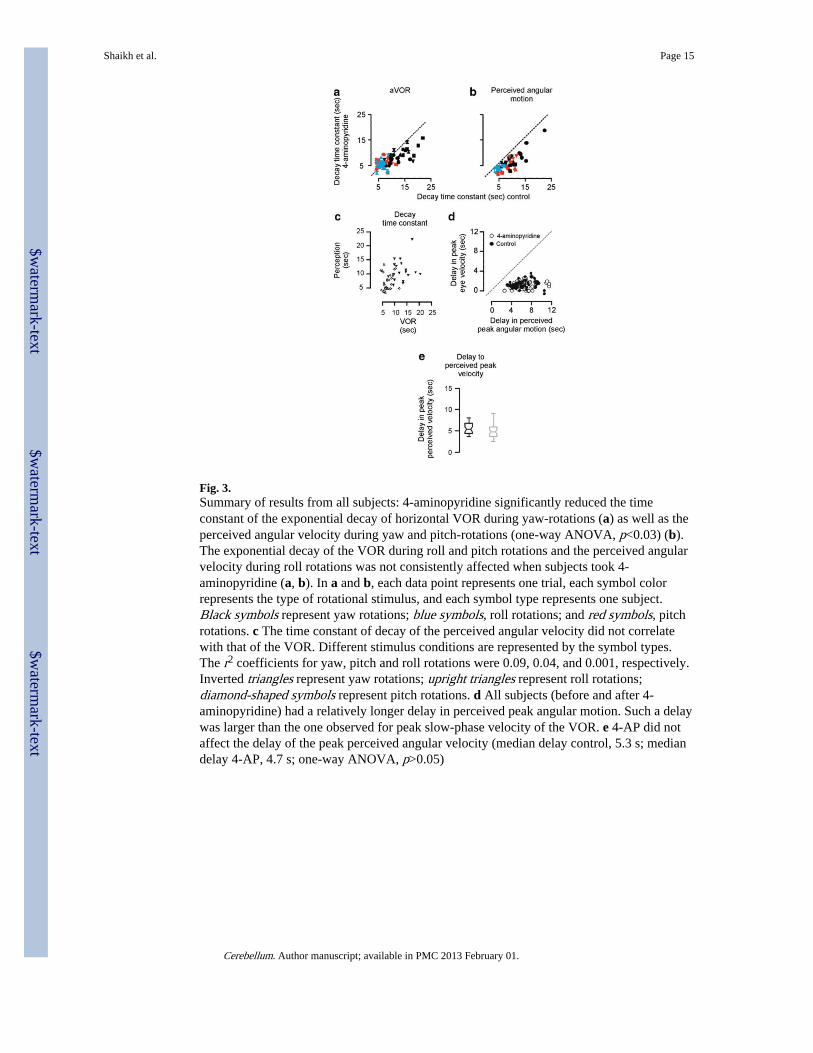
Fig. 3.Summary of results from all subjects: 4-aminopyridine significantly reduced the timeconstant of the exponential decay of horizontal VOR during yaw-rotations (a) as well as theperceived angular velocity during yaw and pitch-rotations (one-way ANOVA, p<0.03) (b).The exponential decay of the VOR during roll and pitch rotations and the perceived angularvelocity during roll rotations was not consistently affected when subjects took 4-aminopyridine (a, b). In a and b, each data point represents one trial, each symbol colorrepresents the type of rotational stimulus, and each symbol type represents one subject.Black symbols represent yaw rotations; blue symbols, roll rotations; and red symbols, pitchrotations. c The time constant of decay of the perceived angular velocity did not correlatewith that of the VOR. Different stimulus conditions are represented by the symbol types.The r2 coefficients for yaw, pitch and roll rotations were 0.09, 0.04, and 0.001, respectively.Inverted triangles represent yaw rotations; upright triangles represent roll rotations;diamond-shaped symbols represent pitch rotations. d All subjects (before and after 4-aminopyridine) had a relatively longer delay in perceived peak angular motion. Such a delaywas larger than the one observed for peak slow-phase velocity of the VOR. e 4-AP did notaffect the delay of the peak perceived angular velocity (median delay control, 5.3 s; mediandelay 4-AP, 4.7 s; one-way ANOVA, p>0.05)
Shaikh et al. Page 15
Cerebellum. Author manuscript; available in PMC 2013 February 01.
$waterm
ark-text$w
atermark-text
$waterm
ark-text

Fig. 4.The time constants of decay of the VOR and perceived angular velocity before and after 4-AP during all three planes of rotations are summarized as box and whisker plots. Black datarepresent time constant of decay of the VOR, and gray represent that of perceived angularvelocity. Horizontal line at the center of the box represents median, while notches are 95 %confidence intervals around the median. If notches from two box–whisker plots do notoverlap, the difference between the median in the two populations is statistically significant.The asterisks above the box–whisker plot indicate statistically significant difference
Shaikh et al. Page 16
Cerebellum. Author manuscript; available in PMC 2013 February 01.
$waterm
ark-text$w
atermark-text
$waterm
ark-text
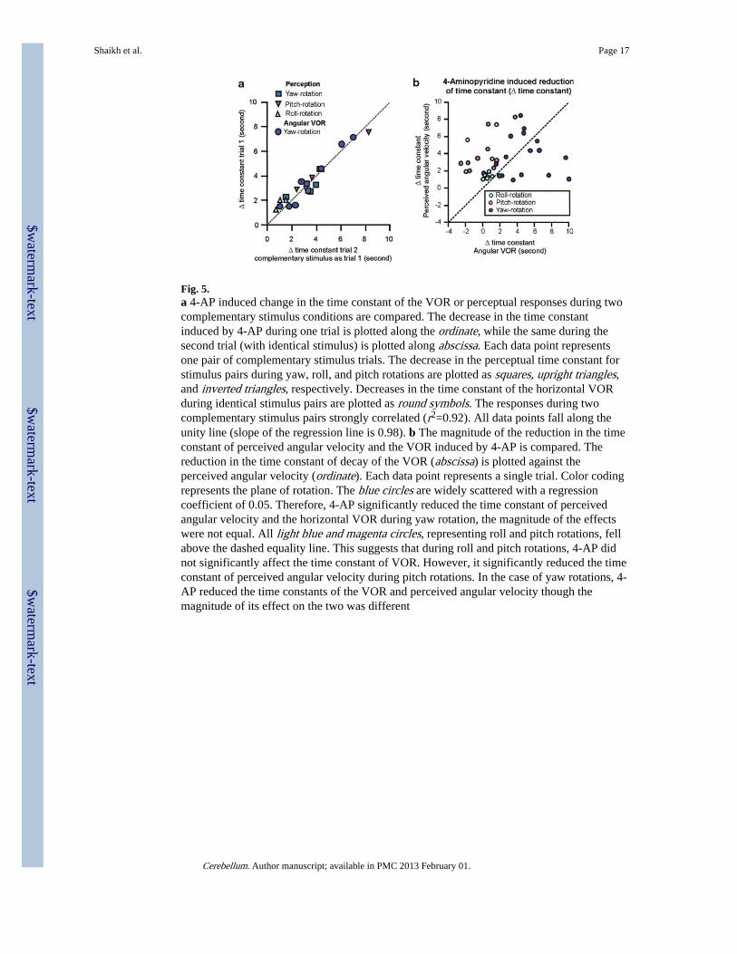
Fig. 5.a 4-AP induced change in the time constant of the VOR or perceptual responses during twocomplementary stimulus conditions are compared. The decrease in the time constantinduced by 4-AP during one trial is plotted along the ordinate, while the same during thesecond trial (with identical stimulus) is plotted along abscissa. Each data point representsone pair of complementary stimulus trials. The decrease in the perceptual time constant forstimulus pairs during yaw, roll, and pitch rotations are plotted as squares, upright triangles,and inverted triangles, respectively. Decreases in the time constant of the horizontal VORduring identical stimulus pairs are plotted as round symbols. The responses during twocomplementary stimulus pairs strongly correlated (r2=0.92). All data points fall along theunity line (slope of the regression line is 0.98). b The magnitude of the reduction in the timeconstant of perceived angular velocity and the VOR induced by 4-AP is compared. Thereduction in the time constant of decay of the VOR (abscissa) is plotted against theperceived angular velocity (ordinate). Each data point represents a single trial. Color codingrepresents the plane of rotation. The blue circles are widely scattered with a regressioncoefficient of 0.05. Therefore, 4-AP significantly reduced the time constant of perceivedangular velocity and the horizontal VOR during yaw rotation, the magnitude of the effectswere not equal. All light blue and magenta circles, representing roll and pitch rotations, fellabove the dashed equality line. This suggests that during roll and pitch rotations, 4-AP didnot significantly affect the time constant of VOR. However, it significantly reduced the timeconstant of perceived angular velocity during pitch rotations. In the case of yaw rotations, 4-AP reduced the time constants of the VOR and perceived angular velocity though themagnitude of its effect on the two was different
Shaikh et al. Page 17
Cerebellum. Author manuscript; available in PMC 2013 February 01.
$waterm
ark-text$w
atermark-text
$waterm
ark-text

$waterm
ark-text$w
atermark-text
$waterm
ark-text
Shaikh et al. Page 18
Table 1
Summary of time constants
Condition Mean (seconds) Variance (seconds)
Yaw VOR control 13.4 16.6
Yaw VOR 4AP 9.1 5.7
Yaw perception control 10.6 12.8
Yaw perception 4AP 7.5 11.9
Roll VOR control 6.3 1.0
Roll VOR 4AP 5.8 1.7
Roll perception control 6.1 8.0
Roll perception 4AP 4.2 1.0
Pitch VOR control 7.8 4.8
Pitch VOR 4AP 5.9 2.8
Pitch perception control 7.9 7.2
Pitch perception 4AP 4.5 5.3
Cerebellum. Author manuscript; available in PMC 2013 February 01.
Copyright © 2022 FDOKUMEN