Biomechanical considerations of animal models used in tissue engineering of bone
Upload
khangminh22Category
view
0download
0
Mukherjee et al. Laboratory Animal Research (2022) 38:18 https://doi.org/10.1186/s42826-022-00128-1
REVIEW
Role of animal models in biomedical research: a reviewP. Mukherjee1†, S. Roy1†, D. Ghosh2 and S. K. Nandi2*
Abstract
The animal model deals with the species other than the human, as it can imitate the disease progression, its’ diagnosis as well as a treatment similar to human. Discovery of a drug and/or component, equipment, their toxicological stud-ies, dose, side effects are in vivo studied for future use in humans considering its’ ethical issues. Here lies the impor-tance of the animal model for its enormous use in biomedical research. Animal models have many facets that mimic various disease conditions in humans like systemic autoimmune diseases, rheumatoid arthritis, epilepsy, Alzheimer’s disease, cardiovascular diseases, Atherosclerosis, diabetes, etc., and many more. Besides, the model has tremendous importance in drug development, development of medical devices, tissue engineering, wound healing, and bone and cartilage regeneration studies, as a model in vascular surgeries as well as the model for vertebral disc regenera-tion surgery. Though, all the models have some advantages as well as challenges, but, present review has emphasized the importance of various small and large animal models in pharmaceutical drug development, transgenic animal models, models for medical device developments, studies for various human diseases, bone and cartilage regen-eration model, diabetic and burn wound model as well as surgical models like vascular surgeries and surgeries for intervertebral disc degeneration considering all the ethical issues of that specific animal model. Despite, the process of using the animal model has facilitated researchers to carry out the researches that would have been impossible to accomplish in human considering the ethical prohibitions.
Keywords: Animal models, Ethical concern, Animal research, Human diseases, Biomedical research
© The Author(s) 2022. Open Access This article is licensed under a Creative Commons Attribution 4.0 International License, which permits use, sharing, adaptation, distribution and reproduction in any medium or format, as long as you give appropriate credit to the original author(s) and the source, provide a link to the Creative Commons licence, and indicate if changes were made. The images or other third party material in this article are included in the article’s Creative Commons licence, unless indicated otherwise in a credit line to the material. If material is not included in the article’s Creative Commons licence and your intended use is not permitted by statutory regulation or exceeds the permitted use, you will need to obtain permission directly from the copyright holder. To view a copy of this licence, visit http://creativecommons.org/licenses/by/4.0/. The Creative Commons Public Domain Dedication waiver (http://creativecom-mons.org/publicdomain/zero/1.0/) applies to the data made available in this article, unless otherwise stated in a credit line to the data.
BackgroundThe animals used in various studies and investigations are related to the evolution of human history. Though there are many shreds of evidence that Aristotle in ancient Greece successfully used animals in understand-ing the human body, the main breakthrough in animal models happened in the eighteenth and nineteenth cen-turies with the scientists like Jean Baptiste Van Helmont, Francesco Redi, John Needham, Lazzaro Spallanzani, Lavoisier and Pasteur who studied the origin of life using
animal models [1]. At the same time, human physiol-ogy, anatomy, pathology as well as pharmacology were also studied using animal models. With the remarkable advancements in drug development, biomedicine and pre-clinical trials, the importance of animal models has increased many folds in the last decades, as the therapeu-tic outcome and drug safety are the foremost important criteria for a drug and medical device considered to be used in the human model [2]. The scientific apply of ani-mal models in the arena of biological research and drug development is an age-old practice because of the notable resemblance in physiology and anatomy between humans and animals, especially mammals [3]. One must consider that the physiological processes of humans, as well as mammals, are complex in terms of circulatory factors, hormones, cellular structures, and tissue systems. Hence,
Open Access
Laboratory Animal Research
†P. Mukherjee and S. Roy equally contributed and joint first author
*Correspondence: [email protected]
2 Department of Veterinary Surgery and Radiology, West Bengal University of Animal and Fishery Sciences, Kolkata, IndiaFull list of author information is available at the end of the article

Page 2 of 17Mukherjee et al. Laboratory Animal Research (2022) 38:18
investigation of various aspects such as molecular struc-tures, cellular and organ functions in physiological and pathological conditions must be taken into consideration.
The process of selection of an animal model for bio-medical research is a very intricate part, as all models are not acceptable due to various limitations. Many fac-tors should be taken into consideration during the selec-tion of an ideal animal model for biomedical trials. The most important criteria are the proper selection of mod-els in terms of resemblance between animal species and humans in terms of physiological and/or pathophysiolog-ical aspects. Detailed evaluation during the application of certain drugs/molecules/devices and their capacity to reproduce the disease or pathology at the same level as that of humans. Availability and the size of animal spe-cies under consideration. Long life duration of the ani-mal species under study. A Large animal population in a model facilitates the availability of multiple sub-species.
Many animal species such as Drosoph-ila (insects), Danio rerio, or zebrafish (fish), Caenorhab-ditis elegans (nematodes), Xenopus (frogs), and mammals such as mice, rabbits, rats, cats, dogs, pigs, and monkeys have been accepted worldwide for their phylogenetic resemblance to humans [4].
Choice of an appropriate animal model is most of the time a tedious job and sometimes depends on assump-tions and convenience of the study and researchers with-out considering whether the model will be appropriate or not. Irrational selection of an inappropriate animal model for scientific investigations will yield incorrect findings, as well as fetch misusage of resources and lives. Moreover, it results in erroneous, duplicative, and inap-propriate experiments [5]. To minimize these problems, recently researchers have advanced their researches to produce animal models that are very specific to the research under consideration. They produced custom-made transgenic animal models by incorporating genetic information directly into the embryo either by injecting foreign DNA or through retroviral vectors [6]. Through the incorporation of human cells into the recipient ani-mals, researchers can study the effects of pathogens simi-lar to the way in the human body [7]. Proper selection of animal models is mainly related to the nature of the drug or medical devices under study. In many instances, a sin-gle animal model is not able to signify a human disease alone, in that case, the combination of several models can potentially signify the procedure [8].
Main textThe significance and challenges of animals in biomedical researchThere has always been a debate among the research-ers about the significance of animal models, as many
experiments yield promising results, whereas, others couldn’t produce desired outcomes, so, that model could be translated to humans too. Owing to their close phy-logenetic closeness to humans, non-human primates are proved to be the most potential candidate. They have genetic, biochemical, and psychological activities similar to humans. In this context, the necessity of non-human primates continues to grow in several areas of research of human diseases viz. AIDS, Parkinson’s disease, hepa-titis, dentistry, orthopaedic surgical techniques, cardio-vascular surgeries, psychological disorders, toxicological studies, drug development, toxicological studies as well as vaccine development [4]. The discovery of vaccines and diagnostic modalities with the animal model does not only benefit humans but also enhances the lifespan of animals and prevents many zoonotic diseases, with the production of many vaccines and drugs like rabies, teta-nus, parvo virus, feline leukemia, etc (Table 1).
Ethical matters on the use of animalsAnimal research adheres to a few dimensions like gov-ernment legislation, public opinion, moral stand, and search for appropriate alternatives for the research. Mahatma Gandhi opined that to judge the greatness and moral progress of a nation, one should judge the way its animals are being treated. Government legislation restricts the researchers and institutes from likely injury, pain, or suffering that may arise during animal research [33]. On the contrary, many modern countries ruled that before human administration, vaccine testing, lethal dose testing should be done on animals [34]. Social accept-ance has also an influential role in animal experiments as it utilizes public money [33]. In their moral view, many people think that dog has more moral impact than pig, rat, fishes, mouse, etc.
Ethical issues on animal experimentation started in 1959, where the emphasis has been given on principles of 3Rs, reduction, refinement, and replacement of animal use [35]. According to this principle, minimum necessary numbers of animals are to be used for scientific experi-ments i.e. reduction. Pain or distress of the animals dur-ing experiments has to be minimized, i.e. refinement. Wherever applicable replacements of the animals are to be done with other non-animal alternatives, i.e. replace-ment. Though these principles are considered as the cornerstone of animal experimentations, but there are questions regarding the implementation of these regula-tions [36].
Laboratory (small) and large animal models for human diseasesThe importance of rat and mouse models has proved their outstanding importance in biomedical research.

Page 3 of 17Mukherjee et al. Laboratory Animal Research (2022) 38:18
Besides, other mammalian and non-mammalian small domestic animals like the guinea pig, hamster, rabbit, fer-rets, birds, amphibians, fishes, flies, worms have equal importance in terms of anatomical and physiological resemblance with humans. Large animal models also proved their uniqueness due to specific anatomical and physiological characteristics pertinent to those specific researches (Table 2).
Transgenic animal models in biomedical researchThe gene rule and role in the biological system of human diseases has improved many folds with the introduction of the transgenic animal model in biomedical research within the last three decades. The early example of most unique biological research started, when structural gene coding for the human growth hormone (GH) was initi-ated into mice after fusion with the regulatory region of mouse metallothionein-I gene, as a result, transgenic mouse produced and showed excess GH production [157].
Linking of the genotype with disease phenotype has been expedited with the genome editing with the intro-duction of the CRISPR–Cas9 system by which disease-causing mutations are done in animal models [158]. Moreover, the production of transgenic animals has been radically changed by the introduction of the CRISPR–Cas9 system. Through the successful use of this model accurate human disease models in animals have been produced and possible therapies have been potenti-ated. Recapitulation of various disease-causing single nucleotide polymorphisms (SNPs) in animal models is achieved by the introduction of gRNA with the combina-tion of Cas9 and donor template DNA [159], viz. mouse model has enormous importance in carrying human genetic traits, developmental similarities as well as dis-ease translation [158, 160–162]. Zhang and Sharp labs at MIT/Broad Institute used CRISPR–Cas9 through AAV and lentivirus [163] both in vivo and ex vivo in neurons as well as endothelial cells of mice for the production of lung cancer model in mice where lung causing genes namely Kras, Tp53, and Lkb1 were mutated. On the other
Table 1 Significance and challenges of different animal models
Disease model/procedure Animal model References
Significance Challenges
Ischemia and reperfusion injury of the spinal cord
Animal models are warranted But, need several models are required (Pig, rabbit, mouse)
[9]
Cartilage defect repair with biomaterials There are murine, ovine, leporine, caprine, porcine, canine, and equine models
In regards to cartilage thickness, joint biomechanics and ethical and licensing matters, caprine models are the best suited
[10]
Monoclonal antibodies for cancer treatment Preclinical trials of monoclonal antibod-ies (mAbs) in animal models are required to reach the clinic
But, mAbs are less adapted to animal studies [11]
Animal models to study of limb restoration Cockroach: similar resemblance within the animal kingdom, cheap, least ethical regulations
Not ideal for the less resemblance with human
[12]
Zebrafish: genome is well identified, ver-tebrate; grow very fast, high regenerative capacity, least ethical regulations
Not ideal for the less resemblance with human
[13, 14]
Mouse: cheap, fast growth, well established genome, many species and transgenic strains, mammalian
Findings not trustworthy for human trials [15–17]
Rat: larger than mice, cheap, fast growth, well established genome, many species and transgenic strains, mammalian
Findings not trustworthy for human trials as well as maintenance cost is more than mice
[18–21]
Dog: large in size, higher physical activity, cheaper than horse, mammalian, good for preclinical trial, results are trustworthy for human trials
More ethical constraints, more maturity period than rodents, expensive rearing cost
[22–26]
Horse: larger mammal than dog, higher physical activity, trial result can easily be transferred to human
More ethical constraints, more maturity period, expensive rearing cost
[27–30]
Development of antibacterials Efficacy and toxicity of antibacterials can be studied
But, animal model can’t predict human response to that component
[31]
Streptozotocin (STZ)—induced diabetes model
STZ produces clinical features in animals that resemble diabetes in humans
But, physiochemical properties and toxici-ties of STZ cause mortality to the animals
[32]

Page 4 of 17Mukherjee et al. Laboratory Animal Research (2022) 38:18
hand, an MIT-Harvard team [164] disrupted the tumor suppressor genes Pten and Tp53, and consequently liver cancer was produced in mice.
Animal models in pharmaceutical drug developmentIn recent advancements, animal models are the most practical tools for pre-clinical drug screening before application into clinical trials. Animal models are con-sidered as most important in vivo models in terms of basic pharmacokinetic parameters like drug efficiency, safety, toxicological studies, as these pre-clinical data are required before translating into humans. Toxicologi-cal tests are performed on a large number of animals like general toxicity, mutagenicity, carcinogenicity, and tera-togenicity and to evaluate whether the drugs are irritant to eyes and skin. In most instances, both in vitro and in vivo models are corroborated before proceeding to medi-cal trials. In vivo models are mostly conducted in mice, rats, and rabbits [2]. Certain stages are involved in pre-clinical trials with animal models: firstly, if the trial drug shows desirable efficacy then only further studies are car-ried out; secondly, if a drug in pre-clinical trials on ani-mals proved to be safe, then it is administered in small human volunteer groups, at the same time, the animal trial will go on to evaluate the effect of the drug when administered for an extended period [8, 165]. Mostly, rodents are used for these trials as they have similar
biological properties to humans and are easy to handle and rear in laboratories. In new regulations, it is manda-tory to carry on the trials on non-rodents such as rab-bits, dogs, cats, or primates simultaneously with rodents [166].
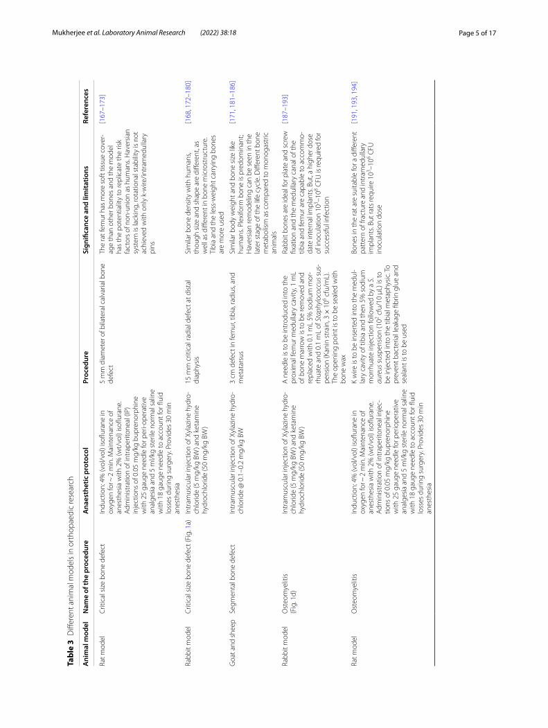
Animal models in orthopedic researchThere are many conditions involving bone pathologies such as osteomyelitis, osteosarcoma, osteoporosis, etc. Being a complex organ, the treatment of bone needs spe-cial care and extensive researches that involves special-ized techniques as well as specific animal models for the studies of specific diseases. Herein, the animal models emphasize mostly related to fracture healing (critical size defect), osteoporosis, osteomyelitis, and osteosarcoma (Table 3).
Animal models in diabetic and burn wound healingType 2 diabetes and associated foot ulcer have turned into an epidemic worldwide in recent years causing severe socio-economic trouble to the patients as well as the health care system of the nation as a whole [208]. Various researches depicted that chance of developing an ulcer in diabetic patients varies between 15–25% [209, 210] and the chance of recurrence is about 20–58% among the patients within a year after recov-ery [211]. Hence, many researchers studied different
Table 2 Biomedical significances and limitations of small animal models
Small animal models Significances and limitations References
Rats (Rattus norvegicus domes-tica) and Mice (Mus musculus) model
Easy breeding, handling, less rearing care, easily interchangeable between rats and mice. They are mostly inbred, so do not have genetic variations like a human, not a suitable model for inflammation study
[37–42]
Guinea pig (Cavia porcellus) Mostly outbred, suitable for cholesterol metabolism, asthma model, feto-placental development and parturition, Alzheimer’s disease study, tuberculosis research, vaccine study. High phenotypic variations, Ebola research in guinea pig is limited due to the poor infectious potential of the virus
[43–63]
Hamster, especially golden hamster (Mesocricetus auretus)
Excellent for reproductive research due to the strict progesterone, but not oestrogen, short gestation period, unique an anatomical feature like loose subcutaneous space, important for micro-circulation studies, cancer model, infection model for leptospirosis, vaccine studies
[64–81]
Rabbit (Oryctolagus cuniculus) Good model for surgically created osteoarthritis, wound healing model, drug study, asthma model, cho-lesterol model, cardiovascular disease model, Alzheimer’s disease model
[82–97]
Equids (Equus) Important for the study of articular defects, orthopaedic models, tendinopathies, asthma model, repro-ductive models. But, more care expenses are required
[98–102]
Cattle (Bos taurus) Important for study of female reproductive model, pregnancy related issues, tuberculosis models. But, more care expenses are required
[103–107]
Goat (Capra hircus) Potential for orthopaedic studies, mechanical circulatory support devices, model for female to male XX sex reversal
[108–116]
Sheep (Ovis aries) Easy to handle, easy sampling, physiological and anatomical nature are similar to humans, good for surgical model for bone and wound healing, asthma model, heart pathology, vaccine development, but, mostly inbred strains
[117–129]
Cat (Felis catus) Important models for asthma, obesity, type-2 diabetes mellitus, HIV, cerebral palsy [130–140]
Dog (Canis familiaris) Narcolepsy, hemophilia B, or hereditary diseases, cancer, musculoskeletal research, etc [141–150]
Pig (Sus scrofa) Large litter size, more similar with human physiology, important for cardiovascular study, Alzheimer’s disease, Atherosclerosis, Type 2 diabetes mellitus, Breast cancer, etc
[151–156]
Page 5 of 17Mukherjee et al. Laboratory Animal Research (2022) 38:18
Tabl
e 3
Diff
eren
t ani
mal
mod
els
in o
rtho
paed
ic re
sear
ch
Ani
mal
mod
elN
ame
of th
e pr
oced
ure
Ana
esth
etic
pro
toco
lPr
oced
ure
Sign
ifica
nce
and
limita
tions
Refe
renc
es
Rat m
odel
Crit
ical
siz
e bo
ne d
efec
tIn
duct
ion:
4%
(vol
/vol
) iso
flura
ne in
ox
ygen
for ~
2 m
in. M
aint
enan
ce o
f an
esth
esia
with
2%
(wt/
vol)
isofl
uran
e.
Adm
inis
trat
ion
of in
trap
erito
neal
(IP)
in
ject
ions
of 0
.05
mg/
kg b
upre
norp
hine
w
ith 2
5 ga
uge
need
le fo
r per
i-ope
rativ
e an
alge
sia
and
5 m
l/kg
ster
ile n
orm
al s
alin
e w
ith 1
8 ga
uge
need
le to
acc
ount
for fl
uid
loss
es d
urin
g su
rger
y. P
rovi
des
30 m
in
anes
thes
ia
5 m
m d
iam
eter
of b
ilate
ral c
alva
rial b
one
defe
ctTh
e ra
t fem
ur h
as m
ore
soft
tiss
ue c
over
-ag
e th
an o
ther
bon
es a
nd th
e m
odel
ha
s th
e po
tent
ialit
y to
repl
icat
e th
e ris
k fa
ctor
s of
non
-uni
on a
s hu
man
s. H
aver
sian
sy
stem
is la
ckin
g, ro
tatio
nal s
tabi
lity
is n
ot
achi
eved
with
onl
y k-
wire
/intr
amed
ulla
ry
pins
[167
–173
]
Rabb
it m
odel
Crit
ical
siz
e bo
ne d
efec
t (Fi
g. 1
a)In
tram
uscu
lar i
njec
tion
of X
ylaz
ine
hydr
o-ch
lorid
e (5
mg/
kg B
W) a
nd k
etam
ine
hydr
ochl
orid
e (5
0 m
g/kg
BW
)
15 m
m c
ritic
al ra
dial
def
ect a
t dis
tal
diap
hysi
sSi
mila
r bon
e de
nsity
with
hum
ans,
thou
gh s
ize
and
shap
e ar
e di
ffere
nt, a
s w
ell a
s di
ffere
nt in
bon
e m
icro
stru
ctur
e.
Tibi
a an
d th
e le
ss-w
eigh
t car
ryin
g bo
nes
are
mor
e us
ed
[168
, 172
–180
]
Goa
t and
she
epSe
gmen
tal b
one
defe
ctIn
tram
uscu
lar i
njec
tion
of X
ylaz
ine
hydr
o-ch
lorid
e @
0.1
–0.2
mg/
kg B
W3
cm d
efec
t in
fem
ur, t
ibia
, rad
ius,
and
met
atar
sus
Sim
ilar b
ody
wei
ght a
nd b
one
size
like
hu
man
s. Pl
exifo
rm b
one
is p
redo
min
ant;
Hav
ersi
an re
mod
elin
g ca
n be
see
n in
the
late
r sta
ge o
f the
life
cyc
le. D
iffer
ent b
one
met
abol
ism
as
com
pare
d to
mon
ogas
tric
an
imal
s
[171
, 181
–186
]
Rabb
it m
odel
Ost
eom
yelit
is(F
ig. 1
d)In
tram
uscu
lar i
njec
tion
of X
ylaz
ine
hydr
o-ch
lorid
e (5
mg/
kg B
W) a
nd k
etam
ine
hydr
ochl
orid
e (5
0 m
g/kg
BW
)
A n
eedl
e is
to b
e in
trod
uced
into
the
prox
imal
fem
ur m
edul
lary
cav
ity, 1
mL
of b
one
mar
row
is to
be
rem
oved
and
re
plac
ed w
ith 0
.1 m
L 5%
sod
ium
mor
-rh
uate
and
0.1
mL
of S
taph
yloc
occu
s sus
-pe
nsio
n (K
anin
str
ain,
3 ×
106 c
fu/m
L).
The
open
ing
poin
t is
to b
e se
aled
with
bo
ne w
ax
Rabb
it bo
nes
are
idea
l for
pla
te a
nd s
crew
fix
atio
n an
d th
e m
edul
lary
can
al o
f the
tib
ia a
nd fe
mur
are
cap
able
to a
ccom
mo-
date
inte
rnal
impl
ants
. But
, a h
ighe
r dos
e of
inoc
ulat
ion
103 –1
08 CFU
is re
quire
d fo
r su
cces
sful
infe
ctio
n
[187
–193
]
Rat m
odel
Ost
eom
yelit
isIn
duct
ion:
4%
(vol
/vol
) iso
flura
ne in
ox
ygen
for ~
2 m
in. M
aint
enan
ce o
f an
esth
esia
with
2%
(wt/
vol)
isofl
uran
e.
Adm
inis
trat
ion
of in
trap
erito
neal
inje
c-tio
ns o
f 0.0
5 m
g/kg
bup
reno
rphi
ne
with
25
gaug
e ne
edle
for p
erio
pera
tive
anal
gesi
a an
d 5
ml/k
g st
erile
nor
mal
sal
ine
with
18
gaug
e ne
edle
to a
ccou
nt fo
r flui
d lo
sses
dur
ing
surg
ery.
Pro
vide
s 30
min
an
esth
esia
K w
ire is
to b
e in
sert
ed in
to th
e m
edul
-la
ry c
avity
of t
ibia
and
then
5%
sod
ium
m
orrh
uate
inje
ctio
n fo
llow
ed b
y a
S.
aure
us s
uspe
nsio
n (1
02 cfu
/10
μL) i
s to
be
inje
cted
into
the
tibia
l met
aphy
sic.
To
prev
ent b
acte
rial l
eaka
ge fi
brin
glu
e an
d se
alan
t is
to b
e us
ed
Bone
s in
the
rat a
re s
uita
ble
for a
diff
eren
t pa
tter
n of
frac
ture
and
intr
amed
ulla
ry
impl
ants
. But
rats
requ
ire 1
03 –106 C
FU
inoc
ulat
ion
dose
[191
, 193
, 194
]

Page 6 of 17Mukherjee et al. Laboratory Animal Research (2022) 38:18
Tabl
e 3
(con
tinue
d)
Ani
mal
mod
elN
ame
of th
e pr
oced
ure
Ana
esth
etic
pro
toco
lPr
oced
ure
Sign
ifica
nce
and
limita
tions
Refe
renc
es
Goa
t mod
elO
steo
mye
litis
Intr
amus
cula
r inj
ectio
n of
Xyl
azin
e hy
dro-
chlo
ride
@ 0
.1–0
.2 m
g/kg
BW
3-m
m d
rill h
ole
is to
be
mad
e in
dis
tal
tibia
and
inje
ctio
n of
1 m
L 5%
sod
ium
m
orrh
uate
, aft
erw
ards
an
inje
ctio
n 10
min
la
ter w
ith S
. aur
eus (
7.05
× 1
04 cfu
). To
pr
even
t bac
teria
l lea
kage
fibr
in g
lue
and
seal
ant i
s to
be
used
They
are
larg
er th
an o
ther
spe
cies
und
er
stud
y he
nce
impl
ants
and
pro
sthe
ses
that
ar
e us
ed in
hum
ans
can
be u
sed
in g
oats
su
cces
sful
ly. B
ut th
ey a
re e
xpen
sive
as
wel
l as
the
rarin
g co
st is
mor
e. In
ocul
atio
n do
se is
103 –1
05 CFU
in g
oat m
odel
s
[191
, 193
, 195
]
Rabb
it m
odel
Ost
eopo
rosi
sIn
tram
uscu
lar i
njec
tion
of X
ylaz
ine
hydr
o-ch
lorid
e (5
mg/
kg B
W) a
nd k
etam
ine
hydr
ochl
orid
e (5
0 m
g/kg
BW
)
Bila
tera
l ova
riect
omy
afte
rwar
ds IM
inje
c-tio
n of
1 m
g/kg
BW
/day
of m
ethy
lpre
dni-
solo
ne fo
r 4 w
eeks
They
ach
ieve
ear
ly s
kele
tal m
atur
ity th
an
othe
r mam
mal
s[1
96–2
00]
Shee
p m
odel
Ost
eopo
rosi
sG
ener
al a
nest
hesi
a w
ith in
tram
uscu
lar
inje
ctio
n of
Xyl
azin
e hy
droc
hlor
ide
@
0.1–
0.2
mg/
kg B
W
Bila
tera
l ova
riect
omy,
low
cal
cium
die
t, w
eekl
y IM
adm
inis
trat
ion
of d
exam
etha
-so
ne fo
r 6 w
eeks
They
are
doc
ile, e
asy
to h
andl
e, a
nd
hous
e. B
one
size
sim
ilar t
o hu
man
. But
, as
they
are
rum
inan
t, he
nce,
ora
l dru
g ad
min
istr
atio
n do
es n
ot y
ield
the
desi
red
resu
lt. S
urgi
cal i
nter
vent
ion
is re
quire
d to
cr
eate
an
abom
asal
fist
ula
[201
–204
]
Mou
se m
odel
Ost
eosa
rcom
aIs
oflur
ane/
oxyg
en-b
ased
ane
sthe
sia
for
indu
ctio
n th
en m
aint
enan
ce b
y IM
adm
in-
istr
atio
n of
Xyl
azin
e @
10 m
g/kg
BW
and
ke
tam
ine
@10
0 m
g/kg
BW
Aft
er th
e pr
epar
atio
n of
ost
eosa
rcom
a ce
lls a
s de
scrib
ed b
y U
luçk
an e
t al.,
a 0.
5 cm
ski
n in
cisi
on is
mad
e ju
st b
elow
th
e kn
ee to
exp
ose
tibia
l tub
eros
ity, t
hen
cells
are
inje
cted
into
the
med
ulla
ry c
avity
w
ith 2
6–28
G s
yrin
ge a
nd s
kin
is s
utur
ed
Che
ap a
vaila
bilit
y, e
asy
to h
andl
e, g
enet
ic
sim
ilarit
y w
ith h
uman
s. H
ence
, bec
ome
impo
rtan
t for
onc
olog
ical
rese
arch
[205
–207
]

Page 7 of 17Mukherjee et al. Laboratory Animal Research (2022) 38:18
materials or drugs to treat diabetic wounds. Similarly, burn wounds occur due to exposure to flames, hot sur-faces, liquids, chemicals, or even cold exposure [212]. Though with the recent modalities like skin grafting prognosis has improved however, the mortality rate is high [213–215].
Diabetic wound rat modelFor developing this model, clinically healthy male Wistar rats (150 ~ 250 g body weight) are used. To induce hyperglycemia, injection nicotinamide (NAD)@ 150 mg/kg BW intraperitoneally, after 15 min injec-tion Streptozotocin (STZ) @ 65 mg/kg BW intraperi-toneally [216] are to be injected. The same procedure has to be repeated after 24 h. Blood is to be collected from the tail after 72 h to check hyperglycemia. Rats having high blood glucose levels (≥ 10 mmol/L) are considered to be diabetic [217]. For wound creation, rats are to be anesthetized with a combination of xyla-zine @10 mg/kg (intramuscular injection) and keta-mine @90 mg/kg (intramuscular injection) [218]. After marking the dorsal back area with methylene blue, the site is to be prepared aseptically after shaving [219]. Full-thickness wound creation is to be done with a sterile 6 mm biopsy punch measuring 6 mm diameter and 2 mm depth and left open [218] (Fig. 1c).
Burn wound modelsBecause of the severity and types of cause, the manage-ment of burn injuries poses a significant challenge to plastic surgeons in humans. In general, primary and sec-ondary burn wounds heal by the primary healing process, but, third-degree burn injuries with the destruction of all the skin layers are resistant to the normal healing pro-cess and necessitate the added surgical procedures, such as skin grafting, and the relevance of advanced wound dressing [220]. Several researchers used the albino Winstar male rats (Rattus norvegicus) model weighing 250 ± 50 g for the study of burn wounds. Anesthesia was achieved with intramuscular administration of atropine sulfate (0.04 mg/kg BW) and after 10 min a combina-tion of 10% ketamine (90 mg/kg) and 2% xylazine (10 mg/kg) intramuscularly produced adequate anesthesia [221]. After aseptic preparation of the dorsal back area, thermal injury has to be made with a 10 mm aluminium rod pre-viously heated with 100 °C boiling water. The aluminium rod has to be kept in situ for 15 s. Immediately after the procedure analgesic is to be provided and to be contin-ued for at least 3 days [222–224]. A hot air blower has been used to produce a 6% third-degree burn injury in a mouse model [225]. In pig, a partial-thickness burn model in the skin was produced by placing a glass bot-tle having heated water at 92 °C for 14 s [226] In other studies, a homemade heating device was placed over the
Fig. 1 a. Bone defect model and implantation of implant b. Vascular graft mode c. Diabetic wound model d. Osteomyelitis model development e. Creation of burn wound model f. Cartilage graft model—All in rabbit

Page 8 of 17Mukherjee et al. Laboratory Animal Research (2022) 38:18
Tabl
e 4
Diff
eren
t ani
mal
mod
els
for c
artil
age
reju
vena
tion
or re
pair
Ani
mal
mod
elA
nest
hesi
aPr
oced
ure
Sign
ifica
nce
and
limita
tions
Refe
renc
es
Rabb
itIn
tram
uscu
lar i
njec
tion
of X
ylaz
ine
hydr
ochl
o-rid
e (5
mg/
kg B
W) a
nd k
etam
ine
hydr
ochl
orid
e (5
0 m
g/kg
BW
)
3 m
m d
iam
eter
crit
ical
siz
e de
fect
at s
houl
der o
r kn
ee, d
epth
0.2
–0.5
mm
at t
he c
hond
ral o
r ost
eo-
chon
dral
site
(Fig
. 1f)
Low
cos
t, ea
sy to
han
dle,
and
hou
se, b
ut d
iffer
ent
from
hum
ans
in re
spec
t of b
iom
echa
nics
due
to
thei
r diff
eren
t hop
ping
and
wal
king
pat
tern
[10,
82,
232
, 237
–239
]
Shee
p/G
oat
Gen
eral
ane
sthe
sia
with
intr
amus
cula
r inj
ectio
n of
Xy
lazi
ne h
ydro
chlo
ride
@ 0
.1–0
.2 m
g/kg
BW
Knee
join
t sur
gica
lly e
xpos
ed a
nd 6
–7 m
m c
ircul
ar
criti
cal d
efec
t is
to b
e cr
eate
d w
ith 0
.4–1
.5 m
m
dept
h at
cho
ndra
l/ost
eoch
ondr
al s
ite
Easy
to ra
re, h
andl
e an
d ha
ve c
lose
ana
tom
ical
si
mila
rity
with
hum
ans
but k
nee
cont
act a
reas
are
di
ffere
nt, h
ence
this
mus
t be
cons
ider
ed
[10,
232
, 240
–245
]
Dog
Gen
eral
ane
sthe
sia
usin
g pr
eane
sthe
tic a
trop
ine
sulp
hate
@0.
04 m
g/kg
BW
SC
, aft
er 1
0 m
in x
ylaz
ine
1–2
mg/
kg B
W IM
. Mai
nten
ance
by
keta
min
e @
5–10
mg/
kg
BW IV
and
dia
zepa
m 0
.5 m
g/ k
g BW
sl
ow IV
Surg
ical
ly c
reat
ed 4
mm
dia
met
er c
ircul
ar c
ritic
al
size
def
ect o
f 0.9
5–1.
3 m
m d
epth
at t
he c
hond
ral/
oste
ocho
ndra
l site
of K
nee,
sho
ulde
r, el
bow
, hip
or
ankl
e jo
int
They
are
a g
ood
mod
el fo
r car
tilag
e re
pair
as th
ey
can
be tr
aine
d fo
r tre
adm
ill w
alki
ng, s
wim
min
g,
etc.
But
, dis
adva
ntag
es a
re th
ere.
Firs
tly, e
thic
al
issu
es in
sev
eral
cou
ntrie
s, m
oreo
ver c
anin
e ca
rti-
lage
is th
inne
r com
pare
d to
hum
an a
nd a
nato
mi-
cal d
iffer
ence
exi
sts
in th
e kn
ee jo
int
[10,
230
, 244
, 246
–248
]

Page 9 of 17Mukherjee et al. Laboratory Animal Research (2022) 38:18
skin for 35 s to create burn wound [227]. In rabbits, it was demonstrated to use a dry-heated brass rod for 10 and 20 s at 90 °C to create a deep partial-thickness burn wound in the ear [228]. In mice, a full-thickness burn was created under 3–5% isoflurane anesthesia and intraperi-toneal caprofen 5 mg/kg as analgesia. Here, a 4 cm2 brass rod attached to a temperature probe was first heated to 260 °C and then cool to 230 °C and finally placed on the dorsum skin for 9 s [229] (Fig. 1e).
Animal models in cartilage repairAnimal models have enormous importance in the study of cartilage repair. Though in vitro models have been
reported, it could not replace the necessity of using ani-mal models prior to clinical implementation [230–236] (Table 4).
Animal models in vascular graftingWith the increase of cardiovascular complications, there is a need for surgical intervention using vascular grafts. Vascular grafting and cardiac valve repair have become important issues to the clinicians for the replacement of damaged vessels [249, 250], hence there is an increased demand for tissue-engineered blood vessel substitute [250, 251]. The main prosthetic options are synthetic grafts such as polytetrafluoroethylene, polyethylene
Table 5 In vivo animal studies of different vascular grafts
Animal species Type of graft Graft diameter (mm)
Graft patency rate In vivo study model References
Ovine EC-seeded xenogenic porcine decellularized carotid artery
5 Common carotid artery/external jugular vein arteriovenous shunt
[254]
Canine PCL + VEGF 2 100% in 4 weeks Femoral artery [255]
Canine P(LLA-CL) + Autologus, EC preendothelialization 4 88.9% in 24 weeks Femoral artery [256]
Canine P(LLA-CL) 4 75% in 3 months Femoral artery [257]
Ovine Decellularized graft derived from fibrin gel and ovine dermal fibroblasts
4 100% in 168 days Carotid artery [258]
Ovine Heparin and VEGF-treated xenogenic porcine dSIS
5 92% in 90 days Carotid artery [259]
Mouse PCL 0.5 53% in 28 days Carotid artery [260]
Rabbit P(LLA-CL) + Collagen + Elastin + VEGF 4 86% in 3 weeks Infrarenal aorta [261]
Ovine PCL electrospun + PLCL sponge 5 100% in 8 weeks Carotid artery [262]
Ovine PHBV/PCL-GF 4 50% in 1 year Carotid artery [263]
Table 6 Different animal models for the study of IVDD
Animal model Anaesthesia Procedure Significance and limitations References
Goat Ketamine (11–33 mg/kg BW) and midazolam (0.5–1.5 mg/kg BW), intra-venously followed by maintenance with an isoflurane-oxygen combina-tion
Following the aseptic technique, the lumbar intervertebral discs were opened via left lateral retroperitoneal, transpsoatic approach. A titanium Kirschner wire was positioned in the L1 or L2 vertebral body to facilitate marking of vertebral levels on radio-graphs
Weight range, disc height, size, and shape are similar to humans. They can withstand the stress of anaesthesia and surgery well. But, goat torse has a different anatomical structure in comparison to a human
[268–272]
Rabbit Intramuscular injection of Xylazine hydrochloride (5 mg/kg BW) and keta-mine hydrochloride (50 mg/kg BW)
After positioning the rabbit in lateral decubitus position a 20 degrees inclination was produced. IVD was exposed with a posterolateral retro-peritoneal approach. After dissecting the skin, subcutaneous tissue, and muscle, the left anterolateral aspect of L1–L5 was exposed. Then, one IVD is punctured between L1–L5 with the help of a 16-gauge needle to a depth of 5 mm in the left anterolateral annulus fibrosus in the annular stab method
Similar to human disc degeneration in biochemical and histological aspects. But, the method causes rapid narrow-ing of the disc space and disc height as well as rapid herniation of nucleus pulposus
[273–280]

Page 10 of 17Mukherjee et al. Laboratory Animal Research (2022) 38:18
terephthalate, and polyurethane [252], and autologous conduits. Although these types of synthetic grafts pro-vide reasonable outcomes in large-diameter vascular applications, long-term patency is questionable as com-pared to autologous conduits in small-diameter (< 6 mm) applications due to their inclination to various compli-cations [253]. Despite the superior outcome of autolo-gous grafts, it has some disadvantages such as limited availability and prior use. Moreover, the determination of a suitable animal model needs considerations of vari-ous factors. The factors for the selection of animal spe-cies depend on diameter and length of conduits, period of implantation, anastomotic site, price, accessibility, reaction to anesthesia and surgery, and flow of blood at sites of graft implantation. Animal applications of these tissue-engineered vessels are, therefore, an utmost neces-sity as pre-clinical studies before use in humans (Fig. 1b, Table 5).
Animal models in disc degenerationIntervertebral disc degeneration (IVDD) and herniation manifested as lower back pain cause a massive socio-economic burden to the patient and society as a whole [264–267]. But there is a lack of treatment modalities to cure mildly to moderate degeneration as well as com-plications associated with surgical interventions asso-ciated with the advanced stage; hence, researchers are enormously trying to reinforce regenerative strategies and to lower the suffering by controlling the pain with the injection of stem cells, growth factors hydrogels for replacement of the disc [268]. Diverse animal models have been reported as a pre-clinical trial to translate the procedure in humans (Table 6).
ConclusionsThe importance of animal models is unquestionable in terms of in vivo study for the implementation of any biomedical research to humans. It serves not only the human race but also well being of veterinary patients. Animal models have not only important roles in drug development, toxicity studies, pharmacokinetic stud-ies of a drug, but also the pre-clinical study of medical and tissue engineering devices that are intended to be used in humans. Laboratory animal models are more cost-effective and agreeable to high throughput test-ing as compared to large animal models. Yet, to obtain preclinical data and to ascertain the clinical potential of vascular graft as well as orthopedic bone plates and implants, large animal models that mimic human anat-omy and physiology are to be developed. Whatever may be the modes of using animal models for biomedical
researches, it should abide by the principles of 3Rs, i.e., reduction, refinement, and replacement of animals.
AbbreviationsBW: Body weight; Cfu: Colony forming unit; ESC: Embryonic stem cell; IVDD: Intervertebral disc degeneration; PCL: Polycaprolactone; STZ: Streptozotocin; VEGF: Vascular endothelial growth factor.
AcknowledgementsThe authors acknowledge the kind support of Vice-Chancellor, West Bengal University of Animal and Fishery Sciences, Kolkata, India.
Author contributionsSKN: Conceptualization, Methodology, Supervision and final correction of draft. PM and SR: Data curation, Writing-Original draft preparation. DG: Editing. All authors have read and approved the final manuscript.
FundingThere was no funding support for this study.
Availability of data and materialsThe data in the present manuscript were collected by searching of literatures as well as involving authors own materials.
Declarations
Competing interestsThe authors declare that there is no competing of interest in this manuscript.
Author details1 Department of Veterinary Clinical Complex, West Bengal University of Animal and Fishery Sciences, Mohanpur, Nadia, India. 2 Department of Veterinary Surgery and Radiology, West Bengal University of Animal and Fishery Sciences, Kolkata, India.
Received: 4 November 2021 Accepted: 21 June 2022
References 1. Oparin AI. The origin of life on the earth. 3rd ed. New York: Academic
Press Inc.; 1957. p. XViii+495. 2. Pehlivanovic B, Dina F, Emina A, Ziga Smajic N, Fahir B. Animal models in
modern biomedical research. Eur J Pharm Med Res. 2019;6(7):35–8. 3. Barré-Sinoussi F, Montagutelli X. Animal models are essential to biologi-
cal research: issues and perspectives. Future Sci OA. 2015;1(4):FSO63. 4. Andersen ML, Winter LMF. Animal models in biological and biomedi-
cal research - experimental and ethical concerns. An Acad Bras Ciênc. 2017;91(suppl 1):e20170238.
5. Gad SC. Animal models in toxicology. In: Wexler P, editor. Encyclopedia of toxicology. Boca Raton: CRC/Taylor & Francis; 2005. p. 138–40.
6. Simmons D. The use of animal models in studying genetic disease: transgenesis and induced mutation. Nat Educ. 2008;1(1):70.
7. Ernst W. Humanized mice in infectious diseases. Comp Immunol Micro-biol Infect Dis. 2016;49:29–38.
8. Dam DV, Deyn PPD. Animal models in the drug discovery pipeline for Alzheimer’s disease. Br J Pharmacol. 2011;164(4):1285–300.
9. Simon F, Oberhuber A, Schelzig H. Advantages and disadvantages of different animal models for studying ischemia/reperfusion injury of the spinal cord. Eur J Vasc Endovasc Surg. 2015;49(6):744.
10. Moran CJ, Ramesh A, Brama PAJ, O’Byrne JM, O’Brien FJ, Levingstone TJ. The benefits and limitations of animal models for translational research in cartilage repair. J Exp Orthop. 2016;3(1):1.
11. Loisel S, Ohresser M, Pallardy M, Daydé D, Berthou C, Cartron G, et al. Relevance, advantages and limitations of animal models used in the

Page 11 of 17Mukherjee et al. Laboratory Animal Research (2022) 38:18
development of monoclonal antibodies for cancer treatment. Crit Rev Oncol Hematol. 2007;62(1):34–42.
12. French V. Leg regeneration in the cockroach, Blatella germanica: II. Regeneration from a non-congruent tibial graft/host junction. J Embryol Exp Morphol. 1976;35(2):267–301.
13. Olsen AS, Sarras MP, Intine RV. Limb regeneration is impaired in an adult zebrafish model of diabetes mellitus. Wound Repair Regen. 2010;18(5):532–42.
14. Pfefferli C, Jaźwińska A. The art of fin regeneration in zebrafish. Regen-eration. 2015;2(2):72–83.
15. Gutpell KM, Hrinivich WT, Hoffman LM. Skeletal muscle fibrosis in the mdx/utrn+/- mouse validates its suitability as a murine model of Duch-enne muscular dystrophy. PLoS ONE. 2015;10(1):e0117306.
16. Heber-Katz E, Leferovich JM, Bedelbaeva K, Gourevitch D. Spallanzani’s mouse: a model of restoration and regeneration. In: Heber-Katz E, editor. Regeneration: stem cells and beyond. Berlin: Springer; 2004. p. 165–89.
17. Zaccagnini G, Palmisano A, Canu T, Maimone B, Russo FML, Ambrogi F, et al. Magnetic resonance imaging allows the evaluation of tissue dam-age and regeneration in a mouse model of critical limb ischemia. PLoS ONE. 2015;10(11): e0142111.
18. Cheng L, Liu Y, Zhao H, Zhang W, Guo Y-J, Nie L. Lentiviral-mediated transfer of CDNF promotes nerve regeneration and functional recovery after sciatic nerve injury in adult rats. Biochem Biophys Res Commun. 2013;440(2):330–5.
19. Leppik LP, Froemel D, Slavici A, Ovadia ZN, Hudak L, Henrich D, et al. Effects of electrical stimulation on rat limb regeneration, a new look at an old model. Sci Rep. 2015;5(1):18353.
20. Oliveira KMC, Barker JH, Berezikov E, Pindur L, Kynigopoulos S, Eischen-Loges M, et al. Electrical stimulation shifts healing/scarring towards regeneration in a rat limb amputation model. Sci Rep. 2019;9(1):11433.
21. Zaccagnini G, Gaetano C, Della Pietra L, Nanni S, Grasselli A, Mangoni A, et al. Telomerase mediates vascular endothelial growth factor-depend-ent responsiveness in a rat model of hind limb ischemia. J Biol Chem. 2005;280(15):14790–8.
22. Barraza-Flores P, Fontelonga TM, Wuebbles RD, Hermann HJ, Nunes AM, Kornegay JN, et al. Laminin-111 protein therapy enhances muscle regeneration and repair in the GRMD dog model of Duchenne muscu-lar dystrophy. Hum Mol Genet. 2019;28(16):2686–95.
23. Cook J, Fox D, Malaviya P, Tomlinson J, Farr J, Kuroki K, et al. Evaluation of small intestinal submucosa grafts for meniscal regeneration in a clinically relevant posterior meniscectomy model in dogs. J Knee Surg. 2006;19(3):159–67.
24. Farah Z, Fan H, Liu Z, He J-Q. A concise review of common ani-mal models for the study of limb regeneration. Organogenesis. 2016;12(3):109–18.
25. Fitzpatrick N, Smith TJ, Pendegrass CJ, Yeadon R, Ring M, Goodship AE, et al. Intraosseous transcutaneous amputation prosthesis (ITAP) for limb salvage in 4 Dogs. Vet Surg. 2011;40(8):909–25.
26. Raske M, McClaran JK, Mariano A. Short-term wound complications and predictive variables for complication after limb amputation in dogs and cats. J Small Anim Pract. 2015;56(4):247–52.
27. Fortier LA, Smith RKW. Regenerative Medicine for tendinous and ligamentous injuries of sport horses. Vet Clin North Am Equine Pract. 2008;24(1):191–201.
28. Kon E, Mutini A, Arcangeli E, Delcogliano M, Filardo G, Aldini NN, et al. Novel nanostructured scaffold for osteochondral regeneration: pilot study in horses. J Tissue Eng Regen Med. 2010;4(4):300–8.
29. Parnell LKS, Volk SW. The evolution of animal models in wound healing research: 1993–2017. Adv Wound Care. 2019;8(12):692–702.
30. Smith RK, Garvican ER, Fortier LA. The current ‘state of play’ of regenera-tive medicine in horses: what the horse can tell the human. Regen Med. 2014;9(5):673–85.
31. Greek R, Hansen L. The strengths and limits of animal models as illustrated by the discovery and development of antibacterials. Biol Syst Open Access. 2013;2(2):109.
32. Goyal SN, Reddy NM, Patil KR, Nakhate KT, Ojha S, Patil CR, et al. Chal-lenges and issues with streptozotocin-induced diabetes – a clinically relevant animal model to understand the diabetes pathogenesis and evaluate therapeutics. Chem Biol Interact. 2016;244:49–63.
33. VandeWoude S, Rollin BE. Practical considerations in regenerative medicine research: IACUCs, ethics, and the use of animals in stem cell studies. ILAR J. 2010;51(1):82–4. https:// doi. org/ 10. 1093/ ilar. 51.1. 82.
34. Kooijman M. Why animal studies are still being used in drug develop-ment. Altern Lab Anim. 2013;41(6):P79-81.
35. Russell WMS, Burch RL. The principles of humane experimental tech-nique. Princ hum exp tech. London: Methuen & Co. Limited; 1960, p. 252.
36. Liguori GR, Jeronimus BF, de Aquinas Liguori TT, Moreira LFP, Harmsen MC. Ethical issues in the use of animal models for tissue engineering: reflections on legal aspects, moral theory, three Rs strategies, and harm-benefit analysis. Tissue Eng Part C Methods. 2017;23(12):850–62.
37. Antony JJ, Sithika MAA, Joseph TA, Suriyakalaa U, Sankarganesh A, Siva D, et al. In vivo antitumor activity of biosynthesized silver nanoparticles using Ficus religiosa as a nanofactory in DAL induced mice model. Col-loids Surf B Biointerfaces. 2013;108:185–90.
38. Liu X, Manzano G, Kim HT, Feeley BT. A rat model of massive rotator cuff tears. J Orthop Res. 2011;29(4):588–95.
39. Shurey S, Akelina Y, Legagneux J, Malzone G, Jiga L, Ghanem AM. The rat model in microsurgery education: classical exercises and new hori-zons. Arch Plast Surg. 2014;41(3):201–8.
40. Soares E, Prediger RD, Nunes S, Castro AA, Viana SD, Lemos C, et al. Spatial memory impairments in a prediabetic rat model. Neuroscience. 2013;250:565–77.
41. von Scheidt M, Zhao Y, Kurt Z, Pan C, Zeng L, Yang X, et al. Applications and limitations of mouse models for understanding human atheroscle-rosis. Cell Metab. 2017;25(2):248–61.
42. Mequanint W, Makonnen E, Urga K. In vivo anti-inflammatory activities of leaf extracts of Ocimum lamiifolium in mice model. J Ethnopharma-col. 2011;134(1):32–6.
43. Antwi AO, Obiri DD, Osafo N. Stigmasterol modulates allergic airway inflammation in guinea pig model of ovalbumin-induced asthma. Mediators Inflamm. 2017;2017:2953930.
44. Bates K, Vink R, Martins R, Harvey A. Aging, cortical injury and Alzhei-mer’s disease-like pathology in the guinea pig brain. Neurobiol Aging. 2014;35(6):1345–51.
45. Buels KS, Jacoby DB, Fryer AD. Non-bronchodilating mechanisms of tiotropium prevent airway hyperreactivity in a guinea-pig model of allergic asthma. Br J Pharmacol. 2012;165(5):1501–14.
46. Cashman KA, Broderick KE, Wilkinson ER, Shaia CI, Bell TM, Shurtleff AC, et al. Enhanced efficacy of a codon-optimized DNA vaccine encod-ing the glycoprotein precursor gene of lassa virus in a guinea pig disease model when delivered by dermal electroporation. Vaccines. 2013;1(3):262–77.
47. Clark S, Hall Y, Williams A. Animal models of tuberculosis: guinea pigs. Cold Spring Harb Perspect Med. 2015;5(5):a018572.
48. deOgburn R, Leite JO, Ratliff J, Volek JS, McGrane MM, Fernandez ML. Effects of increased dietary cholesterol with carbohydrate restriction on hepatic lipid metabolism in guinea pigs. Comp Med. 2012;62(2):109–15.
49. Espinoza J, Montaño LM, Perusquía M. Nongenomic bronchodilating action elicited by dehydroepiandrosterone (DHEA) in a guinea pig asthma model. J Steroid Biochem Mol Biol. 2013;138:174–82.
50. Grover A, Troudt J, Arnett K, Izzo L, Lucas M, Strain K, et al. Assessment of vaccine testing at three laboratories using the guinea pig model of tuberculosis. Tuberculosis. 2012;92(1):105–11.
51. Kondo M, Tsuji M, Hara K, Arimura K, Yagi O, Tagaya E, et al. Chloride ion transport and overexpression of TMEM16A in a guinea-pig asthma model. Clin Exp Allergy. 2017;47(6):795–804.
52. Larrouy-Maumus G, Layre E, Clark S, Prandi J, Rayner E, Lepore M, et al. Protective efficacy of a lipid antigen vaccine in a guinea pig model of tuberculosis. Vaccine. 2017;35(10):1395–402.
53. Maghdessian R, Côté F, Rouleau T, Ouadda ABD, Levy É, Lavoie J-C. Ascorbylperoxide contaminating parenteral nutrition perturbs the lipid metabolism in newborn guinea pig. J Pharmacol Exp Ther. 2010;334(1):278–84.
54. Mahajan SG, Mehta AA. Suppression of ovalbumin-induced Th2-driven airway inflammation by β-sitosterol in a guinea pig model of asthma. Eur J Pharmacol. 2011;650(1):458–64.
55. Ordway DJ, Shanley CA, Caraway ML, Orme EA, Bucy DS, Hascall-Dove L, et al. Evaluation of standard chemotherapy in the guinea pig model of tuberculosis. Antimicrob Agents Chemother. 2010;54(5):1820–33.
Page 12 of 17Mukherjee et al. Laboratory Animal Research (2022) 38:18
56. Orme IM, Ordway DJ. Mouse and guinea pig models of tuberculosis. In: Jacobs WR Jr, McShane H, Mizrahi V, Orme IM, editors. Tuberculosis and the tubercle bacillus. John Wiley & Sons, Ltd; 2017. p. 143–62.
57. Pohanka M, Zemek F, Bandouchova H, Pikula J. Toxicological scoring of Alzheimer’s disease drug huperzine in a guinea pig model. Toxicol Mech Methods. 2012;22(3):231–5.
58. Ryan VE, Bailey TW, Liu D, Vemulapalli T, Cooper B, Cox AD, et al. Listeria adhesion protein-expressing bioengineered probiotics prevent feto-placental transmission of Listeria monocytogenes in a pregnant guinea pig model. Microb Pathog. 2021;151:104752.
59. Salazar C, Valdivia G, Ardiles ÁO, Ewer J, Palacios AG. Genetic variants associated with neurodegenerative Alzheimer disease in natural mod-els. Biol Res. 2016;49(1):14.
60. Sharman MJ, Nik SHM, Chen MM, Ong D, Wijaya L, Laws SM, et al. The guinea pig as a model for sporadic alzheimer’s disease (AD): the impact of cholesterol intake on expression of AD-related genes. PLoS ONE. 2013;8(6):e66235.
61. Valdés G, Acuña S, Schneider D, Ortíz R, Padilla O. Bradykinin exerts independent effects on trophoblast invasion and blood pressure in pregnant guinea pigs. Reprod Sci. 2020;27(8):1648–55.
62. Veselenak RL, Shlapobersky M, Pyles RB, Wei Q, Sullivan SM, Bourne N. A Vaxfectin®-adjuvanted HSV-2 plasmid DNA vaccine is effective for prophylactic and therapeutic use in the guinea pig model of genital herpes. Vaccine. 2012;30(49):7046–51.
63. Yang R, Guo P, Song X, Liu F, Gao N. Hyperlipidemic guinea pig model: mechanisms of triglyceride metabolism disorder and comparison to rat. Biol Pharm Bull. 2011;34(7):1046–51.
64. Barbosa MDCL, Bouskela E, Cyrino FZ, Azevedo APS, Costa MCP, de Souza MDGC, et al. Effects of babassu nut oil on ischemia/reperfusion-induced leukocyte adhesion and macromolecular leakage in the micro-circulation: observation in the hamster cheek pouch. Lipids Health Dis. 2012;11(1):158.
65. Camarozano AC, de Garcia ACFZ, Bottino DA, Bouskela E. Effects of microbubbles and ultrasound on the microcirculation: observation on the hamster cheek pouch. J Am Soc Echocardiogr. 2010;23(12):1323–30.
66. Chanut FJA, Williams AM. The syrian golden hamster estrous cycle: unique characteristics, visual guide to staging, and comparison with the rat. Toxicol Pathol. 2016;44(1):43–50.
67. Cruz ISS, Garabalino MA, Trivillin VA, Itoiz ME, Pozzi ECC, Thorp S, et al. Optimization of the classical oral cancerization protocol in hamster to study oral cancer therapy. Oral Dis. 2020;26(6):1175–84.
68. Evangelista KV, Lourdault K, Matsunaga J, Haake DA. Immunoprotective properties of recombinant LigA and LigB in a hamster model of acute leptospirosis. PLoS ONE. 2017;12(7): e0180004.
69. Ford J, Carnes K, Hess RA. Ductuli efferentes of the male golden Syrian hamster reproductive tract. Andrology. 2014;2(4):510–20.
70. Tsai AG, Intaglietta M, Sakai H, Delpy E, Rochelle CDL, Rousselot M, Zal F. Microcirculation and NO-CO Studies of a natural extracellular hemo-globin developed for an oxygen therapeutic carrier. Curr Drug Discov Technol. 2012;9(3):166–72. https:// doi. org/ 10. 2174/ 15701 63128 02650 814.
71. Gomes-Solecki M, Santecchia I, Werts C. Animal models of leptospirosis: of mice and hamsters. Front Immunol. 2017;8:58.
72. Hirose M, Ogura A. The golden (Syrian) hamster as a model for the study of reproductive biology: past, present, and future. Reprod Med Biol. 2019;18(1):34–9.
73. Julander JG, Trent DW, Monath TP. Immune correlates of protection against yellow fever determined by passive immunization and chal-lenge in the hamster model. Vaccine. 2011;29(35):6008–16.
74. Krötz F, Hellwig N, Bürkle MA, Lehrer S, Riexinger T, Mannell H, et al. A sulfaphenazole-sensitive EDHF opposes platelet–endothelium interac-tions in vitro and in the hamster microcirculation in vivo. Cardiovasc Res. 2010;85(3):542–50.
75. Miao J, Chard LS, Wang Z, Wang Y. Syrian hamster as an animal model for the study on infectious diseases. Front Immunol. 2019;10:2329.
76. Molinari AJ, Aromando RF, Itoiz ME, Garabalino MA, Hughes AM, Heber EM, et al. Blood vessel normalization in the hamster oral cancer model for experimental cancer therapy studies. Anticancer Res. 2012;32(7):2703–9.
77. Molinari AJ, Pozzi ECC, Hughes AM, Heber EM, Garabalino MA, Thorp SI, et al. “Sequential” boron neutron capture therapy (BNCT): A novel
approach to BNCT for the treatment of oral cancer in the hamster cheek pouch model. Radiat Res. 2011;175(4):463–72.
78. Vernel-Pauillac F, Goarant C. Differential Cytokine gene expression according to outcome in a hamster model of leptospirosis. PLoS Negl Trop Dis. 2010;4(1): e582.
79. Walpita P, Cong Y, Jahrling PB, Rojas O, Postnikova E, Yu S, et al. A VLP-based vaccine provides complete protection against Nipah virus chal-lenge following multiple-dose or single-dose vaccination schedules in a hamster model. Npj Vaccines. 2017;2:21.
80. Ye H, Yang K, Tan X-M, Fu X-J, Li H-X. Daily rhythm variations of the clock gene PER1 and cancer-related genes during various stages of carcino-genesis in a golden hamster model of buccal mucosa carcinoma. Onco Targets Ther. 2015;8:1419–26.
81. Zhang W, Xie X, Wang J, Song N, Lv T, Wu D, et al. Increased inflam-mation with crude E. coli LPS protects against acute leptospirosis in hamsters. Emerg Microbes Infect. 2020;9(1):140–7.
82. Bajpayee AG, Scheu M, Grodzinsky AJ, Porter RM. A rabbit model dem-onstrates the influence of cartilage thickness on intra-articular drug delivery and retention within cartilage. J Orthop Res. 2015;33(5):660–7.
83. Brunner AM, Henn CM, Drewniak EI, Lesieur-Brooks A, Machan J, Crisco JJ, et al. High dietary fat and the development of osteoarthritis in a rab-bit model. Osteoarthr Cartil. 2012;20(6):584–92.
84. Camacho P, Fan H, Liu Z, He J-Q. Small mammalian animal models of heart disease. Am J Cardiovasc Dis. 2016;6(3):70–80.
85. Dos Santos RA, Südy R, Peták F, Habre W. Physiologically variable ventilation in a rabbit model of asthma exacerbation. Br J Anaesth. 2020;125(6):1107–16.
86. Elmorsy S, Funakoshi T, Sasazawa F, Todoh M, Tadano S, Iwasaki N. Chon-droprotective effects of high-molecular-weight cross-linked hyaluronic acid in a rabbit knee osteoarthritis model. Osteoarthritis Cartilage. 2014;22(1):121–7.
87. Fan J, Kitajima S, Watanabe T, Xu J, Zhang J, Liu E, et al. Rabbit models for the study of human atherosclerosis: from pathophysiological mech-anisms to translational medicine. Pharmacol Ther. 2015;146:104–19.
88. Hu C-H, Tseng Y-W, Chiou C-Y, Lan K-C, Chou C-H, Tai C-S, et al. Bone marrow concentrate-induced mesenchymal stem cell conditioned medium facilitates wound healing and prevents hypertrophic scar formation in a rabbit ear model. Stem Cell Res Ther. 2019;10(1):275.
89. Kamaruzaman NA, Sulaiman SA, Kaur G, Yahaya B. Inhalation of honey reduces airway inflammation and histopathological changes in a rab-bit model of ovalbumin-induced chronic asthma. BMC Complement Altern Med. 2014;14(1):176.
90. Kobayashi T, Ito T, Shiomi M. Roles of the WHHL rabbit in translational research on hypercholesterolemia and cardiovascular diseases. J Biomed Biotechnol. 2011;2011: 406473.
91. Laverty S, Girard CA, Williams JM, Hunziker EB, Pritzker KPH. The OARSI histopathology initiative – recommendations for histological assess-ments of osteoarthritis in the rabbit. Osteoarthr Cartil. 2010;18:S53-65.
92. Liang H, Baudouin C, Daull P, Garrigue J-S, Brignole-Baudouin F. Ocular safety of cationic emulsion of cyclosporine in an in vitro corneal wound-healing model and an acute in vivo rabbit model. Mol Vis. 2012;18:2195–204.
93. Liu QY, Koukiekolo R, Zhang DL, Smith B, Ly D, Lei JX, et al. Molecular events linking cholesterol to Alzheimer’s disease and inclusion body myositis in a rabbit model. Am J Neurodegener Dis. 2016;5(1):74–84.
94. Ludwig JM, Xing M, Gai Y, Sun L, Zeng D, Kim HS. Targeted yttrium 89-doxorubicin drug-eluting bead—a safety and feasibility pilot study in a rabbit liver cancer model. Mol Pharm. 2017;14(8):2824–30.
95. Schoenberg ED, Blake DA, Swann FB, Parlin AW, Zurakowski D, Margo CE, et al. Effect of two novel sustained-release drug delivery systems on bleb fibrosis: an in vivo glaucoma drainage device study in a rabbit model. Transl Vis Sci Technol. 2015;4(3):4.
96. Schreurs BG, Smith-Bell CA, Lemieux SK. Dietary cholesterol increases ventricular volume and narrows cerebrovascular diameter in a rabbit model of Alzheimer’s disease. Neuroscience. 2013;254:61–9.
97. Shirai T, Kobayashi M, Nishitani K, Satake T, Kuroki H, Nakagawa Y, et al. Chondroprotective effect of alendronate in a rabbit model of osteoar-thritis. J Orthop Res. 2011;29(10):1572–7.
98. Colbath AC, Frisbie DD, Dow SW, Kisiday JD, McIlwraith CW, Goodrich LR. Equine models for the investigation of mesenchymal stem cell ther-apies in orthopaedic disease. Oper Tech Sports Med. 2017;25(1):41–9.

Page 13 of 17Mukherjee et al. Laboratory Animal Research (2022) 38:18
99. Gastal EL, de Gastal MO, Wischral Á, Davis J. The equine model to study the influence of obesity and insulin resistance in human ovarian func-tion. Acta Sci Vet. 2011;39(Suppl 1):s57-70.
100. Kajabi AW, Casula V, Sarin JK, Ketola JH, Nykänen O, Te Moller NCR, et al. Evaluation of articular cartilage with quantitative MRI in an equine model of post-traumatic osteoarthritis. J Orthop Res. 2021;39(1):63–73.
101. Nixon AJ, Begum L, Mohammed HO, Huibregtse B, O’Callaghan MM, Matthews GL. Autologous chondrocyte implantation drives early chon-drogenesis and organized repair in extensive full- and partial-thickness cartilage defects in an equine model. J Orthop Res. 2011;29(7):1121–30.
102. Vargas A, Boivin R, Cano P, Murcia Y, Bazin I, Lavoie JP. Neutrophil extra-cellular traps are downregulated by glucocorticosteroids in lungs in an equine model of asthma. Respir Res. 2017;18(1):207.
103. Brooks-Pollock E, Wood JLN. Eliminating bovine tuberculosis in cattle and badgers: insight from a dynamic model. Proc Biol Sci. 1808;2015(282):20150374.
104. Chen Z, Robbins KM, Wells KD, Rivera RM. Large offspring syndrome. Epigenetics. 2013;8(6):591–601.
105. Hoeck VV, Sturmey RG, Bermejo-Alvarez P, Rizos D, Gutierrez-Adan A, Leese HJ, et al. Elevated non-esterified fatty acid concentrations during bovine oocyte maturation compromise early embryo physiology. PLoS ONE. 2011;6(8): e23183.
106. Villarreal-Ramos B, Berg S, Chamberlain L, McShane H, Hewinson RG, Clifford D, et al. Development of a BCG challenge model for the testing of vaccine candidates against tuberculosis in cattle. Vaccine. 2014;32(43):5645–9.
107. Waters WR, Palmer MV, Thacker TC, Davis WC, Sreevatsan S, Coussens P, et al. Tuberculosis immunity: opportunities from studies with cattle. Clin Dev Immunol. 2010;2011: e768542.
108. Csepe TA, Kilic A. Advancements in mechanical circulatory sup-port for patients in acute and chronic heart failure. J Thorac Dis. 2017;9(10):4070–83.
109. Elzaiat M, Jouneau L, Thépot D, Klopp C, Allais-Bonnet A, Cabau C, et al. High-throughput sequencing analyses of XX genital ridges lacking FOXL2 reveal DMRT1 Up-regulation before SOX9 expression during the sex-reversal process in goats. Biol Reprod. 2014;91(6):153.
110. Guan Y, Karkhanis T, Wang S, Rider A, Koenig SC, Slaughter MS, et al. Physiologic benefits of pulsatile perfusion during mechanical circula-tory support for the treatment of acute and chronic heart failure in adults. Artif Organs. 2010;34(7):529–36.
111. Howarth WR, Brochard K, Campbell SE, Grogan BF. Effect of microfrac-ture on meniscal tear healing in a goat (Capra hircus) model. Ortho-pedics. 2016;39(2):105–10. https:// doi. org/ 10. 3928/ 01477 447- 20160 119- 04.
112. Miyamoto T, Karimov JH, Xanthopoulos A, Starling RC, Fukamachi K. Large animal models to test mechanical circulatory support devices. Drug Discov Today Dis Models. 2017;24:47–53.
113. Pannetier M, Elzaiat M, Thépot D, Pailhoux E. Telling the story of XX sex reversal in the goat: highlighting the sex-crossroad in domestic mam-mals. Sex Dev. 2012;6(1–3):33–45.
114. Hongli W, Fan Z, Feizhou Lv, Jianyuan J, Dayong L, Xinlei X. Osteoin-ductive activity of ErhBMP-2 after anterior cervical diskectomy and fusion with a ß-TCP interbody cage in a goat model. Orthopedics. 2014;37(2):e123–31.
115. Wang JL, Xu JK, Hopkins C, Chow DH, Qin L. Biodegradable magne-sium-based implants in orthopedics—a general review and perspec-tives. Adv Sci. 2020;7(8):1902443.
116. Wang Z, Zhai C, Fei H, Hu J, Cui W, Wang Z, et al. Intraarticular injection autologous platelet-rich plasma and bone marrow concentrate in a goat osteoarthritis model. J Orthop Res. 2018;36(8):2140–6.
117. Clifton VL, McDonald M, Morrison JL, Holman SL, Lock MC, Saif Z, et al. Placental glucocorticoid receptor isoforms in a sheep model of mater-nal allergic asthma. Placenta. 2019;83:33–6.
118. Emmert MY, Schmitt BA, Loerakker S, Sanders B, Spriestersbach H, Fioretta ES, et al. Computational modeling guides tissue-engineered heart valve design for long-term in vivo performance in a translational sheep model. Sci Transl Med. 2018;10(440):eaan4587.
119. Faburay B, Gaudreault NN, Liu Q, Davis AS, Shivanna V, Sunwoo SY, et al. Development of a sheep challenge model for Rift Valley fever. Virology. 2016;489:128–40.
120. Gerdts V, Wilson HL, Meurens F, van Drunen Littel S, van den Hurk S, Wil-son D, Walker S, Wheler C, Townsend H, Potter AA. Large animal models for vaccine development and testing. ILAR J. 2015;56(1):53–62. https:// doi. org/ 10. 1093/ ilar/ ilv009.
121. Gouveris H, Nousia C, Giatromanolaki A, Riga M, Katotomichelakis M, Ypsilantis P, et al. Inferior nasal turbinate wound healing after submu-cosal radiofrequency tissue ablation and monopolar electrocautery: histologic study in a sheep model. Laryngoscope. 2010;120(7):1453–9.
122. Hosper NA, Eggink AJ, Roelofs LAJ, Wijnen RMH, van Luyn MJA, Bank RA, et al. Intra-uterine tissue engineering of full-thickness skin defects in a fetal sheep model. Biomaterials. 2010;31(14):3910–9.
123. Jäger M, Ott C-E, Grünhagen J, Hecht J, Schell H, Mundlos S, et al. Composite transcriptome assembly of RNA-seq data in a sheep model for delayed bone healing. BMC Genomics. 2011;12(1):158.
124. Lienau J, Schmidt-Bleek K, Peters A, Weber H, Bail HJ, Duda GN, et al. Insight into the molecular pathophysiology of delayed bone healing in a sheep model. Tissue Eng Part A. 2009;16(1):191–9.
125. Malhotra A, Pelletier MH, Yu Y, Christou C, Walsh WR. A sheep model for cancellous bone healing. Front Surg. 2014;1:37.
126. Martinello T, Gomiero C, Perazzi A, Iacopetti I, Gemignani F, DeBenedic-tis GM, et al. Allogeneic mesenchymal stem cells improve the wound healing process of sheep skin. BMC Vet Res. 2018;14(1):202.
127. Swibel-Rosenthal LH, Benninger MS, Stone CH, Zacharek MA. Wound healing in the paranasal sinuses after coblation, part II: evaluation for endoscopic sinus surgery using a sheep model. Am J Rhinol Allergy. 2010;24(6):464–6.
128. Theodoridis K, Tudorache I, Calistru A, Cebotari S, Meyer T, Sarikouch S, et al. Successful matrix guided tissue regeneration of decellular-ized pulmonary heart valve allografts in elderly sheep. Biomaterials. 2015;52:221–8.
129. Van der Velden J, Harkness LM, Barker DM, Barcham GJ, Ugalde CL, Koumoundouros E, et al. The effects of tumstatin on vascularity, airway inflammation and lung function in an experimental sheep Model of chronic asthma. Sci Rep. 2016;6(1):26309.
130. Asquith CRM, Sil BC, Laitinen T, Tizzard GJ, Coles SJ, Poso A, et al. Novel epidithiodiketopiperazines as anti-viral zinc ejectors of the feline immunodeficiency virus (FIV) nucleocapsid protein as a model for HIV infection. Bioorg Med Chem. 2019;27(18):4174–84.
131. Aun MV, Bonamichi-Santos R, Arantes-Costa FM, Kalil J, Giavina-Bianchi P. Animal models of asthma: utility and limitations. J Asthma Allergy. 2017;10:293–301.
132. Clowry GJ, Basuodan R, Chan F. What are the best animal models for testing early intervention in cerebral palsy? Front Neurol. 2014;5:258.
133. Cohn LA, DeClue AE, Cohen RL, Reinero CR. Effects of fluticasone propi-onate dosage in an experimental model of feline asthma. J Feline Med Surg. 2010;12(2):91–6.
134. Hoenig M. The cat as a model for human obesity and diabetes. J Diabe-tes Sci Technol. 2012;6(3):525–33.
135. Lee-Fowler TM, Guntur V, Dodam J, Cohn LA, DeClue AE, Reinero CR. The tyrosine kinase inhibitor masitinib blunts airway inflammation and improves associated lung mechanics in a feline Model of chronic allergic asthma. Int Arch Allergy Immunol. 2012;158(4):369–74.
136. Martin JH, Chakrabarty S, Friel KM. Harnessing activity-dependent plasticity to repair the damaged corticospinal tract in an animal model of cerebral palsy. Dev Med Child Neurol. 2011;53(s4):9–13.
137. Meeker RB, Hudson L. Feline immunodeficiency virus neuropathogene-sis: a model for HIV-induced Cns inflammation and neurodegeneration. Vet Sci. 2017;4(1):14.
138. Thoms F, Jennings GT, Maudrich M, Vogel M, Haas S, Zeltins A, et al. Immunization of cats to induce neutralizing antibodies against Fel d 1, the major feline allergen in human subjects. J Allergy Clin Immunol. 2019;144(1):193–203.
139. Trzil JE, Masseau I, Webb TL, Chang C-H, Dodam JR, Cohn LA, et al. Long-term evaluation of mesenchymal stem cell therapy in a feline model of chronic allergic asthma. Clin Exp Allergy. 2014;44(12):1546–57.
140. Yamamoto JK, Sanou MP, Abbott JR, Coleman JK. Feline immunode-ficiency virus model for designing HIV/AIDS vaccines. Curr HIV Res. 2010;8(1):14–25.
141. Brown DC, Agnello K, Iadarola MJ. Intrathecal resiniferatoxin in a dog model: efficacy in bone cancer pain. Pain. 2015;156(6):1018–24.

Page 14 of 17Mukherjee et al. Laboratory Animal Research (2022) 38:18
142. Cantore A, Ranzani M, Bartholomae CC, Volpin M, Valle PD, Sanvito F, et al. Liver-directed lentiviral gene therapy in a dog model of hemo-philia B. Sci Transl Med. 2015;7(277):277ra28.
143. Correard S, Plassais J, Lagoutte L, Botherel N, Thibaud J-L, Hédan B, et al. Canine neuropathies: powerful spontaneous models for human hereditary sensory neuropathies. Hum Genet. 2019;138(5):455–66.
144. Elbadawy M, Usui T, Mori T, Tsunedomi R, Hazama S, Nabeta R, et al. Establishment of a novel experimental model for muscle-invasive blad-der cancer using a dog bladder cancer organoid culture. Cancer Sci. 2019;110(9):2806–21.
145. French RA, Samelson-Jones BJ, Niemeyer GP, Lothrop CD Jr, Merricks EP, Nichols TC, et al. Complete correction of hemophilia B phenotype by FIX-Padua skeletal muscle gene therapy in an inhibitor-prone dog model. Blood Adv. 2018;2(5):505–8.
146. John J, Thannickal TC, McGregor R, Ramanathan L, Ohtsu H, Nishino S, et al. Greatly increased numbers of histamine cells in human narcolepsy with cataplexy. Ann Neurol. 2013;74(6):786–93.
147. Mohammed SI, Utturkar S, Lee M, Yang HH, Cui Z, Atallah Lanman N, et al. Ductal carcinoma in situ progression in dog model of breast cancer. Cancers. 2020;12(2):418.
148. Stark H, Fischer MS, Hunt A, Young F, Quinn R, Andrada E. A three-dimensional musculoskeletal model of the dog. Sci Rep. 2021;11(1):11335.
149. Story BD, Miller ME, Bradbury AM, Million ED, Duan D, Taghian T, et al. Canine models of inherited musculoskeletal and neurodegenerative diseases. Front Vet Sci. 2020;7:80.
150. Switonski M. Dog as a model in studies on human hereditary diseases and their gene therapy. Reprod Biol. 2014;14(1):44–50.
151. Acharya NK, Qi X, Goldwaser EL, Godsey GA, Wu H, Kosciuk MC, et al. Retinal pathology is associated with increased blood–retina barrier permeability in a diabetic and hypercholesterolaemic pig model: Beneficial effects of the LpPLA2 inhibitor Darapladib. Diab Vasc Dis Res. 2017;14(3):200–13.
152. Bassols A, Costa C, Eckersall PD, Osada J, Sabrià J, Tibau J. The pig as an animal model for human pathologies: a proteomics perspective. Proteomics Clin Appl. 2014;8(9–10):715–31.
153. Bolli R, Tang X-L, Sanganalmath SK, Rimoldi O, Mosna F, Abdel-Latif A, et al. Intracoronary delivery of autologous cardiac stem cells improves cardiac function in a porcine model of chronic ischemic cardiomyopa-thy. Circulation. 2013;128(2):122–31.
154. Luo Y, Li J, Liu Y, Lin L, Du Y, Li S, et al. High efficiency of BRCA1 knockout using rAAV-mediated gene targeting: developing a pig model for breast cancer. Transgenic Res. 2011;20(5):975–88.
155. van Hout GPJ, Bosch L, Ellenbroek GHJM, de Haan JJ, van Solinge WW, Cooper MA, et al. The selective NLRP3-inflammasome inhibitor MCC950 reduces infarct size and preserves cardiac function in a pig model of myocardial infarction. Eur Heart J. 2017;38(11):828–36.
156. Wahlberg LU, Lind G, Almqvist PM, Kusk P, Tornøe J, Juliusson B, et al. Targeted delivery of nerve growth factor via encapsulated cell biodelivery in Alzheimer disease: a technology platform for restorative neurosurgery: clinical article. J Neurosurg. 2012;117(2):340–7.
157. Palmiter RD, Norstedt G, Gelinas RE, Hammer RE, Brinster RL. Metal-lothionein-human GH fusion genes stimulate growth of mice. Science. 1983;222(4625):809–14.
158. Kato T, Takada S. In vivo and in vitro disease modeling with CRISPR/Cas9. Brief Funct Genomics. 2017;16(1):13–24.
159. Yang L, Guell M, Byrne S, Yang JL, De Los AA, Mali P, et al. Optimiza-tion of scarless human stem cell genome editing. Nucleic Acids Res. 2013;41(19):9049–61.
160. Seruggia D, Fernández A, Cantero M, Pelczar P, Montoliu L. Functional validation of mouse tyrosinase non-coding regulatory DNA ele-ments by CRISPR–Cas9-mediated mutagenesis. Nucleic Acids Res. 2015;43(10):4855–67.
161. Mei Y, Wang Y, Chen H, Sun ZS, Ju X-D. Recent progress in CRISPR/Cas9 technology. J Genet Genomics. 2016;43(2):63–75.
162. Dow LE. Modeling disease in vivo with CRISPR/Cas9. Trends Mol Med. 2015;21(10):609–21.
163. Platt RJ, Chen S, Zhou Y, Yim MJ, Swiech L, Kempton HR, et al. CRISPR-Cas9 knockin mice for genome editing and cancer modeling. Cell. 2014;159(2):440–55.
164. Xue W, Chen S, Yin H, Tammela T, Papagiannakopoulos T, Joshi NS, et al. CRISPR-mediated direct mutation of cancer genes in the mouse liver. Nature. 2014;514(7522):380–4.
165. Badyal DK, Desai C. Animal use in pharmacology education and research: the changing scenario. Indian J Pharmacol. 2014;46(3):257–65.
166. Fox JG, Barthold SW, Davisson MT, Newcomer CE, Quimby FW, Smith AL. The mouse in biomedical research. Elsevier Inc.; 2007.
167. Lee DJ, Diachina S, Lee YT, Zhao L, Zou R, Tang N, et al. Decellular-ized bone matrix grafts for calvaria regeneration. J Tissue Eng. 2016;7:2041731416680306.
168. Liu M, Lv Y. Reconstructing bone with natural bone graft: a review of in vivo studies in bone defect animal model. Nanomaterials. 2018;8(12):999.
169. Spicer PP, Kretlow JD, Young S, Jansen JA, Kasper FK, Mikos AG. Evalua-tion of bone regeneration using the rat critical size calvarial defect. Nat Protoc. 2012;7(10):1918–29.
170. Ball AN, Donahue SW, Wojda SJ, McIlwraith CW, Kawcak CE, Ehrhart N, et al. The challenges of promoting osteogenesis in segmental bone defects and osteoporosis. J Orthop Res. 2018;36(6):1559–72.
171. Liebschner MAK. Biomechanical considerations of animal models used in tissue engineering of bone. Biomaterials. 2004;25(9):1697–714.
172. Bagi CM, Berryman E, Moalli MR. Comparative bone anatomy of com-monly used laboratory animals: implications for drug discovery. Comp Med. 2011;61(1):76–85.
173. Peric M, Dumic-Cule I, Grcevic D, Matijasic M, Verbanac D, Paul R, et al. The rational use of animal models in the evaluation of novel bone regenerative therapies. Bone. 2015;70:73–86.
174. Begam H, Nandi SK, Chanda A, Kundu B. Effect of bone morphogenetic protein on Zn-HAp and Zn-HAp/collagen composite: a systematic in vivo study. Res Vet Sci. 2017;115:1–9.
175. Dasgupta S, Maji K, Nandi SK. Investigating the mechanical, physi-ochemical and osteogenic properties in gelatin-chitosan-bioactive nanoceramic composite scaffolds for bone tissue regeneration: in vitro and in vivo. Mater Sci Eng C Mater Biol Appl. 2019;94:713–28.
176. Kasten P, Vogel J, Geiger F, Niemeyer P, Luginbühl R, Szalay K. The effect of platelet-rich plasma on healing in critical-size long-bone defects. Biomaterials. 2008;29(29):3983–92.
177. Khan PK, Mahato A, Kundu B, Nandi SK, Mukherjee P, Datta S, et al. Influence of single and binary doping of strontium and lithium on in vivo biological properties of bioactive glass scaffolds. Sci Rep. 2016;6(1):32964.
178. Mukherjee S, Nandi SK, Kundu B, Chanda A, Sen S, Das PK. Enhanced bone regeneration with carbon nanotube reinforced hydroxyapatite in animal model. J Mech Behav Biomed Mater. 2016;60:243–55.
179. Mapara M, Thomas BS, Bhat KM. Rabbit as an animal model for experi-mental research. Dent Res J. 2012;9(1):111–8.
180. Pearce AI, Richards RG, Milz S, Schneider E, Pearce SG. Animal models for implant biomaterial research in bone: a review. Eur Cell Mater. 2007;13:1–10.
181. Ewing KK. Anesthesia techniques in sheep and goats. Vet Clin North Am Food Anim Pract. 1990;6(3):759–78.
182. Liu G, Zhao L, Zhang W, Cui L, Liu W, Cao Y. Repair of goat tibial defects with bone marrow stromal cells and β-tricalcium phosphate. J Mater Sci Mater Med. 2008;19(6):2367–76.
183. Nandi SK, Kundu B, Datta S, De DK, Basu D. The repair of segmental bone defects with porous bioglass: an experimental study in goat. Res Vet Sci. 2009;86(1):162–73.
184. Nandi SK, Kundu B, Ghosh SK, De DK, Basu D. Efficacy of nano-hydroxyapatite prepared by an aqueous solution combustion tech-nique in healing bone defects of goat. J Vet Sci. 2008;9(2):183–91.
185. Wang L, Fan H, Zhang Z-Y, Lou A-J, Pei G-X, Jiang S, et al. Osteogenesis and angiogenesis of tissue-engineered bone constructed by prevascu-larized β-tricalcium phosphate scaffold and mesenchymal stem cells. Biomaterials. 2010;31(36):9452–61.
186. Aerssens J, Boonen S, Lowet G, Dequeker J. Interspecies differences in bone composition, density, and quality: potential implications for in vivo bone research. Endocrinology. 1998;139(2):663–70.
187. Kundu B, Nandi SK, Roy S, Dandapat N, Soundrapandian C, Datta S, et al. Systematic approach to treat chronic osteomyelitis through ceftriax-one–sulbactam impregnated porous β-tri calcium phosphate localized delivery system. Ceram Int. 2012;38(2):1533–48.

Page 15 of 17Mukherjee et al. Laboratory Animal Research (2022) 38:18
188. Nandi SK, Shivaram A, Bose S, Bandyopadhyay A. Silver nanoparticle deposited implants to treat osteomyelitis. J Biomed Mater Res B Appl Biomater. 2018;106(3):1073–83.
189. Nandi SK, Kundu B, Mukherjee P, Mandal TK, Datta S, De DK, et al. In vitro and in vivo release of cefuroxime axetil from bioactive glass as an implantable delivery system in experimental osteomyelitis. Ceram Int. 2009;35(8):3207–16.
190. Nandi SK, Kundu B, Ghosh SK, Mandal TK, Datta S, De DK, et al. Cefuro-xime-impregnated calcium phosphates as an implantable delivery system in experimental osteomyelitis. Ceram Int. 2009;35(4):1367–76.
191. Patel M, Rojavin Y, Jamali AA, Wasielewski SJ, Salgado CJ. Animal models for the study of osteomyelitis. Semin Plast Surg. 2009;23(2):148–54.
192. Schulz S, Steinhart H, Mutters R. Chronic osteomyelitis in a new rabbit model. J Invest Surg. 2001;14(2):121–31.
193. Reizner W, Hunter JG, O’Malley NT, Southgate RD, Schwarz EM, Kates SL. A systematic review of animal models for Staphylococcus aureus osteomyelitis. Eur Cell Mater. 2014;27:196–212.
194. Lucke M, Schmidmaier G, Sadoni S, Wildemann B, Schiller R, Stemberger A, et al. A new model of implant-related osteomyelitis in rats. J Biomed Mater Res B Appl Biomater. 2003;67(1):593–602.
195. Wenke JC, Owens BD, Svoboda SJ, Brooks DE. Effectiveness of commer-cially-available antibiotic-impregnated implants. J Bone Joint Surg Br. 2006;88(8):1102–4.
196. Baofeng L, Zhi Y, Bei C, Guolin M, Qingshui Y, Jian L. Characterization of a rabbit osteoporosis model induced by ovariectomy and glucocorticoid. Acta Orthop. 2010;81(3):396–401.
197. Kaveh K, Ibrahim R, AbuBakar MZ, Ibrahim TA. Osteoporosis induction in animal model. Am J Anim Vet Sci. 2010;5(2):139–45.
198. Oue H, Doi K, Oki Y, Makihara Y, Kubo T, Perrotti V, et al. Influence of implant surface topography on primary stability in a standardized osteoporosis rabbit model study. J Funct Biomater. 2015;6(1):143–52.
199. Wanderman NR, Mallet C, Giambini H, Bao N, Zhao C, An K-N, et al. An ovariectomy-induced rabbit osteoporotic model: a new perspective. Asian Spine J. 2018;12(1):12–7.
200. Permuy M, López-Peña M, Muñoz F, González-Cantalapiedra A. Rabbit as model for osteoporosis research. J Bone Miner Metab. 2019;37(4):573–83.
201. Lill CA, Hesseln J, Schlegel U, Eckhardt C, Goldhahn J, Schneider E. Biomechanical evaluation of healing in a non-critical defect in a large animal model of osteoporosis. J Orthop Res. 2003;21(5):836–42.
202. Zarrinkalam MR, Beard H, Schultz CG, Moore RJ. Validation of the sheep as a large animal model for the study of vertebral osteoporosis. Eur Spine J. 2009;18(2):244–53.
203. Egermann M, Goldhahn J, Schneider E. Animal models for fracture treatment in osteoporosis. Osteoporos Int. 2005;16(2):S129–38.
204. Turner AS. The sheep as a model for osteoporosis in humans. Vet J. 2002;163(3):232–9.
205. Blattmann C, Thiemann M, Stenzinger A, Roth EK, Dittmar A, Witt H, et al. Establishment of a patient-derived orthotopic osteosarcoma mouse model. J Transl Med. 2015;13(1):136.
206. Uluçkan Ö, Segaliny A, Botter S, Santiago JM, Mutsaers AJ. Preclinical mouse models of osteosarcoma. BoneKEy Rep. 2015;4:670.
207. Ek ETH, Dass CR, Choong PFM. Commonly used mouse models of osteosarcoma. Crit Rev Oncol Hematol. 2006;60(1):1–8.
208. Sriyani KA, Wasalathanthri S, Hettiarachchi P, Prathapan S. Predictors of diabetic foot and leg ulcers in a developing country with a rapid increase in the prevalence of diabetes mellitus. PLoS ONE. 2013;8(11): e80856.
209. Haji Zaine N, Burns J, Vicaretti M, Fletcher JP, Begg L, Hitos K. Character-istics of diabetic foot ulcers in Western Sydney, Australia. J Foot Ankle Res. 2014;7(1):39.
210. Singh N, Armstrong DG, Lipsky BA. Preventing foot ulcers in patients with diabetes. JAMA. 2005;293(2):217–28.
211. Wu S, Armstrong DG. Risk assessment of the diabetic foot and wound. Int Wound J. 2005;2(1):17–24.
212. Jorge SA, Dantas SRPE. Abordagem multiprofissional do tratamento de feridas. 2003;378. São Paulo; Atheneu; 2005. p 378.
213. Barret JP, Herndon DN. Modulation of inflammatory and catabolic responses in severely burned children by early burn wound excision in the first 24 hours. Arch Surg. 2003;138(2):127–32.
214. Ramos-e-Silva M, de Castro MCR. New dressings, including tissue-engineered living skin. Clin Dermatol. 2002;20(6):715–23.
215. Sheridan RL, Hinson MI, Liang MH, Nackel AF, Schoenfeld DA, Ryan CM, et al. Long-term Outcome of children surviving massive burns. JAMA. 2000;283(1):69–73.
216. Pimple B, Kadam P, Patil MJ. Ulcer healing properties of different extracts of Origanum majorana in streptozotocin-nicotinamide induced diabetic rats. Asian Pac J Trop Dis. 2012;2(4):312–8.
217. Masiello P, Broca C, Gross R, Roye M, Manteghetti M, Hillaire-Buys D, et al. Experimental NIDDM: development of a new model in adult rats administered streptozotocin and nicotinamide. Diabetes. 1998;47(2):224–9.
218. Muhammad AA, Arulselvan P, Cheah PS, Abas F, Fakurazi S. Evaluation of wound healing properties of bioactive aqueous fraction from Moringa oleifera Lam on experimentally induced diabetic animal model. Drug Des Devel Ther. 2016;10:1715–30.
219. Khorshid F, Ali Abdulhadi S, Alsufyani T, Albar H. Plectranthus tenuiflorus (Shara) promotes wound healing: in vitro and in vivo studies. Int J Bot. 2010;6(2):69–80.
220. Wang Y, Beekman J, Hew J, Jackson S, Issler-Fisher AC, Parungao R, et al. Burn injury: challenges and advances in burn wound healing, infection, pain and scarring. Adv Drug Deliv Rev. 2018;123:3–17.
221. Massone F. Anestesiologia veterinária: farmacologia e técnicas. In: Anestesiol Veterinária Farmacol E Téc., Rio de Janeiro: Guanabara; 1988. p 235.
222. Campelo APBS, Campelo MWS, de Britto GAC, Ayala AP, Guimarães SB, de Vasconcelos PRL. An optimized animal model for partial and total skin thickness burns studies. Acta Cirúrgica Bras. 2011;26:38–42.
223. Shukla SK, Sharma AK, Shaw P, Kalonia A, Yashavarddhan MH, Singh S. Creation of rapid and reproducible burn in animal model with a newly developed burn device. Burns. 2020;46(5):1142–9.
224. dos Tavares Pereira DS, Lima-Ribeiro MHM, de Pontes-Filho NT, dos Carneiro-Leão AMA, dos Correia MTS. Development of animal model for studying deep second-degree thermal burns. J Biomed Biotechnol. 2012;2012:e460841.
225. Calum H, Høiby N, Moser C. Burn mouse models. Methods Mol Biol Clifton NJ. 2014;1149:793–802.
226. Cuttle L, Kempf M, Phillips GE, Mill J, Hayes MT, Fraser JF, et al. A porcine deep dermal partial thickness burn model with hypertrophic scarring. Burns. 2006;32(7):806–20.
227. Deng X, Chen Q, Qiang L, Chi M, Xie N, Wu Y, et al. Development of a porcine full-thickness burn hypertrophic scar model and investiga-tion of the effects of shikonin on hypertrophic scar remediation. Front Pharmacol. 2018;9:590.
228. Friedrich EE, Niknam-Bienia S, Xie P, Jia S-X, Hong SJ, Mustoe TA, et al. Thermal injury model in the rabbit ear with quantifiable burn progres-sion and hypertrophic scar. Wound Repair Regen. 2017;25(2):327–37.
229. Hew JJ, Parungao RJ, Shi H, Tsai KH-Y, Kim S, Ma D, Malcolm J, Li Z, Maitz PK, Wang Y. Mouse models in burns research: characterisation of the hypermetabolic response to burn injury. Burns. 2020;46(3):663–74. https:// doi. org/ 10. 1016/j. burns. 2019. 09. 014.
230. Chu CR, Szczodry M, Bruno S. Animal Models for cartilage regeneration and repair. Tissue Eng Part B Rev. 2010;16(1):105–15.
231. Chung JY, Song M, Ha C-W, Kim J-A, Lee C-H, Park Y-B. Comparison of articular cartilage repair with different hydrogel-human umbilical cord blood-derived mesenchymal stem cell composites in a rat model. Stem Cell Res Ther. 2014;5(2):39.
232. Gregory MH, Capito N, Kuroki K, Stoker AM, Cook JL, Sherman SL. A review of translational animal models for knee osteoarthritis. Arthritis. 2012;2012:e764621.
233. Hurtig MB, Buschmann MD, Fortier LA, Hoemann CD, Hunziker EB, Jurvelin JS, et al. Preclinical studies for cartilage repair: recommen-dations from the international cartilage repair society. Cartilage. 2011;2(2):137–52.
234. McGowan KB, Stiegman G. Regulatory challenges for cartilage repair technologies. Cartilage. 2013;4(1):4–11.
235. Mithoefer K, Saris DBF, Farr J, Kon E, Zaslav K, Cole BJ, et al. Guidelines for the design and conduct of clinical studies in knee articular cartilage repair: international cartilage repair society recommendations based on current scientific evidence and standards of clinical care. Cartilage. 2011;2(2):100–21.

Page 16 of 17Mukherjee et al. Laboratory Animal Research (2022) 38:18
236. Moyer HR, Wang Y, Farooque T, Wick T, Singh KA, Xie L, et al. A new animal model for assessing cartilage repair and regeneration at a non-articular site. Tissue Eng Part A. 2010;16(7):2321–30.
237. Das P, Mishra R, Devi B, Rajesh K, Basak P, Roy M, et al. Decellular-ized xenogenic cartilage extracellular matrix (ECM) scaffolds for the reconstruction of osteochondral defects in rabbits. J Mater Chem B. 2021;9(24):4873–94.
238. Orth P, Zurakowski D, Wincheringer D, Madry H. Reliability, reproduc-ibility, and validation of five major histological scoring systems for experimental articular cartilage repair in the rabbit model. Tissue Eng Part C Methods. 2012;18(5):329–39.
239. Qi Y, Du Y, Li W, Dai X, Zhao T, Yan W. Cartilage repair using mesenchy-mal stem cell (MSC) sheet and MSCs-loaded bilayer PLGA scaffold in a rabbit model. Knee Surg Sports Traumatol Arthrosc. 2014;22(6):1424–33.
240. Desando G, Giavaresi G, Cavallo C, Bartolotti I, Sartoni F, Nicoli Aldini N, et al. Autologous bone marrow concentrate in a sheep model of osteo-arthritis: new perspectives for cartilage and meniscus repair. Tissue Eng Part C Methods. 2016;22(6):608–19.
241. Marmotti A, Bruzzone M, Bonasia DE, Castoldi F, Degerfeld MMV, Big-nardi C, et al. Autologous cartilage fragments in a composite scaffold for one stage osteochondral repair in a goat model. Eur Cell Mater. 2013;26:15–32.
242. Miot S, Brehm W, Dickinson S, Sims T, Wixmerten A, Longinotti C, et al. Influence of in vitro maturation of engineered cartilage on the outcome of osteochondral repair in a goat model. Eur Cell Mater. 2012;23:222–36.
243. Ude CC, Sulaiman SB, Min-Hwei N, Hui-Cheng C, Ahmad J, Yahaya NM, et al. Cartilage regeneration by chondrogenic induced Adult stem cells in osteoarthritic sheep model. PLoS ONE. 2014;9(6): e98770.
244. Proffen BL, McElfresh M, Fleming BC, Murray MM. A comparative anatomical study of the human knee and six animal species. Knee. 2012;19(4):493–9.
245. LaPrade RF, Kimber KA, Wentorf FA, Olson EJ. Anatomy of the postero-lateral aspect of the goat knee. J Orthop Res. 2006;24(2):141–8.
246. Cook JL, Hung CT, Kuroki K, Stoker AM, Cook CR, Pfeiffer FM, et al. Animal models of cartilage repair. Bone Jt Res. 2014;3(4):89–94.
247. Sasaki A, Mizuno M, Mochizuki M, Sekiya I. Mesenchymal stem cells for cartilage regeneration in dogs. World J Stem Cells. 2019;11(5):254–69.
248. Ahern BJ, Parvizi J, Boston R, Schaer TP. Preclinical animal models in sin-gle site cartilage defect testing: a systematic review. Osteoarthr Cartil. 2009;17(6):705–13.
249. Go AS, Mozaffarian D, Roger VL, Benjamin EJ, Berry JD, Blaha MJ, et al. Executive summary: heart disease and stroke statistics—2014 update. Circulation. 2014;129(3):399–410.
250. Li S, Sengupta D, Chien S. Vascular tissue engineering: from in vitro to in situ. WIREs Syst Biol Med. 2014;6(1):61–76.
251. Nerem RM, Seliktar D. Vascular tissue engineering. Annu Rev Biomed Eng. 2001;3(1):225–43.
252. Kannan RY, Salacinski HJ, Butler PE, Hamilton G, Seifalian AM. Current status of prosthetic bypass grafts: a review. J Biomed Mater Res B Appl Biomater. 2005;74B(1):570–81.
253. Ziegler KR, Muto A, Eghbalieh SDD, Dardik A. Basic data related to surgi-cal infrainguinal revascularization procedures: a twenty year update. Ann Vasc Surg. 2011;25(3):413–22.
254. Tillman BW, Yazdani SK, Neff LP, Corriere MA, Christ GJ, Soker S, et al. Bio-engineered vascular access maintains structural integrity in response to arteriovenous flow and repeated needle puncture. J Vasc Surg. 2012;56(3):783–93.
255. Ye L, Wu X, Mu Q, Chen B, Duan Y, Geng X, et al. Heparin-conjugated pcl scaffolds fabricated by electrospinning and loaded with fibroblast growth factor 2. J Biomater Sci Polym Ed. 2011;22(1–3):389–406.
256. Wang S, Mo XM, Jiang BJ, Gao CJ, Wang HS, Zhuang YG, et al. Fabrica-tion of small-diameter vascular scaffolds by heparin-bonded P(LLA-CL) composite nanofibers to improve graft patency. Int J Nanomedicine. 2013;8:2131–9.
257. Huang C, Wang S, Qiu L, Ke Q, Zhai W, Mo X. Heparin loading and pre-endothelialization in enhancing the patency rate of electrospun small-diameter vascular grafts in a canine model. ACS Appl Mater Interfaces. 2013;5(6):2220–6.
258. Syedain ZH, Meier LA, Lahti MT, Johnson SL, Tranquillo RT. Implan-tation of completely biological engineered grafts following
decellularization into the sheep femoral artery. Tissue Eng Part A. 2014;20(11–12):1726–34.
259. Koobatian MT, Row S, Smith RJ Jr, Koenigsknecht C, Andreadis ST, Swartz DD. Successful endothelialization and remodeling of a cell-free small-diameter arterial graft in a large animal model. Biomaterials. 2016;76:344–58.
260. Chan AHP, Tan RP, Michael PL, Lee BSL, Vanags LZ, Ng MKC, et al. Evalua-tion of synthetic vascular grafts in a mouse carotid grafting model. PLoS ONE. 2017;12(3): e0174773.
261. Hu Y-T, Pan X-D, Zheng J, Ma W-G, Sun L-Z. In vitro and in vivo evalua-tion of a small-caliber coaxial electrospun vascular graft loaded with heparin and VEGF. Int J Surg. 2017;44:244–9.
262. Matsuzaki Y, Miyamoto S, Miyachi H, Iwaki R, Shoji T, Blum K, et al. Improvement of a novel small-diameter tissue-engineered arterial graft with heparin conjugation. Ann Thorac Surg. 2021;111(4):1234–41.
263. Antonova LV, Mironov AV, Yuzhalin AE, Krivkina EO, Shabaev AR, Rezvova MA, et al. A brief report on an implantation of small-caliber biodegradable vascular grafts in a carotid artery of the sheep. Pharma-ceuticals. 2020;13(5):101.
264. Cappello R, Bird JLE, Pfeiffer D, Bayliss MT, Dudhia J. Notochordal cell produce and assemble extracellular matrix in a distinct manner, which may be responsible for the maintenance of healthy nucleus pulposus. Spine. 2006;31(8):873–82. https:// doi. org/ 10. 1097/ 01. brs. 00002 09302. 00820. fd.
265. Centers for Disease Control and Prevention (CDC). Prevalence and most common causes of disability among adults–United States 2005. MMWR Morb Mortal Wkly Rep. 2009;58(16):421–6.
266. Webb AA. Potential sources of neck and back pain in clinical conditions of dogs and cats: a review. Vet J Lond Engl 1997. 2003;165(3):193–213.
267. Yelin E, Weinstein S, King T. The burden of musculoskeletal diseases in the United States. Semin Arthritis Rheum. 2016;46(3):259–60.
268. Gullbrand SE, Malhotra NR, Schaer TP, Zawacki Z, Martin JT, Bendigo JR, et al. A large animal model that recapitulates the spectrum of human intervertebral disc degeneration. Osteoarthritis Cartilage. 2017;25(1):146–56.
269. Zhang C, Gullbrand SE, Schaer TP, Lau YK, Jiang Z, Dodge GR, et al. Inflammatory cytokine and catabolic enzyme expression in a goat model of intervertebral disc degeneration. J Orthop Res. 2020;38(11):2521–31.
270. Zhang Y, Drapeau S, An HS, Markova D, Lenart BA, Anderson DG. Histological features of the degenerating intervertebral disc in a goat disc-injury model. Spine. 2011;36(19):1519–27.
271. Patel SA, Kepler CK, Schaer TP, Anderson DG. Large animal models of disc degeneration. In: Shapiro IM, Risbud MV, editors. The Intervertebral Disc: molecular and structural studies of the disc in health and disease. Vienna: Springer; 2014. p. 291–303.
272. Malli SE, Kumbhkarn P, Dewle A, Srivastava A. Evaluation of tissue engineering approaches for intervertebral disc regeneration in relevant animal models. ACS Appl Bio Mater. 2021;4(11):7721–37.
273. Ashinsky BG, Gullbrand SE, Bonnevie ED, Mandalapu SA, Wang C, Elliott DM, et al. Multiscale and multimodal structure–function analysis of intervertebral disc degeneration in a rabbit model. Osteoarthr Cartil. 2019;27(12):1860–9.
274. Kroeber MW, Unglaub F, Wang H, Schmid C, Thomsen M, Nerlich A, et al. New in vivo animal model to create intervertebral disc degeneration and to investigate the effects of therapeutic strategies to stimulate disc regeneration. Spine. 2002;27(23):2684–90.
275. Leckie SK, Bechara BP, Hartman RA, Sowa GA, Woods BI, Coelho JP, et al. Injection of AAV2-BMP2 and AAV2-TIMP1 into the nucleus pulposus slows the course of intervertebral disc degeneration in an in vivo rabbit model. Spine J. 2012;12(1):7–20.
276. Mwale F, Masuda K, Pichika R, Epure LM, Yoshikawa T, Hemmad A, et al. The efficacy of Link N as a mediator of repair in a rabbit model of intervertebral disc degeneration. Arthritis Res Ther. 2011;13(4):R120.
277. Kong MH, Do DH, Miyazaki M, Wei F, Yoon S-H, Wang JC. Rabbit Model for in vivo Study of intervertebral disc degeneration and regeneration. J Korean Neurosurg Soc. 2008;44(5):327–33.
278. Masuda K, Aota Y, Muehleman C, Imai Y, Okuma M, Thonar EJ, et al. A novel rabbit model of mild, reproducible disc degeneration by an anu-lus needle puncture: correlation between the degree of disc injury and

Page 17 of 17Mukherjee et al. Laboratory Animal Research (2022) 38:18
• fast, convenient online submission
•
thorough peer review by experienced researchers in your field
• rapid publication on acceptance
• support for research data, including large and complex data types
•
gold Open Access which fosters wider collaboration and increased citations
maximum visibility for your research: over 100M website views per year •
At BMC, research is always in progress.
Learn more biomedcentral.com/submissions
Ready to submit your researchReady to submit your research ? Choose BMC and benefit from: ? Choose BMC and benefit from:
radiological and histological appearances of disc degeneration. Spine. 2005;30(1):5–14.
279. Lipson SJ, Muir H. Proteoglycans in experimental intervertebral disc degeneration. Spine. 1981;6(3):194–210. https:// doi. org/ 10. 1097/ 00007 632- 19810 5000- 00002.
280. Sobajima S, Kompel JF, Kim JS, Wallach CJ, Robertson DD, Vogt MT, et al. A slowly progressive and reproducible animal model of intervertebral disc degeneration characterized by MRI, X-ray, and histology. Spine. 2005;30(1):15–24.
Publisher’s NoteSpringer Nature remains neutral with regard to jurisdictional claims in pub-lished maps and institutional affiliations.
Copyright © 2022 FDOKUMEN