
Identification of fat-cell enhancer regions in Drosophila melanogaster
doi:10.1182/blood-2006-02-001461Prepublished online April 20, 2006;
Josette-Renee Landry, Dominic J Wells and David A LaneLuigina R Mollica, James T.B. Crawley, Ke Liu, James B Rance, Peter N Cockerill, George A Follows, endothelial cell protein C receptor geneRole of a 5' -enhancer in the transcriptional regulation of the human
(2497 articles)Hemostasis, Thrombosis, and Vascular Biology � (510 articles)Gene Therapy �
Articles on similar topics can be found in the following Blood collections
http://bloodjournal.hematologylibrary.org/site/misc/rights.xhtml#repub_requestsInformation about reproducing this article in parts or in its entirety may be found online at:
http://bloodjournal.hematologylibrary.org/site/misc/rights.xhtml#reprintsInformation about ordering reprints may be found online at:
http://bloodjournal.hematologylibrary.org/site/subscriptions/index.xhtmlInformation about subscriptions and ASH membership may be found online at:
digital object identifier (DOIs) and date of initial publication. theindexed by PubMed from initial publication. Citations to Advance online articles must include
final publication). Advance online articles are citable and establish publication priority; they areappeared in the paper journal (edited, typeset versions may be posted when available prior to Advance online articles have been peer reviewed and accepted for publication but have not yet
Copyright 2011 by The American Society of Hematology; all rights reserved.20036.the American Society of Hematology, 2021 L St, NW, Suite 900, Washington DC Blood (print ISSN 0006-4971, online ISSN 1528-0020), is published weekly by
For personal use only. by guest on November 5, 2012. bloodjournal.hematologylibrary.orgFrom
Mollica et al – EPCR Gene Regulation
1
Role of a 5’-Enhancer in the Transcriptional Regulation of the Human Endothelial
Cell Protein C Receptor Gene
Luigina R. Mollica1, James T. B. Crawley1, Ke Liu2, James B. Rance1, Peter N. Cockerill3, George A.
Follows4, Josette-Renée Landry4, Dominic J. Wells2 and David A. Lane1
1) Department of Haematology, Imperial College London, London W12 0NN, UK
2) Department of Cellular & Molecular Neuroscience, Imperial College London, London W6 8RP,
UK
3) Experimental Haematology, Leeds Institute of Molecular Medicine, University of Leeds, Leeds
LS9 7TF, UK
4) Department of Haematology, University of Cambridge, Cambridge CB2 2XY, UK
Corresponding authors:
L.R.M and J.T.B.C, Department of Haematology, Imperial College London, 5th Floor Commonwealth
Building, Hammersmith Hospital Campus, Du Cane Road, London W12 0NN, UK
Tel ++442083832295; Fax ++442083832296;
e-mail: [email protected] and [email protected]
Short Title: EPCR gene regulation
Scientific Heading: Hemostasis, Thrombosis and Vascular Biology
Abstract 194 words; Word Count 5000 words; 7 Figures, 1 Table
This work was supported by grants from the British Heart Foundation, the Quebec Research Society
Foundation, the Canadian Haemophilia Society, the Association of Haemophilia Clinic Directors of
Canada, and Novo Nordisk Canada
Blood First Edition Paper, prepublished online April 20, 2006; DOI 10.1182/blood-2006-02-001461
Copyright © 2006 American Society of Hematology
For personal use only. by guest on November 5, 2012. bloodjournal.hematologylibrary.orgFrom

Mollica et al – EPCR Gene Regulation
2
Abstract
The endothelial cell protein C receptor (EPCR) is expressed by endothelial cells of large blood vessels
and by haematopoietic stem cells. DNaseI hypersensitive (DH) site mapping across 38 kb of the human
EPCR gene (hEPCR) locus identified three potential regulatory elements. By itself, the DH region
spanning the proximal promoter (PP) was unable to direct cell-specific transcription in transgenic mice.
A second DH element, located upstream of PP and termed -5.5HS, was hypersensitive only in EC and
immature haematopoietic cell lines. Transgenes expressing LacZ under the control of -5.5HS coupled
to either PP or the SV40 promoter were able to direct β-galactosidase activity to the endothelium of
large vessels during embryogenesis and adulthood. The -5.5HS exhibited enhancer activity that was
conferred by the interplay of transcription factors interacting with conserved Ets and composite
GATA/Tal1 motifs. The third DH element, located in intron 2, was primarily hypersensitive in EPCR-
negative cells, and capable of initiating antisense transcription, suggesting a role in hEPCR silencing.
This study identifies critical elements required for the tissue-specificity of hEPCR, and suggests a
mechanism for endothelial and haematopoietic stem cell-specific transcriptional regulation that reflects
the common origin of these cell types.
Introduction
The protein C (PC) anticoagulant pathway plays a crucial role in the regulation of blood coagulation
and inflammation1. Activated PC (APC) generated in this pathway serves to confine the haemostatic
plug to the site of vascular injury by inhibiting the cofactor function of clotting factors Va and VIIIa on
intact endothelium. In addition, APC may also exert anti-apoptotic and neuroprotective functions that
influence inflammatory responses2,3. The important regulatory roles of APC in coagulation and
inflammation are illustrated by the increased risk of thrombosis incurred by individuals with
deficiencies in components of the PC pathway4, and by the improved outcome of patients with severe
sepsis treated with APC5.
PC is activated by thrombin, but only when thrombin is bound to its receptor, thrombomodulin, present
on endothelial cells (EC). Activation is further enhanced by another EC receptor, the endothelial cell
protein C receptor (EPCR)6. By binding PC, EPCR helps present PC to the thrombin:thrombomodulin
complex and so reduces the Km for PC activation7. In this way, EPCR enhances APC generation by at
least 5-fold in vitro7,8. In vivo, blocking protein C-EPCR interactions results in a 88% decrease in
circulating APC levels generated in response to thrombin infusion9.
EPCR function is critical for embryo development as EPCR knockout mice die in mid-gestation10. The
normal distribution of EPCR is highly tissue-specific. During embryogenesis, EPCR is expressed by
For personal use only. by guest on November 5, 2012. bloodjournal.hematologylibrary.orgFrom
Mollica et al – EPCR Gene Regulation
3
the embryonic giant trophoblast cells from ~E7.5, and by certain embryonic EC from ~E11.511. In
adults, EPCR is expressed almost exclusively by EC, particularly those of larger blood vessels12. Its
expression in microvascular EC is either very low, or absent. EPCR is, however, also expressed by
primitive haematopoietic stem cells (HSC)13. Initial reports on the transcriptional regulation of the
murine EPCR (mEPCR)14 and human EPCR (hEPCR)15 genes have failed to define the molecular
mechanisms responsible for the rather unique in vivo cellular distribution of EPCR.
hEPCR consists of four exons that span ~5.4 kb of genomic DNA on chromosome 20, at position
q11.216. mEPCR is structurally homologous to its human counterpart17. Transcription of hEPCR is
initiated from two major sites located at -79 and -82 bp relative to the translation initiation point, and
from an additional minor site at -162 bp16,18. Previous investigations of the hEPCR 5’-flanking region
characterised the 2.3 kb region upstream of the translation initiation site15, referred to as the proximal
promoter (PP). In transfection studies, PP (and also truncations of this region down to -572 bp) was
transcriptionally active in EC (HUVEC and EA.hy926), but not in non-EC (HepG2). Whereas these
results suggested the presence of important regulatory sequences within PP that were responsible for
EC-specific gene expression in vitro, it remained unclear whether PP was sufficient to confer cell-
specific transcription in vivo. In this study we have identified an additional regulatory element 5.5 kb
upstream of the hEPCR translation start site, which is essential for driving hEPCR expression in EC
and primitive haemopoietic cells - cell types that share a common precursor.
Materials and methods
DNaseI hypersensitive site mapping
DNaseI hypersensitive (DH) sites were assayed in cultured human cells and cell lines (HUVEC, U937,
HeLa, HEL, HepG2, CEM, KG1, HMC-1, peripheral blood T cells (PB-T), peripheral blood monocytes
and Raji) as previously described19. In this study, additional DH site mapping was performed between
EcoRI sites at -3.8 and -20.3 kb, using the same probes as previously used for BamHI (Figure 1A).
Sequence analysis
Genomic sequences from around the human and murine EPCR loci were obtained from the Ensembl
genome browser (www.ensembl.org). Sequence alignment was performed by the MAVID server and
analysed using GeneDoc. Putative transcription factor binding sites within -5.5HS were identified using
Transfac, Alibaba 2.1 and Transcription Element Search System
(http://www.cbil.upenn.edu/tess/index.html) programmes.
In vivo dimethylsulphate (DMS) footprinting
For personal use only. by guest on November 5, 2012. bloodjournal.hematologylibrary.orgFrom

Mollica et al – EPCR Gene Regulation
4
In vivo DNA footprinting was carried out as previously described15. Briefly, naked genomic DNA or
cultured cells (HUVEC and HepG2) were treated with DMS, following which methylated guanines
were digested with piperidine20. DMS reactivity was visualised by ligation-mediated PCR, using Pfu
Turbo DNA polymerase (Stratagene). Experiments were performed in triplicate. The sequences of
primers are provided in Supplementary Data.
Plasmid constructs
pSEAP2-basic (pSB) and pSEAP2-promoter (pSP) vectors (Clontech), both containing the secreted
alkaline phosphatase (SEAP) reporter gene, were used for in vitro reporter gene analysis. pSB does not
contain any eukaryotic promoter, whereas pSP contains the SV40 promoter (SV40P) upstream of
SEAP. The previously generated PP-pSB construct, containing –2336 to -9 bp of the hEPCR PP cloned
upstream of SEAP was also used15. DNA sequences under investigation were PCR-amplified from
either genomic DNA or the PAC clone 212-C5 (which contains hEPCR and its 5’-flanking region)
using primers that introduced XhoI or SalI restriction sites for cloning. Mutation of transcription factor
binding sites (Ets#1 C-5609T, GATA A-5598G, Tal1 C-5586G/T-5582C, Ets#2 C-5547T) was
performed using the Quick Change XL site-directed mutagenesis kit (Stratagene). Deletion and
insertion mutants were generated by inverse PCR.
For hEPCR promoter transgenes, various lengths (-1194 to -9 bp, -2336 to -9 bp, or -6 kb to -9 bp) of
the hEPCR 5’-flanking region or -5.5HS/SV40P were PCR-amplified from PAC 212-C5 and cloned
upstream of LacZ. Endofree Plasmid Maxiprep kits (Qiagen) were used for large-scale preparations of
all reporter constructs.
Cell culture, transfection, and reporter assays
HUVEC (PromoCell), EA.hy926 (Dr Cora-Jean Edgell), MS1 and HepG2 (European Collection of Cell
Cultures) cells were maintained as previously described15. For reporter gene assays, cells were grown
in 24-well plates and transfected with the SEAP reporter constructs using Lipofectamine 2000
transfection reagent (Invitrogen). For normalisation, cells were co-transfected with pGL3-control
vector (Promega) encoding firefly luciferase under the control of the SV40 promoter and enhancer15.
All transfections were performed in duplicate. Values reported are the mean of at least six independent
experiments. Statistical significance of variation in reporter activity was determined by two-tailed
paired t-test. All reported changes were statistically significant (p < 0.05).
Chromatin immunoprecipitation (ChIP)
ChIP assays were performed essentially as previously described21. Briefly, EAhy926, HepG2 or MS1
cells (106 cells per ChIP) were treated with 0.4% formaldehyde for 10 minutes, cells and nuclei were
lysed, and cross-linked chromatin was sonicated to ~500 bp. The DNA-protein complexes were
For personal use only. by guest on November 5, 2012. bloodjournal.hematologylibrary.orgFrom
Mollica et al – EPCR Gene Regulation
5
incubated overnight with antibodies against human or murine Fli1, Elf1, Ets1, Ets2, Erg, GATA2,
human Tal1 (Santa Cruz Biotechnology Inc.), murine Tal1 (Dr C. Porcher, Weatherall Institute of
Molecular Medicine, Oxford, UK) or acetyl H3 (Upstate). Immunoprecipitations were performed using
Protein G-Sepharose beads (Roche). Cross-linking between DNA and protein was reversed at 67°C for
5 hours, the protein was digested with proteinase K (Sigma), and the DNA extracted. Enrichment was
determined by real-time PCR using SYBR Green intercalant dye (Applied Biosystems) according to
the manufacturer’s instructions. Primers were designed to amplify the -5.5HSCR, the homologous
murine -8.3 kb region, or control regions (-14.5 kb from hEPCR or -14 kb from mEPCR)
(Supplementary Data).
Antisense transcription analysis
Total RNA was isolated from EA.hy926 and HepG2 cells using the RNeasy mini kit (Qiagen). RT-
PCR was performed using the SuperScript III First-Strand Synthesis System (Invitrogen) and primers
that specifically annealed to either sense or antisense sequences in hEPCR exons 1 and 2. Thereafter
nested PCR was performed on all first strands using the same primer pair. The identity of amplified
sequences was confirmed by sequencing. The 5’ origin of the hEPCR antisense transcripts was
determined by 5’-RACE (Invitrogen). The sequences of primers are presented in Supplementary Data.
Transgene preparation and transgenic mice
For generation of transgenic mice, transgene sequences were excised from vectors, purified and
microinjected at 4 ng/ml in 10 mM Tris (pH7.4), 0.1 mM EDTA into pronuclei of fertilized eggs from
C57B1/10 x CBA/Ca mice using standard techniques22. For analysis of reporter gene expression during
embryogenesis, recipient females were sacrificed at E11.5-13.5, and the embryos were whole-mount
stained using X-gal. Thereafter, X-gal positive embryos were dehydrated, wax-embedded, sectioned,
dewaxed, and counterstained with eosin (BDH). For generating transgenic lines, the offspring
following microinjections were biopsied 3-4 weeks after birth and genotyped for integration of the
transgene using PCR. Transgenic founders were crossed with wild-type mice to generate non-chimeric
transgenic mice. For tissue analysis, adult transgenic mice were sacrificed by cervical dislocation, and
organs harvested. Tissues were rinsed in PBS/2 mM MgCl2, fixed in 4% paraformaldehyde/PBS at 4°C
for 3 hours, cryoprotected in 30% sucrose/PBS/MgCl2 at 4°C for 4 hours, mounted in OCT (BDH),
snap-frozen in liquid nitrogen-cooled isopentane, and stored at –70°C. 10 µm cryosections were taken
for X-gal staining.
Results
For personal use only. by guest on November 5, 2012. bloodjournal.hematologylibrary.orgFrom
Mollica et al – EPCR Gene Regulation
6
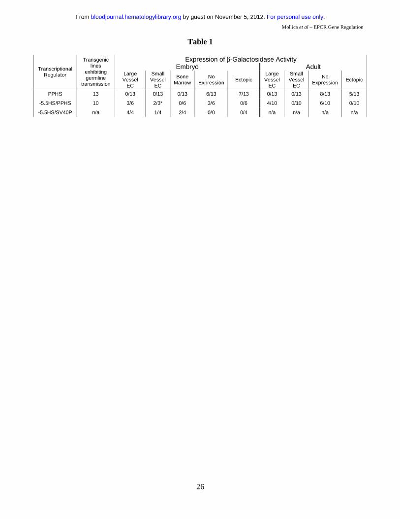
The proximal promoter (PP) alone is incapable of directing cell-specific gene expression in vivo.
To assess PP transcriptional activity in vivo, transgenic mice were generated in which LacZ expression
was directed by PP (-2336 to -9 bp relative to the translation start site), or truncations thereof. Of
seventeen separate transgenic lines, thirteen exhibited germ line transmission of the transgene (Table
1). In eight of these, no β-galactosidase activity was detected in adult transgenic tissues. In the five
remaining lines, LacZ expression was ectopic and non-reproducible. Similarly, LacZ expression was
either ectopic or absent from embryonic transgenic tissues. Failure of PP to direct EPCR-like
transcription in transgenic mice suggested that additional DNA elements outside of this 2.3 kb region
are required for EC specificity in vivo.
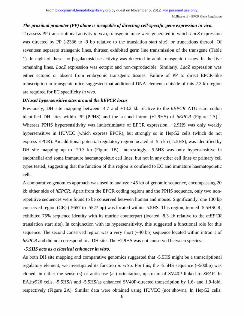
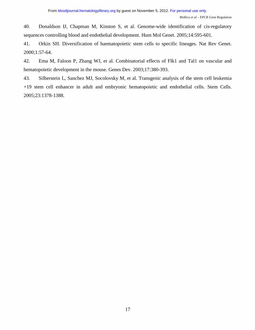
DNaseI hypersensitive sites around the hEPCR locus
Previously, DH site mapping between -4.7 and +18.2 kb relative to the hEPCR ATG start codon
identified DH sites within PP (PPHS) and the second intron (+2.9HS) of hEPCR (Figure 1A)15.
Whereas PPHS hypersensitivity was indiscriminate of EPCR expression, +2.9HS was only weakly
hypersensitive in HUVEC (which express EPCR), but strongly so in HepG2 cells (which do not
express EPCR). An additional potential regulatory region located at -5.5 kb (-5.5HS), was identified by
DH site mapping up to -20.3 kb (Figure 1B). Interestingly, -5.5HS was only hypersensitive in
endothelial and some immature haematopoietic cell lines, but not in any other cell lines or primary cell
types tested, suggesting that the function of this region is confined to EC and immature haematopoietic
cells.
A comparative genomics approach was used to analyze ~45 kb of genomic sequence, encompassing 20
kb either side of hEPCR. Apart from the EPCR coding regions and the PPHS sequence, only two non-
repetitive sequences were found to be conserved between human and mouse. Significantly, one 130 bp
conserved region (CR) (-5657 to -5527 bp) was located within -5.5HS. This region, termed -5.5HSCR,
exhibited 75% sequence identity with its murine counterpart (located -8.3 kb relative to the mEPCR
translation start site). In conjunction with its hypersensitivity, this suggested a functional role for this
sequence. The second conserved region was a very short (~40 bp) sequence located within intron 1 of
hEPCR and did not correspond to a DH site. The +2.9HS was not conserved between species.
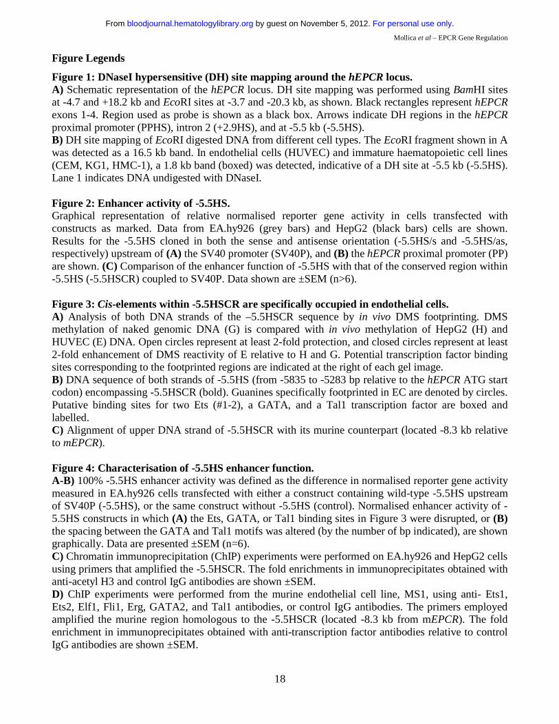
-5.5HS acts as a classical enhancer in vitro.
As both DH site mapping and comparative genomics suggested that -5.5HS might be a transcriptional
regulatory element, we investigated its function in vitro. For this, the -5.5HS sequence (~500bp) was
cloned, in either the sense (s) or antisense (as) orientation, upstream of SV40P linked to SEAP. In
EA.hy926 cells, -5.5HS/s and -5.5HS/as enhanced SV40P-directed transcription by 1.6- and 1.9-fold,
respectively (Figure 2A). Similar data were obtained using HUVEC (not shown). In HepG2 cells,
For personal use only. by guest on November 5, 2012. bloodjournal.hematologylibrary.orgFrom
Mollica et al – EPCR Gene Regulation
7
SV40P-driven transcription was increased 1.8- and 2.1-fold by -5.5HS/s and -5.5HS/as, respectively. In
EA.hy926 cells, -5.5HS/s and -5.5HS/as also enhanced PP-directed transcription by 2.2- and 2-fold
respectively (Figure 2B). -5.5HS did not affect the transcriptional activity of PP in HepG2 cells (not
shown), as PP was inactive in these cells. These results demonstrate that -5.5HS acts as a classical
enhancer in vitro.
-5.5HSCR is responsible for –5.5HS activity.
To delineate the region(s) within -5.5HS responsible for its enhancer activity, we assessed the function
of various -5.5HS fragments. Whereas the fragments encompassing the 130 bp -5.5HSCR were all
similarly active to the ~500 bp -5.5HS (Figure 2C), those lacking -5.5HSCR lost all enhancer activity
(not shown). We thus surmised that its function was primarily attributable to cis-elements contained
within this conserved region.
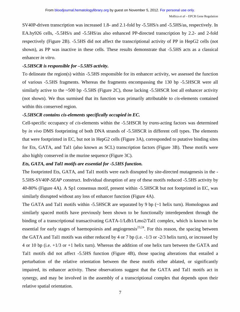
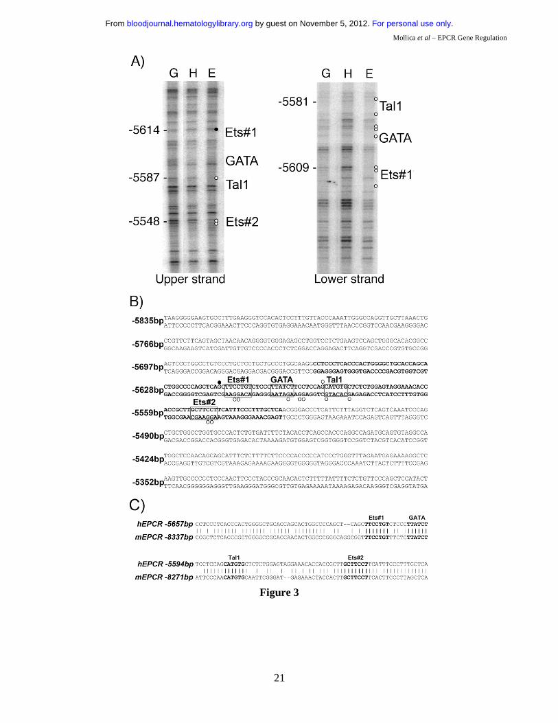
-5.5HSCR contains cis-elements specifically occupied in EC.
Cell-specific occupancy of cis-elements within the -5.5HSCR by trans-acting factors was determined
by in vivo DMS footprinting of both DNA strands of -5.5HSCR in different cell types. The elements
that were footprinted in EC, but not in HepG2 cells (Figure 3A), corresponded to putative binding sites
for Ets, GATA, and Tal1 (also known as SCL) transcription factors (Figure 3B). These motifs were
also highly conserved in the murine sequence (Figure 3C).
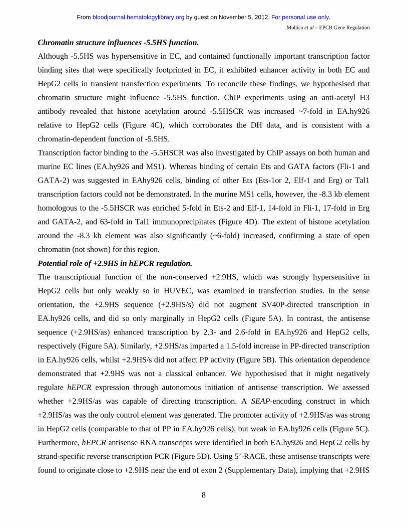
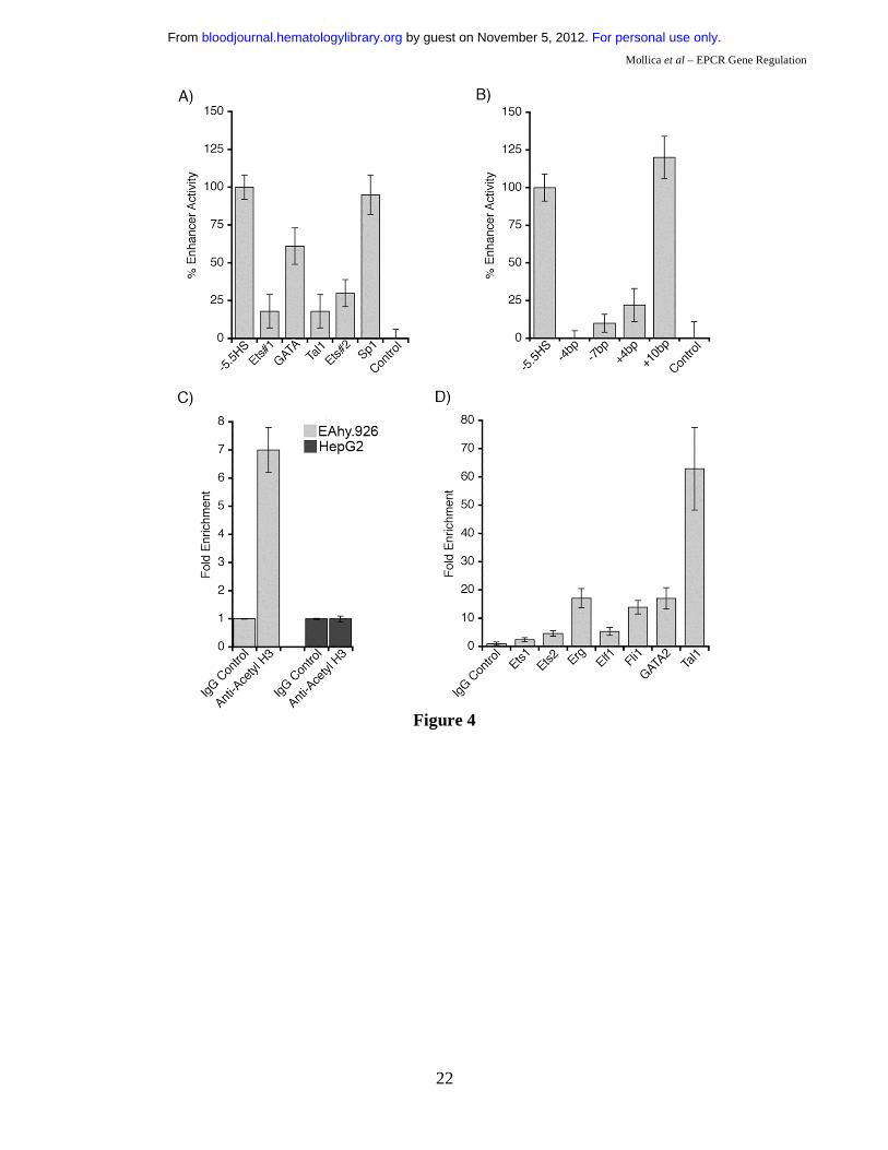
Ets, GATA, and Tal1 motifs are essential for -5.5HS function.
The footprinted Ets, GATA, and Tal1 motifs were each disrupted by site-directed mutagenesis in the -
5.5HS-SV40P-SEAP construct. Individual disruption of any of these motifs reduced -5.5HS activity by
40-80% (Figure 4A). A Sp1 consensus motif, present within -5.5HSCR but not footprinted in EC, was
similarly disrupted without any loss of enhancer function (Figure 4A).
The GATA and Tal1 motifs within -5.5HSCR are separated by 9 bp (~1 helix turn). Homologous and
similarly spaced motifs have previously been shown to be functionally interdependent through the
binding of a transcriptional transactivating GATA-1/Ldb1/Lmo2/Tal1 complex, which is known to be
essential for early stages of haemopoiesis and angiogenesis23,24. For this reason, the spacing between
the GATA and Tal1 motifs was either reduced by 4 or 7 bp (i.e. -1/3 or -2/3 helix turn), or increased by
4 or 10 bp (i.e. +1/3 or +1 helix turn). Whereas the addition of one helix turn between the GATA and
Tal1 motifs did not affect -5.5HS function (Figure 4B), those spacing alterations that entailed a
perturbation of the relative orientation between the these motifs either ablated, or significantly
impaired, its enhancer activity. These observations suggest that the GATA and Tal1 motifs act in
synergy, and may be involved in the assembly of a transcriptional complex that depends upon their
relative spatial orientation.
For personal use only. by guest on November 5, 2012. bloodjournal.hematologylibrary.orgFrom
Mollica et al – EPCR Gene Regulation
8
Chromatin structure influences -5.5HS function.
Although -5.5HS was hypersensitive in EC, and contained functionally important transcription factor
binding sites that were specifically footprinted in EC, it exhibited enhancer activity in both EC and
HepG2 cells in transient transfection experiments. To reconcile these findings, we hypothesised that
chromatin structure might influence -5.5HS function. ChIP experiments using an anti-acetyl H3
antibody revealed that histone acetylation around -5.5HSCR was increased ~7-fold in EA.hy926
relative to HepG2 cells (Figure 4C), which corroborates the DH data, and is consistent with a
chromatin-dependent function of -5.5HS.
Transcription factor binding to the -5.5HSCR was also investigated by ChIP assays on both human and
murine EC lines (EA.hy926 and MS1). Whereas binding of certain Ets and GATA factors (Fli-1 and
GATA-2) was suggested in EAhy926 cells, binding of other Ets (Ets-1or 2, Elf-1 and Erg) or Tal1
transcription factors could not be demonstrated. In the murine MS1 cells, however, the -8.3 kb element
homologous to the -5.5HSCR was enriched 5-fold in Ets-2 and Elf-1, 14-fold in Fli-1, 17-fold in Erg
and GATA-2, and 63-fold in Tal1 immunoprecipitates (Figure 4D). The extent of histone acetylation
around the -8.3 kb element was also significantly (~6-fold) increased, confirming a state of open
chromatin (not shown) for this region.
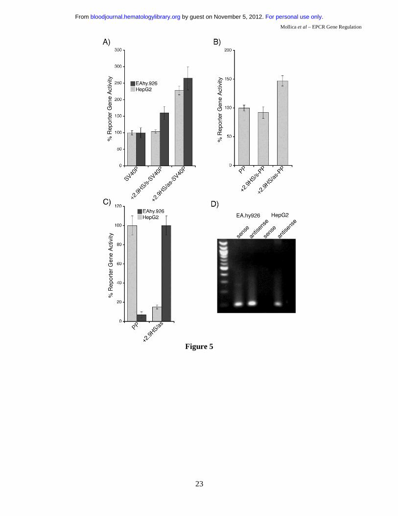
Potential role of +2.9HS in hEPCR regulation.
The transcriptional function of the non-conserved +2.9HS, which was strongly hypersensitive in
HepG2 cells but only weakly so in HUVEC, was examined in transfection studies. In the sense
orientation, the +2.9HS sequence (+2.9HS/s) did not augment SV40P-directed transcription in
EA.hy926 cells, and did so only marginally in HepG2 cells (Figure 5A). In contrast, the antisense
sequence (+2.9HS/as) enhanced transcription by 2.3- and 2.6-fold in EA.hy926 and HepG2 cells,
respectively (Figure 5A). Similarly, +2.9HS/as imparted a 1.5-fold increase in PP-directed transcription
in EA.hy926 cells, whilst +2.9HS/s did not affect PP activity (Figure 5B). This orientation dependence
demonstrated that +2.9HS was not a classical enhancer. We hypothesised that it might negatively
regulate hEPCR expression through autonomous initiation of antisense transcription. We assessed
whether +2.9HS/as was capable of directing transcription. A SEAP-encoding construct in which
+2.9HS/as was the only control element was generated. The promoter activity of +2.9HS/as was strong
in HepG2 cells (comparable to that of PP in EA.hy926 cells), but weak in EA.hy926 cells (Figure 5C).
Furthermore, hEPCR antisense RNA transcripts were identified in both EA.hy926 and HepG2 cells by
strand-specific reverse transcription PCR (Figure 5D). Using 5’-RACE, these antisense transcripts were
found to originate close to +2.9HS near the end of exon 2 (Supplementary Data), implying that +2.9HS
For personal use only. by guest on November 5, 2012. bloodjournal.hematologylibrary.orgFrom
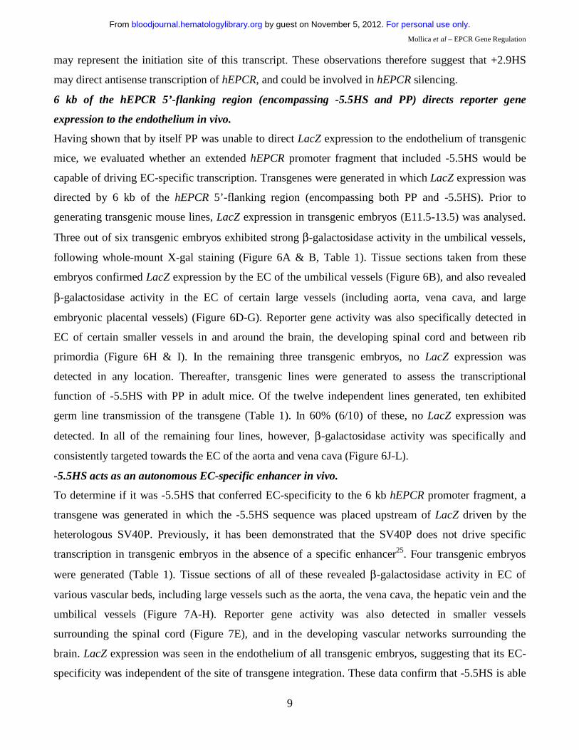
Mollica et al – EPCR Gene Regulation
9
may represent the initiation site of this transcript. These observations therefore suggest that +2.9HS
may direct antisense transcription of hEPCR, and could be involved in hEPCR silencing.
6 kb of the hEPCR 5’-flanking region (encompassing -5.5HS and PP) directs reporter gene
expression to the endothelium in vivo.
Having shown that by itself PP was unable to direct LacZ expression to the endothelium of transgenic
mice, we evaluated whether an extended hEPCR promoter fragment that included -5.5HS would be
capable of driving EC-specific transcription. Transgenes were generated in which LacZ expression was
directed by 6 kb of the hEPCR 5’-flanking region (encompassing both PP and -5.5HS). Prior to
generating transgenic mouse lines, LacZ expression in transgenic embryos (E11.5-13.5) was analysed.
Three out of six transgenic embryos exhibited strong β-galactosidase activity in the umbilical vessels,
following whole-mount X-gal staining (Figure 6A & B, Table 1). Tissue sections taken from these
embryos confirmed LacZ expression by the EC of the umbilical vessels (Figure 6B), and also revealed
β-galactosidase activity in the EC of certain large vessels (including aorta, vena cava, and large
embryonic placental vessels) (Figure 6D-G). Reporter gene activity was also specifically detected in
EC of certain smaller vessels in and around the brain, the developing spinal cord and between rib
primordia (Figure 6H & I). In the remaining three transgenic embryos, no LacZ expression was
detected in any location. Thereafter, transgenic lines were generated to assess the transcriptional
function of -5.5HS with PP in adult mice. Of the twelve independent lines generated, ten exhibited
germ line transmission of the transgene (Table 1). In 60% (6/10) of these, no LacZ expression was
detected. In all of the remaining four lines, however, β-galactosidase activity was specifically and
consistently targeted towards the EC of the aorta and vena cava (Figure 6J-L).
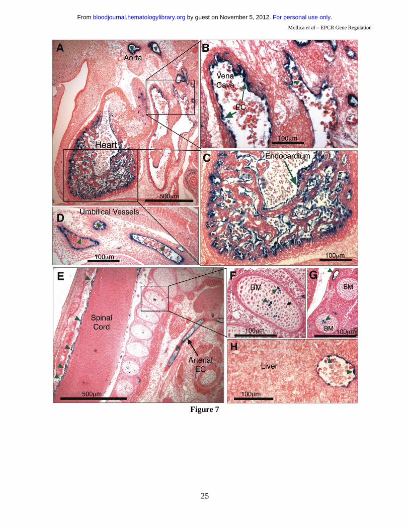
-5.5HS acts as an autonomous EC-specific enhancer in vivo.
To determine if it was -5.5HS that conferred EC-specificity to the 6 kb hEPCR promoter fragment, a
transgene was generated in which the -5.5HS sequence was placed upstream of LacZ driven by the
heterologous SV40P. Previously, it has been demonstrated that the SV40P does not drive specific
transcription in transgenic embryos in the absence of a specific enhancer25. Four transgenic embryos
were generated (Table 1). Tissue sections of all of these revealed β-galactosidase activity in EC of
various vascular beds, including large vessels such as the aorta, the vena cava, the hepatic vein and the
umbilical vessels (Figure 7A-H). Reporter gene activity was also detected in smaller vessels
surrounding the spinal cord (Figure 7E), and in the developing vascular networks surrounding the
brain. LacZ expression was seen in the endothelium of all transgenic embryos, suggesting that its EC-
specificity was independent of the site of transgene integration. These data confirm that -5.5HS is able
For personal use only. by guest on November 5, 2012. bloodjournal.hematologylibrary.orgFrom
Mollica et al – EPCR Gene Regulation
10
to autonomously direct transcription to the EC in vivo. In addition, one of the four transgenic embryos
also presented strong X-gal staining in the endocardium lining the chambers of the developing heart
(Figure 7A & C). Two also presented strong LacZ expression in a small number of cells within the
bone marrow (Figure 7E-G, Table 1).
Parallel analyses of the function of the 40 bp conserved sequence in intron 1 failed to reveal any role
for this element in specific gene regulation (data not shown).
Discussion
Despite considerable progress in our knowledge of EC gene regulation and vascular bed specificity26-29,
there has been incomplete understanding of the mechanisms targeting the expression of genes such as
EPCR to large vessel EC. In a study by Gu et al, transgenic mice were generated which expressed the
green fluorescent protein under the control of mEPCR promoter fragments (extending up to -1080 bp).
However, that study failed to observe any fluorescence of this reporter protein that might allow the
assessment of cellular specificity in vivo14. The present study investigates the transcriptional regulation
of hEPCR, and is the first to elucidate the mechanisms underlying its EC-specific expression. Our
survey of the chromatin structure around the hEPCR locus (from -20.3 to +18.2 kb relative to the
hEPCR ATG start codon) identified three DH regions: PPHS, +2.9HS, and -5.5HS. Although the PP,
which encompasses the PPHS, was previously shown to mediate EC-specific transcription in
transfected EC15, this region alone was insufficient to direct cell-specific reporter gene expression in
transgenic mice. In this study, we demonstrate that EC-specific expression of hEPCR in vivo requires
the cooperation of -5.5HS.
Although we have not identified a clear function for the intronic +2.9HS, this element was more
hypersensitive to DNaseI in cells that do not express EPCR (HepG2) than in cells that do (HUVEC). In
its antisense orientation, +2.9HS exhibited autonomous promoter activity, which was greater in HepG2
than in EA.hy926 cells. Given the increasingly recognised role of antisense transcripts in gene
regulation30, we hypothesise that +2.9HS could help silence hEPCR expression in EPCR negative cells,
or modulate its levels in EC, by directing antisense transcription of hEPCR from intron 2 towards exon
1. Our hypothesis is supported by the detection of hEPCR antisense transcripts originating near
+2.9HS. As the +2.9HS sequence is not conserved between humans and mice, this limits the possibility
of probing its functionality in a murine model.
The -5.5HS was only hypersensitive to DNaseI in EC and immature haematopoietic cells. In transient
transfection experiments, -5.5HS behaved as a classical enhancer, whose activity was not cell-specific.
This does not, however, preclude a cell-specific function for -5.5HS in vivo, as this enhancer could
For personal use only. by guest on November 5, 2012. bloodjournal.hematologylibrary.orgFrom
Mollica et al – EPCR Gene Regulation
11
require chromosomal/chromatin integration to exert its cellular specificity, as suggested by its
hypersensitivity. Histone acetylation around the -5.5HSCR, reflecting disruption of higher-order
chromatin structure, was increased in cells that express EPCR (EA.hy926) compared to cells that do
not (HepG2), corroborating the contention that -5.5HS function is chromatin-dependent. Other
examples of chromatin-dependent enhancers involved in EC- or HSC-specific gene regulation include
those of the Flk131, Tal1 (also known as Scl)32, and CD3433 genes.
The -5.5HS encompasses ~130 bp (-5.5HSCR) that is conserved between humans and mice and that
was both necessary and sufficient for -5.5HS enhancer function. Its activity relied on the interplay of
transcription factors acting upon Ets, GATA, and Tal1-binding motifs. Disruption of any of these
motifs resulted in marked loss of enhancer function, suggesting that the bound transcription factors
may be components of an oligomeric complex. This observation is reminiscent of the Tal1
enhanceosome whose specific function in EC and HSC requires a combination of Ets and GATA
family transcription factors32. Perturbation of the spatial orientation between the GATA and Tal1
motifs almost completely abrogated -5.5HS activity, whereas addition of 10 bp (one helix turn)
between these motifs did not affect enhancer function. These findings are consistent with the concept
that the GATA-Tal1 bipartite DNA motif is involved in the assembly of an enhanceosome that can only
form with the GATA and Tal1 motifs in a specific relative orientation on the DNA helix. A GATA-
Tal1 bipartite DNA motif analogous to that present in the -5.5HS has previously been shown to be
capable of binding an oligomeric transactivation complex essential for both haematopoiesis and
angiogenesis23,24.
Ets, GATA, and Tal1 transcription factors are expressed in both EC and HSC34-36, suggesting that they
might contribute to a common mechanism of hEPCR regulation. In vivo DMS footprinting of the -
5.5HSCR revealed that the Ets, GATA, and Tal1 motifs were occupied by trans-acting factors in EC
but not in HepG2 cells. ChIP assays for these factors performed with the murine MS1 EC line
demonstrated binding of the Ets (Ets2, Erg, Elf1, and Fli1), GATA2, and Tal1 transcription factors to
their cognate motifs in the murine region homologous to -5.5HSCR.
The hEPCR PP failed to drive cell-specific transcription in vivo. However, using 6 kb of the hEPCR 5’-
flanking region that included both PP and -5.5HS, strong, specific, and reproducible reporter gene
expression was directed to the large vessel endothelium. This included the aorta, umbilical vessels, and
selected small vessels surrounding the developing spinal cord and brain. The contrast between these
findings and the findings obtained with PP alone strongly suggested that -5.5HS played a critical role in
targeting EC-specific transgene expression. In each of the four independent adult transgenic lines
expressing LacZ, β-galactosidase activity was limited to the endothelium of the aorta and vena cava,
For personal use only. by guest on November 5, 2012. bloodjournal.hematologylibrary.orgFrom

Mollica et al – EPCR Gene Regulation
12
and in no other location. Although LacZ was not expressed in all vascular beds known to express
EPCR, the 6 kb promoter fragment was capable of discriminating between large and small vessels, a
functionality that has not been demonstrated for any other promoter fragment. The more restricted
transgene expression in adult mice possibly reflects differences in gene regulation between embryonic
and adult tissues37.
To confirm that it was the -5.5HS that conferred EC-specificity to the 6 kb hEPCR promoter, -5.5HS
was coupled to the heterologous SV40P. In transgenic embryos, high-level reporter gene expression
was reproducibly directed to EC, demonstrating that, by itself, the -5.5HS enhancer contains the
regulatory sequences necessary to impart vascular specificity, and thus functions as an autonomous EC-
specific enhancer. Other enhancers have been shown to autonomously direct transcription to the
endothelium of transgenic embryos27,32,38-40, but -5.5HS is the first human enhancer capable of targeting
transcription to both embryonic and adult EC. Moreover, to our knowledge, -5.5HS is the first
regulatory element capable of specifically targeting transcription to large vessel EC. As such, it may
provide a valuable tool for the development of a gene expression unit that specifically targets large
vessels, which are the principal site of atherosclerosis and many vascular diseases.
A recent study by Balazs et al. identified abundant EPCR expression on primitive HSC, and
demonstrated it to be an excellent marker for the identification of such cells from murine bone
marrow13. We hypothesise that -5.5HS contains the regulatory elements necessary to target gene
expression not only to EC, but potentially also to HSC. The hypersensitivity of -5.5HS in immature
haematopoietic cells, and the detection of β-galactosidase in small numbers of cells within the bone
marrow of transgenic mice generated with the -5.5HS-SV40P-LacZ construct, both support this
hypothesis. An ontogenic rationale is available for the proposed dual endothelial-haematopoietic
function of -5.5HS as the development of blood and endothelium are known to be intertwined41. In the
early embryo, the fate of differentiation of the haemangioblast into either an HSC or an EC is thought
to be dictated by the combinatorial effects of the Tal1 and Flk1 genes42. Together with these genes, the
Fli1 and PRH genes contribute to the transcriptional regulation of embryonic HSC and EC formation40.
Interestingly, the Tal1, Flk1, Fli1 and PRH genes are all governed by autonomous enhancers with dual
HSC/EC activity32,38-40. Furthermore, the activity of each of these enhancers is dependent on cis-
elements similar to those present within -5.5HS, namely Ets, GATA, and Tal1 motifs. In light of this,
we propose a model in which -5.5HS targets hEPCR expression to primitive stem cells with dual
specification potential, and in which hEPCR expression is progressively lost as these cells embark into
haematopoietic specification. Of the autonomous HSC/EC enhancers mentioned above, only the
murine Tal1 +19 kb enhancer has been shown capable of targeting transgene expression to adult
For personal use only. by guest on November 5, 2012. bloodjournal.hematologylibrary.orgFrom

Mollica et al – EPCR Gene Regulation
13
HSC43. The Tal1 +19 kb enhancer is not, however, entirely HSC/EC-specific, as it also targets
transgene expression to mast cells, megakaryocytes, and thymocytes43. By comparison, -5.5HS may be
more specific for primitive cells, and thereby potentially represents a more specific HSC gene targeting
tool.
Acknowledgements
The authors would like to thank Dr Rachel Simmonds, Dr George Bou-Gharios, Dr Bertie Göttgens
and Dr Alain Chan for their help with this work.
For personal use only. by guest on November 5, 2012. bloodjournal.hematologylibrary.orgFrom

Mollica et al – EPCR Gene Regulation
14
References
1. Esmon CT. Inflammation and thrombosis. J Thromb Haemost. 2003;1:1343-1348.
2. Riewald M, Petrovan RJ, Donner A, Mueller BM, Ruf W. Activation of endothelial cell
protease activated receptor 1 by the protein C pathway. Science. 2002;296:1880-1882.
3. Cheng T, Liu D, Griffin JH, et al. Activated protein C blocks p53-mediated apoptosis in
ischemic human brain endothelium and is neuroprotective. Nat Med. 2003;9:338-342.
4. Lane DA, Mannucci PM, Bauer KA, et al. Inherited thrombophilia: Part 1. Thromb Haemost.
1996;76:651-662.
5. Bernard GR, Vincent JL, Laterre PF, et al. Efficacy and safety of recombinant human activated
protein C for severe sepsis. N Engl J Med. 2001;344:699-709.
6. Fukudome K, Esmon CT. Identification, cloning, and regulation of a novel endothelial cell
protein C/activated protein C receptor. J Biol Chem. 1994;269:26486-26491.
7. Stearns-Kurosawa DJ, Kurosawa S, Mollica JS, Ferrell GL, Esmon CT. The endothelial cell
protein C receptor augments protein C activation by the thrombin-thrombomodulin complex. Proc Natl
Acad Sci U S A. 1996;93:10212-10216.
8. Xu J, Esmon NL, Esmon CT. Reconstitution of the human endothelial cell protein C receptor
with thrombomodulin in phosphatidylcholine vesicles enhances protein C activation. J Biol Chem.
1999;274:6704-6710.
9. Taylor FB, Jr., Peer GT, Lockhart MS, Ferrell G, Esmon CT. Endothelial cell protein C receptor
plays an important role in protein C activation in vivo. Blood. 2001;97:1685-1688.
10. Gu JM, Crawley JT, Ferrell G, et al. Disruption of the endothelial cell protein C receptor gene
in mice causes placental thrombosis and early embryonic lethality. J Biol Chem. 2002;277:43335-
43343.
11. Crawley JT, Gu JM, Ferrell G, Esmon CT. Distribution of endothelial cell protein C/activated
protein C receptor (EPCR) during mouse embryo development. Thromb Haemost. 2002;88:259-266.
12. Laszik Z, Mitro A, Taylor FB, Jr., Ferrell G, Esmon CT. Human protein C receptor is present
primarily on endothelium of large blood vessels: implications for the control of the protein C pathway.
Circulation. 1997;96:3633-3640.
13. Balazs AB, Fabian AJ, Esmon CT, Mulligan RC. Endothelial protein C receptor (CD201)
explicitly identifies hematopoietic stem cells in murine bone marrow. Blood. 2005.
14. Gu JM, Fukudome K, Esmon CT. Characterization and regulation of the 5'-flanking region of
the murine endothelial protein C receptor gene. J Biol Chem. 2000;275:12481-12488.
For personal use only. by guest on November 5, 2012. bloodjournal.hematologylibrary.orgFrom
Mollica et al – EPCR Gene Regulation
15
15. Rance JB, Follows GA, Cockerill PN, Bonifer C, Lane DA, Simmonds RE. Regulation of the
human endothelial cell protein C receptor gene promoter by multiple Sp1 binding sites. Blood.
2003;101:4393-4401.
16. Simmonds RE, Lane DA. Structural and functional implications of the intron/exon organization
of the human endothelial cell protein C/activated protein C receptor (EPCR) gene: comparison with the
structure of CD1/major histocompatibility complex alpha1 and alpha2 domains. Blood. 1999;94:632-
641.
17. Liang Z, Rosen ED, Castellino FJ. Nucleotide structure and characterization of the murine gene
encoding the endothelial cell protein C receptor. Thromb Haemost. 1999;81:585-588.
18. Hayashi T, Nakamura H, Okada A, et al. Organization and chromosomal localization of the
human endothelial protein C receptor gene. Gene. 1999;238:367-373.
19. Cockerill PN, Bert AG, Roberts D, Vadas MA. The human granulocyte-macrophage colony-
stimulating factor gene is autonomously regulated in vivo by an inducible tissue-specific enhancer.
Proc Natl Acad Sci U S A. 1999;96:15097-15102.
20. Kontaraki J, Chen HH, Riggs A, Bonifer C. Chromatin fine structure profiles for a
developmentally regulated gene: reorganization of the lysozyme locus before trans-activator binding
and gene expression. Genes Dev. 2000;14:2106-2122.
21. Forsberg EC, Downs KM, Bresnick EH. Direct interaction of NF-E2 with hypersensitive site 2
of the beta-globin locus control region in living cells. Blood. 2000;96:334-339.
22. Nagy A, Gertsenstein, M., Vintersten, K., and Behringer, R. Manipulating the Mouse Embryo
(ed Third Edition): Cold Spring Harbor Laboratory Press, Cold Spring Harbour, NY; 2003.
23. Wadman IA, Osada H, Grutz GG, et al. The LIM-only protein Lmo2 is a bridging molecule
assembling an erythroid, DNA-binding complex which includes the TAL1, E47, GATA-1 and
Ldb1/NLI proteins. Embo J. 1997;16:3145-3157.
24. Yamada Y, Warren AJ, Dobson C, Forster A, Pannell R, Rabbitts TH. The T cell leukemia LIM
protein Lmo2 is necessary for adult mouse hematopoiesis. Proc Natl Acad Sci U S A. 1998;95:3890-
3895.
25. Sinclair AM, Gottgens B, Barton LM, et al. Distinct 5' SCL enhancers direct transcription to
developing brain, spinal cord, and endothelium: neural expression is mediated by GATA factor binding
sites. Dev Biol. 1999;209:128-142.
26. Aird WC, Jahroudi N, Weiler-Guettler H, Rayburn HB, Rosenberg RD. Human von Willebrand
factor gene sequences target expression to a subpopulation of endothelial cells in transgenic mice. Proc
Natl Acad Sci U S A. 1995;92:4567-4571.
For personal use only. by guest on November 5, 2012. bloodjournal.hematologylibrary.orgFrom
Mollica et al – EPCR Gene Regulation
16
27. Schlaeger TM, Bartunkova S, Lawitts JA, et al. Uniform vascular-endothelial-cell-specific gene
expression in both embryonic and adult transgenic mice. Proc Natl Acad Sci U S A. 1997;94:3058-
3063.
28. Schlaeger TM, Qin Y, Fujiwara Y, Magram J, Sato TN. Vascular endothelial cell lineage-
specific promoter in transgenic mice. Development. 1995;121:1089-1098.
29. Minami T, Aird WC. Endothelial cell gene regulation. Trends Cardiovasc Med. 2005;15:174-
184.
30. Lavorgna G, Dahary D, Lehner B, Sorek R, Sanderson CM, Casari G. In search of antisense.
Trends Biochem Sci. 2004;29:88-94.
31. Kappel A, Schlaeger TM, Flamme I, Orkin SH, Risau W, Breier G. Role of SCL/Tal-1, GATA,
and ets transcription factor binding sites for the regulation of flk-1 expression during murine vascular
development. Blood. 2000;96:3078-3085.
32. Gottgens B, Nastos A, Kinston S, et al. Establishing the transcriptional programme for blood:
the SCL stem cell enhancer is regulated by a multiprotein complex containing Ets and GATA factors.
Embo J. 2002;21:3039-3050.
33. May G, Enver T. Targeting gene expression to haemopoietic stem cells: a chromatin-dependent
upstream element mediates cell type-specific expression of the stem cell antigen CD34. Embo J.
1995;14:564-574.
34. Sato Y. Role of ETS family transcription factors in vascular development and angiogenesis.
Cell Struct Funct. 2001;26:19-24.
35. Dorfman DM, Wilson DB, Bruns GA, Orkin SH. Human transcription factor GATA-2.
Evidence for regulation of preproendothelin-1 gene expression in endothelial cells. J Biol Chem.
1992;267:1279-1285.
36. Kallianpur AR, Jordan JE, Brandt SJ. The SCL/TAL-1 gene is expressed in progenitors of both
the hematopoietic and vascular systems during embryogenesis. Blood. 1994;83:1200-1208.
37. Santos KF, Mazzola TN, Carvalho HF. The prima donna of epigenetics: the regulation of gene
expression by DNA methylation. Braz J Med Biol Res. 2005;38:1531-1541.
38. Gottgens B, Broccardo C, Sanchez MJ, et al. The scl +18/19 stem cell enhancer is not required
for hematopoiesis: identification of a 5' bifunctional hematopoietic-endothelial enhancer bound by Fli-1
and Elf-1. Mol Cell Biol. 2004;24:1870-1883.
39. Kappel A, Ronicke V, Damert A, Flamme I, Risau W, Breier G. Identification of vascular
endothelial growth factor (VEGF) receptor-2 (Flk-1) promoter/enhancer sequences sufficient for
angioblast and endothelial cell-specific transcription in transgenic mice. Blood. 1999;93:4284-4292.
For personal use only. by guest on November 5, 2012. bloodjournal.hematologylibrary.orgFrom
Mollica et al – EPCR Gene Regulation
17
40. Donaldson IJ, Chapman M, Kinston S, et al. Genome-wide identification of cis-regulatory
sequences controlling blood and endothelial development. Hum Mol Genet. 2005;14:595-601.
41. Orkin SH. Diversification of haematopoietic stem cells to specific lineages. Nat Rev Genet.
2000;1:57-64.
42. Ema M, Faloon P, Zhang WJ, et al. Combinatorial effects of Flk1 and Tal1 on vascular and
hematopoietic development in the mouse. Genes Dev. 2003;17:380-393.
43. Silberstein L, Sanchez MJ, Socolovsky M, et al. Transgenic analysis of the stem cell leukemia
+19 stem cell enhancer in adult and embryonic hematopoietic and endothelial cells. Stem Cells.
2005;23:1378-1388.
For personal use only. by guest on November 5, 2012. bloodjournal.hematologylibrary.orgFrom
Mollica et al – EPCR Gene Regulation
18
Figure Legends
Figure 1: DNaseI hypersensitive (DH) site mapping around the hEPCR locus. A) Schematic representation of the hEPCR locus. DH site mapping was performed using BamHI sites at -4.7 and +18.2 kb and EcoRI sites at -3.7 and -20.3 kb, as shown. Black rectangles represent hEPCR exons 1-4. Region used as probe is shown as a black box. Arrows indicate DH regions in the hEPCR proximal promoter (PPHS), intron 2 (+2.9HS), and at -5.5 kb (-5.5HS). B) DH site mapping of EcoRI digested DNA from different cell types. The EcoRI fragment shown in A was detected as a 16.5 kb band. In endothelial cells (HUVEC) and immature haematopoietic cell lines (CEM, KG1, HMC-1), a 1.8 kb band (boxed) was detected, indicative of a DH site at -5.5 kb (-5.5HS). Lane 1 indicates DNA undigested with DNaseI. Figure 2: Enhancer activity of -5.5HS. Graphical representation of relative normalised reporter gene activity in cells transfected with constructs as marked. Data from EA.hy926 (grey bars) and HepG2 (black bars) cells are shown. Results for the -5.5HS cloned in both the sense and antisense orientation (-5.5HS/s and -5.5HS/as, respectively) upstream of (A) the SV40 promoter (SV40P), and (B) the hEPCR proximal promoter (PP) are shown. (C) Comparison of the enhancer function of -5.5HS with that of the conserved region within -5.5HS (-5.5HSCR) coupled to SV40P. Data shown are ±SEM (n>6). Figure 3: Cis-elements within -5.5HSCR are specifically occupied in endothelial cells. A) Analysis of both DNA strands of the –5.5HSCR sequence by in vivo DMS footprinting. DMS methylation of naked genomic DNA (G) is compared with in vivo methylation of HepG2 (H) and HUVEC (E) DNA. Open circles represent at least 2-fold protection, and closed circles represent at least 2-fold enhancement of DMS reactivity of E relative to H and G. Potential transcription factor binding sites corresponding to the footprinted regions are indicated at the right of each gel image. B) DNA sequence of both strands of -5.5HS (from -5835 to -5283 bp relative to the hEPCR ATG start codon) encompassing -5.5HSCR (bold). Guanines specifically footprinted in EC are denoted by circles. Putative binding sites for two Ets (#1-2), a GATA, and a Tal1 transcription factor are boxed and labelled. C) Alignment of upper DNA strand of -5.5HSCR with its murine counterpart (located -8.3 kb relative to mEPCR). Figure 4: Characterisation of -5.5HS enhancer function. A-B) 100% -5.5HS enhancer activity was defined as the difference in normalised reporter gene activity measured in EA.hy926 cells transfected with either a construct containing wild-type -5.5HS upstream of SV40P (-5.5HS), or the same construct without -5.5HS (control). Normalised enhancer activity of -5.5HS constructs in which (A) the Ets, GATA, or Tal1 binding sites in Figure 3 were disrupted, or (B) the spacing between the GATA and Tal1 motifs was altered (by the number of bp indicated), are shown graphically. Data are presented ±SEM (n=6). C) Chromatin immunoprecipitation (ChIP) experiments were performed on EA.hy926 and HepG2 cells using primers that amplified the -5.5HSCR. The fold enrichments in immunoprecipitates obtained with anti-acetyl H3 and control IgG antibodies are shown ±SEM. D) ChIP experiments were performed from the murine endothelial cell line, MS1, using anti- Ets1, Ets2, Elf1, Fli1, Erg, GATA2, and Tal1 antibodies, or control IgG antibodies. The primers employed amplified the murine region homologous to the -5.5HSCR (located -8.3 kb from mEPCR). The fold enrichment in immunoprecipitates obtained with anti-transcription factor antibodies relative to control IgG antibodies are shown ±SEM.
For personal use only. by guest on November 5, 2012. bloodjournal.hematologylibrary.orgFrom
Mollica et al – EPCR Gene Regulation
19
Figure 5: Functional characterisation of +2.9HS. (A-B) Enhancer function of the +2.9HS. The sense (+2.9HS/s) and antisense (+2.9HS/as) sequences of +2.9HS were coupled to either the SV40P (A) or the hEPCR PP (B). Normalised reporter gene activity obtained following transfection of constructs into EA.hy926 (grey bars) and HepG2 (black bars) cells are presented, ±SEM (n=6). (C) Promoter function of +2.9HS/as compared to that of PP in EA.hy926 and HepG2 cells. +2.9HS/as and PP were cloned upstream of a reporter gene in a construct without any other promoter. Normalised reporter gene activity are presented relative to that of PP in EA.hy926 cells, ±SEM (n=6). (D) Detection of antisense hEPCR transcripts. RT-PCR was performed on total RNA from EA.hy926 or HepG2 cells using strand-specific primers that specifically amplified transcripts spanning hEPCR exons 1 and 2. Nested PCR and electrophoresis was used to visualise amplified sequences. The identity of the amplified RNA transcripts (sense and antisense) was confirmed by sequencing. Figure 6: 6 kb of the hEPCR 5’-flanking region transcriptionally targets EC in transgenic mice. Transgenic mice containing 6 kb of the hEPCR 5’-flanking region coupled to LacZ were generated. Embryos (A-I) and adult tissues (J-L) were analysed. Embryos were harvested at E12.5 and whole-mount stained with X-gal (A & B). Thereafter stained embryos were wax embedded, sectioned and counter-stained with eosin (C-I). Sites of LacZ expression are dark blue (green arrows). In whole-mount stained embryos, staining in the umbilical vessels was apparent (A & B), which was confirmed as endothelial in tissue sections (C). Transgene expression was also specifically detected in EC of large placental veins (D), aorta (E & F), and certain other larger veins and arteries (G). EC-specific expression was also seen in small vessels surrounding the spinal cord (H) and in the developing brain (I). Tissues from adult transgenic mice were stained with X-gal and counter-stained with haematoxylin (J-L). Reporter gene expression was specifically detected in the endothelium lining the aorta and vena cava (J-L). Figure 7: -5.5HS functions as an autonomous endothelial enhancer in transgenic mice. Transgenic embryos generated with LacZ under the control of -5.5HS/SV40P were harvested at ~E12.5, whole-mount stained with X-gal, sectioned, and counter-stained with eosin. Sites of LacZ expression are dark blue (green arrows). In 4/4 transgenic embryos reporter gene expression was specifically detected in EC, including those of the aorta and vena cava (A & B). In one embryo, very strong staining of the endocardium lining the chambers of the developing heart was seen (A & C). Transgene expression was also specifically detected in EC of umbilical vessels (D), small vessels surrounding the spinal cord (E), and in the hepatic vein (H). In 2/4 embryos, strong staining in a small number of bone marrow (BM) cells was seen (E-G). Table 1: Summary of reporter gene distribution in transgenic mouse lines. The number of transgenic mouse lines exhibiting germline transmission are given. Of these, the number demonstrating reporter gene expression in given locations are stated (* of the 3 embryos exhibiting large vessel EC expression, 2 of these also showed small vessel EC reporter gene activity). For the -5.5HS/SV40P analysis, only transgenic embryos generated following microinjection were analysed.
For personal use only. by guest on November 5, 2012. bloodjournal.hematologylibrary.orgFrom
Mollica et al – EPCR Gene Regulation
20
Figure 1
Figure 2
For personal use only. by guest on November 5, 2012. bloodjournal.hematologylibrary.orgFrom
Mollica et al – EPCR Gene Regulation
21
Figure 3
For personal use only. by guest on November 5, 2012. bloodjournal.hematologylibrary.orgFrom
Mollica et al – EPCR Gene Regulation
22
Figure 4
For personal use only. by guest on November 5, 2012. bloodjournal.hematologylibrary.orgFrom
Mollica et al – EPCR Gene Regulation
23
Figure 5
For personal use only. by guest on November 5, 2012. bloodjournal.hematologylibrary.orgFrom

Mollica et al – EPCR Gene Regulation
24
Figure 6
For personal use only. by guest on November 5, 2012. bloodjournal.hematologylibrary.orgFrom
Mollica et al – EPCR Gene Regulation
25
Figure 7
For personal use only. by guest on November 5, 2012. bloodjournal.hematologylibrary.orgFrom
Mollica et al – EPCR Gene Regulation
26
Table 1
Expression of β-Galactosidase Activity Embryo Adult Transcriptional
Regulator
Transgenic lines
exhibiting germline
transmission
Large Vessel
EC
Small Vessel
EC
Bone Marrow
No Expression Ectopic
Large Vessel
EC
Small Vessel
EC
No Expression Ectopic
PPHS 13 0/13 0/13 0/13 6/13 7/13 0/13 0/13 8/13 5/13
-5.5HS/PPHS 10 3/6 2/3* 0/6 3/6 0/6 4/10 0/10 6/10 0/10
-5.5HS/SV40P n/a 4/4 1/4 2/4 0/0 0/4 n/a n/a n/a n/a
For personal use only. by guest on November 5, 2012. bloodjournal.hematologylibrary.orgFrom
Copyright © 2022 FDOKUMEN