Role for Human SIRT2 NAD-Dependent Deacetylase Activity in Control of Mitotic Exit in the Cell Cycle
14
10.1128/MCB.23.9.3173-3185.2003. 2003, 23(9):3173. DOI: Mol. Cell. Biol. Anton-Scott Goustin and Michael A. Tainsky Sylvia C. Dryden, Fatimah A. Nahhas, James E. Nowak, Exit in the Cell Cycle Deacetylase Activity in Control of Mitotic Role for Human SIRT2 NAD-Dependent http://mcb.asm.org/content/23/9/3173 Updated information and services can be found at: These include: REFERENCES http://mcb.asm.org/content/23/9/3173#ref-list-1 at: This article cites 43 articles, 19 of which can be accessed free CONTENT ALERTS more» articles cite this article), Receive: RSS Feeds, eTOCs, free email alerts (when new http://journals.asm.org/site/misc/reprints.xhtml Information about commercial reprint orders: http://journals.asm.org/site/subscriptions/ To subscribe to to another ASM Journal go to: on January 28, 2014 by guest http://mcb.asm.org/ Downloaded from on January 28, 2014 by guest http://mcb.asm.org/ Downloaded from
-
Upload
independent -
Category
Documents
-
view
4 -
download
0
Transcript of Role for Human SIRT2 NAD-Dependent Deacetylase Activity in Control of Mitotic Exit in the Cell Cycle
10.1128/MCB.23.9.3173-3185.2003.
2003, 23(9):3173. DOI:Mol. Cell. Biol. Anton-Scott Goustin and Michael A. TainskySylvia C. Dryden, Fatimah A. Nahhas, James E. Nowak, Exit in the Cell CycleDeacetylase Activity in Control of Mitotic Role for Human SIRT2 NAD-Dependent
http://mcb.asm.org/content/23/9/3173Updated information and services can be found at:
These include:
REFERENCEShttp://mcb.asm.org/content/23/9/3173#ref-list-1at:
This article cites 43 articles, 19 of which can be accessed free
CONTENT ALERTS more»articles cite this article),
Receive: RSS Feeds, eTOCs, free email alerts (when new
http://journals.asm.org/site/misc/reprints.xhtmlInformation about commercial reprint orders: http://journals.asm.org/site/subscriptions/To subscribe to to another ASM Journal go to:
on January 28, 2014 by guesthttp://m
cb.asm.org/
Dow
nloaded from
on January 28, 2014 by guesthttp://m
cb.asm.org/
Dow
nloaded from
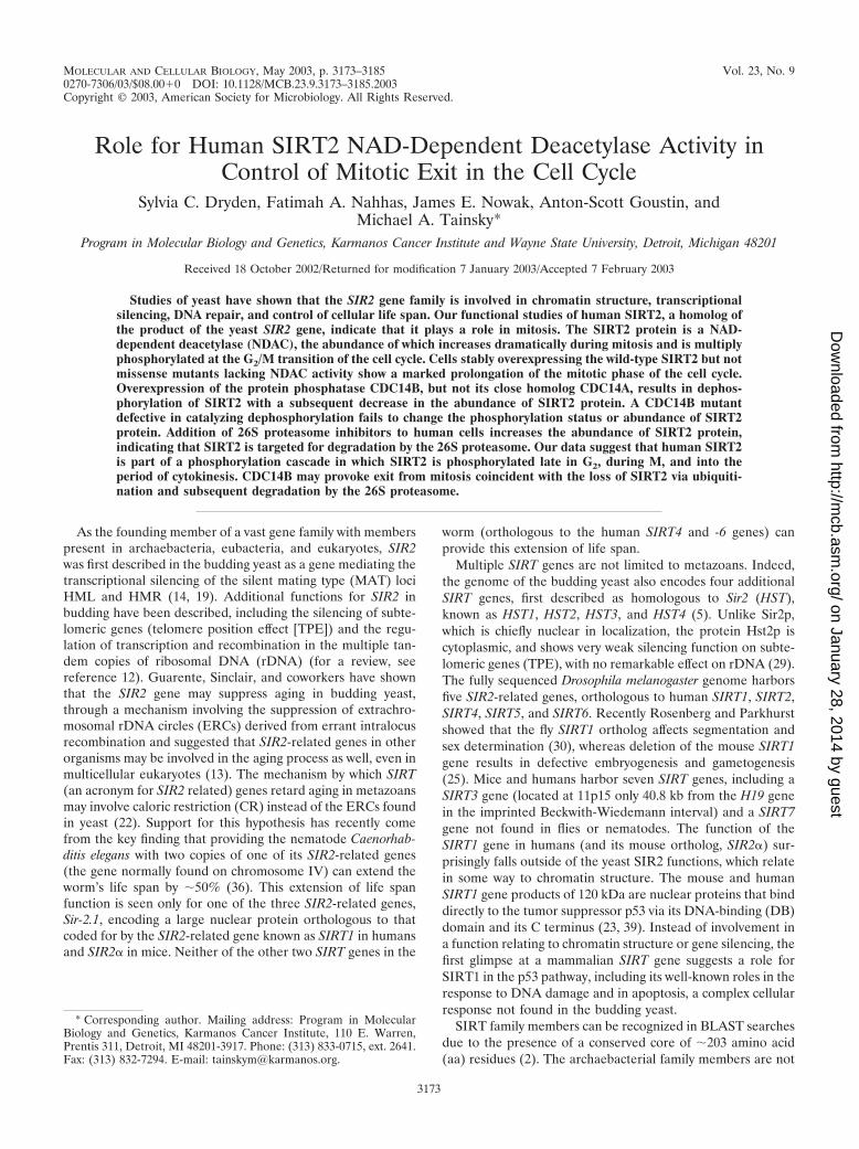
MOLECULAR AND CELLULAR BIOLOGY, May 2003, p. 3173–3185 Vol. 23, No. 90270-7306/03/$08.00�0 DOI: 10.1128/MCB.23.9.3173–3185.2003Copyright © 2003, American Society for Microbiology. All Rights Reserved.
Role for Human SIRT2 NAD-Dependent Deacetylase Activity inControl of Mitotic Exit in the Cell Cycle
Sylvia C. Dryden, Fatimah A. Nahhas, James E. Nowak, Anton-Scott Goustin, andMichael A. Tainsky*
Program in Molecular Biology and Genetics, Karmanos Cancer Institute and Wayne State University, Detroit, Michigan 48201
Received 18 October 2002/Returned for modification 7 January 2003/Accepted 7 February 2003
Studies of yeast have shown that the SIR2 gene family is involved in chromatin structure, transcriptionalsilencing, DNA repair, and control of cellular life span. Our functional studies of human SIRT2, a homolog ofthe product of the yeast SIR2 gene, indicate that it plays a role in mitosis. The SIRT2 protein is a NAD-dependent deacetylase (NDAC), the abundance of which increases dramatically during mitosis and is multiplyphosphorylated at the G2/M transition of the cell cycle. Cells stably overexpressing the wild-type SIRT2 but notmissense mutants lacking NDAC activity show a marked prolongation of the mitotic phase of the cell cycle.Overexpression of the protein phosphatase CDC14B, but not its close homolog CDC14A, results in dephos-phorylation of SIRT2 with a subsequent decrease in the abundance of SIRT2 protein. A CDC14B mutantdefective in catalyzing dephosphorylation fails to change the phosphorylation status or abundance of SIRT2protein. Addition of 26S proteasome inhibitors to human cells increases the abundance of SIRT2 protein,indicating that SIRT2 is targeted for degradation by the 26S proteasome. Our data suggest that human SIRT2is part of a phosphorylation cascade in which SIRT2 is phosphorylated late in G2, during M, and into theperiod of cytokinesis. CDC14B may provoke exit from mitosis coincident with the loss of SIRT2 via ubiquiti-nation and subsequent degradation by the 26S proteasome.
As the founding member of a vast gene family with memberspresent in archaebacteria, eubacteria, and eukaryotes, SIR2was first described in the budding yeast as a gene mediating thetranscriptional silencing of the silent mating type (MAT) lociHML and HMR (14, 19). Additional functions for SIR2 inbudding have been described, including the silencing of subte-lomeric genes (telomere position effect [TPE]) and the regu-lation of transcription and recombination in the multiple tan-dem copies of ribosomal DNA (rDNA) (for a review, seereference 12). Guarente, Sinclair, and coworkers have shownthat the SIR2 gene may suppress aging in budding yeast,through a mechanism involving the suppression of extrachro-mosomal rDNA circles (ERCs) derived from errant intralocusrecombination and suggested that SIR2-related genes in otherorganisms may be involved in the aging process as well, even inmulticellular eukaryotes (13). The mechanism by which SIRT(an acronym for SIR2 related) genes retard aging in metazoansmay involve caloric restriction (CR) instead of the ERCs foundin yeast (22). Support for this hypothesis has recently comefrom the key finding that providing the nematode Caenorhab-ditis elegans with two copies of one of its SIR2-related genes(the gene normally found on chromosome IV) can extend theworm’s life span by �50% (36). This extension of life spanfunction is seen only for one of the three SIR2-related genes,Sir-2.1, encoding a large nuclear protein orthologous to thatcoded for by the SIR2-related gene known as SIRT1 in humansand SIR2� in mice. Neither of the other two SIRT genes in the
worm (orthologous to the human SIRT4 and -6 genes) canprovide this extension of life span.
Multiple SIRT genes are not limited to metazoans. Indeed,the genome of the budding yeast also encodes four additionalSIRT genes, first described as homologous to Sir2 (HST),known as HST1, HST2, HST3, and HST4 (5). Unlike Sir2p,which is chiefly nuclear in localization, the protein Hst2p iscytoplasmic, and shows very weak silencing function on subte-lomeric genes (TPE), with no remarkable effect on rDNA (29).The fully sequenced Drosophila melanogaster genome harborsfive SIR2-related genes, orthologous to human SIRT1, SIRT2,SIRT4, SIRT5, and SIRT6. Recently Rosenberg and Parkhurstshowed that the fly SIRT1 ortholog affects segmentation andsex determination (30), whereas deletion of the mouse SIRT1gene results in defective embryogenesis and gametogenesis(25). Mice and humans harbor seven SIRT genes, including aSIRT3 gene (located at 11p15 only 40.8 kb from the H19 genein the imprinted Beckwith-Wiedemann interval) and a SIRT7gene not found in flies or nematodes. The function of theSIRT1 gene in humans (and its mouse ortholog, SIR2�) sur-prisingly falls outside of the yeast SIR2 functions, which relatein some way to chromatin structure. The mouse and humanSIRT1 gene products of 120 kDa are nuclear proteins that binddirectly to the tumor suppressor p53 via its DNA-binding (DB)domain and its C terminus (23, 39). Instead of involvement ina function relating to chromatin structure or gene silencing, thefirst glimpse at a mammalian SIRT gene suggests a role forSIRT1 in the p53 pathway, including its well-known roles in theresponse to DNA damage and in apoptosis, a complex cellularresponse not found in the budding yeast.
SIRT family members can be recognized in BLAST searchesdue to the presence of a conserved core of �203 amino acid(aa) residues (2). The archaebacterial family members are not
* Corresponding author. Mailing address: Program in MolecularBiology and Genetics, Karmanos Cancer Institute, 110 E. Warren,Prentis 311, Detroit, MI 48201-3917. Phone: (313) 833-0715, ext. 2641.Fax: (313) 832-7294. E-mail: [email protected].
3173
on January 28, 2014 by guesthttp://m
cb.asm.org/
Dow
nloaded from
much larger than this core, ranging in size from 245 to 253 aain length. The additional �45 aa in the archaebacterial SIRTproteins occur as N- and C-terminal extensions flanking theconserved core. The eubacterial members are more divergentin length, ranging in size from 208 residues (Actinobacillusactinomycetemcomitans) to 299 residues (Streptomyces), withmore variation in the N- and C-terminal extensions. Mamma-lian SIRT2, the focus of this work, is a protein not much largerthan the largest prokaryotic SIRT protein. It is, however, con-siderably smaller than the founding member, Sir2, which is 562residues in length. SIRT2 in mice and humans can be synthe-sized in two different forms (352 and 381 residues) as the resultof alternative splicing: these forms are similar in size to Hst2from budding yeast (357 residues). Like Hst2, mammalianSIRT2 is a cytoplasmic protein (1, 29, 43). The conserved coreof SIRT proteins (�203 residues, approximately 24 kDa) foldsinto an NAD�-binding protein with intrinsic protein deacety-lase activity capable of removing the acetyl moiety from theε-amino group of lysine residues in protein substrates, includ-ing the N terminus of histone H4 and the C terminus of p53.This apparent deacetylase activity of SIRT proteins differsfrom the histone deacetylase (HDAC) activity of other mam-malian and yeast HDACs in its insensitivity to trichostatin A(TSA), insensitivity to sodium butyrate, and strict requirementfor NAD� as a cofactor. The proposed product of the enzy-matic activity of Sir2, O-acetyl ADP-ribose (OAAR), is formedwhen Sir2 removes an acetyl group from a protein target andtransfers the moiety to NAD. This final product as well asADP-ribose, when injected into starfish oocytes or blas-tomeres, resulted in the delay or complete blockage of the cellcycle during development (4). Production of OAAR by SIRTproteins is coupled closely to NAD-dependent deacetylase(NDAC) activity, which raises the possibility that OAAR mayact as a second messenger, the de novo generation by SIRTproteins of which is focal in the cell at the site where SIRTproteins interact with their acetylated substrates (6).
The protein deacetylase function presumed to be intrinsic toall SIRT proteins may be their only functional commonality.Thus, the chromatin remodeling properties of Sir2 in the bud-ding yeast may be atypical of SIRT proteins, as might beexpected from the fact that SIRT genes exist in prokaryotesthat are devoid of histones. Presumably, eukaryotic SIRT pro-teins all share the NDAC activity, but differ in their cellularfunction due to general subcellular distribution and specificprotein-protein interactions with their acetylated protein sub-strates, properties that would be unique to each SIRT orthologand presumably determined by the folding of the N-and C-terminal extensions as Avalos et al. (2) have recently sug-gested. It would not be surprising to find functions for mam-malian SIRT proteins that supercede chromatin remodeling,and indeed, we find this to be the case for SIRT2.
We find that human SIRT2 is a cytoplasmic protein thatincreases in abundance during mitosis (M phase). Using ahighly specific rabbit antibody raised to the C terminus ofhuman SIRT2, we have been able to resolve SIRT2 proteinsinto a family of isoforms that, according to sodium dodecylsulfate-polyacrylamide gel electrophoresis (SDS-PAGE), dif-fer in their extent of phosphorylation. Using cell synchroniza-tion techniques, we show that the hyperphosphorylated formsof SIRT2 are confined to the M phase of the cell cycle, coin-
cident with the G2/M transition, and maintained throughoutthe M phase. We have derived cell lines expressing wild-typeand NDAC-defective SIRT2 and found that the presence ofexcess SIRT2 NDAC activity severely delays cell cycle progres-sion through mitosis. Because in budding yeast the CDC14dual-specificity phosphatase (DSP) lies at the head of a signal-ing cascade regulating mitotic exit, we tested the ability of thetwo mammalian CDC14-related DSPs to regulate SIRT2 phos-phorylation and/or abundance. We found that overexpressionof CDC14B, but not CDC14A, leads to the loss of hyperphos-phorylated SIRT2, and this effect is abolished by site-specificmutation of CDC14B that eliminates its phosphatase activity.Finally, we found that like other mitotic regulators such as theB-type cyclins, the human SIRT2 protein becomes ubiquitiny-lated and turns over via the 26S proteasome in a pathwaydownstream from CDC14B. These findings suggest a novelrole for a SIRT protein: namely as a regulator of mitoticprogression, presumably acting downstream from CDC14B ina pathway regulating mitotic exit or subsequent cytokinesis.
MATERIALS AND METHODS
Cell culture and transfections. Saos2 cells (American Type Culture Collec-tion) were cultured in minimum essential medium (Invitrogen) containing 10%fetal bovine serum (HyClone) and 1% penicillin–streptomycin solution (Invitro-gen). For both transient and stable transfections, cells were seeded 24 h prior totransfection at 2.2 � 106 cells per 100-mm-diameter plate. Lipofectamine andPlus reagent (Invitrogen) were used for transfections according to the manufac-turer’s instructions. Unless otherwise indicated, 12 �g of DNA (purified with aQiagen Maxiprep kit) was transfected per 100-mm-diameter culture dish. Tran-sient transfections were harvested after 48 h. Epoxomicin (Biomol) treatmentswere initiated 3 h after transfection. For experiments involving epoxomicin, thetransfection complexes were removed, and medium containing the inhibitors wasadded to the cells. Cultures were incubated for 20 h and then harvested. Trans-fected cells were very sensitive to the proteasome inhibitors. Lactacystin andclasto-lactacystin �-lactone (Sigma) caused extensive cell death and could not beused in transfected cells. For untransfected cells, lactacystin, clasto-lactacystin�-lactone, and epoxomicin (each dissolved in dimethyl sulfoxide [DMSO]) wereadded to cell cultures at the concentrations indicated. After 8 h, fresh mediumcontaining proteasome inhibitors was added, and cells were incubated for anadditional 12 h prior to harvesting for Western blots.
Plasmid constructs and generation of stable clones. The pEGFP-hCdc14Aand pEGFP-hCdc14B plasmids were kindly provided by M. Ljungman (Univer-sity of Michigan at Ann Arbor) and were used in transient transfections. Theubiquitin expression plasmid, pMT123, was provided by Y. Haupt (HebrewUniversity, Jerusalem, Israel). A cDNA containing the entire SIRT2 codingregion (Research Genetics, clone T66100) was inserted into the mammalianexpression vector pcDNA3.1/HisC (Invitrogen). The resultant plasmid,pcDNA3.1-SIRT2, was sequenced to confirm that the SIRT2 open reading frame(ORF) was in-frame to allow expression of a His-tagged SIRT2 protein from thecytomegalovirus (CMV) promoter encoded on the vector. The resulting plasmidORF predicted a 434-residue polypeptide with a calculated molecular mass of 48kDa, tagged with six His residues. Stable transfectants were generated fromSaos2 cells by selection in 100 �g of G418/Geneticin per ml (Invitrogen), initi-ated 72 h posttransfection. Cells were transfected with either pcDNA3.1-SIRT2or the empty vector as a control. Individual clones were isolated with cloningcylinders approximately 3 to 4 weeks after selection was initiated and thenexpanded individually. Potential stable transfectants were analyzed by reversetranscription-PCR (RT-PCR) to confirm the presence of pcDNA3.1-SIRT2 orthe empty vector. The primers used for RT-PCR were T7 (on the vector) and aprimer internal to the SIRT2 gene (primer d2r-CTATGTTCTGCGTGTAGCAGCG) or T7 and the BGH reverse primer (Invitrogen) for the vector controls.When stable transfectants were analyzed for the presence of SIRT2 transcripts,four of eight transfectants showed increased RNA expression of the SIRT2 geneas determined by quantitative RT-PCRs (data not shown). Although these fourclones showed elevated SIRT2 mRNA levels, Western blot analysis of the clonesshowed various levels of the different isoforms of SIRT2 protein (see Fig. 3).
Site-directed mutagenesis of hCDC14B and SIRT2. The forward primer GCCATTGCAGTACATTCCAAAGCTGGCCTTGGTCGC and a plasmid-encoded
3174 DRYDEN ET AL. MOL. CELL. BIOL.
on January 28, 2014 by guesthttp://m
cb.asm.org/
Dow
nloaded from
reverse primer, TGATCAGTTATCTAGATCCGGTGGA, were used in a PCRwith pEGFP-hCdc14B to generate a 0.5-kb product. The forward primer had asingle base change from the wild-type CDC14B sequence (underlined above);this changed the active-site cysteine (TGC) to serine (TCC). The 0.5-kb PCRproduct was gel purified (Qiagen gel purification kit). A second PCR was per-formed with the 0.5-kb fragment used as a reverse primer and an enhanced greenfluorescent protein (EGFP) forward primer, GATCACATGGTCCTGCTGGAGTTC—again with pEGFP-hCdc14B as the template. The final 1.6-kb PCRproduct was digested with BamHI and KpnI and gel purified. The pEGFP-hCdc14B plasmid was also digested with BamHI and KpnI; this removed thewild-type CDC14B insert from the vector. Finally, the digested 1.6-kb PCRfragment was ligated into the BamHI-KpnI-linearized pEGFP vector and trans-formed into Escherichia coli TOP10F� cells (Invitrogen). The resulting Cdc14BC314S mutant was sequenced to confirm that the mutation in the Cdc14B ORFwas as intended (codon 314) and no other inadvertant mutations were intro-duced.
Mutagenesis of SIRT2 was performed in two sequential steps. Two primerpairs (H232Y forward, GGAGGCGTACGGCACCTTCTACACATCACAC;pcDNA3.1 reverse, GCAAACAACAGATGGCTGGCAAC; and pcDNA3.1forward, GAACCCACTGCTTACTGGCTTATC; H232Y reverse, GTGTAGAAGGTGCCGTACGCCTCCTCC) were used in PCRs to produce 1.3- and0.7-kb fragments, respectively. The H232Y forward and reverse primers have asingle base change from the wild-type SIRT2 sequence (underlined above); thischanged the conserved histidine (CAC) to tyrosine (TAC). The PCR productswere gel purified and then reamplified with the pcDNA3.1 forward and reverseprimers (shown above) to produce a final 2.0-kb PCR product. This was digestedwith Bst98I and NotI, ligated into Bst98I-NotI-linearized pcDNA3.1/HisC vector,and transformed into E. coli TOP10F� cells. The resulting SIRT2 H232Y mutantwas confirmed by sequencing. SIRT2 H232Y stable transfectants were generatedas described above. Clones for further analysis were identified by Western blot-ting and RT-PCR.
Cell synchronization. In initial experiments, cells were synchronized by dou-ble-thymidine block and then treated with nocodazole or Colcemid. Cells at�60% confluency were treated with 2 mM thymidine in complete mediumwithout antibiotics for 17 h and then washed with phosphate-buffered saline(PBS) and incubated in fresh drug-free medium for 9 h. The cells were thenretreated with 2 mM thymidine for 16 h and again allowed to recover for 9 h infresh medium. Such treatment resulted in cells primarily (�90%) in the G1 phaseof the cell cycle. The cells were then incubated in medium containing either 0.4�g of nocodazole per ml (arrest at the G2/M boundary) or 17 ng of Colcemid perml (arrest in mid-M phase) for 16 h. The cells were prepared in duplicate foranalysis by both flow cytometry and immunoblotting. For analysis of cell recoveryfrom a nocodazole block, 2.2 � 106 cells seeded in 100-mm-diameter dishes 24 hprior to nocodazole addition were grown in complete medium and synchronizedby the addition of 0.4 �g of nocodazole per ml for 20 h. Nocodazole-containingmedium was then removed, and the cells were incubated in fresh normal mediumat 37°C for 5 min. The medium was removed, and cells were gently washed twicewith PBS. Fresh drug-free medium was then added, and the cells were incubatedat 37°C. The cells were harvested at the times indicated and analyzed by flowcytometry or Western blot.
Flow cytometry. Cells were washed with PBS, treated with trypsin-EDTA,washed off the dish with normal medium, and centrifuged at 800 � g for 5 min.The cells were washed with 5 ml of PBS, centrifuged, and resuspended in 0.5 mlof PBS. For fixation, the resuspended cells were added to 4.5 ml of ice-cold 70%ethanol and incubated for at least 2 h at 20°C. Fixed cells were then pelletedand stained with propidium iodide (Molecular Probes) as described previously(7). Flow cytometry was performed by the Wayne State University Flow Cytom-etry Core Facility by using a BD FACSCalibur flow cytometer and ModFit LT v.2.0 software. Each profile was compiled from at least 10,000 gated events.
Antibodies. Antibody to SIRT2 was raised in rabbits by Zymed Laboratorieswith a synthetic peptide antigen corresponding to the carboxy-terminal 12 aaresidues of the human SIRT2 protein (DEARTTEREKPQ). The rabbit anti-serum was used at the indicated dilutions for all experiments. Actin antibody waspurchased from Santa Cruz. The Ha-11 antibody, specific for the influenza virushemagglutinin (HA) epitope, was purchased from Covance. The secondary an-tibodies used were horseradish peroxidase-conjugated goat anti-rabbit or sheepanti-mouse (Pierce) immunoglobulin G (IgG) and fluorescein isothiocyanate(FITC)-conjugated donkey anti-rabbit IgG (Santa Cruz Biotechnology).
Confocal microscopy. Saos2 cells were grown on glass coverslips in 24-welltissue culture clusters (4 � 104 cells per well). After 24 h, the cells were washedeight times with Dulbecco’s PBS (Invitrogen) and then washed for 10 min inPBS, fixed with 3.7% formaldehyde (in PBS) for 10 min, and then washed threetimes with PBS at room temperature. The fixed cells were incubated for 45 min
in PBS with 0.2% bovine serum albumin and then washed three times with PBScontaining 0.1% saponin. SIRT2 antibody (1:400 dilution in PBS-0.1% saponin)was added to the coverslips for 3 h at room temperature, and then the coverslipswere washed six times with PBS–0.1% saponin. The secondary antibody (FITC-conjugated donkey anti-rabbit) diluted 1:1,000, 4�,6�-diamidino-2-phenylindole(DAPI) (Molecular Probes) diluted 1:1,000, and 5% normal donkey serum(Santa Cruz Biotechnology) in PBS–0.1% saponin were added to the coverslips,and the mixture was incubated for 1 h. The coverslips were again washed sixtimes with PBS–0.1% saponin, refixed in 3.7% formaldehyde, washed with water,and mounted with Slow Fade (Molecular Probes) onto slides. As a control, fixedcells were also incubated in SIRT antibody that had been preincubated with thesynthetic peptide antigen. No fluorescence was detected when such slides wereimaged (data not shown). The slides were imaged at the Wayne State UniversityMicroscopy and Imaging Resources Laboratory with a Zeiss LSM-310 confocalmicroscope.
Western blot analysis. Cells grown on 100-mm-diameter culture dishes werewashed with PBS and then lysed directly on plates with RIPA buffer (1% NonidetP-40, 0.5% deoxycholate, 0.1% SDS in PBS, supplemented with 1 �g of phenyl-methylsulfonyl fluoride [PMSF] per ml, 10 �l of Sigma Protease inhibitor cock-tail per ml, 0.1 M Na3VO4, 10 mM NaF, 1 mM NaPPO4). For lysates to beassayed with lambda protein phosphatase (PPase), the phosphatase inhibitorswere omitted. After shearing of genomic DNA through a 21-gauge syringeneedle, lysates were incubated on ice for 30 min to 1 h. Debris was pelleted bycentrifugation for 20 min in a microcentrifuge. The crude cell lysate was thenconcentrated with a 50-min spin at 6,300 rpm (Sorvall SS34 rotor) in a Centricon30 filter unit (Millipore). For Western analysis, crude protein lysates in Laemmlisample buffer were heated to 95°C for 5 min and then separated on 18- by 16-cmSDS-PAGE (10% polyacrylamide) gels with an acrylamide/bisacrylamide massratio of 150:1. Gels were electrophoretically transferred to nitrocellulose mem-branes, blocked for 1 h in Tris-buffered saline-Tween 20 (TBST; 10 mM Tris [pH7.6], 150 mM NaCl, 0.2% Tween 20)–0.5% powdered milk. SIRT2 antibody(1:1,000 dilution) in TBST–0.5% powdered milk was added for 1 h. Membranesto be probed with actin antibody (1:500 dilution) were incubated with the anti-body overnight. The blots were washed twice for 15 min in TBST–0.05% milk,and then goat anti-rabbit or sheep anti-mouse horseradish peroxidase (HRP)-conjugated secondary antibody was added for 1 h (1:10,000 dilution). Blots wereagain washed twice for 15 min each in TBST–0.05% milk and then visualizedwith the Pierce Super Signal Pico West detection reagents per the manufactur-er’s instructions. The BenchMark prestained protein ladder (Invitrogen) was runon every gel; the fastest-migrating bands reactive with SIRT2 antibody migratedwith an apparent molecular mass of 48 kDa.
�PPase treatments. PPase was purchased from New England Biolabs, andassays were performed as suggested by the manufacturer. Briefly, 10 �g of crudeprotein extract was incubated with 1� reaction buffer, 1� MnCl2, and with orwithout PPase, as indicated (20-�l reaction volumes), for 30 min at 30°C. Whereindicated, 0.1 mM Na3VO4 was added to inhibit the PPase. Reactions wereterminated by the addition of 2 �l of 250 mM EDTA. Laemmli sample buffer,containing 5% �-mercaptoethanol, was added, and samples were heated to 95°Cfor 5 min prior to loading on SDS-PAGE gels.
Nickel bead purification of SIRT2 and immunoprecipitations. Ni-nitrilotri-acetic acid (NTA) magnetic agarose beads (Qiagen) were used to isolate theHis-tagged SIRT2 protein according to the manufacturer’s directions. Proteinwas purified under native conditions with 0.5 mg of crude lysate used for eachisolation. The protein was eluted from the magnetic beads with 250 mM imida-zole. Immunoprecipitations were performed with Ezview red protein A affinitygel (Sigma) according to the manufacturer’s instructions with the followingchanges. The crude lysates were precleared with 20 �l of the affinity gel for 10min at 4°C. The supernatant was transferred to a fresh tube, 7 �l of SIRT2antibody was added, and this mixture was then incubated for 2 h at 4 C withgentle, but thorough mixing. Incubation with affinity beads (20 �l) was per-formed overnight instead of for 1 h.
Peptide labeling and HDAC assay. NDAC activity was measured with anHDAC kit (Upstate Biotechnology) by using 3H-labeled histone H4 peptide(supplied in the kit) per the manufacturer’s instructions. Protein lysates fromtransient transfections of SIRT2 clones were assayed by incubating them in thepresence of 100,000 cpm of 3H-labeled peptide according to the manufacturer’sdirections. Each deacetylase reaction mixture contained 100 �g of crude proteinlysate in 200 �l of HDAC assay buffer. Where indicated, 250 mM sodiumbutyrate, 5 mM nicotinamide, and/or 0.5 mM NAD was added to a reactionmixture. All reactions were performed in duplicate, and the background for eachexperiment was determined with a reaction mixture that contained no protein.
VOL. 23, 2003 SIRT2 NDAC AND MITOTIC EXIT 3175
on January 28, 2014 by guesthttp://m
cb.asm.org/
Dow
nloaded from
RESULTS
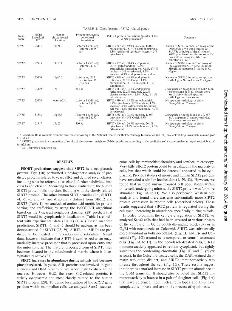
PSORT predictions suggest that SIRT2 is a cytoplasmicprotein. Frye (10) performed a phylogenetic analysis of pre-dicted proteins related to yeast SIR2 and defined seven classes,including what he referred to as class I, further subdivided intoclass Ia and class Ib. According to this classification, the humanSIRT2 protein falls into class Ib, along with the closely relatedSIRT3 protein. The other human SIRT proteins (i.e., SIRT1,-4, -5, -6, and -7) are structurally distinct from SIRT2 andSIRT3 (Table 1). An analysis of amino acid motifs for proteinsorting and trafficking by using the P-SORT-II algorithmsbased on the k-nearest neighbors classifier (28) predicts thatSIRT2 would be cytoplasmic in localization (Table 1), consis-tent with experimental data (Fig. 1) (1, 43). Based on thesepredictions, SIRT1, -6, and -7 should be nuclear, as has beendemonstrated for SIRT1 (23, 39). SIRT3 and SIRT4 are pre-dicted to be located in the endoplasmic reticulum. Recentdata, however, indicate that SIRT3 is synthesized as an enzy-matically inactive precursor that is processed upon entry intothe mitochondria. The mature, processed form of SIRT3 thenbecomes located in the mitochondrial matrix, where it is en-zymatically active (31).
SIRT2 increases in abundance during mitosis and becomesphosphorylated. In yeast, SIR proteins are involved in genesilencing and DNA repair and are accordingly localized to thenucleus. However, Hst2, the yeast Sir2-related protein, isstrictly cytoplasmic and most closely related to the humanSIRT2 protein (29). To define localization of the SIRT2 geneproduct within mammalian cells, we analyzed Saos2 osteosar-
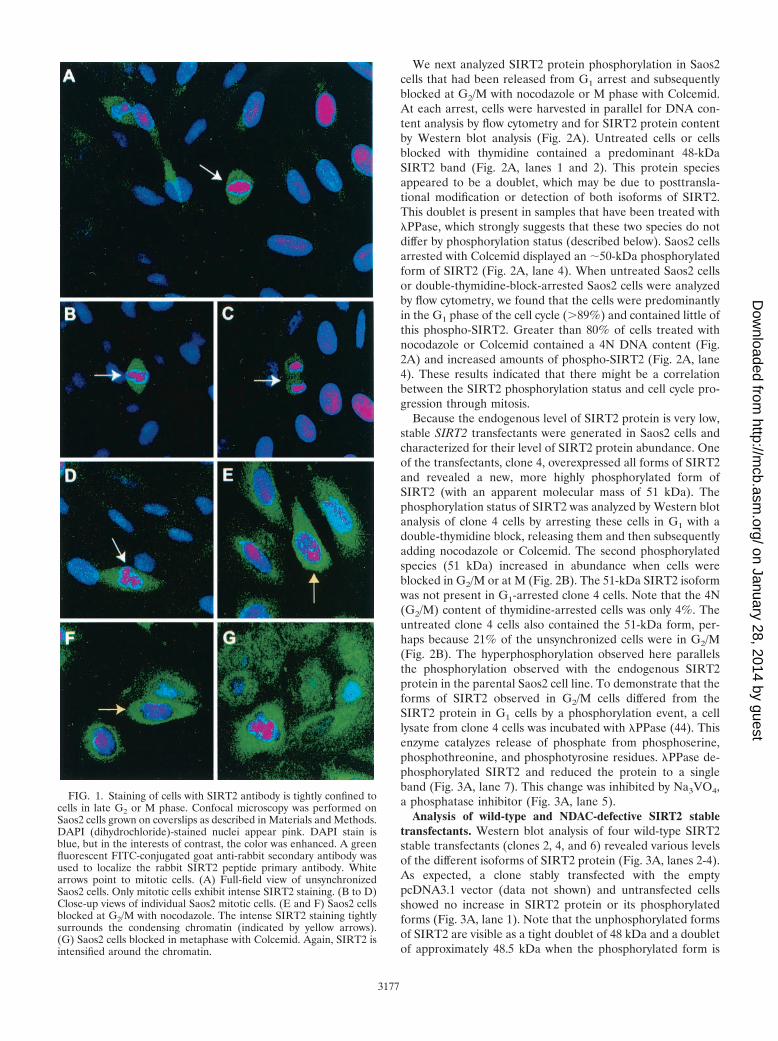
coma cells by immunohistochemistry and confocal microscopy.Very little SIRT2 protein could be visualized in the majority ofcells, but that which could be detected appeared to be cyto-plasmic. Previous studies of mouse and human SIRT2 proteinshave revealed a similar localization (1, 29, 43). However, wefound that in these unsynchronized cell populations, withinthose cells undergoing mitosis, the SIRT2 protein was far moreabundant (Fig. 1A to D). We also performed Western blotanalysis and found there was also substantially more SIRT2protein expression in mitotic cells (described below). Theseresults suggested that SIRT2 protein is regulated during thecell cycle, increasing in abundance specifically during mitosis.
In order to confirm the cell cycle regulation of SIRT2, weanalyzed Saos2 cells that had been arrested at various phasesof the cell cycle; in G1 by double-thymidine block (37) or inG2/M with nocodazole or Colcemid. SIRT2 was substantiallymore abundant in both nocodazole (Fig. 1E and F)- and Col-cemid (Fig. 1G)-treated cells compared to control untreatedcells (Fig. 1A to D). In the nocodazole-treated cells, SIRT2immunoreactivity appeared to remain cytoplasmic but tightlysurrounds the condensing chromatin (Fig. 1E and F, yellowarrows). In the Colcemid-treated cells, the DAPI-stained chro-matin was quite distinct, and SIRT2 immunoreactivity wasintense throughout the cell (Fig. 1G). These results suggestthat there is a marked increase in SIRT2 protein abundance atthe G2/M transition. It should also be noted that SIRT2 im-munoreactivity is intense in a pair of daughter cells (Fig. 1A)that have reformed their nuclear envelopes and thus havecompleted telophase and are in the process of cytokinesis.
TABLE 1. Classification of SIR2-related genes
Genename
NCBILocusLink
IDa
Humanchromosomal
location
Protein product(s),conceptualtranslation
PSORT protein predictions (results of thek-NN prediction)b Comments
SIRT1 23411 10q21.3 Isoform 1 (747 aa),isoform 2 (555aa)
SIRT1 (747 aa): 69.9% nuclear, 13.0%mitochondrial, 8.7% plasma membrane,4.3% vesicles of secretory system, 4.3%cytoplasmic
Known as Sir1� in mice; ortholog of theDrosophila SIRT gene located at34A7-8; ortholog of the C. elegansSIRT gene found on chromosome IV;probable ortholog identified inzebrafish as ESTc
SIRT2 22933 19q13.1 Isoform 1 (389 aa),isoform 2 (352aa)
SIRT2 (352 aa): 30.4% cytoplasmic,21.7% mitochondrial, 17.4%extracellular (including cell wall), 17.4%nuclear, 4.3% cytoskeletal, 4.3%vacuolar, 4.3% endoplasmic reticulum
Known as SIR2L2 in mice ortholog ofthe Drosophila SIRT gene found on3R92E; no apparent ortholog in C.elegans
SIRT3 23410 11p15.5 Isoform A, (257aa), isoform B(346 aa)
SIRT3 (399 aa): 44.4% endoplasmicreticulum, 22.2% Golgi, 11.1%mitochondrial, 11.1% nuclear, 11.1%cytoplasmic
Known as SIR2L3 in mice; no apparentortholog in Drosophila or C. elegans
SIRT4 23409 12q 314 aa SIRT4 (314 aa): 33.3% endoplasmicreticulum, 22.2% vacuolar, 22.2%plasma membrane, 11.1% Golgi, 11.1%mitochondrial
Drosophila ortholog found at 65E5-6 onchromosome 3; in C. elegans thereare 2 closely linked apparentorthologs on chromosome
SIRT5 23408 6pter-p25.1 Isoform 1 (310 aa),isoform 2 (299aa)
SIRT5 (310 aa): 65.2% mitochondrial,8.7% cytoplasmic, 8.7% nuclear, 4.3%vacuolar, 4.3% extracellular (includingcell wall) 4.3% plasma membrane, 4.3%peroxisomal
No apparent orthologs in eitherDrosophila or C. elegans
SIRT6 51548 19p13.3 Isoform 1 (355 aa),isoform 2 (237aa)
SIRT6 (355 aa): 78.3% nuclear, 13.0%cytoskeletal, 4.3% Golgi, 4.3%mitochondrial
Drosophila ortholog found at 3R 85F-86A; apparent C. elegans orthologfound on chromosome I
SIRT7 51547 17q25 400 aa SIRT7 (400 aa): 56.5% nuclear, 26.1%cytoplasmic, 13.0% mitochondrial, 4.3%cytoskeletal
No apparent orthologs in eitherDrosophila or C. elegans
a LocusLink ID is available from the electronic repository at the National Center for Biolotechnology Information (NCBI), available at http://www.ncbi.nlm.nih.gov/LocusLink.
b PSORT prediction is a summation of results of the k-nearest neighbor (k-NN) prediction according to the predictive software accessible at http://psort.nibb.ac.jp/form2.html.
c EST, expressed sequence tag.
3176 DRYDEN ET AL. MOL. CELL. BIOL.
on January 28, 2014 by guesthttp://m
cb.asm.org/
Dow
nloaded from
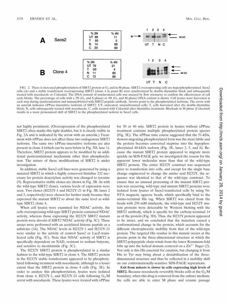
We next analyzed SIRT2 protein phosphorylation in Saos2cells that had been released from G1 arrest and subsequentlyblocked at G2/M with nocodazole or M phase with Colcemid.At each arrest, cells were harvested in parallel for DNA con-tent analysis by flow cytometry and for SIRT2 protein contentby Western blot analysis (Fig. 2A). Untreated cells or cellsblocked with thymidine contained a predominant 48-kDaSIRT2 band (Fig. 2A, lanes 1 and 2). This protein speciesappeared to be a doublet, which may be due to posttransla-tional modification or detection of both isoforms of SIRT2.This doublet is present in samples that have been treated withPPase, which strongly suggests that these two species do notdiffer by phosphorylation status (described below). Saos2 cellsarrested with Colcemid displayed an �50-kDa phosphorylatedform of SIRT2 (Fig. 2A, lane 4). When untreated Saos2 cellsor double-thymidine-block-arrested Saos2 cells were analyzedby flow cytometry, we found that the cells were predominantlyin the G1 phase of the cell cycle (�89%) and contained little ofthis phospho-SIRT2. Greater than 80% of cells treated withnocodazole or Colcemid contained a 4N DNA content (Fig.2A) and increased amounts of phospho-SIRT2 (Fig. 2A, lane4). These results indicated that there might be a correlationbetween the SIRT2 phosphorylation status and cell cycle pro-gression through mitosis.
Because the endogenous level of SIRT2 protein is very low,stable SIRT2 transfectants were generated in Saos2 cells andcharacterized for their level of SIRT2 protein abundance. Oneof the transfectants, clone 4, overexpressed all forms of SIRT2and revealed a new, more highly phosphorylated form ofSIRT2 (with an apparent molecular mass of 51 kDa). Thephosphorylation status of SIRT2 was analyzed by Western blotanalysis of clone 4 cells by arresting these cells in G1 with adouble-thymidine block, releasing them and then subsequentlyadding nocodazole or Colcemid. The second phosphorylatedspecies (51 kDa) increased in abundance when cells wereblocked in G2/M or at M (Fig. 2B). The 51-kDa SIRT2 isoformwas not present in G1-arrested clone 4 cells. Note that the 4N(G2/M) content of thymidine-arrested cells was only 4%. Theuntreated clone 4 cells also contained the 51-kDa form, per-haps because 21% of the unsynchronized cells were in G2/M(Fig. 2B). The hyperphosphorylation observed here parallelsthe phosphorylation observed with the endogenous SIRT2protein in the parental Saos2 cell line. To demonstrate that theforms of SIRT2 observed in G2/M cells differed from theSIRT2 protein in G1 cells by a phosphorylation event, a celllysate from clone 4 cells was incubated with PPase (44). Thisenzyme catalyzes release of phosphate from phosphoserine,phosphothreonine, and phosphotyrosine residues. PPase de-phosphorylated SIRT2 and reduced the protein to a singleband (Fig. 3A, lane 7). This change was inhibited by Na3VO4,a phosphatase inhibitor (Fig. 3A, lane 5).
Analysis of wild-type and NDAC-defective SIRT2 stabletransfectants. Western blot analysis of four wild-type SIRT2stable transfectants (clones 2, 4, and 6) revealed various levelsof the different isoforms of SIRT2 protein (Fig. 3A, lanes 2-4).As expected, a clone stably transfected with the emptypcDNA3.1 vector (data not shown) and untransfected cellsshowed no increase in SIRT2 protein or its phosphorylatedforms (Fig. 3A, lane 1). Note that the unphosphorylated formsof SIRT2 are visible as a tight doublet of 48 kDa and a doubletof approximately 48.5 kDa when the phosphorylated form is
FIG. 1. Staining of cells with SIRT2 antibody is tightly confined tocells in late G2 or M phase. Confocal microscopy was performed onSaos2 cells grown on coverslips as described in Materials and Methods.DAPI (dihydrochloride)-stained nuclei appear pink. DAPI stain isblue, but in the interests of contrast, the color was enhanced. A greenfluorescent FITC-conjugated goat anti-rabbit secondary antibody wasused to localize the rabbit SIRT2 peptide primary antibody. Whitearrows point to mitotic cells. (A) Full-field view of unsynchronizedSaos2 cells. Only mitotic cells exhibit intense SIRT2 staining. (B to D)Close-up views of individual Saos2 mitotic cells. (E and F) Saos2 cellsblocked at G2/M with nocodazole. The intense SIRT2 staining tightlysurrounds the condensing chromatin (indicated by yellow arrows).(G) Saos2 cells blocked in metaphase with Colcemid. Again, SIRT2 isintensified around the chromatin.
3177
on January 28, 2014 by guesthttp://m
cb.asm.org/
Dow
nloaded from
not highly prominent. (Overexpression of the phosphorylatedSIRT2 often masks this tight doublet, but it is clearly visible inFig. 2A and is indicated by the arrow with an asterisk.) Treat-ment with PPase does not affect these two endogenous SIRT2isoforms. The same two PPase-insensitive isoforms are alsopresent in clone 4 (which can be seen below in Fig. 5D, lane 1).Therefore, SIRT2 protein appears to be modified by an addi-tional posttranslational mechanism other than phosphoryla-tion. The nature of these modifications of SIRT2 is underinvestigation.
In addition, stable Saos2 cell lines were generated by using amutated SIRT2 in which a highly conserved histidine 232 nec-essary for protein deacetylase activity was changed to tyrosine(9). Representative stable clones are shown in Fig. 3B. As withthe wild-type SIRT2 clones, various levels of expression wereseen. Two clones (H232Y-1 and H232Y-21 in Fig. 3B, lanes 2and 3, respectively) were chosen for further study because theyexpressed the mutant SIRT2 to about the same level as wild-type SIRT2 clone 4.
When cell lysates were examined for NDAC activity, thecells overexpressing wild-type SIRT2 protein contained NDACactivity, whereas those expressing the H232Y SIRT2 mutantprotein were devoid of SIRT2 NDAC activity (Fig. 3C). Theseassays were performed with an acetylated histone peptide as asubstrate (16). The NDAC levels in H232Y-1 and H232Y-21were similar to the activity of control Saos2 or LacZ-trans-fected cells (Fig. 3C). Note that NDAC activity of SIRT2 isspecifically dependent on NAD, resistant to sodium butyrate,and sensitive to nicotinimide (Fig. 3C).
The H232Y SIRT2 protein is phosphorylated in a similarfashion to the wild-type SIRT2 in clone 4. The SIRT2 proteinin the H232Y stable transfectants appeared to be phosphory-lated following treatment with nocodazole, although to a lesserextent than the SIRT2 protein in clone 4 cells (Fig. 3E). Inorder to analyze this phosphorylation, lysates were isolatedfrom clone 4, H232Y-1, and H232Y-21 cells following G2/Marrest with nocodazole. These lysates were treated with PPase
for 30 or 60 min. SIRT2 protein in lysates without PPasetreatment contains multiple phosphorylated protein species(Fig. 3E). The PPase time course suggested that the 51-kDa,slowest-migrating phosphorylated form was the most labile andthe protein becomes converted stepwise into the hypophos-phorylated 48-kDA isoform (Fig. 3E, lanes 2, 5, and 8). Be-cause the mutant SIRT2 protein appeared to migrate morequickly on SDS-PAGE gels, we investigated the reason for theapparent lower molecular mass than that of the wild-typeSIRT2 protein. The entire H232Y construct was sequencedprior to transfection into cells, and except for the single basechange engineered to change the amino acid H232Y, the se-quence was identical to that of the wild-type construct. Toensure that no unusual processing of the mutant SIRT2 pro-tein was occurring, wild-type and mutant SIRT2 proteins wereisolated from lysates of Saos2-transfected cells by using Ni-NTA magnetic agarose beads, which specifically bind to theamino-terminal His tag. When SIRT2 was eluted from thebeads with 250 mM imidazole, the wild-type and H232Y mu-tant proteins were detectable by Western blotting with theSIRT2 antibody, which is specific for the carboxy-terminal 14aa of the protein (Fig. 3D). Thus, the H232Y protein appearedto be intact, and we concluded that the mutation caused aconformational change in the protein, which accounts for thedifferent electrophoretic mobility from that of the wild-typeprotein. The targeted His residue in this mutant occurs at theprecise point in the three-dimensional structure at which theSIRT2 polypeptide chain winds from the lower Rossmann-foldlobe up into the helical domain centered on a Zn2� finger (2).Not only is this His essential for catalysis, but changing it fromHis to Tyr may bring about a destabilization of the three-dimensional structure and thus be reflected in a mobility shifton our conformationally-sensitive SDS-PAGE separations.
Exit from mitosis is slower in cells expressing high levels ofSIRT2. Because nocodazole reversibly blocks cells at the G2/Mboundary, when this drug is removed from the culture medium,the cells are able to enter M phase and resume passage
FIG. 2. There is increased phosphorylation of SIRT2 protein at G2 and in M phase. SIRT2-overexpressing cells are hyperphosphorylated. Saos2cells (A) and a stable transfectant overexpressing SIRT2 (clone 4 in panel B) were synchronized by double-thymidine block and subsequentlytreated with nocodazole or Colcemid. The DNA content of synchronized cells was assayed by flow cytometry to confirm the effectiveness of cellcycle blocks. The percentage of cells with a 2N (G1 and S phase) or 4N (G2 and M phase) DNA content is shown. Cell lysates were harvested ateach step during synchronization and immunoblotted with SIRT2 peptide antibody. Arrows point to the phosphorylated isoforms. The arrow withan asterisk indicates PPase-insensitive isoforms of SIRT2. UT, untreated, unsynchronized cells; T, cells harvested after the double-thymidineblock; N, cells subsequently treated with nocodazole; C, cells treated with Colcemid after thymidine treatment. Blockade in M phase (Colcemid)results in a more pronounced shift of SIRT2 to the phosphorylated isoform in Saos2 cells.
3178 DRYDEN ET AL. MOL. CELL. BIOL.
on January 28, 2014 by guesthttp://m
cb.asm.org/
Dow
nloaded from
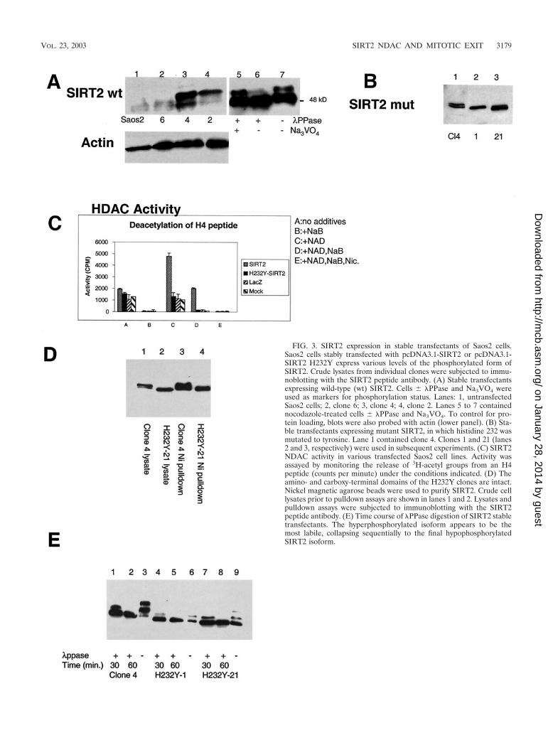
FIG. 3. SIRT2 expression in stable transfectants of Saos2 cells.Saos2 cells stably transfected with pcDNA3.1-SIRT2 or pcDNA3.1-SIRT2 H232Y express various levels of the phosphorylated form ofSIRT2. Crude lysates from individual clones were subjected to immu-noblotting with the SIRT2 peptide antibody. (A) Stable transfectantsexpressing wild-type (wt) SIRT2. Cells � PPase and Na3VO4 wereused as markers for phosphorylation status. Lanes: 1, untransfectedSaos2 cells; 2, clone 6; 3, clone 4; 4, clone 2. Lanes 5 to 7 containednocodazole-treated cells � PPase and Na3VO4. To control for pro-tein loading, blots were also probed with actin (lower panel). (B) Sta-ble transfectants expressing mutant SIRT2, in which histidine 232 wasmutated to tyrosine. Lane 1 contained clone 4. Clones 1 and 21 (lanes2 and 3, respectively) were used in subsequent experiments. (C) SIRT2NDAC activity in various transfected Saos2 cell lines. Activity wasassayed by monitoring the release of 3H-acetyl groups from an H4peptide (counts per minute) under the conditions indicated. (D) Theamino- and carboxy-terminal domains of the H232Y clones are intact.Nickel magnetic agarose beads were used to purify SIRT2. Crude celllysates prior to pulldown assays are shown in lanes 1 and 2. Lysates andpulldown assays were subjected to immunoblotting with the SIRT2peptide antibody. (E) Time course of PPase digestion of SIRT2 stabletransfectants. The hyperphosphorylated isoform appears to be themost labile, collapsing sequentially to the final hypophosphorylatedSIRT2 isoform.
VOL. 23, 2003 SIRT2 NDAC AND MITOTIC EXIT 3179
on January 28, 2014 by guesthttp://m
cb.asm.org/
Dow
nloaded from
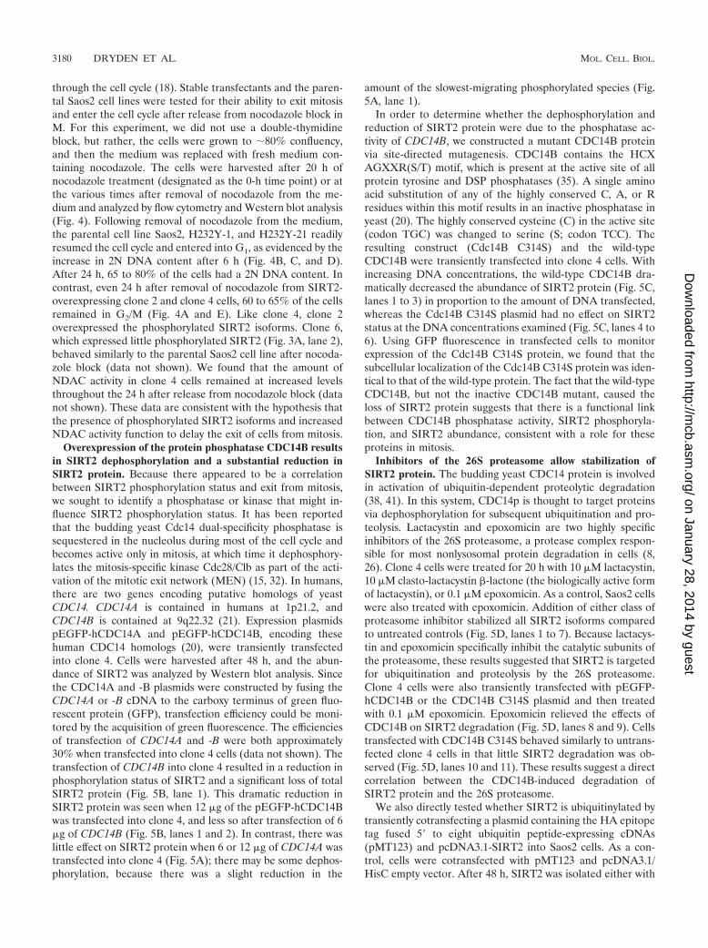
through the cell cycle (18). Stable transfectants and the paren-tal Saos2 cell lines were tested for their ability to exit mitosisand enter the cell cycle after release from nocodazole block inM. For this experiment, we did not use a double-thymidineblock, but rather, the cells were grown to �80% confluency,and then the medium was replaced with fresh medium con-taining nocodazole. The cells were harvested after 20 h ofnocodazole treatment (designated as the 0-h time point) or atthe various times after removal of nocodazole from the me-dium and analyzed by flow cytometry and Western blot analysis(Fig. 4). Following removal of nocodazole from the medium,the parental cell line Saos2, H232Y-1, and H232Y-21 readilyresumed the cell cycle and entered into G1, as evidenced by theincrease in 2N DNA content after 6 h (Fig. 4B, C, and D).After 24 h, 65 to 80% of the cells had a 2N DNA content. Incontrast, even 24 h after removal of nocodazole from SIRT2-overexpressing clone 2 and clone 4 cells, 60 to 65% of the cellsremained in G2/M (Fig. 4A and E). Like clone 4, clone 2overexpressed the phosphorylated SIRT2 isoforms. Clone 6,which expressed little phosphorylated SIRT2 (Fig. 3A, lane 2),behaved similarly to the parental Saos2 cell line after nocoda-zole block (data not shown). We found that the amount ofNDAC activity in clone 4 cells remained at increased levelsthroughout the 24 h after release from nocodazole block (datanot shown). These data are consistent with the hypothesis thatthe presence of phosphorylated SIRT2 isoforms and increasedNDAC activity function to delay the exit of cells from mitosis.
Overexpression of the protein phosphatase CDC14B resultsin SIRT2 dephosphorylation and a substantial reduction inSIRT2 protein. Because there appeared to be a correlationbetween SIRT2 phosphorylation status and exit from mitosis,we sought to identify a phosphatase or kinase that might in-fluence SIRT2 phosphorylation status. It has been reportedthat the budding yeast Cdc14 dual-specificity phosphatase issequestered in the nucleolus during most of the cell cycle andbecomes active only in mitosis, at which time it dephosphory-lates the mitosis-specific kinase Cdc28/Clb as part of the acti-vation of the mitotic exit network (MEN) (15, 32). In humans,there are two genes encoding putative homologs of yeastCDC14. CDC14A is contained in humans at 1p21.2, andCDC14B is contained at 9q22.32 (21). Expression plasmidspEGFP-hCDC14A and pEGFP-hCDC14B, encoding thesehuman CDC14 homologs (20), were transiently transfectedinto clone 4. Cells were harvested after 48 h, and the abun-dance of SIRT2 was analyzed by Western blot analysis. Sincethe CDC14A and -B plasmids were constructed by fusing theCDC14A or -B cDNA to the carboxy terminus of green fluo-rescent protein (GFP), transfection efficiency could be moni-tored by the acquisition of green fluorescence. The efficienciesof transfection of CDC14A and -B were both approximately30% when transfected into clone 4 cells (data not shown). Thetransfection of CDC14B into clone 4 resulted in a reduction inphosphorylation status of SIRT2 and a significant loss of totalSIRT2 protein (Fig. 5B, lane 1). This dramatic reduction inSIRT2 protein was seen when 12 �g of the pEGFP-hCDC14Bwas transfected into clone 4, and less so after transfection of 6�g of CDC14B (Fig. 5B, lanes 1 and 2). In contrast, there waslittle effect on SIRT2 protein when 6 or 12 �g of CDC14A wastransfected into clone 4 (Fig. 5A); there may be some dephos-phorylation, because there was a slight reduction in the
amount of the slowest-migrating phosphorylated species (Fig.5A, lane 1).
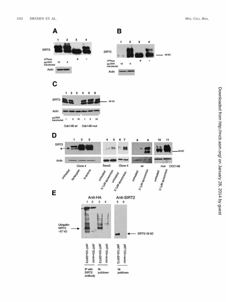
In order to determine whether the dephosphorylation andreduction of SIRT2 protein were due to the phosphatase ac-tivity of CDC14B, we constructed a mutant CDC14B proteinvia site-directed mutagenesis. CDC14B contains the HCXAGXXR(S/T) motif, which is present at the active site of allprotein tyrosine and DSP phosphatases (35). A single aminoacid substitution of any of the highly conserved C, A, or Rresidues within this motif results in an inactive phosphatase inyeast (20). The highly conserved cysteine (C) in the active site(codon TGC) was changed to serine (S; codon TCC). Theresulting construct (Cdc14B C314S) and the wild-typeCDC14B were transiently transfected into clone 4 cells. Withincreasing DNA concentrations, the wild-type CDC14B dra-matically decreased the abundance of SIRT2 protein (Fig. 5C,lanes 1 to 3) in proportion to the amount of DNA transfected,whereas the Cdc14B C314S plasmid had no effect on SIRT2status at the DNA concentrations examined (Fig. 5C, lanes 4 to6). Using GFP fluorescence in transfected cells to monitorexpression of the Cdc14B C314S protein, we found that thesubcellular localization of the Cdc14B C314S protein was iden-tical to that of the wild-type protein. The fact that the wild-typeCDC14B, but not the inactive CDC14B mutant, caused theloss of SIRT2 protein suggests that there is a functional linkbetween CDC14B phosphatase activity, SIRT2 phosphoryla-tion, and SIRT2 abundance, consistent with a role for theseproteins in mitosis.
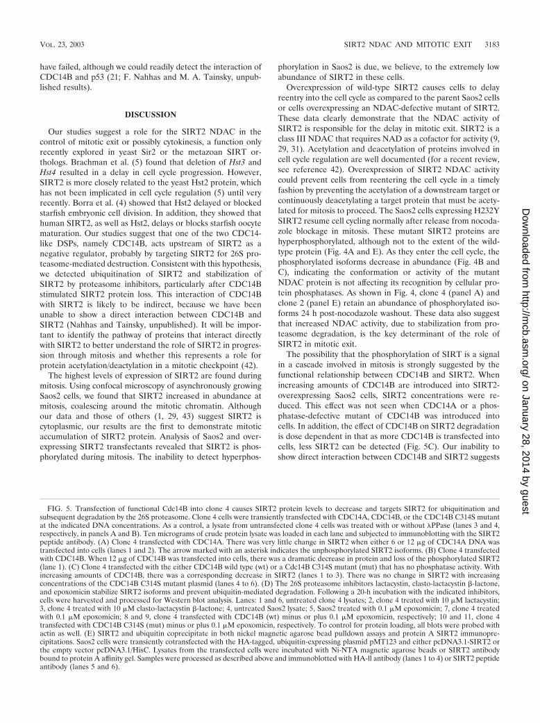
Inhibitors of the 26S proteasome allow stabilization ofSIRT2 protein. The budding yeast CDC14 protein is involvedin activation of ubiquitin-dependent proteolytic degradation(38, 41). In this system, CDC14p is thought to target proteinsvia dephosphorylation for subsequent ubiquitination and pro-teolysis. Lactacystin and epoxomicin are two highly specificinhibitors of the 26S proteasome, a protease complex respon-sible for most nonlysosomal protein degradation in cells (8,26). Clone 4 cells were treated for 20 h with 10 �M lactacystin,10 �M clasto-lactacystin �-lactone (the biologically active formof lactacystin), or 0.1 �M epoxomicin. As a control, Saos2 cellswere also treated with epoxomicin. Addition of either class ofproteasome inhibitor stabilized all SIRT2 isoforms comparedto untreated controls (Fig. 5D, lanes 1 to 7). Because lactacys-tin and epoxomicin specifically inhibit the catalytic subunits ofthe proteasome, these results suggested that SIRT2 is targetedfor ubiquitination and proteolysis by the 26S proteasome.Clone 4 cells were also transiently transfected with pEGFP-hCDC14B or the CDC14B C314S plasmid and then treatedwith 0.1 �M epoxomicin. Epoxomicin relieved the effects ofCDC14B on SIRT2 degradation (Fig. 5D, lanes 8 and 9). Cellstransfected with CDC14B C314S behaved similarly to untrans-fected clone 4 cells in that little SIRT2 degradation was ob-served (Fig. 5D, lanes 10 and 11). These results suggest a directcorrelation between the CDC14B-induced degradation ofSIRT2 protein and the 26S proteasome.
We also directly tested whether SIRT2 is ubiquitinylated bytransiently cotransfecting a plasmid containing the HA epitopetag fused 5� to eight ubiquitin peptide-expressing cDNAs(pMT123) and pcDNA3.1-SIRT2 into Saos2 cells. As a con-trol, cells were cotransfected with pMT123 and pcDNA3.1/HisC empty vector. After 48 h, SIRT2 was isolated either with
3180 DRYDEN ET AL. MOL. CELL. BIOL.
on January 28, 2014 by guesthttp://m
cb.asm.org/
Dow
nloaded from
Ni-NTA magnetic beads or immunoprecipitated with proteinA and SIRT2 antibody and analyzed by Western blotting. An-tibodies to SIRT2 and to HA were used to detect the SIRT2levels and to determine whether the protein was ubiquitiny-lated. Cotransfection of pcDNA3.1-SIRT2 and pMT123yielded a 57- to 60-kDa band that was not present in controllysates (Fig. 5E). There was some nonspecific background,
especially from Ni-NTA isolations, but the predominate ubiq-uitin-SIRT2 band was not present in the controls (compareFig. 5E, lanes 1 and 3, with lanes 2 and 4). Upon prolongedexposure of lane 5, a band comigrating with ubiquitinatedSIRT2 (about 57 kDa) was visible. In summary, consistent withour observation that proteasome inhibitors affect the stabilityof SIRT2 and as such it is a ubiquitinylated protein, bothimmunoprecipitation and Ni-NTA isolations resulted in coas-sociation of HA-tagged ubiquitin and SIRT2.
Our data are highly suggestive that proteasome-mediatedproteolysis of SIRT2 results from the action of CDC14B eitherdirectly or indirectly. However, attempts to demonstrate a di-rect physical interaction between CDC14B and SIRT2 in vitro
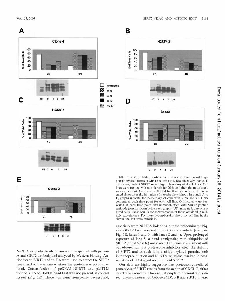
FIG. 4. SIRT2 stable transfectants that overexpress the wild-typephosphorylated forms of SIRT2 return to G1 less effectively than cellsexpressing mutant SIRT2 or nonhyperphosphorylated cell lines. Celllines were treated with nocodazole for 20 h, and then the nocodazolewas washed out. Cells were collected for flow cytometry at the indi-cated times after the initiation of nocodazole washout. In panels A toE, graphs indicate the percentage of cells with a 2N and 4N DNAcontents at each time point for each cell line. Cell lysates were har-vested at each time point and immunoblotted with SIRT2 peptideantibody (results shown below each graph). UT, untreated, unsynchro-nized cells. These results are representative of those obtained in mul-tiple experiments. The more hyperphosphorylated the cell line is, theslower the exit from mitosis is.
VOL. 23, 2003 SIRT2 NDAC AND MITOTIC EXIT 3181
on January 28, 2014 by guesthttp://m
cb.asm.org/
Dow
nloaded from
3182 DRYDEN ET AL. MOL. CELL. BIOL.
on January 28, 2014 by guesthttp://m
cb.asm.org/
Dow
nloaded from
have failed, although we could readily detect the interaction ofCDC14B and p53 (21; F. Nahhas and M. A. Tainsky, unpub-lished results).
DISCUSSION
Our studies suggest a role for the SIRT2 NDAC in thecontrol of mitotic exit or possibly cytokinesis, a function onlyrecently explored in yeast Sir2 or the metazoan SIRT or-thologs. Brachman et al. (5) found that deletion of Hst3 andHst4 resulted in a delay in cell cycle progression. However,SIRT2 is more closely related to the yeast Hst2 protein, whichhas not been implicated in cell cycle regulation (5) until veryrecently. Borra et al. (4) showed that Hst2 delayed or blockedstarfish embryonic cell division. In addition, they showed thathuman SIRT2, as well as Hst2, delays or blocks starfish oocytematuration. Our studies suggest that one of the two CDC14-like DSPs, namely CDC14B, acts upstream of SIRT2 as anegative regulator, probably by targeting SIRT2 for 26S pro-teasome-mediated destruction. Consistent with this hypothesis,we detected ubiquitination of SIRT2 and stabilization ofSIRT2 by proteasome inhibitors, particularly after CDC14Bstimulated SIRT2 protein loss. This interaction of CDC14Bwith SIRT2 is likely to be indirect, because we have beenunable to show a direct interaction between CDC14B andSIRT2 (Nahhas and Tainsky, unpublished). It will be impor-tant to identify the pathway of proteins that interact directlywith SIRT2 to better understand the role of SIRT2 in progres-sion through mitosis and whether this represents a role forprotein acetylation/deactylation in a mitotic checkpoint (42).
The highest levels of expression of SIRT2 are found duringmitosis. Using confocal microscopy of asynchronously growingSaos2 cells, we found that SIRT2 increased in abundance atmitosis, coalescing around the mitotic chromatin. Althoughour data and those of others (1, 29, 43) suggest SIRT2 iscytoplasmic, our results are the first to demonstrate mitoticaccumulation of SIRT2 protein. Analysis of Saos2 and over-expressing SIRT2 transfectants revealed that SIRT2 is phos-phorylated during mitosis. The inability to detect hyperphos-
phorylation in Saos2 is due, we believe, to the extremely lowabundance of SIRT2 in these cells.
Overexpression of wild-type SIRT2 causes cells to delayreentry into the cell cycle as compared to the parent Saos2 cellsor cells overexpressing an NDAC-defective mutant of SIRT2.These data clearly demonstrate that the NDAC activity ofSIRT2 is responsible for the delay in mitotic exit. SIRT2 is aclass III NDAC that requires NAD as a cofactor for activity (9,29, 31). Acetylation and deacetylation of proteins involved incell cycle regulation are well documented (for a recent review,see reference 42). Overexpression of SIRT2 NDAC activitycould prevent cells from reentering the cell cycle in a timelyfashion by preventing the acetylation of a downstream target orcontinuously deacetylating a target protein that must be acety-lated for mitosis to proceed. The Saos2 cells expressing H232YSIRT2 resume cell cycling normally after release from nocoda-zole blockage in mitosis. These mutant SIRT2 proteins arehyperphosphorylated, although not to the extent of the wild-type protein (Fig. 4A and E). As they enter the cell cycle, thephosphorylated isoforms decrease in abundance (Fig. 4B andC), indicating the conformation or activity of the mutantNDAC protein is not affecting its recognition by cellular pro-tein phosphatases. As shown in Fig. 4, clone 4 (panel A) andclone 2 (panel E) retain an abundance of phosphorylated iso-forms 24 h post-nocodazole washout. These data also suggestthat increased NDAC activity, due to stabilization from pro-teasome degradation, is the key determinant of the role ofSIRT2 in mitotic exit.
The possibility that the phosphorylation of SIRT is a signalin a cascade involved in mitosis is strongly suggested by thefunctional relationship between CDC14B and SIRT2. Whenincreasing amounts of CDC14B are introduced into SIRT2-overexpressing Saos2 cells, SIRT2 concentrations were re-duced. This effect was not seen when CDC14A or a phos-phatase-defective mutant of CDC14B was introduced intocells. In addition, the effect of CDC14B on SIRT2 degradationis dose dependent in that as more CDC14B is transfected intocells, less SIRT2 can be detected (Fig. 5C). Our inability toshow direct interaction between CDC14B and SIRT2 suggests
FIG. 5. Transfection of functional Cdc14B into clone 4 causes SIRT2 protein levels to decrease and targets SIRT2 for ubiquitination andsubsequent degradation by the 26S proteasome. Clone 4 cells were transiently transfected with CDC14A, CDC14B, or the CDC14B C314S mutantat the indicated DNA concentrations. As a control, a lysate from untransfected clone 4 cells was treated with or without PPase (lanes 3 and 4,respectively, in panels A and B). Ten micrograms of crude protein lysate was loaded in each lane and subjected to immunoblotting with the SIRT2peptide antibody. (A) Clone 4 transfected with CDC14A. There was very little change in SIRT2 when either 6 or 12 �g of CDC14A DNA wastransfected into cells (lanes 1 and 2). The arrow marked with an asterisk indicates the unphosphorylated SIRT2 isoforms. (B) Clone 4 transfectedwith CDC14B. When 12 �g of CDC14B was transfected into cells, there was a dramatic decrease in protein and loss of the phosphorylated SIRT2(lane 1). (C) Clone 4 transfected with the either CDC14B wild type (wt) or a Cdc14B C314S mutant (mut) that has no phosphatase activity. Withincreasing amounts of CDC14B, there was a corresponding decrease in SIRT2 (lanes 1 to 3). There was no change in SIRT2 with increasingconcentrations of the CDC14B C314S mutant plasmid (lanes 4 to 6). (D) The 26S proteasome inhibitors lactacystin, clasto-lactacystin �-lactone,and epoxomicin stabilize SIRT2 isoforms and prevent ubiquitin-mediated degradation. Following a 20-h incubation with the indicated inhibitors,cells were harvested and processed for Western blot analysis. Lanes: 1 and 6, untreated clone 4 lysates; 2, clone 4 treated with 10 �M lactacystin;3, clone 4 treated with 10 �M clasto-lactacystin �-lactone; 4, untreated Saos2 lysate; 5, Saos2 treated with 0.1 �M epoxomicin; 7, clone 4 treatedwith 0.1 �M epoxomicin; 8 and 9, clone 4 transfected with CDC14B (wt) minus or plus 0.1 �M epoxomicin, respectively; 10 and 11, clone 4transfected with CDC14B C314S (mut) minus or plus 0.1 �M epoxomicin, respectively. To control for protein loading, all blots were probed withactin as well. (E) SIRT2 and ubiquitin coprecipitate in both nickel magnetic agarose bead pulldown assays and protein A SIRT2 immunopre-cipitations. Saos2 cells were transiently cotransfected with the HA-tagged, ubiquitin-expressing plasmid pMT123 and either pcDNA3.1-SIRT2 orthe empty vector pcDNA3.1/HisC. Lysates from the transfected cells were incubated with Ni-NTA magnetic agarose beads or SIRT2 antibodybound to protein A affinity gel. Samples were processed as described above and immunoblotted with HA-ll antibody (lanes 1 to 4) or SIRT2 peptideantibody (lanes 5 and 6).
VOL. 23, 2003 SIRT2 NDAC AND MITOTIC EXIT 3183
on January 28, 2014 by guesthttp://m
cb.asm.org/
Dow
nloaded from
that either the interaction is very transient or CDC14B isacting as a signal to activate an as-yet-unidentified phosphataseor inhibit a putative SIRT2 protein kinase. In yeast, CDC14 isactive only during mitosis and is involved in activation of themitotic exit network (15, 32). Recent work (24) has shown thatboth overexpression and down regulation of human CDC14Adisrupt centrosome separation and chromosome segregation.These authors demonstrated that CDC14B was localized to thenucleolus and when overexpressed showed little effect on thecell cycle, whereas CDC14A induced apoptosis. Our data im-plicating CDC14B in mitotic exit would suggest a conservationof function through eukaryotic evolution with as-yet-unknowntargets.
The human CDC14A and the yeast Cdc14 phosphatase haveboth been implicated in the regulation of the anaphase-pro-moting complex, which mediates the degradation of cell cycleregulators via ubiquitination (3, 35, 38). Since CDC14B pro-motes the disappearance of SIRT2, the likelihood that ubiq-uitination and degradation by the 26S proteasome are bothinvolved in SIRT2 turnover is clearly indicated. Therefore, weanalyzed the effects of protease inhibitors on the abundance ofSIRT2 protein. Lactacystin; its active form, clasto-lactocystin-�-lactone; and epoxomicin have all been shown to be highlypotent and specific inhibitors of the 26S proteasome (8, 26).The link between SIRT2 and CDC14B is further suggested bythe fact that these proteasome inhibitors stabilize SIRT2. Ourobservation that SIRT2 is ubiquitinated further supports thenotion that CDC14B phosphatase can provoke SIRT2 degra-dation via the 26S proteasome-dependent pathway. We pro-pose that phosphorylation stabilizes SIRT2 and that CDC1Bcauses destabilization, thereby controlling the cell cycle-depen-dent abundance of SIRT2, its NDAC activity, and mitotic exit.
What sense can be made (if any) of the apparent p53-di-rected function of SIRT1, the developmental regulation of theDrosophila dSir2, and the M phase regulatory function ofSIRT2? Could it be that these proteins have acquired a cellcycle regulatory function during metazoan evolution, whichfocuses an NAD�-dependent deacetylase intrinsic to SIRTprotein onto evolutionarily innovative targets enabling diver-sification of cell cycle parameters that distinguish higher eu-karyotes? Both the timing and spatial organization of mitosisin early development are key to the diversification of the pat-terns of metazoan development. For example, in frogs, flies,and echinoderms, the first 10 to 12 mitotic cycles are rapid andsynchronous, are missing the G1 and G2 phases, and regulatethe cell cycle at later stages. This occurs in early mammalianembryonic development as well. Spatial diversification of mi-tosis is also key to taxonomic diversification of embryonic pat-tern. Unlike insects, both nematodes and echinoderms rapidlyinstitute asymmetric cell divisions in early development, a phe-nomenon that seems to be enabled in C. elegans by an interplaybetween astral microtubules and the juxtamembrane cytoskel-etal network known as the cortex (11). In addition, the cellularunderpinnings of asymmetric mitoses involve asymmetric ac-tivity of microtubule-associated molecular motors (dyneins, ki-nesins, and myosin V) and other microtubule-associated pro-teins (MAPs), which may regulate “microtubule catastrophe”(27).
How might SIRT2 play into this process? Hyperphosphory-lated SIRT2 is confined to M phase in cycling cultured cells,
and we believe that SIRT2’s enzymatic activity is confined tothis period. We hypothesize that a specific acetylated proteintarget in the spindle apparatus (including the centrosome) isthe target of SIRT2, and SIRT2 might provide a continuouslevel of deacetylation of this target during M phase to coun-teract a continuous “on-reaction” (i.e., acetylation). Such apartnering of SIRT2 with a spindle apparatus protein not onlymight result in deacetylation of this spindle protein, but alsomight provide a distinct subcellular focal source point in thespindle apparatus for generation of the novel metaboliteOAAR (34). In this model, CDC14B release late in M phasewould spread a wave of CDC14B throughout the cell, therebyremoving SIRT2 function. In yeast, Cdc14p is believed to beregulated by its subcellular location. It is sequestered in thenucleolus until its release during early anaphase by the Cdc14early anaphase release (FEAR) network, and then in late an-aphase, the MEN prevents Cdc14p from relocalizing to thenucleolus, as part of this network, Cdc14p dephosphorylates itsmitotic cyclin and Cdk targets, allowing reentry into the cellcycle (32, 40). In humans, CDC14A interacts with centrosomesand is involved in correct centrosome separation and chromo-some segregation, but no in vivo function has been identifiedfor CDC14B (17, 24). It is tempting to speculate that in hu-mans, CDC14B is in a phosphorylation pathway upstream ofSIRT2 and that SIRT2 NDAC activity serves as a late mitoticcheckpoint to ensure correct segregation of chromosomes dur-ing cytokinesis. Once this function has been served, CDC14Btargets SIRT2 for dephosphorylation and ubiquitination, thusturning off SIRT2’s NDAC activity and allowing acetylation ofa target protein, which would then stimulate transcription offactors required for entry into G1 of the cell cycle. This func-tion of SIRT2 would put it in a parallel pathway to that ofSIRT1 and its interactions with p53 (23, 33, 39), whereinSIRT1 deacetylation of p53 is involved in the regulation of thep53 checkpoint induced by DNA damage or stress.
ACKNOWLEDGMENTS
We thank George Brush for help with the PPase experiments andgel conditions for separating phosphorylated isoforms of SIRT2 andGabriel Fenteany for hints on how to use the proteasome inhibitors, aswell as Y. Haupt for the use of pMT123. We also thank George Brushand Gen Sheng Wu for critical reading of the manuscript and JudithAbrams for statistical analysis of flow cytometry data.
This work was supported by the Barbara and Fred Erb EndowedChair in Cancer Genetics to M.A.T.; funds from the Karmanos CancerInstitute; and the Applied Genomics Core, Core Flow Cytometry,Biostatistics Core, and Confocal Microscopy facilities of the KarmanosCancer Institute and Wayne State University (P30CA022453).
ADDENDUM IN PROOF
Recently North et al. showed that the human SIRT2 is anNAD�-dependent tubulin deacetylase and colocalizes with mi-crotubules; these data further support our evidence for a rolefor SIRT2 in the control of mitosis (B. J. North, B. L. Marshall,M. T. Borra, J. M. Denu, and E. Verdin, Mol. Cell 11:437–444,2003).
REFERENCES
1. Afshar, G., and J. P. Murnane. 1999. Characterization of a human gene withsequence homology to Saccharomyces cerevisiae SIR2. Gene 234:161–168.
2. Avalos, J. L., I. Celic, S. Muhammad, M. S. Cosgrove, J. D. Boeke, and C.Wolberger. 2002. Structure of a Sir2 enzyme bound to an acetylated p53peptide. Mol. Cell 10:523–535.
3184 DRYDEN ET AL. MOL. CELL. BIOL.
on January 28, 2014 by guesthttp://m
cb.asm.org/
Dow
nloaded from
3. Bembenek, J., and H. Yu. 2001. Regulation of the anaphase-promotingcomplex by the dual specificity phosphatase human Cdc14a. J. Biol. Chem.276:48237–48242.
4. Borra, M. T., F. J. O’Neill, M. D. Jackson, B. Marshall, E. Verdin, K. R.Foltz, and J. M. Denu. 2002. Conserved enzymatic production and biologicaleffect of O-acetyl-ADP-ribose by silent information regulator 2-like NAD�-dependent deacetylases. J. Biol. Chem. 277:12632–12641.
5. Brachmann, C. G., J. M. Sherman, S. E. Devine, E. E. Cameron, L. Pillus,and J. D. Boeke. 1995. The Sir2 gene family, conserved from bacteria tohumans, functions in silencing, cell cycle progression, and chromosome sta-bility. Genes Dev. 9:2888–2902.
6. Chang, J.-H., H.-C. Kim, K.-Y. Hwang, J.-W. Lee, S. P. Jackson, S. D. Bell,and Y. Cho. 2002. Structural basis for the NAD-dependent deacetylasemechanism of Sir2. J. Biol. Chem. 277:34489–34498.
7. Darzynkiewicz, Z., and G. Juan. 1997. DNA content measurement for DNAploidy and cell cycle analysis, 2nd ed., vol. 1. John Wiley & Sons, Inc., NewYork, N.Y.
8. Fenteany, G., and S. L. Schreiber. 1998. Lactacystin, proteasome function,and cell fate. J. Biol. Chem. 273:8545–8548.
9. Finnin, M. S., J. R. Donigian, and N. P. Pavletich. 2001. Structure of thehistone deacetylase SIRT2. Nat. Struct. Biol. 8:621–625.
10. Frye, R. 2000. Phylogenetic classification of prokaryotic and eukaryotic Sir2-like proteins. Biochem. Biophys. Res. Commun. 273:793–798.
11. Gonczy, P., J. M. Bellanger, M. Kirkham, A. Pozniakowski, K. Baumer, J. B.Phillips, and A. A. Hyman. 2001. zyg-8, a gene required for spindle posi-tioning in C. elegans, encodes a double cortin-related kinase that promotesmicrotubule assembly. Dev. Cell 1:363–375.
12. Guarente, L. 1999. Diverse and dynamic functions of the Sir silencing com-plex. Nat. Genet. 23:281–285.
13. Guarente, L. 2000. Sir2 links chromatin silencing, metabolism, and aging.Genes Dev. 14:1021–1026.
14. Ivy, J. M., A. J. S. Klar, and J. B. Hicks. 1986. Cloning and characterizationof four SIR genes of Saccharomyces cerevisiae. Mol. Cell. Biol. 6:688–702.
15. Jaspersen, S. L., J. F. Charles, R. L. Tinker-Kulberg, and D. O. Morgan.1998. A late mitotic regulatory network controlling cyclin destruction inSaccharomyces cerevisiae. Mol. Biol. Cell 9:2803–2817.
16. Juan, L. J., W. J. Shia, M. H. Chen, W. M. Yang, E. Seto, Y. S. Lin, and C. W.Wu. 2000. Histone deacetylases specifically down-regulate p53-dependentgene activation. J. Biol. Chem. 275:20436–20443.
17. Kaiser, B. K., Z. A. Zimmerman, H. Charbonneau, and P. K. Jackson. 2002.Disruption of centrosome structure, chromosome segregation, and cytoki-nesis by misexpression of human Cdc14A phosphatase. Mol. Biol. Cell 13:2289–2300.
18. Kaur, G., M. Stetler-Stevenson, S. Sebers, P. Worland, H. Sedlacek, C.Myers, J. Czech, R. Naik, and E. Sausville. 1992. Growth inhibition withreversible cell cycle arrest of carcinoma cells by flavone L86–8275. J. Natl.Cancer Inst. 84:1736–1740.
19. Klar, A. J., S. Fogel, and K. Macleod. 1979. Mar1-A regulator of the HMaand HMa loci in Saccharomyces cerevisiae. Genetics 93:37–50.
20. Li, L., B. R. Ernsting, M. J. Wishart, D. L. Lohse, and J. E. Dixon. 1997. Afamily of putative tumor suppressors is structurally and functionally con-served in humans and yeast. J. Biol. Chem. 272:29403–29406.
21. Li, L., M. Ljungman, and J. E. Dixon. 2000. The human Cdc14 phosphatasesinteract with and dephosphorylate the tumor suppressor protein p53. J. Biol.Chem. 275:2410–2414.
22. Lin, S. J., P. A. Defossez, and L. Guarente. 2000. Requirement of NAD andSIR2 for life-span extension by calorie restriction in Saccharomyces cerevi-siae. Science 289:2126–2128.
23. Luo, J., A. Y. Nikolaev, S.-I. Imai, D. Chen, F. Su, A. Shiloh, L. Guarente,and W. Gu. 2001. Negative control of p53 by Sir2a promotes cell survivalunder stress. Cell 107:137–148.
24. Mailand, N., C. Lukas, B. K. Kaiser, P. K. Jackson, J. Bartek, and J. Lukas.2002. Deregulated human Cdc14A phosphatase disrupts centrosome sepa-ration and chromosome segregation. Nat. Cell Biol. 4:317–322.
25. McBurney, M. W., Y. Yang, K. Jardine, M. Hixon, K. Boekelheide, J. R.Webb, P. M. Lansdorp, and M. Lemieux. 2003. The mammalian SIR2aprotein has a role in embryogenesis and gametogenesis. Mol. Biol. Cell23:38–54.
26. Meng, L., R. Mohan, B. H. Kwok, M. Elofsson, N. Sin, and C. M. Crews.1999. Epoxomicin, a potent and selective proteasome inhibitor, exhibits invivo antiinflammatory activity. Proc. Natl. Acad. Sci. USA 96:10403–10408.
27. Mitchison, T., and M. Kirschner. 1984. Dynamic instability of microtubulegrowth. Nature 312:237–242.
28. Nakai, K., and P. Horton. 1999. PSORT: a program for detecting sortingsignals in proteins and predicting their subcellular localization. Trends Bio-chem. Sci. 24:34–36.
29. Perrod, S., M. M. Cockell, T. Laroche, H. Renauld, A. L. Ducrest, C. Bon-nard, and S. M. Gasser. 2001. A cytosolic NAD-dependent deacetylase,Hst2p, can modulate nucleolar and telomeric silencing in yeast. EMBO J.20:197–209.
30. Rosenberg, M. I., and S. M. Parkhurst. 2002. Drosophila Sir2 is required forheterochromatic silencing and by euchromatic Hairy/E(Spl) bHLH repres-sors in segmentation and sex determination. Cell 109:447–458.
31. Schwer, B., B. J. North, R. A. Frye, M. Ott, and E. Verdin. 2002. The humansilent information regulator (Sir)2 homologue hSIRT3 is a mitochondrialnicotinamide adenine dinucleotide-dependent deacetylase. PG. J. Cell Biol.158:647–657.
32. Shou, W., J. H. Seol, A. Shevchenko, C. Baskerville, D. Moazed, Z. W. Chen,J. Jang, H. Charbonneau, and R. J. Deshaies. 1999. Exit from mitosis istriggered by Tem1-dependent release of the protein phosphatase Cdc14from nucleolar RENT complex. Cell 97:233–244.
33. Smith, J. 2002. Human Sir2 and the �silencing’ of p53 activity. Trends CellBiol. 12:404–406.
34. Tanny, J. C., and D. Moazed. 2001. Coupling of histone deacetylation toNAD breakdown by the yeast silencing protein Sir2: evidence for acetyltransfer from substrate to an NAD breakdown product. Proc. Natl. Acad.Sci. USA 98:415–420.
35. Taylor, G. S., Y. Liu, C. Baskerville, and H. Charbonneau. 1997. The activityof Cdc14p, an oligomeric dual specificity protein phosphatase from Saccha-romyces cerevisiae, is required for cell cycle progression. J. Biol. Chem.272:24054–24063.
36. Tissenbaum, H. A., and L. Guarente. 2001. Increased dosage of a sir-2 geneextends lifespan in Caenorhabditis elegans. Nature 410:227–230.
37. Toyoshima, F., T. Moriguchi, A. Wada, M. Fukuda, and E. Nishida. 1998.Nuclear export of cyclin B1 and its possible role in the DNA damage-inducedG2 checkpoint. EMBO J. 17:2728–2735.
38. Traverso, E. E., C. Baskerville, Y. Liu, W. Shou, P. James, R. J. Deshaies,and H. Charbonneau. 1931. 2001. Characterization of the net1 cell cycle-dependent regulator of the Cdc14 phosphatase from budding yeast. J. Biol.Chem. 276:21924–21931.
39. Vaziri, H., S. K. Dessaln, E. N. Eaton, S.-I. Imai, R. A. Frye, T. K. Pandita,L. Guarente, and R. A. Weinberg. 2001. hSir2SIRT1 functions as an NAD-dependent p53 deacetylase. Cell 107:149–159.
40. Visintin, R., and A. Amon. 2000. The nucleolus: the magician’s hat for cellcycle tricks. Curr. Opin. Cell Biol. 12:752.
41. Visintin, R., E. S. Hwang, and A. Amon. 1999. Cfi1 prevents premature exitfrom mitosis by anchoring Cdc14 phosphatase in the nucleolus. Nature398:818–823.
42. Wang, C., M. Fu, S. Mani, S. Wadler, A. M. Senderowicz, and R. G. Pestell.2001. Histone acetylation and the cell-cycle in cancer. Front. Biosci. 6:D610–D629.
43. Yang, Y. H., Y. H. Chen, C. Y. Zhang, M. A. Nimmakayalu, D. C. Ward, andS. Weissman. 2000. Cloning and characterization of two mouse genes withhomology to the yeast Sir2 gene. Genomics 69:355–369.
44. Zhuo, S., J. C. Clemens, D. J. Hakes, D. Barford, and J. E. Dixon. 1993.Expression, purification, crystallization, and biochemical characterization ofa recombinant protein phosphatase. J. Biol. Chem. 268:17754–17761.
VOL. 23, 2003 SIRT2 NDAC AND MITOTIC EXIT 3185
on January 28, 2014 by guesthttp://m
cb.asm.org/
Dow
nloaded from