RNA folding affects the recruitment of SR proteins by mouse and human polypurinic enhancer elements...
14
MOLECULAR AND CELLULAR BIOLOGY, Feb. 2004, p. 1387–1400 Vol. 24, No. 3 0270-7306/04/$08.000 DOI: 10.1128/MCB.24.3.1387–1400.2004 Copyright © 2004, American Society for Microbiology. All Rights Reserved. RNA Folding Affects the Recruitment of SR Proteins by Mouse and Human Polypurinic Enhancer Elements in the Fibronectin EDA Exon Emanuele Buratti, Andre ´s F. Muro, Maurizio Giombi, Daniel Gherbassi,† Alessandra Iaconcig, and Francisco E. Baralle* International Centre for Genetic Engineering and Biotechnology, I-34012 Trieste, Italy Received 21 July 2003/Returned for modification 22 September 2003/Accepted 24 October 2003 In humans, inclusion or exclusion of the fibronectin EDA exon is mainly regulated by a polypurinic enhancer element (exonic splicing enhancer [ESE]) and a nearby silencer element (exonic splicing silencer [ESS]). While human and mouse ESEs behave identically, mutations introduced into the homologous mouse ESS sequence result either in no change in splicing efficiency or in complete exclusion of the exon. Here, we show that this apparently contradictory behavior cannot be simply accounted for by a localized sequence variation between the two species. Rather, the nucleotide differences as a whole determine several changes in the respective RNA secondary structures. By comparing how the two different structures respond to homologous deletions in their putative ESS sequences, we show that changes in splicing behavior can be accounted for by a differential ESE display in the two RNAs. This is confirmed by RNA-protein interaction analysis of levels of SR protein binding to each exon. The immunoprecipitation patterns show the presence of complex multi-SR protein-RNA interactions that are lost with secondary-structure variations after the introduction of ESE and ESS variations. Taken together, our results demonstrate that the sequence context, in addition to the primary sequence identity, can heavily contribute to the making of functional units capable of influencing pre-mRNA splicing. The splicing process is a very flexible and critical step in gene expression. In fact, selected removal or inclusion of individual exons from nascent mRNA molecules allows a single gene to generate multiple proteins with different primary structures; in these cases, the process is known as alternative splicing (1, 38). Constitutive and alternative splicing processes are catalyzed by the spliceosome, a very complex RNA-protein aggregate that has been recently estimated to contain approximately 145 dif- ferent proteins in addition to the five spliceosomal snRNAs (1, 38, 41, 68). These factors are responsible for accurate position- ing of the spliceosome on the 5 and 3 splice sequences that define the exon. However, correct positioning of the spliceo- some is a very complex process owing to the degeneracy of the splice site consensus sequences, the presence of cryptic splice sites in large introns, and the fact that most pre-mRNAs con- tain multiple introns (26). Therefore, the action of several different proteins is required to achieve accurate positioning of the spliceosome on the splice site. Not surprisingly, alterations in the splicing process have been increasingly reported as being involved in many genetic diseases (5, 8, 13, 23, 58). Among the well-known factors that may heavily influence the identifica- tion of intron-exon boundaries by the spliceosome are the exon length (3, 65), the presence of splicing enhancer and silencer elements (5, 38), the strength of splicing signals (26), and the promoter architecture (19, 33). In addition to these factors, it has been proposed that the natural tendency of RNAs to fold in highly stable secondary and tertiary structures can poten- tially influence splicing activity (2, 4, 17, 21, 43, 49, 50). Re- cently, changes in RNA structure have been suggested to play a role in pathogenetic processes involving the tau gene (27, 31, 66) and the dystrophin gene (48). In the fibronectin (FN) gene, the influence of secondary structure has been suggested to play an important role in determining alternative splicing efficiency (52). This gene is a useful system for investigation of alternative splicing because it occurs in three developmentally regulated but noncoordinated sites (EDA and EDB exons and the IIICS region), giving rise to at least 20 different polypeptides in humans (24, 37, 47). Furthermore, the functional relevance of these alternative splicing events has been recently shown in a genetically engi- neered mouse model devoid of regulated EDA splicing (53). In particular, inclusion of the EDA exon is totally dependent on the presence of an 81-nucleotide (nt) sequence located within its central region (42) that was subsequently found to contain two cis-acting elements, a purine-rich human exonic splicing enhancer (hESE [5 GAAGAAGA 3]) and a human exonic splicing silencer (hESS [5 CAAGG 3]), located 13 nt down- stream of the ESE (10). Aside from their enhancing or silenc- ing function, these two elements are also very different in context dependence. In fact, while the function of the hESE element can be exported to at least some different exonic contexts, the hESS function was observed to strictly depend on the complete EDA exonic context (52). Even more strikingly, we have reported that in a hybrid exon containing both EDA and EDB exon sequences, the hESS element acquires the properties of an enhancer sequence (52). Our results thus raised the possibility that the function of the hESS element in the EDA context might be to maintain the correct display of the ESE sequence rather than to bind specific splicing regula- tory proteins (52). Indeed, the importance of RNA secondary * Corresponding author. Mailing address: ICGEB, Padriciano 99, I-34012 Trieste, Italy. Phone: (39) 040-3757337. Fax: (39) 040-3757361. E-mail: [email protected]. † Present address: Institut fu ¨r Anatomie und Zellbiologie III, Uni- versita ¨t Heidelberg, 69120 Heidelberg, Germany. 1387 on June 1, 2016 by guest http://mcb.asm.org/ Downloaded from
-
Upload
independent -
Category
Documents
-
view
2 -
download
0
Transcript of RNA folding affects the recruitment of SR proteins by mouse and human polypurinic enhancer elements...
MOLECULAR AND CELLULAR BIOLOGY, Feb. 2004, p. 1387–1400 Vol. 24, No. 30270-7306/04/$08.00�0 DOI: 10.1128/MCB.24.3.1387–1400.2004Copyright © 2004, American Society for Microbiology. All Rights Reserved.
RNA Folding Affects the Recruitment of SR Proteins by Mouse andHuman Polypurinic Enhancer Elements in the Fibronectin EDA Exon
Emanuele Buratti, Andres F. Muro, Maurizio Giombi, Daniel Gherbassi,†Alessandra Iaconcig, and Francisco E. Baralle*
International Centre for Genetic Engineering and Biotechnology, I-34012 Trieste, Italy
Received 21 July 2003/Returned for modification 22 September 2003/Accepted 24 October 2003
In humans, inclusion or exclusion of the fibronectin EDA exon is mainly regulated by a polypurinic enhancerelement (exonic splicing enhancer [ESE]) and a nearby silencer element (exonic splicing silencer [ESS]).While human and mouse ESEs behave identically, mutations introduced into the homologous mouse ESSsequence result either in no change in splicing efficiency or in complete exclusion of the exon. Here, we showthat this apparently contradictory behavior cannot be simply accounted for by a localized sequence variationbetween the two species. Rather, the nucleotide differences as a whole determine several changes in therespective RNA secondary structures. By comparing how the two different structures respond to homologousdeletions in their putative ESS sequences, we show that changes in splicing behavior can be accounted for bya differential ESE display in the two RNAs. This is confirmed by RNA-protein interaction analysis of levels ofSR protein binding to each exon. The immunoprecipitation patterns show the presence of complex multi-SRprotein-RNA interactions that are lost with secondary-structure variations after the introduction of ESE andESS variations. Taken together, our results demonstrate that the sequence context, in addition to the primarysequence identity, can heavily contribute to the making of functional units capable of influencing pre-mRNAsplicing.
The splicing process is a very flexible and critical step in geneexpression. In fact, selected removal or inclusion of individualexons from nascent mRNA molecules allows a single gene togenerate multiple proteins with different primary structures; inthese cases, the process is known as alternative splicing (1, 38).Constitutive and alternative splicing processes are catalyzed bythe spliceosome, a very complex RNA-protein aggregate thathas been recently estimated to contain approximately 145 dif-ferent proteins in addition to the five spliceosomal snRNAs (1,38, 41, 68). These factors are responsible for accurate position-ing of the spliceosome on the 5� and 3� splice sequences thatdefine the exon. However, correct positioning of the spliceo-some is a very complex process owing to the degeneracy of thesplice site consensus sequences, the presence of cryptic splicesites in large introns, and the fact that most pre-mRNAs con-tain multiple introns (26). Therefore, the action of severaldifferent proteins is required to achieve accurate positioning ofthe spliceosome on the splice site. Not surprisingly, alterationsin the splicing process have been increasingly reported as beinginvolved in many genetic diseases (5, 8, 13, 23, 58). Among thewell-known factors that may heavily influence the identifica-tion of intron-exon boundaries by the spliceosome are the exonlength (3, 65), the presence of splicing enhancer and silencerelements (5, 38), the strength of splicing signals (26), and thepromoter architecture (19, 33). In addition to these factors, ithas been proposed that the natural tendency of RNAs to foldin highly stable secondary and tertiary structures can poten-
tially influence splicing activity (2, 4, 17, 21, 43, 49, 50). Re-cently, changes in RNA structure have been suggested to playa role in pathogenetic processes involving the tau gene (27, 31,66) and the dystrophin gene (48).
In the fibronectin (FN) gene, the influence of secondarystructure has been suggested to play an important role indetermining alternative splicing efficiency (52). This gene is auseful system for investigation of alternative splicing because itoccurs in three developmentally regulated but noncoordinatedsites (EDA and EDB exons and the IIICS region), giving riseto at least 20 different polypeptides in humans (24, 37, 47).Furthermore, the functional relevance of these alternativesplicing events has been recently shown in a genetically engi-neered mouse model devoid of regulated EDA splicing (53). Inparticular, inclusion of the EDA exon is totally dependent onthe presence of an 81-nucleotide (nt) sequence located withinits central region (42) that was subsequently found to containtwo cis-acting elements, a purine-rich human exonic splicingenhancer (hESE [5� GAAGAAGA 3�]) and a human exonicsplicing silencer (hESS [5� CAAGG 3�]), located 13 nt down-stream of the ESE (10). Aside from their enhancing or silenc-ing function, these two elements are also very different incontext dependence. In fact, while the function of the hESEelement can be exported to at least some different exoniccontexts, the hESS function was observed to strictly depend onthe complete EDA exonic context (52). Even more strikingly,we have reported that in a hybrid exon containing both EDAand EDB exon sequences, the hESS element acquires theproperties of an enhancer sequence (52). Our results thusraised the possibility that the function of the hESS element inthe EDA context might be to maintain the correct display ofthe ESE sequence rather than to bind specific splicing regula-tory proteins (52). Indeed, the importance of RNA secondary
* Corresponding author. Mailing address: ICGEB, Padriciano 99,I-34012 Trieste, Italy. Phone: (39) 040-3757337. Fax: (39) 040-3757361.E-mail: [email protected].
† Present address: Institut fur Anatomie und Zellbiologie III, Uni-versitat Heidelberg, 69120 Heidelberg, Germany.
1387
on June 1, 2016 by guesthttp://m
cb.asm.org/
Dow
nloaded from
1388 BURATTI ET AL. MOL. CELL. BIOL.
on June 1, 2016 by guesthttp://m
cb.asm.org/
Dow
nloaded from

structure in the regulation of FN alternative splicing has alsobeen proposed for another region of the EDA exon (64) and areport investigating the splicing of the EDB exon has indicatedthe presence of an exonic splicing enhancer element(s) thatmay be context dependent (39) in addition to the well-charac-terized intronic enhancer element (29).
Comparative study of functional sequences through evolu-tion has been a very useful tool with which to understandmechanisms and structures that otherwise elude interpretation(25, 36). Recently, this approach has yielded important infor-mation regarding the evolution of the FGF-R2 gene throughalternative splicing of the mutually exclusive IIIb and IIIc units(49). Hence, our interest in analyzing the EDA ESE functionin a highly homologous system, such as the mouse. As ex-pected, the mouse and human exons have a very high level ofnucleotide sequence similarity and the difference between thetwo sequences consists of only eight nucleotide substitutions.This similarity is also reflected at the functional level, as thehESE and mouse ESE (mESE) sequences have already beenreported to be interchangeable (54).
In the present study, we have characterized the effects ofintroducing similar mutations into the mouse and hESS ho-mologous regions. Our results show that these mutations in-troduced into the mouse and human sequences do not have thesame effect on the regulation of alternative splicing, with theputative mouse ESS (mESS) sequence behaving as a splicingenhancer. This unexpected finding can be accounted for by theexistence of significant differences in the two RNA secondarystructures that, following mutation, can differentially affectESE display. These results were confirmed by immunoprecipi-tation (IP) analysis, which showed a correlation between SRprotein occupancy of the ESE sequences and RNA secondary-structure predictions.
MATERIALS AND METHODSPlasmid construction. Constructs hTot, h�2e, h�4, mTot, and m�A have been
previously described (10, 54). The m�B5, m�B6, Ah�B5, Ah�B5AhTh,Ah�B53h, Am�B53h, and m/h�4Cm mutations were introduced by PCR-di-rected mutagenesis starting from the mouse EDA minigene as previously de-scribed (54). Plasmid ESEx2 was obtained by cloning into the PstI site of pBlue-script II SK� the sequence 5� CTGATGGTGAAGAAGACACTGCACCTGATGGTGAAGAAGACACTGCAG 3� in such a way as to allow multipleinsertions. Sequence analysis confirmed the presence of two inserts in the correctorientation. Plasmid NF-1 IVS37 was obtained by amplification of a 170-nt-longintronic region from nt 131 to nt 305 of IVS37 in the NF-1 gene by using senseand antisense primers 5� CCTCTTAAAGTTTAGTTGTT 3� and 5� ACAGTACTTGGCAATAGCAGATAA 3�.
Cell culture, transfection, and RT-PCR analysis. NIH 3T3 cells were main-tained in Dulbecco’s modified Eagle medium supplemented with 10% fetal calfserum, 50 mg of gentamicin per ml, and 4 mM L-glutamine. Approximately 1.6 �106 cells were transfected with 5 �g of specific plasmid purified by CsCl gradientcentrifugation and 3 �g of T antigen expression plasmid (pb5�sVBglII) by meansof a modified calcium phosphate precipitation method (32) or DOTAP reagent
(Boehringer Mannheim) as suggested by the manufacturer. Cells were harvested36 h after transfection. Total RNA was then prepared from the transfected cellsby a single-step extraction method with RNAzol TMB (TEL-TEST Inc.). Astandard reverse transcription (RT) protocol with Moloney murine leukemiavirus from Gibco/BRL was used. To perform the RT-PCR assay for analysis ofthe alternatively spliced products, the cDNA was synthesized with an oligo(dT)primer and then amplified by PCR with the primers previously described for thehuman and mouse minigenes (10, 54). Four independent transfections werecarried out for each construct.
Enzymatic analysis of RNA secondary structure. Single-strand-specific (S1nuclease and T1 RNase) and double-strand-specific (V1 RNase) enzymes wereused to analyze the secondary structure of the in vitro-transcribed RNAs. Single-stranded regions were identified by cleavage with RNase T1 (which cleaves incorrespondence to guanosine residues present in single-strand regions) and nu-clease S1 (which cleaves single-stranded nucleic acids without base specificity).Double-stranded or stacked regions were also identified by cleavage with RNaseV1 (which does not possess any base specificity). A sequencing reaction per-formed with the same primer used in the RT analysis was used to fit all of theobserved cleavage sites in the primary nucleotide sequence. The complete mousesequences originally inserted into the pSVED-A vectors were cloned into theSmaI site of the pBluescript II SK� plasmid and transcribed with T7 RNApolymerase (Pharmacia Biotech). Enzymatic digestion was performed essentiallyas previously described (52). Briefly, reaction mixtures contained 1 �g of RNAand 0.02 U of RNase V1 (Pharmacia Biotech), 0.5 U of RNase T1 (Sigma), or20 U of S1 nuclease (Pharmacia Biotech) and were incubated at 30°C for 15 min.A control aliquot of RNA without the addition of RNases was processed simul-taneously with the digested samples. The RNase cleavage sites were identified byprimer extension with a 32P-end-labeled oligonucleotide primer (5� GGGTGGTGGGTGCGTTAA 3�), and the RT reaction products were loaded onto a 6%polyacrylamide sequencing gel and exposed to Kodak X-Omat AR film for 12 to24 h. The results were then used to optimize the RNA secondary-structurepredictions performed by the mFold program (available at The European mFoldServer [http://bibiserv.techfak.uni-bielefeld.de/mfold/]) (70).
IP of SR proteins following UV cross-linking. The RNA probes used in thisstudy (hTot, h�2e, h�4, mTot, m�A, m�B5, m�B6, and NF-1 IVS37) wereobtained by cloning into pBluescript KS� the human and mouse sequences bothwild type and carrying the different mutations. In order to avoid possible inter-ference with other factors binding to the first half of the mouse and human EDAexon, only the sequences from nt 107 to nt 270 of the human exon and from nt103 to nt 270 of the mouse exon were cloned. Each plasmid was then linearizedwith the appropriate restriction enzyme and transcribed with T7 RNA polymer-ase in accordance with standard procedures. Plasmid ESEx2 was transcribed withT3 RNA polymerase and T7 RNA polymerase (to obtain a control RNA). UVcross-linking of [�-32P]UTP-labeled RNAs with commercial HeLa nuclear ex-tract (C4; Biotech) was performed as described before (7). To each sample weadded 150 �l of IP buffer (20 mM Tris [pH 8.0], 300 mM NaCl, 1 mM EDTA,0.25% NP-40) together with 1 �g of monoclonal antibodies (MAbs) and incu-bated the mixture for 2 h at 4°C on a rotator wheel. Anti-SF2/ASF (MAb 96) andanti-SR phosphorylated domain (MAb 1H4) MAbs were purchased from ZymedLaboratories Inc., while anti-SC35 MAb was purchased from Sigma. After the2-h incubation, we added to each sample 30 �l of Protein A/G-Plus Agarose(Santa Cruz Biotechnologies) and incubated the mixture at 4°C overnight. Onthe following morning, the beads were subjected to four washing cycles with 1.5ml of IP buffer and then loaded onto a sodium dodecyl sulfate (SDS)–11%polyacrylamide gel electrophoresis (PAGE) gel. Gels were run at a constant 30mA for approximately 3.5 h, dried, and then exposed for 4 to 6 days with aBioMax Screen (Kodak). Three independent IP assays were carried out for eachantibody. Competition experiments were performed by adding unlabeled RNAin a 5 M excess to the reaction mixture before addition of the labeled hTot RNA.IPs were then performed as already described.
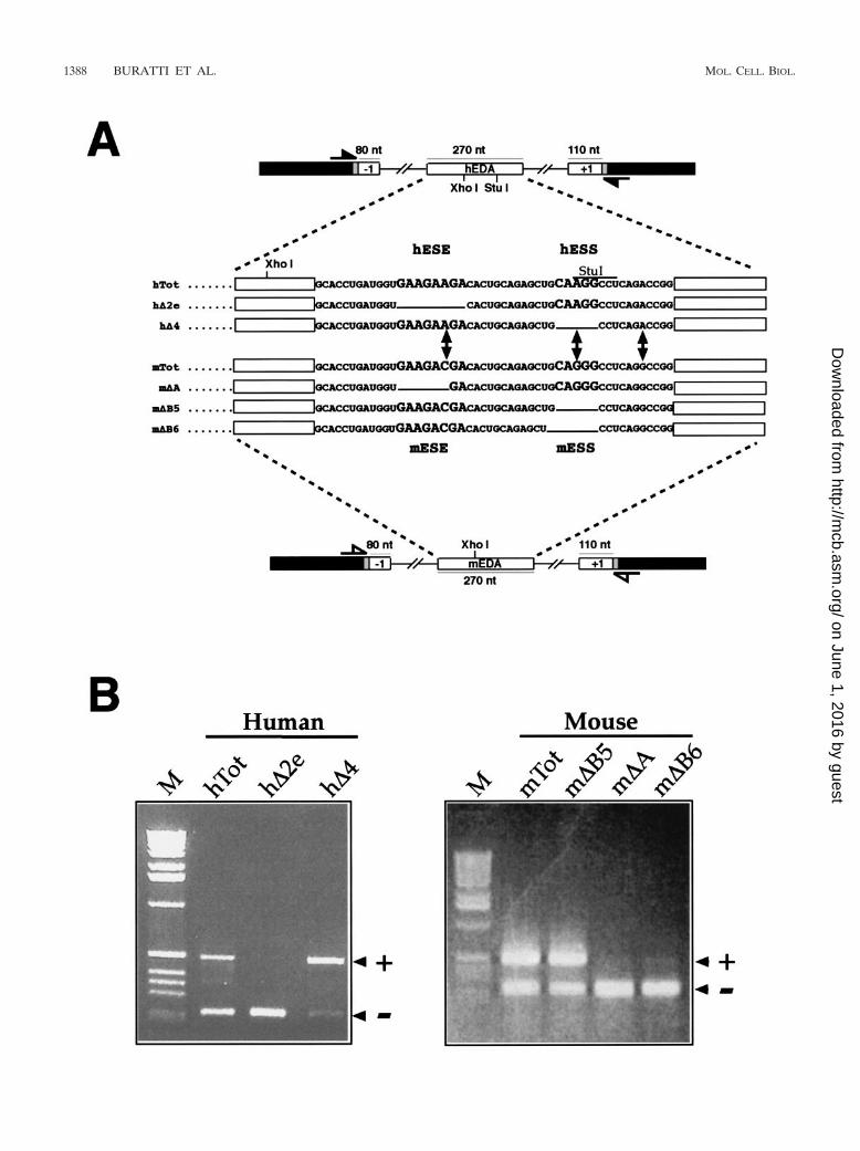
FIG. 1. Changes in mouse and human FN EDA alternative splicing caused by the introduction of deletions into the ESE and ESS regulatoryregions. (A) Schematic representation of the human and mouse minigenes. The FN exons and introns are labeled by white boxes and lines, linkersequences are shadowed, and �-globin exon 3 is indicated by black boxes. The locations of the primers used in the RT-PCR assay are shown: closedarrows indicate human-specific primers, while open arrows indicate mouse-specific primers. The sequence of the exonic region involved in EDAsplicing regulation is reported for the human wild-type minigene (hTot) and mutants carrying a deletion of the ESE (h�2e), ESS (h�4) or for themouse wild-type minigene (mTot) and mutants carrying a deletion of the mESE (m�A) or mESS (m�B5 and m�B6). The respective ESE (hESEand mESE) and ESS (hESS and mESS) sequences are in bigger characters. (B) RT-PCR analysis of total RNA from cells expressing each of theindicated constructs in the NIH 3T3 cell line. PCR products either containing (�) or lacking (�) the FN EDA exon in the messenger transcribedfrom the transfected minigene are indicated. M, molecular weight markers (1 kb; BRL).
VOL. 24, 2004 MOUSE FN EDA EXON ALTERNATIVE SPLICING REGULATION 1389
on June 1, 2016 by guesthttp://m
cb.asm.org/
Dow
nloaded from

1390 BURATTI ET AL. MOL. CELL. BIOL.
on June 1, 2016 by guesthttp://m
cb.asm.org/
Dow
nloaded from
RESULTS
Comparison of alternative splicing regulation in the mouseand human EDA sequences following homologous deletions.To compare the alternative splicing regulation of the humanand mouse FN EDA exons, we used an FN–�-globin hybridminigene, named pSV�Glob (10), into which we inserted themouse and human EDA exons (mTot and hTot, respectively)together with flanking introns and exons (Fig. 1A). This is awell-proven system for measurement of the levels of in vivoalternative splicing of the FN EDA exon in different cellularsystems and has been used extensively in previous studies ofboth human and mouse EDA exons (9, 10, 12, 52, 54). Figure1B shows the effect on the alternative splicing of the EDA exonof analogous deletions in the region surrounding the mESE,mESS, hESE, and hESS elements. It can be seen that deletionof both hESE and mESE polypurinic sequences (h�2e andm�A mutants) causes complete exon skipping from the result-ing mRNA. Thus, both hESE and mESE act as exonic splicingenhancers, a result that has been previously described by ourlaboratory (10, 54). However, when we deleted the putativemouse silencer element (identified solely on the basis of ho-mology with the human sequence), we did not observe thesame effects. In fact, while a deletion in the hESS element (h�4mutant) causes almost total exon inclusion, the identical mu-tation in mESS (m�B5 mutant) does not appear to have anyeffect on the levels of exon inclusion (Fig. 1B). Even morestrikingly, deletion of a single additional nucleotide in themESS element (m�B6) causes total exon exclusion. This resultwas identical to that observed when the mESE element hadbeen deleted (m�A mutant), thus identifying the putativemESS sequence as possessing enhancer characteristics.
Effects of stepwise changes in the m�B5 sequences towardtheir h�4 homologue. The observed changes in splicing behav-ior may be caused by the effects of one (or more) nucleotidesubstitutions on putative SR binding sites. In fact, in manyexperimental systems, single nucleotide substitutions havebeen known to affect ESE elements (11, 13, 14, 44). This maybe particularly relevant for the EDA exon, as proteins belong-ing to the SR protein family have a well-known ability to alterEDA inclusion in the mature mRNA (19, 40, 42). The use ofSR scoring matrices to identify potential SR binding sites hasbeen very useful in functionally characterizing several disease-causing mutations in the BRCA1 and SMN1 genes, as recentlyreviewed (13). Therefore, we decided to apply the availablescoring matrices for SF2/ASF, SC35, SRp40, and SRp55 to thehuman and mouse sequences (scoring matrices are available athttp://exon.cshl.org/ESE/ESEmatrix.html) (15). The resultsshowed that the 8-nt differences between the human andmouse EDA exon sequences may have resulted in changes ineither the positioning or scoring result of several putative SRprotein binding sites (data not shown).
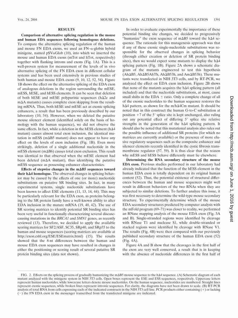
In order to evaluate experimentally the importance of thesepotential binding site changes, we decided to progressively“humanize” the exon sequence of m�B5 toward the h�4 se-quence. The rationale for this mutagenesis approach was thatif any of these exonic single-nucleotide substitutions was re-sponsible for the observed changes in splicing behavior(through either creation or deletion of SR protein bindingsites), then we would expect some mutants to display the h�4splicing pattern (Fig. 1B). Figure 2A shows a schematic dia-gram of the mutants engineered to test this hypothesis(Ah�B5, Ah�B5AhTh, Ah�B53h, and Am�B53h). These mu-tants were transfected in NIH 3T3 cells, and by RT-PCR, weanalyzed the effect on EDA exon inclusion. Figure 2B showsthat none of the mutants acquires the h�4 splicing pattern (allincluded) and that the nucleotide substitutions, at most, causesmall shifts in the EDA � ratio. Only a complete change of allof the exonic nucleotides to the human sequence restores theh�4 pattern, as shown for the m/h�4Cm mutant. It should benoted that in this construct the human G-to-C substitution atposition �7 of the 5� splice site is kept unchanged, also rulingout any potential effect of differing 5� splice site relativestrengths in the generation of the h�4 splicing pattern. Itshould also be noted that this mutational analysis also rules outthe possible influence of additional SR proteins (for which nomatrices are currently available) or the presence of more elu-sive regulatory sequences such as the composite enhancer andsilencer elements recently identified in the cystic fibrosis trans-membrane regulator (57, 59). It is thus clear that the reasonwhy mESS and hESS behave differently must lie elsewhere.
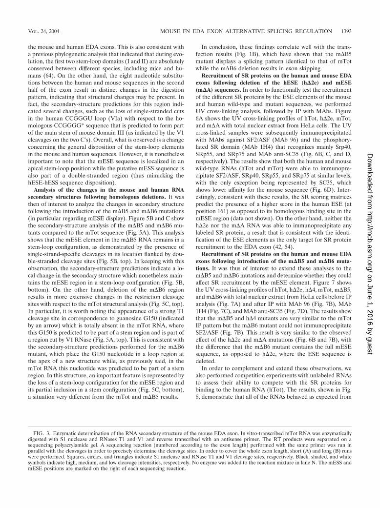
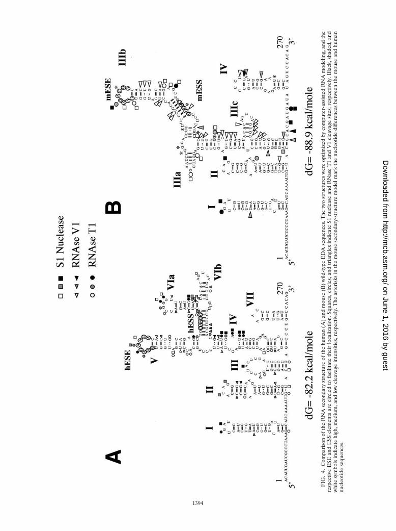
Determining the RNA secondary structure of the mouseEDA exon. Previous studies performed in our laboratory hadalready indicated that the function of the hESS element in thehuman EDA exon is totally dependent on its original humancontext (52). Thus, the potential existence of structural differ-ences between the human and mouse sequences might wellresult in different behaviors of the two RNAs when they aresubjected to similar deletions. To further analyze this issue, itwas first necessary to determine the wild-type mouse secondarystructure. To experimentally determine which of the mouseEDA secondary structures predicted by computer analysis withthe mFold program (69–71) was closer to reality, we performedan RNase mapping analysis of the mouse EDA exon (Fig. 3Aand B). Single-stranded regions were identified by cleavagewith RNase T1 and nuclease S1, while double-stranded orstacked regions were identified by cleavage with RNase V1.The results (Fig. 4B) were then compared with our previouslypublished secondary structure of the human EDA exon (52)(Fig. 4A).
Figure 4A and B show that the cleavages in the first half ofthe exon are very well conserved, a result that is in keepingwith the absence of nucleotide differences in the first half of
FIG. 2. Effects on the splicing process of gradually humanizing the m�B5 mouse sequence to the h�4 sequence. (A) Schematic diagram of eachmutant analyzed with the minigene system in NIH 3T3 cells. Open boxes represent the ESE and ESS sequences, respectively. Uppercase lettersrepresent human nucleotides, while lowercase letters denote mouse nucleotides. On the human sequence, nucleotides are numbered. Straight linesrepresent exonic sequences, while broken lines represent intronic sequences. For clarity, the diagrams have not been drawn to scale. (B) RT-PCRanalysis of total RNA from cells expressing each of the indicated constructs in the NIH 3T3 cell line. PCR products either containing (�) or lacking(�) the FN EDA exon in the messenger transcribed from the transfected minigene are indicated.
VOL. 24, 2004 MOUSE FN EDA EXON ALTERNATIVE SPLICING REGULATION 1391
on June 1, 2016 by guesthttp://m
cb.asm.org/
Dow
nloaded from

1392 BURATTI ET AL. MOL. CELL. BIOL.
on June 1, 2016 by guesthttp://m
cb.asm.org/
Dow
nloaded from
the mouse and human EDA exons. This is also consistent witha previous phylogenetic analysis that indicated that during evo-lution, the first two stem-loop domains (I and II) are absolutelyconserved between different species, including mice and hu-mans (64). On the other hand, the eight nucleotide substitu-tions between the human and mouse sequences in the secondhalf of the exon result in distinct changes in the digestionpattern, indicating that structural changes may be present. Infact, the secondary-structure predictions for this region indi-cated several changes, such as the loss of single-stranded cutsin the human CCGGGU loop (VIa) with respect to the ho-mologous CCGGGG* sequence that is predicted to form partof the main stem of mouse domain III (as indicated by the V1cleavages on the two C’s). Overall, what is observed is a changeconcerning the general disposition of the stem-loop elementsin the mouse and human sequences. However, it is nonethelessimportant to note that the mESE sequence is localized in anapical stem-loop position while the putative mESS sequence isalso part of a double-stranded region (thus mimicking thehESE-hESS sequence disposition).
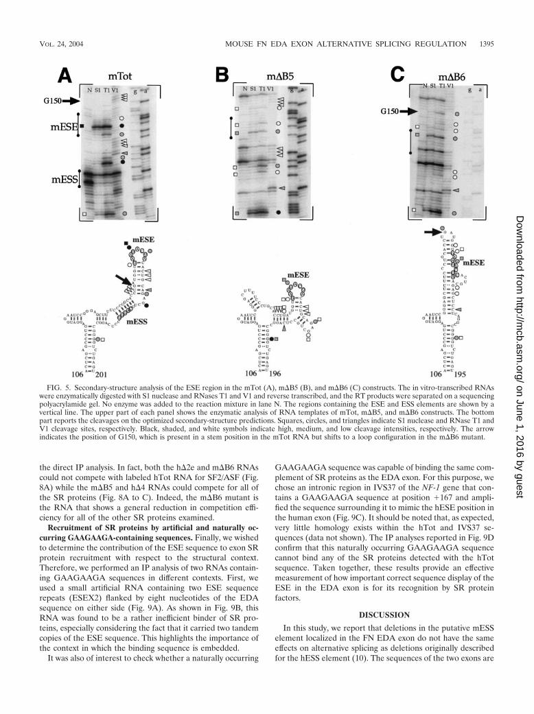
Analysis of the changes in the mouse and human RNAsecondary structures following homologous deletions. It wasthen of interest to analyze the changes in secondary structurefollowing the introduction of the m�B5 and m�B6 mutations(in particular regarding mESE display). Figure 5B and C showthe secondary-structure analysis of the m�B5 and m�B6 mu-tants compared to the mTot sequence (Fig. 5A). This analysisshows that the mESE element in the m�B5 RNA remains in astem-loop configuration, as demonstrated by the presence ofsingle-strand-specific cleavages in its location flanked by dou-ble-stranded cleavage sites (Fig. 5B, top). In keeping with thisobservation, the secondary-structure predictions indicate a lo-cal change in the secondary structure which nonetheless main-tains the mESE region in a stem-loop configuration (Fig. 5B,bottom). On the other hand, deletion of the m�B6 regionresults in more extensive changes in the restriction cleavagesites with respect to the mTot structural analysis (Fig. 5C, top).In particular, it is worth noting the appearance of a strong T1cleavage site in correspondence to guanosine G150 (indicatedby an arrow) which is totally absent in the mTot RNA, wherethis G150 is predicted to be part of a stem region and is part ofa region cut by V1 RNase (Fig. 5A, top). This is consistent withthe secondary-structure predictions performed for the m�B6mutant, which place the G150 nucleotide in a loop region atthe apex of a new structure while, as previously said, in themTot RNA this nucleotide was predicted to be part of a stemregion. In this structure, an important feature is represented bythe loss of a stem-loop configuration for the mESE region andits partial inclusion in a stem configuration (Fig. 5C, bottom),a situation very different from the mTot and m�B5 results.
In conclusion, these findings correlate well with the trans-fection results (Fig. 1B), which have shown that the m�B5mutant displays a splicing pattern identical to that of mTotwhile the m�B6 deletion results in exon skipping.
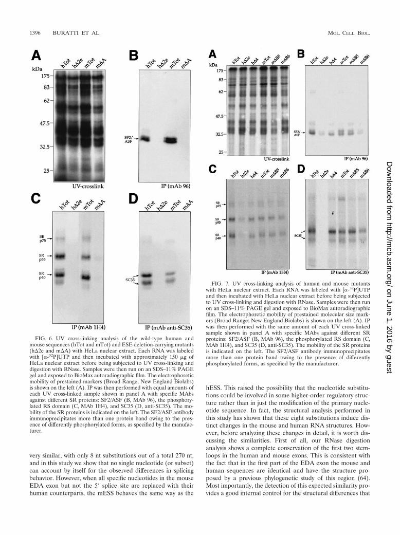
Recruitment of SR proteins on the human and mouse EDAexons following deletion of the hESE (h�2e) and mESE(m�A) sequences. In order to functionally test the recruitmentof the different SR proteins by the ESE elements of the mouseand human wild-type and mutant sequences, we performedUV cross-linking analysis, followed by IP with MAbs. Figure6A shows the UV cross-linking profiles of hTot, h�2e, mTot,and m�A with total nuclear extract from HeLa cells. The UVcross-linked samples were subsequently immunoprecipitatedwith MAbs against SF2/ASF (MAb 96) and the phosphory-lated SR domain (MAb 1H4) that recognizes mainly Srp40,SRp55, and SRp75 and MAb anti-SC35 (Fig. 6B, C, and D,respectively). The results show that both the human and mousewild-type RNAs (hTot and mTot) were able to immunopre-cipitate SF2/ASF, SRp40, SRp55, and SRp75 at similar levels,with the only exception being represented by SC35, whichshows lower affinity for the mouse sequence (Fig. 6D). Inter-estingly, consistent with these results, the SR scoring matricespredict the presence of a higher score in the human ESE (atposition 161) as opposed to its homologous binding site in themESE region (data not shown). On the other hand, neither theh�2e nor the m�A RNA was able to immunoprecipitate anylabeled SR protein, a result that is consistent with the identi-fication of the ESE elements as the only target for SR proteinrecruitment to the EDA exon (42, 54).
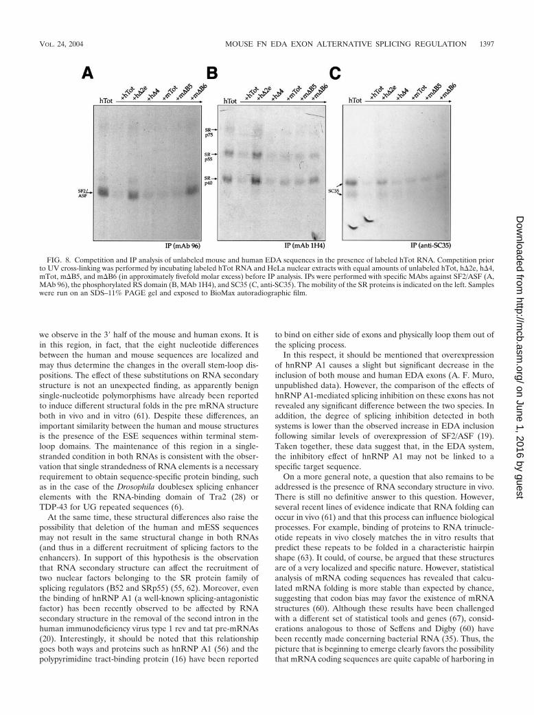
Recruitment of SR proteins on the human and mouse EDAexons following introduction of the m�B5 and m�B6 muta-tions. It was thus of interest to extend these analyses to them�B5 and m�B6 mutations and determine whether they couldaffect SR recruitment by the mESE element. Figure 7 showsthe UV cross-linking profiles of hTot, h�2e, h�4, mTot, m�B5,and m�B6 with total nuclear extract from HeLa cells before IPanalysis (Fig. 7A) and after IP with MAb 96 (Fig. 7B), MAb1H4 (Fig. 7C), and MAb anti-SC35 (Fig. 7D). The results showthat the m�B5 and h�4 mutants are very similar to the mTotIP pattern but the m�B6 mutant could not immunoprecipitateSF2/ASF (Fig. 7B). This result is very similar to the observedeffect of the h�2e and m�A mutations (Fig. 6B and 7B), withthe difference that the m�B6 mutant contains the full mESEsequence, as opposed to h�2e, where the ESE sequence isdeleted.
In order to complement and extend these observations, wealso performed competition experiments with unlabeled RNAsto assess their ability to compete with the SR proteins forbinding to the human RNA (hTot). The results, shown in Fig.8, demonstrate that all of the RNAs behaved as expected from
FIG. 3. Enzymatic determination of the RNA secondary structure of the mouse EDA exon. In vitro-transcribed mTot RNA was enzymaticallydigested with S1 nuclease and RNases T1 and V1 and reverse transcribed with an antisense primer. The RT products were separated on asequencing polyacrylamide gel. A sequencing reaction (numbered according to the exon length) performed with the same primer was run inparallel with the cleavages in order to precisely determine the cleavage sites. In order to cover the whole exon length, short (A) and long (B) runswere performed. Squares, circles, and triangles indicate S1 nuclease and RNase T1 and V1 cleavage sites, respectively. Black, shaded, and whitesymbols indicate high, medium, and low cleavage intensities, respectively. No enzyme was added to the reaction mixture in lane N. The mESS andmESE positions are marked on the right of each sequencing reaction.
VOL. 24, 2004 MOUSE FN EDA EXON ALTERNATIVE SPLICING REGULATION 1393
on June 1, 2016 by guesthttp://m
cb.asm.org/
Dow
nloaded from
FIG
.4.
Com
pari
son
ofth
eR
NA
seco
ndar
yst
ruct
ure
ofth
ehu
man
(A)
and
mou
se(B
)w
ild-t
ype
ED
Ase
quen
ces.
The
two
stru
ctur
esw
ere
optim
ized
byco
mpu
ter-
assi
sted
RN
Am
odel
ing,
and
the
resp
ectiv
eE
SEan
dE
SSel
emen
tsar
eci
rcle
dto
faci
litat
eth
eir
loca
lizat
ion.
Squa
res,
circ
les,
and
tria
ngle
sin
dica
teS1
nucl
ease
and
RN
ase
T1
and
V1
clea
vage
site
s,re
spec
tivel
y.B
lack
,sha
ded,
and
whi
tesy
mbo
lsin
dica
tehi
gh,m
ediu
m,a
ndlo
wcl
eava
gein
tens
ities
,res
pect
ivel
y.T
heas
teri
sks
inth
em
ouse
seco
ndar
y-st
ruct
ure
mod
elm
ark
the
nucl
eotid
edi
ffere
nces
betw
een
the
mou
sean
dhu
man
nucl
eotid
ese
quen
ces.
1394
on June 1, 2016 by guesthttp://m
cb.asm.org/
Dow
nloaded from
the direct IP analysis. In fact, both the h�2e and m�B6 RNAscould not compete with labeled hTot RNA for SF2/ASF (Fig.8A) while the m�B5 and h�4 RNAs could compete for all ofthe SR proteins (Fig. 8A to C). Indeed, the m�B6 mutant isthe RNA that shows a general reduction in competition effi-ciency for all of the other SR proteins examined.
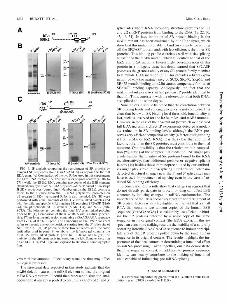
Recruitment of SR proteins by artificial and naturally oc-curring GAAGAAGA-containing sequences. Finally, we wishedto determine the contribution of the ESE sequence to exon SRprotein recruitment with respect to the structural context.Therefore, we performed an IP analysis of two RNAs contain-ing GAAGAAGA sequences in different contexts. First, weused a small artificial RNA containing two ESE sequencerepeats (ESEX2) flanked by eight nucleotides of the EDAsequence on either side (Fig. 9A). As shown in Fig. 9B, thisRNA was found to be a rather inefficient binder of SR pro-teins, especially considering the fact that it carried two tandemcopies of the ESE sequence. This highlights the importance ofthe context in which the binding sequence is embedded.
It was also of interest to check whether a naturally occurring
GAAGAAGA sequence was capable of binding the same com-plement of SR proteins as the EDA exon. For this purpose, wechose an intronic region in IVS37 of the NF-1 gene that con-tains a GAAGAAGA sequence at position �167 and ampli-fied the sequence surrounding it to mimic the hESE position inthe human exon (Fig. 9C). It should be noted that, as expected,very little homology exists within the hTot and IVS37 se-quences (data not shown). The IP analyses reported in Fig. 9Dconfirm that this naturally occurring GAAGAAGA sequencecannot bind any of the SR proteins detected with the hTotsequence. Taken together, these results provide an effectivemeasurement of how important correct sequence display of theESE in the EDA exon is for its recognition by SR proteinfactors.
DISCUSSION
In this study, we report that deletions in the putative mESSelement localized in the FN EDA exon do not have the sameeffects on alternative splicing as deletions originally describedfor the hESS element (10). The sequences of the two exons are
FIG. 5. Secondary-structure analysis of the ESE region in the mTot (A), m�B5 (B), and m�B6 (C) constructs. The in vitro-transcribed RNAswere enzymatically digested with S1 nuclease and RNases T1 and V1 and reverse transcribed, and the RT products were separated on a sequencingpolyacrylamide gel. No enzyme was added to the reaction mixture in lane N. The regions containing the ESE and ESS elements are shown by avertical line. The upper part of each panel shows the enzymatic analysis of RNA templates of mTot, m�B5, and m�B6 constructs. The bottompart reports the cleavages on the optimized secondary-structure predictions. Squares, circles, and triangles indicate S1 nuclease and RNase T1 andV1 cleavage sites, respectively. Black, shaded, and white symbols indicate high, medium, and low cleavage intensities, respectively. The arrowindicates the position of G150, which is present in a stem position in the mTot RNA but shifts to a loop configuration in the m�B6 mutant.
VOL. 24, 2004 MOUSE FN EDA EXON ALTERNATIVE SPLICING REGULATION 1395
on June 1, 2016 by guesthttp://m
cb.asm.org/
Dow
nloaded from
very similar, with only 8 nt substitutions out of a total 270 nt,and in this study we show that no single nucleotide (or subset)can account by itself for the observed differences in splicingbehavior. However, when all specific nucleotides in the mouseEDA exon but not the 5� splice site are replaced with theirhuman counterparts, the mESS behaves the same way as the
hESS. This raised the possibility that the nucleotide substitu-tions could be involved in some higher-order regulatory struc-ture rather than in just the modification of the primary nucle-otide sequence. In fact, the structural analysis performed inthis study has shown that these eight substitutions induce dis-tinct changes in the mouse and human RNA structures. How-ever, before analyzing these changes in detail, it is worth dis-cussing the similarities. First of all, our RNase digestionanalysis shows a complete conservation of the first two stem-loops in the human and mouse exons. This is consistent withthe fact that in the first part of the EDA exon the mouse andhuman sequences are identical and have the structure pro-posed by a previous phylogenetic study of this region (64).Most importantly, the detection of this expected similarity pro-vides a good internal control for the structural differences that
FIG. 6. UV cross-linking analysis of the wild-type human andmouse sequences (hTot and mTot) and ESE deletion-carrying mutants(h�2e and m�A) with HeLa nuclear extract. Each RNA was labeledwith [�-32P]UTP and then incubated with approximately 150 �g ofHeLa nuclear extract before being subjected to UV cross-linking anddigestion with RNase. Samples were then run on an SDS–11% PAGEgel and exposed to BioMax autoradiographic film. The electrophoreticmobility of prestained markers (Broad Range; New England Biolabs)is shown on the left (A). IP was then performed with equal amounts ofeach UV cross-linked sample shown in panel A with specific MAbsagainst different SR proteins: SF2/ASF (B, MAb 96), the phosphory-lated RS domain (C, MAb 1H4), and SC35 (D, anti-SC35). The mo-bility of the SR proteins is indicated on the left. The SF2/ASF antibodyimmunoprecipitates more than one protein band owing to the pres-ence of differently phosphorylated forms, as specified by the manufac-turer.
FIG. 7. UV cross-linking analysis of human and mouse mutantswith HeLa nuclear extract. Each RNA was labeled with [�-32P]UTPand then incubated with HeLa nuclear extract before being subjectedto UV cross-linking and digestion with RNase. Samples were then runon an SDS–11% PAGE gel and exposed to BioMax autoradiographicfilm. The electrophoretic mobility of prestained molecular size mark-ers (Broad Range; New England Biolabs) is shown on the left (A). IPwas then performed with the same amount of each UV cross-linkedsample shown in panel A with specific MAbs against different SRproteins: SF2/ASF (B, MAb 96), the phosphorylated RS domain (C,MAb 1H4), and SC35 (D, anti-SC35). The mobility of the SR proteinsis indicated on the left. The SF2/ASF antibody immunoprecipitatesmore than one protein band owing to the presence of differentlyphosphorylated forms, as specified by the manufacturer.
1396 BURATTI ET AL. MOL. CELL. BIOL.
on June 1, 2016 by guesthttp://m
cb.asm.org/
Dow
nloaded from
we observe in the 3� half of the mouse and human exons. It isin this region, in fact, that the eight nucleotide differencesbetween the human and mouse sequences are localized andmay thus determine the changes in the overall stem-loop dis-positions. The effect of these substitutions on RNA secondarystructure is not an unexpected finding, as apparently benignsingle-nucleotide polymorphisms have already been reportedto induce different structural folds in the pre mRNA structureboth in vivo and in vitro (61). Despite these differences, animportant similarity between the human and mouse structuresis the presence of the ESE sequences within terminal stem-loop domains. The maintenance of this region in a single-stranded condition in both RNAs is consistent with the obser-vation that single strandedness of RNA elements is a necessaryrequirement to obtain sequence-specific protein binding, suchas in the case of the Drosophila doublesex splicing enhancerelements with the RNA-binding domain of Tra2 (28) orTDP-43 for UG repeated sequences (6).
At the same time, these structural differences also raise thepossibility that deletion of the human and mESS sequencesmay not result in the same structural change in both RNAs(and thus in a different recruitment of splicing factors to theenhancers). In support of this hypothesis is the observationthat RNA secondary structure can affect the recruitment oftwo nuclear factors belonging to the SR protein family ofsplicing regulators (B52 and SRp55) (55, 62). Moreover, eventhe binding of hnRNP A1 (a well-known splicing-antagonisticfactor) has been recently observed to be affected by RNAsecondary structure in the removal of the second intron in thehuman immunodeficiency virus type 1 rev and tat pre-mRNAs(20). Interestingly, it should be noted that this relationshipgoes both ways and proteins such as hnRNP A1 (56) and thepolypyrimidine tract-binding protein (16) have been reported
to bind on either side of exons and physically loop them out ofthe splicing process.
In this respect, it should be mentioned that overexpressionof hnRNP A1 causes a slight but significant decrease in theinclusion of both mouse and human EDA exons (A. F. Muro,unpublished data). However, the comparison of the effects ofhnRNP A1-mediated splicing inhibition on these exons has notrevealed any significant difference between the two species. Inaddition, the degree of splicing inhibition detected in bothsystems is lower than the observed increase in EDA inclusionfollowing similar levels of overexpression of SF2/ASF (19).Taken together, these data suggest that, in the EDA system,the inhibitory effect of hnRNP A1 may not be linked to aspecific target sequence.
On a more general note, a question that also remains to beaddressed is the presence of RNA secondary structure in vivo.There is still no definitive answer to this question. However,several recent lines of evidence indicate that RNA folding canoccur in vivo (61) and that this process can influence biologicalprocesses. For example, binding of proteins to RNA trinucle-otide repeats in vivo closely matches the in vitro results thatpredict these repeats to be folded in a characteristic hairpinshape (63). It could, of course, be argued that these structuresare of a very localized and specific nature. However, statisticalanalysis of mRNA coding sequences has revealed that calcu-lated mRNA folding is more stable than expected by chance,suggesting that codon bias may favor the existence of mRNAstructures (60). Although these results have been challengedwith a different set of statistical tools and genes (67), consid-erations analogous to those of Seffens and Digby (60) havebeen recently made concerning bacterial RNA (35). Thus, thepicture that is beginning to emerge clearly favors the possibilitythat mRNA coding sequences are quite capable of harboring in
FIG. 8. Competition and IP analysis of unlabeled mouse and human EDA sequences in the presence of labeled hTot RNA. Competition priorto UV cross-linking was performed by incubating labeled hTot RNA and HeLa nuclear extracts with equal amounts of unlabeled hTot, h�2e, h�4,mTot, m�B5, and m�B6 (in approximately fivefold molar excess) before IP analysis. IPs were performed with specific MAbs against SF2/ASF (A,MAb 96), the phosphorylated RS domain (B, MAb 1H4), and SC35 (C, anti-SC35). The mobility of the SR proteins is indicated on the left. Sampleswere run on an SDS–11% PAGE gel and exposed to BioMax autoradiographic film.
VOL. 24, 2004 MOUSE FN EDA EXON ALTERNATIVE SPLICING REGULATION 1397
on June 1, 2016 by guesthttp://m
cb.asm.org/
Dow
nloaded from
vivo variable amounts of secondary structure that may affectbiological processes.
The structural data reported in this study indicate that them�B6 deletion causes the mESE element to lose the originalmTot RNA structure. It could then represent a situation anal-ogous to that already reported to occur in a variety of 5� and 3�
splice sites where RNA secondary structure prevents the U1and U2 snRNP proteins from binding to the RNA (18, 22, 30,45, 46, 51). In fact, inhibition of SR protein binding in them�B6 mutant has been confirmed by our IP analyses, whichshow that this mutant is unable to bind (or compete for bindingof) the SF2/ASF protein and, with less efficiency, the other SRproteins. This binding profile correlates well with the splicingbehavior of the m�B6 mutant, which is identical to that of theh�2e and m�A mutants. Interestingly, overexpression of thisprotein in a minigene assay has demonstrated that SF2/ASFpossesses the greatest ability of any SR protein family memberto stimulate EDA inclusion (19). This provides a likely expla-nation of why the maintenance of SC35, SRp40, SRp55, andSRp75 protein binding to m�B6 cannot compensate for loss ofSF2/ASF binding capacity. Analogously, the fact that them�B5 mutant possesses an SR protein IP profile identical tothat of mTot is consistent with the observation that both RNAsare spliced to the same degree.
Nonetheless, it should be noted that the correlation betweenSR binding levels and splicing efficiency is not complete. It isclear that below an SR binding level threshold, functionality islost, such as observed for the h�2e, m�A, and m�B6 mutants.However, in the case of the h�4 mutant (for which we observedfull EDA inclusion), direct IP experiments detected a moder-ate reduction in SR binding levels, although the RNA pre-serves very efficient competitor activity (a factor distinguishingit from m�B6 or h�2e RNA). It is thus clear that additionalfactors, other than the SR proteins, must contribute to the finaloutcome. One possibility is that the relative protein composi-tion (“quality”) of the complex that binds the ESE might playa role besides the quantity of SR proteins bound to the RNAor, alternatively, that additional positive or negative splicingfactors (34) besides those immunoprecipitated by our antibod-ies might play a role in h�4 splicing. Furthermore, other un-detected structural changes near the 5� and 3� splice sites mayhave caused improvement of splicing even in the case of re-duced SR binding efficiency.
In conclusion, our results show that changes in regions thatdo not directly participate in protein binding can affect ESEelements by inducing changes in mRNA conformation. Theimportance of the RNA secondary structure for recruitment ofSR protein factors is also highlighted by the fact that a smallRNA that contains two tandem copies of the human ESEsequence (GAAGAAGA) is considerably less efficient in bind-ing the SR proteins detected by a single copy of the samesequence in its original context (the EDA exon). In this re-spect, an even more striking result is the inability of a naturallyoccurring intronic GAAGAAGA sequence to immunoprecipi-tate any of the SR proteins pulled down by the same humansequence in its original context. The results highlight the im-portance of the local context in determining a functional effecton mRNA processing. Taken together, our data demonstratethat the sequence context, in addition to primary sequenceidentity, can heavily contribute to the making of functionalunits capable of influencing pre-mRNA splicing.
ACKNOWLEDGMENT
This work was supported by grants from the Telethon Onlus Foun-dation (grant E1038 awarded to F.E.B.).
FIG. 9. IP analysis comparing the recruitment of SR proteins byhuman ESE sequences alone (GAAGAAGA) as opposed to the fullEDA exon. (A) Comparison of the two RNAs used in this experiment:the hTot RNA contains the ESE within its original context (nt 107 to270), while the ESEx2 RNA contains two copies of the ESE element(flanked only by 8 nt of the EDA sequence) at the 3� end of pBluescriptII SK� sequences (dotted line). Numbering on the ESEx2 constructrefers to the distance from the T3 RNA polymerase promoter onpBluescript II SK�. A control RNA is also included. IPs (B) wereperformed with equal amounts of the UV cross-linked samples andwith the different specific MAbs against SR proteins: SF2/ASF (MAb96), the phosphorylated RS domain (MAb 1H4), and SC35 (anti-SC35). The leftmost gel contains the total UV cross-linked proteinsprior to IP. (C) Comparison of the hTot RNA with a naturally occur-ring, 170-nt-long intronic region containing a GAAGAAGA sequencefrom IVS37 of the NF-1 gene. The numbering on the IVS37 constructrefers to intronic nucleotide positions starting from the 5� splice site ofNF-1 exon 37. (D) IP profile of these two sequences with the sameantibodies used in panel B. As above, the leftmost gel contains thetotal UV cross-linked proteins prior to IP. In all of the gels, themobility of the SR proteins is indicated on the left. Samples were runon an SDS–11% PAGE gel and exposed to BioMax autoradiographicfilm.
1398 BURATTI ET AL. MOL. CELL. BIOL.
on June 1, 2016 by guesthttp://m
cb.asm.org/
Dow
nloaded from

REFERENCES
1. Adams, M. D., D. Z. Rudner, and D. C. Rio. 1996. Biochemistry and regu-lation of pre-mRNA splicing. Curr. Opin. Cell Biol. 8:331–339.
2. Balvay, L., D. Libri, and M. Y. Fiszman. 1993. Pre-mRNA secondary struc-ture and the regulation of splicing. Bioessays 15:165–169.
3. Black, D. L. 1991. Does steric interference between splice sites block thesplicing of a short c-src neuron-specific exon in non-neuronal cells? GenesDev. 5:389–402.
4. Blanchette, M., and B. Chabot. 1997. A highly stable duplex structure se-questers the 5� splice site region of hnRNP A1 alternative exon 7B. RNA3:405–419.
5. Blencowe, B. J. 2000. Exonic splicing enhancers: mechanism of action, di-versity and role in human genetic diseases. Trends Biochem. Sci. 25:106–110.
6. Buratti, E., and F. E. Baralle. 2001. Characterization and functional impli-cations of the RNA binding properties of nuclear factor TDP-43, a novelsplicing regulator of CFTR exon 9. J. Biol. Chem. 276:36337–36343.
7. Buratti, E., T. Dork, E. Zuccato, F. Pagani, M. Romano, and F. E. Baralle.2001. Nuclear factor TDP-43 and SR proteins promote in vitro and in vivoCFTR exon 9 skipping. EMBO J. 20:1774–1784.
8. Caceres, J. F., and A. R. Kornblihtt. 2002. Alternative splicing: multiplecontrol mechanisms and involvement in human disease. Trends Genet. 18:186–193.
9. Caputi, M., F. E. Baralle, and C. A. Melo. 1995. Analysis of the linkagebetween fibronectin alternative spliced sites during ageing in rat tissues.Biochim. Biophys. Acta 1263:53–59.
10. Caputi, M., G. Casari, S. Guenzi, R. Tagliabue, A. Sidoli, C. A. Melo, andF. E. Baralle. 1994. A novel bipartite splicing enhancer modulates the dif-ferential processing of the human fibronectin EDA exon. Nucleic Acids Res.22:1018–1022.
11. Caputi, M., R. J. Kendzior, Jr., and K. L. Beemon. 2002. A nonsensemutation in the fibrillin-1 gene of a Marfan syndrome patient induces NMDand disrupts an exonic splicing enhancer. Genes Dev. 16:1754–1759.
12. Caputi, M., C. A. Melo, and F. E. Baralle. 1995. Regulation of fibronectinexpression in rat regenerating liver. Nucleic Acids Res. 23:238–243.
13. Cartegni, L., S. L. Chew, and A. R. Krainer. 2002. Listening to silence andunderstanding nonsense: exonic mutations that affect splicing. Nat. Rev.Genet. 3:285–298.
14. Cartegni, L., and A. R. Krainer. 2002. Disruption of an SF2/ASF-dependentexonic splicing enhancer in SMN2 causes spinal muscular atrophy in theabsence of SMN1. Nat. Genet. 30:377–384.
15. Cartegni, L., J. Wang, Z. Zhu, M. Q. Zhang, and A. R. Krainer. 2003.ESEfinder: a web resource to identify exonic splicing enhancers. NucleicAcids Res. 31:3568–3571.
16. Chou, M. Y., J. G. Underwood, J. Nikolic, M. H. Luu, and D. L. Black. 2000.Multisite RNA binding and release of polypyrimidine tract binding proteinduring the regulation of c-src neural-specific splicing. Mol. Cell 5:949–957.
17. Clouet d’Orval, B., Y. d’Aubenton carafa, P. Sirand-Pugnet, M. Gallego, E.Brody, and J. Marie. 1991. RNA secondary structure repression of a muscle-specific exon in HeLa cell nuclear extracts. Science 252:1823–1828.
18. Coleman, T. P., and J. R. Roesser. 1998. RNA secondary structure: animportant cis-element in rat calcitonin/CGRP pre-messenger RNA splicing.Biochemistry 37:15941–15950.
19. Cramer, P., J. F. Caceres, D. Cazalla, S. Kadener, A. F. Muro, F. E. Baralle,and A. R. Kornblihtt. 1999. Coupling of transcription with alternative splic-ing: RNA pol II promoters modulate SF2/ASF and 9G8 effects on an exonicsplicing enhancer. Mol. Cell 4:251–258.
20. Damgaard, C. K., T. O. Tange, and J. Kjems. 2002. hnRNP A1 controlsHIV-1 mRNA splicing through cooperative binding to intron and exonsplicing silencers in the context of a conserved secondary structure. RNA8:1401–1415.
21. Del Gatto, F., A. Plet, M. C. Gesnel, C. Fort, and R. Breathnach. 1997.Multiple interdependent sequence elements control splicing of a fibroblastgrowth factor receptor 2 alternative exon. Mol. Cell. Biol. 17:5106–5116.
22. Estes, P. A., N. E. Cooke, and S. A. Liebhaber. 1992. A native RNA second-ary structure controls alternative splice-site selection and generates twohuman growth hormone isoforms. J. Biol. Chem. 267:14902–14908.
23. Faustino, N. A., and T. A. Cooper. 2003. Pre-mRNA splicing and humandisease. Genes Dev. 17:419–437.
24. Ffrench-Constant, C. 1995. Alternative splicing of fibronectin—many differ-ent proteins but few different functions. Exp. Cell Res. 221:261–271.
25. Frazer, K. A., L. Elnitski, D. M. Church, I. Dubchak, and R. C. Hardison.2003. Cross-species sequence comparisons: a review of methods and avail-able resources. Genome Res. 13:1–12.
26. Green, M. R. 1991. Biochemical mechanisms of constitutive and regulatedpre-mRNA splicing. Annu. Rev. Cell Biol. 7:559–599.
27. Grover, A., H. Houlden, M. Baker, J. Adamson, J. Lewis, G. Prihar, S.Pickering-Brown, K. Duff, and M. Hutton. 1999. 5� splice site mutations intau associated with the inherited dementia FTDP-17 affect a stem-loopstructure that regulates alternative splicing of exon 10. J. Biol. Chem. 274:15134–15143.
28. Hertel, K. J., K. W. Lynch, E. C. Hsiao, E. H. Liu, and T. Maniatis. 1996.
Structural and functional conservation of the Drosophila doublesex splicingenhancer repeat elements. RNA 2:969–981.
29. Huh, G. S., and R. O. Hynes. 1994. Regulation of alternative pre-mRNAsplicing by a novel repeated hexanucleotide element. Genes Dev. 8:1561–1574.
30. Jacquenet, S., D. Ropers, P. S. Bilodeau, L. Damier, A. Mougin, C. M.Stoltzfus, and C. Branlant. 2001. Conserved stem-loop structures in theHIV-1 RNA region containing the A3 3� splice site and its cis-regulatoryelement: possible involvement in RNA splicing. Nucleic Acids Res. 29:464–478.
31. Jiang, Z., J. Cote, J. M. Kwon, A. M. Goate, and J. Y. Wu. 2000. Aberrantsplicing of tau pre-mRNA caused by intronic mutations associated with theinherited dementia frontotemporal dementia with parkinsonism linked tochromosome 17. Mol. Cell. Biol. 20:4036–4048.
32. Jordan, M., A. Schallhorn, and F. M. Wurm. 1996. Transfecting mammaliancells: optimization of critical parameters affecting calcium-phosphate precip-itate formation. Nucleic Acids Res. 24:596–601.
33. Kadener, S., J. P. Fededa, M. Rosbash, and A. R. Kornblihtt. 2002. Regu-lation of alternative splicing by a transcriptional enhancer through RNA polII elongation. Proc. Natl. Acad. Sci. USA 99:8185–8190.
34. Kashima, T., and J. L. Manley. 2003. A negative element in SMN2 exon 7inhibits splicing in spinal muscular atrophy. Nat. Genet. 34:460–463.
35. Katz, L., and C. B. Burge. 2003. Widespread selection for local RNA sec-ondary structure in coding regions of bacterial genes. Genome Res. 13:2042–2051.
36. Klaff, P., D. Riesner, and G. Steger. 1996. RNA structure and the regulationof gene expression. Plant Mol. Biol. 32:89–106.
37. Kornblihtt, A. R., K. Umezawa, K. Vibe-Pedersen, and F. E. Baralle. 1985.Primary structure of human fibronectin: differential splicing may generate atleast 10 polypeptides from a single gene. EMBO J. 4:1755–1759.
38. Krainer, A. R. 1997. Eukaryotic mRNA processing. Oxford University PressInc., New York, N.Y.
39. Kuo, B. A., and P. A. Norton. 1999. Accurate selection of a 5� splice siterequires sequences within fibronectin alternative exon B. Nucleic Acids Res.27:3945–3952.
40. Kuo, B. A., T. M. Uporova, H. Liang, V. D. Bennett, R. S. Tuan, and P. A.Norton. 2002. Alternative splicing during chondrogenesis: modulation offibronectin exon EIIIA splicing by SR proteins. J. Cell. Biochem. 86:45–55.
41. Lamond, A. I. 1993. The spliceosome. Bioessays 15:595–603.42. Lavigueur, A., H. La Branche, A. R. Kornblihtt, and B. Chabot. 1993. A
splicing enhancer in the human fibronectin alternate ED1 exon interacts withSR proteins and stimulates U2 snRNP binding. Genes Dev. 7:2405–2417.
43. Libri, D., A. Piseri, and M. Y. Fiszman. 1991. Tissue-specific splicing in vivoof the beta-tropomyosin gene: dependence on an RNA secondary structure.Science 252:1842–1845.
44. Liu, H. X., L. Cartegni, M. Q. Zhang, and A. R. Krainer. 2001. A mechanismfor exon skipping caused by nonsense or missense mutations in BRCA1 andother genes. Nat. Genet. 27:55–58.
45. Liu, H. X., G. J. Goodall, R. Kole, and W. Filipowicz. 1995. Effects ofsecondary structure on pre-mRNA splicing: hairpins sequestering the 5� butnot the 3� splice site inhibit intron processing in Nicotiana plumbaginifolia.EMBO J. 14:377–388.
46. Loeb, D. D., A. A. Mack, and R. Tian. 2002. A secondary structure thatcontains the 5� and 3� splice sites suppresses splicing of duck hepatitis B viruspregenomic RNA. J. Virol. 76:10195–10202.
47. MacLeod, J. N., N. Burton-Wurster, D. N. Gu, and G. Lust. 1996. Fibronec-tin mRNA splice variant in articular cartilage lacks bases encoding the V,III-15, and I-10 protein segments. J. Biol. Chem. 271:18954–18960.
48. Matsuo, M., H. Nishio, Y. Kitoh, U. Francke, and H. Nakamura. 1992.Partial deletion of a dystrophin gene leads to exon skipping and to loss of anintra-exon hairpin structure from the predicted mRNA precursor. Biochem.Biophys. Res. Commun. 182:495–500.
49. Mistry, N., W. Harrington, E. Lasda, E. J. Wagner, and M. A. Garcia-Blanco. 2003. Of urchins and men: evolution of an alternative splicing unit infibroblast growth factor receptor genes. RNA 9:209–217.
50. Muh, S. J., R. H. Hovhannisyan, and R. P. Carstens. 2002. A non-sequence-specific double-stranded RNA structural element regulates splicing of twomutually exclusive exons of fibroblast growth factor receptor 2 (FGFR2).J. Biol. Chem. 277:50143–50154.
51. Munroe, S. H. 1984. Secondary structure of splice sites in adenovirus mRNAprecursors. Nucleic Acids Res. 12:8437–8456.
52. Muro, A. F., M. Caputi, R. Pariyarath, F. Pagani, E. Buratti, and F. E.Baralle. 1999. Regulation of fibronectin EDA exon alternative splicing: pos-sible role of RNA secondary structure for enhancer display. Mol. Cell. Biol.19:2657–2671.
53. Muro, A. F., A. K. Chauhan, S. Gajovic, A. Iaconcig, F. Porro, G. Stanta, andF. E. Baralle. 2003. Regulated splicing of the fibronectin EDA exon isessential for proper skin wound healing and normal lifespan. J. Cell Biol.162:149–160.
54. Muro, A. F., A. Iaconcig, and F. E. Baralle. 1998. Regulation of the fibronec-tin EDA exon alternative splicing. Cooperative role of the exonic enhancerelement and the 5� splicing site. FEBS Lett. 437:137–141.
VOL. 24, 2004 MOUSE FN EDA EXON ALTERNATIVE SPLICING REGULATION 1399
on June 1, 2016 by guesthttp://m
cb.asm.org/
Dow
nloaded from

55. Nagel, R. J., A. M. Lancaster, and A. M. Zahler. 1998. Specific binding of anexonic splicing enhancer by the pre-mRNA splicing factor SRp55. RNA4:11–23.
56. Nasim, F. U., S. Hutchison, M. Cordeau, and B. Chabot. 2002. High-affinityhnRNP A1 binding sites and duplex-forming inverted repeats have similareffects on 5� splice site selection in support of a common looping out andrepression mechanism. RNA 8:1078–1089.
57. Pagani, F., E. Buratti, C. Stuani, and F. E. Baralle. 2003. Missense, non-sense, and neutral mutations define juxtaposed regulatory elements of splic-ing in cystic fibrosis transmembrane regulator exon 9. J. Biol. Chem. 278:26580–26588.
58. Pagani, F., E. Buratti, C. Stuani, R. Bendix, T. Dork, and F. E. Baralle. 2002.A new type of mutation causes a splicing defect in ATM. Nat. Genet.30:426–429.
59. Pagani, F., C. Stuani, M. Tzetis, E. Kanavakis, A. Efthymiadou, S. Doudou-nakis, T. Casals, and F. E. Baralle. 2003. New type of disease causingmutations: the example of the composite exonic regulatory elements ofsplicing in CFTR exon 12. Hum. Mol. Genet. 12:1111–1120.
60. Seffens, W., and D. Digby. 1999. mRNAs have greater negative folding freeenergies than shuffled or codon choice randomized sequences. Nucleic AcidsRes. 27:1578–1584.
61. Shen, L. X., J. P. Basilion, and V. P. Stanton, Jr. 1999. Single-nucleotidepolymorphisms can cause different structural folds of mRNA. Proc. Natl.Acad. Sci. USA 96:7871–7876.
62. Shi, H., B. E. Hoffman, and J. T. Lis. 1997. A specific RNA hairpin loopstructure binds the RNA recognition motifs of the Drosophila SR proteinB52. Mol. Cell. Biol. 17:2649–2657.
63. Sobczak, K., M. De Mezer, G. Michlewski, J. Krol, and W. J. Krzyzosiak.
2003. RNA structure of trinucleotide repeats associated with human neuro-logical diseases. Nucleic Acids Res. 31:5469–5482.
64. Staffa, A., N. H. Acheson, and A. Cochrane. 1997. Novel exonic elements thatmodulate splicing of the human fibronectin EDA exon. J. Biol. Chem. 272:33394–33401.
65. Stamm, S., J. Zhu, K. Nakai, P. Stoilov, O. Stoss, and M. Q. Zhang. 2000. Analternative-exon database and its statistical analysis. DNA Cell Biol. 19:739–756.
66. Varani, L., M. Hasegawa, M. G. Spillantini, M. J. Smith, J. R. Murrell, B.Ghetti, A. Klug, M. Goedert, and G. Varani. 1999. Structure of tau exon 10splicing regulatory element RNA and destabilization by mutations of fron-totemporal dementia and parkinsonism linked to chromosome 17. Proc.Natl. Acad. Sci. USA 96:8229–8234.
67. Workman, C., and A. Krogh. 1999. No evidence that mRNAs have lowerfolding free energies than random sequences with the same dinucleotidedistribution. Nucleic Acids Res. 27:4816–4822.
68. Zhou, Z., L. J. Licklider, S. P. Gygi, and R. Reed. 2002. Comprehensiveproteomic analysis of the human spliceosome. Nature 419:182–185.
69. Zuker, M. 1989. Computer prediction of RNA structure. Methods Enzymol.180:262–288.
70. Zuker, M. 2003. Mfold web server for nucleic acid folding and hybridizationprediction. Nucleic Acids Res. 31:3406–3415.
71. Zuker, M., D. H. Mathews, and D. H. Turner. 1999. Algorithms and ther-modynamics for RNA secondary structure prediction: a practical guide, p.11–43. In W. J. Barciszewski and B. F. C. Clark (ed.), RNA biochemistry andbiotechnology. NATO ASI series. Kluwer Academic Publishers, Dordrecht,The Netherlands.
1400 BURATTI ET AL. MOL. CELL. BIOL.
on June 1, 2016 by guesthttp://m
cb.asm.org/
Dow
nloaded from