On retaining vowel colour in derived roots: Blocked imbrication in Bemba
Upload
independentCategory
view
0download
0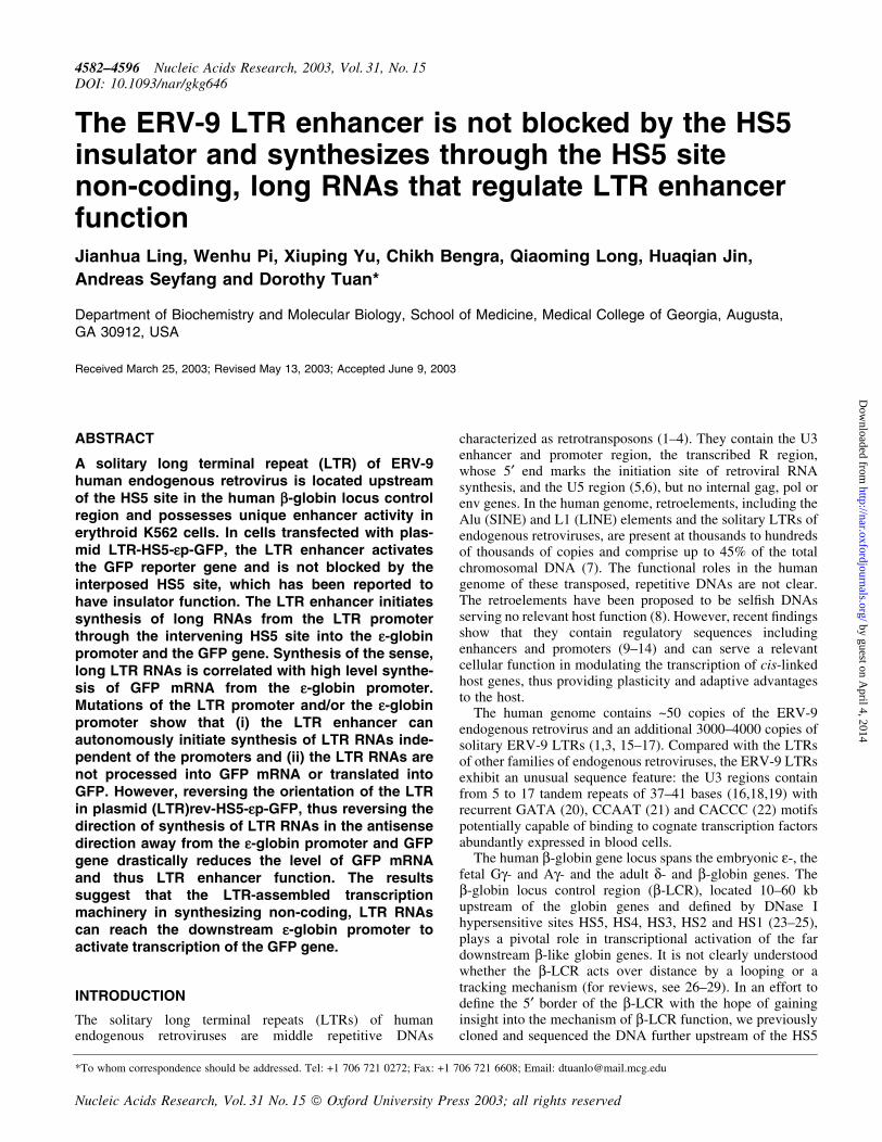
The ERV-9 LTR enhancer is not blocked by the HS5insulator and synthesizes through the HS5 sitenon-coding, long RNAs that regulate LTR enhancerfunctionJianhua Ling, Wenhu Pi, Xiuping Yu, Chikh Bengra, Qiaoming Long, Huaqian Jin,
Andreas Seyfang and Dorothy Tuan*
Department of Biochemistry and Molecular Biology, School of Medicine, Medical College of Georgia, Augusta,GA 30912, USA
Received March 25, 2003; Revised May 13, 2003; Accepted June 9, 2003
ABSTRACT
A solitary long terminal repeat (LTR) of ERV-9human endogenous retrovirus is located upstreamof the HS5 site in the human b-globin locus controlregion and possesses unique enhancer activity inerythroid K562 cells. In cells transfected with plas-mid LTR-HS5-ep-GFP, the LTR enhancer activatesthe GFP reporter gene and is not blocked by theinterposed HS5 site, which has been reported tohave insulator function. The LTR enhancer initiatessynthesis of long RNAs from the LTR promoterthrough the intervening HS5 site into the e-globinpromoter and the GFP gene. Synthesis of the sense,long LTR RNAs is correlated with high level synthe-sis of GFP mRNA from the e-globin promoter.Mutations of the LTR promoter and/or the e-globinpromoter show that (i) the LTR enhancer canautonomously initiate synthesis of LTR RNAs inde-pendent of the promoters and (ii) the LTR RNAs arenot processed into GFP mRNA or translated intoGFP. However, reversing the orientation of the LTRin plasmid (LTR)rev-HS5-ep-GFP, thus reversing thedirection of synthesis of LTR RNAs in the antisensedirection away from the e-globin promoter and GFPgene drastically reduces the level of GFP mRNAand thus LTR enhancer function. The resultssuggest that the LTR-assembled transcriptionmachinery in synthesizing non-coding, LTR RNAscan reach the downstream e-globin promoter toactivate transcription of the GFP gene.
INTRODUCTION
The solitary long terminal repeats (LTRs) of humanendogenous retroviruses are middle repetitive DNAs
characterized as retrotransposons (1±4). They contain the U3enhancer and promoter region, the transcribed R region,whose 5¢ end marks the initiation site of retroviral RNAsynthesis, and the U5 region (5,6), but no internal gag, pol orenv genes. In the human genome, retroelements, including theAlu (SINE) and L1 (LINE) elements and the solitary LTRs ofendogenous retroviruses, are present at thousands to hundredsof thousands of copies and comprise up to 45% of the totalchromosomal DNA (7). The functional roles in the humangenome of these transposed, repetitive DNAs are not clear.The retroelements have been proposed to be sel®sh DNAsserving no relevant host function (8). However, recent ®ndingsshow that they contain regulatory sequences includingenhancers and promoters (9±14) and can serve a relevantcellular function in modulating the transcription of cis-linkedhost genes, thus providing plasticity and adaptive advantagesto the host.
The human genome contains ~50 copies of the ERV-9endogenous retrovirus and an additional 3000±4000 copies ofsolitary ERV-9 LTRs (1,3, 15±17). Compared with the LTRsof other families of endogenous retroviruses, the ERV-9 LTRsexhibit an unusual sequence feature: the U3 regions containfrom 5 to 17 tandem repeats of 37±41 bases (16,18,19) withrecurrent GATA (20), CCAAT (21) and CACCC (22) motifspotentially capable of binding to cognate transcription factorsabundantly expressed in blood cells.
The human b-globin gene locus spans the embryonic e-, thefetal Gg- and Ag- and the adult d- and b-globin genes. Theb-globin locus control region (b-LCR), located 10±60 kbupstream of the globin genes and de®ned by DNase Ihypersensitive sites HS5, HS4, HS3, HS2 and HS1 (23±25),plays a pivotal role in transcriptional activation of the fardownstream b-like globin genes. It is not clearly understoodwhether the b-LCR acts over distance by a looping or atracking mechanism (for reviews, see 26±29). In an effort tode®ne the 5¢ border of the b-LCR with the hope of gaininginsight into the mechanism of b-LCR function, we previouslycloned and sequenced the DNA further upstream of the HS5
*To whom correspondence should be addressed. Tel: +1 706 721 0272; Fax: +1 706 721 6608; Email: [email protected]
4582±4596 Nucleic Acids Research, 2003, Vol. 31, No. 15DOI: 10.1093/nar/gkg646
Nucleic Acids Research, Vol. 31 No. 15 ã Oxford University Press 2003; all rights reserved
by guest on April 4, 2014
http://nar.oxfordjournals.org/D
ownloaded from
site (19) and discovered a solitary ERV-9 LTR located 1.5 kbupstream of the HS5 site in the 5¢ boundary area of the humanb-LCR. The 5¢ HS5 LTR possesses prominent enhanceractivity in erythroid cells (19,30). Here, functional dissectionof the 9 kb boundary area of the b-LCR shows that the LTRU3 region spanning 660 bases of DNA possessed uniqueenhancer and promoter activities not shared by the other DNAin the boundary area. However, the 5¢ HS5 LTR is not linkedto immediately downstream retroviral or cellular genes(GenBank accession no. AF064190) that could be activatedby the LTR enhancer and promoter. This suggests that theERV-9 LTR enhancer may interact with the proximal HS5 sitein modulating transcription of the b-LCR in erythroid cells.
However, like the chicken HS4 insulator in the 5¢ boundaryof the chicken b-globin gene locus, the HS5 site in the 5¢boundary of the human b-LCR has been shown to possessinsulator function (31±33) and bind to CTCF protein that isessential for insulator activity (34). In recombinant plasmidsin which the HS5 site is placed between the HS3 or HS2enhancer of the b-LCR and a promoter, the interposed HS5site blocks enhancer±promoter communication and drasticallydiminishes enhancer function in activating mRNA synthesisfrom the cis-linked reporter gene. It is thus possible that theHS5 site located naturally downstream of the ERV-9 LTR (seeFig. 1a) could similarly block the LTR enhancer, thusprecluding any functional interactions of the LTR enhancerwith the further downstream HS4, HS3, HS2 and HS1 sites inthe b-LCR and the globin genes.
To investigate this possibility, we constructed recombinantplasmid LTR-HS5-ep-GFP, in which the ERV-9 LTR and theHS5 site in their natural genomic order were coupled to thee-globin promoter, the nearest downstream globin promoterand the GFP reporter gene. Transfection experiments showthat contrary to the HS2 and HS3 enhancers, the ERV-9 LTRenhancer synergized with and was not blocked by the HS5 site.This indicates that manifestation of HS5 insulator activity isnot universal and depends on speci®c interactions of the HS5site with the individual enhancer. To investigate the molecularbasis of ERV-9 LTR enhancer function and its interaction withthe HS5 site, we used 5¢-RACE (35) to analyze the RNAstranscribed from the transfected plasmid. The results showthat the LTR enhancer initiated synthesis of long LTR RNAsfrom multiple sites in the LTR through the intervening HS5site into the e-globin promoter and the GFP gene. Disablingthe LTR promoter and/or the e-globin promoter in plasmidLTR-HS5-ep-GFP demonstrates that (i) synthesis of the longLTR RNAs was initiated autonomously by the LTR enhancerindependent of the LTR and e-globin promoters and (ii) thelong LTR RNAs were associated with enhanced synthesis ofGFP mRNA initiated from the e-globin promoter but werethemselves not processed into GFP mRNA. In plasmid(LTR)rev-HS5-ep-GFP, reversing the orientation of the LTRwith respect to the e-globin promoter and GFP gene, thusreversing the direction of synthesis of the long LTR RNAsfrom the sense to the antisense direction away from thee-globin promoter and GFP gene, caused a drastic drop in thelevel of GFP mRNA and thus in LTR enhancer activity. Wediscuss the possibility that the LTR-assembled transcriptionmachinery may mediate enhancer function over a longdistance by a tracking and transcription mechanism.
MATERIALS AND METHODS
Construction of recombinant GFP/CAT plasmids
The GFP plasmids (Fig. 1) were made from pEGFP-C1(Clontech) which was digested with AseI and NheI to generatethe vector backbone containing the GFP reporter gene and theSV40 poly(A) signal downstream of the GFP gene. The insertswere generated by PCR with forward and reverse primerscontaining corresponding AseI and NheI ends from either aphage template spanning the 5¢ boundary area of the humanb-LCR (19) or K562 genomic DNA. The positions inGenBank accession no. AF064190 of the forward and reversePCR primers to generate the following PCR DNAs are asfollows. For the complete LTR, 2650±2672 and 4325±4350;for (E-P-r), 2650±2672 and 3965±3987; for (P-r), 3849±3867and 3965±3987; for (E), 3248±3263 and 3849±3867; 5¢boundary fragment I, 6695±6717 and 8253±8278 (the sameas 65±89, GenBank accession no. U01317); II, 4482±4502 and6695±6717; III, 1021±1045 and 2650±2672; and for borderfragment IV, 128±150 (GenBank accession no. AF149710)and 1157±1176 (GenBank accession no. AF064190). Theplasmid (HS2-P-r)-GFP contained the 740 bp HS2 sequencebetween BamHI and BglII sites (36) spliced into a BglII site atthe 5¢ end of the P-r sequence in the (P-r)-GFP plasmid. In theplasmids (E-P-r)-, (P-r)- and HS2-P-r-GFP (Fig. 1), the 3¢ end40 bases of the R region including the AATAAA motif (see19) were not included. The authenticities of the PCRfragments were con®rmed by DNA sequencing. The referenceGFP plasmid was made by re-circularizing the AseI + NheIcleaved vector with an AseI-NheI adapter.
The construction of the plasmids LTR-CAT, LTR-HS5L-ep-CAT, HS5L-ep-CAT and ep-CAT, shown in Figure 2 andTable 1, have been described (19). To make plasmids LTR-HS5-ep-CAT and HS5-ep-CAT, the 0.5 kb HS5, made by PCRfrom the 1.2 kb HS5L with primers containing a BamHIcloning site, was used to replace HS5L in the correspondingLTR-HS5L-ep-CAT and HS5L-ep-CAT plasmids. The PCRprimers for making HS5 were: forward, bases 6115±6139;reverse, bases 6576±6600 (GenBank accession no.AF064190). To make HS4-ep-CAT, a HS4 fragment of0.9 kb spanning the NF-E2, Ap-1 and GATA-1 sites in theHS4 core (37) was made by PCR from K562 DNA with PCRprimers: forward, 1020±1050; reverse, 1888±1910 (GenBankaccession no. U01317). The forward primer contained a SalIcloning site and the reverse primer a BamHI cloning site. TheHS4 fragment was inserted into the corresponding sitesupstream of the e-globin promoter in plasmid ep-CAT. Tomake HS5L-HS4-ep-CAT, HS5L containing SalI cloning sitesmade by PCR was inserted into the SalI site upstream of HS4in HS4-ep-CAT. Construction of HS2-ep-CAT containing a0.74 kb HS2 DNA fragment has been described (36). PlasmidHS2-HS5L-ep-CAT was made by inserting HS5L with BamHIcloning sites into the BamHI site between HS2 and ep inplasmid HS2ep-CAT.
To construct HS5-ep-GFP containing 0.5 kb of HS5, shownin Table 1 and Figure 3, a PCR fragment spanning HS5 and thee-globin promoter between 5¢ AseI and 3¢ AgeI cloning siteswas made by PCR from plasmid HS5-ep-CAT (19) andinserted in clone II (Fig. 1a) in which the 1.2 kb HS5L and thegenomic DNA 5¢ of HS5L in clone II were deleted by AseI and
Nucleic Acids Research, 2003, Vol. 31, No. 15 4583
by guest on April 4, 2014
http://nar.oxfordjournals.org/D
ownloaded from
AgeI digestion. To create the plasmid LTR-HS5-ep-GFP(construct 1, Fig. 3; construct 13, Table 1), plasmid HS5-ep-GFP, serving as the vector, was cleaved with AseI and SacI toremove the 5¢-half of the HS5 site. The LTR between the AseIand AgeI sites excised from the (LTR)-GFP plasmid (Fig. 1b)and the 5¢-half of HS5 between the AgeI and SacI sitesgenerated by PCR were inserted into the cleaved vector. Thisplasmid contained the ERV-9 LTR of 1.7 kb spanning the U3GC-rich region of 590 bp, U3 enhancer of 570 bp, U3 promoterof 90 bp, the R region of 96 bp and U5 of 344 bp (19), HS5 of
0.5 kb (bases 6100±6600, GenBank accession no. AF064190)containing a CTCF binding site, CCACTAGAG-GGAAGAA, 50 bases from its 5¢ end, the 0.2 kb e-globinpromoter (bases 19307±19505, GenBank accession no.U01317) and the 0.7 kb GFP gene in pEGFP-C1 (Clontech).In this plasmid, the SV40 splice site (Promega Bulletin 80) wasinserted between the EcoRI and SalI sites in the polycloningregion at the 3¢ end of the GFP gene; the SV40 enhancer andpromoter 5¢ of the neomycin/kanamycin resistance gene weredeleted by SspI and StuI double digestion.
4584 Nucleic Acids Research, 2003, Vol. 31, No. 15
by guest on April 4, 2014
http://nar.oxfordjournals.org/D
ownloaded from
To make constructs 2 and 5, shown in Figure 3, containing,respectively, the mutated e-globin and LTR promoters,multiple base substitutions in the promoter were madesimultaneously with the following multiple sites mutagenesisprotocol (H.Jin and A.Seyfang, in preparation). Brie¯y, tomutate each promoter using construct 1 as the template, a setof three reverse primers was synthesized. The middle primer,the mutagenesis primer, was complementary to the region inthe promoter to be mutated and contained the desired basesubstitutions. The 5¢ and 3¢ primers were complementary toappropriate plasmid DNAs ¯anking the promoter and con-tained, respectively, a 5¢ or a 3¢ tail of a unique DNA sequencenot found in the plasmid. The primers were annealed todenatured plasmid DNA and the gaps between them weresynthesized with T4 DNA polymerase. The DNAs were thenligated with T4 ligase to generate the mutagenized DNAstrand. Subsequently, the DNA was ampli®ed by PCR usingthe unique 5¢ and 3¢ tails as primers, the ampli®ed DNA wascleaved at unique restriction enzyme sites present in the¯anking DNA and ligated with similarly cleaved construct 1.The mutagenesis primer for the LTR promoter was 5¢-GGG-CAGCCTGCTTTTTTTCTCTTTTCTGGCCCCTCCCACC-ATCCTGCTG-3¢; for the e-globin promoter to mutate theAATAAA box, 5¢-GTCTGGCCTTTTTTTCTTTACTGCC-3¢, and to mutate the CCAAT and CACCC motifs, 5¢-CTT-AAAAGTCATGGGTCAAGGCTGACCTGTGTCCTCAG-GGGAGGAGTCAGGTCC-3¢ (the bold letters representmutated bases in the respective underlined motifs) (seeFig. 3b). Correct base substitutions were con®rmed by DNAsequencing of the ®nal plasmids. Constructs 4 and 6, (LTR)*-ep* and (LTR)**-ep*, were made by replacing the wild-typee-globin promoter in constructs 3 and 5 with the basesubstituted e-globin promoter excised from construct 2 bySacI cleavage in HS5 and EcoRI cleavage in the multi-cloningsites downstream of the GFP gene. In constructs 3±6 (Fig. 3)the R region mutations (AATAAA to AGTAAG) were madefrom construct 1 with a Quikchange site-directed mutagenesiskit (Stratagene).
To create the (LTR)rev-HS5-ep-GFP plasmid, shown inFigure 4, the LTR was generated from plasmid LTR-HS5-ep-GFP by PCR to contain an AgeI site at the 5¢ end and AseI siteat the 3¢ end and together with the 5¢-half of HS5 between theAgeI and SacI sites was inserted into the AseI and AgeI
cleaved vector for construct 1 (Fig. 3) as described above. The(LTR)**rev-HS5-ep-GFP plasmid was made from (LTR)**-HS5-ep-GFP (construct 5, Fig. 3) by the same cloning strategyas plasmid (LTR)rev-HS5-ep-GFP.
Transfection assays and ¯uorescent ¯ow cytometryanalyses
For transient transfections (Fig. 1), 10 mg of circular plasmidsin duplicate were transfected by electroporation into 4 3 106
host cells in 400 ml of medium without fetal calf serum at240 V, 960 mF in a Gene Pulser II (Bio-Rad). All othertransient transfections (Figs 3 and 4) were carried out withlinearized plasmids. Enhancer activities of the transfectedplasmids were analyzed 48 h later in a FACScalibur withCellquest software (Becton-Dickinson). Samples of 2 3 104
live cells were analyzed for each sample; necrotic cells wereexcluded from the FACS analyses by propidium iodidestaining. The expression levels of the GFP gene in thetransfected plasmids were calculated as shown in Figure 1c.The calculated GFP levels were then corrected with respect tothe copy numbers of the transfected GFP gene as described(30). For transient transfections (Figs 3 and 4), 20 mg each ofthe plasmids linearized at a unique ApaLI site in the vectorupstream of the LTR were electroporated and analyzed asdescribed above. To screen for K562 cells harboring inte-grated plasmids (Fig. 3 and Table 1), the linearized plasmidswere co-transfected with an expression plasmid for theneomycin resistance gene as described (36). The cellsharboring integrated plasmids were harvested for FACS andRNA analyses after 3 weeks of G418 selection. The protocolsfor creating K562 cells harboring integrated CAT plasmidslinearized at a unique AlwNI or NdeI site in the vector (Fig. 2and Table 1) and CAT assays were previously described (19).
RNA isolation and 5¢-RACE
Total cellular RNAs were isolated with a Totally RNA kit(Ambion). Before use, the RNAs were treated with RNase-free DNase I to eliminate possible DNA contamination. The5¢-RACE kit (Gibco BRL) was used according to the vendorprotocol as described (30). Total cellular RNA, 5 mg/sample,was used in cDNA synthesis. After puri®cation, 1 mg of cDNAper sample was used for oligo(dC) tailing. Finally, 0.2 mg ofC-tailed cDNA per sample was used in nested PCR. After the
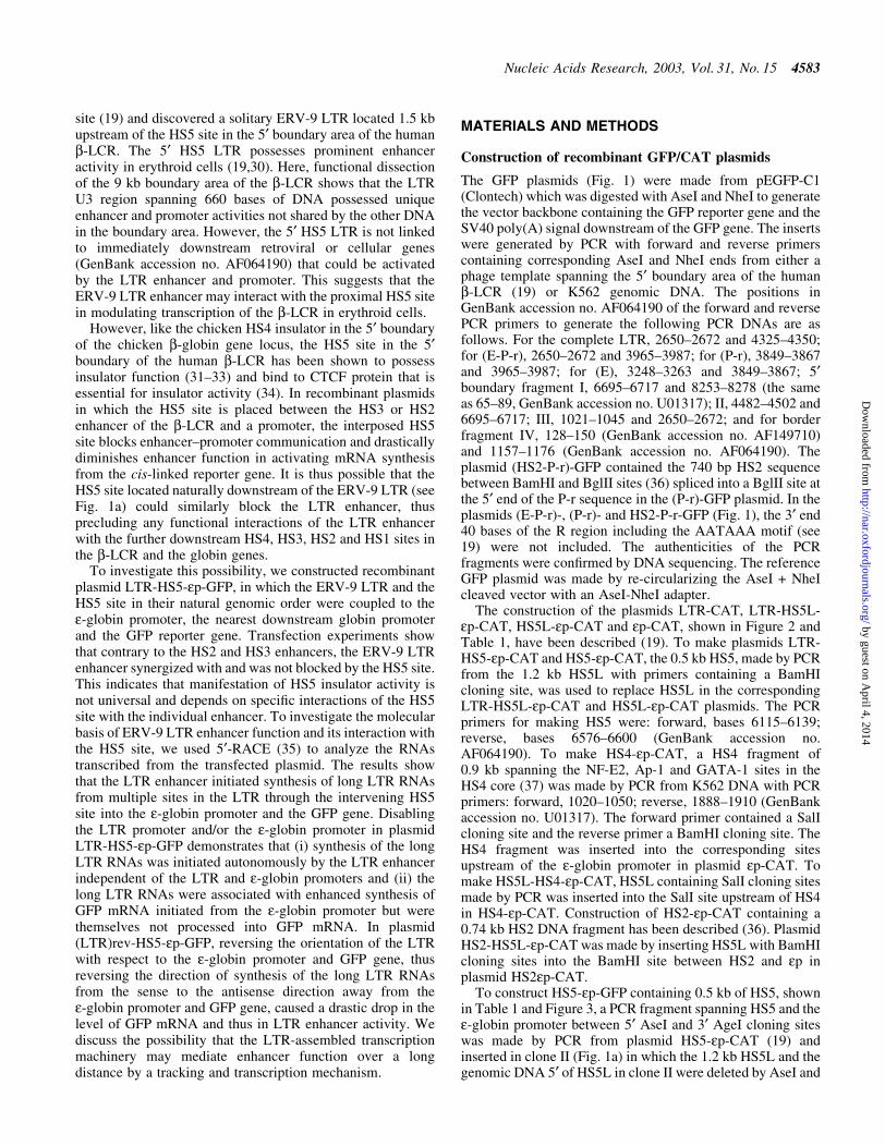
Figure 1. (Opposite) Structural and functional analyses of the 5¢ boundary area of the human b-LCR. (a) Map of the ERV-9 LTR and the 5¢ boundary area ofthe human b-LCR. Hatched box, LTR; stippled box, HS5L, the 1.2 kb HS5 site; vertical bar, CTCF binding site; *, NF-E2 binding site; HS5, the 0.5 kb HS5site (see Table 1 for constructs containing HS5L or HS5); solid boxes, DNase I hypersensitive sites HS4, HS3, HS2 and HS1 of the b-LCR. The 9 kb DNAbetween // signs is drawn to scale; the 65 kb DNA to the right of // spans the b-LCR and the human b-like globin genes and is not drawn to scale. DNAfragments I, II, III and IV, DNAs in the boundary area that ¯ank the LTR. The LTR is enlarged to show the U3, R and U5 regions. E, the U3 enhancerspanning 14 tandem repeats of 40 bp each denoted by the arrowheads; P, the 90 bp U3 promoter; open box to the left of E, the 590 bp of U3 GC-rich DNA5¢ of the enhancer; horizontal arrows, the three tandem repeats of ~80 bp each in U5. (b) Maps of the recombinant GFP plasmids containing subfragments ofthe 5¢ boundary area, I-, II-, III- and IV-, the LTR and components of the LTR. (HS2-P-r)-GFP contained the HS2 enhancer, the U3 promoter and the 5¢ halfof the R region of the 5¢HS5 LTR coupled to the GFP gene. GFP, the reference enhancerless and promoterless GFP plasmid. The third column gives the GFPlevels of the test plasmids relative to that of the reference GFP plasmid in transfected cells; values are averages of two determinations. For the I-, II-, III- andIV-GFP plasmids, the representative values of the III-GFP plasmid are presented; the GFP levels of the I-, II- and IV-GFP plasmids were even lower, in the0.2±0.4 range, and are not shown. The three numbers in parentheses are, respectively, the percentage of ¯uorescent cells, the mean ¯uorescence intensities ofthe ¯uorescent cells [see (c)] and the ratio of the plasmid copy number of the transfected test plasmid to that of the reference GFP plasmid. (c) Samplecalculation of the enhancer/promoter activity of the transfected (E-P-r)-GFP plasmid. (Left, middle and right) Dot plots by FACS analyses of K562 cellstransfected with Tris buffer and GFP and (E-P-r)-GFP plasmids, respectively. x-axis, GFP ¯uorescence intensities of the transfected cells; y-axis, FL2channel. The dot plots are the same whether FL2 or side scatter was used as the y-axis in the Cellquest program. However, using FL2 as the y-axis producedmore compact and thus more easily gated ¯uorescent and non-¯uorescent cell populations. R2 region, non-¯uorescent cells; R3 region, ¯uorescent cells. Thetable below the dot plots gives quantitative analysis by the Cellquest program of the dot plot data. Xmean, mean ¯uorescence intensities of the gated cells.(Bottom) Calculation of the enhancer/promoter activity of (E-P-r)-GFP in K562 cells.
Nucleic Acids Research, 2003, Vol. 31, No. 15 4585
by guest on April 4, 2014
http://nar.oxfordjournals.org/D
ownloaded from
second round of nested PCR, 10±15 ml samples from 50 mlwere resolved in agarose gels. The amplicons were puri®edfrom agarose gels and sequenced by the Molecular BiologyCore Laboratory. Alternatively, the authenticity of the5¢-RACE bands was con®rmed with restriction enzymedigestions. The positions of the gene-speci®c, nested reverseprimers used for (i) cDNA synthesis, (ii) and (iii) two roundsof PCR ampli®cations and (iv) DNA sequencing are asfollows. In the CAT gene in plasmid pCAT-Basic (Fig. 2):
(i) 2635±2655; (ii) 2575±2595; (iii) 2381±2401; (iv) 2328±2348 (Promega Manual). In the GFP gene in plasmid pEGFP-C1 (Fig. 3): (i) 826±850; (ii) 736±757; (iii) 617±640; (iv) 617±640 (Clontech).
RT±PCR and directional RT±PCR
Between 2 and 5 mg of total cellular RNAs isolated fromtransfected cells was used as the template for cDNA synthesisin each reverse transcription (RT) reaction. In directional RT±
4586 Nucleic Acids Research, 2003, Vol. 31, No. 15
by guest on April 4, 2014
http://nar.oxfordjournals.org/D
ownloaded from

PCR (Fig. 4), to synthesize cDNAs from the sense RNAs, thereverse primers of the primer pairs spanning the regions ofinterest were used in RT. To synthesize cDNAs from theantisense RNAs, the forward primers of the primer pairs wereused in RT. Aliquots of cDNAs transcribed from 400 ng ofRNA were used in the subsequent PCRs with each of theforward and reverse primer pairs used in cDNA synthesis.The RT step was carried out with M-MLV reversetranscriptase (BRL) at 42°C for 60 min. The PCR con-ditions were denaturation at 94°C for 1 min, annealing at 58°Cfor 1 min and extension at 72°C for 1 min, repeated for32 cycles, which were chosen from pilot PCRs carried out for28, 30, 32, 34 and 36 cycles. At 32 cycles, the PCR ingredientswere not exhausted and the PCR product was within the linearrange suitable for semi-quantitative analyses. Aliquots of thePCR products (5 ml of 50 ml) were analyzed in 2% agarosegels.
Note that in order to amplify only the RNAs transcribedfrom the transfected plasmids, the RT±PCR primer pairs weredesigned to span speci®c junction sequences not found in theK562 genome (see Fig. 4a and c for primer pair locations). Inprimer pair 1a, the forward primer spanned the junctionbetween the LTR and the vector sequence; in primer pair 1b,the reverse primer spanned the junction between U5 and HS5,which in the genome were separated by >3 kb of DNA, adistance not ef®ciently ampli®ed under the current RT±PCRconditions; the forward primer in primer pair 1c was located atthe junction between U5 and the vector; primer pair 1dspanned the LTR enhancer in reverse orientation with respectto HS5, a con®guration not found in the K562 genome; primerpair 2 spanned HS5 and the e-globin promoter, which in thegenome were separated by >20 kb of DNA; primer pair 3ampli®ed the GFP reporter gene which was a non-humansequence derived from jelly®sh. The sequences of the primerpairs were as follows. 1a: forward, 4712±0008 (pEGFP-C1,Clontech); reverse, 3260±3281 (GenBank accession no.AF064190). 1b: forward, 4004±4028 (AF064190); reverse,5¢-TGACATACTGTGACCGGTAGCGCTAGCATT-3¢ (theAgeI site at the junction between the LTR and HS5 isunderlined). 1c: forward, 4712±0008 (pEGFP-C1, Clontech);reverse, 4004±4028 (GenBank accession no. AF064190). 1d:forward, 3260±3281; reverse, 6258±6280 (GenBank accessionno. AF064190). 2: 6268±6292 (GenBank accession no.AF064190); reverse, 19338±19361 (U01317). 3: 616±631,956±981 (pEGFP-C1).
An internal control was generated by a primer pair in thee-globin mRNA (forward, 19957±19975; reverse, 21021±21045; GenBank accession no. U01317) as follows. Thereverse primer in e-globin mRNA was added together witheach of the above test primers in the same tube as for RT toalso synthesize cDNA from the endogenous e-globin mRNApresent in all the RNA samples. Equal aliquots of the cDNAstock were ampli®ed in separate PCR tubes with the testprimer pairs or the e-globin primer pair for 32 and 30 cyclesrespectively. Samples (10±15 ml) from the 50 ml of PCRproducts were resolved in 2% agarose gels. The authenticity ofthe RT±PCR bands was con®rmed either by DNA sequencingor restriction enzyme digestion. To quantify the RNAtemplates, the intensities of the RT±PCR bands were quan-ti®ed with an IS1000 Image Analyzer (Alpha Innotech) andnormalized with respect to the intensities of the bandgenerated by endogenous e-globin mRNA.
Northern blot
Aliquots of 15 mg of total cellular RNA isolated from thetransfected cells (Fig. 4a) were resolved in 1.5% denaturingagarose gels, blotted and hybridized to 32P-labeled probeaccording to published protocols (38). Radioactive intensitiesof the RNA bands were quanti®ed with a PhosphorImager.
RESULTS
Functional dissections of the 5¢ boundary of the humanb-LCR and the ERV-9 LTR
Using a newly developed transfection assay with the GFP geneas the reporter followed by FACS analyses (30), wedetermined the enhancer and promoter functions of the DNAin the 5¢ boundary area of the b-LCR. The 9 kb boundary DNAincluding 3 kb of DNA spanning the HS5 site was sub-clonedinto ®ve recombinant GFP plasmids, clones I, II, LTR, III andIV (Fig. 1a), which were transfected separately into K562erythroid cells. In a previous report (30), we observed that thepercentages of ¯uorescent cells (®rst numbers in parentheses,Fig. 1b) correlated positively with the enhancer and promoterstrengths as measured by the mean ¯uorescence intensities ofthe transfected cells (second numbers in parentheses, Fig. 1b).Hence, we took the product of the percentage of ¯uorescentcells multiplied by the mean ¯uorescence intensity of the¯uorescent cells as a quantitative measure of the combinedenhancer and promoter strengths of the LTR (see Fig. 1c).
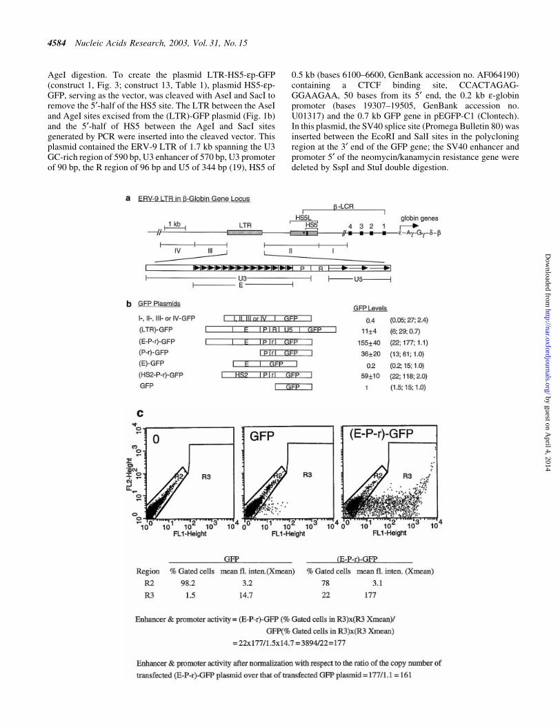
Figure 2. (Opposite) Transcription of integrated plasmid LTR-HS5-ep-CAT in K562 cells. (a) Maps of the integrated LTR-HS5-ep-CAT and HS5-ep-CATplasmids and transcription of the plasmids analyzed by 5¢-RACE. Angled arrows, locations of transcriptional initiation sites in the LTR, HS5 site and thee-globin promoter. Left to right arrows, RNAs initiated from within the LTR, HS5 and e-globin promoter, with the thickness of the arrows showing therelative abundance of the RNAs. Right to left arrows, cDNAs reverse transcribed from the respective RNAs and PCR fragments subsequently ampli®ed fromthe cDNAs by the 5¢-RACE protocol. The PCR fragments depicted were the sequenced DNA strand; the arrows are aligned with the plasmid map on top.(C)n, poly(dC) tails added to the 3¢ end of the cDNAs by the TdT enzyme. The arrowheads at the 5¢ ends of the cDNA or the PCR fragment indicate thereverse primers used for cDNA synthesis, PCR ampli®cation and DNA sequencing. Numbers are the sizes in nucleotides of the PCR fragments determined bygel electrophoresis and DNA sequencing [see (b), (c) and (d)]. CAT levels are relative levels of CAT enzyme produced by the LTR-HS5-ep-CAT plasmid (at8 copies/cell) and HS5-ep-CAT (at 14 copies/cell) determined previously by CAT assay (19). (b) PCR products of 5¢-RACE. Lanes 1 and 2, PCR bandsgenerated by RNAs transcribed, respectively, from the LTR-HS5-ep-CAT and HS5-ep-CAT plasmids; M, 100 bp size markers. Numbers on the right and leftmargins are sizes in bp of the PCR fragments and the size markers. (c) DNA seqence of the 210 bp PCR fragment generated from CAT mRNA. The basemarked by the arrow is the 5¢ end/initiation site of CAT mRNA. (d) DNA sequence of the 1300 bp PCR fragment generated from the long LTR RNA; onlythe 5¢ end of the sequence is shown. Arrow, 5¢ end/initiation site of the LTR RNA. (e) DNA sequences near the transcriptional initiation sites of the LTRRNA and CAT mRNA. AATAAA, TATA box in the U3 or the e-globin promoter; angled arrows, locations of transcriptional initiation sites; bold bases,transcribed bases.
Nucleic Acids Research, 2003, Vol. 31, No. 15 4587
by guest on April 4, 2014
http://nar.oxfordjournals.org/D
ownloaded from
Consistent with previous assays using the CAT reporter gene(19), the (LTR)-GFP plasmid exhibited enhancer/promoteractivities ~10-fold above the reference GFP plasmid (Fig. 1b).Clones I±IV, including clone II, which spans the HS5 site,produced very few ¯uorescent cells and possessed nodetectable enhancer/promoter activities.
We next determined the function of the U3, R and U5regions of the ERV-9 LTR as demarcated by DNA sequenceanalyses (19). The U3 region spanning the 14 tandem repeatsof 40 bases/repeat exhibited enhancer activity: plasmid (E-P-r)-GFP, containing the U3 enhancer and promoter togetherwith the 5¢ end of the R region spanning the retroviral
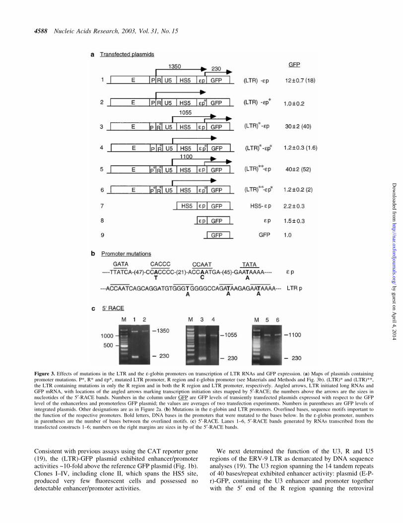
Figure 3. Effects of mutations in the LTR and the e-globin promoters on transcription of LTR RNAs and GFP expression. (a) Maps of plasmids containingpromoter mutations. P*, R* and ep*, mutated LTR promoter, R region and e-globin promoter (see Materials and Methods and Fig. 3b). (LTR)* and (LTR)**,the LTR containing mutations in only the R region and in both the R region and LTR promoter, respectively. Angled arrows, LTR initiated long RNAs andGFP mRNA, with locations of the angled arrows marking transcription initiation sites mapped by 5¢-RACE; the numbers above the arrows are the sizes innucleotides of the 5¢-RACE bands. Numbers in the column under GFP are GFP levels of transiently transfected plasmids expressed with respect to the GFPlevel of the enhancerless and promoterless GFP plasmid; the values are averages of two transfection experiments. Numbers in parentheses are GFP levels ofintegrated plasmids. Other designations are as in Figure 2a. (b) Mutations in the e-globin and LTR promoters. Overlined bases, sequence motifs important tothe function of the respective promoters. Bold letters, DNA bases in the promoters that were mutated to the bases below. In the e-globin promoter, numbersin parentheses are the number of bases between the overlined motifs. (c) 5¢-RACE. Lanes 1±6, 5¢-RACE bands generated by RNAs transcribed from thetransfected constructs 1±6; numbers on the right margins are sizes in bp of the 5¢-RACE bands.
4588 Nucleic Acids Research, 2003, Vol. 31, No. 15
by guest on April 4, 2014
http://nar.oxfordjournals.org/D
ownloaded from
transcriptional initiation site, expressed GFP 150-fold higherthan the GFP plasmid (Fig. 1b). However, in the absence of theU3 promoter, the 14 U3 repeats by themselves in plasmid (E)-GFP did not activate the GFP gene (Fig. 1b), indicating arequirement for the LTR promoter in LTR enhancer function.
Plasmid (LTR)-GFP, containing the entire LTR includingthe U5 region, showed 15-fold lower enhancer activity thanplasmid (E-P-r)-GFP (Fig. 1b). The reduction in GFP expres-sion was probably due to the extra 400 bases of the R and U5DNA that may contain transcriptional or translationalinhibitors. In comparison with the LTR enhancer, the strongHS2 enhancer of the b-LCR (36) in plasmid (HS2-P-r)-GFPactivated the GFP gene to a level 40% that of the (E-P-r)-GFPconstruct (Fig. 1b). These results indicate that among the DNAfragments in the 5¢ boundary area of the b-LCR, the ERV-9LTR possessed unique enhancer/promoter activity that was2- to 3-fold higher than that of the HS2 enhancer as assayed inthese GFP constructs.
ERV-9 LTR enhancer activity is not blocked by the HS5site
We previously reported that in recombinant plasmid LTR-HS5L-ep-CAT containing the 1.2 kb HS5 fragment, theERV-9 LTR enhancer activity was not blocked by theinterposed HS5L (see constructs 1 and 3, Table 1) (19).However, the 1.2 kb HS5L spanned not only the CTCFbinding site essential to HS5 insulator function (34) but alsomultiple binding sites for positive transcription factors such aserythroid NF-E2 and GATA-1 (39) (see Fig. 1a), whichexhibited weak enhancer activity in transfected plasmidHS5L-ep-CAT (construct 2, Table 1). Thus, it is possiblethat the failure of HS5L to block ERV-9 LTR enhanceractivity was due to the presence of these positive regulatoryelements and/or the special vector backbone of the CATplasmid that combined to negate the insulation effect of CTCFbound at the HS5 site. To investigate these possibilities, weused the pEGFP plasmid (Clontech) with a different vectorbackbone to construct the plasmid LTR-HS5-ep-GFP, inwhich the HS5 site was truncated to 0.5 kb to delete the NF-E2site and nine GATA sites present in the ®rst 700 bases of the1.2 kb HS5L. The truncated HS5 core contained the CTCFbinding site at its 5¢ end and only 4 of the 13 GATA sites inHS5L (see Fig. 1a). For comparison, recombinant CATplasmids containing the truncated 0.5 kb HS5 or the 1.2 kbHS5L coupled to the b-LCR HS4 or HS2 enhancers were alsoconstructed (constructs 4±10, Table 1).
The plasmids were linearized at a site in the vector upstreamof the LTR enhancer so that the enhancer could not act on thelinked reporter gene from a location downstream of the geneas in a circular plasmid, thus by-passing the potential blockingeffect of the HS5 insulator. To ensure that the linearizedplasmids were not integrated into K562 cells in long tandemarrays so that the LTR enhancer in the downstream plasmidcould directly interact with and activate the reporter gene ofthe upstream plasmid without an interposed HS5 site, thelinearized plasmids were transfected into K562 cells byelectroporation. Southern blots of the integrated plasmidsfollowing digestion by a restriction enzyme with a singlecleavage site in the plasmids showed that the majority of theplasmids were intergrated in single copies into multiple,separate host sites (blots not shown) (40).
In integrated plasmid HS5-ep-CAT, the 0.5 kb HS5 did notexhibit appreciable enhancer activity (construct 4, Table 1).However, it still did not block ERV-9 LTR enhancer activityin LTR-HS5-ep-CAT, nor did it block LTR enhancer activityin LTR-HS5-ep-GFP (see constructs 1 and 5 and 11 and 13,Table 1). Furthermore, in the plasmid HS5L-HS4-ep-CAT, inwhich HS5L was coupled to the natural downstream HS4 sitein the b-LCR (see Fig. 1a), HS5 strongly stimulated HS4, withinherently weak enhancer activity, to exhibit prominentenhancer activity comparable to that exhibited by the strongHS2 enhancer (compare constructs 6, 7 and 9, Table 1).
The failure of the HS5 site to exhibit insulator activity inthese plasmids did not appear to be an artifact of the particularexperimental conditions. In the plasmid HS2-HS5L-ep-CAT,similarly integrated into K562 cells with an identicaltransfection protocol, the HS5 site inserted downstream ofthe strong HS2 enhancer exhibited insulator properties inblocking HS2 enhancer activity (see constructs 9 and 10,Table 1). Taken together, the results indicate that whencoupled in the genomic order to its natural neighbors in theb-LCR, i.e. either downstream of the ERV-9 LTR or upstreamof the HS4 site, the HS5 site did not exhibit insulatorproperties. This indicates that manifestation of HS5 insulatoractivity is not universal and appears to depend on speci®cinteractions between HS5 and the individual LTR, HS2 orHS3 enhancers.
In the LTR-HS5-ep-CAT plasmid the LTR enhanceractivates synthesis of long RNAs that are initiated fromthe LTR promoter and extended through the HS5 siteand the e-globin promoter into the CAT gene
To investigate the molecular basis of ERV-9 LTR enhanceractivity and its synergistic interaction with HS5, we used5¢-RACE to analyze the RNAs transcribed from plasmid LTR-HS5-ep-CAT (Fig. 2a). We detected long LTR RNAs thatproduced a 5¢-RACE band of 1300 nt (Fig. 2b, lane 1). DNAsequencing of the entire 1300 bp band showed that the longLTR RNA was initiated from the LTR at the C base located 25bases downstream of the AATAAA box in the U3 promoter(Fig. 2d and e) and extended through HS5 and the e-globinpromoter into the CAT gene (Fig. 2a). Correlated with thepresence of the long LTR RNA, CAT mRNA, producing a5¢-RACE band of 210 bp, was synthesized more abundantlyfrom plasmid LTR-HS5-ep-CAT than from plasmid HS5-ep-CAT (compare the intensities of the bands of 210 nt in Fig. 2b,lanes 1 and 2; the intensities of the 410 nt bands in these lanesproduced by RNA initiated from within HS5 in the respectiveplasmids were comparable and served as the internal referencefor the quality of the RNA templates and sample loading).DNA sequencing showed that the 210 nt band was producedby CAT mRNA initiated at the A base 22 bases downstream ofthe AATAAA (TATA) box in the e-globin promoter (Fig. 2cand e). The enhanced synthesis of CAT mRNA from theintegrated plasmid LTR-HS5-ep-CAT was observed repeat-edly in three 5¢-RACE assays using two different RNApreparations. The correlation between transcription of the longLTR RNAs and enhanced synthesis of CAT mRNA suggeststhat the LTR-initiated long RNA could be processed into CATmRNA. The enhanced level of CAT enzyme synthesized fromthe plasmid LTR-HS5-ep-CAT suggests that the long LTRRNA could also be directly translated into the CAT enzyme.
Nucleic Acids Research, 2003, Vol. 31, No. 15 4589
by guest on April 4, 2014
http://nar.oxfordjournals.org/D
ownloaded from
The LTR-initiated long RNAs are not processed intomRNA or translated into protein products
To investigate the possibilities that the long LTR RNAs mightbe processed into CAT mRNA or directly translated into theprotein product, we created three pairs of reference and testplasmids (constructs 1 and 2, 3 and 4 and 5 and 6, Fig. 3a) tostudy the relationship between syntheses of long LTR RNAsand mRNA. In these plasmids, we attempted to abrogatesyntheses of the long LTR RNA or mRNA through disablingthe LTR or e-globin promoter. The e-globin promoter wasdisabled by base substitutions in the AATAAA (TATA),CACCC and CCAAT motifs previously shown to be importantfor globin promoter function (41); the LTR promoter wasdisabled by similar base substitutions (Fig. 3b). The GFPreporter gene replaced the CAT gene in these constructs forease of carrying out reporter gene assays by FACS analysis.
The linearized plasmids were transiently or stably transfectedinto K562 cells by electroporation. GFP protein and mRNA inthe transfected cells were analyzed by FACS and 5¢-RACE.
In constructs 1 and 2, the reference plasmid LTR-HS5-ep-GFP, (LTR)-ep, either transiently transfected or stably inte-grated into K562 cells, expressed GFP at a level 12- to 18-foldhigher than the enhancerless and promoterless GFP plasmid(construct 1, Fig. 3a). As with the integrated plasmid LTR-HS5-ep-CAT (Fig. 2), (LTR)-ep synthesized both the longLTR RNA and the short GFP mRNA, which produced,respectively, the 1350 and 230 nt bands in 5¢-RACE (Fig. 3c,lane 1). Restriction enzyme digestions of the 1350 nt band (notshown) con®rmed that this band was generated by LTR-GFPRNA initiated from within the LTR and extended through theHS5 and e-globin promoter into the GFP gene. Sequencing ofthe 230 nt band (not shown) indicated that this band was
4590 Nucleic Acids Research, 2003, Vol. 31, No. 15
by guest on April 4, 2014
http://nar.oxfordjournals.org/D
ownloaded from
generated by GFP mRNA initiated from the same speci®c site
in the e-globin promoter as CAT mRNA (Fig. 2c and e).In contrast, the test plasmid LTR-HS5-ep*-GFP, (LTR)-
ep*, containing the mutated e-globin promoter, expressed GFPat a background level similar to that of the enhancerless andpromoterless GFP plasmid (construct 2, Fig. 3a). In 5¢-RACE,the 230 nt band produced by the GFP mRNA was notdetectable, indicating that the mutant e-globin promoter didnot initiate synthesis of GFP mRNA. On the other hand, thelong LTR RNA initiated from the LTR and extended throughHS5 into the GFP gene was still synthesized and produced the1350 nt band (Fig. 3c, lane 2). The LTR RNA extended intothe 3¢ end of the GFP gene as indicated by RT±PCR with aprimer pair spanning the entire GFP gene (not shown). Theclear absence of GFP mRNA from the test plasmid (Fig. 3c,lane 2) could not be due to less RNA template from the test
plasmid used in the RT step or the PCR conditions of the5¢-RACE protocol, since an equal amount of RNA templatefrom the reference plasmid, processed under an identicalexperimental protocol, produced not only the GFP mRNAband but also the LTR RNA band at a similar intensity to thetest plasmid (compare intensities of the LTR RNA bands inlanes 1 and 2 and also in lanes 3 and 4 and lanes 5 and 6,Fig. 3c). These results, reproducibly observed in two inde-pendent experiments, indicate that the long LTR RNA was notprocessed into GFP mRNA nor was it ef®ciently translatedinto GFP.
Constructs 3 and 4, (LTR)*-ep and (LTR)*-ep* (Fig. 3a),con®rmed these observations. These two plasmids contained(LTR)* in which the R region AATAAA motif was mutated toAGTAAG. FACS analysis showed surprisingly that construct3, (LTR)*-ep, as a result of the two A®G base mutations in
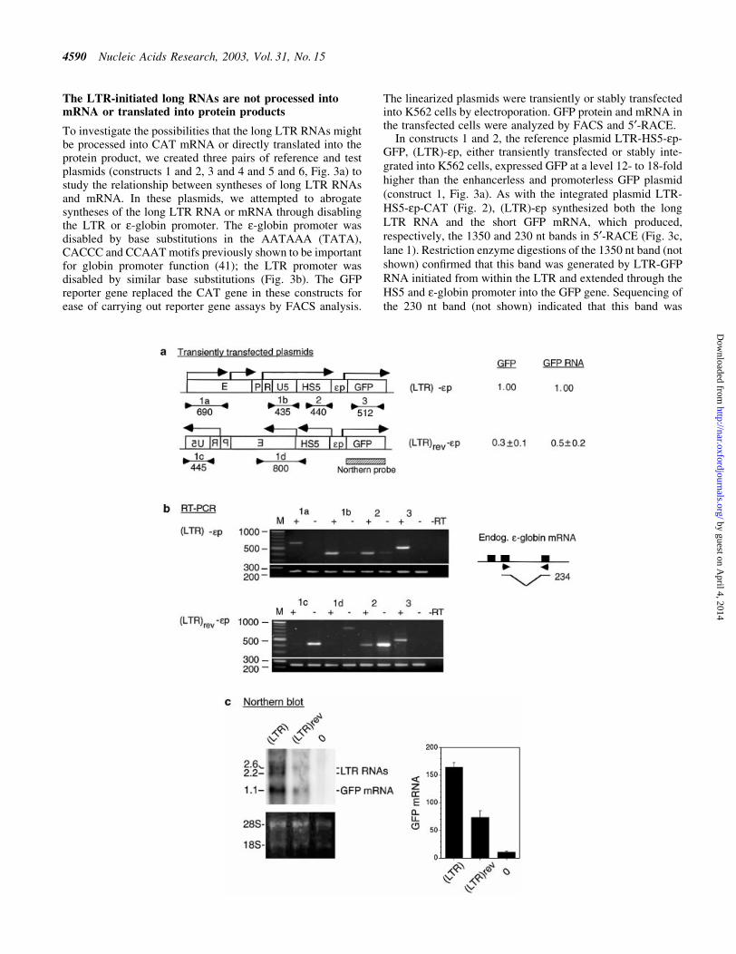
Figure 4. (Opposite and above) Effects of LTR orientation on GFP mRNA synthesis and GFP expression. (a) Maps of the reference (LTR)-ep and test(LTR)rev-ep plasmids. Locations and direction of angled arrows show the locations of RNA initiation sites and direction of RNA synthesis. The locationsmarked by heavy stems are initiation sites mapped by 5¢-RACE and northern blot; locations marked by thin stems are initiation sites postulated to exist fromthe RT±PCR bands. Horizontal lines marked 1a, 1b, 1c, 1d, 2 and 3 are RT±PCR products generated by the respective primer pairs 1a±1d, 2 and 3; the linesare aligned with the plasmid maps on top; numbers underneath the lines are sizes (bp) of the RT±PCR bands. Hatched box, northern blot probe. GFP andGFP RNA, levels of GFP expression and GFP RNA of the test relative to those of the reference plasmids. Numbers in parentheses are GFP levels of the plas-mids relative to that of the enhancerless and promoterless GFP plasmid. (b) Directional RT±PCR. Lanes marked 1a±1d, 2 and 3 are RT±PCR bands generatedby the respective primer pairs; + and ± lanes, RT±PCR bands generated by each primer pair from the sense and the antisense RNAs; ±RT lane, no reversetranscriptase in the RT step to show absence of DNA contamination in the RNA samples; bottom rows of RT±PCR bands were generated from the endo-genous e-globin mRNA with a primer pair as depicted in the inset to its right and served as internal controls. (Inset) Solid boxes, the three exons of thee-globin gene; arrowheads, the location of the primer pair spanning the second intron; horizontal lines, the spliced e-globin mRNA ampli®ed by the primerpair; the bent line, spliced out second intron; number to the right, size in base pairs of the RT±PCR band ampli®ed by the primer pair from the e-globinmRNA. (c) Northern blot of total cellular RNAs isolated from K562 cells transfected with (LTR)-ep plasmid [lane (LTR)], (LTR)rev-ep plasmid [lane(LTR)rev] and a non-transfected K562 control (lane 0). (Top left) Northern blot hybridized to the GFP gene probe. Numbers in the left margin are sizes inkilobases of the LTR RNAs and GFP mRNA. (Bottom left) Ethidium bromide stained bands of 28S and 18S RNAs to show equal loading of the RNAsamples in different lanes in the agarose gel. Bar graphs are mean values of the radioactive counts, measured in arbitrary pixel units, of GFP mRNA fromblots of three separate RNA preparations. (d and e) Maps of reference (LTR)**-ep and test (LTR)**rev-ep plasmids. Designations as in (a) and (b).(f) 5¢-RACE of LTR RNAs and GFP mRNA transcribed from (LTR)**-ep (lane 1) and (LTR)**rev-ep (lane 2).
Nucleic Acids Research, 2003, Vol. 31, No. 15 4591
by guest on April 4, 2014
http://nar.oxfordjournals.org/D
ownloaded from

the transcribed R region, expressed GFP at a level almost3-fold that of construct 1, (LTR)-ep (Fig. 3a). 5¢-RACEshowed that (LTR)*-ep synthesized the long LTR* RNA andalso GFP, which produced, respectively, the bands of 1055and 230 nt (Fig. 3c, lane 3). In contrast, the test plasmid(LTR)*-ep* containing the disabled e-globin promotersynthesized no GFP mRNA but only the long LTR* RNAthat produced the 1055 nt band and expressed GFP at nearbackground level, 1.2-fold that of the GFP plasmid (Fig. 3a).These analyses again indicate that the long LTR* RNA wasnot processed into GFP mRNA or translated into GFP.
The size of the 1055 nt band produced by LTR* RNA wasnearly 300 bases shorter than the 1350 nt band produced by theLTR RNA transcribed from constructs 1 and 2. The LTR*RNA was thus initiated not from the 5¢ border of the R regionbut from a cryptic promoter 300 bases downstream in the U5region within the intervening DNA between the second andthird U5 repeats (see Fig. 1a), which contained no recogniz-able TATA box (30). Apparently, the mutated AGTAAGmotif in the transcribed R region interacted with thetranscriptional machinery assembled at the AATAAA(TATA) box located 80 bp further upstream in the LTRpromoter to shift the major transcription site of the LTR* RNAby 300 bases into the U5 region.
The LTR enhancer can autonomously initiate RNAsynthesis independent of the LTR promoter
To determine whether synthesis of the long LTR RNAs couldbe abrogated by disabling the LTR promoter, constructs 5 and6, (LTR)**-ep and (LTR)**-ep*, were made (Fig. 3a). Inthese plasmids the LTR promoter was disabled by basesubstitutions in the AATAAA, GATA and CACCC motifs(Fig. 3b). In addition, the AATAAA motif in the R region wasmutated to AGTAAG to prevent it from serving as a surrogateTATA box after the AATAAA (TATA) box in the LTRpromoter was mutated. In construct 5, (LTR)**-ep, the LTRenhancer was surprisingly very active in the presence of the
disabled LTR promoter and activated synthesis of GFP fromthe e-globin promoter to the highest level among constructs1±6, at 40-fold that of the GFP plasmid (Fig. 3a). Thisindicates that optimal LTR enhancer activity was achieved inthe absence of a functional LTR promoter, which apparentlydampened LTR enhancer activity in these plasmids.
In construct 5, the LTR** enhancer synthesized the longLTR** RNA and GFP mRNA. The LTR** RNA produced a5¢-RACE band of 1100 nt which, as in (LTR)*-ep (construct 3),was initiated from the cryptic promoter in the U5 region; theGFP mRNA produced a 5¢-RACE band of 230 nt initiatedfrom the e-globin promoter (Fig. 3c, lane 5). The resultsindicate that the LTR** enhancer could initiate synthesis ofboth long LTR RNAs and GFP mRNA and exhibit elevatedenhancer activity independent of the LTR promoter.
In construct 6, (LTR)**-ep*, which contained not only adisabled LTR promoter but also a disabled e-globin promoter,LTR enhancer activity was not detectable, as indicated by thebackground level of GFP expression (Fig. 3a). Corres-pondingly, the band of 230 nt produced by GFP mRNA wasnot detected, although the long LTR** RNA producing theband of 1100 nt was present (Fig. 3c, lane 6). These resultsindicate that the LTR**RNA transcribed through the HS5 siteand the e-globin promoter into the GFP gene was notprocessed into GFP mRNA or translated into GFP.Furthermore, they showed that the LTR** enhancer couldautonomously initiate synthesis of long LTR RNAs fromdownstream cryptic promoters in U5 and HS5 (see 1100, 900and 800 bp bands in lanes 5 and 6, Fig. 3c). However, thesecryptic promoters produced non-coding RNAs and appearednot to be truly functional promoters, which should producemRNA that could be translated into the protein product of thegene. Hence, expression of the GFP gene required a functionale-globin promoter proximal to the GFP gene to synthesizetranslatable GFP mRNA.
Taken together, the results indicate that (i) the ability of theLTR enhancer to initiate synthesis of long LTR RNAs did notrequire the LTR and e-globin promoters, (ii) the long LTRRNAs synthesized autonomously by the LTR enhancerthrough the HS5 site and the e-globin promoter into theCAT/GFP gene were not processed into mRNA or translatedinto protein products, and (iii) manifestation of LTR enhanceractivity in recombinant plasmids required a functionale-globin promoter from which to initiate mRNA synthesis.
Reversing the orientation of the LTR with respect to thee-globin promoter and GFP gene, thus causing the LTRRNA to be synthesized in the antisense direction awayfrom the e-globin promoter and gene, diminishes LTRenhancer function
To assess the functional signi®cance of the synthesis of longLTR RNAs in LTR enhancer function, we determined whetherinterfering with this transcription process affected LTRenhancer function. To this end, we created the plasmid(LTR)rev-HS5-ep-GFP, (LTR)rev-ep (Fig. 4a), in which theorientation of the LTR was reversed with respect to thee-globin promoter and GFP gene. This plasmid and thereference plasmid (LTR)-HS5-ep-GFP, (LTR) -ep, containingthe LTR inserted in the sense orientation, were linearized at asite in the vector upstream of the LTR (see Materials andMethods) to ensure that the LTR enhancer acted only from a
Table 1. ERV-9 LTR enhancer activity is not blocked by the HS5 site inplasmids transiently transfected or stably integrated into K562 cells
Plasmid CAT/GFP level
1. LTR-CATa 8 6 1.62. HS5L-ep-CATa 5 6 2.33. LTR-HS5L-ep-CATa 27 6 4.54. HS5-ep-CAT 2 6 0.55. LTR-HS5-ep-CAT 12 6 26. HS4-ep-CAT 4 6 1.57. HS5L-HS4-ep-CAT 28 6 108. ep-CAT 19. HS2-ep-CAT 22 6 5
10. HS2-HS5L-ep-CAT 8 6 411. LTR-GFP 9 6 3.3 (116 4)12. HS5-ep-GFP 1.2 6 0.4 (2 6 0.3)13. LTR-HS5-ep-GFP 18 6 5 (12 6 0.7)14. ep-GFP 1 (1)
HS5L, the 1.2 kb HS5 DNA; HS5, the truncated 0.5 kb HS5 DNA thatcontained the binding site for CTCF but not for NF-E2 (see Fig. 1a).CAT levels were averages of three determinations with standard deviations.GFP levels were averages of two determinations. Numbers in parenthesesare GFP levels of transiently transfected plasmids.aCAT levels were determined in a previous study (19).
4592 Nucleic Acids Research, 2003, Vol. 31, No. 15
by guest on April 4, 2014
http://nar.oxfordjournals.org/D
ownloaded from

location upstream of the HS5 site and the GFP gene but notdownstream of the GFP gene by-passing the HS5 site. Thelinearized plasmids were transiently transfected into K562cells. The level of GFP expression in the transfected cells wasdetermined by FACS analyses. The sense versus antisense(+ versus ±) direction of the RNAs transcribed from theplasmids was determined by directional RT±PCR with primerpairs 1a±1d, 2 and 3, which ampli®ed regions of interest in theplasmids but not the corresponding regions in the K562genome (see Fig. 4a and Materials and Methods). In direc-tional RT±PCR, the difference in band intensities betweeneach pair of + and ± lanes should faithfully re¯ect the relativeabundance of the sense versus antisense RNAs of the region,since the difference in band intensities could not be due todifferent ampli®cation ef®ciencies of the primer pair, as ineach pair of + and ± lanes, the bands were generated by thesame primer pair (see Materials and Methods). Furthermore,to show an equal amount of RNA template used in each lane ofRT±PCR, an internal control band was generated from theendogenous e-globin mRNA (see Materials and Methods).
FACS analysis showed that the transfected (LTR)rev-epexpressed GFP at a greatly reduced level at 30% that of thereference (LTR)-ep (Fig. 4a). Directional RT±PCR showedthat the LTR enhancer now initiated synthesis of LTR RNApredominantly in the antisense direction (compare + and ±lanes ampli®ed by primer pair 1c, Fig. 4b). Interestingly, theHS5 site in (LTR)rev-ep, now located upstream of the LTR,was also transcribed predominantly in the antisense direction(compare + and ± lanes ampli®ed by 1d and 2, Fig. 4b). Incontrast, in the reference (LTR)-ep plasmid containing theLTR inserted in the sense orientation, the LTR and also thedownstream HS5 site were transcribed predominantly in thesense direction, co-linear with synthesis of GFP mRNA(compare + and ± lanes ampli®ed by primer pairs 1a, 1b and 2in (LTR)-ep panel, Fig. 4b). Since the forward primers in 1aand 1c were located in the vector sequence, the LTRtranscription machinery could drive transcription from thevector sequence as well as HS5 in either the antisense or sensedirection, co-linear with synthesis of the LTR RNAs (Fig. 4a).
In (LTR)rev-ep, in association with transcription of the LTRand the HS5 site in the antisense direction away from thee-globin promoter and the GFP gene, the level of GFP RNAwas reduced by 50% as compared with that from the reference(LTR)-ep plasmid (compare the intensities of the GFP RNAbands in + lanes ampli®ed by primer pair 3, Fig. 4b). Thereduction in transcription of GFP mRNA from (LTR)rev-epcould not have been due to the transcription of antisense GFPRNA resulting from elongation of the antisense LTR RNAthrough the vector DNA into the GFP gene, thus forming GFPRNA duplexes which could trigger the mechanism of RNAinterference (42) to degrade GFP mRNA. This RNA interfer-ence was ruled out because (i) the transfected plasmid waslinearized at a site in the vector such that the antisense LTRRNAs could not elongate through the vector DNA into theGFP gene and (ii) directional RT±PCR con®rmed that the GFPgene was not transcribed in the antisense direction (seeabsence of bands in ± lanes ampli®ed by primer pair 3, Fig. 4b).
The 50% reduction in GFP mRNA transcribed from the(LTR)rev-ep plasmid might not have been accurately estim-ated from the intensities of RT±PCR bands, which producedonly semi-quantitative measurements. In addition, since the
RNA sample contained both GFP mRNA and LTR RNA thatextended into the GFP gene, the RT±PCR band of GFP RNAwas ampli®ed not only from GFP mRNA but also the longLTR RNAs. Hence, we performed northern blots in which theGFP mRNA of 1.1 kb and the long LTR RNAs of 2.6 and2.2 kb, initiated, respectively, from within the LTR enhancerand from the LTR promoter at the 5¢ border of the R region,were resolved into separate bands (Fig. 4c), the radioactiveintensities of which could be estimated without relying onPCR ampli®cation. Quanti®cation of the GFP mRNA bandsshowed that GFP mRNA produced by the (LTR)rev-ep testplasmid was 40% of that produced by the reference (LTR)-epplasmid (see bar graph, Fig. 4c). Thus, the reduction in GFPmRNA transcribed from the (LTR)rev-ep plasmid, as estim-ated by both RT±PCR and northern blots, was in a similarrange of 50±60%.
In (LTR)rev-ep the 50±60% reduction in transcription ofGFP mRNA associated with antisense transcription of theLTR and the HS5 site suggests that the LTR transcriptionmachinery, in synthesizing antisense LTR and HS5 RNAsaway from the e-globin promoter, could not reach the e-globinpromoter to activate GFP mRNA synthesis, thus resulting in alower level of GFP mRNA. On the other hand, in (LTR)-ep theenhanced transcription of GFP mRNA associated with sensetranscription of the LTR and the HS5 site suggests that theLTR transcription machinery through synthesizing sense LTRand HS5 RNAs into the e-globin promoter could reach thee-globin promoter to activate GFP mRNA synthesis, thusresulting in a higher level of GFP mRNA.
In contrast, (LTR)**rev-ep, containing the LTR enhancerand the disabled LTR promoter inserted in the antisenseorientation, activated GFP expression to a very high level,comparable to that exhibited by the reference (LTR)**-epcontaining (LTR)** in the sense orientation (Fig. 4d).Correlating with the active LTR enhancer in both(LTR)**rev-ep and (LTR)**-ep, directional RT±PCR showedthat the (LTR)** enhancer, regardless of its genomic orreverse genomic orientation, now initiated sense transcriptionof LTR RNAs through the HS5 site into the e-globin promoterand the GFP gene [see lanes 1a, 1b, 1d and 2 in the (LTR)**-ep panel and lanes 1d and 2 in the (LTR)**rev-ep panel,Fig. 4e]. In addition, the (LTR)** enhancer in (LTR)**rev-epalso activated synthesis of sense RNAs from within the LTRenhancer (see 1300 and 950 bp bands, Fig. 4f, lane 2) and fromHS5, which generated a strong 5¢-RACE band of 580 bp(Fig. 4f, lane 2).
Thus, in the presence of a disabled LTR promoter in(LTR)**-ep and (LTR)**rev-ep, the LTR enhancer initiatedtranscription in the sense direction towards the functionale-globin promoter and exhibited very high enhancer activity.On the other hand, in the presence of both a functional LTRpromoter and a functional e-globin promoter in (LTR)rev-ep,the LTR enhancer initiated transcription preferentially in theantisense direction towards the LTR promoter and exhibitedgreatly reduced enhancer activity. Together, the resultsindicate that the LTR transcriptional machinery, in synthesiz-ing non-coding, sense, long RNAs through the interveningHS5 site, could reach the e-globin promoter to activate GFPmRNA synthesis and thus mediate enhancer function over adistance.
Nucleic Acids Research, 2003, Vol. 31, No. 15 4593
by guest on April 4, 2014
http://nar.oxfordjournals.org/D
ownloaded from
DISCUSSION
In this study, functional dissection of the 5¢ boundary area ofthe b-LCR shows that the ERV-9 LTR located within 1.5 kbupstream of the HS5 site in the b-LCR exhibited uniqueenhancer and promoter activities in K562 erythroid cells notshared by other DNA fragments in the 9 kb boundary area.Since the LTR is not linked to immediately downstreamretroviral or cellular genes, it is possible that the strong ERV-9LTR enhancer may interact with the proximal HS5 site inmodulating transcription of the b-LCR in erythroid cells. Onthe other hand, the HS5 site has been reported to exhibitinsulator properties, in being able to block the enhancerfunction of the HS2 and HS3 sites when HS5 was placeddownstream of these enhancers (31,33,34). Hence, the HS5site located naturally downstream of the ERV-9 LTR couldsimilarly block LTR enhancer activity, thus precluding anyfunctional interactions of the LTR enhancer with the b-LCR orthe further downstream globin genes. However, transfectionexperiments in this study showed that in LTR-HS5-ep-GFP/CAT plasmids, the HS5 site located downstream of the LTRenhancer did not block LTR enhancer activity. Furthermore, inintegrated plasmid HS5L-HS4-ep-CAT, the HS5 site insertedupstream of HS4 and the e-globin promoter dramaticallystimulated the weak HS4 enhancer to exhibit very highenhancer activity. This is contrary to the reported behavior ofthe insulator, which does not signi®cantly inhibit or stimulateenhancer activity when it is placed upstream of both theenhancer and the promoter in integrated plasmids (31±33,43).Thus, our results indicate that when the HS5 site was linked inthe genomic order to its natural neighbors in the b-LCR, i.e.downstream of the ERV-9 LTR or upstream of the HS4 site, itsynergized with the neighboring enhancers and did not exhibitinsulator function. Through such synergistic interactions withthe neighboring HS5 and HS4 sites, ERV-9 LTR enhanceractivity could potentially be transmitted over a distance intothe b-LCR to regulate LCR transcription in erythroid cells.
It has been postulated that the b-LCR could act over longintervening DNA of 6±45 kb to activate transcription of the fardownstream globin genes by a looping mechanism, in whichthe LCR complex, the LCR DNA and its associatedtranscription factors, loops over the intervening DNA todirectly interact with the globin promoters (28,29), or by atracking mechanism, in which the LCR complex or its proteincomponents track along the intervening DNA to reach andactivate the far downstream gene (40,44), or by a combinationof the two mechanisms (27,45). Recently, the b-LCR has beenshown to be in physical proximity to the downstream globingene that is being transcribed (46,47). These studies indicatethat long range LCR function could be mediated by a loopingmechanism, although how the LCR complex translocatesthrough the nucleoplasm space to precisely loop with the cis-linked globin genes without encountering, and thus loopingwith and activating, nearby, unlinked heterologous genes isstill not clear.
Viewed within the looping model, the ERV-9 LTRenhancer could apparently loop over the HS5 site with theassembled CTCF complex to reach the e-globin promoter andactivate the CAT/GFP gene. Thus, the LTR enhancer inplasmid (LTR)-HS5-ep-GFP may loop with either the LTR orthe e-globin promoter to activate transcription of the GFP gene
through a ¯ip-¯op looping mechanism (48). In the (LTR)**-HS5-ep-GFP and (LTR)**rev-HS5-ep-GFP plasmids contain-ing a disabled LTR promoter, the LTR** enhancer, regardlessof orientation, interacted exclusively with the functionale-globin promoter without competition from the disabled LTRpromoter and thus exhibited enhancer activity over 3-foldhigher than the LTR in (LTR)-HS5-ep-GFP (Fig. 4a and d).
The looping model would thus predict that the (LTR)revenhancer, inserted in reverse genomic orientation in(LTR)rev-HS5-ep-GFP, should also be able to loop withthe e-globin promoter with similar ef®ciency and exhibitcomparable enhancer activity as the LTR enhancer inserted inthe genomic orientation in (LTR)-HS5-ep-GFP. However,transfection results showed that (LTR)rev exhibited anenhancer activity only 30±50% that of (LTR) in the twoplasmids. This reduction in enhancer activity could not beattributed to differences in distance or sequence compositionof the intervening DNA between the LTR enhancer and thee-globin promoter, since the intervening DNAs in these twoplasmids were identical (Fig. 4a and d). The lack of completeconsistency when solely using the looping model to interpretthe results of LTR enhancer assays suggests that othermechanisms may also participate in LTR enhancer functionin these plasmids.
Indeed, transcriptional analyses by directional RT±PCRshowed that the (LTR)rev enhancer in (LTR)rev-HS5-ep-GFPinitiated transcription of both the LTR and the HS5 sitepredominantly in the antisense direction, away from thee-globin promoter, which correlated with reduced enhanceractivity. In contrast, the (LTR) enhancer in (LTR)-HS5-ep-GFP and also the (LTR)** enhancers in (LTR)**rev-HS5-ep-GFP and (LTR)**-HS5-ep-GFP initiated transcription pre-dominantly in the sense direction through the intervening HS5site into the e-globin promoter, which correlated with elevatedenhancer activities of these plasmids (Fig. 4a±f)
These observations suggest that the transcription machineryassembled by the LTR enhancer contains RNA polymerase,which could track and transcribe through the intervening HS5site to reach the downstream e-globin promoter and activateGFP mRNA synthesis. The RNA polymerase associated withthe LTR transcriptional machinery appeared to be RNApolymerase II (pol II), since synthesis of LTR RNAs could beinitiated from the LTR promoter, a pol II promoter containinga TATA box (Fig. 2e) (30) and their synthesis could beinhibited by a low concentration of a-amenitin (49).Consistent with a transcription mechanism of enhancerfunction, it has been shown that in yeast the major functionof the enhancer is to deliver pol II to the cis-linked promoter(50), since the promoter packaged in nucleosomes is unable torecruit pol II and associated transcription factors to thepromoter site (51). Thus, it is possible that pol II associatedwith the LTR enhancer complex, in transcribing long LTRRNAs through the intervening DNA and the e-globinpromoter, may guide the enhancer complex through thistracking and transcription process to ensure that the enhancercomplex interacts and forms a loop not with nearbyheterologous genes in trans but only with the cis-linked targetgene. In view of the long processivity of pol II which cantranscribe genes of over 100 kb, it is possible to envision thatthe LTR transcription machinery could transcribe longdistances to bring the LTR enhancer complex to far
4594 Nucleic Acids Research, 2003, Vol. 31, No. 15
by guest on April 4, 2014
http://nar.oxfordjournals.org/D
ownloaded from
downstream target sequences in the b-LCR and the globingenes, thus forming an increasingly enlarging loop betweenthe enhancer complex and the intervening DNA beingtranscribed and ultimately forming a loop between theenhancer complex and the promoter of the downstreamgene. Hence, the LTR enhancer may act through a combin-ation of the tracking and the looping mechanisms, with thetracking and transcription process establishing precise loopformation between the enhancer complex and its downstreamtarget (27,45).
This study also revealed several novel features in thesynthesis of LTR RNAs.
Synthesis of the LTR RNAs in the plasmids appeared to bean autonomous transcription process initiated by the LTRenhancer independent of the LTR promoter. In the presence ofa disabled LTR promoter (constructs 5 and 6, Figs 3 and 4d±f),the LTR enhancer initiated synthesis of non-coding LTRRNAs from multiple cryptic promoters within the LTRenhancer (see 1300 and 950 bp bands, Fig. 4f, lane 2), in thevector sequence (Fig. 4e, lanes 1a and 1c) and in HS5 (800 and900 bp bands, Fig. 3c, lanes 5 and 6; 580 bp band, Fig. 4f,lane 2). In contrast, HS5 by itself, correlating with its weakenhancer activity, initiated synthesis of only low levels of HS5RNAs that were not consistently detectable (Fig. 2b, lanes 1and 2, and Fig. 3c, lane 1). However, these cryptic promotersdid not appear to be functional promoters, which by de®nitionshould produce mRNA that could be translated into the proteinproduct of the gene. Providing additional support for thisobservation, the LTR enhancer containing cryptic promoters,when coupled directly to the GFP gene in an (E)-GFP plasmid,did not produce mRNAs that could be translated into GFP(Fig. 1b). These observations indicate that the crypticpromoters initiated syntheses of non-coding RNAs.
While the LTR enhancer could autonomously initiatesynthesis of LTR RNAs from multiple cryptic promoters inthe absence of a functional LTR promoter, the LTR promoterwhen present in the LTR-HS5-ep-GFP plasmid appeared tospecify a preferred initiation site for the LTR RNA, at the 5¢border of the R region located 25 bases downstream of theAATAAA (TATA) box of the LTR promoter (Fig. 2e).Although longer LTR RNAs initiated from the vectorsequence (Fig. 4a and b) and the LTR enhancer (Fig. 4c)were also synthesized by this plasmid, the shorter LTR RNAinitiated from the 5¢ border of R by the LTR promoter was theonly RNA detectable by 5¢-RACE (Figs 2 and 3, lanes 1),apparently because it could more ef®ciently compete for andwas thus preferentially ampli®ed by the common PCR primersused in 5¢-RACE (see illustration of 5¢-RACE, Fig. 2a).Consistent with this ®nding, the endogenous ERV-9 LTR inthe b-globin gene locus in K562 cells also synthesizedendogenous LTR RNA from the same site at the 5¢ border ofthe R region that was detectable by 5¢-RACE (30,49). We andothers suggest that synthesis of the endogenous ERV-9 LTRRNAs represents the initiating event in transcriptionalactivation of the b-LCR and may thus regulate the transcrip-tional status of the globin gene locus (19,30,49).
The LTR promoter could also specify the direction ofsynthesis of LTR RNAs. Thus, in (LTR)-HS5-ep-GFP theLTR RNAs were synthesized predominantly in the sensedirection towards the LTR promoter, whereas in (LTR)rev-HS5-ep-GFP the LTR RNAs were synthesized predominantly
in the antisense direction towards the LTR promoter insertedin the antisense orientation (Fig. 4a and b). Earlier, it wasreported that the LTRs of murine intracisternal A particles,isolated from different gene loci in the mouse genome andtested in either orientation in reporter gene assays, also showLTR transcription and enhancer activity that are dependent onthe orientation of the LTR promoters (52).
The long LTR RNAs transcribed from the cryptic promotersand also the LTR promoter through the HS5 site into thee-globin promoter and the GFP gene did not serve as mRNAand was not translated into GFP (Fig. 3). In contrast, when theLTR was linked directly to the GFP gene in a (LTR)-GFPplasmid, the LTR-GFP RNA initiated from the LTR promoterand elongated directly into the GFP gene served as mRNA andwas translated into GFP (Fig. 1b). Sequence analysis of thelong LTR-HS5-ep-GFP RNA revealed that the 0.5 kb HS5contained eight start codons and an extraordinary 59 stopcodons, which is up to 6- to 8-fold higher than those in the U5and e-globin promoter, each of which contained one start and10±11 stop codons. It is possible that in the short LTR-GFPRNA the ribosomes could escape the limited number of startand stop codons in the U5 leader sequence to translate thedownstream GFP gene (53). However, in the long LTR-HS5-ep-GFP RNA, the ribosomes might not be able to escape themany AUG codons in the long leader sequence, which couldinitiate out-of-frame translation, and the many stop codons inHS5, which could prematurely terminate translation, thusresulting in truncated translation products without GFPactivity. Other factors contributing to the translation blockof the long LTR RNAs may also exist and await furtherinvestigation.
Large-scale transcriptional analysis of human chromosomes21 and 22 indicates that >90% of the transcribed DNA islocated in the non-coding regions of the chromosomes (54).Non-coding RNAs have been reported to participate in variouscellular functions, including regulation of translation and Xchromosome dosage compensation (55). In this study, weshowed that synthesis of non-coding LTR RNAs by theenhancer-assembled transcription machinery could yet con-tribute to a new function in regulating LTR enhancer functionin plasmids. The biological signi®cance of the synthesis ofLTR RNAs from the ERV-9 LTR in the b-globin gene locusand from other ERV-9 LTRs in the human genome (30)remains to be elucidated.
ACKNOWLEDGEMENT
This work was supported in part by NIH grants HL 39948 and62308.
REFERENCES
1. Wilkison,D., Mager,D. and Leong,J. (1994) Endogenous humanretroviruses. In Levy,J. (ed.), The Retroviridae. Plenum Press, New York,NY, Vol. 3, pp. 465±535.
2. Smit,A.F. (1996) The origin of interspersed repeats in the humangenome. Curr. Opin. Genet. Dev., 6, 743±748.
3. Lower,R., Lower,J. and Kurth,R. (1996) The viruses in all of us:characteristics and biological signi®cance of human endogenousretrovirus sequences. Proc. Natl Acad. Sci. USA, 93, 5177±5184.
4. Henikoff,S., Greene,E., Pietrokovski,S., Bork,P., Attwood,T. andHood,L. (1997) Gene families: the taxonomy of protein paralogs andchimeras. Science, 278, 609±614.
Nucleic Acids Research, 2003, Vol. 31, No. 15 4595
by guest on April 4, 2014
http://nar.oxfordjournals.org/D
ownloaded from

5. Temin,H.M. (1981) Structure, variation and synthesis of retrovirus longterminal repeat. Cell, 27, 1±3.
6. Cof®n,J., Hughes,S. and Varmus,H. (1997) Retroviruses. Cold SpringHarbor Laboratory Press, Cold Spring Harbor, NY.
7. International Human Genome Sequencing Consortium (2001) Initialsequencing and analysis of the human genome. Nature, 409, 860±921.
8. Doolittle,W.F. and Sapienza,C. (1980) Sel®sh genes, the phenotypeparadigm and genome evolution. Nature, 284, 601±603.
9. Medstrand,P., Landry,J.R. and Mager,D.L. (2001) Long terminal repeatsare used as alternative promoters for the endothelin B receptor andapolipoprotein C-I genes in humans. J. Biol. Chem., 276, 1896±1903.
10. Strazzullo,M., Parisi,T., Di Cristofano,A., Rocchi,M. and La Mantia,G.(1998) Characterization and genomic mapping of chimeric ERV9endogenous retroviruses-host gene transcripts. Gene, 5, 77±83.
11. Perez-Stable,C., Ayres,T.M. and Shen,C.J. (1984) Distinctive sequenceorganization and functional programming of an Alu repeat promoter.Proc. Natl Acad. Sci. USA, 81, 5291±5295.
12. Schmid,C.W. (1996) Alu: structure, origin, evolution, signi®cance andfunction of one tenth of human DNA. Prog. Nucleic Acid Res. Mol. Biol.,53, 283±319.
13. Britten,R.J. (1996) DNA sequence insertion and evolutionary variation ingene regulation. Proc. Natl Acad. Sci. USA, 93, 9374±9377.
14. Moran,J.V., DeBerardinis,R.J. and Kazazian,H.H. (1999) Exon shuf¯ingby L1 retrotransposition. Science, 283, 1530±1533.
15. Henthorn,P.S., Mager,D., Huisman,D. and Smithies,O. (1986) A genedeletion ending within a complex array of repeated sequences 3¢ to thehuman beta-globin gene cluster. Proc. Natl Acad. Sci. USA, 83, 5194±5198.
16. La Mantia,G., Maglione,D., Pengue,G., Di Cristofano,A., Simeone,A.,Lanfrancone,L. and Lania,L. (1991) Identi®cation and characterization ofnovel human endogenous retroviral sequences prefentially expressed inundifferentiated embryonal carcinoma cells. Nucleic Acids Res., 19,1513±1520.
17. Zucchi,I. and Schlessinger,D. (1992) Distribution of moderatelyrepetitive sequences pTR5 and LF1 in Xq24-q28 human DNA and theiruse in assembling YAC contigs. Genomics, 12, 264±275.
18. Di Cristofano,A., Strazzullo,M., Parisi,T. and LaMantia,G. (1995)Mobilization of an ERV9 human endogenous retroviral element duringprimate evolution. Virology, 213, 271±275.
19. Long,Q., Bengra,C., Li,C., Kutlar,F. and Tuan,D. (1998) A long terminalrepeat of the human endogenous retrovirus ERV-9 is located in the 5¢boundary area of the human b-globin locus control region. Genomics, 54,542±555.
20. Orkin,S.H. (1992) GATA-binding transcription factors in hematopoieticcells. Blood, 80, 575±581.
21. Tenen,D.G., Hromas,R., Licht,J. and Zhang,D. (1997) Transcriptionfactors, normal myeloid development and leukemia. Blood, 90, 489±519.
22. Miller,I. and Bieker,J. (1993) A novel, erythroid cell-speci®c murinetranscription factor that binds to the CACCC element and is related to theKruppel family of nuclear proteins. Mol. Cell. Biol., 13, 2776±2786.
23. Tuan,D., Solomon,W., Li,Q. and London,I. (1985) The "b-like-globin"gene domain in human erythroid cells. Proc. Natl Acad. Sci. USA, 82,6384±6388.
24. Forrester,W., Takegawa,S. Papayannopoulou,T., Stamatoyannopoulos,G.and Groudine,M. (1987) Evidence for a locus activation region: theformation of developmentally stable hypersensitive sites in globin-expressing hybrids. Nucleic Acids Res., 15, 10159±10177.
25. Grosveld,F., van Assendelft,G., Greaves,D. and Kollias,G. (1987)Position-independent, high-level expression of the human b-globin genein transgenic mice. Cell, 51, 975±985.
26. Higgs,D.R. (1998) Do LCRs open chromatin domains? Cell, 95,299±302.
27. Li,Q. and Peterson,K. (1999) Locus control regions coming of age at adecade plus. Trends Genet., 10, 403±408.
28. Bulger,M. and Groudine,M. (1999) Looping versus linking: toward amodel for long-distance gene activation. Genes Dev., 13, 2465±2477.
29. Engel,J.D. and Tanimoto,K. (2000) Looping, linking and chromatinactivity: new insights into b-globin locus regulation. Cell, 100, 499±502.
30. Ling,J., Pi,W., Bollag,R., Zeng,S., Keskintepe,M., Saliman,H., Krantz,S.,Whitney,B. and Tuan,D. (2002) The solitary long terminal repeats ofERV-9 endogenous retrovirus are conserved during primate evolutionand possess enhancer activities in embryonic and hematopoietic cells.J. Virol., 76, 2410±2434.
31. Chung,J., Whiteley,M. and Felsenfeld,G. (1993) A 5¢ element of thechicken beta-globin domain serves as an insulator in human erythroidcells and protects against position effect in Drosophila. Cell, 74,505±514.
32. Yu,J., Bock,J., Slightom,J. and Villeponteau,B. (1994) A 5¢ b-globinmatrix-attachment region and the polyoma enhancer together conferposition-independent transcription. Gene, 139, 139±145.
33. Li,Q. and Stamatoyannopoulos,G. (1995) Hypersensitive site 5 of thehuman b locus control region functions as a chromatin insulator. Blood,84, 1399±1401.
34. Farrell,C., West,A. and Felsenfeld,G. (2002) Conserved CTCF insulatorelements ¯ank the mouse and human b-globin loci. Mol. Cell. Biol., 22,3820±3831.
35. Frohman,A. (1993) Rapid ampli®cation of complementary DNA ends forgeneration of full-length complementary DNAs: thermal RACE. MethodsEnzymol., 218, 340±356.
36. Tuan,D., Solomon,W., London,I. and Lee,D. (1989) An erythroid-speci®c, developmental-stage-independent enhancer far upstream of thehuman `b-like globin' genes. Proc. Natl Acad. Sci. USA, 86, 2554±2558.
37. Stamatoyannopoulos,J., Goodwin,A., Joyce T. and Lowrey,C. (1995)NF-E2 and GATA binding motifs are required for the formation ofDNase I hypersensitive site 4 of the human beta-globin locus controlregion. EMBO J., 14, 106±116.
38. Sambrook,J., Fritsch,E. and Maniatis,T. (1989) Molecular Cloning:A Laboratory Manual, 2nd Edn. Cold Spring Harbor Laboratory Press,Cold Spring Harbor, NY, Vol. 1, Ch. 7.
39. Li,Q., Zhang,M., Duan,Z. and Stamatoyannopoulos,G. (1999) Structuralanalysis and mapping of DNase I hypersensitivity of HS5 of the beta-globin locus control region. Genomics, 6, 183±193.
40. Kong,S., Bohl,D., Li,C. and Tuan,D. (1997) Transcription of the HS2enhancer toward a cis-linked gene is independent of the orientation,position and distance of the enhancer relative to the gene. Mol. Cell.Biol., 17, 3955±3965.
41. Nienhuis,A., Anagnou,N. and Ley,T. (1984) Advances in thalassemiaresearch. Blood, 63, 738±758.
42. Hannon,G. (2002) RNA interference. Nature, 418, 244±250.43. Cai,H. and Levine,M. (1995) Modulation of enhancer-promoter
interactions by insulators in the Drosophila embryo. Nature, 376,533±536.
44. Hatzis,P. and Tallanidis,I. (2002) Dynamics of enhancer-promotercommunication during differentiation-induced gene activation. Mol. Cell,10, 1467±1477.
45. Tuan,D., Kong,S. and Hu,K. (1992). Transcription of the hypersensitiveHS2 enhancer in erythroid cells. Proc. Natl Acad. Sci. USA, 89, 11219±11223.
46. Carter,D., Chakalova,L., Osborne,C., Dai,Y. and Fraser,P. (2002) Long-range chromatin regulatory interactions in vivo. Nature Genet., 32, 1±4.
47. Tolhuis,B., Palstra,R., Splinter,E., Grosveld,F. and Laat,W. (2002)Looping and interaction between hypersensitive sites in the activeb-globin locus. Mol. Cell, 10, 1453±1465.
48. Wijgerde,M., Grosveld,F. and Fraser,P. (1995) Transcriptional complexstability and chromatin dynamics in vivo. Nature, 377, 209±213.
49. Plant,K., Routledge,S. and Proudfoot,N. (2001) Intergenic transcriptionin the human b-globin gene cluster. Mol. Cell. Biol., 21, 6507±6514.
50. Keaveney,M. and Struhl,K. (1998) Activator-mediated recruitment ofRNA polymerase II is the predominant mechanism for transcriptionalactivation in yeast. Mol. Cell, 1, 917±924.
51. Imbalzano,A., Kwon,H., Green,M.R. and Kingston,R.E. (1994)Facilitated binding of TATA-binding protein to nucleosomal DNA.Nature, 370, 481±485.
52. Christy,R. and Huang,R.C. (1988) Functional analysis of the longterminal repeats of intracisternal A particle genes: sequences within theU3 region determine both the ef®ciency and direction of a promoteractivity. Mol. Cell. Biol., 8, 1093±1102.
53. Kozak,M. (1999) Initiation of translation in prokaryotes and eukaryotes.Gene, 234, 187±208.
54. Kapranov,P., Cawley,S., Drenkow,J., Bekiranov,S,. Strausberg,R.,Fodor,S. and Gingeras,T. (2002) Large-scale transcriptional activity inchromosomes 21 and 22. Science, 296, 916±919.
55. Erdmann,V.A., Szymanski,M., Hochberg,A., de Groot,N. andBarciszewski,J. (1999) Collection of mRNA-like non-coding RNAs.Nucleic Acids Res., 27, 192±195.
4596 Nucleic Acids Research, 2003, Vol. 31, No. 15
by guest on April 4, 2014
http://nar.oxfordjournals.org/D
ownloaded from
Copyright © 2022 FDOKUMEN