Review SNAREs and traffic
25
Review SNAREs and traffic Wanjin Hong * Membrane Biology Laboratory, Institute of Molecular and Cell Biology, Proteos, 61 Biopolis Drive, Singapore 138673, Singapore Received 14 December 2004; received in revised form 24 March 2005; accepted 28 March 2005 Available online 21 April 2005 Abstract SNAREs (soluble N-ethylmaleimide-sensitive factor attachment protein receptors) are now generally accepted to be the major players in the final stage of the docking and the subsequent fusion of diverse vesicle-mediated transport events. The SNARE-mediated process is conserved evolutionally from yeast to human, as well as mechanistically and structurally across different transport events in eukaryotic cells. In the post-genomic era, a fairly complete list of ‘‘all’’ SNAREs in several organisms (including human) can now be made. This review aims to summarize the key properties and the mechanism of action of SNAREs in mammalian cells. D 2005 Published by Elsevier B.V. Keywords: Soluble N-ethylmaleimide-sensitive factor attachment protein receptor; Vesicle fusion; Membrane traffic; Synaptic transmission; Exocytosis; Endocytosis 1. Introduction The highly-organized eukaryotic cell contains many membrane-enclosed intracellular organelles/compartments and it requires precise mechanisms to govern protein transport between different organelles, particularly in the secretory and endocytic pathways. Small shuttling vesicles (such as synaptic vesicles of neurons) or larger transport containers (such as zymogen granules of pancreatic acinar cells) are the major intermediates in anterograde or retrograde translocation of proteins between various compartments in the secretory and endocytic pathways. The basic steps underlying vesicle-mediated transport are vesicle/container formation from a donor compartment, translocation of transport intermediates to a target compartment, tethering of transport intermediates with the target compartment, and, finally, the docking and fusion of vesicles/containers with the target compartment [1]. SNAREs function in the final event of docking of vesicles/containers with the target compartment and cata- lyze the fusion of the apposing membranes of the transport intermediate and the target compartment [2–4]. Function- ally, SNAREs can be classified into v-SNAREs that are associated with the vesicle/container and t-SNAREs that are associated with the target compartment (Table 1). Specific interaction of v-SNARE on the transport inter- mediate with the cognate t-SNARE on the receiving target compartment underlies the central event of docking and fusion process of vesicle-mediated transport. Our current knowledge is that v-SNARE usually consists of a tail- anchored SNARE having a single SNARE motif, while a t-SNARE consists of either two or three polypeptides [5]. A heterodimeric t-SNARE is usually comprised of a member of the syntaxin (Syn) subfamily, which contributes one SNARE motif as the t-SNARE heavy chain, and a member of the SNAP-25 subfamily, which contributes two SNARE motifs as two t-SNARE light chains. A hetero- trimeric t-SNARE is formed by one member of the Syn subfamily (as the heavy chain), one member of the SNARE subfamily related to the N-terminal half of SNAP-25 (as one of the two light chains), and one member of the SNARE subfamily related to the C-terminal half of SNAP-25 (as the other light chain). Interaction between v-SNARE and t-SNARE leads to the formation of the trans -SNARE complex (or SNAREpin), in which the 0167-4889/$ - see front matter D 2005 Published by Elsevier B.V. doi:10.1016/j.bbamcr.2005.03.014 * Tel.: +65 6568 9606; fax: +65 6779 1117. E-mail address: [email protected]. Biochimica et Biophysica Acta 1744 (2005) 120 – 144 http://www.elsevier.com/locate/bba
-
Upload
independent -
Category
Documents
-
view
2 -
download
0
Transcript of Review SNAREs and traffic
http://www.elsevier.com/locate/bba
Biochimica et Biophysica Act
Review
SNAREs and traffic
Wanjin Hong*
Membrane Biology Laboratory, Institute of Molecular and Cell Biology, Proteos, 61 Biopolis Drive, Singapore 138673, Singapore
Received 14 December 2004; received in revised form 24 March 2005; accepted 28 March 2005
Available online 21 April 2005
Abstract
SNAREs (soluble N-ethylmaleimide-sensitive factor attachment protein receptors) are now generally accepted to be the major players in
the final stage of the docking and the subsequent fusion of diverse vesicle-mediated transport events. The SNARE-mediated process is
conserved evolutionally from yeast to human, as well as mechanistically and structurally across different transport events in eukaryotic cells.
In the post-genomic era, a fairly complete list of ‘‘all’’ SNAREs in several organisms (including human) can now be made. This review aims
to summarize the key properties and the mechanism of action of SNAREs in mammalian cells.
D 2005 Published by Elsevier B.V.
Keywords: Soluble N-ethylmaleimide-sensitive factor attachment protein receptor; Vesicle fusion; Membrane traffic; Synaptic transmission; Exocytosis;
Endocytosis
1. Introduction
The highly-organized eukaryotic cell contains many
membrane-enclosed intracellular organelles/compartments
and it requires precise mechanisms to govern protein
transport between different organelles, particularly in the
secretory and endocytic pathways. Small shuttling vesicles
(such as synaptic vesicles of neurons) or larger transport
containers (such as zymogen granules of pancreatic acinar
cells) are the major intermediates in anterograde or
retrograde translocation of proteins between various
compartments in the secretory and endocytic pathways.
The basic steps underlying vesicle-mediated transport are
vesicle/container formation from a donor compartment,
translocation of transport intermediates to a target
compartment, tethering of transport intermediates with
the target compartment, and, finally, the docking and
fusion of vesicles/containers with the target compartment
[1].
SNAREs function in the final event of docking of
vesicles/containers with the target compartment and cata-
0167-4889/$ - see front matter D 2005 Published by Elsevier B.V.
doi:10.1016/j.bbamcr.2005.03.014
* Tel.: +65 6568 9606; fax: +65 6779 1117.
E-mail address: [email protected].
lyze the fusion of the apposing membranes of the transport
intermediate and the target compartment [2–4]. Function-
ally, SNAREs can be classified into v-SNAREs that are
associated with the vesicle/container and t-SNAREs that
are associated with the target compartment (Table 1).
Specific interaction of v-SNARE on the transport inter-
mediate with the cognate t-SNARE on the receiving target
compartment underlies the central event of docking and
fusion process of vesicle-mediated transport. Our current
knowledge is that v-SNARE usually consists of a tail-
anchored SNARE having a single SNARE motif, while a
t-SNARE consists of either two or three polypeptides [5].
A heterodimeric t-SNARE is usually comprised of a
member of the syntaxin (Syn) subfamily, which contributes
one SNARE motif as the t-SNARE heavy chain, and a
member of the SNAP-25 subfamily, which contributes two
SNARE motifs as two t-SNARE light chains. A hetero-
trimeric t-SNARE is formed by one member of the Syn
subfamily (as the heavy chain), one member of the
SNARE subfamily related to the N-terminal half of
SNAP-25 (as one of the two light chains), and one
member of the SNARE subfamily related to the C-terminal
half of SNAP-25 (as the other light chain). Interaction
between v-SNARE and t-SNARE leads to the formation of
the trans-SNARE complex (or SNAREpin), in which the
a 1744 (2005) 120 – 144

Table 1
Classification of SNAREs. Functionally, SNAREs can be classified into
v-SNAREs associated with the vesicle (or other forms of transport
intermediates) and t-SNARE associated with the target compartment
A t-SNARE is generally assembled from one heavy chain and two light
chains of SNARE domains. The two light chains can come from one or two
proteins. Based on the residue in the 0 layer in the four-helical SNARE
bundle of the SNARE domain, SNAREs can be structurally divided into
Q-SNAREs (those having a Q/Gln residue) and R-SNARE (those having an
Arg/R residue). The Q SNAREs can be further subdivided into Qa-, Qb-,
and Qc-SNAREs based on amino acid sequence of the SNARE domain.
W. Hong / Biochimica et Biophysica Acta 1744 (2005) 120–144 121
four SNARE motifs assemble as a twisted parallel four-
helical bundle, which catalyzes the apposition and fusion
of the vesicle with the target compartment [6–8].
Fig. 1. The general mode of SNARE action using t
2. The general mode of SNARE action
All newly-made SNAREs are first delivered to their
hosting compartment(s) via the secretory and endocytic
pathways. The general mode of action of SNAREs in
vesicle-mediated transport is highlighted in Fig. 1. First, the
v-SNARE is packaged together with other cargo proteins
into the budding vesicle so that the resulting transport
intermediate is competent to fuse with the target compart-
ment. SNAREs may also play an active role in the formation
of the vesicle through direct interaction with coat proteins,
as exemplified by the interaction of SNAREs with COPII
coat proteins during the formation of vesicles from the
endoplasmic reticulum (ER) [9,10]. Interaction of SNAREs
with the COPI machinery has also been observed [11].
Similarly, the interaction of Vti1b with epsinR is involved in
the formation of shuttling vesicles between the late endo-
some and trans-Golgi network (TGN) [12]. A role for
VAMP2 in rapid endocytosis of synaptic vesicles also
suggests that SNAREs function in driving the formation of
transport vesicles [13].
Next, during the tethering event mediated by various
tethering factors [14,15], vesicles are positioned precisely at
the region of the target compartment where the t-SNAREs
he synaptic SNARE complex as an example.

W. Hong / Biochimica et Biophysica Acta 1744 (2005) 120–144122
are located. The tethering factors, which act over a longer
distance than the SNAREs, interact with both the vesicle
and the target compartment to facilitate the subsequent
pairing of the v-SNARE with the cognate t-SNARE. For
example, the tethering factor p115 enhances the formation
of two SNARE complexes in the early secretory pathway
[16].
In the third stage, the interaction of v-SNAREs and
t-SNAREs on the two opposing membranes mediates the
short-range docking of the vesicle with the target compart-
ment followed by the formation of a trans-SNARE complex
[17–20]. The SNARE motifs are believed to be ‘‘unstruc-
tural’’ before complex assembly and become highly
organized into a four-helical bundle during the formation
of the trans-SNARE complex. The energy released during
the SNARE complex assembly (which functions like a
zipper and the zippering starts from the N-terminal side and
progresses toward the C-terminal end) may overcome the
energy barrier for membrane opposition created largely by
the negative charges of phospholipid headgroups of the lipid
bilayers. The trans-SNARE complex may thus directly
catalyze the fusion of the two apposing membranes. After
fusion, the complex becomes a cis-SNARE complex in the
target compartment.
Fig. 2. The general structural frame
Finally, to be ready for the subsequent rounds of tran-
sport, the cis-SNARE complex needs to be disassembled.
This is catalyzed by the combined action of a-SNAP
(soluble N-ethylmaleimide-sensitive factor attachment pro-
tein) and NSF (N-ethylmaleimide-sensitive factor) which is
an ATPase. Interaction of NSF (in the form of a hexamer)
and three a-SNAPs with the cis-SNARE complex leads to
the formation of a transient 20 S complex [21–24]. ATP
hydrolysis by NSF leads to the disassembly of the 20 S
complex as well as the cis-SNARE complex. The freed
v-SNAREs can then be recycled to the donor compartment
by retrograde transport, while the t-SNARE subunits can be
re-organized into functional t-SNAREs for the next round of
docking and fusion events.
3. General structural features of SNAREs
3.1. Most SNAREs are anchored to the cytoplasmic side of
the membrane via a carboxyl (C)-terminal membrane
anchor
SNAREs are generally small proteins of around 100–
300 amino acids in length (Fig. 2, Table 1). The core
works of different SNAREs.

W. Hong / Biochimica et Biophysica Acta 1744 (2005) 120–144 123
structural feature of SNAREs is an evolutionally-conserved
SNARE motif of about 60 residues that is present in all
SNAREs (Figs. 2 and 4) [25,26]. Around 36 distinct
SNAREs are known in mammalian cells and the major
features of the 36 human SNAREs are summarized in Table
2 [17,27,28]. The majority (31 out of the 36 SNAREs) is
also characterized by a C-terminal hydrophobic region that
functions as a membrane anchor (Fig. 1), anchoring the
polypeptide to the cytoplasmic side of the membrane and
oriented the rest of the polypeptide towards the cytoplasm.
The other 5 SNAREs (SNAP-23, SNAP-25, SNAP-29,
Syn11, and Ykt6) do not have a C-terminal membrane
spanning domain, but are instead attached to the membrane
by prenylation (Ykt6) [29], palmitoylation of Cys residues
(SNAP-25, Ykt6, and Syn11) [29–31], and/or interaction
Table 2
List of human SNAREs and their properties
Name Yeast homolog Locations AA SNARE motif
Syntaxin1 Sso1p/Sso2p PM 288 202–254
Syntaxin2 PM 288 201–253
Syntaxin3 PM 289 201–253
Syntaxin4 PM 297 210–262
Syntaxin5 Sed5p Go 301 219–271
Syntaxin6 Tlg1p TGN and End 255 173–225
Syntaxin7 Pep12p EE and LE 261 175–227
Syntaxin8 Syn8p EE and LE 236 155–207
Syntaxin10 TGN 249 167–219
Syntaxin11 TGN and LE 287 214–266
Syntaxin13 EE 276 188–240
Syntaxin16 Tlg2p TGN 325 240–292
Syntaxin17 ER 302 172–224
Syntaxin18 Ufe1p ER 335 253–305
SNAP-23 Sec9p PM 211 24–76 and 156–208
SNAP-25 Spo20p PM 206 29–81 and 150–202
SNAP-29 Go and End 258 60–112 and 206–258
VAMP1 Snc1p/Snc2p SV 118 34–86
VAMP2 SV 116 32–84
VAMP3 EE and RE 100 15–67
VAMP4 TGN and EE 141 53–105
VAMP5 PM 116 6–58
VAMP7 Nyv1p LE and Ly and PM 220 126–178
VAMP8 EE and LE 100 13–65
Ykt6 Ykt6p Go 198 139–191
Sec22a ER and IC 282 135–187
Sec22b Sec22p IC and cis-Go 215 135–187
Sec22c ER and IC 250 135–185
Bet1 Bet1p IC, cis-Go 118 36–88
GS15 Sft1p Go 111 25–77
GS27 Bos1p IC and Go 212 130–182
GS28 Gos1p Go 250 170–222
Vti1a Vti1p trans-Go 217 132–184
Vti1b EE and LE 232 146–198
Slt1 Slt1p/Use1p ER 259 173–225
Sec20 Sec20p ER 228 132–184
Mammalian homologues of yeast Vam3p and Vam7p have not been defined yet.
retrieval signal [146] and the longer form of human Syn5 has the GenBank access
Go: cis-Golgi compartments; trans-Go: trans-Golgi compartments; TGN: trans-G
RE: recycling endosomes; Ly: Lysosomes; ER: endoplasmic reticulum; IC: ER-Go
acid residues; TM: transmembrane domain.
with other SNAREs that are anchored by C-terminal tails
[32].
3.2. Segregation of SNARE motifs into Qa/Syn, Qb(S25N),
Qc(S25C), and R(VAMP) subfamilies
Although SNAREs are functionally classified as v-
SNAREs or t-SNAREs, they can be structurally also
distinguished as Q or R types [26] (Table 1). Most
SNAREs (33 out of the 36) contain only one SNARE
motif near the C-terminal tail anchor or the C-terminus,
but 3 of these (SNAP-23, SNAP-25, and SNAP-29)
contain two tandem SNARE motifs separated by a linker
region. The N-terminal SNARE motifs of these three
SNAREs are more homologous to each other than to the
TM domain GenBank acc # Synonyms Type
266–288 Q16623 HPC-1 Qa
265–286 P32856 Epimorphin Qa
264–288 NM_004177 Qa
274–296 Q12846 Qa
280–301 U26648 Qa
235–255 AJ002078 Qc
238–259 U77942 Qa
216–233 NP_004844 Qc
229–249 AF035531 Qc
No O75558 Qa
251–273 NP_803173 Syntaxin12 Qa
302–322 NP_001001433 Qa
230–250 NP_060389 Qa
314–330 Q9P2W9 Qa
No NP_003816 Syndet Qb and Qc
No NP_003072 Qb and Qc
No O95721 GS32 Qb and Qc
97–117 P23763 Synaptobrevin1 R
95–114 NP_055047 Synaptobrevin2 R
78–98 NP_004772 Cellubrevin R
119–137 NP_973723 R
73–93 NP_006625 R
189–214 NP_005629 Ti-VAMP R
76–99 NP_003752 Endobrevin R
Prenyl AAB81131 R
190–208 AAD43013 ?
196–215 NP_004883 ERS-24 R
186–204 AAD02171 ?
96–115 NP_005859 Qc
87–106 AAF37877 Qc
192–212 O14653 Membrin, Gos-27 Qb
231–250 O95249 GOS-28 Qb
193–214 BI830707 and BF805294 Vti1-rp2 Qb
207–229 NP_006361 Vti1-rp1 Qb
232–252 BC008455 Use1, p31 Qc
203–220 NP_001196 Bnip1 ?
Syn5 has a longer version with N-terminal extension which harbors an ER
ion number CAD97668. PM: plasma membrane; Go: Golgi apparatus; cis-
olgi network; End: endosomes; EE: early endosomes; LE: late endosomes;
lgi intermediate compartments; SV: synaptic vesicles; AA: number of amino

W. Hong / Biochimica et Biophysica Acta 1744 (2005) 120–144124
C-terminal SNARE motif of the same protein. The same
is also true for the C-terminal SNARE motif. Accord-
ingly, the N-terminal SNARE motif of SNAP-25 defines
a subfamily (S25N) of SNARE domains, while the C-
terminal SNARE motif of SNAP-25 defines another
subfamily (S25C). The SNARE motifs of GS27, GS28,
Vti1a, and Vti1b are more structurally similar to the
S25N motif, while the SNARE motifs of Syn6, Syn8,
Syn10, GS15, Bet1, and Slt1 conform structurally to the
S25C motif. SNARE motifs of the remaining SNAREs
belong to either the Syn subfamily (Syn1, 2, 3, 4, 5, 7,
11, 13, 16, 17, and 18) or the VAMP subfamily
(VAMP1, 2, 3, 4, 5, 7, 8, Sec22b, and Ykt6). The
SNARE-like motifs of Sec22a, Sec22c, and Sec20/BNIP1
segregate together in the phylogenetics analysis and are
more divergent (Fig. 3). In summary, the SNARE
proteins may be segregated into four major subfamilies
based on the amino acid sequence homologies of the
SNARE domains, as shown in Fig. 3. The Syn subfamily
is also referred to as Qa subfamily, while the S25N and
S25C are the Qb and Qc subfamilies, respectively.
Members of the VAMP subfamily are all R-SNAREs
[7,8,17,28]. The classification of Qa, Qb, Qc, and R
SNAREs is based on the residue at the position of the
zero ionic layer of the 4-helical bundle (see Section 3.3).
Analysis of SNARE complex formation in reconstituted
lipid vesicles has provided another designation for t-
SNAREs. Qa/Syn SNARE is considered to be the heavy
chain, while Qb and Qc SNAREs (either from the same
protein such as SNAP-25 or from two different proteins)
are considered as the light chains [5,7,8].
3.3. A SNARE complex consists of a four-helical bundle
assembled from one member from each of the four
subfamilies
The crystal structure of the synaptic SNARE complex
consisting of the SNARE domain of Syn1, S25N, S25C, and
VAMP2 (Fig. 3B, upper panel) reveals that the four SNARE
domains form a twisted parallel 4-helical bundle with each
SNARE domain contributing one helix [7]. In this manner,
the formation of the trans-SNARE complex will serve to
‘‘zipper up’’ (from the N- to the C-terminal end) the two
apposing membranes by bringing closer the tail anchor of
VAMP2 to that of Syn1. The energy generated during the
formation of the four-helical bundle has been proposed to be
the driving force for the fusion process [33]. Within the
interior of the SNARE bundle, the four helices are
connected by 16 layers of interacting surfaces mediated by
the side chain of the residues which are mostly hydrophobic
and are arranged perpendicular to the axis of the four-helical
bundle. The middle of the bundle is usually characterized by
a layer (defined as the 0 layer) of interaction mediated by
hydrophilic residues of three Gln (Q) residues (contributed
each by Syn1, S25N and S25C), and one Arg (R) residue
(contributed by VAMP2) (Fig. 3C) [7,26]. The 7 layers of
interaction preceding the 0 layer are defined as �7 to �1,
while the 8 layers C-terminal to the 0 layer are defined as 1
to 8. As mentioned above, SNAREs are thus structurally
classified as Q-SNAREs and R-SNAREs dependent on the
presence of either Q or R at this position, with the
Q-SNAREs further divided into Qa (for Syn subfamily),
Qb (for S25N subfamily), and Qc (for S25C subfamily)
SNAREs [26,28]. Solution of the crystal structure of an
endosomal SNARE complex consisting of the SNARE
domains of Syn7 (Qa), Vti1b (Qb), Syn8 (Qc), and VAMP8
(R) (Fig. 3B, lower panel) supports this stoichiometric
principle derived from analysis of the neuronal SNARE
complex [8]. It is now generally believed that one member
of each subfamily contributes a single SNARE motif,
resulting in the Qa:Qb:Qc:R configuration of a SNARE
complex. Thus, in general, most v-SNAREs are R-SNAREs
(such as VAMP1, 2, 3, 4, 5, 7, and 8) although some
R-SNAREs (such as Sec22b and Ykt6) may function as one
of the two light chains of t-SNARE. Qa SNAREs are always
the heavy chain of a t-SNARE and Qb SNAREs usually
participate as one of the light chain. Most Qc SNAREs act
as a light chain of t-SNARE, but some Qc SNAREs (such as
GS15, Bet1 and likely Slt1) may function as v-SNAREs
[3,5,34,35].
3.4. Classification of the N-terminal region of SNAREs
Most SNAREs (except for VAMP1, 2, 3, 5, 8, GS15,
Bet1, SNAP-23, SNAP-25, and SNAP-29) are also charac-
terized by an extended N-terminal domain with coiled-coil
regions (Figs. 2 and 4) [36]. The N-terminal region of these
SNAREs can be divided into 5 major types. The first is
represented by Qa/Syn SNAREs and has a three-helical
(consisting of Ha, Hb, and Hc regions) bundle preceded by
an N-terminal domain (Figs. 2, 5 and 6). The Habc 3-helical
bundle in some Qa SNAREs such as Syn1 and Syn7 can
fold back to interact with the C-terminal SNARE motif,
generating a closed conformation [33,36–38], while the
Habc bundle of other Qa SNAREs such as Syn5 and Syn16
does not interact with the C-terminal SNARE motif and has
an open conformation [36,39–42]. The closed conformation
of Qa SNAREs needs to be opened by some regulators
before and/or during the assembly of t-SNARE. The N-
terminal domain of some Qa SNAREs is involved in the
interaction with such regulators. SLY1 binds with the N-
terminus of Syn5 and Syn18 [40–42] and VPS45 with the
N-terminus of Syn16 [39]. The N-terminal helical regions of
Vti1b, Syn6, and Syn8 have been shown to form a three-
helical bundle [38,43]. The sequence similarities of Vti1a,
GS27 and GS28 with Vti1b (Fig. 6A) and bioinformatics
analysis [43] predict that the N-terminal region of Vit1a,
GS27 and GS28 also forms a three-helical bundle. Syn10 is
highly homologous to Syn6 (Fig. 6B) and its N-terminal
region may also adopt a structure similar to that of Syn6.
The third type of N-terminal extension is characterized by
those of Ykt6 and Sec22b and it has been referred to as
Tarte
강조

Fig. 3. Phylogenetic tree of SNAREs. (A) Segregation of human SNAREs into four major groups (Qa/Syn; Qb/S25N, Qc/S25C, and R/VAMP) according to
their amino acid relatedness of their SNARE motifs. (B) Similar four-helical bundle of synaptic (upper) [7] and endosomal (lower) [8] SNARE complexes, as
depicted using the PyMol program (http://pymol.sourceforge.net/). (C) The 0-layer of neuronal SNARE complex as depicted also using PyMol program.
W. Hong / Biochimica et Biophysica Acta 1744 (2005) 120–144 125
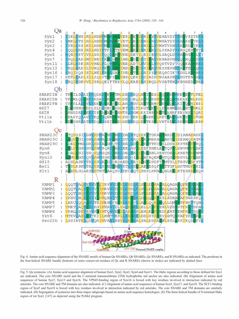
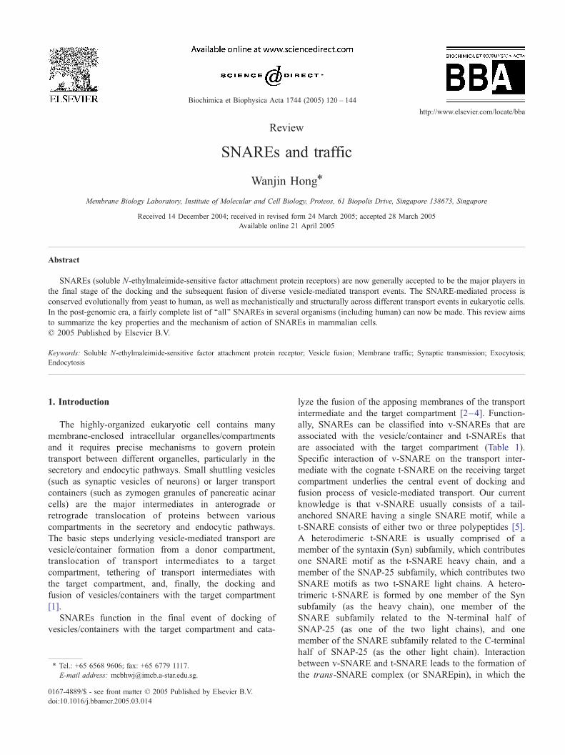
Fig. 4. Amino acid sequence alignment of the SNARE motifs of human Qa SNAREs, Qb SNAREs, Qc SNAREs, and R SNAREs as indicated. The positions in
the four-helical SNARE bundle (bottom) of some conserved residues of Qc and R SNAREs (shown in sticks) are indicated by dashed lines.
Fig. 5. Qa syntaxins. (A) Amino acid sequence alignment of human Syn1, Syn2, Syn3, Syn4 and Syn11. The Habc regions according to those defined for Syn1
are indicated. The core SNARE motif and the C-terminal transmembrane (TM) hydrophobic tail anchor are also indicated. (B) Alignment of amino acid
sequences of human Syn7, Syn13 and Syn16. The VPS45-binding region of Syn16 is boxed with key residues involved in interaction indicated by red
asterisks. The core SNARE and TM domains are also indicated. (C) Alignment of amino acid sequences of human Syn5, Syn17, and Syn18. The SLY1-binding
region of Syn5 and Syn18 is boxed with key residues involved in interaction indicated by red asterisks. The core SNARE and TM domains are similarly
indicated. (D) Segregation of syntaxins into three major subgroups based on amino acid sequence homologies. (E) The three-helical bundle of N-terminal Habc
region of rat Syn1 [147] as depicted using the PyMol program.
W. Hong / Biochimica et Biophysica Acta 1744 (2005) 120–144126

W. Hong / Biochimica et Biophysica Acta 1744 (2005) 120–144 127
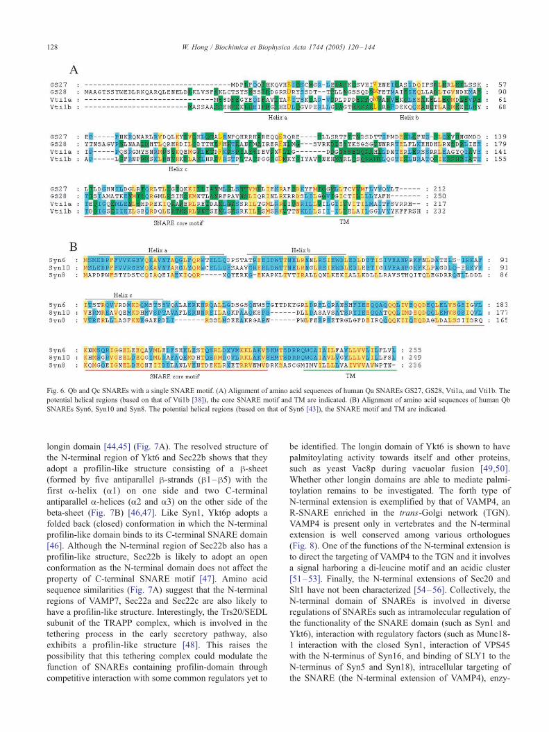
Fig. 6. Qb and Qc SNAREs with a single SNARE motif. (A) Alignment of amino acid sequences of human Qa SNAREs GS27, GS28, Vti1a, and Vti1b. The
potential helical regions (based on that of Vti1b [38]), the core SNARE motif and TM are indicated. (B) Alignment of amino acid sequences of human Qb
SNAREs Syn6, Syn10 and Syn8. The potential helical regions (based on that of Syn6 [43]), the SNARE motif and TM are indicated.
W. Hong / Biochimica et Biophysica Acta 1744 (2005) 120–144128
longin domain [44,45] (Fig. 7A). The resolved structure of
the N-terminal region of Ykt6 and Sec22b shows that they
adopt a profilin-like structure consisting of a h-sheet(formed by five antiparallel h-strands (h1–h5) with the
first a-helix (a1) on one side and two C-terminal
antiparallel a-helices (a2 and a3) on the other side of the
beta-sheet (Fig. 7B) [46,47]. Like Syn1, Ykt6p adopts a
folded back (closed) conformation in which the N-terminal
profilin-like domain binds to its C-terminal SNARE domain
[46]. Although the N-terminal region of Sec22b also has a
profilin-like structure, Sec22b is likely to adopt an open
conformation as the N-terminal domain does not affect the
property of C-terminal SNARE motif [47]. Amino acid
sequence similarities (Fig. 7A) suggest that the N-terminal
regions of VAMP7, Sec22a and Sec22c are also likely to
have a profilin-like structure. Interestingly, the Trs20/SEDL
subunit of the TRAPP complex, which is involved in the
tethering process in the early secretory pathway, also
exhibits a profilin-like structure [48]. This raises the
possibility that this tethering complex could modulate the
function of SNAREs containing profilin-domain through
competitive interaction with some common regulators yet to
be identified. The longin domain of Ykt6 is shown to have
palmitoylating activity towards itself and other proteins,
such as yeast Vac8p during vacuolar fusion [49,50].
Whether other longin domains are able to mediate palmi-
toylation remains to be investigated. The forth type of
N-terminal extension is exemplified by that of VAMP4, an
R-SNARE enriched in the trans-Golgi network (TGN).
VAMP4 is present only in vertebrates and the N-terminal
extension is well conserved among various orthologues
(Fig. 8). One of the functions of the N-terminal extension is
to direct the targeting of VAMP4 to the TGN and it involves
a signal harboring a di-leucine motif and an acidic cluster
[51–53]. Finally, the N-terminal extensions of Sec20 and
Slt1 have not been characterized [54–56]. Collectively, the
N-terminal domain of SNAREs is involved in diverse
regulations of SNAREs such as intramolecular regulation of
the functionality of the SNARE domain (such as Syn1 and
Ykt6), interaction with regulatory factors (such as Munc18-
1 interaction with the closed Syn1, interaction of VPS45
with the N-terminus of Syn16, and binding of SLY1 to the
N-terminus of Syn5 and Syn18), intracellular targeting of
the SNARE (the N-terminal extension of VAMP4), enzy-

Fig. 7. Longin domains. (A) Amino acid sequence alignment of N-terminal Login domains of VAMP7, Ykt6, Sec22b, Sec22a, and Sec22c. The regions with
defined secondary structures based on Sec22b [47] are indicated. (B) The profilin-like fold of the N-terminal login domain of Sec22b as depicted using the
PyMol program with the secondary structures indicated.
W. Hong / Biochimica et Biophysica Acta 1744 (2005) 120–144 129
matic activity (Ykt6), and/or other roles that remain to be
defined.
4. The emerging concept of SNARE action
The current concept of the action of SNAREs is that
combinatory use of the various members of the Qa-, Qb-,
Qc-, and R-SNAREs will give rise to a wide array of
SNARE complexes, whose functions are determined in part
by the subcellular targeting of newly-made SNAREs. A
given SNARE such as Syn5 can be incorporated into (and is
indeed found in) several different SNARE complexes
(Syn5/GS27/Bet1/Sec22b, Syn5/GS28/Bet1/Ykt6, and
Syn5/GS28/GS15/Ykt6) to mediate different transport
events (Fig. 9) [16,57,58]. Similarly, the same t-SNARE
such as Syn4/SNAP-23 interacts with different v-SNAREs
such as VAMP2 [59], VAMP3 [60], VAMP7 [61], or
VAMP8 [62] depending on the cell types. The same v-
SNARE such as VAMP8 interacts with different t-SNAREs
in different transport events of different cells: VAMP8 is
present mainly in endocytic SNARE complexes (with Syn7/
Vti1b/Syn8) in liver hepatocytes [63], but it interacts
primarily with the surface t-SNARE (Syn4/SNAP-23) in
pancreatic acinar cells [62]. The t-SNARE in the TGN
assembled from Syn16, Vti1a, and Syn6 interact with either
VAMP3 or VAMP4 as the v-SNARE [64]. Similarly, the
endosomal t-SNARE assembled from Syn7, Vti1b, and
Syn8 interacts with either VAMP7 or VAMP8 [62,65].
5. Known SNARE complexes (SNAREpins) in
mammalian cells
Several SNARE complexes have been defined to
function in various transport events in the secretory and/
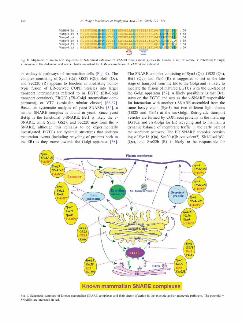
Fig. 8. Alignment of amino acid sequences of N-terminal extension of VAMP4 from various species (h: human; r: rat; m: mouse; z: zebrafish; f: Fugu;
x: Xenopus). The di-leucine and acidic cluster important for TGN accumulation of VAMP4 are indicated.
W. Hong / Biochimica et Biophysica Acta 1744 (2005) 120–144130
or endocytic pathways of mammalian cells (Fig. 9). The
complex consisting of Syn5 (Qa), GS27 (Qb), Bet1 (Qc),
and Sec22b (R) appears to function in mediating homo-
typic fusion of ER-derived COPII vesicles into larger
transport intermediates referred to as EGTC (ER-Golgi
transport container), ERGIC (ER-Golgi intermediate com-
partment), or VTC (vesicular tubular cluster) [66,67].
Based on systematic analysis of yeast SNAREs [34], a
similar SNARE complex is found in yeast. Since yeast
Bet1p is the functional v-SNARE, Bet1 is likely the v-
SNARE, while Syn5, GS27, and Sec22b may form the t-
SNARE, although this remains to be experimentally
investigated. EGTCs are dynamic structures that undergo
maturation events (including recycling of proteins back to
the ER) as they move towards the Golgi apparatus [68].
Fig. 9. Schematic summary of known mammalian SNARE complexes and their s
SNAREs are indicated in red.
The SNARE complex consisting of Syn5 (Qa), GS28 (Qb),
Bet1 (Qc), and Ykt6 (R) is suggested to act in the late
stage of transport from the ER to the Golgi and is likely to
mediate the fusion of matured EGTCs with the cis-face of
the Golgi apparatus [57]. A likely possibility is that Bet1
stays on the EGTC and acts as the v-SNARE responsible
for interaction with another t-SNARE assembled from the
same heavy chain (Syn5) but two different light chains
(GS28 and Ykt6) at the cis-Golgi. Retrograde transport
vesicles are formed by COPI coat proteins in the maturing
EGTCs and cis-Golgi for ER recycling and to maintain a
dynamic balance of membrane traffic in the early part of
the secretory pathway. The ER SNARE complex consist-
ing of Syn18 (Qa), Sec20 (Qb-equivalent?), Slt1/Use1/p31
(Qc), and Sec22b (R) is likely to be responsible for
ite(s) of action in the exocytic and/or endocytic pathways. The potential v-

W. Hong / Biochimica et Biophysica Acta 1744 (2005) 120–144 131
receiving the recycling traffic [54–56,69]. In view of its
segregation with Bet1 and GS15 (Fig. 3), Slt1 may act as
the v-SNARE for the recycling vesicle to interact with t-
SNARE assembled from Syn18, Sec20 and Sec22b in the
ER. Like the interaction of yeast Sec20p with its regulator
TIP20 [70], the mammalian Sec20 also interacts with
RINT-1, a mammalian protein homologous to TIP20 (54,
69). The SNARE complex consisting of Syn5 (Qa), GS28
(Qb), GS15 (Qc), and Ykt6 (R) functions in intra-Golgi
traffic and a similar complex is also found in yeast (16, 35,
58). Based on analysis in yeast, GS15 acts as the v-
SNARE, interacting with t-SNARE assembled from Syn5,
GS28, and Ykt6 [35]. In addition, a recent study suggests
that this SNARE complex also mediates traffic from the
endosomal compartments to the Golgi apparatus [71] and
that GS15 is shifted to the endosomes when the endosomal
function is perturbed. The endosomal compartments are
known to be integrated with the secretory pathway by
retrograde traffic from various endosomal compartments to
the TGN. The major SNARE complex functioning in the
retrograde traffic from early/recycling endosomes to the
TGN consists of Syn16 (Qa), Vti1a (Qb), Syn6 (Qc), and
VAMP4 (R) [64]. VAMP4 is most likely the v-SNARE for
transport intermediates derived from early/recycling endo-
somes and it interacts with t-SNARE assembled from
Syn16, Vti1a, and Syn6 at the TGN. The same TGN t-
SNARE interacts also with VAMP3, which plays a minor
role in this retrograde recycling pathway [64]. Although
retrograde transport from the late endosome to the TGN
has been well established, the exact nature of the SNARE
complex involved in this event is currently unknown.
Several SNARE complexes are implicated in the
endocytic pathway. Syn13 is likely the major Qa SNARE
functioning in the early/sorting endosome and it interacts
with SNAP-25 (Qb and Qc) and VAMP2 (R) to regulate
fusion of early/sorting endosomes [72,73]. VAMP2 is the v-
SNARE in this fusion event. Syn7 (Qa) is distributed
throughout the entire endocytic pathway and is enriched in
the later compartments of the endocytic pathway. Syn7
interacts with Vti1b (Qb) and Syn8 (Qc) to form the t-
SNARE, which acts both in the late endosomes and
lysosomes. By interacting with VAMP8, this t-SNARE
may regulate fusion events in the late endosome [63].
Fusion of the late endosome with the lysosome or
homotypic lysosome fusion might use the same t-SNARE
but a different member of the VAMP subfamily (VAMP7) as
the v-SNARE [65].
Exocytic traffic from the TGN to the cell surface is not
only important for constitutive secretion and biogenesis of
the plasma membrane but also for regulated traffic in
diverse physiological processes. The neuronal SNARE
complex consisting of Syn1, SNAP-25, and VAMP2 is the
best studied and has served as a paradigm for other vesicular
transport events [2,7,18,19]. The functionality of VAMP2 as
the v-SNARE, the assembly of the t-SNARE from Syn1 and
SNAP-25, as well as the activity of the assembled trans-
SNARE complex in mediating synaptic vesicle fusion with
the plasma membrane are subjected to diverse regulatory
mechanisms. This SNARE complex is also the converging
point for many cellular regulations on presynaptic events in
the brain.
A major mode of insulin action is to mobilize glucose
transporter 4 (GLUT4) by stimulating fusion of GLUT4-
containing intracellular vesicles with the plasma membrane.
In this case, VAMP2 functions as a v-SNARE for GLUT4-
containing vesicles and it interacts with t-SNARE
assembled from Syn4 and SNAP-23 [59]. VAMP8 was
recently shown to be the premier v-SNARE of zymogen
granules in pancreatic acinar cells and it mediates regulated
fusion with the apical surface, interacting with t-SNARE
assembled from Syn4 and SNAP-23 [62]. The Syn4/SNAP-
23 t-SNARE may also mediate the fusion of secretory
lysosomes with the plasma membrane by interacting with
VAMP7 as the v-SNARE [61]. The exact SNARE complex
involved in constitutive transport from the TGN to the
surface remains to be defined, although ubiquitously-
expressed Syn2, 3, and/or 4 are likely the heavy chains of
the t-SNARE. These Syns have been shown to exhibit
differential distributions in either the apical or the baso-
lateral domain of polarized epithelial cells and may be a
contributing factor to polarized traffic from the TGN to
various surface domains [74,75]. The v-SNARE for general
TGN-surface transport remains unknown, although VAMP7
is implicated in apical transport in polarized epithelial cells
by acting as a v-SNARE [76].
6. Regulation of SNARE function
The function of SNAREs is subjected to diverse
regulation at various stages of their generation and action.
These include transcriptional regulation of gene expression,
targeting of SNAREs to the correct compartment (s), the
functionality of v-SNARE in the vesicle, the assembly and
functional status of t-SNARE in the target compartment,
long-range interaction of vesicles with the target compart-
ment during tethering, and the assembly and activity of the
trans-SNARE complex. Many regulators are being uncov-
ered and they play either positive and/or negative roles in
the functionality of SNAREs or SNARE complexes. In
addition, post-translational modifications such as phosphor-
ylation [53,77–79], palmitoylation [29–31], and prenyla-
tion [29,80] are also likely to regulate the function of
SNAREs. The major regulators are summarized in Figs.
10–12, and/or Table 3. Examples of these regulators are
briefly described here.
6.1. NSF and a-SNAP
NSF and a-SNAP represent the most essential regulators
of SNAREs as they enable the disassembly of cis-SNARE
complexes. The concerted action of a-SNAP and NSF

Fig. 10. The schematic illustration of the 20 S complex formed by the cis-SNARE complex, three molecules of a-SNAP and an NSF hexamer. The structure of
yeast a-SNAP (1QQE) depicted by PyMol is used to illustrate that the N-terminal sheet and C-terminal globular domain interact with the cis-SNARE complex
and the N-terminal domain of NSF, respectively. Hydrolysis of ATP by NSF (D1 domain) leads to the disassembly of the complex into free SNAREs. The D2
domain of NSF mediates the hexamer formation.
W. Hong / Biochimica et Biophysica Acta 1744 (2005) 120–144132
causes dissociation of cis-SNARE complexes formed after
SNARE-mediated fusion events, releasing free SNAREs for
repeated use. Three molecules of a-SNAP act as bridges to
link up the cis-SNARE complex with a hexameric NSF to
form a transient 20 S complex (Fig. 10) [21–24]. NSF
contains an N-terminal (N) domain of about 200 residues
followed by two ATPase domains of about 280 residues
each (designated D1 and D2 for the first and C-terminal
domains, respectively). The D2 ATPase domain mediates
the hexamerization of NSF, while the N domain interacts
with a C-terminal globular domain (consisting of C-terminal
5 a-helices) of a-SNAP. The D1 ATPase domain effects the
dissociation of the SNARE complex through hydrolysis of
ATP and conformational changes of the hexamer. The first 9
a-helices of a-SNAP arrange in antiparallel to form an N-
terminal sheet whose positively charged residues interact
with the acidic surfaces of the SNARE complex [81].
Association of NSF with a-SNAP into the 20 S complex
stimulates the ATPase activity of the former. The functional
importance of a-SNAP is underscored by genetic analysis
of hyh (hydrocephalus with hop gait) mouse and the
discovery that the hyh phenotype is due to a mutation of
a-SNAP gene [82]. A role for a-SNAP in apical protein
localization and control of neural cell fate has been
suggested. Besides a-SNAP, two homologous proteins
referred to as h-SNAP and g-SNAP are known [83].
h-SNAP is highly homologous to a-SNAP and is expressed
selectively in neurons. h-SNAP can act together with or

Fig. 11. Mammalian SM proteins. (A) List of 7 known mammalian SM
proteins and their major features. (B) Amino acid sequence identities
among the 7 mammalian SM proteins. (C) Phylogenetic tree showing the
segregation of the 7 mammalian SM proteins into four major subtypes.
W. Hong / Biochimica et Biophysica Acta 1744 (2005) 120–144 133
regulate a-SNAP function during regulated exocytosis
[84,85]. Unlike a-SNAP, g-SNAP (which is as widely
expressed as a-SNAP) does not interact with SNAREs,
although it interacts with NSF [86], suggesting that it has a
different role. Consistent with this, a-SNAP but not
g-SNAP is essential for ER-Golgi transport [87].
6.2. Sec1/Munc18-like (SM) proteins
There are at least seven mammalian members of the SM
protein family: Munc18-1, Munc18-2, and Munc18-3,
VPS33A, VPS33B, VPS45, and SLY1 (Fig. 10). Munc18-
1, Munc18-2, and Munc18-3 are functionally homologous
to yeast Sec1p and function at the plasma membrane (PM).
VPS33A and VPS33B correspond to yeast Vps33p and act
in the endocytic pathway. VPS45 and SLY1 correspond to
yeast Vps45p and Sly1p, respectively, and are involved in
traffic at the trans- and cis-faces of the Golgi apparatus
[88,89]. Interactions with Syns occur through the approx-
imately 140 residue N-terminal region of SM proteins [42],
leaving the rest of the molecule for other functions.
Munc18-1, Munc18-2, and Munc18-3 bind to the closed
conformation of Syn1-4. This interaction is dependent on
both the N-terminal Habc region and the SNARE motif of
Syns [37]. SLY1 and VPS45 interact with a short N-terminal
region of Syn5/18 and Syn16, respectively, without the
involvement of the Habc region or the SNARE motif [39–
41]. This differential binding properties of Munc18s vs.
SLY1 and VPS45 result from the Syn-binding site of
Munc18s being present on the opposite side of the folded
surface in comparison to SLY1 and VPS45 [42]. Although
Munc18-1, Munc18-2, and Munc18-3 are all related to
Sec1p and act at the PM, they are likely to have distinct
functions paralleling the more sophisticated cellular phys-
iology of the vertebrate. Munc18-1 is most abundantly
expressed in the neuron and is involved in synaptic vesicle
fusion [19,88], while Munc18-2 is more widely expressed
and acts to regulate Syn2 and 3 [90,91]. Munc18-3 is
believed to interact preferentially with Syn4 and has been
implicated in GLUT4 translocation to the PM in response to
insulin [59,92]. VPS33A and VPS33B are likely to serve
unique functions as mutations of VPS33A in mice lead to a
buff phenotype [93], while mutation of VPS33B in human is
associated with the arthrogryposis-renal dysfunction-choles-
tasis (ARC) syndrome [94].
6.3. Munc13s
Munc13-1, Munc13-2, Munc13-3, and Munc13-4 are
homologous proteins containing several Ca2+-binding C2
domains implicated in interactions with diacylglycerol,
Ca2+, and phospholipids [95–97]. Munc13-1, Munc13-2,
and Munc13-3 are expressed in different cells/regions in the
brain. Knockout of both Munc13-1 and Munc13-2 abolishes
spontaneous and evoked synaptic transmissions [98].
Munc13-1/2 is proposed to be essential for the priming
process for synaptic vesicles tethered onto the presynaptic
plasma membrane [19]. The priming process empowers the
tethered/docked vesicles with the competence of evoked
fusion. Mechanistically, Munc13-1/2 might facilitate the
dissociation of Munc18-1 from Syn1, to open-up Syn1 so
that it can interact with SNAP-25 to form t-SNARE, and/or
to facilitate the formation of a partial complex of Syn1/
SNAP-25 on the presynaptic membrane with VAMP2 on the
synaptic vesicle [19,99]. A small region (residues 1181-
1345) in the C-terminal part of Munc13-1 has been shown
to interact with N-terminal region (residues 53–79) of Syn1
[99]. The tethering of synaptic vesicles is regulated by Rab3
and its effector RIM1 [19,100]. Since Munc13-1 interacts
with RIM1 [101], the progression from the tethering to the
priming event is therefore regulated by the Rab3–RIM1–
Munc13-1 interaction cascade, meaning that Munc13-1/2
effectively facilitates the formation of the partial trans-
SNARE complex implicated in the early priming event [19].
An important role for Munc13-4 in the regulated secretion
of cytolytic granules at the immunological synapse of
cytotoxic lymphocytes has been revealed. Familial hemo-
phagocytic lymphohistiocytosis (FHL) is associated with
defective cytotoxic lymphocytes. A subtype of FHL (FHL3)
is caused by mutations of Munc13-4, resulting in defective
exocytosis of cytolytic granules. Munc13-4 is suggested to
prime tethered/docked cytolytic granules, rendering them

Fig. 12. Examples of regulators of the neuronal SNARE complex. (A) Alignment of amino acid sequences of the SNARE domains of human amisyn, tomosyn,
tomosyn-like KIAA1006 with those of human VAMP1, VAMP4, and VAMP8. The position of the 0 layer residue is indicated. (B) Alignment of amino acid
sequences of complexin I, complexin II, and two candidate members (complexin III and complexin IV) that are yet to be characterized. The region of a-helix
involved in interacting with the SNARE complex is indicated. The potential prenylation sites of complexin III and complexin IV are indicated. (C) Schematic
illustration of the helix region of complexin I sealing the groove between Syn1 and VAMP2 in the neuronal SNARE complex. The D179, D186, and D193
residues of SNAP25 important for Ca2+-dependent interaction with synaptotagmin I [114] are indicated in red.
W. Hong / Biochimica et Biophysica Acta 1744 (2005) 120–144134
competent for regulated exocytosis, via undefined interac-
tions with SNAREs [102].
6.4. Complexins
Complexins I and II are small proteins (134 residues)
(Fig. 11B, Table 3) regulating synaptic transmission
[103,104]. They are enriched in neurons where they
colocalize with Syn1 and SNAP-25, although complexin
II is also expressed ubiquitously at lower levels. Analysis of
neurons from knockout mice lacking both complexins I and
II suggests that complexins act at, or following, the Ca2+-
triggered step of fast synchronous transmitter release [105].
Recent structural studies suggest that complexins might
stabilize the trans-SNARE complex formed between the
synaptic vesicle and the presynaptic surface membrane, thus
regulating a late step in Ca2+-triggered neurotransmitter
release [104,106,107]. A complexin I a-helix (residues 32–
72 as indicated in Fig. 11B) binds in an antiparallel
orientation to the groove formed between the Syn1 and
VAMP2 helices in the second half of the SNARE complex
(starting from �3 layer onwards), thus stabilizing the
zippered of the complex (Fig. 11C). Complexins thus act
like ‘‘adhesive tape’’ to seal the groove formed by Syn1 and
VAMP2, ensuring that synaptic vesicles are in the fully-
primed state. Accordingly, complexins are proposed to
complete the priming process of synaptic vesicles by
facilitating the transition the Munc 13-1/2-mediated partially
assembled SNARE complex to the fully assembled state
[19]. In addition, complexins seemingly facilitate interaction
of the transmembrane domains of VAMP2 and Syn1 [108].
Two proteins homologous to complexins I and II, tentatively
named complexins III and IV (Fig. 11B), have been
identified, but their biochemical properties and cellular
roles in traffic have not been investigated. Interestingly,
complexins III and IV contain C-terminal consensus motifs
for prenylation, suggesting that, like Ykt6, they might be
anchored to the cytoplasmic side of the membrane via a
prenyl group.
6.5. Synaptotagmin I
Synaptotagmin I is the premier member of the synapto-
tagmin protein family consisting of at least 13 members
[109]. It is preferentially expressed in neurons and is
associated with the synaptic vesicle as a type I membrane

Table 3
A list of some other human regulators of SNAREs (in addition to SM proteins)
Name of human protein AA # GenBank acc # Interacting SNAREs Reference
a-SNAP 295 NP_003818 SNARE complex [81,83]
h-SNAP 298 NP_071363 SNARE complex [83,84]
g-SNAP 312 NP_003817 SNARE complex [83,86]
NSF 744 NP_006169 a-SNAP-SNAREs [21–24]
Amisyn 210 Q8NFX7 Syn1-SNAP25 [118]
Tomosyn 1115 NP_640337 Syn1-SNAP25 [116,117]
KIAA1006 1186 BAA76850 Syn1-SNAP25 [117]
Pallidin 172 NP_036520 Syn13 [148]
Snapin 136 NP_036569 SNAP-25 [149]
Synip 245 NP_848604 Syn4 [150]
Complexin I 134 NP_006642 SNARE complex [103,107]
Complexin II 134 Q6PUV4 SNARE complex [103,107]
Complexin III 158 AAP41127 ?
Complexin IV 160 NP_857637 ?
Munc13-1 1665 XP_038604 Syn1 [95,99]
Munc13-2 1591 O14795 Syn1 [95]
Munc13-3 1954 XP_085234 Syn1 [95]
Munc13-4 1090 NP_954712 ? [96,97,102]
Synaptophysin 313 NP_003170 VAMP2 [119]
Synaptotagmin I 422 NP_005630 SNARE complex [19,112]
GATE-16 117 NP_009209 GS28 [132,133]
EpsinR 625 Q14677 Vti1b [12]
p115 962 NP_003706 GS28, Syn5 [16]
VCIP135 1222 NP_079330 Syn5/p97/47 [151]
Septin5 369 AAC39779 Syn1 [152]
Granuphilin 671 NP_542775 Syn1 [153]
Hrs 777 NP_004703 SNAP-25 [73,154]
FIG 462 NP_065132 Syn6 [130]
EEA1 1411 NP_003557 Syn6 [129]
Syntaphilin 494 NP_055538 Syn1 [155]
Syntabulin 663 AAU93914 Syn1 [156]
CDC42 191 NP_001782 VAMP2 [136]
SIP30 266 Q8VIL3 SNAP-25 [157]
RINT-1/TIP20 821 NP_068749 Sec20/BNIP1 [54,69]
Taxilin 546 NP_787048 Syn1, Syn3, Syn4 [158]
W. Hong / Biochimica et Biophysica Acta 1744 (2005) 120–144 135
protein with its short N-terminal region oriented towards the
lumen of the synaptic vesicle. Its larger C-terminal
cytoplasmic domain contains two tandem Ca2+-binding C2
(C2A and C2B) domains. Exocytosis of synaptic vesicles in
the synapse is strictly regulated by Ca2+ concentrations and
synaptotagmin I is probably the Ca2+ sensor that couples
this ion flux to the exocytosis of synaptic vesicles that
occurs on account of an action potential [110–112].
Synaptotagmin I binds directly to Syn1 and SNAP-25 of
the t-SNARE. The tandem C2 (C2A and C2B) domains
cooperate to enhance the penetration of some hydrophobic
residues into the lipid bilayer of the target compartment in
response to Ca2+. The C2B domain apparently also interacts
with phosphatidyl inositol 4,5-biphosphate (PtdIns4,5P) in
response to Ca2+ binding, thus steering the penetration of
hydrophobic residues of C2A and C2B into the target
membrane [112,113]. Several acidic residues in the
C-terminal region of SNAP-25 (red-colored surfaces indi-
cated in Fig. 12) are known to be important for its Ca2+-
triggered interaction with synaptotagmin-1 [114]. Through
the simultaneously enhanced interactions with t-SNARE
(such as through the acidic residues of SNAP-25),
PtdIns4,5P, and the lipid bilayer, synaptotagmin I triggers
the complexin-stabilized trans-SNARE complex to catalyze
the fusion process in the presynaptic membrane in response
to a rise in Ca2+ levels [112,115]. Complexins also appear to
act with synaptotagmin I at this fusion stage by facilitating
the interaction of the transmembrane domains of the vesicle-
located VAMP2 and Syn1 on the pre-synaptic membrane
[108].
6.6. Amisyn, tomosyn, and KIAA1006
The C-terminal region of tomosyn, its closely-related
protein KIAA1006 and amisyn contain a 60-residue region
characteristic of R-SNAREs (Fig. 11A). It has been shown
that the R-SNARE domain of tomosyn and amisyn can
replace VAMP2 and interact with Syn1 and SNAP-25 to
form a non-fusogenic SNARE complex [116–118]. As
tomosyn, KIAA1006 and amisyn do not contain a hydro-
phobic transmembrane domain [116–118], they act as
competitive inhibitors of synaptic vesicle VAMP2 by vying
for the same t-SNARE. In addition, the SNARE complex
formed with tomosyn cannot interact with complexins
W. Hong / Biochimica et Biophysica Acta 1744 (2005) 120–144136
[117]. The potential modulation of the R-SNARE domains
of tomosyn, KIAA1006 and amisyn by factors that may
bind to their respective N-terminal regions provides a
possible avenue for coupling various regulatory processes
to synaptic transmission.
6.7. Synaptophysin
Synaptophysin is one of the major proteins of synaptic
vesicles and is preferentially expressed in neurons. It spans
the membrane four times with both N- and C-termini facing
the cytoplasm [119]. Association of synaptophysin with
VAMP2 inhibits the ability of VAMP2 to interact with
t-SNARE [19,119,120]. A role for synaptophysin in
directing the targeting of newly-made VAMP2 to the
synaptic vesicle has also been proposed [121]. Synaptophy-
sin may therefore regulate the targeting and functionality of
VAMP2. No major defect in synaptic transmission is
observed when synaptophysin is absent in its knockout mice
[122], suggesting that its role in regulating the functional
status of VAMP2 is minor or very subtle. Alternatively, the
lack of a discernible phenotype might reflect the functional
replacement of the absent synaptophysin by its homolog,
synaptoporin, or the more distantly-related proteins such as
synaptogyrin (neuron-enriched) or cellugyrin (ubiquitously-
expressed form of synaptogyrin) [123].
6.8. p115
p115 is a tethering factor that functions by simultaneous
interaction with giantin on COPI-generated vesicles and
with GM130 on the cis-Golgi [124]. A SNARE-related
coiled-coil region of p115 interacts with many SNAREs
(Syn5, GS28, GS27, Ykt6, GS15, Bet1, Sec22b) of the
Golgi apparatus, and its direct interaction with Syn5 and
GS28 has also been demonstrated. Accordingly, p115
stimulates the formation of at least two Syn5-containing
SNARE complexes (Syn5-GS28-GS15-Ykt6 and Syn5-
GS27-Bet1-Sec22b), suggesting that p115 modulates the
formation of trans-SNARE complexes involved in several
transport events in the Golgi apparatus [16]. Mutational
analysis of p115 suggests that the SNARE-modulating
activity of p115 is more important than its tethering activity
in maintaining the structure and function of the Golgi
apparatus [125].
6.9. Hrs
Hrs interacts with SNAP-25 and functions in regulating
the sorting event at the interface between early and late
endosomes [126,127]. Like p115, a SNARE-like coiled coil
region of Hrs is shown to inhibit the incorporation of an R-
SNARE into the SNARE complex through competitive
binding with a t-SNARE complex. Through this activity,
Hrs inhibits early endosomal fusion mediated by Syn13,
SNAP-25, and VAMP2 [73].
6.10. EEA1
Rab5 is a major player regulating the sorting endosome
and it acts via several downstream effectors [128]. EEA1 is
one such Rab5 effector and it functions as a tethering factor
regulating fusion of the early endosome [72]. EEA1 has
been shown to interact with Syn6 via its C-terminal region
which is also involved in interaction with Rab5 [129].
Although the majority of Syn6 is detected at the TGN, a
fraction of Syn6 is also found in EEA1-containing sorting
endosomes. The biological consequence of EEA1–Syn6
interaction remains to be investigated.
6.11. FIG
Another protein interacting with Syn6 is the TGN-
localized FIG (also called CAL, PIST, GOPC1) [130].
FIG contains two coiled-coil regions followed by a single
PDZ domain. The second coiled-coil region and its C-
terminal flanking region interact with Syn6. Again, the
biological consequences of this interaction remain to be
investigated. Knockout of the FIG gene in mice results in
selective ablation of acrosome formation during spermato-
genesis [131]. The acrosome is believed to form from the
Golgi apparatus and the absence of FIG leads to fragmented
acrosomal vesicles, suggesting a role for FIG in the fusion
of these vesicles into the acrosome.
6.12. GATE-16
GATE-16 is a small Golgi protein that interacts with both
NSF and GS28 [132]. In addition to disassembling cis-
SNARE complexes through ATP hydrolysis, NSF also
exhibits an ATPase-independent activity during in vitro
Golgi formation. NSF/a-SNAP facilitates the interaction of
GATE-16 with GS28 in a manner that requires ATP-binding
but not ATP hydrolysis. Interestingly, GATE-16 binding
prevents GS28 from interacting with Syn5 [133]. Since
Syn5 is the heavy chain of t-SNARE and GS28 is a Qb-
SNARE that serves as one of the light chains, GATE-16 has
the property of preventing the assembly of a functional
t-SNARE. Interestingly, GATE-16 is a member of a family
of autophagy-related ubiquitin-like proteins (GATE-16,
MAP1-LC3, GABARAP, and Apg8L) that are substrates
of Apg4B protease [133a]. How this property is related to its
role in Golgi function remains elusive.
7. Physiological studies of SNAREs
Recent studies on mammalian SNAREs have advanced
from cellular studies to systematic functions in mice,
including the generation of gene knockouts and genetic
analysis of mutant mice. In addition, genetic analyses of
human diseases resulting from mutations in SNAREs and
regulators have also provided additional understanding. The
W. Hong / Biochimica et Biophysica Acta 1744 (2005) 120–144 137
physiological roles of some SNAREs and their regulators in
the context of the whole organism, as deduced from
targeted gene knockout analysis in mice, genetic analysis
of mutant mice, and/or genetic analysis of human diseases,
are listed in Table 4. Some of these studies are briefly
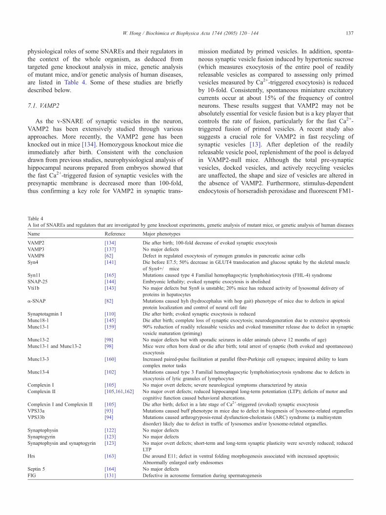
described below.
7.1. VAMP2
As the v-SNARE of synaptic vesicles in the neuron,
VAMP2 has been extensively studied through various
approaches. More recently, the VAMP2 gene has been
knocked out in mice [134]. Homozygous knockout mice die
immediately after birth. Consistent with the conclusion
drawn from previous studies, neurophysiological analysis of
hippocampal neurons prepared from embryos showed that
the fast Ca2+-triggered fusion of synaptic vesicles with the
presynaptic membrane is decreased more than 100-fold,
thus confirming a key role for VAMP2 in synaptic trans-
Table 4
A list of SNAREs and regulators that are investigated by gene knockout experime
Name Reference Major phenotypes
VAMP2 [134] Die after birth; 100-fold d
VAMP3 [137] No major defects
VAMP8 [62] Defect in regulated exocy
Syn4 [141] Die before E7.5; 50% dec
of Syn4+/� mice
Syn11 [165] Mutations caused type 4 F
SNAP-25 [144] Embryonic lethality; evok
Vti1b [143] No major defects but Syn
proteins in hepatocytes
a-SNAP [82] Mutations caused hyh (hy
protein localization and co
Synaptotagmin I [110] Die after birth; evoked syn
Munc18-1 [145] Die after birth; complete l
Munc13-1 [159] 90% reduction of readily
vesicle maturation (primin
Munc13-2 [98] No major defects but with
Munc13-1 and Munc13-2 [98] Mice were often born dea
exocytosis
Munc13-3 [160] Increased paired-pulse fac
complex motor tasks
Munc13-4 [102] Mutations caused type 3 F
exocytosis of lytic granule
Complexin I [105] No major overt defects; se
Complexin II [105,161,162] No major overt defects; re
cognitive function caused
Complexin I and Complexin II [105] Die after birth; defect in a
VPS33a [93] Mutations caused buff phe
VPS33b [94] Mutations caused arthrogr
disorder) likely due to def
Synaptophysin [122] No major defects
Synaptogyrin [123] No major defects
Synaptophysin and synaptogyrin [123] No major overt defects; sh
LTP
Hrs [163] Die around E11; defect in
Abnormally enlarged early
Septin 5 [164] No major defects
FIG [131] Defective in acrosome for
mission mediated by primed vesicles. In addition, sponta-
neous synaptic vesicle fusion induced by hypertonic sucrose
(which measures exocytosis of the entire pool of readily
releasable vesicles as compared to assessing only primed
vesicles measured by Ca2+-triggered exocytosis) is reduced
by 10-fold. Consistently, spontaneous miniature excitatory
currents occur at about 15% of the frequency of control
neurons. These results suggest that VAMP2 may not be
absolutely essential for vesicle fusion but is a key player that
controls the rate of fusion, particularly for the fast Ca2+-
triggered fusion of primed vesicles. A recent study also
suggests a crucial role for VAMP2 in fast recycling of
synaptic vesicles [13]. After depletion of the readily
releasable vesicle pool, replenishment of the pool is delayed
in VAMP2-null mice. Although the total pre-synaptic
vesicles, docked vesicles, and actively recycling vesicles
are unaffected, the shape and size of vesicles are altered in
the absence of VAMP2. Furthermore, stimulus-dependent
endocytosis of horseradish peroxidase and fluorescent FM1-
nts, genetic analysis of mutant mice, or genetic analysis of human diseases
ecrease of evoked synaptic exocytosis
tosis of zymogen granules in pancreatic acinar cells
rease in GLUT4 translocation and glucose uptake by the skeletal muscle
amilial hemophagocytic lymphohistiocytosis (FHL-4) syndrome
ed synaptic exocytosis is abolished
8 is unstable; 20% mice has reduced activity of lysosomal delivery of
drocephalus with hop gait) phenotype of mice due to defects in apical
ntrol of neural cell fate
aptic exocytosis is reduced
oss of synaptic exocytosis; neurodegeneration due to extensive apoptosis
releasable vesicles and evoked transmitter release due to defect in synaptic
g)
sporadic seizures in older animals (above 12 months of age)
d or die after birth; total arrest of synaptic (both evoked and spontaneous)
ilitation at parallel fiber-Purkinje cell synapses; impaired ability to learn
amilial hemophagocytic lymphohistiocytosis syndrome due to defects in
s of lymphocytes
vere neurological symptoms characterized by ataxia
duced hippocampal long-term potentiation (LTP); deficits of motor and
behavioral altercations.
late stage of Ca2+-triggered (evoked) synaptic exocytosis
notype in mice due to defect in biogenesis of lysosome-related organelles
yposis-renal dysfunction-cholestasis (ARC) syndrome (a multisystem
ect in traffic of lysosomes and/or lysosome-related organelles.
ort-term and long-term synaptic plasticity were severely reduced; reduced
ventral folding morphogenesis associated with increased apoptosis;
endosomes
mation during spermatogenesis
W. Hong / Biochimica et Biophysica Acta 1744 (2005) 120–144138
43 is delayed. VAMP2 acts thus as a nucleating factor for
prompt endocytosis leading to quick reuse of synaptic
vesicles following rapid exocytosis triggered by Ca2+.
VAMP2 participates also in regulated secretion of other
cells such as fat and endocrine cells. VAMP2 acts as a v-
SNARE of storage vesicles containing GLUT4 and interacts
with t-SNARE assembled from Syn4 and SNAP-23,
mediating fusion with the plasma membrane in response
to insulin [59,92]. In addition, VAMP2 is implicated in
vasopressin-regulated translocation of aquaporin 2-contain-
ing vesicles [135]. VAMP2 might also regulate secretion of
endocrine cells, such as insulin secretion by pancreatic h-cells. Interaction of VAMP2 with CDC42 is recently shown
to be involved in coordinating secretion through actin
cytoskeleton arrangement in h-cells [136].
7.2. VAMP3
The amino acid sequence of ubiquitously expressed
VAMP3 is 75% identical to that of VAMP2. It is
preferentially associated with sorting/early and recycling
endosomes. Remarkably, deletion of the VAMP3 gene in
mice exhibits little effects on development or various
physiological processes (such as GLUT4 translocation or
endocytic traffic) [137]. VAMP3 is not necessary for either
regulated GLUT4 translocation or general constitutive
membrane recycling, suggesting that one or more proteins
may provide functional redundancy. VAMP8, present in the
early and late endosomes [63,65], is a prime candidate for
functional overlap with VAMP3, a possibility that awaits
experimental verification.
7.3. VAMP8
The amino acid sequence of VAMP8 is only 32%
identical to that of VAMP2 [138]. The intracellular
distribution of VAMP8 in the endocytic pathway is similar
to VAMP3. Although VAMP8 is ubiquitously expressed, it
is enriched in tissues with epithelial cells such as the
kidney, intestine, pancreas, and lung [139]. VAMP8
mediates homotypic fusion of early and late endosomes
by functioning as a v-SNARE to interact with a t-SNARE
assembled from Syn7, Vti1b, and Syn8 [140]. Deletion of
VAMP8 gene in mice does not significantly affect develop-
ment [62]. Analysis of the endocytic pathway using
VAMP8-null embryonic fibroblasts suggests that the endo-
cytic pathway is not grossly altered in the absence of
VAMP8. However, one striking observation is that VAMP-
null pancreatic acinar cells are filled with an excess number
of zymogen granules (the total number is increased about
3-fold). VAMP8 is enriched in the membrane of zymogen
granules and is necessary for regulated secretion of
zymogen granules, suggesting that VAMP8 is the major
v-SNARE of zymogen granules in pancreatic acinar cells
[62]. The role of VAMP8 in other regulated secretions
remains to be investigated and the hypothesis that it has
overlapping role with VAMP3 in the endocytic pathway
needs to be tested by experiments.
7.4. Syn4
Syn4 is a widely expressed t-SNARE heavy chain on the
plasma membrane and is enriched in the basolateral surface
of polarized epithelial cells, whereas Syn2 and Syn3 are
targeted preferentially to the apical surface [74,75]. Syn4
may also function at the apical surface as it is detectable
there and has been implicated in apical secretion of
zymogen granules of pancreatic acinar cells [62,75]. Syn4
is apparently essential for early embryonic development as
Syn4-null embryos die before E7.5 [141]. Heterozygous
knockout mice (Syn4+/�) developed normally. Interest-
ingly, the Syn4+/� mice manifest impaired glucose
tolerance with a 50% reduction in whole-body glucose
uptake due to a 50% reduction in glucose transport in the
skeletal muscle. Mechanistically, insulin-stimulated GLUT4
translocation in skeletal muscle is also significantly reduced.
However, GLUT4 translocation and glucose uptake are not
obviously affected in the adipose tissue and liver, suggesting
a critical and selective role of Syn4 in insulin-stimulated
GLUT4 deployment and glucose uptake in skeletal muscle
[141]. Syn4 has also been implicated in lipopolysaccharide
(LPS)-induced secretion of tumor necrosis factor (TNF) in
macrophages [142]. Protein levels of Syn4, together with its
interacting proteins (SNAP-23 and Munc18-3), are signifi-
cantly increased by LPS in a temporal pattern coinciding
with TNF secretion. This suggests that Syn4 acts at a rate-
limiting step during TNFa secretion.
7.5. Vti1b
As a Qb light chain, Vit1b interacts with Syn7 (the Qa
heavy chain) and Syn8 (the Qc light chain) to form the
endosomal t-SNARE; thus regulating homotypic fusion of
late endosome through interaction with VAMP8 as the v-
SNARE [63,65]. It is also implicated in the fusion of late
endosomes with lysosomes, and the homotypic fusion of
lysosomes [65]. Knockout of the Vti1b gene does not affect
embryo development and the majority of Vti1b-null mice
behave normally [143]. Interestingly, Syn8 levels are
selectively reduced in Vti1b-null cells, suggesting that
interaction with Vti1b can stabilize Syn8. About 20% of
Vti1b-null mice are smaller and lysosomal degradation of an
endocytosed protein is slightly delayed in hepatocytes of
these mice. Multivesicular bodies and autophagic vacuoles
accumulate in hepatocytes of these smaller Vti1b-null mice.
Whether Vti1a compensates for the loss of Vti1b in these
mice remains to be investigated.
7.6. SNAP-25
SNAP-25 contributes both Qb and Qc SNARE motifs to
the synaptic SNARE complex and thus plays a key role in
W. Hong / Biochimica et Biophysica Acta 1744 (2005) 120–144 139
mediating synaptic vesicle fusion [7,19]. Ca2+-regulated
interaction of SNAP-25 with synaptotagmin I is important
for Ca2+-triggered fast exocytosis [114]. Complete ablation
of the SNAP-25 gene results in embryonic lethality of the
mutant mice [144]. The SNAP-25-null embryos are clearly
morphologically abnormal compared to wild type around
E17.5–E18.5, although major brain structures appear
unaltered. Electrophysiological analysis suggests that
SNAP-25 is essential for Ca2+-evoked synaptic transmission
at neuromuscular junctions and central synapses, although
stimulus-independent spontaneous neurotransmitter release
is not dramatically affected. This phenotype is similar to that
of VAMP2-null mice with regards to evoked exocytosis in
the neuron [134].
7.7. Munc18-1
Munc18-1 is believed to bind to Syn1 in its closed
conformation involving both the N-terminal Habc region
and the SNARE motif, thus modulating its interaction with
SNAP-25 and t-SNARE assembly [19,37]. Mice lacking
Munc18-1 develop normally but die immediately after birth
[145]. Brain development and synaptogenesis occur nor-
mally. However, synaptic exocytosis (both Ca2+-evoked and
spontaneous events) is completely lost in the absence of
Munc18-1. This abolition of synaptic transmission leads to
neurodegeneration as a result of extensive apoptosis. The
severe effect of Munc18-1 ablation contrasts with the partial
spontaneous synaptic exocytosis that is observed in the
absence of SNAP-25 or VAMP2. The complete dependence
of synaptic transmission on Munc18-1 cannot be explained
solely by its role in binding Syn1. Munc18-1 must perform
other crucial functions, such as proper folding and intra-
cellular targeting of Syn1, the assembly of the presynaptic
t-SNARE, formation of trans-SNARE complex, and/or
interaction with other regulatory factors.
7.8. Synaptotagmin I
Mice lacking synaptotagmin I develop normally, but the
newborns die within 48 h after birth [110]. Electrophysio-
logical analysis of hippocampal neurons derived from
synaptotagmin I-null mice reveals severe impairment of
synaptic transmission. Ca2+-triggered synchronized synaptic
exocytosis is specifically decreased, whereas asynchronous
slow release processes, such as spontaneous synaptic activity
(miniature excitatory postsynaptic current frequency) and
release triggered by hypertonic solution or alpha-latrotoxin,
are unaffected. This demonstrates a physiological role for
synaptotagmin I in Ca2+-evoked synchronous neurotrans-
mitter release. Subsequent studies using the genetic approach
in mice [111] and biochemical reconstitution in vitro [115]
establish that synaptotagmin I is a Ca2+-sensor that couples
action potential to fast exocytosis, a function dependent on its
Ca2+-regulated interaction with phospholipids, PtdIns4,5P2,
and the trans-SNARE complex [19,112].
8. Future perspectives
Although the majority of SNAREs have presumably
been identified and their general principles of action
resolved, much more work is required for a precise
understanding of their various physiological roles, molec-
ular mechanisms of action, and regulation. A higher level of
understanding will likely be achieved through biophysical
approaches that yield quantitative descriptions of SNARE
formation and action in terms of time, energy, space, and
geometry. Formation of the trans-SNARE complex is
presumably the core event underlying diverse fusion events
and serves as the converging point for various regulatory
processes. More regulators of SNAREs are expected to be
identified and understanding their precise roles and mech-
anisms of action are essential to link up vesicle fusion with
the rest of the cellular regulatory networks. The role of
many other SNAREs at the organism level, explored
through gene ablation approach with comprehensive assess-
ment of diverse developmental and physiological processes,
will provide more precise understanding of SNARE
functions under physiological settings. Small chemicals that
selectively modulate the interaction of SNAREs and their
regulators, or even SNARE complex formation, should
prove useful as tools to reversibly perturb the action and
regulation of SNAREs in intact cells. This approach will not
only allow us to gain further insights into the SNARE
function but also provide a foundation for regulating cellular
and physiological processes that are governed by SNAREs.
The emerging interface (Chemical Biology) between bio-
logy and chemistry will definitely facilitate this process.
More genetic analysis of human diseases is likely to uncover
new associations of loss or partial loss of function of
SNAREs and their regulators in connection with diseases.
More exciting new discoveries in structural, molecular,
mechanistic, biophysical, regulatory, and conceptual aspects
of SNAREs and regulators will advance our understanding
of their cellular, developmental, and physiological roles.
References
[1] J.S. Bonifacino, B.S. Glick, The mechanisms of vesicle budding and
fusion, Cell 116 (2004) 153–166.
[2] T. Sollner, S.W. Whiteheart, M. Brunner, H. Erdjument-Bromage, S.
Geromanos, P. Tempst, J.E. Rothman, SNAP receptors implicated in
vesicle targeting and fusion, Nature 362 (1993) 318–324.
[3] J.A. McNew, F. Parlati, R. Fukuda, R.J. Johnston, K. Paz, F. Paumet,
T.H. Sollner, J.E. Rothman, Compartmental specificity of cellular
membrane fusion encoded in SNARE proteins, Nature 407 (2000)
153–159.
[4] C. Hu, M. Ahmed, T.J. Melia, T.H. Sollner, T. Mayer, J.E. Rothman,
Fusion of cells by flipped SNAREs, Science 300 (2003) 1745–1749.
[5] R. Fukuda, J.A. McNew, T. Weber, F. Parlati, T. Engel, W. Nickel,
J.E. Rothman, T.H. Sollner, Functional architecture of an intracellular
membrane t-SNARE, Nature 407 (2000) 198–202.
[6] T. Weber, B.V. Zemelman, J.A. McNew, B. Westermann, M. Gmachl,
F. Parlati, T.H. Sollner, J.E. Rothman, SNAREpins: minimal
machinery for membrane fusion, Cell 92 (1998) 759–772.

W. Hong / Biochimica et Biophysica Acta 1744 (2005) 120–144140
[7] R.B. Sutton, D. Fasshauer, R. Jahn, A.T. Brunger, Crystal structure of
a SNARE complex involved in synaptic exocytosis at 2.4 A
resolution, Nature 395 (1998) 347–353.
[8] W. Antonin, D. Fasshauer, S. Becker, R. Jahn, T.R. Schneider,
Crystal structure of the endosomal SNARE complex reveals
common structural principles of all SNAREs, Nat. Struct. Biol. 9
(2002) 107–111.
[9] E. Mossessova, L.C. Bickford, J. Goldberg, SNARE selectivity of the
COPII coat, Cell 114 (2003) 483–495.
[10] E.A. Miller, T.H. Beilharz, P.N. Malkus, M.C. Lee, S. Hamamoto, L.
Orci, R. Schekman, Multiple cargo binding sites on the COPII
subunit Sec24p ensure capture of diverse membrane proteins into
transport vesicles, Cell 114 (2003) 497–509.
[11] U. Rein, U. Andag, R. Duden, H.D. Schmitt, A. Spang, ARF-GAP-
mediated interaction between the ER-Golgi v-SNAREs and the COPI
coat, J. Cell Biol. 157 (2002) 395–404.
[12] J. Hirst, S.E. Miller, M.J. Taylor, G.F. von Mollard, M.S. Robinson,
EpsinR Is an adaptor for the SNARE Protein Vti1b, Mol. Biol. Cell
15 (2004) 5593–5602.
[13] F. Deak, S. Schoch, X. Liu, T.C. Sudhof, E.T. Kavalali, Synapto-
brevin is essential for fast synaptic-vesicle endocytosis, Nat. Cell
Biol. 6 (2004) 1102–1108.
[14] A.K. Gillingham, S. Munro, Long coiled-coil proteins and membrane
traffic, Biochim. Biophys. Acta 1641 (2003) 71–85.
[15] J.R. Whyte, S. Munro, Vesicle tethering complexes in membrane
traffic, J. Cell Sci. 115 (2002) 2627–2637.
[16] J. Shorter, M.B. Beard, J. Seemann, A.B. Dirac-Svejstrup, G.
Warren, Sequential tethering of Golgins and catalysis of SNAREpin
assembly by the vesicle-tethering protein p115, J. Cell Biol. 157
(2002) 45–62.
[17] Y.A. Chen, R.H. Scheller, SNARE-mediated membrane fusion, Nat.
Rev. Mol. Cell Biol. 2 (2001) 98–106.
[18] R. Jahn, T. Lang, T.C. Sudhof, Membrane fusion, Cell 112 (2003)
519–533.
[19] T.C. Sudhof, The synaptic vesicle cycle, Annu. Rev. Neurosci. 27
(2004) 509–547.
[20] J.E. Rothman, Lasker basic medical research award. The machinery
and principles of vesicle transport in the cell, Nat. Med. 8 (2002)
1059–1062.
[21] T.M. Hohl, F. Parlati, C. Wimmer, J.E. Rothman, T.H. Sollner, H.
Engelhardt, Arrangement of subunits in 20 S particles consisting of
NSF, SNAPs, and SNARE complexes, Mol. Cell 2 (1998) 539–548.
[22] C. Wimmer, T.M. Hohl, C.A. Hughes, S.A. Muller, T.H. Sollner, A.
Engel, J.E. Rothman, Molecular mass, stoichiometry, and assembly
of 20 S particles, J. Biol. Chem. 276 (2001) 29091–29097.
[23] J. Furst, R.B. Sutton, J. Chen, A.T. Brunger, N. Grigorieff, Electron
cryomicroscopy structure of N-ethyl maleimide sensitive factor at 11
A resolution, EMBO J. 22 (2003) 4365–4374.
[24] A.T. Brunger, B. DeLaBarre, NSF and p97/VCP: Similar at first,
different at last, FEBS Lett. 555 (2003) 126–133.
[25] T. Weimbs, S.H. Low, S.J. Chapin, K.E. Mostov, P. Bucher, K.
Hofmann, A conserved domain is present in different families of
vesicular fusion proteins: a new superfamily, Proc. Natl. Acad. Sci.
94 (1997) 3046–3051.
[26] D. Fasshauer, R.B. Sutton, A.T. Brunger, R. Jahn, Conserved
structural features of the synaptic fusion complex: SNARE proteins
reclassified as Q- and R-SNAREs, Proc. Natl. Acad. Sci. 95 (1998)
15781–15786.
[27] R. Jahn, T.C. Sudhof, Membrane fusion and exocytosis, Annu. Rev.
Biochem. 68 (1999) 863–911.
[28] J.B. Bock, H.T. Matern, A.A. Peden, R.H. Scheller, A genomic
perspective on membrane compartment organization, Nature 409
(2001) 839–841.
[29] M. Fukasawa, O. Varlamov, W.S. Eng, T.H. Sollner, J.E. Rothman,
Localization and activity of the SNARE Ykt6 determined by its
regulatory domain and palmitoylation, Proc. Natl. Acad. Sci. 101
(2004) 4815–4820.
[30] M. Veit, T.H. Sollner, J.E. Rothman, Multiple palmitoylation of
synaptotagmin and the t-SNARE SNAP-25, FEBS Lett. 385 (1996)
119–123.
[31] R. Prekeris, J. Klumperman, R.H. Scheller, Syntaxin 11 is an atypical
SNARE abundant in the immune system, Eur. J. Cell Biol. 79 (2000)
771–780.
[32] K. Vogel, J.P. Cabaniols, P.A. Roche, Targeting of SNAP-25 to
membranes is mediated by its association with the target SNARE
syntaxin, J. Biol. Chem. 275 (2000) 2959–2965.
[33] D. Fasshauer, Structural insights into the SNARE mechanism,
Biochim. Biophys. Acta 1641 (2003) 87–97.
[34] F. Parlati, J.A. McNew, R. Fukuda, R. Miller, T.H. Sollner, J.E.
Rothman, Topological restriction of SNARE-dependent membrane
fusion, Nature 407 (2000) 194–198.
[35] F. Parlati, O. Varlamov, K. Paz, J.A. McNew, D. Hurtado, T.H.
Sollner, J.E. Rothman, Distinct SNARE complexes mediating
membrane fusion in Golgi transport based on combinatorial
specificity, Proc. Natl. Acad. Sci. U. S. A. 99 (2002) 5424–5429.
[36] L.E. Dietrich, C. Boeddinghaus, T.J. LaGrassa, C. Ungermann,
Control of eukaryotic membrane fusion by N-terminal domains of
SNARE proteins, Biochim. Biophys. Acta 1641 (2003) 111–119.
[37] K.M. Misura, R.H. Scheller, W.I. Weis, Three-dimensional structure
of the neuronal-Sec1–syntaxin 1a complex, Nature 404 (2000)
355–362.
[38] W. Antonin, I. Dulubova, D. Arac, S. Pabst, J. Plitzner, J. Rizo, R.
Jahn, The N-terminal domains of syntaxin 7 and vti1b form three-
helix bundles that differ in their ability to regulate SNARE complex
assembly, J. Biol. Chem. 277 (2002) 36449–36456.
[39] I. Dulubova, T. Yamaguchi, Y. Gao, S.W. Min, I. Huryeva, T.C.
Sudhof, J. Rizo, How Tlg2p/syntaxin 16 Fsnares_ Vps45, EMBO J.
21 (2002) 3620–3631.
[40] T. Yamaguchi, I. Dulubova, S.W. Min, X. Chen, J. Rizo, T.C.
Sudhof, Sly1 binds to Golgi and ER syntaxins via a conserved N-
terminal peptide motif, Dev. Cell 2 (2002) 295–305.
[41] A. Bracher, W. Weissenhorn, Structural basis for the Golgi
membrane recruitment of Sly1p by Sed5p, EMBO J. 21 (2002)
6114–6124.
[42] I. Dulubova, T. Yamaguchi, D. Arac, H. Li, I. Huryeva, S.W. Min,
J. Rizo, T.C. Sudhof, Convergence and divergence in the mechanism
of SNARE binding by Sec1/Munc18-like proteins, Proc. Natl. Acad.
Sci. U. S. A. 100 (2003) 32–37.
[43] K.M. Misura, J.B. Bock, L.C. Gonzalez Jr., R.H. Scheller, W.I. Weis,
Three-dimensional structure of the amino-terminal domain of
syntaxin 6, a SNAP-25 C homolog, Proc. Natl. Acad. Sci. 99
(2002) 9184–9189.
[44] F. Filippini, V. Rossi, T. Galli, A. Budillon, M. D’Urso, M.
D’Esposito, Longins: a new evolutionary conserved VAMP family
sharing a novel SNARE domain, Trends Biochem. Sci. 26 (2001)
407–409.
[45] V. Rossi, D.K. Banfield, M. Vacca, L.E. Dietrich, C. Ungermann, M.
D’Esposito, T. Galli, F. Filippini, Longins and their longin domains:
regulated SNAREs and multifunctional SNARE regulators, Trends
Biochem. Sci. 29 (2004) 682–688.
[46] H. Tochio, M.M. Tsui, D.K. Banfield, M. Zhang, An autoinhibitory
mechanism for nonsyntaxin SNARE proteins revealed by the
structure of Ykt6p, Science 293 (2001) 698–702.
[47] L.C. Gonzalez Jr., W.I. Weis, R.H. Scheller, A novel snare N-
terminal domain revealed by the crystal structure of Sec22b, J. Biol.
Chem. 276 (2001) 24203–24211.
[48] S.B. Jang, Y.G. Kim, Y.S. Cho, P.G. Suh, K.H. Kim, B.H. Oh,
Crystal structure of SEDL and its implications for a genetic disease
spondyloepiphyseal dysplasia tarda, J. Biol. Chem. 277 (2002)
49863–49869.
[49] L.E. Dietrich, R. Gurezka, M. Veit, C. Ungermann, The SNARE
Ykt6 mediates protein palmitoylation during an early stage of
homotypic vacuole fusion, EMBO J. 23 (2004) 45–53.
[50] M. Veit, The human SNARE protein Ykt6 mediates its own
W. Hong / Biochimica et Biophysica Acta 1744 (2005) 120–144 141
palmitoylation at C-terminal cysteine residues, Biochem. J. 384
(2004) 233–237.
[51] A.A. Peden, G.Y. Park, R.H. Scheller, The Di-leucine motif of
vesicle-associated membrane protein 4 is required for its localization
and AP-1 binding, J. Biol. Chem. 276 (2001) 49183–49187.
[52] Q. Zeng, T.T. Tran, H.X. Tan, W. Hong, The cytoplasmic domain of
Vamp4 and Vamp5 is responsible for their correct subcellular
targeting: the N-terminal extenSion of VAMP4 contains a dominant
autonomous targeting signal for the trans-Golgi network, J. Biol.
Chem. 278 (2003) 23046–23054.
[53] I. Hinners, F. Wendler, H. Fei, L. Thomas, G. Thomas, S.A. Tooze,
AP-1 recruitment to VAMP4 is modulated by phosphorylation-
dependent binding of PACS-1, EMBO Rep. 4 (2003) 1182–1189.
[54] K. Nakajima, H. Hirose, M. Taniguchi, H. Kurashina, K. Arasaki, M.
Nagahama, K. Tani, A. Yamamoto, M. Tagaya, Involvement of
BNIP1 in apoptosis and endoplasmic reticulum membrane fusion,
EMBO J. 23 (2004) 3216–3226.
[55] L. Burri, O. Varlamov, C.A. Doege, K. Hofmann, T. Beilharz, J.E.
Rothman, T.H. Sollner, T. Lithgow, A SNARE required for
retrograde transport to the endoplasmic reticulum, Proc. Natl. Acad.
Sci. U. S. A. 100 (2003) 9873–9877.
[56] M. Dilcher, B. Veith, S. Chidambaram, E. Hartmann, H.D. Schmitt,
G. Fischer von Mollard, Use1p is a yeast SNARE protein required
for retrograde traffic to the ER, EMBO J. 22 (2003) 3664–3674.
[57] T. Zhang, W. Hong, Ykt6 forms a SNARE complex with syntaxin 5,
GS28, and Bet1 and participates in a late stage in endoplasmic
reticulum-Golgi transport, J. Biol. Chem. 276 (2001) 27480–27487.
[58] Y. Xu, S. Martin, D.E. James, W. Hong, GS15 forms a SNARE
complex with syntaxin 5, GS28, and Ykt6 and is implicated in traffic
in the early cisternae of the Golgi apparatus, Mol. Biol. Cell 13
(2002) 3493–3507.
[59] N.J. Bryant, R. Govers, D.E. James, Regulated transport of the
glucose transporter GLUT4, Nat. Rev., Mol. Cell Biol. 3 (2002)
267–277.
[60] R. Flaumenhaft, K. Croce, E. Chen, B. Furie, B.C. Furie, Proteins of
the exocytotic core complex mediate platelet alpha-granule secretion.
Roles of vesicle-associated membrane protein, SNAP-23, and
syntaxin 4, J. Biol. Chem. 274 (1999) 2492–2501.
[61] S.K. Rao, C. Huynh, V. Proux-Gillardeaux, T. Galli, N.W. Andrews,
Identification of SNAREs involved in synaptotagmin VII-regulated
lysosomal exocytosis, J. Biol. Chem. 279 (2004) 20471–20479.
[62] C.C. Wang, C.P. Ng, L. Lu, V. Atlashkin, W. Zhang, L.F. Seet, W.
Hong, A role of VAMP8/endobrevin in regulated exocytosis of
pancreatic acinar cells, Dev. Cell 7 (2004) 359–371.
[63] W. Antonin, C. Holroyd, D. Fasshauer, S. Pabst, G.F. Von Mollard,
R. Jahn, A SNARE complex mediating fusion of late endosomes
defines conserved properties of SNARE structure and function,
EMBO J. 19 (2000) 6453–6464.
[64] F. Mallard, B.L. Tang, T. Galli, D. Tenza, A. Saint-Pol, X. Yue, C.
Antony, W. Hong, B. Goud, L. Johannes, Early/recycling endo-
somes-to-TGN transport involves two SNARE complexes and a
Rab6 isoform, J. Cell Biol. 156 (2002) 653–664.
[65] P.R. Pryor, B.M. Mullock, N.A. Bright, M.R. Lindsay, S.R. Gray,
S.C. Richardson, A. Stewart, D.E. James, R.C. Piper, J.P. Luzio,
Combinatorial SNARE complexes with VAMP7 or VAMP8 define
different late endocytic fusion events, EMBORep. 5 (2004) 590–595.
[66] T. Zhang, S.H. Wong, B.L. Tang, Y. Xu, F. Peter, V.N. Subramaniam,
W. Hong, The mammalian protein (rbet1) homologous to yeast Bet1p
is primarily associated with the pre-Golgi intermediate compartment
and is involved in vesicular transport from the endoplasmic reticulum
to the Golgi apparatus, J. Cell Biol. 139 (1997) 1157–1168.
[67] T. Zhang, S.H. Wong, B.L. Tang, Y. Xu, W. Hong, Morphological
and functional association of Sec22b/ERS-24 with the pre-Golgi
intermediate compartment, Mol. Biol. Cell 10 (1999) 435–453.
[68] H. Horstmann, C.P. Ng, B.L. Tang, W. Hong, Ultrastructural
characterization of endoplasmic reticulum-Golgi transport containers
(EGTC), J. Cell Sci. 115 (2002) 4263–4273.
[69] H. Hirose, K. Arasaki, N. Dohmae, K. Takio, K. Hatsuzawa, M.
Nagahama, K. Tani, A. Yamamoto, M. Tohyama, M. Tagaya,
Implication of ZW10 in membrane trafficking between the endo-
plasmic reticulum and Golgi, EMBO J. 23 (2004) 1267–1278.
[70] D.J. Sweet, H.R. Pelham, The TIP1 gene of Saccharomyces
cerevisiae encodes an 80 kDa cytoplasmic protein that interacts with
the cytoplasmic domain of Sec20p, EMBO J. 12 (1993) 2831–2840.
[71] G. Tai, L. Lu, T.L. Wang, B.L. Tang, B. Goud, L. Johannes, W.
Hong, Participation of the syntaxin 5/Ykt6/GS28/GS15 SNARE
complex in transport from the early/recycling endosome to the trans-
Golgi network, Mol. Biol. Cell 15 (2004) 4011–4022.
[72] H.M. McBride, V. Rybin, C. Murphy, A. Giner, R. Teasdale, M.
Zerial, Oligomeric complexes link Rab5 effectors with NSF and
drive membrane fusion via interactions between EEA1 and syntaxin
13, Cell 98 (1999) 377–3786.
[73] W. Sun, Q. Yan, T.A. Vida, A.J. Bean, Hrs regulates early endosome
fusion by inhibiting formation of an endosomal SNARE complex,
J. Cell Biol. 162 (2003) 125–137.
[74] S.H. Low, S.J. Chapin, T. Weimbs, L.G. Komuves, M.K. Bennett,
K.E. Mostov, Differential localization of syntaxin isoforms in
polarized Madin–Darby canine kidney cells, Mol. Biol. Cell 7
(1996) 2007–2018.
[75] H. Fujita, P.L. Tuma, C.M. Finnegan, L. Locco, A.L. Hubbard,
Endogenous syntaxins 2, 3 and 4 exhibit distinct but overlapping
patterns of expression at the hepatocyte plasma membrane, Biochem.
J. 329 (1998) 527–538.
[76] F. Lafont, P. Verkade, T. Galli, C. Wimmer, D. Louvard, K. Simons,
Raft association of SNAP receptors acting in apical trafficking in
Madin–Darby canine kidney cells, Proc. Natl. Acad. Sci. 96 (1999)
3734–3738.
[77] N.J. Xu, Y.X. Yu, J.M. Zhu, H. Liu, L. Shen, R. Zeng, X. Zhang, G.
Pei, Inhibition of SNAP-25 phosphorylation at Ser187 is involved in
chronic morphine-induced down-regulation of SNARE complex
formation, J. Biol. Chem. 279 (2004) 40601–40608.
[78] G. Nagy, K. Reim, U. Matti, N. Brose, T. Binz, J. Rettig, E. Neher,
J.B. Sorensen, Regulation of releasable vesicle pool sizes by protein
kinase A-dependent phosphorylation of SNAP-25, Neuron 41 (2004)
417–429.
[79] J. Polgar, W.S. Lane, S.H. Chung, A.K. Houng, G.L. Reed,
Phosphorylation of SNAP-23 in activated human platelets, J. Biol.
Chem. 278 (2003) 44369–44376.
[80] H. Hasegawa, Z. Yang, L. Oltedal, S. Davanger, J.C. Hay, Intra-
molecular protein–protein and protein– lipid interactions control the
conformation and subcellular targeting of neuronal Ykt6, J. Cell Sci.
117 (2004) 4495–4508.
[81] K.E. Marz, J.M. Lauer, P.I. Hanson, Defining the SNARE complex
binding surface of alpha-SNAP: implications for SNARE complex
disassembly, J. Biol. Chem. 278 (2003) 27000–27008.
[82] T.H. Chae, S. Kim, K.E. Marz, P.I. Hanson, C.A. Walsh, The hyh
mutation uncovers roles for alpha Snap in apical protein localization
and control of neural cell fate, Nat. Genet. 36 (2004) 264–270.
[83] S.W. Whiteheart, I.C. Griff, M. Brunner, D.O. Clary, T. Mayer, S.A.
Buhrow, J.E. Rothman, SNAP family of NSF attachment proteins
includes a brain-specific isoform, Nature 362 (1993) 353–355.
[84] A.W. Sudlow, B.W. McFerran, H. Bodill, R.J. Barnard, A. Morgan,
R.D. Burgoyne, Similar effects of alpha- and beta-SNAP on Ca(2+)-
regulated exocytosis, FEBS Lett. 393 (1996) 185–188.
[85] J. Xu, Y. Xu, G.C. Ellis-Davies, G.J. Augustine, F.W. Tse, Differ-
ential regulation of exocytosis by alpha- and beta-SNAPs, Neuro-
science 22 (2002) 53–61.
[86] K. Tani, M. Shibata, K. Kawase, H. Kawashima, K. Hatsuzawa, M.
Nagahama, M. Tagaya, Mapping of functional domains of gamma-
SNAP, J. Biol. Chem. 278 (2003) 13531–13538.
[87] F. Peter, S.H. Wong, V.N. Subramaniam, B.L. Tang, W. Hong,
Alpha-SNAP but not gamma-SNAP is required for ER-Golgi
transport after vesicle budding and the Rab1-requiring step but
before the EGTA-sensitive step, J. Cell Sci. 111 (1998) 2625–2633.

W. Hong / Biochimica et Biophysica Acta 1744 (2005) 120–144142
[88] J. Rizo, T.C. Sudhof, Snares and Munc18 in synaptic vesicle fusion,
Nat. Rev., Neurosci. 3 (2002) 641–653.
[89] D. Gallwitz, R. Jahn, The riddle of the Sec1/Munc-18 proteins—New
twists added to their interactions with SNAREs, Trends Biochem.
Sci. 28 (2003) 113–116.
[90] Y. Hata, T.C. Sudhof, A novel ubiquitous form of Munc-18 interacts
with multiple syntaxins. Use of the yeast two-hybrid system to study
interactions between proteins involved in membrane traffic, J. Biol.
Chem. 270 (1995) 13022–13028.
[91] M. Kauppi, G. Wohlfahrt, V.M. Olkkonen, Analysis of the
Munc18b-syntaxin binding interface. Use of a mutant Munc18b to
dissect the functions of syntaxins 2 and 3, J. Biol. Chem. 277 (2002)
43973–43979.
[92] R.T. Watson, M. Kanzaki, J.E. Pessin, Regulated membrane traffick-
ing of the insulin-responsive glucose transporter 4 in adipocytes,
Endocr. Rev. 25 (2004) 177–204.
[93] T. Suzuki, N. Oiso, R. Gautam, E.K. Novak, J.J. Panthier, P.G.
Suprabha, T. Vida, R.T. Swank, R.A. Spritz, The mouse organellar
biogenesis mutant buff results from a mutation in Vps33a, a
homologue of yeast vps33 and Drosophila carnation, Proc. Natl.
Acad. Sci. 100 (2003) 1146–1150.
[94] P. Gissen, C.A. Johnson, N.V. Morgan, J.M. Stapelbroek, T. Forshew,
W.N. Cooper, P.J. McKiernan, L.W. Klomp, A.A. Morris, J.E.
Wraith, P. McClean, S.A. Lynch, R.J. Thompson, B. Lo, O.W.
Quarrell, M. Di Rocco, R.C. Trembath, H. Mandel, S. Wali, F.E.
Karet, A.S. Knisely, R.H. Houwen, D.A. Kelly, E.R. Maher,
Mutations in VPS33B, encoding a regulator of SNARE-dependent
membrane fusion, cause arthrogryposis-renal dysfunction-cholestasis
(ARC) syndrome, Nat. Genet. 36 (2004) 400–404.
[95] N. Brose, K. Hofmann, Y. Hata, T.C. Sudhof, Mammalian
homologues of Caenorhabditis elegans unc-13 gene define novel
family of C2-domain proteins, J. Biol. Chem. 270 (1995)
25273–25280.
[96] H. Koch, K. Hofmann, N. Brose, Definition of Munc13-homology—
Domains and characterization of a novel ubiquitously expressed
Munc13 isoform, Biochem. J. 349 (2000) 247–253.
[97] R. Shirakawa, T. Higashi, A. Tabuchi, A. Yoshioka, H. Nishioka, M.
Fukuda, T. Kita, H. Horiuchi, Munc13-4 is a GTP-Rab27-binding
protein regulating dense core granule secretion in platelets, J. Biol.
Chem. 279 (2004) 10730–10737.
[98] F. Varoqueaux, A. Sigler, J.S. Rhee, N. Brose, C. Enk, K. Reim, C.
Rosenmund, Total arrest of spontaneous and evoked synaptic
transmission but normal synaptogenesis in the absence of
Munc13-mediated vesicle priming, Proc. Natl. Acad. Sci. 99
(2002) 9037–9042.
[99] A. Betz, M. Okamoto, F. Benseler, N. Brose, Direct interaction of the
rat unc-13 homologue Munc13-1 with the N terminus of syntaxin,
J. Biol. Chem. 272 (1997) 2520–2526.
[100] Y. Wang, M. Okamoto, F. Schmitz, K. Hofmann, T.C. Sudhof, Rim is
a putative Rab3 effector in regulating synaptic-vesicle fusion, Nature
388 (1997) 593–598.
[101] S. Schoch, P.E. Castillo, T. Jo, K. Mukherjee, M. Geppert, Y. Wang,
F. Schmitz, R.C. Malenka, T.C. Sudhof, RIM1alpha forms a protein
scaffold for regulating neurotransmitter release at the active zone,
Nature 415 (2002) 321–326.
[102] J. Feldmann, I. Callebaut, G. Raposo, S. Certain, D. Bacq, C.
Dumont, N. Lambert, M. Ouachee-Chardin, G. Chedeville, H.
Tamary, V. Minard-Colin, E. Vilmer, S. Blanche, F. Le Deist, A.
Fischer, G. de Saint Basile, Munc13-4 is essential for cytolytic
granules fusion and is mutated in a form of familial hemophagocytic
lymphohistiocytosis (FHL3), Cell 115 (2003) 461–473.
[103] H.T. McMahon, M. Missler, C. Li, T.C. Sudhof, Complexins:
cytosolic proteins that regulate SNAP receptor function, Cell 83
(1995) 111–119.
[104] K.E. Marz, P.I. Hanson, Sealed with a twist: complexin and the
synaptic SNARE complex, Trends Neurosci. 25 (2002) 381–383.
[105] K. Reim, M. Mansour, F. Varoqueaux, H.T. McMahon, T.C. Sudhof,
N. Brose, C. Rosenmund, Complexins regulate a late step in Ca2+-
dependent neurotransmitter release, Cell 104 (2001) 71–81.
[106] A. Bracher, J. Kadlec, H. Betz, W. Weissenhorn, A. Bracher, J.
Kadlec, H. Betz, W. Weissenhorn, X-ray structure of a neuronal
complexin–SNARE complex from squid, J. Biol. Chem. 277 (2002)
26517–26523.
[107] X. Chen, D.R. Tomchick, E. Kovrigin, D. Arac, M. Machius, T.C.
Sudhof, J. Rizo, Three-dimensional structure of the complexin/
SNARE complex, Neuron 33 (2002) 397–409.
[108] K. Hu, J. Carroll, C. Rickman, B. Davletov, Action of complexin on
SNARE complex, J. Biol. Chem. 277 (2002) 41652–41656.
[109] T.C. Sudhof, Synaptotagmins: why so many? J. Biol. Chem. 277
(2002) 7629–7632.
[110] M. Geppert, Y. Goda, R.E. Hammer, C. Li, T.W. Rosahl, C.F.
Stevens, T.C. Sudhof, Synaptotagmin I: a major Ca2+ sensor for
transmitter release at a central synapse, Cell 79 (1994) 717–727.
[111] R. Fernandez-Chacon, A. Konigstorfer, S.H. Gerber, J. Garcia, M.F.
Matos, C.F. Stevens, N. Brose, J. Rizo, C. Rosenmund, T.C. Sudhof,
Synaptotagmin I functions as a calcium regulator of release
probability, Nature 410 (2001) 41–49.
[112] J. Bai, E.R. Chapman, The C2 domains of synaptotagmin—Partners
in exocytosis, Trends Biochem. Sci. 29 (2004) 143–151.
[113] J. Bai, W.C. Tucker, E.R. Chapman, PIP2 increases the speed of
response of synaptotagmin and steers its membrane-penetration
activity toward the plasma membrane, Nat. Struct. Mol. Biol. 11
(2004) 36–44.
[114] X. Zhang, M.J. Kim-Miller, M. Fukuda, J.A. Kowalchyk, T.F.
Martin, Ca2+-dependent synaptotagmin binding to SNAP-25 is
essential for Ca2+-triggered exocytosis, Neuron 34 (2002) 599–611.
[115] W.C. Tucker, T. Weber, E.R. Chapman, Reconstitution of Ca2+-
regulated membrane fusion by synaptotagmin and SNAREs, Science
304 (2004) 435–438.
[116] K. Hatsuzawa, T. Lang, D. Fasshauer, D. Bruns, R. Jahn, The
R-SNARE motif of tomosyn forms SNARE core complexes
with syntaxin 1 and SNAP-25 and down-regulates exocytosis,
J. Biol. Chem. 278 (2003) 31159–31166.
[117] A.V. Pobbati, A. Razeto, M. Boddener, S. Becker, D. Fasshauer,
Structural basis for the inhibitory role of tomosyn in exocytosis,
J. Biol. Chem. 279 (2004) 47192–47200.
[118] S.J. Scales, B.A. Hesser, E.S. Masuda, R.H. Scheller, Amisyn, a
novel syntaxin-binding protein that may regulate SNARE complex
assembly, J. Biol. Chem. 277 (2002) 28271–28279.
[119] F. Valtorta, M. Pennuto, D. Bonanomi, F. Benfenati, Synaptophysin:
leading actor or walk-on role in synaptic vesicle exocytosis?
BioEssays 26 (2004) 445–453.
[120] M.V. Khvotchev, T.C. Sudhof, Stimulus-dependent dynamic homo-
and heteromultimerization of synaptobrevin/VAMP and synaptophy-
sin, Biochemistry 43 (2004) 15037–15043.
[121] M. Pennuto, D. Bonanomi, F. Benfenati, F. Valtorta, Synaptophysin I
controls the targeting of VAMP2/synaptobrevin II to synaptic
vesicles, Mol. Biol. Cell 14 (2003) 4909–4919.
[122] H.T. McMahon, V.Y. Bolshakov, R. Janz, R.E. Hammer, S.A.
Siegelbaum, T.C. Sudhof, Synaptophysin, a major synaptic vesicle
protein, is not essential for neurotransmitter release, Proc. Natl. Acad.
Sci. 93 (1996) 4760–4764.
[123] R. Janz, T.C. Sudhof, R.E. Hammer, V. Unni, S.A. Siegelbaum, V.Y.
Bolshakov, Essential roles in synaptic plasticity for synaptogyrin I
and synaptophysin I, Neuron 24 (1999) 687–700.
[124] J. Shorter, G. Warren, Golgi architecture and inheritance, Annu. Rev.
Cell Dev. Biol. 18 (2002) 379–420.
[125] M.A. Puthenveedu, A.D. Linstedt, Gene replacement reveals that
p115/SNARE interactions are essential for Golgi biogenesis, Proc.
Natl. Acad. Sci. 101 (2004) 1253–1256.
[126] J. Gruenberg, H. Stenmark, The biogenesis of multivesicular endo-
somes, Nat. Rev., Mol. Cell Biol. 5 (2004) 317–323.
[127] M.J. Clague, S. Urbe, Hrs function: viruses provide the clue, Trends
Cell Biol. 13 (2003) 603–606.

W. Hong / Biochimica et Biophysica Acta 1744 (2005) 120–144 143
[128] M. Zerial, H. McBride, Rab proteins as membrane organizers, Nat.
Rev., Mol. Cell Biol. 2 (2001) 107–117.
[129] A. Simonsen, J.M. Gaullier, A. D’Arrigo, H. Stenmark, The Rab5
effector EEA1 interacts directly with syntaxin-6, J. Biol. Chem. 274
(1999) 28857–28860.
[130] A. Charest, K. Lane, K. McMahon, D.E. Housman, Association of a
novel PDZ domain-containing peripheral Golgi protein with the Q-
SNARE (Q-soluble N-ethylmaleimide-sensitive fusion protein (NSF)
attachment protein receptor) protein syntaxin 6, J. Biol. Chem. 276
(2001) 29456–29465.
[131] R. Yao, C. Ito, Y. Natsume, Y. Sugitani, H. Yamanaka, S. Kuretake,
K. Yanagida, A. Sato, K. Toshimori, T. Noda, Lack of acrosome
formation in mice lacking a Golgi protein, GOPC, Proc. Natl. Acad.
Sci. 99 (2002) 11211–11216.
[132] Y. Sagiv, A. Legesse-Miller, A. Porat, Z. Elazar, GATE-16, a
membrane transport modulator, interacts with NSF and the Golgi
v-SNARE GOS-28, EMBO J. 19 (2000) 1494–1504.
[133] J.M. Muller, J. Shorter, R. Newman, K. Deinhardt, Y. Sagiv, Z.
Elazar, G. Warren, D.T. Shima, Sequential SNARE disassembly and
GATE-16-GOS-28 complex assembly mediated by distinct NSF
activities drives Golgi membrane fusion, J. Cell Biol. 157 (2002)
1161–1173.
[133a] J. Hemelaar, V.S. Lelyveld, B.M. Kessler, H.L. Ploegh, A single
protease, Apg4B, is specific for the autophagy-related ubiquitin-like
proteins GATE-16, MAP1-LC3, GABARAP, and Apg8L, J. Biol.
Chem. 278 (2003) 51841–61850.
[134] S. Schoch, F. Deak, A. Konigstorfer, M. Mozhayeva, Y. Sara, T.C.
Sudhof, E.T. Kavalali, SNARE function analyzed in synaptobrevin/
VAMP knockout mice, Science 294 (2001) 1117–1122.
[135] S. Gouraud, A. Laera, G. Calamita, M. Carmosino, G. Procino, O.
Rossetto, R. Mannucci, W. Rosenthal, M. Svelto, G. Valenti,
Functional involvement of VAMP/synaptobrevin-2 in cAMP-stimu-
lated aquaporin 2 translocation in renal collecting duct cells, J. Cell
Sci. 115 (2002) 3667–3674.
[136] A.K. Nevins, D.C. Thurmond, A direct interaction between Cdc42
and VAMP2 regulates SNARE-dependent insulin exocytosis, J. Biol.
Chem. 280 (3) (2005) 1944–1952.
[137] C. Yang, S. Mora, J.W. Ryder, K.J. Coker, P. Hansen, L.A. Allen,
J.E. Pessin, VAMP3 null mice display normal constitutive, insulin-
and exercise-regulated vesicle trafficking, Mol. Cell. Biol. (2001)
1573–1580.
[138] S.H. Wong, T. Zhang, Y. Xu, V.N. Subramaniam, G. Griffiths, W.
Hong, Endobrevin, a novel synaptobrevin/VAMP-like protein pref-
erentially associated with the early endosome, Mol. Biol. Cell 9
(1998) 1549–1563.
[139] M. Steegmaier, K.C. Lee, R. Prekeris, R.H. Scheller, SNARE
protein trafficking in polarized MDCK cells, Traffic 1 (2000)
553–560.
[140] W. Antonin, C. Holroyd, R. Tikkanen, S. Honing, R. Jahn, The
R-SNARE endobrevin/VAMP-8 mediates homotypic fusion of
early endosomes and late endosomes, Mol. Biol. Cell 11 (2000)
3289–3298.
[141] C. Yang, K.J. Coker, J.K. Kim, S. Mora, D.C. Thurmond, A.C.
Davis, B. Yang, R.A. Williamson, G.I. Shulman, J.E. Pessin,
Syntaxin 4 heterozygous knockout mice develop muscle insulin
resistance, J. Clin. Invest. 107 (2001) 1311–1318.
[142] J.K. Pagan, F.G. Wylie, S. Joseph, C. Widberg, N.J. Bryant, D.E.
James, J.L. Stow, The t-SNARE syntaxin 4 is regulated during
macrophage activation to function in membrane traffic and cytokine
secretion, Curr. Biol. 13 (2003) 156–160.
[143] V. Atlashkin, V. Kreykenbohm, E.L. Eskelinen, D. Wenzel, A.
Fayyazi, G. Fischer von Mollard, Deletion of the SNARE vti1b in
mice results in the loss of a single SNARE partner, syntaxin 8, Mol.
Cell. Biol. 23 (2003) 5198–5207.
[144] P. Washbourne, P.M. Thompson, M. Carta, E.T. Costa, J.R. Mathews,
G. Lopez-Bendito, Z. Molnar, M.W. Becher, C.F. Valenzuela, L.D.
Partridge, M.C. Wilson, Genetic ablation of the t-SNARE SNAP-25
distinguishes mechanisms of neuroexocytosis, Nat. Neurosci. 5
(2002) 19–26.
[145] M. Verhage, A.S. Maia, J.J. Plomp, A.B. Brussaard, J.H. Heeroma,
H. Vermeer, R.F. Toonen, R.E. Hammer, T.K. van den Berg, M.
Missler, H.J. Geuze, T.C. Sudhof, Synaptic assembly of the brain
in the absence of neurotransmitter secretion, Science 287 (2000)
864–869.
[146] N. Hui, N. Nakamura, B. Sonnichsen, D.T. Shima, T. Nilsson, G.
Warren, An isoform of the Golgi t-SNARE, syntaxin 5, with an
endoplasmic reticulum retrieval signal, Mol. Biol. Cell 8 (1997)
1777–1787.
[147] I. Fernandez, J. Ubach, I. Dulubova, X. Zhang, T.C. Sudhof, J.
Rizo, Three-dimensional structure of an evolutionarily conserved
N-terminal domain of syntaxin 1A, Cell 94 (1998) 841–849.
[148] L. Huang, Y.M. Kuo, J. Gitschier, The pallid gene encodes a novel,
syntaxin 13-interacting protein involved in platelet storage pool
deficiency, Nat. Genet. 23 (1999) 329–332.
[149] J.M. Ilardi, S. Mochida, Z.H. Sheng, Snapin: a SNARE-associated
protein implicated in synaptic transmission, Nat. Neurosci. 2 (1999)
119–124.
[150] T. Saito, S. Okada, E. Yamada, K. Ohshima, H. Shimizu, K.
Shimomura, M. Sato, J.E. Pessin, M. Mori, Syntaxin 4 and Synip
(syntaxin 4 interacting protein) regulate insulin secretion in the
pancreatic beta HC-9 cell, J. Biol. Chem. 278 (2003) 36718–36725.
[151] K. Uchiyama, E. Jokitalo, F. Kano, M. Murata, X. Zhang, B. Canas,
R. Newman, C. Rabouille, D. Pappin, P. Freemont, H. Kondo,
VCIP135, a novel essential factor for p97/p47-mediated membrane
fusion, is required for Golgi and ER assembly in vivo, J. Cell Biol.
159 (2002) 855–866.
[152] C.L. Beites, H. Xie, R. Bowser, W.S. Trimble, The septin CDCrel-1
binds syntaxin and inhibits exocytosis, Nat. Neurosci. 2 (1999)
434–439.
[153] S. Torii, T. Takeuchi, S. Nagamatsu, T. Izumi, Rab27 effector
granuphilin promotes the plasma membrane targeting of insulin
granules via interaction with syntaxin 1a, J. Biol. Chem. 279 (2004)
22532–22538.
[154] Q. Yan, W. Sun, J.A. McNew, T.A. Vida, A.J. Bean, Ca2+ and N-
ethylmaleimide-sensitive factor differentially regulate disassembly of
SNARE complexes on early endosomes, J. Biol. Chem. 279 (2004)
18270–18276.
[155] G. Lao, V. Scheuss, C.M. Gerwin, Q. Su, S. Mochida, J. Rettig, Z.H.
Sheng, Syntaphilin: a syntaxin-1 clamp that controls SNARE
assembly, Neuron 25 (2000) 191–201.
[156] Q. Su, Q. Cai, C. Gerwin, C.L. Smith, Z.H. Sheng, Syntabulin is a
microtubule-associated protein implicated in syntaxin transport in
neurons, Nat. Cell Biol. 6 (2004) 941–953.
[157] H.K. Lee, S. Safieddine, R.S. Petralia, R.J. Wenthold, Identification
of a novel SNAP25 interacting protein (SIP30), J. Neurochem. 81
(2002) 1338–1347.
[158] S. Nogami, S. Satoh, M. Nakano, H. Shimizu, H. Fukushima, A.
Maruyama, A. Terano, H. Shirataki, Taxilin; a novel syntaxin-
binding protein that is involved in Ca2+-dependent exocytosis in
neuroendocrine cells, Genes Cells 8 (2003) 17–28.
[159] I. Augustin, C. Rosenmund, T.C. Sudhof, N. Brose, Munc13-1 is
essential for fusion competence of glutamatergic synaptic vesicles,
Nature 400 (1999) 457–461.
[160] I. Augustin, S. Korte, M. Rickmann, H.A. Kretzschmar, T.C. Sudhof,
J.W. Herms, N. Brose, The cerebellum-specific Munc13 isoform
Munc13-3 regulates cerebellar synaptic transmission and motor
learning in mice, J. Neurosci. 21 (2001) 10–17.
[161] S. Takahashi, H. Ujihara, G.Z. Huang, K.I. Yagyu, M. Sanbo, H.
Kaba, T. Yagi, Reduced hippocampal LTP in mice lacking a
presynaptic protein: complexin II, Eur. J. Neurosci. 11 (1999)
2359–2366.
[162] D. Glynn, R.A. Bortnick, A.J. Morton, Complexin II is essential for
normal neurological function in mice, Hum. Mol. Genet. 12 (2003)
2431–2448.

W. Hong / Biochimica et Biophysica Acta 1744 (2005) 120–144144
[163] M. Komada, P. Soriano, Hrs, a FYVE finger protein localized to early
endosomes, is implicated in vesicular traffic and required for ventral
folding morphogenesis, Genes Dev. 13 (1999) 1475–1485.
[164] X.R. Peng, Z. Jia, Y. Zhang, J. Ware, W.S. Trimble, The septin
CDCrel-1 is dispensable for normal development and neurotransmit-
ter release, Mol. Cell. Biol. 22 (2002) 378–387.
[165] U. Zur Stadt, S. Schmidt, B. Kasper, K. Beutel, A.S. Diler, J.I.
Henter, H. Kabisch, R. Schneppenheim, P. Nurnberg, G. Janka,
H.C. Hennies, Linkage of familial hemophagocytic lymphohis-
tiocytosis (FHL) type-4 to chromosome 6q24 and identification
of mutations in syntaxin 11, Hum. Mol. Genet. 14 (6) (2005)
827–834.