Review of techniques and spectrum of the ureteric diseases
13
Original Article 64 MS-CTU: Review of techniques and spectrum of the ureteric diseases Mahmoud Agha a,b,⇑ , Ahmed Fathi Eid c a Medical Research Institute, Alexandria University, Egypt b Almana General Hospital, Saudi Arabia c National Guard Hospital, Saudi Arabia article info Article history: Received 15 March 2017 Revised 4 June 2017 Accepted 7 July 2017 Available online 27 July 2017 abstract Objective: The study aims to clarify the sensitivity of the CTU, and if is it coast effective and time effective to be used as first and the one-stop shop imaging modality for the diagnosis of the different ureteric dis- eases. Patients and methods: 400 patients with different urinary tract complaints (hematuria and/or renal colic) did triphasic CTU examinations, for diagnosis of suspected obstructive or traumatic ureteric uropathy from January 2014 to October 2016. These patients were filtered from a larger number of patients – who were presented with urinary tract complaints by plain KUB X ray and US, which showed no explain- ing kidneys or urinary bladder pathology. Results: Ureteric duplication was detected in 5 (1.25%) patients, ectopic ureter in one patient (0.25%), UPJ stricture in 4 patients (1%), PUJ vascular impression in 2 patients (0.5%), ureteric calculus in 103 patients (25.75%), pyogenic ureteritis in 8 patients (2%), ureteritis cystica in one patient (0.25%) TCC in 3 patients (0.75%), PRPF in one patient (0.025%) and Trauma in one patient (0.025%). Conclusion: CTU is very sensitive tool of imaging and could be confidently considered the one-stop shop imaging tool for accurate diagnosis of the different ureteric lesions. Ó 2017 Alexandria University Faculty of Medicine. Production and hosting by Elsevier B.V. This is an open access article under the CC BY-NC-ND license (http://creativecommons.org/licenses/by-nc-nd/4.0/). 1. Introduction The ureter is responsible for a considerable share of the urinary tract complaints, which are renal colic or pain, hematuria and impaired renal functions. Being retroperitoneal in position, of thin caliber and long course; it is not always easy to diagnose ureteric lesions, by conventional imaging methods like plain KUB, US or even IVP. So, it is not uncommon to miss some ureteric lesions at its early presentation, which will be definitely reflected upon the outcome and prognosis of such disease. Also, its small caliber makes it possible for small ureteric lesions to completely obstruct its lumen causing obstructive nephropathy, with resultant impaired renal function. 1 Anatomically, the ureter is a long thin duct which connects urine from the kidney to the urinary bladder. Proximally, it arises from the renal pelvis at the pelviureteric junction (PUJ) and ends distally into the urinary bladder at the vesicoureteric junction (VUJ) (Fig. 1). It leads a complete retroperitoneal course, having around 25 cm length, with less than 3 mm caliber width. Histolog- ically, it is lines with a layer of urothelium surrounded by a smooth muscle layer that allows peristaltic contractions. So, it may be normally not thoroughly opacified at CTU, as there may be some contracted non opacified segments at the time of the scan. How- ever, the absence of an obstructive cause and lack of proximal dilatation is the clue to consider it a physiological not pathological phenomenon. 2 After advent of the multislic CT scan, CTU had been increasingly requested during the last decade as the first urinary tract imaging modality. With the continuously developing MSCT scan advances of CT urography, it is crucial for radiologists to be acquainted with the normal ureteral anatomy and the different ureteral patholo- gies. These pathologic findings at CT urography include congenital abnormalities, urolithiasis, inflammations, neoplastic lesions and http://dx.doi.org/10.1016/j.ajme.2017.07.002 2090-5068/Ó 2017 Alexandria University Faculty of Medicine. Production and hosting by Elsevier B.V. This is an open access article under the CC BY-NC-ND license (http://creativecommons.org/licenses/by-nc-nd/4.0/). Abbreviations: AGH, Al-Mana General Hospital; CTU, computed tomography of the urinary tract; ESWL, extracorporeal shock wave lithotripsy; HIV, human immune-deficiency virus; HU, Hounsfield unit; IVU, intravenous urography; MSCT, multislic CT scan; PRPF, primary retroperitoneal fibrosis; PUJ, pelviureteric junc- tion; RTA, road traffic accidents; TCC, transitional cell carcinoma; UT, urinary tract; VUJ, vesicoureteric junction; VUR, vesicoureteric reflux. Peer review under responsibility of Alexandria University Faculty of Medicine. ⇑ Corresponding author at: Medical Research Institute, Alexandria University, Egypt. E-mail address: [email protected] (M. Agha). Alexandria Journal of Medicine 54 (2018) 215–227 Contents lists available at ScienceDirect Alexandria Journal of Medicine journal homepage: http://www.elsevier.com/locate/ajme
-
Upload
khangminh22 -
Category
Documents
-
view
1 -
download
0
Transcript of Review of techniques and spectrum of the ureteric diseases
Alexandria Journal of Medicine 54 (2018) 215–227
Contents lists available at ScienceDirect
Alexandria Journal of Medicine
journal homepage: ht tp : / /www.elsevier .com/locate /a jme
Original Article
64 MS-CTU: Review of techniques and spectrum of the ureteric diseases
http://dx.doi.org/10.1016/j.ajme.2017.07.0022090-5068/� 2017 Alexandria University Faculty of Medicine. Production and hosting by Elsevier B.V.This is an open access article under the CC BY-NC-ND license (http://creativecommons.org/licenses/by-nc-nd/4.0/).
Abbreviations: AGH, Al-Mana General Hospital; CTU, computed tomography ofthe urinary tract; ESWL, extracorporeal shock wave lithotripsy; HIV, humanimmune-deficiency virus; HU, Hounsfield unit; IVU, intravenous urography; MSCT,multislic CT scan; PRPF, primary retroperitoneal fibrosis; PUJ, pelviureteric junc-tion; RTA, road traffic accidents; TCC, transitional cell carcinoma; UT, urinary tract;VUJ, vesicoureteric junction; VUR, vesicoureteric reflux.
Peer review under responsibility of Alexandria University Faculty of Medicine.⇑ Corresponding author at: Medical Research Institute, Alexandria University,
Egypt.E-mail address: [email protected] (M. Agha).
Mahmoud Agha a,b,⇑, Ahmed Fathi Eid c
aMedical Research Institute, Alexandria University, EgyptbAlmana General Hospital, Saudi ArabiacNational Guard Hospital, Saudi Arabia
a r t i c l e i n f o a b s t r a c t
Article history:Received 15 March 2017Revised 4 June 2017Accepted 7 July 2017Available online 27 July 2017
Objective: The study aims to clarify the sensitivity of the CTU, and if is it coast effective and time effectiveto be used as first and the one-stop shop imaging modality for the diagnosis of the different ureteric dis-eases.Patients and methods: 400 patients with different urinary tract complaints (hematuria and/or renal colic)did triphasic CTU examinations, for diagnosis of suspected obstructive or traumatic ureteric uropathyfrom January 2014 to October 2016. These patients were filtered from a larger number of patients –who were presented with urinary tract complaints by plain KUB X ray and US, which showed no explain-ing kidneys or urinary bladder pathology.Results: Ureteric duplication was detected in 5 (1.25%) patients, ectopic ureter in one patient (0.25%), UPJstricture in 4 patients (1%), PUJ vascular impression in 2 patients (0.5%), ureteric calculus in 103 patients(25.75%), pyogenic ureteritis in 8 patients (2%), ureteritis cystica in one patient (0.25%) TCC in 3 patients(0.75%), PRPF in one patient (0.025%) and Trauma in one patient (0.025%).Conclusion: CTU is very sensitive tool of imaging and could be confidently considered the one-stop shopimaging tool for accurate diagnosis of the different ureteric lesions.� 2017 Alexandria University Faculty of Medicine. Production and hosting by Elsevier B.V. This is an open
access article under the CC BY-NC-ND license (http://creativecommons.org/licenses/by-nc-nd/4.0/).
1. Introduction
The ureter is responsible for a considerable share of the urinarytract complaints, which are renal colic or pain, hematuria andimpaired renal functions. Being retroperitoneal in position, of thincaliber and long course; it is not always easy to diagnose uretericlesions, by conventional imaging methods like plain KUB, US oreven IVP. So, it is not uncommon to miss some ureteric lesions atits early presentation, which will be definitely reflected upon theoutcome and prognosis of such disease. Also, its small calibermakes it possible for small ureteric lesions to completely obstruct
its lumen causing obstructive nephropathy, with resultantimpaired renal function.1
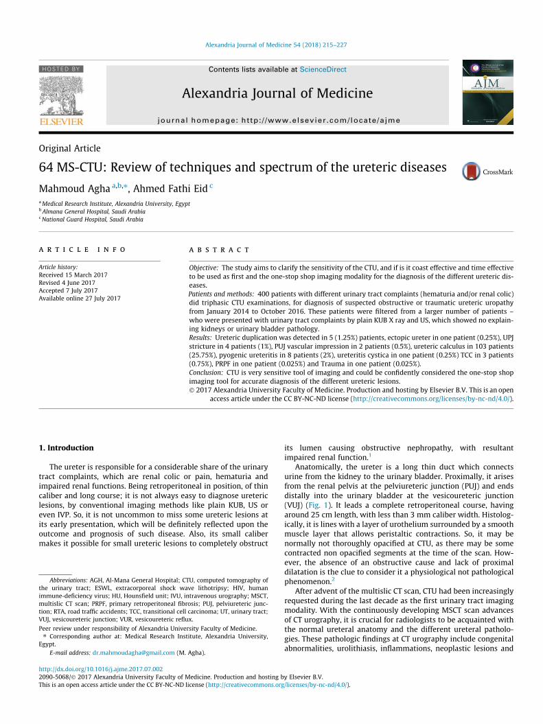
Anatomically, the ureter is a long thin duct which connectsurine from the kidney to the urinary bladder. Proximally, it arisesfrom the renal pelvis at the pelviureteric junction (PUJ) and endsdistally into the urinary bladder at the vesicoureteric junction(VUJ) (Fig. 1). It leads a complete retroperitoneal course, havingaround 25 cm length, with less than 3 mm caliber width. Histolog-ically, it is lines with a layer of urothelium surrounded by a smoothmuscle layer that allows peristaltic contractions. So, it may benormally not thoroughly opacified at CTU, as there may be somecontracted non opacified segments at the time of the scan. How-ever, the absence of an obstructive cause and lack of proximaldilatation is the clue to consider it a physiological not pathologicalphenomenon.2
After advent of the multislic CT scan, CTU had been increasinglyrequested during the last decade as the first urinary tract imagingmodality. With the continuously developing MSCT scan advancesof CT urography, it is crucial for radiologists to be acquainted withthe normal ureteral anatomy and the different ureteral patholo-gies. These pathologic findings at CT urography include congenitalabnormalities, urolithiasis, inflammations, neoplastic lesions and
A B C
Fig. 1. (A) CRF, (B) SRF& (C) VR CTU showing: Left short segmental upper ureteric duplication (Arrows).
A
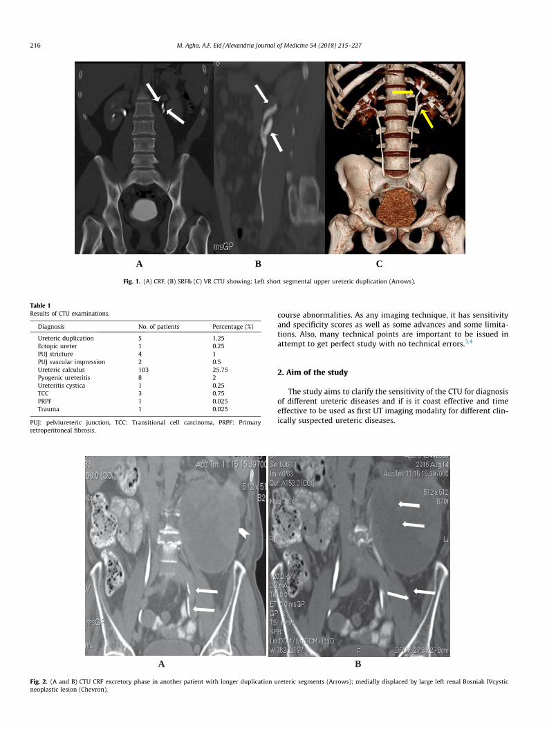
Fig. 2. (A and B) CTU CRF excretory phase in another patient with longer duplication uneoplastic lesion (Chevron).
Table 1Results of CTU examinations.
Diagnosis No. of patients Percentage (%)
Ureteric duplication 5 1.25Ectopic ureter 1 0.25PUJ stricture 4 1PUJ vascular impression 2 0.5Ureteric calculus 103 25.75Pyogenic ureteritis 8 2Ureteritis cystica 1 0.25TCC 3 0.75PRPF 1 0.025Trauma 1 0.025
PUJ: pelviureteric junction, TCC: Transitional cell carcinoma, PRPF: Primaryretroperitoneal fibrosis.
216 M. Agha, A.F. Eid / Alexandria Journal of Medicine 54 (2018) 215–227
course abnormalities. As any imaging technique, it has sensitivityand specificity scores as well as some advances and some limita-tions. Also, many technical points are important to be issued inattempt to get perfect study with no technical errors.3,4
2. Aim of the study
The study aims to clarify the sensitivity of the CTU for diagnosisof different ureteric diseases and if is it coast effective and timeeffective to be used as first UT imaging modality for different clin-ically suspected ureteric diseases.
B
reteric segments (Arrows); medially displaced by large left renal Bosniak IVcystic
M. Agha, A.F. Eid / Alexandria Journal of Medicine 54 (2018) 215–227 217
3. Patients and methods
3.1. Patients
400 patients with different urinary tract complaints (hematuriaand/or renal colic) were referred to CT scan division of the radiol-ogy department of Al-Mana General Hospital (AGH), with a sus-pected ureteric obstructive uropathy from January 2014 toOctober 2016. These patients were filtered from a larger numberof patients – who were presented with urinary tract complaintsby plain KUB X ray and US, which showed no explaining kidneysor urinary bladder pathology. The study protocol was approvedby the scientific and ethics committee in Al-Mana General Hospital(AGH). A signed consent was obtained from all study candidates,including detailed description of the technique with the rare possi-ble drawbacks.
A B
D
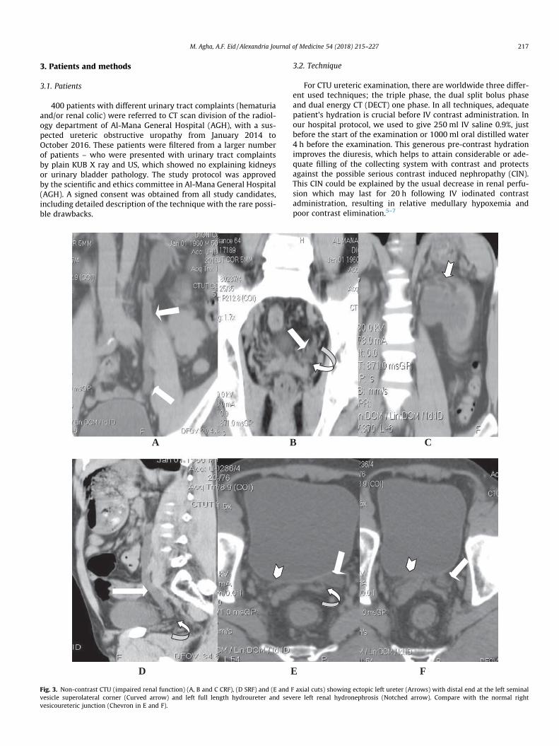
Fig. 3. Non-contrast CTU (impaired renal function) (A, B and C CRF), (D SRF) and (E andvesicle superolateral corner (Curved arrow) and left full length hydroureter and sevvesicoureteric junction (Chevron in E and F).
3.2. Technique
For CTU ureteric examination, there are worldwide three differ-ent used techniques; the triple phase, the dual split bolus phaseand dual energy CT (DECT) one phase. In all techniques, adequatepatient’s hydration is crucial before IV contrast administration. Inour hospital protocol, we used to give 250 ml IV saline 0.9%, justbefore the start of the examination or 1000 ml oral distilled water4 h before the examination. This generous pre-contrast hydrationimproves the diuresis, which helps to attain considerable or ade-quate filling of the collecting system with contrast and protectsagainst the possible serious contrast induced nephropathy (CIN).This CIN could be explained by the usual decrease in renal perfu-sion which may last for 20 h following IV iodinated contrastadministration, resulting in relative medullary hypoxemia andpoor contrast elimination.5–7
C
E F
F axial cuts) showing ectopic left ureter (Arrows) with distal end at the left seminalere left renal hydronephrosis (Notched arrow). Compare with the normal right

218 M. Agha, A.F. Eid / Alexandria Journal of Medicine 54 (2018) 215–227
We used to apply the triple phase technique as a routine CTUprotocol in our department. First, a non-contrast phase was done,and then automated injection of 1.5 ml/kg intravenous nonioniccontrast media (Omnipaque 300) as a single bolus. Nephrographicphase was allowed to run 100 s after the bolus, then after 10–15 min we used to run the last excretory phase. The whole studyCT examinations were done through Philips Brilliance 64 CT Scan-ner, Philips Medical System, Nederland. B.V. Veenpulis 4–5, 4684PC Best, Netherlands.
3.3. Post-processing and image interpretation
There are different post-processing data reconstructions pro-grams that vary from place to place for the urinary tract CT exam-ination, however there are some commonly shared and agreedprotocols for ureteric examinations. Starting with evaluation ofthe initial non-contrast phase is the rule, as it detects the stonesand the initially hyperdense abnormalities like blood clots. Also,it delineated the base line density of the lesion to determine thedegree of its enhancement. After evaluation of the non-contrastphase, you could stop the examination with no going furtherahead, if there is a hyperdense ureteric stone explaining the com-
A
C
Fig. 4. CTU Excretory phase (A and B) CRF, (C and D) SRF: showing localized PUJ stricturlevel is seen layering posteriorly in the dilated calyces. (Notched arrows in C and D). No
plaint of the patient, who was presented by acute renal colic.Hence, the importance for the technique to be supervised by theradiologist.8,9
Revision of the source images of the axial excretory phase is thesecond step we used to do after checking the non-contrast phase. Itis advised to use wide window measures- like bone window sets-to review this excretory phase, as this helps to perfectly diagnosesubtle lesions that may be hidden by contrast material at the nar-row soft tissue window sets. In our protocols, we used to do man-ual windows adjustment with different window length and widthfigures to attain different densities of the excreted contrast mate-rial, which helps to minimize the messing errors. Also routinelywe used to do different reformat sets in coronal and sagittal recon-structed images with the use of maximum intensity projection(MIP) tools. Despite time consuming, this multiplanar differentwindows revision helps much to increase the sensitivity of theexaminations, in comparison with source images revision only.9,10
4. Results
Ureteric duplication was detected in 5 (1.25%) patients, ectopicureter in one patient (0.25%), UPJ stricture in 4 patients (1%), PUJ
B
D
e (Arrow). Marked dilatation of the right renal pelvis and calyces. A urine–contrastrmal caliber non- opacified ureter is seen (Chevron in D).
A B
C D
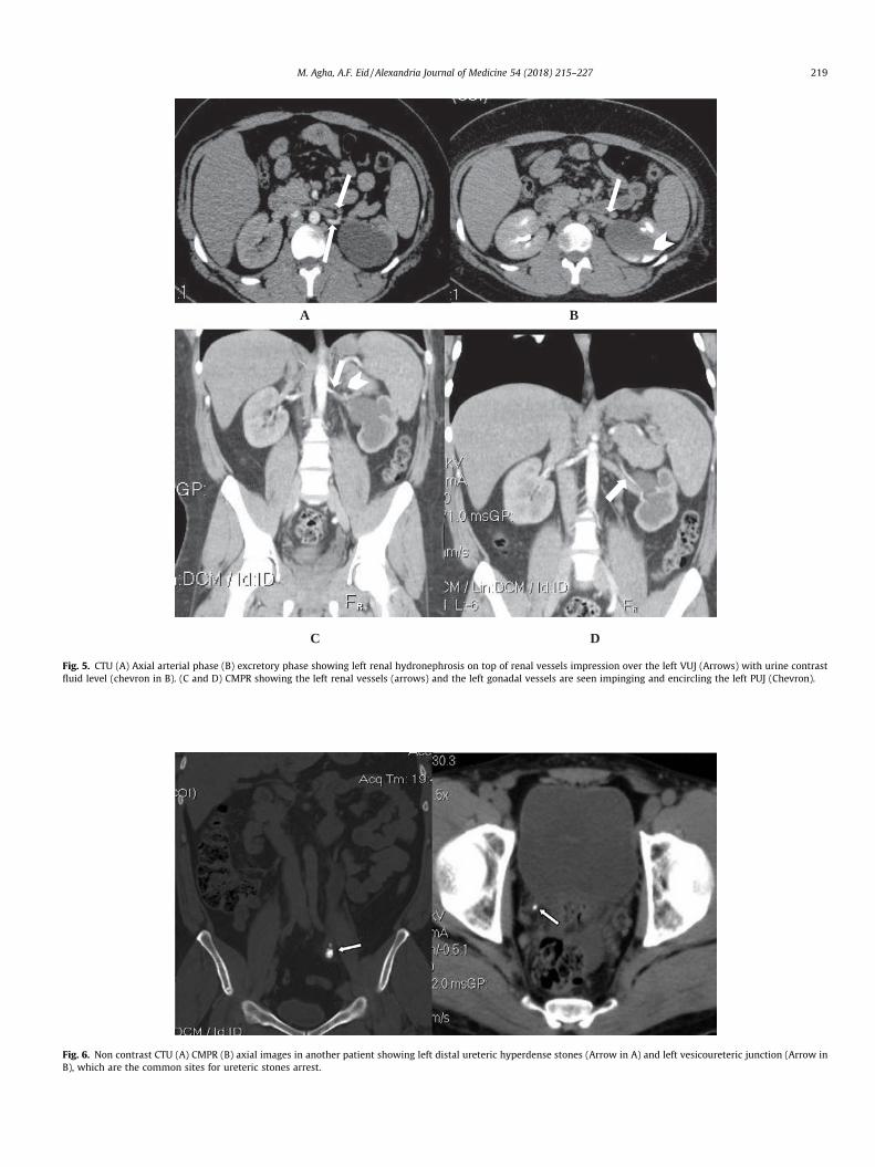
Fig. 5. CTU (A) Axial arterial phase (B) excretory phase showing left renal hydronephrosis on top of renal vessels impression over the left VUJ (Arrows) with urine contrastfluid level (chevron in B). (C and D) CMPR showing the left renal vessels (arrows) and the left gonadal vessels are seen impinging and encircling the left PUJ (Chevron).
Fig. 6. Non contrast CTU (A) CMPR (B) axial images in another patient showing left distal ureteric hyperdense stones (Arrow in A) and left vesicoureteric junction (Arrow inB), which are the common sites for ureteric stones arrest.
M. Agha, A.F. Eid / Alexandria Journal of Medicine 54 (2018) 215–227 219
220 M. Agha, A.F. Eid / Alexandria Journal of Medicine 54 (2018) 215–227
vascular impression in 2 patients (0.5%), ureteric calculus in 103patients (25.75%), pyogenic ureteritis in 8 patients (2%), ureteritiscystica in one patient (0.25%) TCC in 3 patients (0.75%), PRPF inone patient (0.025%) and Trauma in one patient (0.025%) (seeTable.1).
5. Discussion
Different congenital anomalies and acquired ureteric abnormal-ities were diagnosed by triple phase CTU at this study. The congen-ital ureteric lesions included variable types of ureteric duplications,as they were reported in 5 patients (1.25%) (Fig. 1). Thepartial duplication is more common, and occurs when there is fail-ure of fusion of the primitive separate upper and lower poleureters. This leads to partially separated upper ureters with vari-able length, which unite before the vesicoureteric junction. Fortu-nately, partial duplications are much more common than completeduplications.11,12 The partial type is usually discovered incidentallywith other abnormalities e.g. infection, calculus or neoplastic(Fig. 2).
Complete duplication of the ureters down to VUJ, where thereare separate distal ureteric openings, is relatively uncommon. Itis a known predisposing factor for some urinary tract diseases; likeincreased risk of repeated urinary tract infection, obstruction, andvesicoureteric reflux (VUR). According to Weigert-Meyer rule, thelower moiety ureter ends distally at a urinary bladder superolat-eral abnormal position, while the upper moiety ureter ends at anabnormal inferiomedial location. The lower moiety ureter is com-monly associated with vesicoureteric reflux, due to abnormalhigher position with lack of sphincteric control. The upper moietydistal ureteric end could be at the normal trigonal site, or notuncommonly at other ectopic vesical or extravesical sites.13,14
This ectopic distal ureteric termination may be also seen in non-duplicated ureters; as seen in one patient of this study (0.25%)(Fig. 3). The ectopic ureter in females usually presents with urinaryincontinence due to the usual insertion of the ectopic feminineureter below the external urethral sphincter. In contrast to males,whose ectopic ureter may insert on the posterior urethra, seminalvesicle, vas deferens, bladder neck, or prostate, hence it commonlyhas obstructed distal end. Males with ectopic ureter do not presentwith incontinence because unlike females, their ectopic ureter hasa distal end superior to the level of the external urethral sphincter.
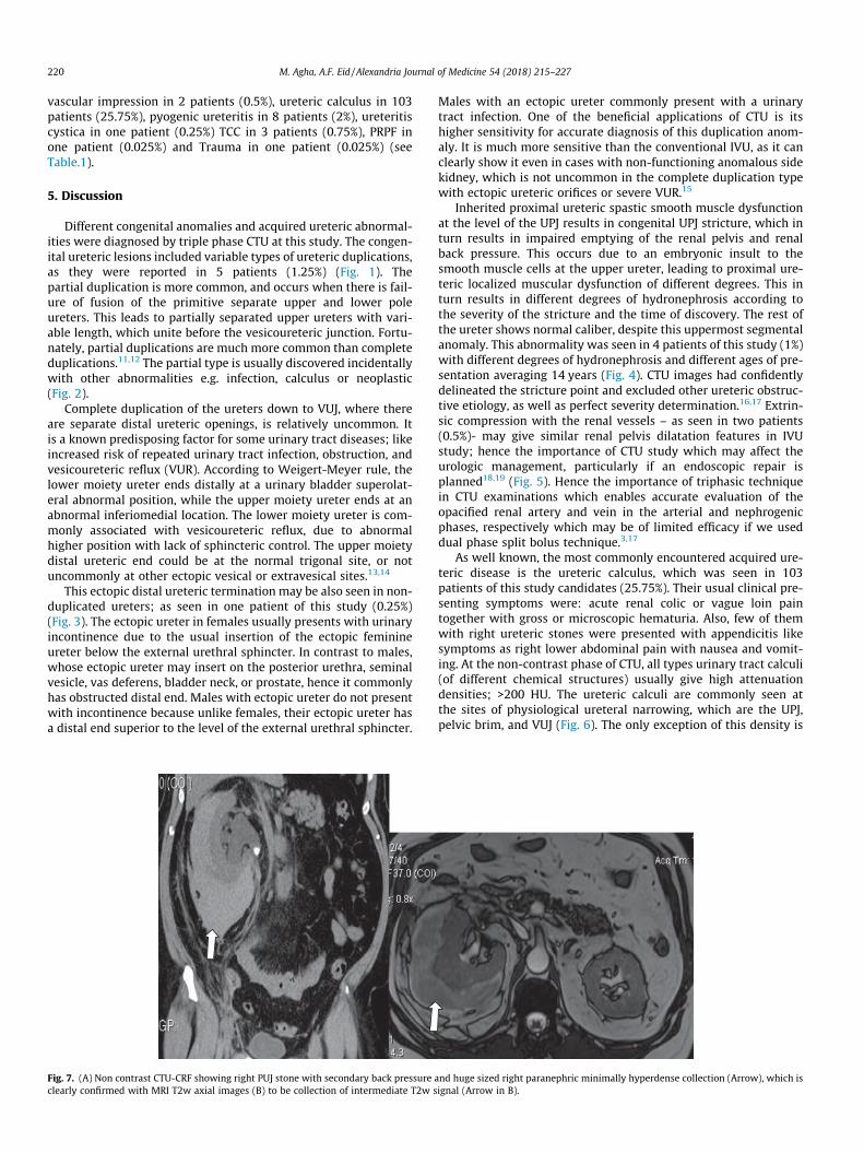
Fig. 7. (A) Non contrast CTU-CRF showing right PUJ stone with secondary back pressure aclearly confirmed with MRI T2w axial images (B) to be collection of intermediate T2w s
Males with an ectopic ureter commonly present with a urinarytract infection. One of the beneficial applications of CTU is itshigher sensitivity for accurate diagnosis of this duplication anom-aly. It is much more sensitive than the conventional IVU, as it canclearly show it even in cases with non-functioning anomalous sidekidney, which is not uncommon in the complete duplication typewith ectopic ureteric orifices or severe VUR.15
Inherited proximal ureteric spastic smooth muscle dysfunctionat the level of the UPJ results in congenital UPJ stricture, which inturn results in impaired emptying of the renal pelvis and renalback pressure. This occurs due to an embryonic insult to thesmooth muscle cells at the upper ureter, leading to proximal ure-teric localized muscular dysfunction of different degrees. This inturn results in different degrees of hydronephrosis according tothe severity of the stricture and the time of discovery. The rest ofthe ureter shows normal caliber, despite this uppermost segmentalanomaly. This abnormality was seen in 4 patients of this study (1%)with different degrees of hydronephrosis and different ages of pre-sentation averaging 14 years (Fig. 4). CTU images had confidentlydelineated the stricture point and excluded other ureteric obstruc-tive etiology, as well as perfect severity determination.16,17 Extrin-sic compression with the renal vessels – as seen in two patients(0.5%)- may give similar renal pelvis dilatation features in IVUstudy; hence the importance of CTU study which may affect theurologic management, particularly if an endoscopic repair isplanned18,19 (Fig. 5). Hence the importance of triphasic techniquein CTU examinations which enables accurate evaluation of theopacified renal artery and vein in the arterial and nephrogenicphases, respectively which may be of limited efficacy if we useddual phase split bolus technique.3,17
As well known, the most commonly encountered acquired ure-teric disease is the ureteric calculus, which was seen in 103patients of this study candidates (25.75%). Their usual clinical pre-senting symptoms were: acute renal colic or vague loin paintogether with gross or microscopic hematuria. Also, few of themwith right ureteric stones were presented with appendicitis likesymptoms as right lower abdominal pain with nausea and vomit-ing. At the non-contrast phase of CTU, all types urinary tract calculi(of different chemical structures) usually give high attenuationdensities; >200 HU. The ureteric calculi are commonly seen atthe sites of physiological ureteral narrowing, which are the UPJ,pelvic brim, and VUJ (Fig. 6). The only exception of this density is
nd huge sized right paranephric minimally hyperdense collection (Arrow), which isignal (Arrow in B).
M. Agha, A.F. Eid / Alexandria Journal of Medicine 54 (2018) 215–227 221
the indinavir induced soft stones which are induced by such pro-tease inhibitor anti HIV drug. So these softer stones may be missedin the non-contrast scans and seen only in the excretory phase asfilling defects that got some other differential diagnosis excludedby clear clinical and pharmacological history intake.20–22
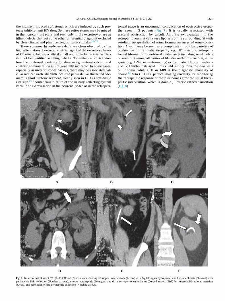
These common hyperdense calculi are often obscured by thehigh attenuation of excreted contrast agent at the excretory phasesof CT urography, especially if small and non-obstructive, as theywill not be identified as filling defects. Non-enhanced CT is there-fore the preferred modality for diagnosing ureteral calculi, andcontrast administration is not generally indicated. In some cases,especially in ureteric stones passers, there may be associated cal-cular induced ureteritis with localized peri-calcular thickened ede-matous short ureteric segment, clearly seen in CTU as soft-tissuerim sign.23 Spontaneous rupture of the urinary collecting systemwith urine extravasation in the perirenal space or in the retroperi-
A B
D
Fig. 8. Non contrast phase of CTU (A–C) CRF and (D) axial cuts showing left upper ureteriperinephric fluid collection (Notched arrows), anterior paranephric (Pentagon) and dista(Arrow) and resolution of the perinephric collection (Notched arrow).
toneal space is an uncommon complication of obstructive uropa-thy, seen in 2 patients (Fig. 7). It is usually associated withureteral obstruction by calculi. As urine extravasates into theretroperitoneum, it can cause lipolysis of the surrounding fat withresultant encapsulation of urine, forming an encysted urine collec-tion. Also, it may be seen as a complication to other varieties ofobstructive or traumatic uropathy e.g. UPJ stricture, retroperi-toneal fibrosis, retroperitoneal malignancy including renal pelvisor ureteric tumors, all causes of bladder outlet obstruction, iatro-genic (e.g. ESWL or ureteroscopy) or traumatic. US examinationsand IVU without delayed films could simply miss the diagnosisof urinoma, while CTU or MRI is the diagnostic modality ofchoice.24 Also CTU is a perfect imaging modality for monitoringthe therapeutic response of these urinomas after the usual thera-peutic intervention, which is double J ureteric catheter insertion(Fig. 8).
zz
C
E F
c stone (Arrow) with 2ry left upper hydroureter and hydronephrosis (Chevron) withl retroperitoneal urinoma (Curved arrow). (E&F) Post ureteric DJ catheter insertion
A B
C D
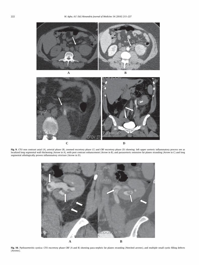
Fig. 9. CTU non contrast axial (A), arterial phase (B), zoomed excretory phase (C) and CRF excretory phase (D) showing: left upper ureteric inflammatory process see aslocalized long segmental wall thickening (Arrow in A), with post contrast enhancement (Arrow in B), and paraureteric extensive fat planes stranding (Arrow in C) and longsegmental athologically proven inflammatory stricture (Arrow in D).
A B
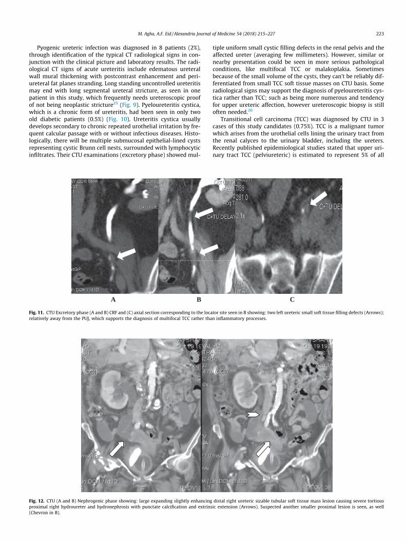
Fig. 10. Pyeloureteritis cystica: CTU excretory phase CRF (A and B) showing para-nephric fat planes stranding (Notched arrows), and multiple small cystic filling defects(Arrows).
222 M. Agha, A.F. Eid / Alexandria Journal of Medicine 54 (2018) 215–227
M. Agha, A.F. Eid / Alexandria Journal of Medicine 54 (2018) 215–227 223
Pyogenic ureteric infection was diagnosed in 8 patients (2%),through identification of the typical CT radiological signs in con-junction with the clinical picture and laboratory results. The radi-ological CT signs of acute ureteritis include edematous ureteralwall mural thickening with postcontrast enhancement and peri-ureteral fat planes stranding. Long standing uncontrolled ureteritismay end with long segmental ureteral stricture, as seen in onepatient in this study, which frequently needs ureteroscopic proofof not being neoplastic stricture25 (Fig. 9). Pyeloureteritis cystica,which is a chronic form of ureteritis, had been seen in only twoold diabetic patients (0.5%) (Fig. 10). Ureteritis cystica usuallydevelops secondary to chronic repeated urothelial irritation by fre-quent calcular passage with or without infectious diseases. Histo-logically, there will be multiple submucosal epithelial-lined cystsrepresenting cystic Brunn cell nests, surrounded with lymphocyticinfiltrates. Their CTU examinations (excretory phase) showed mul-
A B
Fig. 11. CTU Excretory phase (A and B) CRF and (C) axial section corresponding to the locarelatively away from the PUJ, which supports the diagnosis of multifocal TCC rather tha
Fig. 12. CTU (A and B) Nephrogenic phase showing: large expanding slightly enhancingproximal right hydroureter and hydronephrosis with punctate calcification and extrins(Chevron in B).
tiple uniform small cystic filling defects in the renal pelvis and theaffected ureter (averaging few millimeters). However, similar ornearby presentation could be seen in more serious pathologicalconditions, like multifocal TCC or malakoplakia. Sometimesbecause of the small volume of the cysts, they can’t be reliably dif-ferentiated from small TCC soft tissue masses on CTU basis. Someradiological signs may support the diagnosis of pyeloureteritis cys-tica rather than TCC; such as being more numerous and tendencyfor upper ureteric affection, however ureteroscopic biopsy is stilloften needed.26
Transitional cell carcinoma (TCC) was diagnosed by CTU in 3cases of this study candidates (0.75%). TCC is a malignant tumorwhich arises from the urothelial cells lining the urinary tract fromthe renal calyces to the urinary bladder, including the ureters.Recently published epidemiological studies stated that upper uri-nary tract TCC (pelviureteric) is estimated to represent 5% of all
C
tor site seen in B showing: two left ureteric small soft tissue filling defects (Arrows);n inflammatory processes.
distal right ureteric sizable tubular soft tissue mass lesion causing severe tortiousic extension (Arrows). Suspected another smaller proximal lesion is seen, as well

224 M. Agha, A.F. Eid / Alexandria Journal of Medicine 54 (2018) 215–227
urothelial cancers and in less than 10% of all renal tumors.27 Evi-dences indicate that the frequency of upper urinary tract malig-nancies is increasing. The diagnosis of TCC in this study wasbased on the fact that it the commonest type of malignant ureterictumors. TCC of the ureter could be radiologically seen at CTU excre-tory phase as intraluminal polypoidal filling defects or annularstricture at its early presentation (Fig. 11). Also, it is not uncom-mon to have simultaneous multifocal primary lesions at the timeof presentation or later on at time intervals of months or evenyears i.e. synchronous or metachronus multiplicity. If missed fora while, as one of our patient, it could be presented with severeobstructive nephropathy secondary to a large ureteric expandingsoft tissue ureteric mass lesion, which may infiltrate the adjacentstructures. There is a described CTU sign named the goblet sign;which is highly suggestive (Not 100% pathognomonic) of uretericTCC. This goblet sign is a cup like filling defect due to the soft tissuegrowth inside the ureter; however it is more conspicuous in retro-grade not antegrade CT or conventional pyeloureterography.28,29
The clinical and/or the laboratory presentation of painlesshematuria are very important signs to consider pelviureteric andurinary bladder tumors, of which TCC is the most common. Still,there is a conflict in small ureteric lesions seen in CTU, as theymay simulate ureteritis or malakoplakia. However, the distal ure-teric location and the paucity of the lesions significantly favorthe neoplastic etiology (Fig. 11). CTU diagnosis of urothelial tumorsis much simpler in neglected cases, as seen in one patient of thisstudy (Fig. 12), due to the looking of the aggressive expanding softtissue swelling and extrinsic extension. CTU had been proven notonly to be perfect imaging modality for detection and diagnosisof TCC but also in TNM staging of the tumor, which is crucial formanagement Table 2.
Primary retroperitoneal fibrosis (PRPF) was diagnosed in onlyone patient (0.025%), during the whole study (Fig. 13). It is a raredisease with an estimated incidence of 1.38/100,000 people. Somemedications, abdominal aortic aneurysm, infections, malignancies,trauma, surgery, and radiation therapy have been accused to becausative pathology of RPF; however, it is commonly idiopathic.PRPF commonly occurs in individuals 40–60 years of age; with amale to female ratio of 3.3:1.30,31 It had been previously describedas chronic periaortitis, as the pathological process is centeredaround the abdominal aorta. Histologically; it is a fibrotic reactionwithin the retroperitoneal tissues that encases the retroperitoneal
Table 2TNM staging of ureteric TCC.
TNM Signs
TTa Non invasive papillary tumorTis In situ (non invasive flat)T1 Through lamina propria into sub-epithelial connective tissuesT2 Into muscularis propriaT3 Invasion into periureteric fatT4 Direct invasion into adjacent organs/structures
NN0 No nodal involvement.N1 Single node involved <2 cmN2 Single node 2–5 cm or multiple nodes all <5 cmN3 One or more nodes >5 cm
MM0 No metastasesM1 Metastases identified
StagingStage 0 Ta or Tis, N0, M0Stage I T1, N0, M0Stage II T2(a or b), N0, M0Stage III T3(a or b) or T4a, N0, M0Stage IV T4b, any N, any M or N1-3, any T, any M
structure causing bilateral medial ureteral traction and constric-tion. IVP can show the secondary effect of PRPF, which is bilateralmedial ureteric deviation and obstructive uropathy. However, CTUwill be more informative imaging tool, as it can clearly show theextent of the paraortic retroperitoneal fibrotic bands with its trac-tion obstructive effect on the ureters. Also, it helps to show if thereis a causative abdominal etiology like abdominal aortic aneurysmor retroperitoneal malignancy which are the common causes ofsecondary RPF.32
Ureteric trauma is a relatively uncommon but it may result inserious complications, due to the usual delayed diagnosis. The clas-sic clinical presentation is post-traumatic loin or flank pain withhematuria. However these symptoms and signs may also be com-pletely absent in up to 25% of patients, who may be lately pre-sented with acute renal failure or severe acute abdomen. Thedetection of ureteric injuries is delayed for days or even weeks inabout 60% of patients. There is a wide range of ureteric injuries;ranging from mucosal injury, ligation, partial or complete ureterictransection up to complete ureteric avulsion.33,34 18 RTA trauma-tized patients with post traumatic hematuria, were included in thisstudy. CTU had diagnosed pelviureteric avulsion in only onepatient, associated with displaced fracture of the lumbar spineand lower polar left renal laceration (Fig. 14). The other 17patients’ CTU results were either normal or kidneys or bladderinjuries. Hematuric blood clots could be seen in the non-contrastphase as hyperdense intraureteric fluid (>50 HU) (Fig. 15). Typi-cally it appears as a vermiform like filling defect in the excretoryphase. Also, the clot does not enhance in the contrast phases andchanges position or completely resolves at follow-up imaging stud-ies. However, in non-traumatized patients with hematuria; a smallclot may be difficult to be distinguished from TCC at initial CTU,which may need ureteroscopy or short-interval follow-up imagingfor confirmation.35,36
Ureteric injuries are more commonly iatrogenic; most com-monly during gynaecological procedures. Penetrating or blunt ure-teric injuries are relatively uncommon; representing <1% of allurological trauma. Anatomically, ureteric injury could be classifiedinto pelviureteric - upper third ureter (mostly due to blunt abdom-inal trauma and RTA), middle and distal thirds ureteric trauma,which is often following iatrogenic injury. CTU with intravenouscontrast is the examination of choice for evaluation of ureterictrauma, as it clearly demonstrates the site and the extent of theinjury. Also, it can accurately verify its type; whether avulsion ortrans-section or ligation. In addition to its role in detection of asso-ciated other abdominal organ injuries and the resultant complica-tions e.g. retroperitoneal hematoma, hydronephrosis, stricturesand the resultant obstructive nephropathy ending if neglected intorenal failure. Early diagnosis and appropriate correction of thecause- if possible- (e.g. removal of suture on tied ureter or recon-struction of induced ureteric strictures) will result in satisfactoryoutcome. Temporary ureteric stents or even percutaneousnephrostomy may be needed until full ureteric repair is done.37
Despite the sensitivity of IVP in ureteric imaging, it has limita-tions of being in need for good renal excretory poor, lack of extra-ureteric adjacent structures evaluation and wide differential diag-nosis of filling defects. Based upon the results of this study we rec-ommend the use of CTU as a one-stop shop imaging technique forpatients with suspected ureteric pathology, thereby saving timeand hospital visits and leading to earlier diagnosis. This result isaccepted at the expanse of the high dose radiation exposure, whichmay be not suitable for some patients, like pregnant females. How-ever, the American Urological Association guidelines still equallyrecommend IV or CT urography as the initial imaging test forpatients with asymptomatic microscopic hematuria.3 In contrastto, the recent guidelines from the European Society of UrogenitalRadiology which had supported the use of CTU. Contraindications

A B
C D E
Fig. 13. A 58 years male with PRPF; (A) MIP & (B) VR CRF CTU excretory phase showing asymmetrical medially deviated and kinked right ureter with secondary right sidedminimal obstructive uropathy. (Arrows) (C–E) CTU arterial phase axial cuts showing entrapment of the aortic bifurcation and right common iliac vessels by retroperitonealprevertebral irregular fibrous soft tissue swelling. The right ureter is significantly deviated medially and partially entrapped by the mass (notched arrows), in comparisonwith the normal posed left ureter, which is seen in front of the left psoas muscle (Chevrons).
M. Agha, A.F. Eid / Alexandria Journal of Medicine 54 (2018) 215–227 225
to CTU are generally limited to those patients who cannot receiveiodinated contrast because of renal insufficiency, prior severe reac-tion, or pregnancy.38
6. Conclusion
CTU could be trusty used as a very sensitive diagnostic one-stopshop imaging tool in patients with clinically suspected uretericlesions.
7. Limitations of the study
Pregnant females may suffer from different ureteric complaints;commonly colic due to ureteric stones or ureteric compression bythe pregnant uterus. Considering the fact that, ionizing radiationhas a carcinogenic and teratogenic hazards to the developing fetus,there is a considerable limitations to use CTU, especially in the firsttrimester. US and KUB are recommended as first imaging modali-
ties; however sometimes CTU is mandatory e.g. RTA with sus-pected ureteric trauma.39
Also, there are some considerations for radiation exposure inchildren, as children are considerably more sensitive to radiationthan adults. Children have a longer life expectancy than adults,resulting in a larger window of opportunity for expressing radi-ation damage. Children commonly receive a higher radiationdose than necessary if CT settings are not adjusted for theirsmaller body size. All These facts encourage keeping CTU exam-inations as last resort in children after trials to obtain the diag-nosis by other less hazardous tools. Also, we recommend the useof low kV and dual phase techniques if CTU is going to be donein children, in attempt to minimize the radiation doseexposure.40
Also, due to the large group of ureteric pathologies as well ascoast-effective facts in the private hospitals, there was limitationfor comparison of our study CT results with other imaging modal-ities; as MRU.

A B
C
Fig. 14. CTU after severe RTA injury (A and B) CRF and (C) axial section showing fractured dislocation of the mid-lumbar spine (Notched arrow), lower left renal contusion(Arrow in A) with distorted pelviureteric junction (Arrow in B) and retroperitoneal extravasated contrast in the delayed scans (Arrow in C).
Fig. 15. Post RTA abdominal injury CTU non contrast axial sections show (A) Liver lacerations (Arrows) and (B) left renal pelvis hyperdense hematoma (Notched arrow).
226 M. Agha, A.F. Eid / Alexandria Journal of Medicine 54 (2018) 215–227

M. Agha, A.F. Eid / Alexandria Journal of Medicine 54 (2018) 215–227 227
References
1. Masarani M, Dinneen M. Ureteric colic: new trends in diagnosis andtreatment. Postgrad Med J. 2007;83:469–472. http://dx.doi.org/10.1136/pgmj.2006.055913.
2. Lye CM, Fasano L, Woolf AS. Ureter myogenesis: putting Teashirt into context. JAm Soc Nephrol. 2010;21:24–30.
3. Silverman SG, Leyendecker JR, Amis ES. What is the current role of CT urographyand MR urography in the evaluation of the urinary tract? Radiology.2009;250:309–323.
4. Nawfel RD, Judy PF, Schleipman AR, et al.. Patient radiation dose at CT urographyand conventional urography. Radiology. 2004;232:126–132.
5. Joffe SA, Servaes S, Okon S, et al.. Multi-detector row CT urography in theevaluation of hematuria. Radiographics. 2003;23:1441–1456.
6. Dahlman P, van der Molen AJ, Magnusson M, Magnusson A. How much dose canbe saved in three-phase CT urography? A combination of normal-dosecorticomedullary phase with low-dose unenhanced and excretory phases. AJRAm J Roentgenol. 2012;199:852–860.
7. Kaza RK, Platt JF, Cohan RH, Caoili EM, Al-Hawary MM, Wasnik A. Dual-energyCT with single- and dual-source scanners: current applications in evaluating thegenitourinary tract. RadioGraphics. 2012;32:353–369.
8. Kenney PJ. CT evaluation of urinary lithiasis. Radiol Clin N Am. 2003;41:979–999.9. Sourtzis S, Thibeau JF, Damry N, et al. Radiologic investigation of renal colic:
unenhanced helical CT compared with excretory urography. Am J Roentgentol1999;172:1491–4.
10. Mahmoud M, Moawad Ahmed, El-Zawawy Mohamed S. The role ofmultidetector computed tomography urography in the evaluation ofobstructive uropathy. Korean J Radiol. 2004;5:1–10.
11. Fernbach SK1, Feinstein KA, Spencer K, Lindstrom CA. Ureteral duplication andits complications. Radiographics 1997;17:109–27.
12. Gatti JM, Cendron M. Ureteral duplication, ureteral ectopia, and ureterocele.<http://emedicine.medscape.com/article/1017202-overview> (Updated: Oct09, 2015).
13. Adiego B, Martinez-Ten P, Perez-Pedregosa J, et al.. Antenatally diagnosed renalduplex anomalies: sonographic features and long-term postnatal outcome. JUltrasound Med. 2011;30:809–815.
14. Williams H. Renal revision: from lobulation to duplication—what is normal?Arch Dis Child Educ Pract Ed. 2007;92:152–158.
15. Berrocal T, Lopez-Pereira P, Arjonilla A, Gutierrez J. Anomalies of the distalureter, bladder, and urethra in children: embryologic, radiologic, andpathologic features. RadioGraphics. 2002;22. 1139–116.
16. Klein J, Gonzalez J, Miravete M, et al.. Congenital ureteropelvic junctionobstruction: human disease and animal models. Int J Exp Pathol.2011;92:168–192.
17. Lawler LP, Jarret TW, Corl FM, Fishman EK. Adult ureteropelvic junctionobstruction: insights with three dimensional multi-detector row CT.RadioGraphics. 2005;25:121–134.
18. Lam JS, Breda A, Schulam PG. Ureteropelvic junction obstruction. J Urol.2007;177:1652–1658.
19. Tsai JD, Huang FY, Lin CC, et al.. Intermittent hydronephrosis secondary toureteropelvic junction obstruction: clinical and imaging features. Pediatrics.2006;117:139–146.
20. Tamm EP, Silverman PM, ShumanWP. Evaluation of the patient with flank painand possible ureteral calculus. Radiology. 2003;228:319–329. http://dx.doi.org/10.1148/radiol.2282011726.
21. Matlaga BR, Shah OD, Assimos DG. Drug induced calculi. Rev Urol.2003;5:227–231.
22. Heneghan JP, McGuire KA, Leder RA, et al.. Helical CT for nephrolithiasis andureterolithiasis: comparison of conventional and reduced radiation-dosetechniques. Radiology. 2003;229:575–580. http://dx.doi.org/10.1148/radiol.2292021261.
23. Dyer RB, Chen MY, Zagoria RJ. Classic signs in uroradiology. Radiographics2004;24 Suppl 1 (suppl 1): S247–80. Radiographics (full text). http://dx.doi.org/10.1148/rg.24si045509 – Pubmed citation.
24. Sebastià C, Quiroga S, Boyé R, Cantarell C, Fernandez-Planas M, Alvarez A.Helical CT in renal transplantation: normal findings and early and latecomplications. RadioGraphics. 2001;21:1103–1117.
25. Wasnik AP, Elsayes KM, Kaza RK, et al.. Multimodality imaging in ureteric andperiureteric pathologic abnormalities. AJR Am J Roentgenol. 2011;197. http://dx.doi.org/10.2214/AJR.11.6623.
26. Kawashima A, Vrtiska TJ, Leroy AJ et-al. CT urography. Radiographics. 2004;24Suppl 1 (suppl 1): S35–54.
27. Siegel RL, Miller KD, Jemal A. Cancer statistics. CA Cancer J Clin. 2016;66:7–30.28. Vikram R, Sandler CM, Ng CS. Imaging and staging of transitional cell
carcinoma: part 2, upper urinary tract. AJR Am J Roentgenol.2009;192:1488–1493.
29. Browne RF, Meehan CP, Colville J, et al.. Transitional cell carcinoma of the upperurinary tract: spectrum of imaging findings. Radiographics.2005;25:1609–1627.
30. Vaglio A, Salvarani C, Buzio C. Retroperitoneal fibrosis. Lancet.2006;367:241–251.
31. Van Bommel EF, Jansen I, Hendriksz TR, Aarnoudse AL. Idiopathicretroperitoneal fibrosis: prospective evaluation of incidence andclinicoradiologic presentation. Medicine (Baltimore). 2009;88:193–201.
32. Cronin CG, Lohan DG, Blake MA, et al.. Retroperitoneal fibrosis: a review ofclinical features and imaging findings. AJR Am J Roentgenol. 2008;191:423–431.
33. Pereira B, Ogilvie M, Gomez-Rodriguez J, et al.. A review of ureteral injuriesafter external trauma. Scand J Trauma Resusc Emerg Med. 2010;18:6.
34. Srinivasa RN, Akbar SA, Jafri SZ, Howells GA. Genitourinary trauma: a pictorialessay. Emerg Radiol. 2009;16:21–33.
35. Shinagare A, Sadow C, Sahni, Silverman Stuart G. Urinary bladder: normalappearance and mimics of malignancy at CT urography. Cancer Imaging.2011;11:100–108.
36. Choyke PL. Radiologic evaluation of hematuria: guidelines from the AmericanCollege of Radiology’s appropriateness criteria. Am Fam Phys.2008;78:347–352.
37. Ramchandani P, Buckler PM. Imaging of genitourinary trauma. AJR Am JRoentgenol. 2009;192:1514–1523.
38. Van der Molen AJ, Cowan NC, Mueller-Lisse UG, Nolte-Emsting CC, Takahashi S,Cohan RH. CT urography: definition, indications and techniques-a guideline forclinical practice. EurRadiol. 2008;18:4–17.
39. Sadro C, Dubinsky T. CT in pregnancy: risks and benefits. Appl Radiol.2013;42:6–16.
40. Arch ME, Frush DP. Pediatric body MDCT: a 5-year follow-up survey ofscanning parameters used by pediatric radiologists. AJR. 2008;191:611–617.


![[Characteristics of encrustation of ureteric stents in patients with urinary stones]](https://static.fdokumen.com/doc/165x107/633645e7cd4bf2402c0b6fc8/characteristics-of-encrustation-of-ureteric-stents-in-patients-with-urinary-stones.jpg)