Review of existing grading systems for cervical or lumbar disc and facet joint degeneration
14
Purpose Degeneration of the spine is a very common phenomenon. The morphological changes occurring during degenera- tion have often been described macroscopically [13, 22, 24, 43, 60, 64, 66], histologically [6, 13, 16–18, 20, 22, 33, 64, 66], and using many different imaging techniques such as plain radiography [13, 24, 35, 36, 54], discography [1], Annette Kettler Hans-Joachim Wilke Review of existing grading systems for cervical or lumbar disc and facet joint degeneration Received: 13 October 2004 Revised: 4 April 2005 Accepted: 14 April 2005 Published online: 20 September 2005 Ó Springer-Verlag 2005 Abstract The aim of this literature review was to present and to evalu- ate all grading systems for cervical and lumbar disc and facet joint degeneration, which are accessible from the MEDLINE database. A MEDLINE search was conducted to select all articles presenting own grading systems for cervical or lum- bar disc or facet joint degeneration. To give an overview, these grading systems were listed systematically depending on the spinal region they refer to and the methodology used for grading. All systems were checked for reliability tests and those recommended for use having an in- terobserver Kappa or Intraclass Correlation Coefficient >0.60 if disc degeneration was graded and >0.40 if facet joint degeneration was gra- ded. MEDLINE search revealed 42 different grading systems. Thirty of these were used to grade lumbar spine degeneration, ten were used to grade cervical spine degeneration and two were used to grade both. Thus, the grading systems for the lumbar spine represented the vast majority of all 42 grading systems. Interobserver reliability tests were found for 12 grading systems. Based on their Kappa or Intraclass Corre- lation Coefficients nine of these could be recommended for use and three could not. All other systems could neither be recommended nor not be recommended since reliability tests were missing. These systems should therefore first be tested be- fore use. The design of the grading systems varied considerably. Five grading systems were beginning with the lowest degree of degeneration, 37, however, with the normal, not degenerated state. A 5-grade scale was used in six systems, a 4-grade scale in 24, a 3-grade scale in eight and a 2-grade scale in three systems. In 15 cases the normal, not degen- erated state was assigned to ‘‘grade 0’’, in another 15 cases, however, this state was assigned to ‘‘grade 1’’. This wide variety in the design of the grading systems makes comparisons difficult and may easily lead to con- fusion. We would therefore recom- mend to define certain standards. Our suggestion would be to use a scale of three to five grades, to begin the scale with the not degenerated state and to assign this state to ‘‘grade 0’’. Eur Spine J (2006) 15: 705–718 DOI 10.1007/s00586-005-0954-y REVIEW A. Kettler H.-J. Wilke (&) Institute of Orthopaedic Research and Biomechanics, University of Ulm, Ulm, Germany E-mail: [email protected] Tel.: +49-731-50023482 Fax: +49-731-50023498
-
Upload
independent -
Category
Documents
-
view
4 -
download
0
Transcript of Review of existing grading systems for cervical or lumbar disc and facet joint degeneration
Purpose
Degeneration of the spine is a very commonphenomenon.The morphological changes occurring during degenera-
tion have often been described macroscopically [13, 22,24, 43, 60, 64, 66], histologically [6, 13, 16–18, 20, 22, 33,64, 66], and usingmany different imaging techniques suchas plain radiography [13, 24, 35, 36, 54], discography [1],
Annette Kettler
Hans-Joachim WilkeReview of existing grading systems forcervical or lumbar disc and facet jointdegeneration
Received: 13 October 2004Revised: 4 April 2005Accepted: 14 April 2005Published online: 20 September 2005� Springer-Verlag 2005
Abstract The aim of this literaturereview was to present and to evalu-ate all grading systems for cervicaland lumbar disc and facet jointdegeneration, which are accessiblefrom the MEDLINE database. AMEDLINE search was conducted toselect all articles presenting owngrading systems for cervical or lum-bar disc or facet joint degeneration.To give an overview, these gradingsystems were listed systematicallydepending on the spinal region theyrefer to and the methodology usedfor grading. All systems werechecked for reliability tests and thoserecommended for use having an in-terobserver Kappa or IntraclassCorrelation Coefficient >0.60 if discdegeneration was graded and >0.40if facet joint degeneration was gra-ded. MEDLINE search revealed 42different grading systems. Thirty ofthese were used to grade lumbarspine degeneration, ten were used tograde cervical spine degenerationand two were used to grade both.Thus, the grading systems for thelumbar spine represented the vastmajority of all 42 grading systems.Interobserver reliability tests werefound for 12 grading systems. Based
on their Kappa or Intraclass Corre-lation Coefficients nine of thesecould be recommended for use andthree could not. All other systemscould neither be recommended nornot be recommended since reliabilitytests were missing. These systemsshould therefore first be tested be-fore use. The design of the gradingsystems varied considerably. Fivegrading systems were beginning withthe lowest degree of degeneration,37, however, with the normal, notdegenerated state. A 5-grade scalewas used in six systems, a 4-gradescale in 24, a 3-grade scale in eightand a 2-grade scale in three systems.In 15 cases the normal, not degen-erated state was assigned to ‘‘grade0’’, in another 15 cases, however,this state was assigned to ‘‘grade 1’’.This wide variety in the design of thegrading systems makes comparisonsdifficult and may easily lead to con-fusion. We would therefore recom-mend to define certain standards.Our suggestion would be to use ascale of three to five grades, to beginthe scale with the not degeneratedstate and to assign this state to‘‘grade 0’’.
Eur Spine J (2006) 15: 705–718DOI 10.1007/s00586-005-0954-y REVIEW
A. Kettler Æ H.-J. Wilke (&)Institute of Orthopaedic Research andBiomechanics, University of Ulm,Ulm, GermanyE-mail:[email protected].: +49-731-50023482Fax: +49-731-50023498
computed tomography [11, 22, 54, 71], or magnetic reso-nance imaging [22, 54, 73]. If these techniques have to becompared with each other in their ability to depictdegeneration, grading systems are needed. Degenerationalso has to be graded to investigate the relationship be-tween morphology and functional parameters such asinstability or intradiscal pressure, or to assess the corre-lation between different morphological changes. To an-swer whether disc degeneration causes facet jointdegeneration also requires grading systems.Thus, gradingsystems for disc and facet joint degeneration are useful oreven needed to answer a vide variety of different ques-tions. It is therefore not surprising that more and moregrading systems are reported in the literature.
The aim of this literature review was to collect, sortand evaluate all grading systems for cervical and lumbardisc and facet joint degeneration, which are accessiblefrom the MEDLINE database.
Materials and methods
Literature search
A MEDLINE search was conducted via PubMed usingvarious combinations of the following key words:degeneration, ageing, classification, grade, cervicalspine, lumbar spine, intervertebral disc, and facet joint.Once a new grading system was found, the ‘‘relatedarticles’’ option of PubMed was used to further expandthe search. All extracted articles were reviewed and thoseselected with own grading systems for cervical or lumbardisc or facet joint degeneration. Even though this searchwas carried out thoroughly, it may be not complete.
According to the grading method, the selected articleswere then assigned to one of the following subgroups:macroscopic anatomy, histology, plain radiography,computed tomography, magnetic resonance imaging, ordiscography if disc degeneration was graded, or to mac-roscopic anatomy, histology, plain radiography, con-ventional tomography, computed tomography, ormagnetic resonance imaging if facet joint degenerationwas graded.
Evaluation of grading systems
The evaluation of the grading systems was divided intotwo steps. In the first step, they were checked for reli-ability tests. Focus was put on the interobserver reli-ability, which is generally somewhat lower than theintraobserver reliability. To assess the reliability eitherKappa [12] or Intraclass Correlation Coefficients (ICC)[57] have to be determined. According to the criteriapublished by Landis and Koch [37], a Kappa of <0.00
can be interpreted as poor agreement, 0.00–0.20 as slightagreement, 0.21–0.40 as fair agreement, 0.41–0.60 asmoderate agreement, 0.61–0.80 as substantial agreement,and >0.81 as almost perfect agreement. Based on thisclassification, we defined own criteria for recommenda-tion: a grading system for cervical or lumbar discdegeneration was recommended only if Kappa or ICCwas >0.60 (at least substantial agreement). Since theassessment of facet joint degeneration was assumed to bemore difficult than that of the disc, the facet joint gradingsystems were recommended already at Kappa orICC>0.40 (at least moderate agreement). Articlesdescribing linear correlation coefficients instead of Kap-pa or ICC did not meet our criteria for recommendationsince such correlation coefficients only reflect how strongthe linear relationship but not how strong the agreementbetween two measurements is. If, for example, observer 1would always grade half as high as observer 2, the linearcorrelation would be perfect but the agreement very low.
In the second step, all statistically recommendedgrading systems were additionally evaluated with respectto their clinical feasibility, clinical relevance and aca-demic value.
Results
MEDLINE search revealed 32 articles describing 42different grading systems for cervical or lumbar disc orfacet joint degeneration. Thirty of these were applied tothe lumbar spine, ten to the cervical spine and two toboth spinal regions (Tables 1, 3, 5, 7).
The design of the grading systems varied consider-ably:
– Beginning of the scale: 37 grading systems were begin-ning with the normal, not degenerated state [1–3, 6, 10,19, 26, 28–31, 35, 36, 38, 39, 47, 48, 52, 53, 55, 56, 58, 61–63, 67, 70, 74] while five grading systems were startingwith the lowest degree of degeneration [8, 14, 34, 44].
– Nomenclature of grades: In 35 grading systemsnumerical grades such as grade 0, 1, 2, ... or categoryI, II, III ... [2, 3, 6, 8, 14, 19, 26, 28–31, 34–36, 38, 47,48, 52, 53, 55, 58, 61–63, 67, 70, 74] and in sevensystems terms such as ‘‘normal’’, ‘‘mild’’, ‘‘moderate’’,or ‘‘severe’’ were used [1, 10, 39, 44, 56].
– Nomenclature for the normal, not degenerated state: Ifthe scale was beginning with the normal, not degen-erated state, this state was assigned to ‘‘grade 0’’ (15grading systems [19, 31, 35, 36, 38, 52, 55, 58, 67, 70]),to ‘‘grade 1’’ (15 grading systems [2, 3, 26, 28–30, 34,47, 48, 53, 61–63, 74]), or to a linguistic term such as‘‘normal’’ or ‘‘not degenerated’’ (six grading systems:[10, 39, 56]).
– Number of grades: A 5-grade scale was used in sixsystems [1, 35, 36, 53, 58, 63], a 4-grade scale in 24 [2,
706
3, 19, 26, 28–31, 34, 44, 47, 48, 52, 56, 58, 67, 70], a 3-grade scale in eight [8, 38, 39, 55, 61, 62, 74] and a 2-grade scale in three systems [10, 14]. A score instead ofdistinct grades was used in one grading system [6].
– Structure: In 25 grading systems a certain combinationof several morphological changes was assigned to eachgrade [2, 10, 14, 19, 26, 28, 31, 34, 44, 48, 52, 55, 56, 58,61, 62, 67, 70, 74], in four others this list was divided intoseveral subgroups such as ‘‘annulus’’, ‘‘nucleus’’,‘‘endplates’’ for the disc or ‘‘cartilage’’, ‘‘subchondralbone’’, ‘‘margins’’ for the facet joints [3, 29, 53, 63]. Inseven grading systems each feature of degeneration wasgraded separately and either summed up to a summarygrade [38, 47] or not [8, 30, 39, 67].
Grading systems for lumbar disc degeneration
Twenty-two different grading systems for lumbar discdegeneration were found (Table 1), including five for
macroscopic anatomy, three for histology, six for plainradiography, five for magnetic resonance imaging, andthree for discography. A grading system based on thecomputed tomography scans was not found probablybecause this imaging technique is not suitable to depictchanges within the disc.
Interobserver reliability tests were found only for fivegrading systems (Table 2). Out of these five systems onlythose described by Thompson et al. [63] (Table 10), Booset al. [6] (except for endplate variable ‘‘cells’’) (Table 11),Lane et al. [38] (except for sclerosis) (Table 13), andPfirrmann et al. [53] (Table 14) had substantial to almostperfect agreement and thus fulfilled our criteria for rec-ommendation.Not recommended is the grading systemofMadan et al. [44] since the most interobserver KappaCoefficients were below 0.60. The reliability of the gradingsystem of Gries et al. [29] and Berlemann et al. [3] couldnot be evaluated since Spearman’s correlation coefficientswere determined instead of Kappa or Intraclass Correla-tion Coefficients [3, 29].
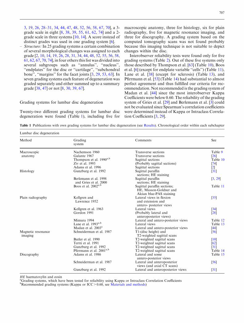
Table 1 Publications with own grading systems for lumbar disc degeneration (see Results). Chronological order within each subchapter
Lumbar disc degeneration
Method Gradingsystem
Comments See
Macroscopicanatomy
Nachemson 1960 Transverse sections Table 9Galante 1967 Transverse sections [26]Thompson et al. 1990a,b Sagittal sections Table 10Ziv et al. 1993 (Probably sagittal sections) [74]Adams et al. 1996 Sagittal sections [2]
Histology Gunzburg et al. 1992 Sagittal paraffinsections; HE staining
[31]
Berlemann et al. 1998and Gries et al. 2000
Sagittal paraffinsections; HE staining
[3, 29]
Boos et al. 2002a,b Sagittal paraffin sections;HE, Masson-Goldner andAlcian blue-PAS staining
Table 11
Plain radiography Kellgren andLawrence 1952
Lateral views in flexionand extension andantero- posterior views
[35]
Kellgren et al. 1963 Lateral views [34]Gordon 1991 (Probably lateral and
anteroposterior views)[28]
Mimura 1994 Lateral and antero-posterior views Table 12Lane et al. 1993a,b Lateral views Table 13Madan et al. 2003a Lateral and antero-posterior views [44]
Magnetic resonanceimaging
Schneiderman et al. 1987 T1-(disc height) andT2-weighted sagittal scans
[56]
Butler et al. 1990 T2-weighted sagittal scans [10]Tertti et al. 1991 T2-weighted sagittal scans [62]Gunzburg et al. 1992 T2-weighted sagittal scans [31]Pfirrmann et al. 2001a,b T2-weighted sagittal scans Table 14
Discography Adams et al. 1986 Lateral and someantero-posterior views
Table 15
Schneiderman et al. 1987 Lateral and anteroposteriorviews (and axial CT scans)
[56]
Gunzburg et al. 1992 Lateral and anteroposterior views [31]
HE haematoxylin and eosinaGrading systems, which have been tested for reliability using Kappa or Intraclass Correlation CoefficientsbRecommended grading systems (Kappa or ICC>0.60, see Materials and methods)
707
The clinical feasibility of the four statistically rec-ommended grading systems of Thompson et al. [63](Table 10), Boos et al. [6] (Table 11), Lane et al. [38](Table 13) and Pfirrmann et al. [53] (Table 14) variesconsiderably: it is the highest for the grading system ofLane et al. since the only material needed are lateralradiographs and the system itself has a clear structureand can very easily be applied. The grading system ofPfirrmann et al. also has a high clinical feasibility,however, magnetic resonance images are needed andsome more time is needed to arrive at the result. Theclinical relevance, however, is higher for the gradingsystem of Pfirrmann et al. than for that of Lane et al.since, on MRI images, the disc itself can directly beassessed, whereas on radiographs, only bony structuresare assessable. The grading systems of Thompson et al.[63] and Boos et al. [6], in contrast, have more academicthan clinical value since both systems can not be appliedon patients. However, both of them are based on thedetailed morphological studies, allow an in depth eval-uation of the disc and are therefore valuable tools tograde disc degeneration in vitro.
Besides these four statistically recommended gradingsystems, another three are of special interest: the mac-roscopic grading system of Nachemson [48] (Table 9),the radiographic system of Mimura et al. [47] (Table 12)
and the discographic system of Adams et al. [1](Table 15). Nachemson was one of the first to grade discdegeneration macroscopically. His grading system hasoften been used in the past. Mimura et al. were the firstwho tried to develop a more objective grading system. Itis more based on numbers such as the percentage de-crease in disc height than on subjective terms such as‘‘mild’’, ‘‘moderate’’, or ‘‘severe’’ . Adams et al. finallyneed to be mentioned since they count among the first todescribe the relationship between the distribution offluid injected into the disc and disc degeneration.
Grading systems for cervical disc degeneration
To grade cervical disc degeneration ten systems werefound (Table 3), two based on macroscopic anatomy,four using plain radiography, two using magnetic reso-nance imaging and two using discography. No histologi-cal or computed tomography grading systemswere found.
Interobserver reliability tests were found only for thegrading system of Kellgren et al. [34] (Tables 4, 16).Since the Kappa Coefficient was 0.71, this grading sys-tem fulfilled our criteria for recommendation.
From a clinical point of view this system has a goodfeasibility since it is based on the lateral radiographs and
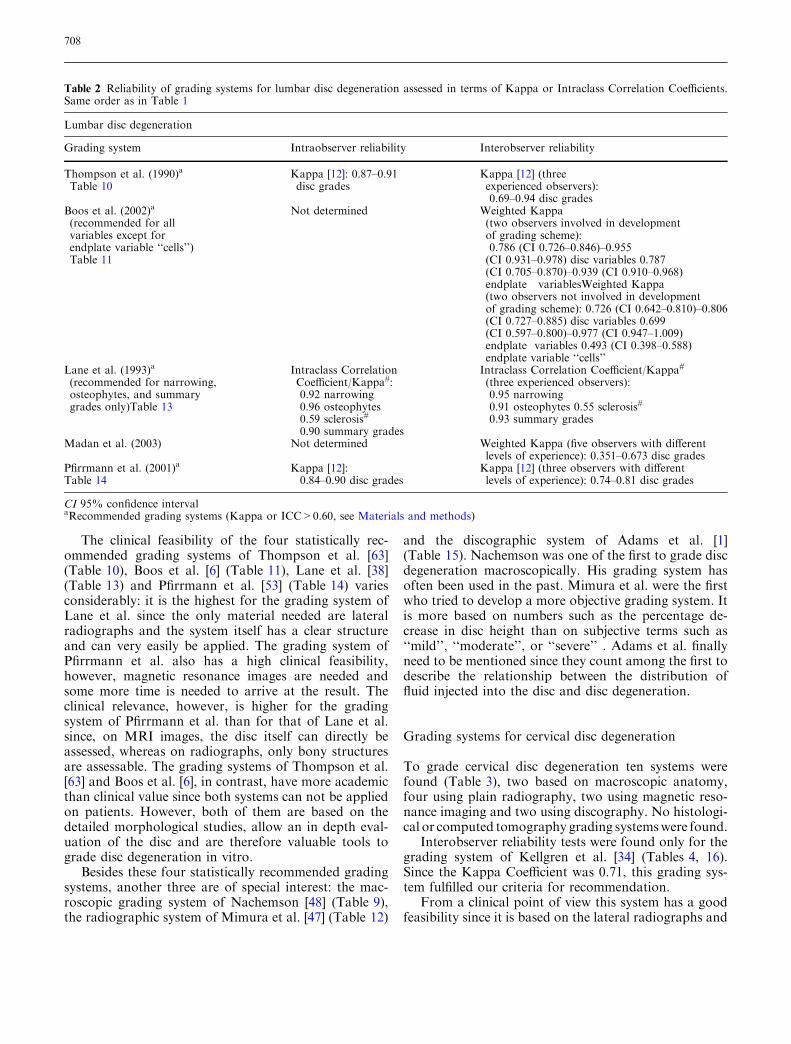
Table 2 Reliability of grading systems for lumbar disc degeneration assessed in terms of Kappa or Intraclass Correlation Coefficients.Same order as in Table 1
Lumbar disc degeneration
Grading system Intraobserver reliability Interobserver reliability
Thompson et al. (1990)a
Table 10Kappa [12]: 0.87–0.91disc grades
Kappa [12] (threeexperienced observers):0.69–0.94 disc grades
Boos et al. (2002)a
(recommended for allvariables except forendplate variable ‘‘cells’’)Table 11
Not determined Weighted Kappa(two observers involved in developmentof grading scheme):0.786 (CI 0.726–0.846)–0.955(CI 0.931–0.978) disc variables 0.787(CI 0.705–0.870)–0.939 (CI 0.910–0.968)endplate variablesWeighted Kappa(two observers not involved in developmentof grading scheme): 0.726 (CI 0.642–0.810)–0.806(CI 0.727–0.885) disc variables 0.699(CI 0.597–0.800)–0.977 (CI 0.947–1.009)endplate variables 0.493 (CI 0.398–0.588)endplate variable ‘‘cells’’
Lane et al. (1993)a
(recommended for narrowing,osteophytes, and summarygrades only)Table 13
Intraclass CorrelationCoefficient/Kappa#:0.92 narrowing0.96 osteophytes0.59 sclerosis#
0.90 summary grades
Intraclass Correlation Coefficient/Kappa#
(three experienced observers):0.95 narrowing0.91 osteophytes 0.55 sclerosis#
0.93 summary grades
Madan et al. (2003) Not determined Weighted Kappa (five observers with differentlevels of experience): 0.351–0.673 disc grades
Pfirrmann et al. (2001)a
Table 14Kappa [12]:0.84–0.90 disc grades
Kappa [12] (three observers with differentlevels of experience): 0.74–0.81 disc grades
CI 95% confidence intervalaRecommended grading systems (Kappa or ICC>0.60, see Materials and methods)
708

can easily be applied. In contrast, its clinical relevance issomewhat lower since the discs can only indirectly beassessed in terms of reactive changes of the surroundingbone and a loss of disc space height.
Grading systems for lumbar facet joint degeneration
Twelve grading systems for lumbar facet joint degener-ation were found via MEDLINE (Table 5). These sys-tems were either based on the macroscopic anatomy,histology, plain radiography, conventional tomography,computed tomography, or magnetic resonance imaging.
Interobserver reliability tests were found for five ofthese twelve grading systems (Table 6). As expected, theKappa or Intraclass Correlation Coefficients were gen-erally lower for the facet joints when compared to theintervertebral discs. Thus, only three grading systemsfulfilled the criteria for recommendation, even thoughthe threshold was lower than for the discs: Pathria et al.[52] (computed tomography only) (Table 17) and We-ishaupt et al. [70] (computed tomography and magneticresonance imaging) (Table 18). The Kappa Coefficientsof the grading systems described by Pathria et al. [52] forplain radiography, and Coste et al. [14] were below 0.40and should therefore not be used. The reliability of thegrading systems of Gries et al. [29] and Kellgren andLawrence [36] could not be assessed since linear corre-lation coefficients were reported instead of Kappa orICC.
From a clinical point of view the statistically rec-ommended grading systems of Pathria et al. [52] (Ta-ble 17) and Weishaupt et al. [70] (Table 18) are bothfeasible and relevant since they are based CT-scans ormagnetic resonance images. In contrast, from an aca-demic point of view, systems such as those of Tannoet al. [61] or Gries et al. [29] could be of interest sincethey allow to assess facet joint degeneration in vitroeither by macroscopic or histologic inspection. How-ever, these systems have not yet been tested for reli-ability.
Table 3 Publications with own grading systems for cervical disc degeneration (see Results). Chronological order within each subchapter
Cervical disc degeneration
Method Grading system Comments See
Macroscopicanatomy
Silberstein (1965) Inspection of surface,no sections
[58]
Viikari–Juntura et al. (1989) Mid-frontal sections [67]Histology No grading systems foundPlain radiography Kellgren and Lawrence (1952) Lateral views in
flexion and extension[35]
Kellgren et al. (1963)a,b
(tested for reliabilityby Cote et al. 1997)
Lateral views Table 16
Brooker and Barter (1965) Antero-posterior views,lateral views in neutralposition, flexion andextension, and bothoblique views
[8]
Silberstein (1965) Antero-posterior, lateraland both oblique views
[58]
Magnetic resonanceimaging
Viikari–Juntura et al. (1989) Sagittal scans [67]Lehto et al. (1994) T2-weighted scans [39]
Discography Viikari–Juntura et al. (1989) Poster-anterior, lateraland axial views
[67]
Schellhas et al. (1996) Antero-posterior and lateral views [55]
HE haematoxylin and eosinaGrading systems, which have been tested for reliability using Kappa or Intraclass Correlation CoefficientsbRecommended grading systems (Kappa or ICC>0.60, see Materials and methods)
Table 4 Reliability of grading systems for cervical disc degenera-tion assessed in terms of Kappa or Intraclass Correlation Coeffi-cients
Cervical disc degeneration
Gradingsystem
Intraobserverreliability
Interobserverreliability
Kellgren et al. (1963)a
(tested forreliabilityby Cote et al. 1997)Table 16 (and 19)
Notdetermined
Intraclass CorrelationCoefficient [21, 59](three experiencedobservers): 0.71(CI 0.23–0.88) discgrades (0.45 (CI 0.09–0.71)facet joint grades)
CI 95% confidence intervalaRecommended grading systems (Kappa or ICC>0.60, see Mate-rials and methods)
709

Grading systems for cervical facet joint degeneration
Cervical facet joint degeneration was only graded in fourarticles (Table 7). One of these four grading systems was
based on the macroscopic anatomy, and three on plainradiography. No histologic grading system and no sys-tems using conventional tomography, computedtomography or magnetic resonance imaging were found.
Table 5 Publications with own grading systems for lumbar facet joint degeneration (see Results). Chronological order within eachsubchapter
Lumbar facet joint degeneration
Method Gradingsystem
Comments See
Macroscopicanatomy
Ziv et al.(1993)
Inspection of joint surface afterIndia ink staining
[74]
Tanno et al. (2003) Inspection of jointsurface
[61]
Histology Gries et al. (2000) Paraffin sectionsperpendicular tothe joint line; HEstaining
[29]
Plainradiography
Kellgren andLawrence (1957)
Lateral views [36]
Pathria et al. (1987)a Both oblique views [52]Conventionaltomography
Demaerel et al. (1992) Both oblique views
Computedtomography
Pathria et al. (1987)a,b Axial scans (0� gantryangulation)
Table 17
Butler et al. (1990) ( probably axialscans)
[10]
Coste et al. (1994)a Axial scans (parallelto disc space)
[14]
Weishaupt et al. (1999)a,b Axial scans Table 18Magneticresonanceimaging
Grogan et al. (1997) T1- and T2-weightedaxial scans
[30]
Weishaupt et al. (1999)a,b T1- and T2-weightedsagittal, andT2-weighted axial scans
Table 18
HE haematoxylin and eosinaGrading systems, which have been tested for reliability using Kappa or Intraclass Correlation CoefficientsbRecommended grading systems (Kappa or ICC>0.40, see Materials and methods)
Table 6 Reliability of grading systems for lumbar facet joint degeneration assessed in terms of Kappa or Intraclass Correlation Coeffi-cients. Same order as in Table 5
Lumbar facet joint degeneration
Gradingsystem
Intraobserverreliability
Interobserver reliability
Pathria et al. (1987)a
(recommended forCT only) Table 17
Not determined Kappa (two experienced observers):0.46 facet joint grades on CT0.26 facet joint grades on oblique radiographs
Coste et al. (1994) Unweighted Kappa [21]:)0.03–0.34 right facet joints at L4-5 level0.30–0.74 right facet joints at L5-S1 level)0.06–0.48 left facet joints at L4-5 level0.26–0.68 left facet joints at L5-S1 level
Unweighted Kappa [21] (four experienced observers):0.02–0.09 right facet joints at L4-5 level0.11–0.39 right facet joints at L5-S1 level)0.11–0.10 left facet joints at L4-5 level0.10–0.39 left facet joints at L5-S1 level
Weishaupt et al. (1999)a
Table 18Weighted Kappa [37]:0.70 facet joint grades on MRI, observer 10.76 facet joint grades on MRI, observer 20.70 facet joint grades on CT, observer 10.77 facet joint grades on CT, observer 2
Weighted Kappa [37] (two experienced observers):0.41 facet joint grades on MRI0.60 facet joint grades on CT
CI 95% Confidence interval, MRI magnetic resonance imaging, CT computed tomographyaRecommended grading systems (Kappa or ICC>0.40, see Materials and methods)
710

The interobserver reliability of the grading system ofKellgren et al. [34] (Table 19) was only one fulfilling thecriteria for recommendation (Table 8). The systemdescribed by Kellgren and Lawrence [36] was only testedfor linear correlation. Thus, its reliability still needs to bedetermined.
From a clinical point of view the recommendedgrading system of Kellgren et al. [34] is feasible sinceonly lateral radiographs are needed.
Discussion
Forty-two different grading systems for cervical andlumbar disc and facet joint degeneration were accessiblefrom the MEDLINE database. This high number is notastonishing since grading systems are needed to answer avide variety of different questions. Several grading sys-tems were for example used to investigate the correlationbetween morphology and flexibility, intradiscal pressure,or pain [8, 19, 25, 31, 35, 47, 48, 55]. Others were neededto compare different diagnostic methods with each other[1, 3, 27, 52, 55, 56, 62, 67, 70], or to investigate therelation between the disc and facet joint degeneration[10, 29, 61, 74].
The majority of all 42 grading systems were used forthe lumbar but only few for the cervical spine. Histologicgrading systems, for example, were only found for thelumbar but not for the cervical spine. This also appliesto grading systems for facet joint degeneration using theconventional tomography, computed tomography ormagnetic resonance imaging. Thus, despite of their highnumber, new grading systems may still be needed espe-cially for the cervical spine.
Regardless of the spinal region the grading systemswere belonging to, the variability of their design wasvery high: Some systems started with the normal, notdegenerated state, others with the lowest degree ofdegeneration; some assigned the not degenerated stateto ‘‘grade 0’’, others to ‘‘grade 1’’; some were using a 5-grade scale, others a 4-grade, 3-grade, or 2-grade scale;some assigned linguistic terms such as ‘‘normal’’,‘‘mild’’, ‘‘severe’’ to each grade, some others numericalvalues (grade 0, grade 1, grade 2, ...). These and severalother variations make comparisons difficult and mayeasily lead to confusion. We would therefore recom-mend to define certain standards. Whether to start with‘‘grade 0’’ or ‘‘grade 1’’ or with the not degenerated orthe least degenerated state and whether to use linguisticterms or numerical values to grade degeneration couldeasily be standardised. Our suggestion would be to
Table 7 Publications with own grading systems for cervical facet joint degeneration (see Results). Chronological order within eachsubchapter
Cervical facet joint degeneration
Method Grading system Comments See
Macroscopic anatomy Silberstein (1965) Probably inspection of surface [58]Histology No grading systems foundPlain radiography Kellgren and Lawrence (1957) Lateral views [36]
Kellgren et al. (1963)a,b (testedfor reliability by Cote et al. 1997)
Lateral views Table 19
Silberstein (1965) Anteroposterior, lateral and both oblique views [58]Conventional tomography No grading systems foundComputed tomography No grading systems foundMagnetic resonance imaging No grading systems found
aGrading systems, which have been tested for reliability using Kappa or Intraclass Correlation CoefficientsbRecommended grading systems (Kappa or ICC>0.40, see Materials and methods)
Table 8 Reliability of grading systems for cervical facet joint degeneration assessed in terms of Kappa or Intraclass Correlation Coef-ficients
Cervical facet joint degeneration
Grading system Intraobserver reliability Interobserver reliability
Kellgren et al. 1963a (tested forreliability by Cote et al. 1997)Table (16 and) 19
Not determined Intraclass correlation coefficient [21, 59](three experienced observers):(0.71 (CI 0.23–0.88) disc grades)0.45 (CI 0.09–0.71) facet joint grades
CI 95% Confidence intervalaRecommended grading systems (Kappa or ICC>0.40, see Materials and methods)
711
begin the scale with the not degenerated state and toassign this state to ‘‘grade 0’’. This is what many au-thors already did. For the number of grades, however,it is critical to define a standard since this numbershould individually be adapted to the purpose of thestudy and the number of discs or facet joints, whichhave to be graded. A small number of discs or facetjoints should be divided into less degrees of degenera-tion than a large number since otherwise, the numberof individual discs or facet joints in each group ofdegeneration may become too small for further evalu-ation. Furthermore, the method used for grading alsoneeds to be considered since most methods have alimited resolution and only cover certain but not allstructures of the discs or facet joints. Thus, if thenumber of discs or facet joints is small and the methoddoes not allow a detailed inspection of all structuressuch as plain radiography, the use of only three or fourdegrees of degeneration can be recommended. If, incontrast, the number of discs or facet joints is high andmethods such as histology or macroscopy are used,which allow an in depth inspection of all tissues, up tofive degrees of degeneration can be recommended.However, in general less than three grades should notbe used since otherwise discs or facet joints with a widevariety of different appearances would all be mixedtogether within one and the same degree of degenera-tion. Sometimes it might even be advantageous not toreport an overall degree of degeneration but rather theappearance of the different structures individually, be-cause otherwise details concerning these individualstructures might get lost.
Reliability is one of the most important character-istics of a grading system. Intra- and interobserverreliability can be measured in terms of Kappa [12] orICC [57]. Unfortunately, only few of all reviewedgrading systems were tested for reliability. Out ofthese, nine grading systems fulfilled our criteria forrecommendation (Kappa or ICC>0.60 for discs and>0.40 for facet joints) but one for lumbar disc andtwo for lumbar facet joint degeneration did not(Tables 2, 4, 6, 8) [14, 44, 52]. These results indicatethat grading systems should always be tested for reli-ability before use. Thresholds for Kappa or ICC mighthelp to decide whether to use or not to use a testedsystem. General thresholds such as ours are suitable,if, for example, a general comparison between severalgrading systems is needed. However, if a specificgrading system is planned to be used, the thresholdsneed to be adjusted to the respective purpose of thestudy and to the degree of experience and training ofthe observers, who will use the system and who hadused it to determine its reliability. The thresholdsshould also be modified if templates will be or wereavailable and if the observers were involved in thedevelopment of the grading system.
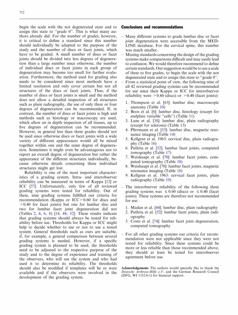
Conclusions and recommendations
– Many different systems to grade lumbar disc or facetjoint degeneration were accessible from the MED-LINE database. For the cervical spine, this numberwas much smaller.
– Missing standards concerning the design of the gradingsystems make comparisons difficult and may easily leadto confusion. We would therefore recommend to definecertain standards.Our suggestionwouldbe tousea scaleof three to five grades, to begin the scale with the notdegenerated state and to assign this state to ‘‘grade 0’’.
– From a statistical point of view, the following nine ofall 42 reviewed grading systems can be recommendedfor use since their Kappa or ICC for interobserverreliability were >0.60 (discs) or >0.40 (facet joints):
1. Thompson et al. [63]: lumbar disc, macroscopicanatomy (Table 10)
2. Boos et al. [6]: lumbar disc, histology (except forendplate variable ‘‘cells’’) (Table 11)
3. Lane et al. [38]: lumbar disc, plain radiography(except for sclerosis) (Table 13)
4. Pfirrmann et al. [53]: lumbar disc, magnetic reso-nance imaging (Table 14)
5. Kellgren et al. 1963: cervical disc, plain radiogra-phy (Table 16)
6. Pathria et al. [52]: lumbar facet joints, computedtomography (Table 17)
7. Weishaupt et al. [70]: lumbar facet joints, com-puted tomography (Table 18)
8. Weishaupt et al. [70]: lumbar facet joints, magneticresonance imaging (Table 18)
9. Kellgren et al. 1963: cervical facet joints, plainradiography (Table 19)
– The interobserver reliability of the following threegrading systems was £ 0.60 (discs) or £ 0.40 (facetjoints). These systems are therefore not recommendedfor use:
1. Madan et al. [44]: lumbar disc, plain radiography2. Pathria et al. [52]: lumbar facet joints, plain radi-
ography3. Coste et al. [74]: lumbar facet joint degeneration,
computed tomography
– For all other grading systems our criteria for recom-mendation were not applicable since they were nottested for reliability. Since these systems could bemore or less reliable than those recommended above,they should at least be tested for interobserveragreement before use.
Acknowledgements The authors would specially like to thank theDeutsche Arthrose-Hilfe e.V. and the German Research Council(DFG, WI 1352/6-1) for financial support.
712
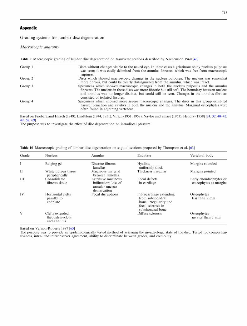
Table 9 Macroscopic grading of lumbar disc degeneration on transverse sections described by Nachemson 1960 [48]
Group 1 Discs without changes visible to the naked eye. In these cases a gelatinous shiny nucleus pulposuswas seen; it was easily delimited from the annulus fibrosus, which was free from macroscopicruptures.
Group 2 Discs which showed macroscopic changes in the nucleus pulposus. The nucleus was somewhatmore fibrous, but could be clearly distinguished from the annulus, which was intact.
Group 3 Specimens which showed macroscopic changes in both the nucleus pulposus and the annulusfibrosus. The nucleus in these discs was more fibrotic but still soft. The boundary between nucleusand annulus was no longer distinct, but could still be seen. Changes in the annulus fibrosusconsisted of isolated fissures.
Group 4 Specimens which showed more severe macroscopic changes. The discs in this group exhibitedfissure formation and cavities in both the nucleus and the annulus. Marginal osteophytes wereoften found in adjoining vertebrae.
Based on Frieberg and Hirsch (1949), Lindblom (1944, 1951), Virgin (1951, 1958), Naylor and Smare (1953), Hendry (1958) [24, 32, 40–42,49, 68, 69]The purpose was to investigate the effect of disc degeneration on intradiscal pressure
Table 10 Macroscopic grading of lumbar disc degeneration on sagittal sections proposed by Thompson et al. [63]
Grade Nucleus Annulus Endplate Vertebral body
I Bulging gel Discrete fibrouslamellas
Hyaline,uniformly thick
Margins rounded
II White fibrous tissueperipherically
Mucinous materialbetween lamellas
Thickness irregular Margins pointed
III Consolidatedfibrous tissue
Extensive mucinousinfiltration; loss ofannular-nucleardemarcation
Focal defectsin cartilage
Early chondrophytes orosteophytes at margins
IV Horizontal cleftsparallel toendplate
Focal disruptions Fibrocartilage extendingfrom subchondralbone; irregularity andfocal sclerosis insubchondral bone
Osteophytesless than 2 mm
V Clefts extendedthrough nucleusand annulus
Diffuse sclerosis Osteophytesgreater than 2 mm
Based on Vernon-Roberts 1987 [65]The purpose was to provide an epidemiologically tested method of assessing the morphologic state of the disc. Tested for comprehen-siveness, intra- and interobserver agreement, ability to discriminate between grades, and credibility
Appendix
Grading systems for lumbar disc degeneration
Macroscopic anatomy
713
Histology
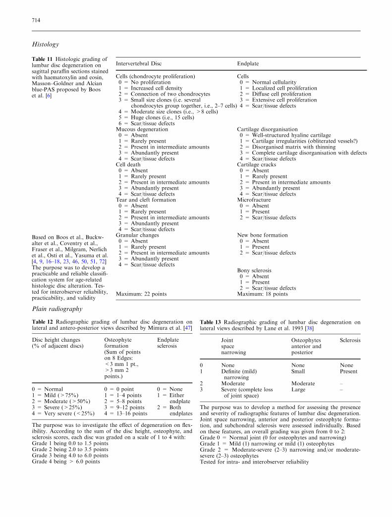
Table 11 Histologic grading oflumbar disc degeneration onsagittal paraffin sections stainedwith haematoxylin and eosin,Masson–Goldner and Alcianblue-PAS proposed by Booset al. [6]
Based on Boos et al., Buckw-alter et al., Coventry et al.,Fraser et al., Milgram, Nerlichet al., Osti et al., Yasuma et al.[4, 9, 16–18, 23, 46, 50, 51, 72]The purpose was to develop apracticable and reliable classifi-cation system for age-relatedhistologic disc alteration. Tes-ted for interobserver reliability,practicability, and validity
Intervertebral Disc Endplate
Cells (chondrocyte proliferation)0 = No proliferation1 = Increased cell density2 = Connection of two chondrocytes3 = Small size clones (i.e. several
chondrocytes group together, i.e., 2–7 cells)4 = Moderate size clones (i.e., >8 cells)5 = Huge clones (i.e., 15 cells)6 = Scar/tissue defects
Cells0 = Normal cellularity1 = Localized cell proliferation2 = Diffuse cell proliferation3 = Extensive cell proliferation4 = Scar/tissue defects
Mucous degeneration0 = Absent1 = Rarely present2 = Present in intermediate amounts3 = Abundantly present4 = Scar/tissue defects
Cartilage disorganisation0 = Well-structured hyaline cartilage1 = Cartilage irregularities (obliterated vessels?)2 = Disorganised matrix with thinning3 = Complete cartilage disorganisation with defects4 = Scar/tissue defects
Cell death0 = Absent1 = Rarely present2 = Present in intermediate amounts3 = Abundantly present4 = Scar/tissue defects
Cartilage cracks0 = Absent1 = Rarely present2 = Present in intermediate amounts3 = Abundantly present4 = Scar/tissue defects
Tear and cleft formation0 = Absent1 = Rarely present2 = Present in intermediate amounts3 = Abundantly present4 = Scar/tissue defects
Microfracture0 = Absent1 = Present2 = Scar/tissue defects
Granular changes0 = Absent1 = Rarely present2 = Present in intermediate amounts3 = Abundantly present4 = Scar/tissue defects
New bone formation0 = Absent1 = Present2 = Scar/tissue defects
Bony sclerosis0 = Absent1 = Present2 = Scar/tissue defects
Maximum: 22 points Maximum: 18 points
Table 12 Radiographic grading of lumbar disc degeneration onlateral and antero-posterior views described by Mimura et al. [47]
Disc height changes(% of adjacent discs)
Osteophyteformation(Sum of pointson 8 Edges:<3 mm 1 pt.,>3 mm 2points.)
Endplatesclerosis
0 = Normal1 = Mild (>75%)2 = Moderate (>50%)3 = Severe (>25%)4 = Very severe (<25%)
0 = 0 point1 = 1–4 points2 = 5–8 points3 = 9–12 points4 = 13–16 points
0 = None1 = Either
endplate2 = Both
endplates
The purpose was to investigate the effect of degeneration on flex-ibility. According to the sum of the disc height, osteophyte, andsclerosis scores, each disc was graded on a scale of 1 to 4 with:Grade 1 being 0.0 to 1.5 pointsGrade 2 being 2.0 to 3.5 pointsGrade 3 being 4.0 to 6.0 pointsGrade 4 being > 6.0 points
Table 13 Radiographic grading of lumbar disc degeneration onlateral views described by Lane et al. 1993 [38]
Jointspacenarrowing
Osteophytesanterior andposterior
Sclerosis
0 None None None1 Definite (mild)
narrowingSmall Present
2 Moderate Moderate –3 Severe (complete loss
of joint space)Large –
The purpose was to develop a method for assessing the presenceand severity of radiographic features of lumbar disc degeneration.Joint space narrowing, anterior and posterior osteophyte forma-tion, and subchondral sclerosis were assessed individually. Basedon these features, an overall grading was given from 0 to 2:Grade 0 = Normal joint (0 for osteophytes and narrowing)Grade 1 = Mild (1) narrowing or mild (1) osteophytesGrade 2 = Moderate-severe (2–3) narrowing and/or moderate-severe (2–3) osteophytesTested for intra- and interobserver reliability
Plain radiography
714

Magnetic resonance imaging
Table 14 Grading of lumbar disc degeneration on T2-weightedsagittal MRI-scans proposed by Pfirrmann et al. [53]
Grade Structure Distinctionof Nucleusand Annulus
Signalintensity
Height ofIntervertebralDisc
I Homogeneous,bright white
Clear Hyperintense,isointense tocerebrospinalfluid
Normal
II Inhomogeneouswith or withouthorizontal bands
Clear Hyperintense,isointense tocerebrospinalfluid
Normal
III Inhomogeneous,grey
Unclear Intermediate Normal toslightlydecreased
IV Inhomogeneous,grey to black
Lost Intermediateto hypointense
Normal tomoderatelydecreased
V Inhomogeneous,black
Lost Hypointense Collapseddisc space
Based on Boos et al., Brant-Zawadtzki et al., Masaryk et al.,Thompson et al. [5, 7, 45, 63]The purpose was to develop an applicable, reliable classificationsystem. Tested for intra- and interobserver reliability and ability todistinct between grades
Table 15 Discographic grading of lumbar disc degeneration in thesagittal plane proposed by Adams et al. [1]
Discogram type Stage of disc degeneration
1. Cottonball No signs of degeneration.Soft, white amorphous nucleus
2. Lobular Mature disc with nucleus startingto coalesce into fibrous lumps
3. Irregular Degenerated disc with fissuresand clefts in the nucleus andinner annulus
4. Fissured Degenerated disc with radialfissure leading to the outer edgeof the annulus
5. Rupture Disc has a complete radial fissurethat allows injected fluid to escape.Can be in any state of degeneration
The purpose was to depict the stages of disc degeneration by meansof discograms
Table 16 Radiographic grading of cervical disc degeneration onlateral views described by Kellgren et al. [34]
Grade 1 Minimal anterior osteophytosisGrade 2 Definite anterior osteophytosis
with possible narrowing of thedisc space and some sclerosisof vertebral plates
Grade 3 Moderate narrowing of thedisc space with definitesclerosis of vertebral platesand osteophytosis
Grade 4 Severe narrowing of the discspace with sclerosis of vertebralplates and multiple large osteophytes
The purpose was to present some agreed system of grading radio-logical changes in the joints. Tested for interobserver reliability byCote et al. [15], who added an additional Grade 0 = Absence ofdegeneration in the disc
Grading systems for cervical disc degeneration
Plain radiography
Grading systems for lumbar facet joint degeneration
Computed tomography
DiscographyTable 17 Grading of lumbar facet joint degeneration on obliqueconventional radiographs and CT scans described by Pathria et al.[52]
Grade 0 NormalGrade 1 Joint space narrowing
(mild degenerative disease)Grade 2 Narrowing plus sclerosis
or hypertrophy (moderatedegenerative disease)
Grade 3 Severe osteoarthrosis withnarrowing, sclerosis, andosteophytes (severedegenerative disease)
The purpose was to assess the accuracy of oblique radiographs inthe detection of lumbar facet joint osteoarthritis. Tested for inte-robserver reliability
715
References
1. Adams MA, Dolan P, Hutton WC(1986) The stages of disc degenerationas revealed by discograms. J Bone JointSurg Br 68(1):36–41
2. Adams MA, McNally DS, Dolan P(1996) ’Stress’ distributions insideintervertebral discs. The effects of ageand degeneration. J Bone Joint Surg Br78(6):965–972
3. Berlemann U, Gries NC, Moore RJ(1998) The relationship between height,shape and histological changes in earlydegeneration of the lower lumbar discs.Eur Spine J 7(3):212–217
4. Boos N, Nerlich AG, Wiest I, von derMark K, Aebi M (1997) Immunolocal-ization of type X collagen in humanlumbar intervertebral discs during age-ing and degeneration. Histochem CellBiol 108(6):471–480
5. Boos N, Wallin A, Schmucker T, AebiM, Boesch C (1994) Quantitative MRimaging of lumbar intervertebral discand vertebral bodies: methodology,reproducibility, and preliminary re-sults. Magn Reson Imaging 12(4):577–587
6. Boos N, Weissbach S, Rohrbach H,Weiler C, Spratt KF, Nerlich AG (2002)Classification of age-related changes inlumbar intervertebral discs: 2002 VolvoAward in basic science. Spine27(23):2631–2644
7. Brant-Zawadzki MN, Jensen MC,Obuchowski N, Ross JS, Modic MT(1995) Interobserver and intraobservervariability in interpretation of lumbardisc abnormalities. A comparison oftwo nomenclatures (discussion 1264).Spine 20(11):1257–1263
8. Brooker AE, Barter RW (1965) Cer-vical spondylosis. A clinical study withcomparative radiology. Brain88(5):925–936
9. Buckwalter JA (1995) Aging anddegeneration of the human interverte-bral disc. Spine 20(11):1307–1314
10. Butler D, Trafimow JH, Andersson GB,McNeill TW, Huckman MS (1990)Discs degenerate before facets. Spine15(2):111–113
11. Carrera GF, Haughton VM, SyvertsenA, Williams AL (1980) Computedtomography of the lumbar facet joints.Radiology 134(1):145–148
12. Cohen J (1968) Weighted kappa: nom-inal scale agreement with provision forscaled disagreement or partial credit.Psychol Bull 70:213–220
13. Collins DH (1949) The pathology ofarticular and spinal diseases. EdwardArnold & Co, London
Magnetic resonance imaging
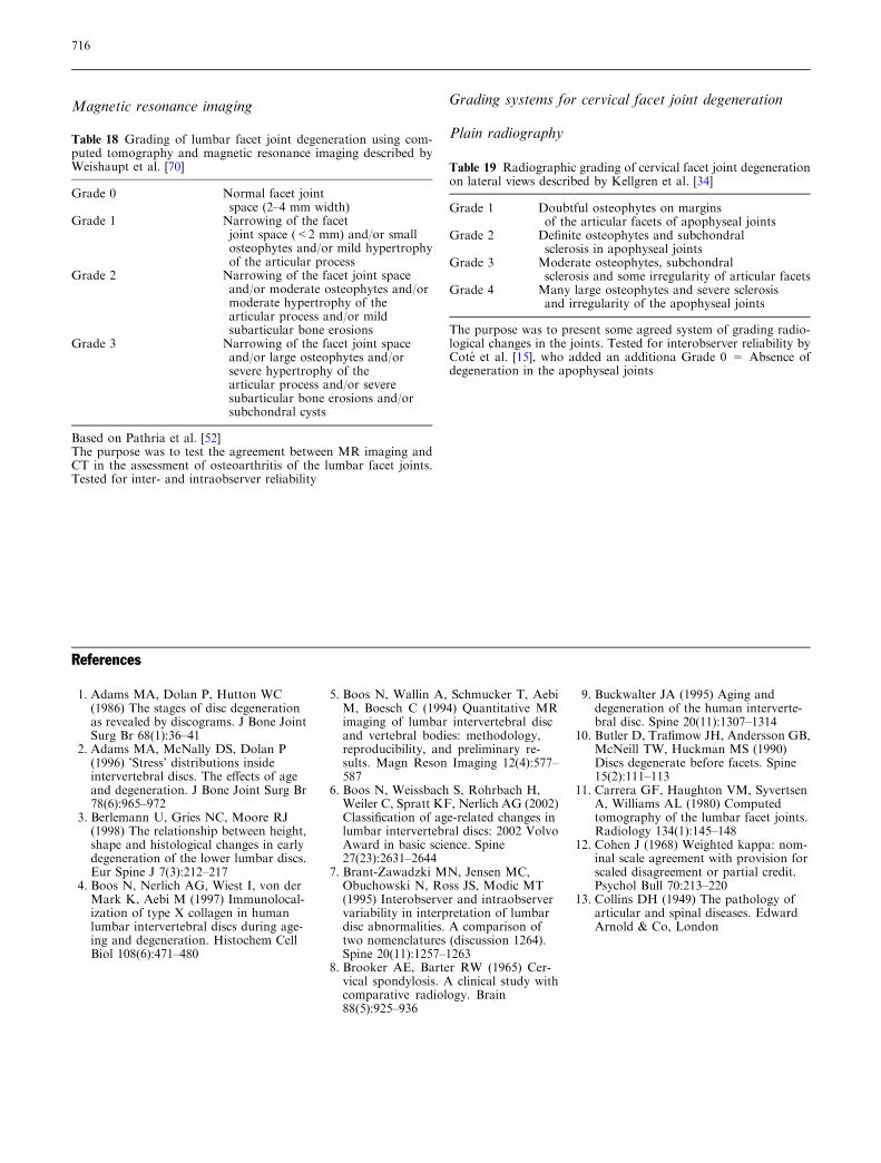
Table 18 Grading of lumbar facet joint degeneration using com-puted tomography and magnetic resonance imaging described byWeishaupt et al. [70]
Grade 0 Normal facet jointspace (2–4 mm width)
Grade 1 Narrowing of the facetjoint space (<2 mm) and/or smallosteophytes and/or mild hypertrophyof the articular process
Grade 2 Narrowing of the facet joint spaceand/or moderate osteophytes and/ormoderate hypertrophy of thearticular process and/or mildsubarticular bone erosions
Grade 3 Narrowing of the facet joint spaceand/or large osteophytes and/orsevere hypertrophy of thearticular process and/or severesubarticular bone erosions and/orsubchondral cysts
Based on Pathria et al. [52]The purpose was to test the agreement between MR imaging andCT in the assessment of osteoarthritis of the lumbar facet joints.Tested for inter- and intraobserver reliability
Grading systems for cervical facet joint degeneration
Plain radiography
Table 19 Radiographic grading of cervical facet joint degenerationon lateral views described by Kellgren et al. [34]
Grade 1 Doubtful osteophytes on marginsof the articular facets of apophyseal joints
Grade 2 Definite osteophytes and subchondralsclerosis in apophyseal joints
Grade 3 Moderate osteophytes, subchondralsclerosis and some irregularity of articular facets
Grade 4 Many large osteophytes and severe sclerosisand irregularity of the apophyseal joints
The purpose was to present some agreed system of grading radio-logical changes in the joints. Tested for interobserver reliability byCote et al. [15], who added an additiona Grade 0 = Absence ofdegeneration in the apophyseal joints
716
14. Coste J, Judet O, Barre O, Siaud JR,Cohen de Lara A, Paolaggi JB (1994)Inter- and intraobserver variability inthe interpretation of computed tomog-raphy of the lumbar spine. J Clin Epi-demiol 47(4):375–381
15. Cote P, Cassidy JD, Yong-Hing K,Sibley J, Loewy J (1997) Apophysialjoint degeneration, disc degeneration,and sagittal curve of the cervical spine.Can they be measured reliably onradiographs? Spine 22(8):859–864
16. Coventry MB (1945) The intervertebraldisc: Its macroscopic anatomy andpathology: Part III. Pathological chan-ges in the intervertebral disc lesion. JBone Joint Surg [Am] 27:460–473
17. Coventry MB (1945) The intervertebraldisc: Its microscopic anatomy andpathology: Part I. Anatomy, develop-ment and pathology. J Bone Joint Surg[Am] 27:105–112
18. Coventry MB (1945) The interverte-bral disc: Its microscopic anatomy andpathology: Part II. Changes in theintervertebral disc concomitant withage. J Bone Joint Surg [Am] 27:233–247
19. Demaerel P, Wilms G, Goffin J, BaertAL (1992) Osteoarthritis of the facetjoints and its role in low-back pain:evaluation with conventional tomogra-phy. J Belge Radiol 75(2):81–86
20. Eisenstein SM, Parry CR (1987) Thelumbar facet arthrosis syndrome. Clini-cal presentation and articular surfacechanges. J Bone Joint Surg Br 69(1):3–7
21. Fleiss JL (1981) Statistical methods forrates and proportions, 2nd edn. Wiley,New York
22. Fletcher G, Haughton VM, Ho KC, YuSW (1990) Age-related changes in thecervical facet joints: studies with cryo-microtomy, MR, and CT. AJR Am JRoentgenol 154(4):817–820
23. Fraser RD, Osti OL, Vernon-Roberts B(1993) Intervertebral disc degeneration.Eur Spine J 1:205–213
24. Friberg S, Hirsch C (1949) Anatomicaland clinical studies on lumbar discdegeneration. Acta Orthop Scand19:222–242
25. Friedenberg ZB, Miller WT (1963)Degenerative disc disease of the cervi-cal spine. J Bone Joint Surg Am45:1171–8
26. Galante JO (1967) Tensile properties ofthe human lumbar annulus fibrosus.Acta Orthop Scand (Suppl100):1–91
27. Gibson MJ, Buckley J, Mawhinney R,Mulholland RC, Worthington BS(1986) Magnetic resonance imaging anddiscography in the diagnosis of discdegeneration. A comparative study of50 discs. J Bone Joint Surg Br68(3):369–373
28. Gordon SJ, Yang KH, Mayer PJ, MaceAH Jr, Kish VL, Radin EL (1991)Mechanism of disc rupture. A pre-liminary report. Spine 16(4):450–456
29. Gries NC, Berlemann U, Moore RJ,Vernon-Roberts B (2000) Early histo-logic changes in lower lumbar discs andfacet joints and their correlation. EurSpine J 9(1):23–29
30. Grogan J, Nowicki BH, Schmidt TA,Haughton VM (1997) Lumbar facetjoint tropism does not acceleratedegeneration of the facet joints. AJNRAm J Neuroradiol 18(7):1325–1329
31. Gunzburg R, Parkinson R, Moore Ret al (1992) A cadaveric study compar-ing discography, magnetic resonanceimaging, histology, and mechanicalbehavior of the human lumbar disc.Spine 17(4):417–426
32. Hendry NG (1958) The hydration of thenucleus pulposus and its relation tointervertebral disc derangement. J BoneJoint Surg Br 40B(1):132–144
33. Hirsch C (1972) Some morphologicalchanges in the cervical spine duringageing. In: Hirsch C, Zotterman Y (eds)Cervical pain. Pergamon Press, NewYork, pp 21–32
34. Kellgren JH, Jeffrey MR, Ball J (1963)The epidemiology of chronic rheuma-tism. vol II: Atlas of standard radio-graphs of arthritis. Blackwell ScientificPublications, Oxford, pp 14–19
35. Kellgren JH, Lawrence JS (1952)Rheumatism in miners. II. X-ray study.Br J Ind Med 9(3):197–207
36. Kellgren JH, Lawrence JS (1957)Radiological assessment of osteo-arthrosis. Ann Rheum Dis 16(4):494–502
37. Landis JR, Koch GG (1977) The mea-surement of observer agreement forcategorical data. Biometrics 33(1):159–174
38. Lane NE, Nevitt MC, Genant HK,Hochberg MC (1993) Reliability of newindices of radiographic osteoarthritis ofthe hand and hip and lumbar discdegeneration. J Rheumatol20(11):1911–1918
39. Lehto IJ, Tertti MO, Komu ME, Pa-ajanen HE, Tuominen J, Kormano MJ(1994) Age-related MRI changes at 0.1T in cervical discs in asymptomaticsubjects. Neuroradiology 36(1):49–53
40. Lindblom K (1944) Protrusions of discsand nerve compression in the lumbarregion. Acta Radiol 25:195
41. Lindblom K (1951) Backache and itsrelation to ruptures of the intravertebraldisks. Radiology 57(5):710–719
42. Lindblom K (1951) Discography ofdissecting transosseous ruptures ofintervertebral discs in the lumbar re-gion. Acta Radiol 36(1):12–16
43. Macnab I (1975) Cervical spondylosis.Clin Orthop (109):69–77
44. Madan SS, Rai A, Harley JM (2003)Interobserver error in interpretation ofthe radiographs for degeneration of thelumbar spine. Iowa Orthop J 23:51–56
45. Masaryk TJ, Ross JS, Modic MT,Boumphrey F, Bohlman H, Wilber G(1988) High-resolution MR imaging ofsequestered lumbar intervertebral disks.AJR Am J Roentgenol 150(5):1155–1162
46. Milgram J (1990) Intervertebral discdisease and degenerative arthritis of thespine. In: Milgram J (ed) Radiologicand histologic pathology of nontumor-ous dieseases of bone and joints.Northbrook Publishing Company, pp519–588
47. Mimura M, Panjabi MM, Oxland TR,Crisco JJ, Yamamoto I, Vasavada A(1994) Disc degeneration affects themultidirectional flexibility of the lumbarspine. Spine 19(12):1371–1380
48. Nachemson A (1960) Lumbar Intradi-scal Pressure. Experimental studies onpost-mortem material. Acta OrthopScand Suppl 43:43
49. Naylor A, Smare DL (1953) Fluidcontent of the nucleus pulposus as afactor in the disk syndrome; preliminaryreport. Br Med J 4843:975–976
50. Nerlich AG, Schleicher ED, Boos N(1997) 1997 Volvo Award winner inbasic science studies. Immunohistologicmarkers for age-related changes of hu-man lumbar intervertebral discs. Spine22(24):2781–2795
51. Osti OL, Vernon-Roberts B, Fraser RD(1990) 1990 Volvo Award in experi-mental studies. Anulus tears and inter-vertebral disc degeneration. Anexperimental study using an animalmodel. Spine 15(8):762–767
52. Pathria M, Sartoris DJ, Resnick D(1987) Osteoarthritis of the facet joints:accuracy of oblique radiographicassessment. Radiology 164(1):227–230
53. Pfirrmann CW, Metzdorf A, Zanetti M,Hodler J, Boos N (2001) Magnetic res-onance classification of lumbar inter-vertebral disc degeneration. Spine26(17):1873–1878
54. Resnick D (1985) Degenerative diseasesof the vertebral column. Radiology156(1):3–14
717
55. Schellhas KP, Smith MD, Gundry CR,Pollei SR (1996) Cervical discogenicpain. Prospective correlation of mag-netic resonance imaging and discogra-phy in asymptomatic subjects and painsufferers (discussion 311–312). Spine21(3):300–311
56. Schneiderman G, Flannigan B, Kings-ton S, Thomas J, Dillin WH, WatkinsRG (1987) Magnetic resonance imagingin the diagnosis of disc degeneration:correlation with discography. Spine12(3):276–281
57. Shrout PE, Fleiss J (1979) Intraclasscorrelations: Uses in Assessing RaterReliability. Psychol Bull 86(2):420–428
58. Silberstein CE (1965) The Evolution ofDegenerative Changes in the CervicalSpine and an Investigation into the‘‘Joints of Luschka’’. Clin Orthop40:184–204
59. Streiner DL, Norman GL (1989) Healthmeasurement scales: a practical guide totheir development and use. OxfordUniversity Press, Oxford
60. Swanepoel MW, Adams LM, Smea-thers JE (1995) Human lumbar apoph-yseal joint damage and intervertebraldisc degeneration. Ann Rheum Dis54(3):182–188
61. Tanno I, Oguma H, Murakami G, SatoS, Yamashita T (2003) Which portion ina facet is specifically affected by articu-lar cartilage degeneration with aging inthe human lumbar zygapophysial joint?Okajimas Folia Anat Jpn 80(1):29–34
62. Tertti M, Paajanen H, Laato M, AhoH, Komu M, Kormano M (1991) Discdegeneration in magnetic resonanceimaging. A comparative biochemical,histologic, and radiologic study in ca-daver spines. Spine 16(6):629–634
63. Thompson JP, Pearce RH, SchechterMT, Adams ME, Tsang IK, Bishop PB(1990) Preliminary evaluation of ascheme for grading the gross morphol-ogy of the human intervertebral disc.Spine 15(5):411–415
64. Tondury G (1972) The behaviour of thecervical discs during life. In: Hirsch C,Zotterman Y (eds) Cervical pain. Perg-amon Press, New York, pp 59–66
65. Vernon-Roberts B (1987) Pathology ofintervertebral discs and apophysealjoints. The lumbar spine and back pain.Churchill-Livingstone, London, pp 37–55
66. Vernon-Roberts B, Pirie CJ (1977)Degenerative changes in the interverte-bral discs of the lumbar spine and theirsequelae. Rheumatol Rehabil 16(1):13–21
67. Viikari-Juntura E, Raininko R, Vid-eman T, Porkka L (1989) Evaluation ofcervical disc degeneration with ultralowfield MRI and discography. An experi-mental study on cadavers. Spine14(6):616–619
68. Virgin WJ (1951) Experimental investi-gations into the physical properties ofthe intervertebral disc. J Bone JointSurg Br 33B(4):607–611
69. Virgin WJ (1958) Anatomical andpathological aspects of the interverte-bral disc. Indian J Surg 20:113
70. Weishaupt D, Zanetti M, Boos N, Ho-dler J (1999) MR imaging and CT inosteoarthritis of the lumbar facet joints.Skeletal Radiol 28(4):215–219
71. Wybier M (2001) Imaging of lumbardegenerative changes involving struc-tures other than disk space. Radiol ClinNorth Am 39(1):101–114
72. Yasuma T, Koh S, Okamura T, Ya-mauchi Y (1990) Histological changesin aging lumbar intervertebral discs.Their role in protrusions and prolapses.J Bone Joint Surg Am 72(2):220–229
73. Yu S, Haughton VM, Sether LA, HoKC, Wagner M (1989) Criteria forclassifying normal and degeneratedlumbar intervertebral disks. Radiology170(2):523–536
74. Ziv I, Maroudas C, Robin G, Marou-das A (1993) Human facet cartilage:swelling and some physicochemicalcharacteristics as a function of age. Part2: age changes in some biophysicalparameters of human facet joint carti-lage. Spine 18(1):136–146
718