Reverse Transcriptase of Moloney Murine Leukemia Virus Binds to Eukaryotic Release Factor 1 to...
13
Cell, Vol. 115, 319–331, October 31, 2003, Copyright 2003 by Cell Press Reverse Transcriptase of Moloney Murine Leukemia Virus Binds to Eukaryotic Release Factor 1 to Modulate Suppression of Translational Termination quences (Jacks, 1990; Jacks et al., 1988b; Jacks and Varmus, 1985). While most ribosomes read the RNA to produce the Gag protein, a small proportion of the ribosomes undergo a minus-one frameshift event and continue in the new reading frame so as to bypass the Marianna Orlova, Andrew Yueh, Juliana Leung, and Stephen P. Goff* Department of Biochemistry and Molecular Biophysics Integrated Program in Cell and Molecular Biology stop codon and produce the longer Gag-Pol protein. In Howard Hughes Medical Institute other retroviruses such as the murine and feline leuke- Columbia University College of Physicians & mia viruses, however, a distinct mechanism is used. The Surgeons gag and pol genes are in the same reading frame and New York, New York 10032 are separated by a UAG stop codon (Shinnick et al., 1981; Yoshinaka et al., 1985b). While most ribosomes terminate synthesis at the stop codon to produce the Summary Gag protein, termination is sometimes suppressed (Hat- field et al., 1992; Murphy and Arlinghaus, 1978; Murphy The pol (for polymerase) gene of the murine leukemia et al., 1978, 1980; Philipson et al., 1978); ribosomes viruses (MuLVs) is expressed in the form of a large incorporate a glutamine residue at the stop codon and Gag-Pol precursor protein by the suppression of trans- continue elongation to form the Gag-Pol product (Yoshi- lational termination, or enhanced readthrough, of a naka et al., 1985a). Translation can also pass through UAG stop codon at the end of gag. A search for cellular UAA or UGA stop codons (Feng et al., 1989b), incorpo- proteins that interact with the reverse transcriptase rating other amino acids at these codons (Feng et al., of Moloney MuLV resulted in the identification of eRF1, 1990). The correct ratio of Gag to Gag-Pol proteins, the eukaryotic translation release factor 1. The pro- whether achieved by frameshifting or suppression of teins bound strongly in vitro, and the overexpression termination, is crucial to the formation of infectious virus of eRF1 resulted in the RT-dependent incorporation particles (Felsenstein and Goff, 1988). of the protein into assembling virion particles. The These processes of frameshifting and suppression overexpression of RT in trans enhanced the transla- of termination are largely controlled by sequences and tional readthrough of a reporter construct containing structures in the retroviral RNA itself (ten Dam et al., the Gag-Pol boundary region. Noninteracting mutants 1990). The frameshift event occurs at a homopolymeric of RT failed to synthesize adequate levels of Gag-Pol sequence called a “slippery site,” and requires a specific and could not replicate. These results suggest that pseudoknot structure downstream of the site (Jacks, RT enhances suppression of termination and that the 1990; Jacks et al., 1988a, 1988b). Suppression of termi- interaction of RT with eRF1 is required for an appro- nation requires a few specific nucleotides immediately priate level of translational readthrough. past the UAG and a pseudoknot (Alam et al., 1999; Felsenstein and Goff, 1992; Feng et al., 1992; Honigman Introduction et al., 1991; Wills et al., 1991). No changes in the pool of aminoacyl tRNAs, or any particular viral proteins, are Reverse transcriptase (RT), encoded by the retroviral required for these events. Frameshifting and transla- pol gene, plays a defining role in the virus life cycle in tional suppression occur even during translation of viral directing the synthesis of viral DNA from RNA (see RNAs in vitro, and when the translation reactions are Skalka and Goff, 1993, for review). RT exhibits two enzy- prepared from either uninfected or virally infected cell matic activities residing in separate domains (Tanese lysates (Feng et al., 1989a; Panganiban, 1988). It is not and Goff, 1988): a DNA polymerase active on RNA and known if the efficiency of readthrough, however, might DNA templates, and an RNase H able to digest RNA in be subtly altered by infection. One report of such a RNA:DNA hybrid form. These two activities of RT medi- change (Kuchino et al., 1987) has not been confirmed. ate the complex process of reverse transcription that The termination of translation at stop codons in eu- results in the formation of a linear double-stranded DNA karyotes is mediated by a complex containing the from the single-stranded RNA genome in the cytoplasm eukaryotic release factors eRF1 and eRF3 (Frolova et of the infected cell (Telesnitsky and Goff, 1997). al., 1994; Zhouravleva et al., 1995; for review see Kis- RT is synthesized as part of a Gag-Pol fusion protein selev and Buckingham, 2000). The process of termina- (Jamjoom et al., 1977). Translation of the viral genomic tion and polypeptide release involves the recognition of RNA results in the formation of both a Gag precursor the stop codon by eRF1 and is accompanied by the protein and, at 10- or 20-fold lower levels, a larger Gag- hydrolysis of GTP by eRF3 (Frolova et al., 1996). The Pol protein (Hatfield et al., 1992; for review, see Levin levels of eRF1 are important in termination; overexpress- et al. 1993). In some retroviruses, such as the avian ing eRF1 reduces readthrough (Drugeon et al., 1997; Le leukosis and human immunodeficiency viruses, the gag Goff et al., 1997), and depleting eRF1 in yeast increases and pol genes are in different reading frames, and the readthrough (Stansfield et al., 1996). The structure of formation of Gag-Pol is mediated by a translational eRF1 is remarkably similar to a folded tRNA (Song et frameshift that occurs near the 3 end of the gag se- al., 2000), suggesting that the factor is a “protein mimic” of a tRNA (Ito et al., 1996). The protein is thought to bind at the A site of the ribosome much like a tRNA, *Correspondence: [email protected]
Transcript of Reverse Transcriptase of Moloney Murine Leukemia Virus Binds to Eukaryotic Release Factor 1 to...

Cell, Vol. 115, 319–331, October 31, 2003, Copyright 2003 by Cell Press
Reverse Transcriptase of Moloney Murine LeukemiaVirus Binds to Eukaryotic Release Factor 1to Modulate Suppression of Translational Termination
quences (Jacks, 1990; Jacks et al., 1988b; Jacks andVarmus, 1985). While most ribosomes read the RNAto produce the Gag protein, a small proportion of theribosomes undergo a minus-one frameshift event andcontinue in the new reading frame so as to bypass the
Marianna Orlova, Andrew Yueh, Juliana Leung,and Stephen P. Goff*Department of Biochemistry and Molecular
BiophysicsIntegrated Program in Cell and Molecular Biology
stop codon and produce the longer Gag-Pol protein. InHoward Hughes Medical Instituteother retroviruses such as the murine and feline leuke-Columbia University College of Physicians &mia viruses, however, a distinct mechanism is used. TheSurgeonsgag and pol genes are in the same reading frame andNew York, New York 10032are separated by a UAG stop codon (Shinnick et al.,1981; Yoshinaka et al., 1985b). While most ribosomesterminate synthesis at the stop codon to produce theSummaryGag protein, termination is sometimes suppressed (Hat-field et al., 1992; Murphy and Arlinghaus, 1978; MurphyThe pol (for polymerase) gene of the murine leukemiaet al., 1978, 1980; Philipson et al., 1978); ribosomesviruses (MuLVs) is expressed in the form of a largeincorporate a glutamine residue at the stop codon andGag-Pol precursor protein by the suppression of trans-continue elongation to form the Gag-Pol product (Yoshi-lational termination, or enhanced readthrough, of anaka et al., 1985a). Translation can also pass throughUAG stop codon at the end of gag. A search for cellularUAA or UGA stop codons (Feng et al., 1989b), incorpo-proteins that interact with the reverse transcriptaserating other amino acids at these codons (Feng et al.,of Moloney MuLV resulted in the identification of eRF1,1990). The correct ratio of Gag to Gag-Pol proteins,the eukaryotic translation release factor 1. The pro-whether achieved by frameshifting or suppression ofteins bound strongly in vitro, and the overexpressiontermination, is crucial to the formation of infectious virusof eRF1 resulted in the RT-dependent incorporationparticles (Felsenstein and Goff, 1988).of the protein into assembling virion particles. The
These processes of frameshifting and suppressionoverexpression of RT in trans enhanced the transla-of termination are largely controlled by sequences andtional readthrough of a reporter construct containingstructures in the retroviral RNA itself (ten Dam et al.,the Gag-Pol boundary region. Noninteracting mutants1990). The frameshift event occurs at a homopolymericof RT failed to synthesize adequate levels of Gag-Polsequence called a “slippery site,” and requires a specificand could not replicate. These results suggest thatpseudoknot structure downstream of the site (Jacks,RT enhances suppression of termination and that the1990; Jacks et al., 1988a, 1988b). Suppression of termi-interaction of RT with eRF1 is required for an appro-nation requires a few specific nucleotides immediatelypriate level of translational readthrough.past the UAG and a pseudoknot (Alam et al., 1999;Felsenstein and Goff, 1992; Feng et al., 1992; HonigmanIntroductionet al., 1991; Wills et al., 1991). No changes in the poolof aminoacyl tRNAs, or any particular viral proteins, areReverse transcriptase (RT), encoded by the retroviralrequired for these events. Frameshifting and transla-pol gene, plays a defining role in the virus life cycle intional suppression occur even during translation of viraldirecting the synthesis of viral DNA from RNA (seeRNAs in vitro, and when the translation reactions areSkalka and Goff, 1993, for review). RT exhibits two enzy-prepared from either uninfected or virally infected cell
matic activities residing in separate domains (Taneselysates (Feng et al., 1989a; Panganiban, 1988). It is not
and Goff, 1988): a DNA polymerase active on RNA andknown if the efficiency of readthrough, however, might
DNA templates, and an RNase H able to digest RNA in be subtly altered by infection. One report of such aRNA:DNA hybrid form. These two activities of RT medi- change (Kuchino et al., 1987) has not been confirmed.ate the complex process of reverse transcription that The termination of translation at stop codons in eu-results in the formation of a linear double-stranded DNA karyotes is mediated by a complex containing thefrom the single-stranded RNA genome in the cytoplasm eukaryotic release factors eRF1 and eRF3 (Frolova etof the infected cell (Telesnitsky and Goff, 1997). al., 1994; Zhouravleva et al., 1995; for review see Kis-
RT is synthesized as part of a Gag-Pol fusion protein selev and Buckingham, 2000). The process of termina-(Jamjoom et al., 1977). Translation of the viral genomic tion and polypeptide release involves the recognition ofRNA results in the formation of both a Gag precursor the stop codon by eRF1 and is accompanied by theprotein and, at 10- or 20-fold lower levels, a larger Gag- hydrolysis of GTP by eRF3 (Frolova et al., 1996). ThePol protein (Hatfield et al., 1992; for review, see Levin levels of eRF1 are important in termination; overexpress-et al. 1993). In some retroviruses, such as the avian ing eRF1 reduces readthrough (Drugeon et al., 1997; Leleukosis and human immunodeficiency viruses, the gag Goff et al., 1997), and depleting eRF1 in yeast increasesand pol genes are in different reading frames, and the readthrough (Stansfield et al., 1996). The structure offormation of Gag-Pol is mediated by a translational eRF1 is remarkably similar to a folded tRNA (Song etframeshift that occurs near the 3� end of the gag se- al., 2000), suggesting that the factor is a “protein mimic”
of a tRNA (Ito et al., 1996). The protein is thought tobind at the A site of the ribosome much like a tRNA,*Correspondence: [email protected]
Cell320
and to recognize the stop codon through an anticodon interaction occurred at physiological salt concentrations(150 mM NaCl) but was disrupted in the presence of 500mimicry element (Bertram et al., 2000).
In a search for partners of the Moloney MuLV RT, we mM NaCl. As much as 10%–20% of the input RT wasrecovered in association with the GST-eRF1 beads. Inused a yeast two-hybrid assay to screen a mouse cDNA
library for proteins that interact with RT, and identified control experiments, RT did not detectably interact withbeads alone, or with beads containing GST alone.the mammalian polypeptide release factor eRF1. Analy-
sis of RT mutants shows that the RT-eRF1 interaction is Both RT and eRF1 interact with nucleic acids, andthus it is conceivable that the interaction would be medi-important for efficient translation of the Gag-Pol protein,
and thus that RT enhances its own synthesis by modu- ated or enhanced by an RNA or DNA bridge. To test thispossibility, the immobilized GST-eRF1 was pretreatedlating translational readthrough.with RNase or DNase and then tested for its ability tobind purified RT. Treatment with high concentrations ofResultsRNase, DNase, or both had no effect on the binding ofRT (Figure 1C), suggesting that binding is likely attribut-Yeast Two-Hybrid Screen For Proteins Interactingable to a direct protein-protein interaction.with the MuLV RT
To identify cellular proteins that interact with the MuLVRT, the LexA DNA binding domain (LexADB) fused to eRF1 Binds to Both the DNA Polymerasethe full-length RT was used as bait to screen a mouse and RNase H Domains of RTcDNA library of sequences expressed as fusions to the MuLV RT contains two distinct domains: an N-terminalGal4 activation domain (Gal4AD). Out of approximately domain containing the DNA polymerase activity, and a106 yeast colonies screened, four positive colonies were C-terminal domain containing RNase H activity. Eachfound to contain independent isolates of cDNAs encod- domain can function independently in vitro (Tanese anding eRF1. Sequence analysis showed that Gal4AD was Goff, 1988). To test whether eRF1 interacts with thefused in frame to four different positions near the 5� end separate domains, beads containing GST-eRF1 wereof the eRF1 sequence; all the isolates extended to the incubated with intact RT, the polymerase domain alone3� end of the cDNA (Figure 1A). All four clones required (RT�RH), or the RNase H domain alone (RH). The boundLexADB-RT to activate the LacZ reporter (Supplemental proteins were examined on gels by Western blot or byTable S1 at http://www.cell.com/cgi/content/full/115/3/ Coomassie stain. eRF1 bound the polymerase domain319/DC1). To confirm that the full-length eRF1 protein as efficiently as the complete RT in 50 mM salt, but thealso interacts with RT, the complete coding region was interaction was more sensitive to higher salt concentra-obtained by PCR amplification from a mouse liver cDNA tions (Figure 1D). eRF1 bound the separated RNase Hlibrary, and was used to generate a plasmid encoding domain as efficiently as the complete RT under thesefull-length Gal4AD-eRF1. LexADB-RT interacted with conditions (not shown). These experiments suggest thatthis protein as well as with the four original isolates. A eRF1 interacts strongly with the RNase H domain andLexADB-Gag-Pro*-Pol construct expressing the entire more weakly with the polymerase domain.uncleaved Gag-Pol protein, carrying a mutation in theprotease active site, also bound Gal4AD-eRF1 equally
Interaction of eRF1 with the DNA Polymerasewell. A LexADB-eRF1 fusion also interacted stronglyDomain of RT Can Be Blocked by Competitionwith Gal4AD-RT.with Excess tRNATo examine the specificity of the interaction of eRF1RT can bind specifically to tRNA and can select tRNAfor MuLV RT, the eRF1 clones were tested for interactionfrom a mixture of other RNAs (Panet and Berliner, 1978).with the p66 and p51 subunits of HIV-1 RT (TachedjianSince the structure of eRF1 is similar to that of tRNAet al., 2000). None of the eRF1 constructs interacted(Song et al., 2000), it is possible that RT binds to eRF1with the HIV-1 RT fusions (Supplemental Table S1). Thus,at the same site that is used to bind tRNA. To test thiseRF1 showed a specific affinity for the MuLV RT andnotion, GST-eRF1 was used to bind RT in the presencenot HIV-1 RT. The Gal4AD-eRF1 proteins failed to inter-of tRNA as competitor. While the interaction of eRF1act with a variety of unrelated LexA fusion proteins (datawith the complete RT could not be competed away bynot shown). None of the lexA-eRF1 proteins interactedtRNA, the weaker interaction with the DNA polymerasewith the Gal4AD-eRF1 proteins, suggesting that eRF1domain was blocked by excess tRNA (Figure 1E). Thisdoes not form homodimers. eRF1 also interacted stronglyresult is consistent with the possibility that the bindingwith its partner eRF3.of the polymerase domain to eRF1 mimics the bindingto structurally similar tRNAs.eRF1 Binds to MuLV RT In Vitro
To test whether RT binds to eRF1 in vitro, eRF1 wasexpressed in bacteria as a fusion protein with glutathi- RT Enzymatic Activities Are Neither Required
for Nor Affected by eRF1 Bindingone S transferase (GST) and isolated by adsorption toglutathione beads. The beads containing GST-eRF1 To test whether the catalytic activities of RT are required
for the interaction, GST-eRF1 was tested for its ability towere incubated with recombinant RT under various con-ditions, and the bound proteins were analyzed by West- bind variants with point mutations affecting the catalytic
sites. eRF1 bound a mutant lacking DNA polymeraseern blots with anti-RT antibody. MuLV RT was efficientlybound by GST-eRF1 (Figure 1B). The binding was resis- activity (mutant DD224NN, lacking the YxDD motif; Tel-
esnitsky and Goff, 1993) and a mutant lacking RNase Htant to nonionic detergents, including NP40, TritonX-100, and Tween 20, at concentrations up to 1%. The activity (mutant D524N; Blain and Goff, 1993) as effi-

RT-eRF1 Interaction321
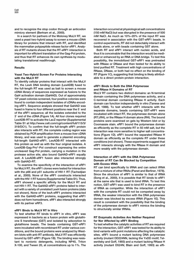
Figure 1. Interaction of eRF1 with RT
(A) Structure of Gal4AD-eRF1 fusion proteins recovered in the two-hybrid system.(B) GST or GST-eRF1 was bound to glutathione beads, and incubated with purified recombinant RT at various salt and detergent concentrationsas indicated. Bound proteins were eluted and analyzed by SDS PAGE and Western blotting with antisera specific for RT (upper panel). Aduplicate gel was stained with Coomassie blue to detect the GST fusion proteins (lower panel).(C) GST-eRF1 on beads was incubated with RT alone, or in the presence of increasing concentrations of RNase or DNase or both. The boundproteins were eluted and analyzed as before.(D) Beads containing GST-eRF1 were used to bind wild-type RT (WT) or the DNA polymerase domain lacking RNase H (RT�RH) in varioussalt concentrations as indicated.(E) Beads containing GST-eRF1 were used to bind RTs in the presence of tRNA as indicated.(F) The wild-type RT (WT), an RNase H-deficient mutant (D524N) and a polymerase-deficient mutant (DD224NN) were bound to GST-eRF1beads and analyzed by Western blot.(G) Upper: DNA polymerase assays were performed with RT or RT�H on poly(rA):oligo(dT) with radiolabeled dTTP in the presence or GST orGST-eRF1 as indicated. The DNA product was detected by autoradiography. Lower: DNA primer extension assays were performed with orwithout competitor trap, and with GST or GST-eRF1 as indicated, and products were analyzed by gel electrophoresis and autoradiography.
Cell322
ciently as the wild-type RT (Figure 1F). Thus, enzyme particles were purified, and the levels of HA-eRF1 wereassessed by Western blot (Figure 2C). Whereas wild-activity is not required for the interaction.type virions and virions lacking the RNase H domainThe interaction of eRF1 with RT might be supposedboth contained high levels of HA-eRF1, mutant virionsto alter or inhibit the enzymatic activities of RT. Additionlacking RT showed almost none of the protein (lane 6).of increasing amounts of purified GST-eRF1 to a con-Thus, the incorporation of eRF1 did not occur with emptystant amount of RT or RT�RH had no effect on RNA-virions but required the presence of RT in the particle.dependent DNA polymerase activity on homopolymerThe DNA polymerase domain even without the RNasesubstrates (Figure 1G). To test for effects on processi-H domain was sufficient to mediate the incorporationvity, a primer extension assay was performed with orof the protein. Control analysis of the blots showed thewithout addition of a competitor trap to prevent enzymepresence of CA and RT proteins in the virions as ex-rebinding. GST-eRF1 had no effect on the yield of longpected, and of HA-eRF1 in the cell lysates at levelsproducts made by either RT or the poorly processiveappropriate for its incorporation into virions.mutant RT�RH (Figure 1G). Assays of RNase H action
eRF1 also bound to the uncleaved Gag-Pol precursor.on an RNA-DNA hybrid substrate showed no effect ofExpression of tagged eRF1 with a mutant provirus inaddition of GST-eRF1 on the digestion pattern (notwhich the viral protease was inactivated by mutation ofshown). To confirm that the complex retained catalyticthe active site showed virion incorporation at a levelactivity, beads containing GST-eRF1 were used to bindequal to that seen in the wild-type virus (data not shown).RT, unbound enzyme was removed by washing, and theThus, the initial translation product, the Gag-Pol precur-bound proteins were assayed for RNA-dependent DNAsor, could interact with eRF1 and in principle could affectpolymerase activity. The complex retained comparableits function cotranslationally.specific activity to the free enzyme (data not shown).
These results suggest that the interaction did not oc-Overexpression of RT Causesclude the active sites.Enhanced Translational Readthroughof a Reporter ConstructeRF1 Associates with Virion Particles In VivoThe interaction of RT with eRF1 could modulate theTo test for an interaction between eRF1 and virions inability of eRF1 to recognize stop codons and directmammalian cells, plasmids expressing HA-tagged eRF1the release of polypeptides from the ribosome. Further,and M-MuLV proviral DNA were used to cotransformsuch an activity could affect the competition betweenCos7 cells. After 48 hr the cells and the culture mediumtRNA and eRF1 that determines the extent of transla-were collected and the virion particles released into thetional readthrough at the Gag-Pol boundary and thusmedium were purified by centrifugation on sucrose den-the level of Pol protein synthesis. To test for the abilitysity step gradients. The virions and associated proteinsof RT to modulate readthrough, 293T cells were trans-were examined by Western blots. HA-eRF1 was readilyformed with a dual luciferase reporter construct (Grentz-detected in the cell lysates, and also in the virion prepa-mann et al., 1998) in the absence or presence of a sepa-rations (Figure 2A, lane 5) at levels one-tenth to one-rate DNA construct expressing high levels of RT in trans.twentieth that of CA, and roughly one-half that of RT,This reporter expresses a large protein containing thejudged in comparisons with other similarly tagged pro-renilla luciferase, the Gag-Pol boundary region includingteins. The expression of HA-eRF1 did not cause athe stop codon, and firefly luciferase. The levels of thechange in the level of CA protein released by the cells.two luciferases expressed from the construct provide aMock virus preparations made from cells expressingmeasure of the readthrough frequency at the gag gene’s
HA-eRF1 without virus contained only very low levelsUAG stop codon.
of the protein (Figure 2, lane 3).Cells were transformed with the reporter and various
To confirm that the tagged eRF1 was associated with levels of DNA expressing HA-RT, and after 48 hr lysatesvirions, purified virus from cotransfected cells was lay- were prepared and assayed first for firefly luciferase;ered onto linear 25%–55% sucrose gradients and centri- the reactions were quenched, and the same lysates werefuged to equilibrium. Fractions were analyzed by West- then assayed for renilla luciferase. The proportion of theern blot. HA-eRF1 and the viral CA protein sedimented ribosomes that readthrough was calculated from the twowith the identical profile and with a peak centered at a activities (Figure 3A). Cells transformed with the reporterdensity of approximately 1.16 g/ml (Figure 2B). These alone gave a basal readthrough frequency correspond-results suggest that eRF1 interacts with viral proteins ing to a ratio of approximately 4.2%. The addition ofin infected cells and, when present at high concentra- increasing levels of RT caused a dose-dependent in-tions, is incorporated into virion particles. crease in the ratio to a maximum of 9%, more than
double the basal level. Significantly, at a fixed level ofIncorporation of eRF1 Into Virions RT, the expression of HA-eRF1 restored the readthroughis RT-Dependent levels to the basal frequency. These experiments sug-The association of eRF1 with virion particles could be gest that overexpression of RT can inhibit eRF1 activitymediated by nonspecific mechanisms or could reflect and so promote an enhanced level of readthrough atspecific interaction with RT. To assess whether virion- the Gag-Pol boundary. This readthrough was blockedassociated RT was required for the incorporation of by excess eRF1. We note that even moderate amountseRF1 into particles, virus particles lacking portions of of the RT expression construct used in these experi-RT were generated. Cos7 cells were cotransformed with ments directed the synthesis of much higher levels ofHA-eRF1 and M-MuLV mutants in which the RNase H RT than are achieved in normal infections. Western blots
confirmed the expression of RT and eRF1 (Figure 3B).domain or the entire RT was deleted. The mutant virion

RT-eRF1 Interaction323
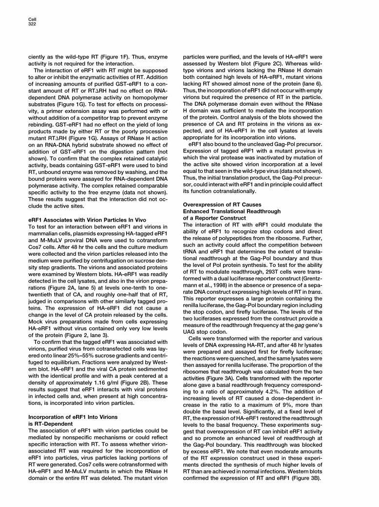
Figure 2. eRF1 Is Incorporated into Virion Particles
(A) 293T cells were transformed with DNAs expressing HA-eRF1 or viral proteins as indicated. Lysates of the purified virions and of the cellswere analyzed by Western blot with anti-HA or anti-CA antisera.(B) eRF1 copurifies with virion particles. Viruses harvested from cells expressing HA-eRF1 were displayed by equilibrium centrifugation onlinear sucrose density gradients, and fractions were analyzed by Western blots with anti-HA or anti-CA antisera. The density of each fractionis graphed below.(C) 293T cells were transformed as before with DNAs encoding HA-eRF1 and proviruses encoding wild-type RT (WT) or mutant RT lackingthe RNase H domain (RT�RH) or no RT (�RT), as indicated. The culture medium was harvested and virion particles were purified and analyzedby Western blot with antisera specific for HA, CA, or RT (upper three blots). Cell lysates were also collected and analyzed with HA antisera(bottom blot).
These readthrough experiments were limited by a pro- the production of virion CA proteins and intracellularGag protein; similar expression of eRF1 or the controlfoundly toxic effect of high-level expression of RT. Inp11 protein had no such effect. The inhibition of CAmany experiments, high levels of the DNA expressingrelease was strongly dose-dependent with increasingRT were not tolerated, causing the bulk of the cells tolevels of the RT plasmid DNA. As intracellular levels ofround up and detach from the plate. The effects of theseRT increased, the levels of released CA and intracellularhigher levels of RT could thus not be assessed.Gag decreased proportionally. These effects were notspecific for the viral proteins, but were manifest as a
Overexpression of RT Is Toxic, and the Toxicity global inhibition of all protein synthesis as detected byIs Relieved by Overexpression of eRF1 reduction in 35S-methionine incorporation (data notIf RT is able to bind and sequester eRF1, the high-level shown).expression of RT might interfere with normal protein If the inhibition by RT were due to its interaction withsynthesis and inhibit cell viability. To test for such ef- eRF1, then it should be possible to reverse the inhibitionfects, 293T cells were transformed with plasmids over- by overexpressing eRF1. At a constant high level ofexpressing HA-tagged RT, eRF1, or an unrelated 11 kDa the RT plasmid (1 �g), cotransformation with increasingcontrol protein (HA-p11), along with the M-MuLV proviral levels of a plasmid encoding HA-eRF1 did indeed relievegenome. After 48 hr, virions were purified from the cul- the block and restored host and viral protein synthesisture medium and cell lysates were prepared. The levels and virion release (Figure 4A). Cotransformation withof viral proteins were then assessed by Western blot increasing levels of an unrelated RT binding protein,
ferritin heavy chain, did not relieve the block (Figure(Figure 4A). Overexpression of RT dramatically inhibited

Cell324
eRF1. In the similar context of a carboxyterminalLexA202, the separated polymerase and RNase H do-main each interacted with eRF1 (Figure 5A). Thus, aswas observed in the in vitro binding experiments, boththe polymerase and RNase H domains independentlybound to eRF1. The RNase H domain bound morestrongly, and the polymerase more weakly, also as seenin vitro. Using the appropriate yeast constructs, it waspossible to monitor each domain’s interaction witheRF1 separately.
Isolation of Noninteracting Mutants of RTTo test the role of the interaction of RT with eRF1 inviral gene expression, we sought to isolate RT mutantsthat could no longer interact with eRF1. These screenswere complicated by the fact that both domains of RTwere independently capable of interacting with eRF1,and based on the virion incorporation results, bothneeded to be eliminated to prevent interaction. A two-step procedure allowed for the recovery, first, of a singlemutation in RNase H (G525E), and, second, of a numberof mutations in the DNA polymerase that togetherblocked binding of RT with eRF1 in yeast (see Supple-mental Data at http://www.cell.com/cgi/content/full/115/3/319/DC1 for details). Six mutant clones were iden-tified which produced high levels of a stable fusion pro-tein with the same apparent size as the parental con-struct (Figure 5B). The mutant RTs in these lysates werethen tested for RNA-dependent DNA polymerase activ-ity on a homopolymeric template. Three of the mutants
Figure 3. High-Level Overexpression of RT Causes an Increase in exhibited a wild-type level of activity (sub34, sub89, andReadthrough of a Reporter RNA In trans
sub114) and were selected for further study (Figure 5B).(A) 293T cells were transformed with the dual luciferase reporter
The DNA sequence of the entire polymerase domain ofDNA plus varying amounts of DNAs encoding HA-RT or HA-eRF1each of the three mutants was determined. The mutantsas indicated, lysates were prepared, and the extent of readthroughcontained a single change in RNase H, and one to threewas determined from the ratio of luciferase activities.
(B) Western blot of proteins from transformed cells. The proteins different substitutions in the DNA polymerase (Supple-were visualized with anti-HA antiserum. mental Table S2).
RT Mutants that Do Not Interact with eRF14B). Furthermore, the enhanced release of virions by Are Replication Defective and Produce Lowoverexpressed eRF1 was only observed in the context Levels of Gag-Pol Protein In Vivoof the block caused by RT. Overexpressed eRF1 did not To test for the consequences for viral gene expressionrelieve the block to virus release caused by Nuc212, a of a failure to interact with eRF1, the noninteracting RTdominant acting inhibitor of virus release acting through mutations were transferred into M-MuLV proviral DNA,an interaction with the NC domain of Gag (Bacharach and the mutant genomes were used to transform 293Tet al., 2000) (Figure 4B). These results indicate that the cells. In this setting, the levels of Gag and Gag-Pol ex-specific interaction of RT and eRF1 was responsible for pression are similar to those in infected cells, and RTthe effects of RT on protein synthesis. expression depends on translational readthrough as it
does in infection. Virions were purified from the culturemedium after 48 hr, and lysates of both cells and virionsMapping the Domains of RT Interacting
with eRF1 in Yeast were prepared. The viral proteins were then analyzedby Western blot with antisera specific for Gag and RTThe bait for the yeast two-hybrid screen consisted of
an aminoterminal lexA joined to the entire RT. To identify (Figure 6A). The wild-type genome induced synthesis ofnormal intracellular levels of the Gag precursor proteinthe regions of RT that interact with eRF1, a number of
constructs were generated (Figure 5A). A LexA fusion Pr65gag and of the Gag-Pol precursor Pr200gag-pol. All themutant viral genomes also induced synthesis of normalto the RNase H domain alone interacted strongly and
specifically with Gal4AD-eRF1, confirming that the intracellular levels of the Gag precursor protein Pr65gag.Virus lacking RT by deletion, as expected, made noRNase H domain contained a binding site. A fusion of an
aminoterminal LexA to the separate polymerase domain Pr200gag-pol. Virus containing only the G525E mutationproduced levels of the Gag-Pol precursor equal to thoseactivated the LacZ reporter gene without a partner and
thus could not be used for these tests. A fusion of an of the wild-type genome. In contrast, the three noninter-acting mutants sub34, 89, and 114 all made dramaticallyaminoterminal RT to a carboxyterminal LexA202, how-
ever, was not self-activating and interacted strongly with reduced levels of the Gag-Pol readthrough protein (Fig-

RT-eRF1 Interaction325
Figure 4. Effects of High-Level Expression ofRT on Protein Synthesis
(A) Overexpression of RT causes inhibition ofviral protein synthesis, which is relieved byoverexpression of eRF1. 293T cells were co-transformed with proviral DNA (pNCA) plusDNAs encoding p11 (an inert control), RT, oreRF1, and lysates of purified virions and cellswere analyzed by Western blots probed withanti-CA, anti-RT, or anti-HA antisera.(B) Relief of RT-mediated inhibition of virusproduction by eRF1 is not seen with other RTbinding proteins, and eRF1 does not relieveother blocks to virus production. 293T cellswere cotransformed with DNAs encoding theindicated proteins, and lysates of the virionpreparations and the cells were prepared andanalyzed by Western blot with anti-CA (upperfour blots) or a mix of anti-RT and anti-HAantisera (bottom two blots). Left blot: TheC-terminal portion of nucleolin (Nuc212)blocked virus assembly (Bacharach et al.,2000) but had no effect on Gag synthesis.Expression of RT blocked both virus produc-tion and Gag synthesis in a dose-dependentmanner. Right blot: At a constant level of RT,increasing eRF1 relieved the inhibition in adose-dependent manner. Increasing ferritinheavy chain (Fer.H) did not relieve the inhibi-tion. At a constant level of nucleolin, increas-ing levels of eRF1 did not relieve the blockto virus assembly. The cell lysates showedthe expected patterns of protein expression.
ure 6A). These results suggest that an interaction with mutants were completely replication defective and didnot generate progeny even after two weeks (Figure 6B).eRF1 is required for efficient synthesis of Pr200gag-pol,
and conversely that the failure to interact with eRF1 The failure of the noninteracting mutants to direct thesynthesis of high levels of Pr200gag-pol is mostly simplydramatically reduces the levels of Gag-Pol protein that
are made in vivo. attributable to a low rate of readthrough synthesis, butan alternative explanation is that the mutations in RTThe production of intracellular Pr65gag resulted in the
release of high levels of virion particles in all cases. In could render the protein unstable, even though they didnot do so in yeast. To address this issue, the UAG stopthe wild-type and G525E mutant virions, most of the
Gag precursor was successfully processed by the viral codon at the end of the gag gene was changed to aCAG codon encoding glutamine, the residue incorpo-protease to yield the mature CA proteins. In the case of
the virions produced by �RT and the three noninter- rated during readthrough, in each of the RT mutant ge-nomes. The DNAs were used to transform 293T cells,acting mutants, the bulk of the Gag protein remained
unprocessed, with only traces cleaved to form CA. This and the cells were then metabolically pulse-labeled with{35S}-methionine and -cysteine. Lysates were preparedlimited cleavage is consistent with the low levels of Gag-
Pol and low levels of the PR protease contributed by after a 30 min or 2 hr chase period with unlabeled aminoacids. The proteins were immunoprecipitated with anti-the Gag-Pol precursor. RT assays of these preparations
showed high levels in the wild-type, moderate levels in Gag antisera, collected on protein A beads, and ana-lyzed by SDS gel electrophoresis followed by autoradio-the G525E mutant, and undetectable levels in the double
mutants (Figure 6B). The virus stocks were then used graphy (Figure 6C). Since no readthrough was requiredfor its synthesis, the wild-type, the G525E mutant, andto infect Rat2 cells, and the ability of the virus to spread
in the cultures was assessed by RT assay. While the the three noninteracting mutants all made high levels ofthe Gag-Pol fusion protein. Further, the Gag-Pol pro-wild-type virus rapidly spread in the cultures, all the

Cell326
factors. Recoding of RNAs during translation is a notableexample (Atkins et al., 1990; Gesteland et al., 1992). Theresults presented here show that the murine leukemiavirus RT interacts specifically with the translational re-lease factor eRF1 to increase its own gene expression.The interaction was detected in yeast, in vitro, and inmammalian cells expressing viral proteins, and appar-ently does not require nucleic acids to bridge the pro-teins. The binding must involve extended interactionsbetween the proteins, with independent contacts involv-ing both the DNA polymerase and RNase H domains ofRT. eRF1 does not enhance or inhibit the polymeraseor RNase H activities of RT.
The structure of eRF1 is similar to that of a maturetRNA (Song et al., 2000), and the interaction with RTmay be similar to the binding of tRNA to RT. tRNA inexcess was able to compete with eRF1 for binding tothe polymerase domain, but not to the intact enzyme.While the RT of some viruses can selectively bind thetRNA used as primer for DNA synthesis—e.g., trypto-phan tRNA for RSV (Panet et al., 1975)—the RT of themurine leukemia viruses seems to bind all tRNAs withoutselectivity (Panet and Berliner, 1978). It is not knownprecisely how tRNAs are bound to any RT, however,and it is not clear that they bind at the active sites orare initially positioned so as to serve as primer for DNAsynthesis. Indeed, complex unfolding and annealing re-actions must occur to place the tRNA on the genomicFigure 5. Mapping of Interaction Domains on RTRNA at the primer binding site. We have no indication(A) Fusion proteins containing an N-terminal LexA87 or a C-terminalthat eRF1 alters any of the catalytic activities of RTLexA202 and the DNA polymerase domain (Pol) or the RNase H
domain (RH) or both, as indicated, were tested for interaction with needed for virus replication, nor that it affects tRNAeither a Gal4AD-eRF1 fusion or Gal4AD alone in the yeast two- incorporation or utilization.hybrid system. Dotted lines indicate regions of RT deleted from the Experiments to probe the effects of overexpressingconstructs. Yeast colonies were lifted to nitrocellulose and stained
RT on host translation revealed that high levels of RTfor reporter activity by staining with X-gal. ���, turned dark blueexpression were extraordinarily toxic to cells. This didin 10 min; ��, turned blue in 1 hr; �, turned blue overnight; �,not involve RT enzyme activities, since inactive pointremained white overnight; S/A, self-activating and could not be
scored. mutants of RT were equally toxic. RT expression con-(B) Isolation of noninteracting mutants of RT. Upper graph: Yeast structs caused a dramatic inhibition of viral protein syn-colonies containing putatively noninteracting mutants of RT-LexA thesis and an equally strong inhibition of virion produc-fusions and GAD-eRF1 were grown into large cultures and lysates tion, though there is no indication that the block iswere tested for �-gal activity. Middle: Lysates of yeast containing
specific to viral assembly beyond the effects on proteincandidate noninteracting mutants were analyzed by Western blotsynthesis. The major toxicity of RT, however, may befor synthesis of stable RT-LexA fusion proteins. All six mutant DNAsdue to the interaction with eRF1, since overexpression ofdirected synthesis of proteins at levels comparable to that of the
parent. Lower: Lysates of yeast expressing candidate noninteracting eRF1 was able to fully reverse the inhibition and restoremutants were scored for RT activity on oligo(dT):poly(rA) templates, normal protein expression. We cannot rule out the possi-and the radiolabeled products were spotted on DEAE cellulose pa- bility that the inhibition of protein synthesis reflects anper and exposed to autoradiography. interaction with some other protein that is relieved by
eRF1. It should be noted that the inhibition of proteinsynthesis was induced only at very high levels of RT
teins of all the mutants were equally stable, persisting expression, much more than would normally bein the cells without significant loss for the 2 hr chase. achieved in an infected cell, and indeed infection doesThus, the low levels of Gag-Pol seen in noninteracting not cause such inhibition. Further, in a normal infection,mutants when translation must pass through the stop RT is only present in the context of the Gag-Pol precur-codon are not due to instability of the product but reflect sor and is quickly encapsidated in nascent virion parti-low levels of synthesis. As control for transfection effi- cles. The cytotoxic effect, even though seen at artificiallyciencies, all the mutants made equal levels of the Env high levels of RT, suggests that this interaction of RTprecursor protein encoded by the spliced viral mRNA. with eRF1 can have important effects on cellular trans-
lation.Discussion In the context of the retroviral genome, we find that
the interaction of RT with eRF1 has a major role in regu-Viruses often make unusual changes to the normal host lating the level of readthrough that mediates formationmachinery for gene expression, activating particular of the Gag-Pol precursor in vivo (Figure 7). Point muta-pathways, removing or inhibiting unnecessary compo- tions in RT that block eRF1 binding significantly reduce
Gag-Pol translation. Thus, the binding of eRF1 by wild-nents, or introducing specific modifications to other host

RT-eRF1 Interaction327
Figure 6. Viral RT Mutants Unable to Interactwith eRF1 Do Not Synthesize Sufficient Gag-Pol Protein for Replication
(A) Phenotype of viral genomes carrying RTmutations that block the normal associationwith eRF1. Proviral DNAs carrying the indi-cated mutations were used to transform 293Tcells, and lysates of the cell cultures and thepurified virions were analyzed by Westernblot. Mutants unable to interact with eRF1(34, 89, and 114) synthesized only very lowlevels of Gag-Pol (upper blot). All mutantsmade normal levels of Pr65gag (second blot).The virions produced by the wild-type ge-nome contained high levels of capsid (CA)and RT; mutants lacking RT showed only par-tial processing of Pr65gag due to defects in PRactivity. While the RTG525E mutant providednormal levels of PR to cleave Gag proteins,the virions released from the noninteractingmutants showed poor Gag cleavage as a con-sequence of the low Gag-Pol expression.(B) Noninteracting RT mutants are replicationdefective. Virus preparations were collectedfrom transfected cells and assayed for RTactivity (top row) and used to infect Rat2 cells.Release of spreading virus was detected byRT assays of culture media on the indicateddays postinfection.(C) Noninteracting mutants of RT synthesizenormal levels of Pr200gag-pol when the needfor translational readthrough is abrogated bymutation of the UAG stop codon. The UAGstop codon at the Gag-Pol junction was re-placed by CAG in the proviral DNA of RT mu-tants unable to interact with eRF1, and theresulting DNAs were used to transform 293Tcells. The cells were pulse-labeled for 30 minwith 35S-methionine, and chased for 2 hr. Ly-sates were prepared and analyzed by immu-noprecipitation followed by autoradiography.Pr80env was monitored as a transfectioncontrol.
type RT upregulates readthrough, creating a positive chronically infected cell, termination at the gag stopcodon still occurs correctly 90%–95% of the time. Thefeedback loop that drives the synthesis of more of the
pol products, including RT itself. The effect may be ratio of Gag to Gag-Pol is known to be critical for efficientvirus replication: mutations in the cis-acting sequencesstrongest in cis, with the nascent RT acting specifically
on the termination of those ribosomes reading the same that increase or decrease this ratio have severe effectson virus assembly and infectivity (Dinman and Wickner,mRNA from which it is made. The observed interaction
of the entire Gag-Pol precursor with eRF1 seen in yeast 1992; Felsenstein and Goff, 1988; Karacostas et al.,1993; Shehu-Xhilaga et al., 2001). In the presence of theis in support of this possibility. This positive feedback,
which could in principle increase readthrough to very nonbinding RT mutants studied here, where only lowlevels of Gag-Pol are formed, virion particles are assem-high levels, must eventually be restricted by limiting
concentrations of some factor involved in the read- bled and released without adequate levels of the prote-ase to fully process the Gag precursor, and withoutthrough. It should be noted that even when RT is present
and promoting readthrough to its natural extent in a normal levels of RT. The mutants are replication defec-

Cell328
that the readthrough efficiency is similar whether theconstructs do or do not retain the coding region forRT (Feng et al., 1989a). Thus, these in vitro translationsystems do not respond strongly to the effects of RTseen in vivo in our transfected cells. Perhaps the levelsof tRNA, eRF1, eRF3, or other factors in the reactionsare sufficiently different from the levels in vivo that read-through is not sensitive to RT inhibition of eRF1 and sois not enhanced in the presence of RT. In addition, thereadthrough of reporter constructs containing the gag-pol junction occurs in uninfected cells as efficiently asin infected cells (Berteaux et al., 1991; Panganiban,1988). The high levels of readthrough observed in thoseexperiments contrast with the low levels of Gag-Pol for-mation seen in vivo here when a mutant RT cannotinteract with eRF1. Perhaps the context of the terminatorregion in the reporter RNA is responsible for this differ-ence, and perhaps RT may not act efficiently on otherRNAs in trans but only on its own RNA in cis whenexpressed at normal levels.
Translational termination in vitro is known to be sensi-tively controlled by eRF1 levels: overexpression of eRF1causes increased termination (Drugeon et al., 1997; Le
Figure 7. Model for the Mechanism of Action of RT in EnhancingGoff et al., 1997), and depletion of eRF1 promotes read-Translational Readthrough of the Stop Codon at the Gag-Pol Borderthrough (Stansfield et al., 1996; Carnes et al., 2000). AsMost ribosomes transiting the genome recognize the stop codonthese observations suggest, eRF1 may be a target ofand terminate protein synthesis with the assistance of eRF1andregulation of translation in several systems. For exam-eRF3 (top panel). A small fraction of the ribosomes read through
the terminator and continue synthesis to form Pr200gag-pol (middle ple, the Upf proteins, components of the surveillancepanel). The RT region of the precursor then interacts with eRF1, complex that scans for aberrant nonsense-containingdisplaces eRF3, and so increases synthesis of elevated levels of mRNAs, interact with the translation release factors tothe readthrough protein (bottom panel). enhance eRF1 termination activity (Czaplinski et al.,
1998; Wang et al., 2001). In another case, the cytomega-lovirus uORF2 protein binds to eRF1 while exiting thetive. Presumably, the evolution of a surface for the in-ribosome and inhibits its own translational termination,teraction of RT with eRF1 has been subject to strongcausing subsequent ribosomal stalling and inhibition ofselective pressure to finely tune the correct level of read-translation of downstream sequences (Alderete et al.,through for efficient virus replication.2001; Janzen et al., 2002). The ability of RT to inhibitWe suspect that RT may act by preventing the normaleRF1 activity provides yet another example of howassociation of eRF1 with eRF3, its essential cofactor.viruses evolve mechanisms to target key host proteinsSeveral experiments suggest that RT may bind to eRF1to fine-tune the regulated expression of their genein competition with eRF3. First, RT binds to the C-ter-products.minal portion of eRF1 (Figure 1A), the same region
known to interact with eRF3. Second, we have selectedmutants of eRF1 that show increased binding to RT and Experimental Proceduresto various RT mutants; tests of these mutants revealedthat they show a similarly strongly increased binding to Yeast Expression Vectors
Plasmid pSH-RT expressing a LexA DNA binding domain (LexA1-eRF3 (data not shown). Thus, the contacts of eRF1 with87) fused to RT was constructed by PCR amplification of the RTRT may involve similar contacts of eRF1 with eRF3,coding sequence from proviral clone pNCA (Colicelli and Goff, 1988)suggesting that RT could compete with eRF3 for accessusing primers 5�-CCGGAATTCACCCTAAATATAGAAGATGAGCA
to eRF1 and so prevent normal assembly of the hetero- TCG-3� and 5�-GGAATTCTACTCGAGGAGGGTAGAGGTGTCTGG-3�,dimeric termination complex. Both factors, including the and cloning into the EcoRI site of pSH2-1 (Hanes and Brent, 1989).
An XhoI site was added upstream of the stop codon. PlasmidsGTPase activity of eRF3, are likely to be required forpSH-RT�RH and pSH-RH that express fusions of LexA1-87 to DNAcorrect termination and polypeptide release.Polymerase (Pol) domain (amino acids 2–498) and RNase H (RH)Numerous studies of Gag-Pol translational read-domain (amino acids 500–671) of RT were constructed by subclon-through have shown that a basal level of readthroughing the corresponding PCR fragments into the EcoRI and XhoI sites
can readily be observed in a number of settings, and is of pSH-RT. A PCR-amplified hygromycin-resistance sequence wasmost critically dependent on RNA structures immedi- inserted at the XhoI site of pSH-RT to generate pSH-RT-Hyg. Plas-
mids pNLexA-RT, pNLexA-RTDRH, and pNLexA-RH expressingately downstream of the stop codon (Felsenstein andC-terminal fusions of RT and its subdomains to the LexA proteinGoff, 1992; Feng et al., 1989a; Jones et al., 1989; Pan-(LexA202) were generated by cloning corresponding PCR amplifiedganiban, 1988; ten Dam et al., 1990; Wills et al., 1991).fragments between the EcoRI and XhoI sites of pNLexA (OriGeneIn vitro translation systems can mediate readthrough ofTechnologies, Rockville, MD). Plasmid pGal4AD-eRF1 expressing
RNAs containing the gag-pol junction, and do so at the mouse eRF1 sequences fused to Gal4AD was generated byefficiencies close to those in infected cells (Honigman cloning a PCR-amplified DNA fragment of eRF1 into the NotI and
SalI sites of pGADNot (Luban et al., 1993).et al., 1991; Murphy et al., 1978). One report shows

RT-eRF1 Interaction329
Yeast Two-Hybrid Screen 3 �g of immobilized GST-eRF1 was incubated with varying amountsof RNase A (Sigma or Pharmacia; 0.5, 5, or 10 �g/reaction) or DNaseThe yeast two-hybrid system and the cDNA library used in this
screen were as described previously (Bacharach et al., 2000). In I (Boehringer; 5, 50, or 100 units/reaction) in 100 �l of TN buffercontaining 10 mM MgCl2. Following incubation for 30 min at 37�C,brief, yeast strain CTY10-5d was transformed with pSHRT and a
Gal4AD-library plasmid pool, and plated on selective media lacking beads were washed three times with TN buffer containing 50 mMEDTA and then used for binding reactions.histidine and leucine for plasmid maintenance. �-galactosidase
(�-gal) expression in the transformants was assayed by the X-galcolony filter lift method (Luban et al., 1993). Mammalian Expression Plasmids
Plasmids expressing HA-tagged mouse eRF1 and M-MuLV RT wereCloning of the Full-Length Mouse eRF1 cDNA constructed by cloning the corresponding PCR-amplified DNA frag-To clone the full length mouse eRF1 cDNA, we performed 5� Race ments into the XbaI and SmaI sites of the mammalian expressionPCR (Marathon cDNA Amplification Kit, Clontech) on a mouse liver vector pCGN (Tanaka and Herr, 1990), resulting in pCGN-eRF1 andcDNA library (Clontech) according to the manufacturer’s instructions pCGN-RT, respectively. pCGN-Nuc212 and pCGN-p11 have beenusing adaptor primer AP1 and the eRF1 sequence-specific primer described (Bacharach et al., 2000).5�-GCTGTCATCTGAAAGTAACGCTGTAAGAGCC-3�. The resultingsingle major PCR product was cloned into pUC18. Based on their Proviral DNAssequences, the complete mouse eRF1 cDNA was amplified by PCR pNCA, pNCS, and pNCS-�RT were described previously (Bacharachfrom the mouse liver cDNA library using primers 5�-ATGGCGGAC et al., 2000; Colicelli and Goff, 1988). pNCS-RT�RH was obtainedGACCCCAGTGCTGCCG-3� and 5�-CTAGTAGTCATCAAGGTCAAA by replacing the BlpI-SalI fragment of pNCS with the correspondingAAA TTCATC-3� and subcloned into the TOPO T-A vector (In- fragment from pRT�RH107-3. pNCA-RTG525E and other RT mu-vitrogen). Alignment of mouse and human eRF1 showed 94% iden- tants were obtained by replacing the BlpI-SalI fragment of pNCS withtity at the DNA level and 99% identity at the protein level. the corresponding fragment from mutant yeast plasmids (pNLexA-
RTG525E or RTG525E noninteracting mutants).Construction and Screening of Mutant Libraries A mutant provirus without the termination codon at the end ofMutants of RT lacking the ability to interact with eRF1 were con- the gag gene was generated using the GeneEditor in vitro site-structed in two steps. Plasmid pSH-RT-Hyg encoding a LexA-RT- directed mutagenesis system (Promega). Mutant proviruses thatHyg fusion was randomly mutagenized by passage through E. coli express Gag-Pol polyprotein with mutations in RT were obtainedmutator strain XL-1Red (Stratagene) according to the manufactur- by substituting the BclI-BlpI fragment of pNCA-Pr200gag-pol for theer’s protocol. Yeast strain CTY10-5d was transformed with pGAD- corresponding fragment from pNLexA-RTG525E and pNLexA-eRF1 and the pSH-RT-Hyg mutant library, plated on media lacking RTG525E-MUT, resulting in pNCA-Pr200gag-pol-RTG525E and pNCA-histidine and leucine and containing 200 mg/ml hygromycin B, and Pr200gag-pol-RTG525E-MUT, respectively.screened by X-gal staining of filter lifts.
The second step utilized PCR mutagenesis of the Pol domain ofTransfection Methods and Virus Purification
RT. First, the domain was PCR-amplified from pNLexA-RTG525ECos7 cells were transfected by the DEAE-Dextran method in 10 cm
using primers 5�-CCTGCAAT TATTATTCTTTTCTTTCCTC-3� (lo-diameter dishes, typically with 5 �g of each plasmid per transfection.
cated in pNLexA �100 bp 5� of the EcoRI site of the polylinker) and293T cells were transfected by LypofectAMINE PLUS reagent (Gib-
5�-CCGTGTACCAGGTGTGGTC GGC-3� (located �100 bp 3� of thecoBRL) in 6-well plates, typically with 1 or 2 �g of each plasmid per
EcoNI site of RT). This PCR fragment was gel purified and used astransfection. Two days after transfection, cells were analyzed for
a template for mutagenic PCR reactions performed under conditionsthe expression of the proteins. Virus purification and linear sucrose
of reduced fidelity for Taq polymerase (Leung et al., 1989). pNLexA-density gradients were as described previously (Bacharach et al.,
RTG525E was digested with EcoRI and EcoNI to delete the Pol2000).
sequence and gel purified. Yeast harboring pGal4AD-eRF1 was co-transformed with the mutagenized DNA and pNLexA-RTG525E de-
Measurement of Readthrough Efficiencyleted for the Pol domain, to generate full-length mutants by gap293T cells were transformed by calcium phosphate with the dualrepair (Muhlrad et al., 1992). The transformed yeast cells were platedluciferase reporter (Grentzmann et al., 1998) containing the gag-onto selective media lacking leucine and histidine and screened bypol readthrough sequences of M-MuLV, the generous gift of R.X-gal filter lift assay. Expression of the RT-LexA fusions was verifiedGesteland. All transfections included empty vector DNA to maintainby Western blotting using anti-LexA.constant total �g of DNA. Lysates were prepared after 48 hr, andassayed first for firefly luciferase activity in the presence of beetleExpression and Purification of GST-eRF1 Fusion Proteinluciferin. The reactions were quenched, the substrate for renillaand Wild-Type and Mutant RTluciferase was added, and the second activity was determined. TheThe mouse eRF1 cDNA PCR fragment was digested with XbaI andrelative efficiencies of readthrough were calculated as Fact/(Ract �EcoRI and cloned in pGEX-2TPL (a derivative of pGEX-2TK, Phar-Fact) without further normalization. Error bars show standard devia-macia) to form plasmid pGEX-eRF1. GST fusion proteins were in-tions from three experiments.duced and isolated as described (Bacharach et al., 2000). Construc-
tion of plasmids expressing wild-type and mutant versions of RTMetabolic Labeling of Proteins(pRTDRH107-3, pRTDD224NN, and pRTD524N) was as described293T cells, two days posttransfection, were starved for 30 min in(Blain and Goff, 1993). Recombinant RT proteins were expressedDMEM lacking methionine and cysteine prior to 30 min pulse within E. coli strain DH5�, and wild-type and RT�RH proteins were200 �Ci {35S} Met-Cys (1200 Ci/mmol; Translabel, ICN), then lysedpurified to 90% homogeneity (Gao et al., 1997).immediately with 1 ml of ice-cold RIPA buffer (50 mM Tris [pH 8.0],150 mM NaCl, 1% NP-40, 0.5% deoxycholate, 0.1% SDS). For theIn Vitro Binding Experimentschase experiments, duplicate cell cultures were washed with PBS,In standard experiments, 20 �l of a 50% slurry of beads with boundincubated with complete DMEM for another 2 hr, and then lysed.GST-eRF1 (typically 2–4 �g) were incubated with 100 ng of purifiedThe labeled proteins were immunoprecipitated with goat polyclonalRT proteins in 500 �l of TN buffer (50 mM Tris [pH 8], 150 mManti-R-MuLV p30gag antisera (NCI #79S-804) for 1 hr at 4�C, analyzedNaCl, 1% NP-40, 10% glycerol) with 1 mg/ml of BSA (Boehringer).by 7% SDS-PAGE, and visualized by PhosphorImager (MolecularFollowing incubation for 1 hr at 4�C, beads were washed three timesDynamics).in TN and the bound proteins were eluted and subjected to SDS-
PAGE and Western blotting. In the experiments with crude bacteriallysates containing RT, mutant protein levels were examined by Antibodies and Western Blotting
Western blots were performed with rabbit polyclonal anti-RT antiserumWestern blotting and an equivalent of 100 ng of RT was added tothe binding reactions. (Blain and Goff, 1993) at 1:3000 dilution, anti-HA mouse monoclonal
(Boehringer Mannheim) at 0.4 mg/ml, goat polyclonal anti-CA (NCIFor RNase and DNase treatment experiments, 20 �l of beads with

Cell330
#79S-804) at 1:5000, and anti-LexA at 1:1000. Bound proteins were Feng, Y.X., Levin, J.G., Hatfield, D.L., Schaefer, T.S., Gorelick, R.J.,and Rein, A. (1989b). Suppression of UAA and UGA terminationvisualized with ECL or ECL plus (Amersham Pharmacia Biotech).codons in mutant murine leukemia viruses. J. Virol. 63, 2870–2873.
Acknowledgments Feng, Y.X., Copeland, T.D., Oroszlan, S., Rein, A., and Levin, J.G.(1990). Identification of amino acids inserted during suppression of
We thank Eran Bacharach and Monica Roth for advice and reagents. UAA and UGA termination codons at the gag-pol junction of MoloneyThis work was partially supported by Public Health Service Grant murine leukemia virus. Proc. Natl. Acad. Sci. USA 87, 8860–8863.CA 30488 from the National Cancer Institute. M.O. and A.Y. are
Feng, Y.X., Yuan, H., Rein, A., and Levin, J.G. (1992). Bipartite signalAssociates, and S.P.G. is an Investigator, of the Howard Hughes
for read-through suppression in murine leukemia virus mRNA: anMedical Institute.
eight-nucleotide purine-rich sequence immediately downstream ofthe gag termination codon followed by an RNA pseudoknot. J. Virol.
Received: May 19, 2003 66, 5127–5132.Revised: September 12, 2003
Frolova, L., Le Goff, X., Rasmussen, H.H., Cheperegin, S., Drugeon,Accepted: September 17, 2003G., Kress, M., Arman, I., Haenni, A.L., Celis, J.E., Philippe, M., et al.Published: October 30, 2003(1994). A highly conserved eukaryotic protein family possessingproperties of polypeptide chain release factor. Nature 372, 701–703.ReferencesFrolova, L., Le Goff, X., Zhouravleva, G., Davydova, E., Philippe, M.,and Kisselev, L. (1996). Eukaryotic polypeptide chain release factorAlam, S.L., Wills, N.M., Ingram, J.A., Atkins, J.F., and Gesteland,eRF3 is an eRF1- and ribosome-dependent guanosine triphospha-R.F. (1999). Structural studies of the RNA pseudoknot required fortase. RNA 2, 334–341.readthrough of the gag-termination codon of murine leukemia virus.
J. Mol. Biol. 288, 837–852. Gao, G., Orlova, M., Georgiadis, M.M., Hendrickson, W.A., and Goff,S.P. (1997). Conferring RNA polymerase activity to a DNA polymer-Alderete, J.P., Child, S.J., and Geballe, A.P. (2001). Abundant earlyase: a single residue in reverse transcriptase controls substrateexpression of gpUL4 from a human cytomegalovirus mutant lackingselection. Proc. Natl. Acad. Sci. USA 94, 407–411.a repressive upstream open reading frame. J. Virol. 75, 7188–7192.
Atkins, J.F., Weiss, R.B., and Gesteland, R.F. (1990). Ribosome gym- Gesteland, R.F., Weiss, R.B., and Atkins, J.F. (1992). Recoding: re-nastics–degree of difficulty 9.5, style 10.0. Cell 62, 413–423. programmed genetic decoding. Science 257, 1640–1641.
Bacharach, E., Gonsky, J., Alin, K., Orlova, M., and Goff, S.P. (2000). Grentzmann, G., Ingram, J.A., Kelly, P.J., Gesteland, R.F., and At-The carboxy-terminal fragment of nucleolin interacts with the nu- kins, J.F. (1998). A dual-luciferase reporter system for studying re-cleocapsid domain of retroviral gag proteins and inhibits virion as- coding signals. RNA 4, 479–486.sembly. J. Virol. 74, 11027–11039. Hanes, S.D., and Brent, R. (1989). DNA specificity of the bicoidBerteaux, V., Rousset, J.P., and Cassan, M. (1991). UAG readthrough activator protein is determined by homeodomain recognition helixis not increased in vivo by Moloney murine leukemia virus infection. residue 9. Cell 57, 1275–1283.Biochimie 73, 1291–1293. Hatfield, D.L., Levin, J.G., Rein, A., and Oroszlan, S. (1992). Transla-Bertram, G., Bell, H.A., Ritchie, D.W., Fullerton, G., and Stansfield, tional suppression in retroviral gene expression. Adv. Virus Res.I. (2000). Terminating eukaryote translation: domain 1 of release 41, 193–239.factor eRF1 functions in stop codon recognition. RNA 6, 1236–1247.
Honigman, A., Wolf, D., Yaish, S., Falk, H., and Panet, A. (1991). cisBlain, S.W., and Goff, S.P. (1993). Nuclease activities of Moloney Acting RNA sequences control the gag-pol translation readthroughmurine leukemia virus reverse transcriptase. Mutants with altered in murine leukemia virus. Virology 183, 313–319.substrate specificities. J. Biol. Chem. 268, 23585–23592.
Ito, K., Ebihara, K., Uno, M., and Nakamura, Y. (1996). ConservedCarnes, J., Frolova, L., Zinnen, S., Drugeon, G., Phillippe, M., Juste- motifs in prokaryotic and eukaryotic polypeptide release factors:sen, J., Haenni, A.L., Leinwand, L., Kisselev, L.L., and Yarus, M. tRNA-protein mimicry hypothesis. Proc. Natl. Acad. Sci. USA 93,(2000). Suppression of eukaryotic translation termination by se- 5443–5448.lected RNAs. RNA 6, 1468–1479.
Jacks, T. (1990). Translational suppression in gene expression inColicelli, J., and Goff, S.P. (1988). Sequence and spacing require- retroviruses and retrotransposons. Curr. Top. Microbiol. Immunol.ments of a retrovirus integration site. J. Mol. Biol. 199, 47–59. 157, 93–124.Czaplinski, K., Ruiz-Echevarria, M.J., Paushkin, S.V., Han, X., Weng, Jacks, T., and Varmus, H.E. (1985). Expression of the Rous sarcomaY., Perlick, H.A., Dietz, H.C., Ter-Avanesyan, M.D., and Peltz, S.W. virus pol gene by ribosomal frameshifting. Science 230, 1237–1242.(1998). The surveillance complex interacts with the translation re-
Jacks, T., Madhani, H.D., Masiarz, F.R., and Varmus, H.E. (1988a).lease factors to enhance termination and degrade aberrant mRNAs.Signals for ribosomal frameshifting in the Rous sarcoma virus gag-Genes Dev. 12, 1665–1677.pol region. Cell 55, 447–458.
Dinman, J.D., and Wickner, R.B. (1992). Ribosomal frameshiftingJacks, T., Power, M.D., Masiarz, F.R., Luciw, P.A., Barr, P.J., andefficiency and gag/gag-pol ratio are critical for yeast M1 double-Varmus, H.E. (1988b). Characterization of ribosomal frameshiftingstranded RNA virus propagation. J. Virol. 66, 3669–3676.in HIV-1 gag-pol expression. Nature 331, 280–283.
Drugeon, G., Jean-Jean, O., Frolova, L., Le Goff, X., Philippe, M.,Jamjoom, G.A., Naso, R.B., and Arlinghaus, R.B. (1977). FurtherKisselev, L., and Haenni, A.L. (1997). Eukaryotic release factor 1characterization of intracellular precursor polyproteins of Rauscher(eRF1) abolishes readthrough and competes with suppressor tRNAsleukemia virus. Virology 78, 11–34.at all three termination codons in messenger RNA. Nucleic Acids
Res. 25, 2254–2258. Janzen, D.M., Frolova, L., and Geballe, A.P. (2002). Inhibition ofTranslation Termination Mediated by an Interaction of EukaryoticFelsenstein, K.M., and Goff, S.P. (1988). Expression of the gag-polRelease Factor 1 with a Nascent Peptidyl-tRNA. Mol. Cell. Biol.fusion protein of Moloney murine leukemia virus without gag protein22, 8562–8570.does not induce virion formation or proteolytic processing. J. Virol.
62, 2179–2182. Jones, D.S., Nemoto, F., Kuchino, Y., Masuda, M., Yoshikura, H.,and Nishimura, S. (1989). The effect of specific mutations at andFelsenstein, K.M., and Goff, S.P. (1992). Mutational analysis of thearound the gag-pol gene junction of Moloney murine leukaemiagag-pol junction of Moloney murine leukemia virus: requirementsvirus. Nucleic Acids Res. 17, 5933–5945.for expression of the gag-pol fusion protein. J. Virol. 66, 6601–6608.
Feng, Y.X., Hatfield, D.L., Rein, A., and Levin, J.G. (1989a). Transla- Karacostas, V., Wolffe, E.J., Nagashima, K., Gonda, M.A., and Moss,B. (1993). Overexpression of the HIV-1 gag-pol polyprotein resultstional readthrough of the murine leukemia virus gag gene amber
codon does not require virus-induced alteration of tRNA. J. Virol. in intracellular activation of HIV-1 protease and inhibition of assem-bly and budding of virus-like particles. Virology 193, 661–671.63, 2405–2410.

RT-eRF1 Interaction331
Kisselev, L.L., and Buckingham, R.H. (2000). Translational termina- Tanaka, M., and Herr, W. (1990). Differential transcriptional activa-tion by Oct-1 and Oct-2: interdependent activation domains inducetion comes of age. Trends Biochem. Sci. 25, 561–566.Oct-2 phosphorylation. Cell 60, 375–386.Kuchino, Y., Beier, H., Akita, N., and Nishimura, S. (1987). NaturalTanese, N., and Goff, S.P. (1988). Domain structure of the MoloneyUAG suppressor glutamine tRNA is elevated in mouse cells infectedmurine leukemia virus reverse transcriptase: mutational analysis andwith Moloney murine leukemia virus. Proc. Natl. Acad. Sci. USAseparate expression of the DNA polymerase and RNase H activities.84, 2668–2672.Proc. Natl. Acad. Sci. USA 85, 1777–1781.Le Goff, X., Philippe, M., and Jean-Jean, O. (1997). OverexpressionTelesnitsky, A., and Goff, S.P. (1993). Two defective forms of reverseof human release factor 1 alone has an antisuppressor effect intranscriptase can complement to restore retroviral infectivity. EMBOhuman cells. Mol. Cell. Biol. 17, 3164–3172.J. 12, 4433–4438.Leung, D.W., Chen, E., and Goeddel, D.V. (1989). A method forTelesnitsky, A., and Goff, S.P. (1997). Reverse transcriptase andrandom mutagenesis of a defined DNA segment using a modifiedthe generation of retroviral DNA. In Retroviruses, J.M. Coffin, S.H.polymerase chain reaction. Technique 1, 11–15.Hughes, and H. E. Varmus, eds. (Cold Spring Harbor, NY, ColdLevin, J.G., Hatfield, D.L., Oroszlan, S., and Rein, A. (1993). Mecha-Spring Harbor Press), pp. 121–160.nisms of translational suppression used in the biosynthesis of re-ten Dam, E.B., Pleij, C.W., and Bosch, L. (1990). RNA pseudoknots:verse transcriptase. In Reverse Transcriptase, A.M. Skalka and S.P.translational frameshifting and readthrough on viral RNAs. VirusGoff, eds. (Cold Spring Harbor, NY, Cold Spring Harbor Press),Genes 4, 121–136.pp. 5–31.Wang, W., Czaplinski, K., Rao, Y., and Peltz, S.W. (2001). The roleLuban, J., Bossolt, K.L., Franke, E.K., Kalpana, G.V., and Goff, S.P.of Upf proteins in modulating the translation read-through of non-(1993). Human immunodeficiency virus type 1 Gag protein binds tosense-containing transcripts. EMBO J. 20, 880–890.cyclophilins A and B. Cell 73, 1067–1078.Wills, N.M., Gesteland, R.F., and Atkins, J.F. (1991). Evidence thatMuhlrad, D., Hunter, R., and Parker, R. (1992). A rapid method fora downstream pseudoknot is required for translational read-throughlocalized mutagenesis of yeast genes. Yeast 8, 79–82.of the Moloney murine leukemia virus gag stop codon. Proc. Natl.Murphy, E.C., Jr., and Arlinghaus, R.B. (1978). Cell-free synthesisAcad. Sci. USA 88, 6991–6995.of Rauscher murine leukemia virus “gag” and “gag-pol” precursorYoshinaka, Y., Katoh, I., Copeland, T.D., and Oroszlan, S. (1985a).polyproteins from virion 35 S RNA in a mRNA-dependent translationMurine leukemia virus protease is encoded by the gag-pol gene andsystem derived from mouse tissue culture cells. Virology 86,is synthesized through suppression of an amber termination codon.329–343.Proc. Natl. Acad. Sci. USA 82, 1618–1622.Murphy, E.C., Jr., Kopchick, J.J., Watson, K.F., and Arlinghaus, R.B.Yoshinaka, Y., Katoh, I., Copeland, T.D., and Oroszlan, S. (1985b).(1978). Cell-free synthesis of a precursor polyprotein containing bothTranslational readthrough of an amber termination codon duringgag and pol gene products by Rauscher murine leukemia virus 35Ssynthesis of feline leukemia virus protease. J. Virol. 55, 870–873.RNA. Cell 13, 359–369.Zhouravleva, G., Frolova, L., Le Goff, X., Le Guellec, R., Inge-Vechto-Murphy, E.C., Jr., Wills, N., and Arlinghaus, R.B. (1980). Suppressionmov, S., Kisselev, L., and Philippe, M. (1995). Termination of transla-of murine retrovirus polypeptide termination: effect of amber sup-tion in eukaryotes is governed by two interacting polypeptide chainpressor tRNA on the cell-free translation of Rauscher murine leuke-release factors, eRF1 and eRF3. EMBO J. 14, 4065–4072.mia virus, Moloney murine leukemia virus, and Moloney murine sar-
coma virus 124 RNA. J. Virol. 34, 464–473.
Panet, A., and Berliner, H. (1978). Binding of tRNA to reverse tran-scriptase of RNA tumor viruses. J. Virol. 26, 214–220.
Panet, A., Haseltine, W.A., Baltimore, D., Peters, G., Harada, F., andDahlberg, J.E. (1975). Specific binding of tryptophan transfer RNAto avian myeloblastosis virus RNA-dependent DNA polymerase (re-verse transcriptase). Proc. Natl. Acad. Sci. USA 72, 2535–2539.
Panganiban, A.T. (1988). Retroviral gag gene amber codon suppres-sion is caused by an intrinsic cis-acting component of the viralmRNA. J. Virol. 62, 3574–3580.
Philipson, L., Andersson, P., Olshevsky, U., Weinberg, R., Baltimore,D., and Gesteland, R. (1978). Translation of MuLV and MSV RNAsin nuclease-treated reticulocyte extracts: enhancement of the gag-pol polypeptide with yeast suppressor tRNA. Cell 13, 189–199.
Shehu-Xhilaga, M., Crowe, S.M., and Mak, J. (2001). Maintenanceof the Gag/Gag-Pol ratio is important for human immunodeficiencyvirus type 1 RNA dimerization and viral infectivity. J. Virol. 75, 1834–1841.
Shinnick, T.M., Lerner, R.A., and Sutcliffe, J.G. (1981). Nucleotidesequence of Moloney murine leukaemia virus. Nature 293, 543–548.
Skalka, A.M., and Goff, S.P. (1993). Reverse Transcriptase (ColdSpring Harbor, NY, Cold Spring Harbor Press).
Song, H., Mugnier, P., Das, A.K., Webb, H.M., Evans, D.R., Tuite,M.F., Hemmings, B.A., and Barford, D. (2000). The crystal structureof human eukaryotic release factor eRF1–mechanism of stop codonrecognition and peptidyl-tRNA hydrolysis. Cell 100, 311–321.
Stansfield, I., Eurwilaichitr, L., Akhmaloka, and Tuite, M.F. (1996).Depletion in the levels of the release factor eRF1 causes a reductionin the efficiency of translation termination in yeast. Mol. Microbiol.20, 1135–1143.
Tachedjian, G., Aronson, H.E., and Goff, S.P. (2000). Analysis ofmutations and suppressors affecting interactions between the sub-units of the HIV type 1 reverse transcriptase. Proc. Natl. Acad. Sci.USA 97, 6334–6339.