Reversal of autism-like behaviors and metabolism in adult mice with single-dose antipurinergic...
11
OPEN ORIGINAL ARTICLE Reversal of autism-like behaviors and metabolism in adult mice with single-dose antipurinergic therapy JC Naviaux 1 , MA Schuchbauer 1 , K Li 2,3 , L Wang 2,3 , VB Risbrough 1,4 , SB Powell 1 and RK Naviaux 2,3,4,5,6 Autism spectrum disorders (ASDs) now affect 1–2% of the children born in the United States. Hundreds of genetic, metabolic and environmental factors are known to increase the risk of ASD. Similar factors are known to influence the risk of schizophrenia and bipolar disorder; however, a unifying mechanistic explanation has remained elusive. Here we used the maternal immune activation (MIA) mouse model of neurodevelopmental and neuropsychiatric disorders to study the effects of a single dose of the antipurinergic drug suramin on the behavior and metabolism of adult animals. We found that disturbances in social behavior, novelty preference and metabolism are not permanent but are treatable with antipurinergic therapy (APT) in this model of ASD and schizophrenia. A single dose of suramin (20 mg kg - 1 intraperitoneally (i.p.)) given to 6-month-old adults restored normal social behavior, novelty preference and metabolism. Comprehensive metabolomic analysis identified purine metabolism as the key regulatory pathway. Correction of purine metabolism normalized 17 of 18 metabolic pathways that were disturbed in the MIA model. Two days after treatment, the suramin concentration in the plasma and brainstem was 7.64 μM pmol μl - 1 (±0.50) and 5.15 pmol mg - 1 (±0.49), respectively. These data show good uptake of suramin into the central nervous system at the level of the brainstem. Most of the improvements associated with APT were lost after 5 weeks of drug washout, consistent with the 1-week plasma half-life of suramin in mice. Our results show that purine metabolism is a master regulator of behavior and metabolism in the MIA model, and that single-dose APT with suramin acutely reverses these abnormalities, even in adults. Translational Psychiatry (2014) 4, e400; doi:10.1038/tp.2014.33; published online 17 June 2014 INTRODUCTION Genetic, 1–3 environmental 4,5 and metabolic 6 factors can contri- bute to the risk of autism to different extents in each affected child. Despite this etiologic heterogeneity, and the well-known clinical variations that make each child unique, clinical studies suggest that a common denominator may underlie the shared behavioral and cognitive features that define autism spectrum disorders (ASDs) as a group. For example, in a prospective study conducted by the Zimmerman group at the Kennedy Krieger Institute in 2007, 83% of children with autism spectrum disorders were found to improve transiently in association with fever. 7 Improvements were lost with the resolution of the fever. This study showed that, despite the many different causes of ASD, the symptoms were not permanent and could be improved in a substantial fraction of children. Transient improvements with fever have also been found in patients with certain forms of post-infection brain syndromes, movement disorders, dementia and schizophrenia in the early 1900s, although those early studies were made complicated by the use of live malarial parasites to produce the fevers. 8,9 In contrast to these beneficial effects, when the exposure to serious infection happens before the onset of disease—during early development, and particularly during pregnancy—the metabolic changes associated with significant fever or infection are known to increase the risk of neurodevelopmental disorders in the offspring. These disorders include schizophrenia, 10 ASDs, 11 attention deficit/hyperactivity disorder, 12 bipolar disorder, 13 epilepsy 14 and cerebral palsy. 15 The nature and developmental timing of the exposure are important. Metabolism and mitochon- drial function change adaptively during and after infection, and are well-known regulators of neurotransmission 16 and synaptic plasticity. 17 Collectively, these studies suggest that, despite many different causes, the symptoms of several neurodevelopmental disorders such as ASD, schizophrenia and bipolar disorder may have a metabolic basis and be acutely responsive to treatment using the right metabolic intervention. The maternal immune activation (MIA) mouse model of neurodevelopmental disorders produces symptoms that are biologically similar to those of ASD 18 and schizophrenia. 19 Pregnant females that are exposed to a simulated viral infection by injection of the double-stranded RNA poly(Inosine:Cytosine) produce offspring with features of ASD 20 and schizophrenia. 21 Exposure to poly(IC) activates an evolutionarily conserved meta- bolic response to a threat called the cell danger response (CDR). 22 Pathological persistence of the CDR, beyond the physical presence of the threat, has been observed in a variety of chronic disorders including ASDs. 22 Purinergic signaling has been hypothesized to be a key regulator of the CDR; 22 however, this has not yet been proven. In support of this hypothesis, we recently showed that antipurinergic therapy (APT) in the MIA mouse model corrected all of the behavioral, molecular and neuropathological abnormalities when weekly treatment with the antipurinergic drug suramin was 1 Department of Psychiatry, University of California San Diego School of Medicine, La Jolla, CA, USA; 2 The Mitochondrial and Metabolic Disease Center, University of California San Diego School of Medicine, San Diego, CA, USA; 3 Department of Medicine, University of California San Diego School of Medicine, La Jolla, CA, USA; 4 Veterans Affairs Center for Excellence in Stress and Mental Health (CESAMH), La Jolla, CA, USA; 5 Department of Pediatrics, University of California San Diego School of Medicine, La Jolla, CA, USA and 6 Department of Pathology, University of California San Diego School of Medicine, La Jolla, CA, USA. Correspondence: Professor RK Naviaux, Departments of Medicine, Pediatrics, and Pathology, University of California San Diego School of Medicine, 214 Dickinson Street, Building CTF, Room C102, San Diego, CA 92103-8467, USA. E-mail: [email protected] Received 13 November 2013; revised 14 March 2014; accepted 16 April 2014 Citation: Transl Psychiatry (2014) 4, e400; doi:10.1038/tp.2014.33 © 2014 Macmillan Publishers Limited All rights reserved 2158-3188/14 www.nature.com/tp
-
Upload
independent -
Category
Documents
-
view
2 -
download
0
Transcript of Reversal of autism-like behaviors and metabolism in adult mice with single-dose antipurinergic...

OPEN
ORIGINAL ARTICLE
Reversal of autism-like behaviors and metabolism in adultmice with single-dose antipurinergic therapyJC Naviaux1, MA Schuchbauer1, K Li2,3, L Wang2,3, VB Risbrough1,4, SB Powell1 and RK Naviaux2,3,4,5,6
Autism spectrum disorders (ASDs) now affect 1–2% of the children born in the United States. Hundreds of genetic, metabolic andenvironmental factors are known to increase the risk of ASD. Similar factors are known to influence the risk of schizophrenia andbipolar disorder; however, a unifying mechanistic explanation has remained elusive. Here we used the maternal immune activation(MIA) mouse model of neurodevelopmental and neuropsychiatric disorders to study the effects of a single dose of theantipurinergic drug suramin on the behavior and metabolism of adult animals. We found that disturbances in social behavior,novelty preference and metabolism are not permanent but are treatable with antipurinergic therapy (APT) in this model of ASD andschizophrenia. A single dose of suramin (20 mg kg− 1 intraperitoneally (i.p.)) given to 6-month-old adults restored normal socialbehavior, novelty preference and metabolism. Comprehensive metabolomic analysis identified purine metabolism as the keyregulatory pathway. Correction of purine metabolism normalized 17 of 18 metabolic pathways that were disturbed in the MIAmodel. Two days after treatment, the suramin concentration in the plasma and brainstem was 7.64 μM pmol μl− 1 (±0.50) and5.15 pmol mg− 1 (±0.49), respectively. These data show good uptake of suramin into the central nervous system at the level of thebrainstem. Most of the improvements associated with APT were lost after 5 weeks of drug washout, consistent with the 1-weekplasma half-life of suramin in mice. Our results show that purine metabolism is a master regulator of behavior and metabolism inthe MIA model, and that single-dose APT with suramin acutely reverses these abnormalities, even in adults.
Translational Psychiatry (2014) 4, e400; doi:10.1038/tp.2014.33; published online 17 June 2014
INTRODUCTIONGenetic,1–3 environmental4,5 and metabolic6 factors can contri-bute to the risk of autism to different extents in each affectedchild. Despite this etiologic heterogeneity, and the well-knownclinical variations that make each child unique, clinical studiessuggest that a common denominator may underlie the sharedbehavioral and cognitive features that define autism spectrumdisorders (ASDs) as a group. For example, in a prospective studyconducted by the Zimmerman group at the Kennedy KriegerInstitute in 2007, 83% of children with autism spectrum disorderswere found to improve transiently in association with fever.7
Improvements were lost with the resolution of the fever. Thisstudy showed that, despite the many different causes of ASD, thesymptoms were not permanent and could be improved in asubstantial fraction of children.Transient improvements with fever have also been found in
patients with certain forms of post-infection brain syndromes,movement disorders, dementia and schizophrenia in the early1900s, although those early studies were made complicated bythe use of live malarial parasites to produce the fevers.8,9 Incontrast to these beneficial effects, when the exposure to seriousinfection happens before the onset of disease—during earlydevelopment, and particularly during pregnancy—the metabolicchanges associated with significant fever or infection are knownto increase the risk of neurodevelopmental disorders in theoffspring. These disorders include schizophrenia,10 ASDs,11
attention deficit/hyperactivity disorder,12 bipolar disorder,13
epilepsy14 and cerebral palsy.15 The nature and developmentaltiming of the exposure are important. Metabolism and mitochon-drial function change adaptively during and after infection, andare well-known regulators of neurotransmission16 and synapticplasticity.17 Collectively, these studies suggest that, despite manydifferent causes, the symptoms of several neurodevelopmentaldisorders such as ASD, schizophrenia and bipolar disorder mayhave a metabolic basis and be acutely responsive to treatmentusing the right metabolic intervention.The maternal immune activation (MIA) mouse model of
neurodevelopmental disorders produces symptoms that arebiologically similar to those of ASD18 and schizophrenia.19
Pregnant females that are exposed to a simulated viral infectionby injection of the double-stranded RNA poly(Inosine:Cytosine)produce offspring with features of ASD20 and schizophrenia.21
Exposure to poly(IC) activates an evolutionarily conserved meta-bolic response to a threat called the cell danger response (CDR).22
Pathological persistence of the CDR, beyond the physical presenceof the threat, has been observed in a variety of chronic disordersincluding ASDs.22 Purinergic signaling has been hypothesized tobe a key regulator of the CDR;22 however, this has not yet beenproven. In support of this hypothesis, we recently showed thatantipurinergic therapy (APT) in the MIA mouse model corrected allof the behavioral, molecular and neuropathological abnormalitieswhen weekly treatment with the antipurinergic drug suramin was
1Department of Psychiatry, University of California San Diego School of Medicine, La Jolla, CA, USA; 2The Mitochondrial and Metabolic Disease Center, University of California SanDiego School of Medicine, San Diego, CA, USA; 3Department of Medicine, University of California San Diego School of Medicine, La Jolla, CA, USA; 4Veterans Affairs Center forExcellence in Stress and Mental Health (CESAMH), La Jolla, CA, USA; 5Department of Pediatrics, University of California San Diego School of Medicine, La Jolla, CA, USA and6Department of Pathology, University of California San Diego School of Medicine, La Jolla, CA, USA. Correspondence: Professor RK Naviaux, Departments of Medicine, Pediatrics,and Pathology, University of California San Diego School of Medicine, 214 Dickinson Street, Building CTF, Room C102, San Diego, CA 92103-8467, USA.E-mail: [email protected] 13 November 2013; revised 14 March 2014; accepted 16 April 2014
Citation: Transl Psychiatry (2014) 4, e400; doi:10.1038/tp.2014.33© 2014 Macmillan Publishers Limited All rights reserved 2158-3188/14
www.nature.com/tp

begun at 1.5 months of age, near the age of reproductive maturityfor mice.23 Significant reductions in mitochondrial oxygenconsumption and body temperature were also found. However,comprehensive metabolomic analysis was not reported in thatstudy.23
In the present study, we tested the hypothesis that thebehavioral manifestations of the MIA model are a consequenceof pathological persistence of the evolutionarily conserved CDR,22
and that the CDR is maintained by dysregulated purinemetabolism and secondary abnormalities in purinergic signaling.We found that a single dose of the antipurinergic drug suramingiven to adult animals about 6 months of age (21–27 weeks)produced the concerted correction of over 90% of the metabolicpathway disturbances, and all of the behavioral abnormalities thatwe tested in the MIA model. Six-month-old mice are the humanbiological age equivalents of about 30 years24 (see Materials andmethods). After washout of the drug, these improvements werelost and the former abnormalities returned. These data show thatpurine metabolism and purinergic signaling represent a novelneurochemical switch that regulates both behavior and metabo-lism in the MIA model of neurodevelopmental disorders such asASD and schizophrenia.
MATERIALS AND METHODSAnimals and husbandryAll studies were conducted at the University of California, San Diego(UCSD) in facilities accredited by the Association for Assessment andAccreditation of Laboratory Animal Care International (AAALAC) under theUCSD Institutional Animal Care and Use Committee-approved animalsubjects protocols, and followed the National Institutes of Health Guide-lines for the use of animals in research. Six- to eight-week-old C57BL/6J(strain no. 000664) mice were obtained from Jackson Laboratories (BarHarbor, ME, USA), given food and water ad libitum, identified by ear tags,and used to produce the timed matings. Animals were housed in atemperature- (22–24 °C) and humidity (40–55%)-controlled vivarium with a12-h light–dark cycle (lights on at 0700 hours). Nulliparous dams weremated at 9–10 weeks of age. The sires were also 9–10 weeks of age. Thehuman biological age equivalent for the C57BL/6J strain of laboratorymouse (Mus musculus) can be estimated from the following equation: 12years for the first month, 6 years for the second month, 3 years for months3–6 and 2.5 years for each month thereafter.24 Therefore, a 6-month-oldmouse would be the biological equivalent of 30 years old ( = 12+6+3×4)on a human timeline.
Poly(IC) preparation and gestational exposureTo initiate the MIA model, pregnant dams were given two intraperitonealinjections of Poly(I:C) (Potassium salt; Sigma-Aldrich, St. Louis, MO, USA, Catno. P9582; >99% pure; o1% mononucleotide content). These werequantified by UV spectrophotometry. One unit (U) of poly(IC) was definedas 1 absorbance unit at 260 nm. Typically, 1U= 12 μg of RNA. 0.25 U/g[3mg kg− 1] of poly(IC) was given on E12.5 and 0.125 U g− 1 (1.5 mg kg− 1)on E17.5 as previously described.23 Contemporaneous control pregnancieswere produced by timed matings and randomized assignment of pregnantdams to saline injection (5 μl g− 1 intraperitoneally (i.p.)) on E12.5and E17.5.
Postnatal handling and antipurinergic therapy (APT)Offspring of timed matings were weaned at 3–4 weeks of age into cages oftwo to four animals. No mice were housed in isolation. Only males wereevaluated in these studies. Littermates were identified by ear tags anddistributed into different cages in order to minimize litter and dam effects.To avoid chance differences in groups selected for single-dose treatment,the saline and poly(IC) exposure groups were each balanced according totheir social approach scores at 2.25 months. At 5.25 or 6.5 months of age,half the animals received a single injection of either saline (5 μl g− 1 i.p.) orsuramin (hexasodium salt, 20mg kg− 1 i.p.; Tocris Bioscience, Bristol, UK,Cat no. 1472). Beginning 2 days later, behaviors were evaluated asdescribed below. After completing the behavioral measurements, half ofthe subjects were killed after a 5-week-washout period for measurement of
suramin tissue levels. For acute suramin levels, the other half was injectedat 7.75 months of age and killed 2 days later for tissue leveldeterminations.
Behavioral testingBehavioral testing began at 2.25 months (9 weeks) of age. Mice weretested in social approach, rotarod, t-maze test of spontaneous alternationand light–dark box test. If abnormalities were found, treatment withsuramin or saline was given at 5.25 months (21 weeks) or 6.5–6.75 months(26–27 weeks) and the testing was repeated. Only male animals weretested.
Social approach. Social behavior was tested as social preference aspreviously described23 with minor modifications (see Full Methods inSupplementary Information; N=19–25, 2.25-month-old males per groupbefore adult treatment with suramin. N=8–13, 6.5-month-old males pergroup).
T-Maze. Novelty preference was tested as spontaneous alternationbehavior in the T-maze by a modification of the methods of Frye andWalf25 (see Full Methods Supplementary Information). N= 19–25, 4-month-old males per group before adult treatment with suramin. N=8–13, 5.25-month-old males per group.
Rotarod. Sensorimotor coordination was tested as latency to fall on therotarod as previously described23 (see Full Methods SupplementaryInformation; N= 19–25, 2.5-month-old males per group before adulttreatment with suramin. N=8–13, 6.75-month-old males per group).
Light–dark box. Certain anxiety-related and light-avoidance behaviorswere tested in the light–dark box paradigm as previously described26 (seeFull Methods Supplementary Information; N= 19–25, 3.5-month-old malesper group).
Absence of abnormal behaviors produced by suramin. This was assessed inthe non-MIA control animals (indicated as the ‘Saline’ group in thepretreatment figures) that were injected with suramin as adults (indicatedas the ‘Sal-Sur’ groups in the single-dose treatment figures) using each ofthe above behavioral paradigms.
Suramin quantitationTissue samples (brainstem, cerebrum and cerebellum) were ground intopowder under liquid nitrogen in a pre-cooled mortar. Powdered tissue(15–50mg) was weighed and mixed with the internal standard trypan blueto a final concentration of 5 μM (pmol mg− 1) and incubated at roomtemperature for 10min to permit metabolite interaction with bindingproteins. Nine volumes of methanol:acetonitrile:H2O (43:43:16) pre-chilledto − 20 °C was added to produce a final solvent ratio of 40:40:20, and thesamples were deproteinated and macromolecules removed by precipita-tion on crushed ice for 30min. The mixture was centrifuged at 16 000 g for10min at 4 °C and the supernatant was transferred to a new tube and keptat − 80 °C for further LC-MS/MS (liquid chromatography-tandem massspectrometry) analysis. For plasma, 90 μl was used, to which 10 μl of 50 μMstock of trypan blue was added to achieve an internal standardconcentration of 5 μM. This was incubated at room temperature for10min to permit metabolite interaction with binding proteins, thenextracted with 4 volumes (400 μl) of pre-chilled methanol:acetonitrile(50:50) to produce a final concentration of 40:40:20 (methanol:acetonitrile:H2O) and precipitated on ice for 10min. Other steps were the same as forsolid tissue extraction.Suramin was analyzed on an AB SCIEX QTRAP 5500 triple quadrupole
mass spectrometer equipped with a Turbo V electrospray ionizationsource, Shimadzu LC-20A UHPLC system, and a PAL CTC autosampler (ABSCIEX, Framingham, MA, USA). Ten microliters of extract were injected ontoa Kinetix pentafluorophenyl column (150 × 2.1 mm, 2.6 μm; Phenomenex,Torrance, CA, USA) held at 30 °C for chromatographic separation. Themobile phase A was water with 20mM ammonium acetate (NH4OAC; pH 7)and mobile phase B was methanol with 20mM NH4OAC (pH 7). Elution wasperformed using the following gradient: 0 min—0% B, 15min—100% B,18min—100% B, 18.1 min—0% B, 23min—end. The flow rate was 300μl min− 1. All the samples were kept at 4 °C during analysis. Suramin andtrypan blue were detected using scheduled multiple reaction monitoring(MRM) with a dwell time of 30ms in negative mode and retention time
Antipurinergic therapy of ASDJC Naviaux et al
2
Translational Psychiatry (2014), 1 – 11 © 2014 Macmillan Publishers Limited

window of 7.5–8.5 min for suramin and 8.4–9.4 min for trypan blue. MRMtransitions for the doubly charged form of suramin were 647.0 m z− 1 (Q1)precursor and 382.0 m z− 1 (Q3) product. MRM transitions for trypan bluewere 435.2 (Q1) and 185.0 (Q3). Absolute concentrations of suramin weredetermined for each tissue using a tissue-specific standard curve toaccount for matrix effects, and the peak area ratio of suramin to theinternal standard trypan blue. The declustering potential, collision energy,entrance potential and collision exit potential were − 104, − 9.5,− 32 and− 16.9, and − 144.58, − 7, − 57.8 and − 20.94 for suramin and trypan blue,respectively. The electrospray ionization source parameters were set asfollows: source temperature 500 °C; curtain gas 30; ion source gas 1, 35; ionsource gas 2 35; spray voltage − 4500 V. Analyst 1.6.1 was used for dataacquisition and analysis. N= 4–6 per tissue. Results are reported asmeans± s.e.m. in absolute μM (pmol μl− 1) concentration for plasma, andpmol mg− 1 wet weight for tissues.
MetabolomicsBroad-spectrum analysis of 478 targeted metabolites from 44 biochemicalpathways in the plasma was performed by a modification of the methodsdescribed by Bajad and Shulaev.27 Only male animals that had beenbehaviorally evaluated were tested. Samples were analyzed on an ABSCIEX QTRAP 5500 triple quadrupole mass spectrometer equipped with aTurbo V electrospray ionization source, Shimadzu LC-20A UHPLC systemand a PAL CTC autosampler (AB SCIEX). Whole blood was collected 2 daysafter a single dose of suramin (20mg kg− 1 i.p.) or saline (5 μl g− 1 i.p.) fromanimals that were lightly anesthetized with isoflurane (Med-Vet Interna-tional, Mettawa, IL, USA, Cat no. RXISO-250) in a drop jar into BDMicrotainer tubes containing lithium heparin (Becton Dickinson, San Diego,CA, USA, Ref no. 365971) by submandibular vein lancet.28 Plasma wasseparated by centrifugation at 600 g× 5min at 20 °C within 1 h ofcollection. Fresh lithium–heparin plasma was transferred to labeled tubesfor storage at − 80 °C for analysis. Typically, 45 μl of plasma was thawed onice and transferred to a 1.7-ml Eppendorf tube. Two and one-half (2.5)microliters of a cocktail containing 35 commercial stable isotope internalstandards (Supplementary Table S3) and 2.5 μl of 310 stable isotopeinternal standards that were custom-synthesized in Escherichia coli andSaccharomyces cerevisiae by metabolic labeling with 13C-glucose and 13C-bicarbonate were added, mixed and incubated for 10min at 20 °C topermit small molecules and vitamins in the internal standards to associatewith plasma-binding proteins. Macromolecules (protein, DNA, RNA and soon) were precipitated by extraction with 4 volumes (200 μl) of cold (−20 °C), acetonitrile:methanol (50:50) (LCMS grade, Cat no. LC015-2.5 andGC230-4, Burdick & Jackson, Honeywell, Muskegon, MI, USA), vortexedvigorously and incubated on crushed ice for 10min, and then removedwith centrifugation at 16 000 g×10min at 4 °C. The supernatants contain-ing the extracted metabolites and internal standards in the resulting40:40:20 solvent mix of acetonitrile:methanol:water were transferred tolabeled cryotubes and stored at − 80 °C for LC-MS/MS (liquidchromatography-tandem mass spectrometry) analysis.LC-MS/MS analysis was performed by MRM under the Analyst v1.6.1
software control in both negative and positive modes with rapid polarityswitching (50 ms). Nitrogen was used for curtain gas (set to 30), collisiongas (set to high) and ion source gases 1 and 2 (set to 35). The sourcetemperature was 500 °C. Spray voltage was set to − 4500 V in negativemode and to 5500 V in positive mode. The values for Q1 and Q3 mass-to-charge ratios (m z− 1), declustering potential, entrance potential, collisionenergy and collision cell exit potential were determined and optimized foreach MRM for each metabolite. Ten microliters of extract were injectedwith PAL CTC autosampler into a 250mm×2.1 mm, 5-μm Luna NH2aminopropyl HPLC column (Phenomenex) held at 25 °C for chromato-graphic separation. The mobile phase was solvent A: 95% water with23.18mM NH4OH (Sigma, Fluka Cat no. 17837-100ML), 20 mM formic acid(Sigma, Fluka Cat no. 09676-100ML) and 5% acetonitrile (pH 9.44); solventB: 100% acetonitrile. Separation was achieved using the following gradient:0 min—95% B, 4 min—B, 19min—2% B, 22min—2% B, 23min—95% B,28min—end. The flow rate was 300 μl min− 1. All the samples were kept at4 °C during analysis. The chromatographic peaks were identified usingMultiQuant v2.1.1 (AB SCIEX), confirmed by manual inspection and thepeak areas were integrated. The median of the peak area of stable isotopeinternal standards was calculated and used for the normalization ofmetabolite concentration across the samples and batches. N= 6, 6.5-month-old males per group. Metabolite data were log-transformed beforemultivariate and univariate analyses.
Data analysisAnimals were randomized into active (suramin) and mock (saline)treatment groups at ~ 6 months of age. Group means and s.e.m. arereported. Behavioral data involving more than two groups were analyzedby two-way analysis of variance (ANOVA) and one-way ANOVAs (GraphPadPrism 5.0d, GraphPad Software Inc., La Jolla, CA, USA). Pair-wise post hoctesting was performed by the method of Tukey. Repeated measuresANOVA with prenatal treatment and drug as between subject factors andstimulus (mouse/cup) on time spent with mouse or cup was used as anadditional test of social preference. Student’s t-test was used forcomparisons involving the two groups. Significance was set at Po0.05.Bonferroni post hoc correction was used to control for multiple hypothesistesting when t-tests were used to test social preference in two or moreexperimental groups. Metabolomic data were analyzed using multivariatepartial least squares discriminant analysis, Ward hierarchical clustering andunivariate one-way ANOVA with pairwise comparisons and post hoccorrection by Fisher’s least significant difference test in MetaboAnalyst.29
RESULTSRestoration of normal social behaviorSocial behavior in mice can be quantified as the time spentinteracting with a novel (‘stranger’) mouse compared with thetotal time spent interacting with either a mouse or a novelinanimate object.30 MIA animals showed social deficits from anearly age (Figure 1a, Supplementary Figure S1a). Single-dose APTwith suramin completely reversed the social abnormalities in 6.5-month-old adults (Figure 1b, Supplementary Figure S1b). Fiveweeks (5 half-lives) after suramin washout, a small residual benefitto social behavior was still detectable (Figure 1c, SupplementaryFigure S1c). The residual social benefit of APT even after 5 weeksfollowing suramin was correlated with retained metabolomicbenefits (see below). This phenomenon was not investigatedfurther but may be related to the development of metabolicmemory and/or somatic epigenetic DNA changes that lastedlonger than the physical presence of the drug.31
Restoration of spontaneous alternation in the T-mazeNovelty preference is an innate feature of normal rodent32 andhuman33 behavior and a predictor of socialization and commu-nication growth in children with ASD.34 The loss or suppression ofnovelty preference in children with ASD is associated with thephenomenon known as insistence on sameness.35 We estimatedpreference for novelty as spontaneous alternation behavior in theT-maze.25 The T-maze can also be used to estimate spatial workingmemory, especially when food-motivated.36 We did not use thefood-motivated variation in our study. We found that MIA animalsshowed deficient novelty preference as reflected by chance (near50%) spontaneous alternation behavior (Figure 1d). These deficitswere normalized after a single dose of suramin (Figure 1e). Fiveweeks after suramin washout, no residual benefit remained(Figure 1f).
Failure to restore rotarod performance in adult animalsPrevious studies have shown age-dependent, postnatal loss ofcerebellar Purkinje cells in the MIA model. This can reach up to60% of Purkinje cells lost by 4 months (16 weeks) of age.23,37
Motor coordination measured by rotarod performance is deficientin the MIA model23 (and Figure 1g) and is critically dependent onthe integrity of Purkinje cell circuits in the cerebellum.38 Wehypothesized that since Purkinje cells are known to be lost in MIAanimals by 4 months (16 weeks) of age, that APT given later in lifewould have no effect. Our results confirmed this. We found that asingle injection of suramin given to 6-month-old adults failed torestore normal motor coordination (Figures 1g and h). Althoughcerebellar Purkinje cell density was not quantified in this study,our results are consistent with the notion that once Purkinje cellsare lost, their function cannot be restored by APT in adult animals.
Antipurinergic therapy of ASDJC Naviaux et al
3
© 2014 Macmillan Publishers Limited Translational Psychiatry (2014), 1 – 11

Figure 1. Single-dose correction of behavioral abnormalities. (a) Social abnormalities in male MIA animals were found at the earliest ages oftesting at 2.25 months of age. (Student’s t-test ****Po0.0002; N= 19 Saline and 25 Poly(IC)). (b) A single dose of suramin given to adult MIAmice restored normal social behavior (PIC-Sur). two-way ANOVA was first used to test for the presence of interaction between drug treatmentand experimental groups. This revealed an interaction consistent with the observation that suramin benefited social behavior in the MIAanimals but had no effect on normal controls (F(1,39)= 13.48); P= 0.0007). We then performed one-way ANOVA to test for suramin effects. Asingle treatment with suramin (20mg kg− 1 i.p.) given 2–4 days before testing restored normal social behavior (one-way ANOVA F(3,40)= 8.95;Po0.0001; Tukey post hoc PIC-Sal versus PIC-Sur ****Po0.0001; N= 8–13 per group). (c) After 5 weeks of suramin washout, the social behaviorremained improved compared with saline-treated animals but was decreased from the first week after treatment. (F(3,40)= 10.5; Tukey posthoc PIC-Sal versus PIC-Sur *Po0.05; N= 8–13 per group). Values are expressed as means± s.e.m. (d) We estimated the strength of noveltypreference32 as spontaneous alternation in the T-maze. MIA mice showed deficits in spontaneous alternation from the age of earliest testingat 4 months of age (Student’s t-test; ****Po0.0001; N= 19 Saline and 25 PIC). (e) Two-way ANOVA was first used to test for the presence ofinteraction between drug treatment and experimental groups. This revealed an interaction consistent with the observation that suraminrestored spontaneous alternation in the MIA animals but had no effect on normal controls (F(1,40)= 7.609; P= 0.0087). We then performedone-way ANOVA to test for suramin effects. A single dose of suramin (20mg kg− 1 i.p.) injected 2–4 days before testing corrected the deficits inyoung adult animals that were 5.25 months of age. (F(3,40)= 9.46; ; Tukey post hoc Sal-Sal versus PIC-Sal **Po0.01; PIC-Sal versus PIC-Sur***Po0.001); N= 8–13 per group). (f) This benefit was lost after a drug washout period of 5 weeks, leaving a significant difference betweencontrol (Sal) and MIA (PIC) groups (F(3,39)= 18.05; Po0.0001), but no remaining effect of suramin by post hoc testing. (Tukey post hoc PIC-Salversus PIC-Sur P=ns; N= 8–13 per group). Values are expressed as means± s.e.m. (g) Motor coordination abnormalities were quantified on therotarod as latency to fall. Performance was abnormal from the earliest age of testing at 2.5 months of age (Student’s t-test ****Po0.0001;N= 19 Saline and 25 Poly(IC)). (h) Suramin did not improve performance after two doses (20mg kg− 1 i.p.) given at 6.5 and 6.75 months of ageand tested 2–4 days after the second dose. (two-way ANOVA interaction F(1,39)= 0.1227; P= 0.728 (ns); Poly(IC) effect F(1,39)= 25.06;****Po0.0001; treatment effect F(1,39)= 0.01; P= 0.908 (ns)). Values are expressed as means± s.e.m.
Antipurinergic therapy of ASDJC Naviaux et al
4
Translational Psychiatry (2014), 1 – 11 © 2014 Macmillan Publishers Limited
Other behaviorsCertain features of ASD and schizophrenia were not captured byour studies of the MIA model and therefore could not beinterrogated for pharmacologic response to APT. For example, wedid not find any abnormalities in the MIA mouse model using ourprotocol when we looked for certain types of anxiety-relatedbehavior in the light–dark box paradigm (Supplementary FigureS2). Likewise, our earlier studies showed no stereotypic repetitivemovements in the C57BL/6J mouse strain either by clinicalobservation or by testing in the hole board exploration beambreak mouse behavioral pattern monitor.23 Finally, no abnormalbehaviors were produced by suramin treatment itself. This wasshown by the absence of behavioral differences between controlmice treated with saline (‘Sal-Sal’) and those treated with suramin(‘Sal-Sur’) in Figures 1b, e and h.
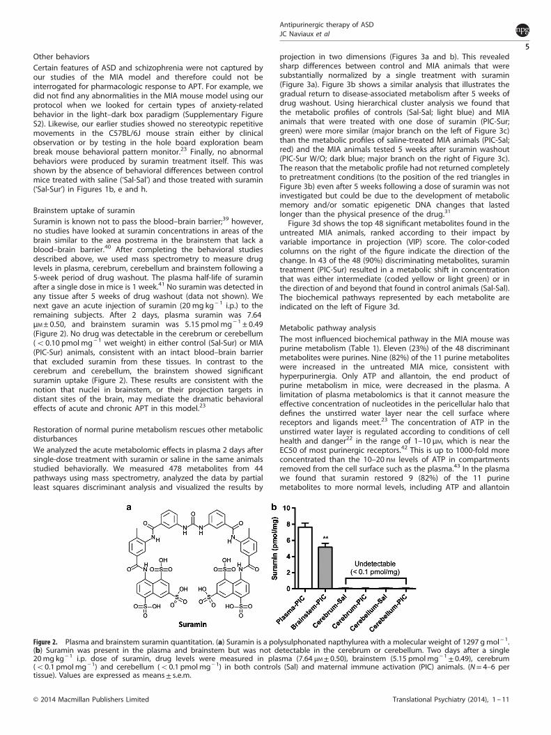
Brainstem uptake of suraminSuramin is known not to pass the blood–brain barrier;39 however,no studies have looked at suramin concentrations in areas of thebrain similar to the area postrema in the brainstem that lack ablood–brain barrier.40 After completing the behavioral studiesdescribed above, we used mass spectrometry to measure druglevels in plasma, cerebrum, cerebellum and brainstem following a5-week period of drug washout. The plasma half-life of suraminafter a single dose in mice is 1 week.41 No suramin was detected inany tissue after 5 weeks of drug washout (data not shown). Wenext gave an acute injection of suramin (20 mg kg− 1 i.p.) to theremaining subjects. After 2 days, plasma suramin was 7.64μM± 0.50, and brainstem suramin was 5.15 pmol mg− 1 ± 0.49(Figure 2). No drug was detectable in the cerebrum or cerebellum(o0.10 pmol mg− 1 wet weight) in either control (Sal-Sur) or MIA(PIC-Sur) animals, consistent with an intact blood–brain barrierthat excluded suramin from these tissues. In contrast to thecerebrum and cerebellum, the brainstem showed significantsuramin uptake (Figure 2). These results are consistent with thenotion that nuclei in brainstem, or their projection targets indistant sites of the brain, may mediate the dramatic behavioraleffects of acute and chronic APT in this model.23
Restoration of normal purine metabolism rescues other metabolicdisturbancesWe analyzed the acute metabolomic effects in plasma 2 days aftersingle-dose treatment with suramin or saline in the same animalsstudied behaviorally. We measured 478 metabolites from 44pathways using mass spectrometry, analyzed the data by partialleast squares discriminant analysis and visualized the results by
projection in two dimensions (Figures 3a and b). This revealedsharp differences between control and MIA animals that weresubstantially normalized by a single treatment with suramin(Figure 3a). Figure 3b shows a similar analysis that illustrates thegradual return to disease-associated metabolism after 5 weeks ofdrug washout. Using hierarchical cluster analysis we found thatthe metabolic profiles of controls (Sal-Sal; light blue) and MIAanimals that were treated with one dose of suramin (PIC-Sur;green) were more similar (major branch on the left of Figure 3c)than the metabolic profiles of saline-treated MIA animals (PIC-Sal;red) and the MIA animals tested 5 weeks after suramin washout(PIC-Sur W/O; dark blue; major branch on the right of Figure 3c).The reason that the metabolic profile had not returned completelyto pretreatment conditions (to the position of the red triangles inFigure 3b) even after 5 weeks following a dose of suramin was notinvestigated but could be due to the development of metabolicmemory and/or somatic epigenetic DNA changes that lastedlonger than the physical presence of the drug.31
Figure 3d shows the top 48 significant metabolites found in theuntreated MIA animals, ranked according to their impact byvariable importance in projection (VIP) score. The color-codedcolumns on the right of the figure indicate the direction of thechange. In 43 of the 48 (90%) discriminating metabolites, suramintreatment (PIC-Sur) resulted in a metabolic shift in concentrationthat was either intermediate (coded yellow or light green) or inthe direction of and beyond that found in control animals (Sal-Sal).The biochemical pathways represented by each metabolite areindicated on the left of Figure 3d.
Metabolic pathway analysisThe most influenced biochemical pathway in the MIA mouse waspurine metabolism (Table 1). Eleven (23%) of the 48 discriminantmetabolites were purines. Nine (82%) of the 11 purine metaboliteswere increased in the untreated MIA mice, consistent withhyperpurinergia. Only ATP and allantoin, the end product ofpurine metabolism in mice, were decreased in the plasma. Alimitation of plasma metabolomics is that it cannot measure theeffective concentration of nucleotides in the pericellular halo thatdefines the unstirred water layer near the cell surface wherereceptors and ligands meet.23 The concentration of ATP in theunstirred water layer is regulated according to conditions of cellhealth and danger22 in the range of 1–10 μM, which is near theEC50 of most purinergic receptors.42 This is up to 1000-fold moreconcentrated than the 10–20 nM levels of ATP in compartmentsremoved from the cell surface such as the plasma.43 In the plasmawe found that suramin restored 9 (82%) of the 11 purinemetabolites to more normal levels, including ATP and allantoin
Figure 2. Plasma and brainstem suramin quantitation. (a) Suramin is a polysulphonated napthylurea with a molecular weight of 1297 gmol− 1.(b) Suramin was present in the plasma and brainstem but was not detectable in the cerebrum or cerebellum. Two days after a single20mg kg− 1 i.p. dose of suramin, drug levels were measured in plasma (7.64 μM± 0.50), brainstem (5.15 pmol mg− 1± 0.49), cerebrum(o0.1 pmol mg− 1) and cerebellum (o0.1 pmol mg− 1) in both controls (Sal) and maternal immune activation (PIC) animals. (N= 4–6 pertissue). Values are expressed as means± s.e.m.
Antipurinergic therapy of ASDJC Naviaux et al
5
© 2014 Macmillan Publishers Limited Translational Psychiatry (2014), 1 – 11

(Figure 3d, right PIC-Sur column, coded yellow or light green) andincreased inosine and deoxyinosine to above normal.Additional pathway analysis revealed a pattern of disturbances
that was remarkably similar to metabolic disturbances that have
been found in children with ASDs (Table 1). Eighteen of the 44pathways were disturbed in the MIA model. The 44 pathwaysinterrogated by this analysis are reported in Supplementary TableS1. After purine metabolism, the next most influenced pathway
Figure 3. Metabolomic analysis. (a) APT rescues widespread metabolic abnormalities. Plasma samples were collected 2 days after a single doseof suramin (20mg kg− 1 i.p.) or saline (5 μl g− 1 i.p.). This analysis shows that a single dose of suramin (PIC-Sur; green) drives the metabolism ofMIA animals (PIC-Sal; red) strongly in the direction of controls (Sal-Sal; blue). Metabolomic profiles consisted of 478 metabolites from 44biochemical pathways measured with LC-MS/MS. N= 6, 6.5-month-old males per group. (b) Metabolic memory preserves metabolic rescue byAPT. This analysis shows that 5 weeks after a single dose of suramin (PIC-Sur W/O; green) the metabolism of treated animals has drifted backtoward that of untreated, MIA animals (PIC-Sal; red; N= 6 males per group). (c) Hierarchical clustering of suramin-treated and suramin-washoutmetabotypes. This analysis illustrates the metabolic similarity of control (Sal-Sal; light blue) and MIA animals treated with one dose of suramin(PIC-Sur; green) compared with saline-treated MIA animals (PIC-Sal; red) and ASD-like animals tested 5 weeks after suramin washout (PIC-SurW/O; dark blue). The numbers listed along the x axis are animal ID numbers. (d) Rank Order of metabolites disturbed in the MIA model.Multivariate analysis across the four treatment groups (PIC-Sal=MIA; PIC-Sur= acute suramin treatment; PIC-Sur w/o= 5 weeks post-suraminwashout; Sal-Sal=Controls). Biochemical pathway assignments are listed on the left. Relative magnitudes of each metabolite disturbance arelisted on the right as high (red), intermediate (yellow or light green) and low (dark green). Variable importance in projection (VIP) scores are amultivariate statistic that reflects the impact of each metabolite on the partial least squares discriminant analysis model. VIP scores above 1.5are significant.
Antipurinergic therapy of ASDJC Naviaux et al
6
Translational Psychiatry (2014), 1 – 11 © 2014 Macmillan Publishers Limited

Table1.
Bioch
emical
pathwayswithmetab
olites
alteredin
theMIA
mouse
model
ofneu
rodevelopmen
taldisorders
No.
Pathway
Measuredmetab
olites
inthepa
thway
(N)
Expected
pathway
prop
ortio
n(P=N/478)
Expected
hits
ina
sampleof
48(P*48)
Observedhitsin
the
top48
metab
olites
Fold-enrichm
ent
(Obs/Exp)
Impa
ct(Σvip)
Fractio
nof
VIP
explained
(%of
116.16)
Pathway
norm
alized
bysing
le-dosesuramin
treatm
ent
1Pu
rinemetab
olism
480.10
044.82
0111
2.3
28.19
24.3%
Yes(9/11)
2Microbiomemetab
olism
320.06
693.21
346
1.9
17.53
15.1%
Yes(6/6)
3Ph
ospholip
idmetab
olism
880.18
418.83
684
0.5
9.76
8.4%
Yes(4/4)
4Bile
saltmetab
olism
40.00
840.40
173
7.5
9.23
7.9%
No(0/3)
5Sp
hingolip
idmetab
olism
720.15
067.23
014
0.6
8.28
7.1%
Yes(4/4)
6Cholesterol,co
rtisol,steroid
metab
olism
190.03
971.90
794
2.1
8.08
7.0%
Yes(4/4)
7Glyco
lysisan
dgluco
neo
gen
esis
170.03
561.70
713
1.8
6.25
5.4%
Yes(3/3)
8Oxalate,g
lyxo
ylate
metab
olism
30.00
630.30
132
6.6
5.02
4.3%
Yes(2/2)
9Tryp
tophan
metab
olism
110.02
301.10
461
0.9
4.11
3.5%
Yes(1/1)
10Krebscycle
180.03
771.80
752
1.1
3.58
3.1%
Yes(2/2)
11Vitam
inB3(niacin/N
AD)
metab
olism
70.01
460.70
291
1.4
3.19
2.7%
Yes(1/1)
12GABA,g
lutamate,
arginine,
ornithine,
prolin
emetab
olism
60.01
260.60
251
1.7
2.33
2.0%
Yes(1/1)
13Py
rimidinemetab
olism
350.07
323.51
461
0.3
2.24
1.9%
Yes(1/1)
14Vitam
inB2(riboflavin)
metab
olism
40.00
840.40
171
2.5
1.97
1.7%
Yes(1/1)
15Th
yroxinemetab
olism
10.00
210.10
041
10.0
1.66
1.4%
Yes(1/1)
16Amino-sugar
andgalactose
metab
olism
100.02
091.00
421
1.0
1.61
1.4%
Yes(1/1)
17SA
M,S
AH,m
ethionine,
cysteine,
glutathione
metab
olism
220.04
602.20
921
0.5
1.57
1.3%
Yes(1/1)
18Biopterin,n
eopterin,
molybdopterinmetab
olism
10.00
210.10
041
10.0
1.56
1.3%
Yes(1/1)
398(0.832
6×47
8)0.83
2640
(0.832
6×48
)48
116.16
100%
94%
(17/18
)
Abbreviation:VIP,v
ariable
importan
cein
projection.Pa
thwayswererankedbytheirim
pactmeasuredbysummed
VIP
(ΣVIP)scores.Atotalof48
metab
olites
werefoundto
discrim
inatetreatm
ent,co
ntrol,
washoutan
dMIA
groupsbymultivariate
partial
leastsquares
discrim
inan
tan
alysis(PLSDA).Significantmetab
olites
had
VIP
scoresof⩾1.5.
Eighteen
(41%
)ofthe44
pathwaysinterrogated
had
atleastone
metab
olitewithVIP
scores⩾1.5.Th
etotalimpactofthese48
metab
olites
corresponded
toasummed
VIP
score
of11
6.16
.Thefractionalim
pactofeach
pathway
isquan
tified
asthepercentofthesummed
VIP
score
anddisplayedin
thefinal
columnontherightin
thetable.Single
dose
APT
withsuramin
notonlyco
rrectedpurinemetab
olism
butalso
norm
alized
17(94%
)of18
metab
olic
pathway
abnorm
alitiesthat
defi
ned
theMIA
model
ofneu
rodev
elopmen
taldisorders.
Antipurinergic therapy of ASDJC Naviaux et al
7
© 2014 Macmillan Publishers Limited Translational Psychiatry (2014), 1 – 11

was the microbiome. Microbiome metabolites are molecules thatare produced by biochemical pathways that are absent inmammalian cells but are present in bacteria that reside in the gutmicrobiome.44 Together, purine and microbiome metabolismaccounted for nearly 40% (ΣVIP = 39.4%) of the impact measuredby VIP scores. The two top discriminant metabolites were productsof the microbiome (Figure 3d). A total of seven pathways eachcontributed 5% or more to the VIP pathway impact scores(Table 1). These top seven pathways were purines, microbiomemetabolism, phospholipids, bile salt metabolism, sphingolipids,cholesterol, cortisol, and steroid metabolism and glycolysis.Seventy-five percent (75%) of the metabolite VIP score impactwas accounted for by metabolites in these seven pathways(Table 1). Univariate statistical analysis was conducted by one-wayANOVA and pairwise group comparisons with post hoc correction(Supplementary Table S2). Forty-six (46) metabolites satisfied afalse discovery rate threshold of less than 10% in this analysis.These were rank ordered by P-values. This univariate analysisidentified 16 (35% of 46) metabolites (shaded yellow,Supplementary Table S3) that were also found by multivariateanalysis across the four groups, and 30 (65%) additionalmetabolites (unshaded in Supplementary Table S2) that werediscriminating only in pairwise group comparisons.Restoration of normal purine metabolism by APT led to the
concerted normalization of 17 (94%) of the 18 biochemicalpathway disturbances that characterized the MIA model (Table 1;far right column). Only the bile salt pathway was not restored bysuramin (Table 1,Figure 3d). The three bile salt metabolites werehighest in the plasma of control animals (Figure 3d; Sal-Sal codedred in the columns on the right), lower in MIA animals (Figure 3d;PIC-Sal coded yellow) and made even lower by suramin (Figure 3d;PIC-Sur, coded dark green). Overall, we found that restoration ofnormal purine metabolism with APT led to the concertedimprovement in both the behavioral and metabolic abnormalitiesin this model.
DISCUSSIONChildren with inborn errors in purine and pyrimidine metabolismhave long been known to have neurodevelopmental andbehavioral abnormalities.45 However, the neurochemical basisfor the brain and behavioral manifestations of these classicdisorders such as Lesch-Nyhan syndrome46 and adenosinedeaminase deficiency45 is not yet understood. Genetic disordersof purine metabolism were some of the first single-gene disordersfound to be associated with ASD behaviors. In 1969, WilliamNyhan described a 3-year-old boy with autistic behavior andhearing impairment caused by a defect in the first committed stepin purine biosynthesis, phosphoribosyl pyrophosphate synthase.47
This mutation created an enzyme that was resistant to feedbackinhibition by ATP, resulting in phosphoribosyl pyrophosphatesynthase superactivity, and excess purine biosynthesis.47–49 Otherreports of purine50 and pyrimidine51 disorders linked to autismsoon followed.The discovery of ATP signaling by Geoffrey Burnstock just a few
years later in 1972 showed for the first time that extracellularnucleotides could act as neurotransmitters.52 However, the fieldsof inborn errors in purine and pyrimidine metabolism andpurinergic signaling developed independently for the next 40years. Our work in autism lies at the confluence of these two riversof investigation. A large number of other single gene disorders,53
genetic variants, chromosomal and copy number variations2,54
and environmental factors6,55 are known to increase the risk ofASD. Most of these have no apparent ties to purine and pyrimidinemetabolism. Our results are consistent with the unifying hypoth-esis that each factor, or mixture of factors, that causes ASD, doesso by triggering a conserved cellular response to stress that wehave called the CDR.22 In the MIA mouse model, the CDR is
maintained by abnormalities in purinergic signaling that can betreated with antipurinergic drugs such as suramin.It is well known that ATP inside the cell acts as an energy carrier.
It is less well known that ATP and other nucleotides locatedoutside the cell can bind to cell surface receptors and act assignaling molecules and neuromodulators that are important ininflammation,56 neurotransmission57 and many other biologicalprocesses. As such, ATP, other nucleotides and certain metaboliteshave been collectively considered as mitokines—signaling mole-cules traceable to mitochondrial function.23 Fifteen differentisoforms of phosphorylated purinergic nucleotide receptors arenow known.58 Four additional purinergic receptors are responsiveto the unphosphorylated nucleoside adenosine, and called the P1,or adenosine receptors.59 The 15 phosphorylated nucleotidereceptors are divided into seven ionotropic P2X1-7 receptors,and eight metabotropic, P2Y G-protein-coupled receptors num-bered 1–14. Six former P2Y receptors (nos 3, 5 and 7–10) havebeen reclassified. Purinergic receptors are expressed in tissue-specific patterns on every cell type in the body. Together, the 19P1, P2X and P2Y receptors control a broad range of biologicalfunctions that have relevance to a number of neurodevelop-mental disorders, including autism, schizophrenia, attentiondeficit/hyperactivity and bipolar disorder. These include synapto-genesis and brain development,58 neuronal plasticity,60,61 sleep,62
regulation of the PI3K/AKT/GSK3β (glycogen synthase kinase-3β)pathway,63 control of immune responses and chronicinflammation,64 gut motility,65 gut permeability,66 taste chemo-sensory transduction,67 sensitivity to food allergies,68 hearing,69
innate immune signaling,70 neuroinflammation, antiviral signaling,microglial activation, neutrophil chemotaxis, autophagy, chronicpain syndromes,58 cerebellar Purkinje cell signaling and motorcoordination,71 and the regulation of autonomic parasympatheticcontrol of heart rate variability.72 The importance of metabolism inregulating behavior has recently been highlighted by thediscovery that acute inhibition of the metabolic control proteinGSK3β restores normal spontaneous alternation in the Y-maze inthe MIA model.73
Our results show that purine metabolism is a master regulatorypathway in the MIA model (Table 1, Figure 3d, SupplementaryTable S1). Correction of purine metabolism with APT restorednormal social behavior (Figure 1b) and novelty preference(Figure 1e). Comprehensive metabolomic analysis revealeddisturbances in several other metabolic pathways relevant tochildren with ASDs. These included disturbances in microbiome,phospholipid, cholesterol/sterol, sphingolipid, glycolytic and bilesalt metabolism (Table 1). The top, non-microbiome-associatedmetabolite was quinolinic acid (Figure 3d), which was decreasedin the MIA model. Quinolinic acid is a product of the indoleamine2,3-dioxygenase pathway of tryptophan metabolism.74 Interest-ingly, abnormalities in purine,47,75 tryptophan,76,77
microbiome,78,79 phospholipid,80 cholesterol/sterol81 andsphingolipid82,83 metabolism have each been reported in childrenwith ASDs. Abnormalities in purine metabolism,84 tryptophan,85
cholesterol/sterol,86 sphingolipid87 and phospholipid88 metabo-lism have also been described in schizophrenia. Although thedetailed metabolic features of ASD and schizophrenia aredifferent, these disorders share biochemical pathway disturbancesthat reveal the persistent activation of the evolutionarilyconserved CDR22 in both ASD and schizophrenia. These datashow that the metabolic disturbances in the MIA model andhuman ASD and schizophrenia are similar and provide strongsupport for the biochemical validity of this animal model.Our results also reveal a novel mechanism by which drugs that
cannot penetrate the blood–brain barrier can still have centralnervous system effects. We show that suramin is taken up into thebrainstem (Figure 2b), an area of the brain known to containcritical collections of chemosensory neurons such as the areapostrema that is not protected by the blood–brain barrier.40
Antipurinergic therapy of ASDJC Naviaux et al
8
Translational Psychiatry (2014), 1 – 11 © 2014 Macmillan Publishers Limited

Purinergic neurons in the area postrema are known to integrateblood-borne signals that activate the hypothalamic–pituitary–-adrenal axis during stress,89 coordinate parasympathetic andsympathetic autonomic balance90 and regulate whole-bodymetabolism and sickness behavior during inflammation91 inresponse to the evolutionarily conserved CDR associated withASDs and other disorders.22 These data provide a plausible newmechanism that could apply to many drugs that have centralnervous system effects but are known not to pass the blood–brainbarrier.Although our results of single-dose correction of abnormal
behaviors in an animal model of autism and schizophrenia areencouraging, there are several caveats that must be consideredbefore extending the results to humans. First, while the MIAmouse model captures several features of ASD and schizophrenia,no animal model can fully capture the complexities of humanbehavior. Second, suramin is a poor drug choice for chronic usebecause of potentially toxic side effects that can occur withprolonged treatment.92 Third, human forms of ASD and schizo-phrenia may occur by mechanisms not captured by the MIAmodel. Mechanisms that do not involve the CDR22 may not beamenable to APT. Human clinical trials will be necessary to answerthese questions.In summary, this study identifies purinergic signaling as a novel
neurochemical switch that regulates both behavior and metabo-lism in the MIA mouse model. Our results provide new tools forrefining current concepts of pathogenesis in autism, schizophreniaand several other neurodevelopmental disorders, and create afresh path forward for the development of newer and safer drugs.The data provide preclinical support for the hypothesis that someof this new class of antipurinergic medicines need not be givenchronically. Rather, new drugs might be given only once, orintermittently, during sensitive windows to unblock metabolism,restore more normal neural network function, improve resilienceand plasticity, and permit improved development in response tobehavioral and interdisciplinary therapies, and to natural play.
CONFLICT OF INTERESTThe authors declare no conflict of interest.
ACKNOWLEDGMENTSWe thank Dewleen Baker, Sophia Colamarino, Richard Haas, William Nyhan, MayaShetreat-Klein, Leanne Chukoskie, Jeanne Townsend, Will Alaynick, Andrea Chiba,Ben Murrell, Jim Adams and Steve Edelson for helpful discussions and comments onthe manuscript. We thank Laura Dugan for providing the rotarod and comments onthe manuscript. We thank two anonymous reviewers for helpful comments. Thisresearch was supported by grants from the Jane Botsford Johnson Foundation (RKN)and National Institute of Health grant MH091407 (SBP), with additional support fromthe UCSD Christini Foundation, the Wright Family Foundation and the It Takes GutsFoundation to RKN. The funders had no role in the study design, data collection andanalysis, decision to publish or preparation of the manuscript.
REFERENCES1 Anney R, Klei L, Pinto D, Almeida J, Bacchelli E, Baird G et al. Individual common
variants exert weak effects on the risk for autism spectrum disorderspi. Hum MolGenet 2012; 21: 4781–4792.
2 Pinto D, Pagnamenta AT, Klei L, Anney R, Merico D, Regan R et al. Functionalimpact of global rare copy number variation in autism spectrum disorders. Nature2010; 466: 368–372.
3 Casey JP, Magalhaes T, Conroy JM, Regan R, Shah N, Anney R et al. A novelapproach of homozygous haplotype sharing identifies candidate genes in autismspectrum disorder. Hum Genet 2012; 131: 565–579.
4 Volk HE, Lurmann F, Penfold B, Hertz-Picciotto I, McConnell R. Traffic-related airpollution, particulate matter, and autism. JAMA Psychiatry 2013; 70: 71–77.
5 Hallmayer J, Cleveland S, Torres A, Phillips J, Cohen B, Torigoe T et al. Geneticheritability and shared environmental factors among twin pairs with autism. ArchGen Psychiatry 2011; 68: 1095–1102.
6 Krakowiak P, Walker CK, Bremer AA, Baker AS, Ozonoff S, Hansen RL et al. Maternalmetabolic conditions and risk for autism and other neurodevelopmental dis-orders. Pediatrics 2012; 129: e1121–e1128.
7 Curran LK, Newschaffer CJ, Lee LC, Crawford SO, Johnston MV, Zimmerman AW.Behaviors associated with fever in children with autism spectrum disorders.Pediatrics 2007; 120: e1386–e1392.
8 Vogel G. Malaria as lifesaving therapy. Science 2013; 342: 686.9 Patterson PH. Infectious Behavior--Brain-Immune Connections in Autism, Schizo-
phrenia, and Depression. The MIT Press,Cambridge, MA, USA, 2011, p 162.10 Mednick SA, Machon RA, Huttunen MO, Bonett D. Adult schizophrenia following
prenatal exposure to an influenza epidemic. Arch Gen Psychiatry 1988; 45:189–192.
11 Zerbo O, Iosif AM, Walker C, Ozonoff S, Hansen RL, Hertz-Picciotto I. Is maternalinfluenza or fever during pregnancy associated with autism or developmentaldelays? Results from the CHARGE (CHildhood Autism Risks from Genetics andEnvironment) Study. J Autism Dev Disord 2013; 43: 25–33.
12 Pineda DA, Palacio LG, Puerta IC, Merchan V, Arango CP, Galvis AY et al. Envir-onmental influences that affect attention deficit/hyperactivity disorder: study of agenetic isolate. Eur Child Adolesc Psychiatry 2007; 16: 337–346.
13 Parboosing R, Bao Y, Shen L, Schaefer CA, Brown AS. Gestational influenza andbipolar disorder in adult offspring. JAMA Psychiatry 2013; 70: 677–685.
14 Sun Y, Vestergaard M, Christensen J, Nahmias AJ, Olsen J. Prenatal exposure tomaternal infections and epilepsy in childhood: a population-based cohort study.Pediatrics 2008; 121: e1100–e1107.
15 Harvey L, Boksa P. Prenatal and postnatal animal models of immune activation:relevance to a range of neurodevelopmental disorders. Dev Neurobiol 2012; 72:1335–1348.
16 Accardi MV, Daniels BA, Brown PM, Fritschy JM, Tyagarajan SK, Bowie D. Mito-chondrial reactive oxygen species regulate the strength of inhibitory GABA-mediated synaptic transmission. Nat Commun 2014; 5: 3168.
17 Pascual O, Casper KB, Kubera C, Zhang J, Revilla-Sanchez R, Sul JY et al. Astrocyticpurinergic signaling coordinates synaptic networks. Science 2005; 310: 113–116.
18 Malkova NV, Yu CZ, Hsiao EY, Moore MJ, Patterson PH. Maternal immune acti-vation yields offspring displaying mouse versions of the three core symptomsof autism. Brain Behav Immun 2012; 26: 607–616.
19 Pacheco-Lopez G, Giovanoli S, Langhans W, Meyer U. Priming of metabolic dys-functions by prenatal immune activation in mice: relevance to schizophrenia.Schizophr Bull 2013; 39: 319–329.
20 Patterson PH. Modeling autistic features in animals. Pediatr Res 2011; 69: 34R–40R.21 Bitanihirwe BK, Peleg-Raibstein D, Mouttet F, Feldon J, Meyer U. Late prenatal
immune activation in mice leads to behavioral and neurochemical abnormalitiesrelevant to the negative symptoms of schizophrenia. Neuropsychopharmacology2010; 35: 2462–2478.
22 Naviaux RK. Metabolic features of the cell danger response. Mitochondrionadvance online publication, 24 August 2013; PMID: 23981537.
23 Naviaux RK, Zolkipli Z, Wang L, Nakayama T, Naviaux JC, Le TP et al. Antipurinergictherapy corrects the autism-like features in the poly(IC) mouse model. PLoS ONE2013; 8: e57380.
24 Flurkey K, Currer JM, Harrison DE. Mouse models in aging research. In: Fox JG ea(ed) The Mouse in Biomedical Research, 2nd edn, vol. 3. Academic Press: San Diego,CA, USA, 2007 pp 637–672.
25 Frye CA, Walf AA. Effects of progesterone administration and APPswe+PSEN1-Deltae9 mutation for cognitive performance of mid-aged mice. Neurobiol LearnMemory 2008; 89: 17–26.
26 Toth M, Gresack JE, Bangasser DA, Plona Z, Valentino RJ, Flandreau EI et al.Forebrain-specific CRF over-production during development is sufficient toinduce enduring anxiety and startle abnormalities in adult mice. Neuropsycho-pharmacology 2014; 39: 1409–1419.
27 Bajad S, Shulaev V. LC-MS-based metabolomics. Methods Mol Biol 2011; 708:213–228.
28 Golde WT, Gollobin P, Rodriguez LL. A rapid, simple, and humane method forsubmandibular bleeding of mice using a lancet. Lab Anim 2005; 34: 39–43.
29 Xia J, Mandal R, Sinelnikov IV, Broadhurst D, Wishart DS. MetaboAnalyst 2.0--acomprehensive server for metabolomic data analysis. Nucleic Acids Res 2012; 40:W127–W133.
30 Moy SS, Nadler JJ, Young NB, Nonneman RJ, Segall SK, Andrade GM et al. Socialapproach and repetitive behavior in eleven inbred mouse strains. Behav Brain Res2008; 191: 118–129.
31 Intine RV, Sarras MP Jr. Metabolic memory and chronic diabetes complications:potential role for epigenetic mechanisms. Curr Diabetes Rep 2012; 12: 551–559.
32 Hughes RN. Neotic preferences in laboratory rodents: issues, assessment andsubstrates. Neurosci Biobehav Rev 2007; 31: 441–464.
33 Vecera SP, Rothbart MK, Posner MI. Development of spontaneous alternation ininfancy. J Cogn Neurosci 1991; 3: 351–354.
Antipurinergic therapy of ASDJC Naviaux et al
9
© 2014 Macmillan Publishers Limited Translational Psychiatry (2014), 1 – 11

34 Munson J, Faja S, Meltzoff A, Abbott R, Dawson G. Neurocognitive predictors ofsocial and communicative developmental trajectories in preschoolers with autismspectrum disorders. J Int Neuropsychol Soc 2008; 14: 956–966.
35 Gotham K, Bishop SL, Hus V, Huerta M, Lund S, Buja A et al. Exploring the rela-tionship between anxiety and insistence on sameness in autism spectrum dis-orders. Autism res 2013; 6: 33–41.
36 Moy SS, Nadler JJ, Young NB, Perez A, Holloway LP, Barbaro RP et al. Mousebehavioral tasks relevant to autism: phenotypes of 10 inbred strains. Behav BrainRes 2007; 176: 4–20.
37 Shi L, Smith SE, Malkova N, Tse D, Su Y, Patterson PH. Activation of the maternalimmune system alters cerebellar development in the offspring. Brain BehavImmun 2009; 23: 116–123.
38 Tsai PT, Hull C, Chu Y, Greene-Colozzi E, Sadowski AR, Leech JM et al. Autistic-likebehaviour and cerebellar dysfunction in Purkinje cell Tsc1 mutant mice. Nature2012; 488: 647–651.
39 Hawking F. Concentration of Bayer 205 (Germanin) in human blood and cere-brospinal fluid after treatment. Trans R Soc Trop Med Hyg 1940; 34: 37–52.
40 Siso S, Jeffrey M, Gonzalez L. Sensory circumventricular organs in health anddisease. Acta Neuropathol2010; 120: 689–705.
41 Sola F, Farao M, Pesenti E, Marsiglio A, Mongelli N, Grandi M. Antitumor activity ofFCE 26644 a new growth-factor complexing molecule. Cancer Chemother Phar-macol 1995; 36: 217–222.
42 Jacobson KA, Balasubramanian R, Deflorian F, Gao ZG. G protein-coupled ade-nosine (P1) and P2Y receptors: ligand design and receptor interactions. PurinergicSignal 2012; 8: 419–436.
43 Adams JB, Audhya T, McDonough-Means S, Rubin RA, Quig D, Geis E et al.Nutritional and metabolic status of children with autism vs neurotypical children,and the association with autism severity. Nutr Metab 2011; 8: 34.
44 Wikoff WR, Anfora AT, Liu J, Schultz PG, Lesley SA, Peters EC et al. Metabolomicsanalysis reveals large effects of gut microflora on mammalian blood metabolites.Proc Natl Acad Sci USA 2009; 106: 3698–3703.
45 Micheli V, Camici M, Tozzi MG, Ipata PL, Sestini S, Bertelli M et al. Neurologicaldisorders of purine and pyrimidine metabolism. Curr Top Med Chem 2011; 11:923–947.
46 Nyhan WL. Disorders of purine and pyrimidine metabolism. Mol Genet Metab2005; 86: 25–33.
47 Nyhan WL, James JA, Teberg AJ, Sweetman L, Nelson LG. A new disorder of purinemetabolism with behavioral manifestations. J Pediatr 1969; 74: 20–27.
48 Becker MA, Raivio KO, Bakay B, Adams WB, Nyhan WL. Variant human phos-phoribosylpyrophosphate synthetase altered in regulatory and catalytic functions.J Clin Invest 1980; 65: 109–120.
49 Nyhan WL. Phosphoribosylpyrophosphate synthetase and its abnormalities. Atlasof Inherited Metabolic Disorders. 3rd edn Hodder Arnold: London, UK, 2012,503–506.
50 Ciardo F, Salerno C, Curatolo P. Neurologic aspects of adenylosuccinate lyasedeficiency. J Child Neurol 2001; 16: 301–308.
51 Berger R, Stoker-de Vries SA, Wadman SK, Duran M, Beemer FA, de Bree PK et al.Dihydropyrimidine dehydrogenase deficiency leading to thymine-uraciluria. Aninborn error of pyrimidine metabolism. Clin Chim Acta 1984; 141: 227–234.
52 Burnstock G, Satchell DG, Smythe A. A comparison of the excitatory and inhibitoryeffects of non-adrenergic, non-cholinergic nerve stimulation and exogenouslyapplied ATP on a variety of smooth muscle preparations from different vertebratespecies. Br J Pharmacol 1972; 46: 234–242.
53 Miles JH. Autism spectrum disorders--a genetics review. Genet Med 2011; 13:278–294.
54 Betancur C. Etiological heterogeneity in autism spectrum disorders: more than100 genetic and genomic disorders and still counting. Brain Res 2011; 1380:42–77.
55 McDonald ME, Paul JF. Timing of increased autistic disorder cumulative incidence.Environ Sci Technol 2010; 44: 2112–2118.
56 Eltzschig HK, Sitkovsky MV, Robson SC. Purinergic signaling during inflammation.New Engl J Med 2012; 367: 2322–2333.
57 Burnstock G. Introduction to purinergic signalling in the brain. Adv Exp Med Biol2013; 986: 1–12.
58 Abbracchio MP, Burnstock G, Verkhratsky A, Zimmermann H. Purinergic signallingin the nervous system: an overview. Trends Neurosci 2009; 32: 19–29.
59 Chen JF, Eltzschig HK, Fredholm BB. Adenosine receptors as drug targets--whatare the challenges? Nat Rev Drug Discov 2013; 12: 265–286.
60 Chen J, Tan Z, Zeng L, Zhang X, He Y, Gao W et al. Heterosynaptic long-termdepression mediated by ATP released from astrocytes. Glia 2013; 61: 178–191.
61 Dias RB, Rombo DM, Ribeiro JA, Henley JM, Sebastiao AM. Adenosine: setting thestage for plasticity. Trends Neurosci 2013; 36: 248–257.
62 Halassa MM. Thalamocortical dynamics of sleep: roles of purinergic neuromo-dulation. Semin Cell Dev Biol 2011; 22: 245–251.
63 Franke H, Sauer C, Rudolph C, Krugel U, Hengstler JG, Illes P. P2 receptor-mediated stimulation of the PI3-K/Akt-pathway in vivo. Glia 2009; 57: 1031–1045.
64 Pelegrin P. Targeting interleukin-1 signaling in chronic inflammation: focus onP2X(7) receptor and Pannexin-1. Drug News Perspect 2008; 21: 424–433.
65 Gallego D, Vanden Berghe P, Farre R, Tack J, Jimenez M. P2Y1 receptors mediateinhibitory neuromuscular transmission and enteric neuronal activation in smallintestine. Neurogastroenterol Motil 2008; 20: 159–168.
66 Matos JE, Sorensen MV, Geyti CS, Robaye B, Boeynaems JM, Leipziger J. Distalcolonic Na(+) absorption inhibited by luminal P2Y(2) receptors. Pflugers Arch Eur JPhysiol 2007; 454: 977–987.
67 Surprenant A, North RA. Signaling at purinergic P2X receptors. Annu Rev Physiol2009; 71: 333–359.
68 Leng Y, Yamamoto T, Kadowaki M. Alteration of cholinergic, purinergic andsensory neurotransmission in the mouse colon of food allergy model. NeurosciLett 2008; 445: 195–198.
69 Housley GD, Jagger DJ, Greenwood D, Raybould NP, Salih SG, Jarlebark LE et al.Purinergic regulation of sound transduction and auditory neurotransmission.Audiol Neurootol 2002; 7: 55–61.
70 Zhang Q, Raoof M, Chen Y, Sumi Y, Sursal T, Junger W et al. Circulating mito-chondrial DAMPs cause inflammatory responses to injury. Nature 2010; 464:104–107.
71 Brockhaus J, Dressel D, Herold S, Deitmer JW. Purinergic modulation of synapticinput to Purkinje neurons in rat cerebellar brain slices. Eur J Neurosci 2004; 19:2221–2230.
72 Kirchhof P, Fabritz L, Fortmuller L, Matherne GP, Lankford A, Baba HA et al. Alteredsinus nodal and atrioventricular nodal function in freely moving mice over-expressing the A1 adenosine receptor. Am J Physiol Heart Circ Physiol 2003; 285:H145–H153.
73 Willi R, Harmeier A, Giovanoli S, Meyer U. Altered GSK3beta signaling in aninfection- based mouse model of developmental neuropsychiatric disease. Neu-ropharmacology 2013; 73: 56–65.
74 Takikawa O. Biochemical and medical aspects of the indoleamine 2,3-dioxygen-ase- initiated L-tryptophan metabolism. Biochem Biophys Res Commun 2005; 338:12–19.
75 Page T, Coleman M. Purine metabolism abnormalities in a hyperuricosuric sub-class of autism. Biochim Biophys Acta 2000; 1500: 291–296.
76 Schain RJ, Freedman DX. Studies on 5-hydroxyindole metabolism in autistic andother mentally retarded children. J Pediatr 1961; 58: 315–320.
77 Mulder EJ, Anderson GM, Kema IP, de Bildt A, van Lang ND, den Boer JA et al.Platelet serotonin levels in pervasive developmental disorders and mentalretardation: diagnostic group differences, within-group distribution, and beha-vioral correlates. J Am Acad Child Adolesc Psychiatry 2004; 43: 491–499.
78 Mulle JG, Sharp WG, Cubells JF. The gut microbiome: a new frontier in autismresearch. Curr Psychiatry Rep 2013; 15: 337.
79 Williams BL, Hornig M, Buie T, Bauman ML, Cho Paik M, Wick I et al. Impairedcarbohydrate digestion and transport and mucosal dysbiosis in the intestines ofchildren with autism and gastrointestinal disturbances. PLoS ONE 2011; 6: e24585.
80 Pastural E, Ritchie S, Lu Y, Jin W, Kavianpour A, Khine Su-Myat K et al. Novelplasma phospholipid biomarkers of autism: mitochondrial dysfunction as aputative causative mechanism. Prostaglandins Leukot Essen Fatty Acids 2009; 81:253–264.
81 Tierney E, Bukelis I, Thompson RE, Ahmed K, Aneja A, Kratz L et al. Abnormalitiesof cholesterol metabolism in autism spectrum disorders. Am J Med Genet 2006;141B: 666–668.
82 Schengrund CL, Ali-Rahmani F, Ramer JC. Cholesterol, GM1, and autism. Neu-rochem Res 2012; 37: 1201–1207.
83 Nordin V, Lekman A, Johansson M, Fredman P, Gillberg C. Gangliosides in cere-brospinal fluid in children with autism spectrum disorders. Dev Med Child Neurol1998; 40: 587–594.
84 Yao JK, Dougherty GG, Reddy RD, Matson WR, Kaddurah-Daouk R, Keshavan MS.Associations between purine metabolites and monoamine neurotransmitters infirst- episode psychosis. Front Cell Neurosci 2013; 7: 90.
85 Yao JK, Dougherty GG Jr., Reddy RD, Keshavan MS, Montrose DM, Matson WRet al. Altered interactions of tryptophan metabolites in first-episode neuroleptic-naive patients with schizophrenia. Mol Psychiatry 2010; 15: 938–953.
86 Bicikova M, Hill M, Ripova D, Mohr P, Hampl R. Determination of steroid meta-bolome as a possible tool for laboratory diagnosis of schizophrenia. J SteroidBiochem Mol Biol 2013; 133: 77–83.
87 Smesny S, Schmelzer CE, Hinder A, Kohler A, Schneider C, Rudzok M et al. Skinceramide alterations in first-episode schizophrenia indicate abnormal sphingoli-pid metabolism. Schizophr Bull 2013; 39: 933–941.
88 Kaddurah-Daouk R, McEvoy J, Baillie R, Zhu H, K Yao J, Nimgaonkar VL et al.Impaired plasmalogens in patients with schizophrenia. Psychiatry Res 2012; 198:347–352.
Antipurinergic therapy of ASDJC Naviaux et al
10
Translational Psychiatry (2014), 1 – 11 © 2014 Macmillan Publishers Limited

89 Lee HY, Whiteside MB, Herkenham M. Area postrema removal abolishes stimu-latory effects of intravenous interleukin-1beta on hypothalamic-pituitary-adrenalaxis activity and c-fos mRNA in the hypothalamic paraventricular nucleus. BrainRes Bull 1998; 46: 495–503.
90 Price CJ, Hoyda TD, Ferguson AV. The area postrema: a brain monitor and inte-grator of systemic autonomic state. Neuroscientist 2008; 14: 182–194.
91 McCusker RH, Kelley KW. Immune-neural connections: how the immune system'sresponse to infectious agents influences behavior. J Exp Biol 2013; 216: 84–98.
92 Voogd TE, Vansterkenburg EL, Wilting J, Janssen LH. Recent research on thebiological activity of suramin. Pharmacol Rev 1993; 45: 177–203.
This work is licensed under a Creative Commons Attribution-NonCommercial-ShareAlike 3.0 Unported License. The images or
other third party material in this article are included in the article’s Creative Commonslicense, unless indicatedotherwise in the credit line; if thematerial is not included underthe Creative Commons license, users will need to obtain permission from the licenseholder to reproduce the material. To view a copy of this license, visit http://creativecommons.org/licenses/by-nc-sa/3.0/
Supplementary Information accompanies the paper on the Translational Psychiatry website (http://www.nature.com/tp)
Antipurinergic therapy of ASDJC Naviaux et al
11
© 2014 Macmillan Publishers Limited Translational Psychiatry (2014), 1 – 11