Oral Diagnostic Methods for the Detection of Periodontal ...
Upload
independentCategory
view
0download
0
Results From the Periodontitis and Vascular Events (PAVE)Study: A Pilot Multicentered, Randomized, Controlled Trial toStudy Effects of Periodontal Therapy in a Secondary PreventionModel of Cardiovascular Disease
Steven Offenbacher*, James D. Beck†, Kevin Moss†, Luisito Mendoza*, David W. Paquette*,David A. Barrow‡, David J. Couper§, Dawn D. Stewart§, Karen L. Falkner||, Susan P.Graham¶, Sara Grossi#, John C. Gunsolley**, Theresa Madden††,‡‡, Gerardo Maupome§§,Maurizio Trevisan||||, Thomas E. Van Dyke¶¶, and Robert J. Genco||
*Center for Oral and Systemic Diseases, Department of Periodontology, University of North Carolinaat Chapel Hill, Chapel Hill, NC †Center for Oral and Systemic Diseases, Department of DentalEcology, University of North Carolina at Chapel Hill ‡Center for Oral and Systemic Diseases, NorthCarolina Oral Health Institute, University of North Carolina at Chapel Hill §Department ofBiostatistics, University of North Carolina at Chapel Hill ||Departments of Oral Biology andMicrobiology, University at Buffalo, Buffalo, NY ¶Department of Clinical Medicine, University atBuffalo #Department of Pediatrics, Brody School of Medicine, East Carolina University, Greenville,NC **Department of Periodontics, Virginia Commonwealth University, Richmond, VA ††Departmentof Periodontology, Oregon Health and Science University, Portland, OR ‡‡Department of PublicHealth and Psychosocial Studies, Auckland University of Technology, Auckland, New Zealand§§Department of Preventive and Community Dentistry, Indiana University, Indianapolis, IN ||||Department of Social and Preventive Medicine, University at Buffalo ¶¶Department ofPeriodontology and Oral Biology, Boston University, Boston, MA
AbstractBackground—In the Periodontitis and Vascular Events (PAVE) pilot study, periodontal therapywas provided as an intervention in a secondary cardiac event prevention model through fivecoordinated cardiac– dental centers.
Methods—Subjects were randomized to either community care or protocol provided scaling androot planing to evaluate effects on periodontal status and systemic levels of high-sensitivity C-reactive protein (hs-CRP).
Results—After 6 months, there was a significant reduction in mean probing depth and extent of 4-or 5-mm pockets. However, there were no significant differences in attachment levels, bleeding uponprobing, or extent of subgingival calculus comparing subjects assigned to protocol therapy (n = 151)to those assigned to community care (n = 152). Using intent-to-treat analyses, there was no significanteffect on serum hs-CRP levels at 6 months. However, 48% of the subjects randomized to communitycare received preventive or periodontal treatments. Secondary analyses demonstrated thatconsideration of any preventive or periodontal care (i.e., any treatment) compared to no treatmentshowed a significant reduction in the percentage of people with elevated hs-CRP (values >3 mg/l)
Correspondence: Dr. Steven Offenbacher, Center for Oral and Systemic Diseases, North Carolina Oral Health Institute, P.O. Box 14290,Durham, NC 27709. [email protected] authors report no conflicts of interest related to this study.
NIH Public AccessAuthor ManuscriptJ Periodontol. Author manuscript; available in PMC 2009 November 17.
Published in final edited form as:J Periodontol. 2009 February ; 80(2): 190–201. doi:10.1902/jop.2009.080007.
NIH
-PA Author Manuscript
NIH
-PA Author Manuscript
NIH
-PA Author Manuscript

at 6 months. However, obesity nullified the periodontal treatment effects on hs-CRP reduction. Theadjusted odds ratio for hs-CRP levels >3 mg/l at 6 months for any treatment versus no treatmentamong non-obese individuals was 0.26 (95%confidence interval: 0.09 to 0.72), adjusting forsmoking, marital status, and gender.
Conclusion—This pilot study demonstrated the critical role of considering obesity as well asrigorous preventive and periodontal care in trials designed to reduce cardiovascular risk.
KeywordsC-reactive protein; cardiovascular diseases; controlled clinical trial; obesity; periodontitis;prevention
Over the last decade, there have been numerous case-control and longitudinal studies thatgenerally, but not unanimously, support a statistically significant association between theclinical presentation of periodontal disease and more severe cardiovascular disease (CVD).Linkages between periodontitis and various CVD diagnostic outcomes include the presence ofcoronary heart disease, peripheral arterial disease, myocardial infarctions, severeatherosclerosis (thick carotid intimal-medial wall thickness as determined by ultrasound andalso with echo-indicated calcification),1–4 acute coronary syndrome,5 and non-hemorrhagicstroke.6 These associations, adjusted for a variety of cardiovascular risk factors, appear to bemild to moderate in magnitude with increased odds ratios (ORs), relative risks, and hazardsratios in studies showing a positive association ranging from 1.2 to 3.9.7,8 Although not allpopulations or studies show statistically significant associations, the majority of the studies,as summarized by meta-analyses, show significant associations even after adjusting fortraditional risk factors including smoking, blood lipids, race, gender and obesity.7,8 Whetherthese associations are causally related or due to underlying genetic or behavioral risk factorsthat are common to both conditions remains unknown.
It has been suggested that clinical periodontal-disease measurements that are used to quantifythe level of exposure in these studies provide only a unidimensional estimate of the burden ofperiodontal disease and may not fully represent the infectious and inflammatory stressassociated with periodontal disease.9 Recent studies10,11 of CVD association, usingmeasurements that capture clinical signs and periodontal infectious and inflammatorymeasurements as exposures, generally show stronger associations than just clinical parametersof disease. This point is important to understand the association between the two conditions,as it is now generally believed that atherogenesis and plaque rupture, two critical elements ofcardiovascular pathogenesis that lead to chronic disease burden and clinical events, are aconsequence of systemic and vascular inflammatory processes.
Inflammation impairs the function of the endothelium, promotes atheroma formation withinthe major elastic arteries, and compromises the structural integrity of the arterial plaque bycreating vascular regions of unstable plaque that lead to susceptibility to thrombotic andembolic events.12 Thus, the traditional role of lipid abnormalities in cardiovascular risk, suchas an increase in blood levels of low-density lipoprotein (LDL) cholesterol and a decrease inhigh-density lipoprotein, comprise one causal pathway leading to cardiovascular events, andit is now clear that inflammation poses a second pathogenic pathway.12 For example, it iswidely cited that about half of all cardiovascular events occur among subjects who do not haveany of the traditional Framingham risk factors12 (e.g., lipid abnormalities including highcholesterol and low LDL). It is in this context that systemic inflammation is thought to promoteatherogenesis and plaque instability, a process that can be reflected by increased levels of serumbiomarkers of inflammation, such as C-reactive protein (CRP), soluble intracellular adhesionmolecule, or interleukin (IL)-6. An infection of oral origin can represent one potential source
Offenbacher et al. Page 2
J Periodontol. Author manuscript; available in PMC 2009 November 17.
NIH
-PA Author Manuscript
NIH
-PA Author Manuscript
NIH
-PA Author Manuscript
of systemic infectious and inflammatory stress that might contribute to the morbidity andmortality of CVD.
Despite the fact that many studies have shown associations that link periodontal disease toCVD, there have been relatively few studies to address the potential effects of periodontaltreatment on surrogate markers of cardiovascular risk or cardiovascular outcomes. Recentstudies have suggested that periodontal treatments can reduce levels of serum high-sensitivity(hs)-CRP (as determined by hs-CRP assay), lower IL-6,13–15 and improve endothelial functionas measured by flow-mediated dilation.15–18
It is not known whether the vascular lesions that are associated with periodontal disease arefully reversible. Indeed, not all multifactorial diseases can be reversed by removing an etiologiccomponent. This is particularly relevant for chronic diseases in which the causative agent waspresent, perhaps insidiously, for decades prior to clinical onset.
Thus, to better understand whether periodontal disease was a potentially modifiable cause ofCVD, the Periodontitis and Vascular Events (PAVE) pilot study was designed as a randomizedcontrolled intervention trial to determine whether we could: 1) recruit patients with CVD intoa periodontal treatment study;2)provide periodontal treatment in a combined university/community setting and measure improvements in periodontal health; 3) determine whetherperiodontal treatment affects the levels of serumhs-CRP;and4) follow-up with subjects to showthat we could evaluate cardiovascular endpoints.
In the following report of the PAVE pilot randomized controlled trial (RCT), we demonstratethat periodontal therapy improved periodontal health but did not reduce the level of hs-CRPcompared to the community care control group using an intent-to-treat analysis. However, weperformed secondary analyses to further examine the importance of community care, theresponse to therapy, and other risk modifiers on study outcomes. The findings from this pilotstudy may provide important insight into how to optimally design future studies aimed atproviding periodontal care to reduce adverse cardiovascular outcomes and lower levels ofsurrogate biomarkers of cardiovascular risk.
MATERIALS AND METHODSStudy Design
In two previous publications,19,20 we described the study aims, the design, patient recruitment,patient demographics, treatments received by both protocol provided and community caregroups, subject retention, clinical periodontal responses to treatment, and adverse eventoutcomes. As described in Couper et al.,19 “the specific aims of the PAVE pilot study were:1) to organize field centers and the administrative infrastructure necessary to perform adefinitive clinical trial of periodontal treatment in the secondary prevention of CVD; 2) to usethese field centers and the clinical trial administrative infrastructure to design and implementa pilot RCT with the primary goal of testing the efficacy of the infrastructure in recruiting andenrolling patients, and in data management; and 3) to obtain information from the pilot trialon feasibility, including data on compliance, periodontal and cardiovascular outcomes, andadverse events.” In this article, we report the effects of periodontal therapy on changes inclinical status (probing depth [PD], bleeding on probing [BOP], and attachment level) and thebiomarker data (serum hs-CRP and the periodontal gingival crevicular fluid [GCF]-IL-1βlevels). Our hypotheses were that periodontal therapy would reduce the levels of PD, BOP,and attachment loss (AL) and lower hs-CRP and GCF-IL-1β levels among the protocoltreatment group compared to the community care group.
Offenbacher et al. Page 3
J Periodontol. Author manuscript; available in PMC 2009 November 17.
NIH
-PA Author Manuscript
NIH
-PA Author Manuscript
NIH
-PA Author Manuscript

Patient Population and TreatmentsThe study, conducted from January 2003 to June 2005, was approved by human subjectcommittees at all five field centers (University at Buffalo, University of North Carolina atChapel Hill, Boston University, Oregon Health and Science University, and University ofMaryland), and written informed consent was obtained prior to subject enrollment. Oversightof the study was provided by a Data Safety Monitoring Board (DSMB) appointed by theNational Institute of Dental and Craniofacial Research(NIDCR), National Institutes of Health(NIH), Bethesda, Maryland, that included both periodontal and cardiology experts. The studyrandomized 303 subjects (beginning in March 2003; enrollment ended in December 2004, withthe last follow-up visits in June 2005) with periodontal disease and a previous history of recentcardiovascular events. For the cardiovascular criteria, participants had to have ≥50% blockageof one coronary artery or, within 3 to 36 months prior to enrollment, have had a coronary event,including myocardial infarction, coronary artery bypass graft surgery, or coronary transluminalangioplasty with or without a stent. The periodontal inclusion criteria were the presence of atleast six natural teeth (not including hopeless teeth), including third molars, with at least threeteeth having PD≥4 mm, at least two teeth with interproximal AL ≥2 mm, and with ≥10% ofsites having BOP. Once subjects were identified as eligible, hopeless teeth were extracted, andsubjects were randomized to a either a community care group, consisting of oral hygieneinstruction plus a letter of referral to seek periodontal care, or a protocol “intensive treatment”group consisting of oral-hygiene instruction plus scaling and root planing under anesthesiausing apiezoelectric ultrasonic scaler,## suturing as needed. All therapists receivedstandardized instruction and training in the use of the piezoelectric scaler. Full-mouthperiodontal examinations were conducted at baseline and at 6 and 12 months collecting dataon the plaque index,21 gingival index,22 calculus index,23 PD, AL, and BOP using trained andcalibrated examiners. Serum and GCF were collected at all three visits.23,24
Laboratory AnalysesGCF measurements of IL-1β were performed measuring at four GCF sampling sites per subject.24 Briefly, immunoreagents*** were used to create standard curves using a an ELISA platereader††† in a range of 0 to 250 pg/ml with a lower sensitivity of 1.5 pg/ml. The coefficient ofvariance (CV) for standard curves was in the rangeof8%to12%, which was within ourlaboratory operational maximum of a CV <15% for this assay.
Serum hs-CRP concentrations were measured by latex-enhanced nephelometry.‡‡‡ The hs-CRP assay uses a monoclonal antibody attached to polystyrene particles and fixed-time kineticnephelometric measurements. This fully automated system creates a seven-point standardcurve from 0.4975 mg/ml (1:40 dilution of Rheumatolgy standard) to 0.0078 mg/ml (1:2,560dilution). The nephelometer makes a 1:400 dilution to measure sample CRP concentrationsbetween 3.5 and 210 mg/l and a 1:20 dilution <3.5 mg/l. This is the only hs-CRP assay thathas been approved by the United States Food and Drug Administration (FDA) for use inassessing the risk of cardiovascular and peripheral vascular disease.
Data AnalysesBaseline characteristics of the protocol treatment and community care groups were expressedas the prevalence for categoric data and differences in proportions by the χ2 test. Continuousvariables were expressed as means, and standard errors were computed. Differences in meanswere tested for statistical significance using t tests. Differences in response variables at 6 and
##Cavitron, Dentsply, York, PA.***Cayman ELISA Kit, Cayman Chemical, Ann Arbor, MI.†††Spectro-Max M2, Molecular Devices, Sunnyvale, CA.‡‡‡BN high-sensitivity CRP assay on a BN II nephelometer, Dade Behring (Siemens), Deerfield, IL.
Offenbacher et al. Page 4
J Periodontol. Author manuscript; available in PMC 2009 November 17.
NIH
-PA Author Manuscript
NIH
-PA Author Manuscript
NIH
-PA Author Manuscript
12 months for community versus protocol treatment were assessed by analysis of covariance(ANCOVA) adjusting for baseline values for each patient. P values <0.05 were consideredstatistically significant.
Three study design issues were identified a priori as potential modifiers of the effects of theplanned experimental intervention: 1) non-protocol preventive and periodontal treatmentsprovided to subjects randomized to the community care group; 2) subject obesity that servesto increase hs-CRP levels irrespective of periodontal disease status;23 and 3) hs-CRP levelswithin the normal range at baseline may be difficult to reduce further, and improvements mayonly be evident in patients with elevated levels at baseline or in preventing an increase in hs-CRP from normal to high during the period of study.
As secondary analyses, we stratified subjects based upon dental-use patterns, obesity, and hs-CRP values. Dental-use patterns were assessed by an administered questionnaire during visitsand follow-up telephone inquiries. Utilizations were categorized by preventive (prophylaxisonly) or scaling and root planing plus prophylaxis. To explore the effects of communitytreatment on outcomes, the community referral group was stratified into those who reportedreceiving any periodontal care (any treatment, i.e., either prophylaxis, scaling and root planing,or gum surgery) or no periodontal treatment. Three subjects had periodontal surgery followingscaling and root planing with prophylaxis and were pooled with the scaling and root planinggroup. Subjects were also stratified by obesity using a body mass index (BMI) value >30 forobesity.
Additional analyses were performed using a three-level categoric definition of hs-CRP asdescribed by Ridker25 with hs-CRP ranges 0 to <1 mg/l = low or normal, 1 to 3 mg/l =intermediate, and >3 mg/l = high. hs-CRP at values >3 mg/l approximates the highest decileas described by Ridker,25 and a high hs-CRP value reflects a “high risk” category for futurecardiovascular events. Differences in the proportions of subjects with high hs-CRP amongvarious treatment and obesity groups were analyzed by the χ2 test. Bivariate associations weredetermined for the relationship between baseline characteristics and elevated hs-CRP. Logisticregression models were developed for high hs-CRP using the subset of the community caregroup who indicated that they did not receive any preventive or periodontal treatment as thereferent population (no treatment). Baseline characteristics were included in the adjusted modelfor hs-CRP if they were significantly (P <0.05) related to elevated hs-CRP. Separate logisticmodels were computed stratifying by obesity to compute odds ratios (OR) and 95% confidenceintervals (CI) for high hs-CRP.
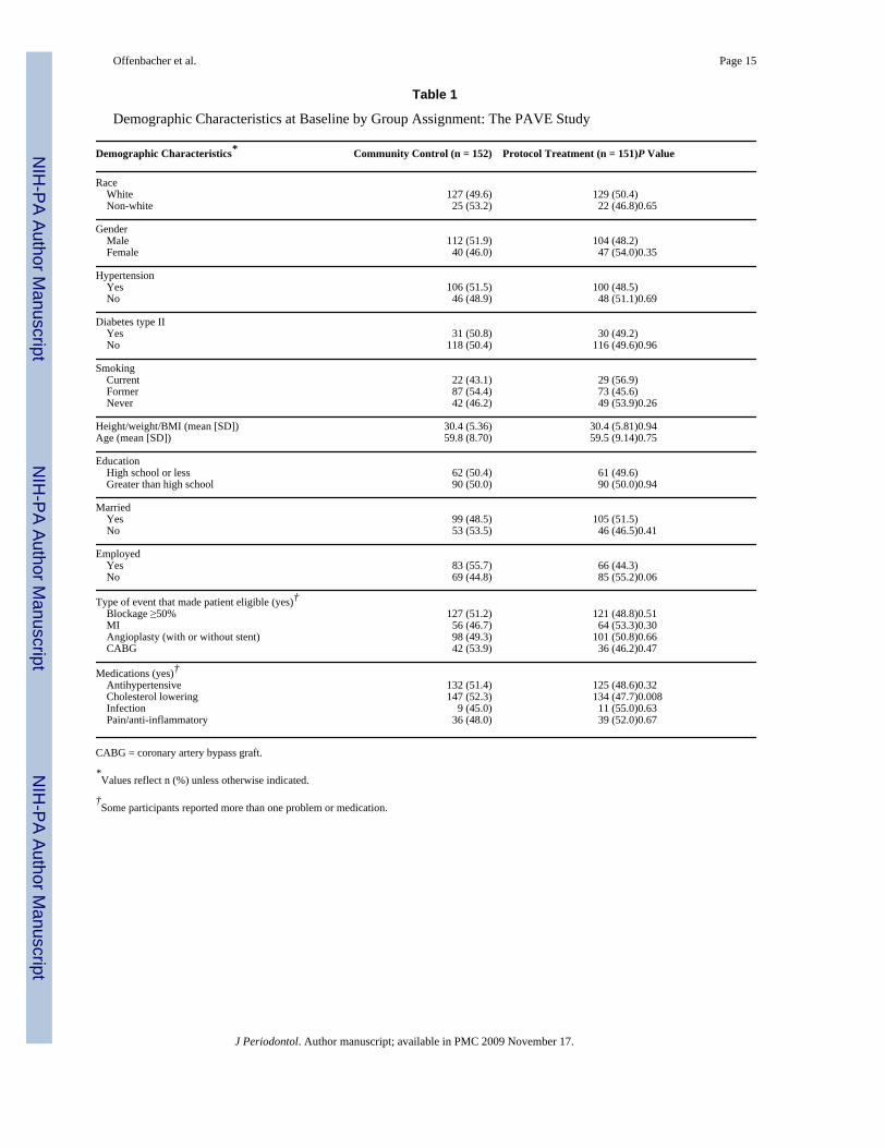
RESULTSThe baseline demographics of the community care and protocol treatment groups appear inTable 1. The baseline characteristics for both groups have similar distributions of race, gender,hypertension, diabetes, smoking history, BMI, mean age, level of education, marital status,employment, and type of CVD event that qualified them for the study. There were asignificantly greater number of subjects on cholesterol-lowering drugs in the community caregroup with 96.7% of subjects (147 of 152) taking these medications compared to 88.7% ofsubjects in the protocol treatment group. Further analyses of this difference showed that thisvariation in medication-usage patterns was not a significant modifier of any of the periodontalor inflammatory outcomes in this study, and no post hoc adjustments were indicated.
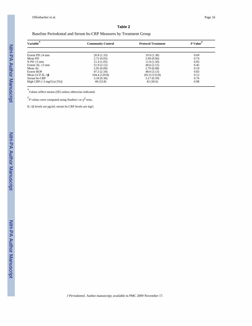
In Table 2, the baseline periodontal measurements for the community care and the protocoltreatment groups show that they were well matched in terms of disease severity at baseline asindicated by measures of extent PD ≥4 mm, mean PD, mean AL, and extent BOP. Plaqueindices and gingival indices were also similar for both groups (data not shown). The meanGCF-
Offenbacher et al. Page 5
J Periodontol. Author manuscript; available in PMC 2009 November 17.
NIH
-PA Author Manuscript
NIH
-PA Author Manuscript
NIH
-PA Author Manuscript

IL-1β levels were not significantly different at baseline comparing the two groups. Similarly,the baseline mean serum hs-CRP levels and the percentage of subjects with high hs-CRP werenot different. Equal distribution of baseline characteristics between the two comparison groupssuggested that randomization was effective.
The effects of periodontal therapy are shown at 6months in Table 3 and at 1 year in Table 4.The treatment protocol had a significant effect on improving periodontal status at 6 monthscompared to community care (Table 3). The P values are statistically significant for extent PD≥4 mm (P = 0.001) and mean PD(P = 0.0009)without significant effects on mean AL(borderlineat P= 0.052) or extent BOP scores (P = 0.16)adjusting for baseline values for each patient usingANCOVA. Overall, there was an improvement in pocket reduction and a trend for slightlylower bleeding scores and less AL at 6 months comparing the protocol intervention group tothe community care group. The GCF-IL-1β levels showed no significant differences betweengroups at 6months. The effects of periodontal intervention on serum hs-CRP show that therewere no effects of protocol therapy on serum hs-CRP levels (Table 3) at 6months.
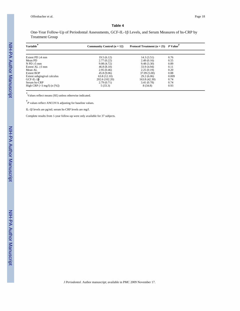
Compared to baseline, both groups showed a trend toward higher values at 6 months, but therewere no significant differences in mean CRP values at 6 months adjusting for baseline valuesfor each subject by ANCOVA. Thus, using intent-to-treat principles, the intervention resultedin an improvement in periodontal health but did not result in a significant change in GCF-IL-1β or serum hs-CRP levels at 6 months. As shown in Table 4, the clinical improvementseen among those treated under protocol was not significantly different from baseline at the 1-year follow-up. At the 1-year visit, GCF and serum data were available for 37 subjects. Table4 demonstrates that, at 1 year, there were no significant differences, using ANCOVA, in anyof the clinical parameters or biomarker values comparing the community care group to theprotocol treatment group after adjusting for baseline values, except for the extent subgingivalcalculus scores (values ≥2), which were significantly lower among those subjects receivingprotocol-mandated care (P = 0.009). Furthermore, the clinical status for these 37 subjects wasnot significantly different from baseline at 1 year, except for lower subgingival calculus scores.However, because relatively few subjects were seen at 1 year, detailed secondary analyses werelimited to the baseline and 6-month data.
Secondary Analyses of hs-CRPOne design issue of the trial that concerned us from the beginning was that subjects assignedto the community care group might receive periodontal care during the study period, anintervention that could potentially diminish any possible differences between the two studygroups. At the onset before randomization, we also extracted hopeless teeth and provided homecare instructions, thereby instituting some level of infection control among those who wereultimately assigned to the community care group. Furthermore, subjects who were identifiedas having periodontal disease and were randomized to the community care group were alsoprovided with referral letters to seek additional care.
Table 5 shows the dental care–use patterns for all study participants. Overall, 48% of thecommunity care control group received one or more preventive or periodontal therapies duringthe 6-month study period, including prophylaxis, scaling and root planing, and periodontalsurgery. Some of this information regarding scaling and root planing was previously published.19 In Table 5, those subjects assigned to the community care group received significantly higherlevels of non-study-care than those receiving therapy on protocol, including total number ofdental visits, numbers of prophylaxis, and subgingival scaling and root planing. These usedifferences suggest that perhaps many who received care as part of the PAVE protocol choseto substitute study-provided care for normal community care dental visits, although they wereadvised to continue to seek routine dental care during the study. The protocol treatmentprovided a net change (SD) of −0.30 (0.45) mm (6-month baseline value) which was more
Offenbacher et al. Page 6
J Periodontol. Author manuscript; available in PMC 2009 November 17.
NIH
-PA Author Manuscript
NIH
-PA Author Manuscript
NIH
-PA Author Manuscript

improvement than that seen among the community care group (−0.13 [0.34] mm; P = 0.001).However, both groups demonstrated a significant improvement in PD at 6 months relative tobaseline (Tables 2 and 3). These findings suggested that secondary analyses to explore theeffects of periodontal care provided to subjects randomized to the community care groupappeared warranted.
As secondary analyses, subjects were regrouped into those who received any periodontaltreatment and those who received no treatment, irrespective of group assignment. Also,subjects were stratified based upon obesity. The community no treatment group was definedas those subjects assigned to the community care group who did not report having anyperiodontal care during the study period (6 months; n = 79). The community care group wasfurther subdivided into those that received a prophylaxis only or a prophylaxis plus scaling androot planing. Three individuals in the community care group received periodontal surgery andwere combined into the community scaling and root planing group. The any treatment group(n = 224) included protocol treatment plus those in the community care group that had eitherprophylaxis, scaling and root planing, or periodontal surgery. In Figure 2, the percentage ofsubjects with high hs-CRP (hs-CRP >3mg/l) are shown for obese and non-obese individualsat 6 months for the community care group, stratified upon type of care provided within thecommunity setting, the protocol treatment group, and the pooled any treatment group. Therewere two striking findings. First, there was no difference in the proportion of subjects whohave elevated hs-CRP irrespective of treatment among those 73 obese individuals (BMI >30).It appears that periodontal treatment of obese individuals with high hs-CRP had no effect inthe prevalence of elevated hs-CRP levels at 6months. Secondly, there was a marked lowerproportion of subjects with hs-CRP ≥3 mg/l at 6 months among those non-obese subjects whoreceived either a community prophylaxis or scaling and root planing, whether assigned tocommunity care or protocol. Among non-obese individuals in the community who receivedno treatment, 43.5% had high hs-CRP at 6 months. In contrast, there was a strikingly lowerproportion of subjects with high hs-CRP among those subjects who received a prophylaxis(6.25%; P = 0.009), scaling and root planing (8.3%; P = 0.03), and protocol therapy (22.2%;P = 0.04). It is noteworthy that these two community treatment groups also demonstratedclinical improvements at 6 months relative to baseline. For example, the mean (SE) PD changein millimeters for the four groups was: no treatment (−0.07 [0.06]), prophylaxis (−1.17 [0.06]),scaling and root planing (−0.20 [0.08]), and protocol treatment (−0.30 [0.03]). Combining alltreated, non-obese subjects into the any treatment group showed, with statistical significance,a lower proportion of subjects with high hs-CRP from43.5% in the no treatment group to 17.6%(P = 0.006) in the any treatment group.
Logistic regression models were used to compute the ORs shown in Table 6 for having highhs-CRP (>3 mg/l) at 6 months based upon periodontal treatment provided either in thecommunity setting or on protocol compared to the community no treatment group as thereferent population stratifying by obesity or using all treated subjects. As illustrated in Figure2, there were no significant findings regarding the risk for high hs-CRP among obeseindividuals irrespective of treatment category. However, any treatment among all subjectsresulted in a statistically significant 2.38-fold lower odds for high hs-CRP (OR = 0.42 [95%CI = 0.18 to 0.995]). The effects were even stronger among the non-obese subjects, with a3.85-fold lower odds for having high hs-CRP at 6 months with any treatment (OR = 0.26 [95%CI = 0.09 to 0.72]). The community-provided prophylaxis showed a significant reduction inOR among non-obese subjects, whereas the lower OR (0.09) seen with community scaling androot planing was not statistically significant among non-obese individuals. The protocoltreatment had no effect among the obese individuals but significantly lowered the OR to 0.34(95% CI = 0.12 to 0.98) among the non-obese subjects, representing a statistically significant2.9-fold reduction in the number of subjects with high hs-CRP at 6 months compared to thosenon-treated individuals in the community setting.
Offenbacher et al. Page 7
J Periodontol. Author manuscript; available in PMC 2009 November 17.
NIH
-PA Author Manuscript
NIH
-PA Author Manuscript
NIH
-PA Author Manuscript
DISCUSSIONGeneral Comments Regarding Study Design and Implementation
This pilot study resulted in several important operational and outcome assessment insights.During the first year of the trial, the PAVE enrollment was inadequate because the periodontaldisease levels for eligibility were initially too stringent, and the logistic engagement ofcardiovascular-qualified potential subjects into the dental component of the investigation wasnot convenient for patients. Periodontal disease criteria were lessened, and we learned thatdental screening must be performed in cardiology settings rather than as a two-step referralprocess. In the PAVE trial, the sample size was not designed to be adequate to test thehypothesis that periodontal treatment reduces the incidence of secondary cardiovascularevents, but it was adequately powered to test the hypothesis that periodontal treatment mayimprove periodontal status, lower GCF-IL-1β, and reduce the serum level of CRP at 6 monthspost-treatment compared to the community care group. Although relatively few subjects wereavailable for analysis at 1 year due to slow initial enrollment, several significant trends can beseen at 1 year that warrant discussion. In addition, the preliminary findings provide furtherguidance for future study design.
Effects of Periodontal Therapy on Periodontal Status and GCF-IL-1β LevelsThe treatment protocol resulted in a significant improvement in periodontal status at 6 monthsas assessed by reduction of PD, but there was little or no change in attachment levels, BOP, orsubgingival calculus. This unexpected result suggested that a more rigorous protocol fortreatment may be necessary in a population of cardiovascular patients. The effects of treatmentwere attenuated at 1 year. This finding is consistent for treatments provided in a communitysetting in which close patient follow-up including oral hygiene and supportive maintenance isnot provided at intervals that are more frequent than 6 months. Subjects were initially identifiedas cardiac patients who were asked to participate in a dental trial and were not initially patientsseeking periodontal care, which may in part explain the relatively modest response toperiodontal therapy. Furthermore, many patients who were randomized to the therapy armapparently skipped community dental appointments, as their non-study dental use wassignificantly lower (specifically, 57% of the rate) than the group randomized to communitycare. This finding points to the need in future trials to provide amore stringent maintenanceprogram to maintain a more stable improvement in periodontal status in this secondary cardiacprevention model.
Although there were not significant differences in GCF-IL-1β as a result of periodontal therapyat 6 months, GCF-IL-1β responses to scaling and root planing are usually short term, occurringshortly after treatment (i.e., 4 to 6 weeks) and are highly correlated to clinical changes.27 Thus,the reestablishment of periodontal pockets and bleeding scores at 6 months would be expectedto result in a reestablishment of GCF-IL-1β levels. The proportion of the community care group(48%) that received some form of preventive or periodontal care and demonstrated clinicalimprovement was an unexpected outcome. In our previous studies on subjects who were inmedical studies but enrolled in secondary dental studies (e.g., the Piedmont Study28 or the OralConditions and Pregnancy Study29), subjects showed much lower rates of dental care–seekingbehaviors when informed of their periodontal disease, closer to arangeof12%to15%(unpublished data). This may be a reflection of this particular patient population who are morehealth care motivated due to their recent cardiovascular event or perhaps amore generalappreciation for the potential linkages between oral health and CVD than in the population atlarge. It is important to note that many patients had some level of infection control prior torandomization, and this alone may have prompted many patients to seek additional communitycare. Irrespective of the reasons, the effects of community care are significant because theyresulted in clinical improvements and a reduction in systemic inflammation among those
Offenbacher et al. Page 8
J Periodontol. Author manuscript; available in PMC 2009 November 17.
NIH
-PA Author Manuscript
NIH
-PA Author Manuscript
NIH
-PA Author Manuscript

individuals who have high hs-CRP. This finding points to an important design issue for futurestudies in that the level of community care provided to control subjects, both preventive andperiodontal therapy, needs to be considered. In a community-based trial, treatments providedby private practitioners cannot be suspended, and ethically, patients who are newly diagnosedas having periodontal disease must be informed and advised to seek care. Thus, the level ofperiodontal care received by patients within the community remains a wild-card variable thatcan be monitored but not readily controlled in community-based studies and is likely to affectall intent-to-treat analyses.
Effects of Periodontal Therapy on Serum hs-CRPFrom an intent-to-treat perspective, the pilot intervention therapy did not result in a significantreduction in mean hs-CRP or a reduction in the proportions of individuals with high hs-CRP(>3 mg/l) compared to the community care group. Several previous cross-sectional studies23,25,30 indicated that periodontal disease was associated with elevated serum levels of hs-CRP.Although serum CRP elevation is a nonspecific marker of the acute phase response that canbe elicited by a broad range of stimuli, elevated hs-CRP has been demonstrated to be predictiveof CVD events including myocardial infarction and stroke.25 Furthermore, several studies13,14,31 that include periodontal treatment demonstrated a lowering of the serum CRP levelfollowing periodontal treatment, but not all studies show reductions in systemic inflammationfollowing periodontal care.32 This evidence suggested that periodontal treatment may reducethe levels of serum CRP in certain circumstances and, therefore, could potentially be evaluatedas an intermediate surrogate endpoint in a pilot intervention trial that would be underpoweredto demonstrate an effect on cardiovascular events. A study by Slade et al.24 demonstrated thatboth periodontal disease and obesity were associated with increased serum hs-CRP levels andthat there was a significant interaction between obesity and periodontal disease on the serumhs-CRP level. The cross-sectional study24 could imply that treating periodontal disease amongpeople with obesity might have no effect on lowering the serum hs-CRP level unless the patientalso lost weight at the same time. In the PAVE study, obesity was associated with higher levelsof hs-CRP as reflected in the baseline differences in hs-CRP levels comparing obese and non-obese subjects (3.81 [2.99 SD] versus 1.91 [2.07 SD] mg/l, respectively; P <0.0001).Furthermore, in the PAVE study, the hs-CRP was non-responsive to the effects of periodontaltherapy among obese individuals, confirming the fact that obesity overwhelms the mildelevation of hs-CRP that is elicited by periodontal disease. This is potentially due to the well-established concept that adipose tissue is both a fat-storage organ as well as a source ofinflammatory adipokines that serve to increase serum markers of inflammation, including hs-CRP.12 Similar concerns were possible for other factors that could potentially modify serumhs-CRP levels, including concomitant infections or inflammatory conditions, gender, smoking,and diabetes. Adjusting for these confounders in our intent-to-treat study design, we found anon-significant overall treatment effect of lowering the prevalence of elevated CRP at 6 months(>3 μg/l; OR = 0.55 [95%CI = 0.24 to 1.24]). Thus, whereas the effect of periodontal treatmenton lowering serum CRP may be demonstrated in a well-controlled clinical trial, it was not clearwhether periodontal treatment could have an impact on hs-CRP levels in a community settingbecause periodontal disease is just one of the potential stressors that can activate the acutephase response. We would suggest that, in clinical trials conducted in university settings, studydesigns select subjects with more severe periodontal disease and higher serum hs-CRP valuesupon randomization, and the level of care provided to controls is minimal in order to maximizethe potential difference between treated and untreated groups. Indeed, the results from thePAVE study suggest that even a single prophylaxis preventative therapy visit could have aneffect on lowering the prevalence of high hs-CRP (Fig. 2) at 6 months in non-obese individuals.Furthermore, in the PAVE study, the proportion of non-obese individuals with high hs-CRPwas lower at 6 months, irrespective of the treatment provided.
Offenbacher et al. Page 9
J Periodontol. Author manuscript; available in PMC 2009 November 17.
NIH
-PA Author Manuscript
NIH
-PA Author Manuscript
NIH
-PA Author Manuscript
The PAVE findings on the effects of treatment on hs-CRP need to be interpreted with cautionbecause they are based on secondary analyses. Nonetheless, several important trends seem tobe emerging regarding the effects of periodontal therapy on hs-CRP that may be important forfuture study design.
First, control subjects must be truly untreated individuals, because even minimal care such asprophylaxis alone appears to have an impact on reducing high hs-CRP. Secondly, the effectsof treatment are not uniform across the spectrum of hs-CRP values. Typically, hs-CRP valueswithin the population are not normally distributed and are often categorized into three levels:hs-CRP values between 0 and <1mg/l are categorized as normal; values from 1 to 3 mg/l areintermediate; and values >3 mg/l are high. Hs-CRP values above 10 mg/l are suggestive ofacute infection or stress that would be transient and not reflective of the chronic level.Therefore, patients with levels >10 mg/l are advised to have repeat values within a week toallow the transient peak to subside to chronic homeostatic levels.33 In some ways, periodontaldisease appears to operate within the range of 1 to 10 mg/l and can contribute to drift in hs-CRP within this range depending upon biofilm load, periodontal status, and periodontal diseaseactivity. Close examination of the data on the effects of treatment suggest that there is littlechange in the chronic steady-state level of hs-CRP following care. The effect of periodontaltherapy appears to prevent any increase in hs-CRP to levels >3 mg/l, a trend that is commonamong patients with periodontal disease. Non-obese patients without periodontal disease orother infectious or inflammatory challenges will typically have hs-CRP levels <1 mg/l andremain in the normal range. Patients with periodontal disease have hs-CRP values that arehigher (with values, on average, of 2.3 mg/l per 10% increase in extent PD ≥4measurements23) and, upon repeat measurements, show time-dependent variations. Thus,preventive and periodontal treatments appear to lower hs-CRP levels below 3 mg/l and alsoprevent the upward drift of hs-CRP above 3 mg/l that occurs among subjects with untreatedperiodontal disease. It appears likely that the upward shifts in hs-CRP levels seen amonguntreated periodontitis patients may be mediated by infectious or inflammatory eventsoccurring within the periodontium, but that remains to be proven. It is interesting to note that,at 1 year, the OR for high hs-CRP is 0.54 (95% CI = 0.10 to 2.78) for any treatment comparedto the community no treatment group, adjusting for BMI. This is not statistically significant,but the trend suggests that the effects of treatment on hs-CRP may potentially persist >6 months.
CONCLUSIONSCollectively, these data suggest that periodontal therapy may lower hs-CRP levels among non-obese cardiovascular patients if the levels are >3mg/l initially, and it may prevent a drift tolevels >3mg/l for those that are in the intermediate range of 1 to 3mg/l. It does not appear thatperiodontal therapy diminishes hs-CRP levels among individuals with hs-CRP levels <3 mg/l in this patient population. The chronic burden of CVD as an inflammatory process may aloneserve to keep a mild elevation of hs-CRP present, irrespective of the added burden ofperiodontal disease. Obesity acts to increase hs-CRP levels and to possibly nullify anyperiodontal treatment effects on hs-CRP levels. These data indicate that, overall, there weresignificant improvements in periodontal clinical health at 6 months following the periodontalintervention compared to community care. In our previous publications of this study,19,34 wedemonstrated that cardiovascular patients can be successfully recruited through cardiologypractices and successfully randomized and that cardiovascular events can be monitored for 1year post-therapy and endpoints adjudicated. Thus, this pilot study establishes feasibility andimportant insight for designing future interventional trials.
Offenbacher et al. Page 10
J Periodontol. Author manuscript; available in PMC 2009 November 17.
NIH
-PA Author Manuscript
NIH
-PA Author Manuscript
NIH
-PA Author Manuscript
AcknowledgmentsThe authors acknowledge the support of NIDCR grant U01 DE13940 awarded to Dr. Genco, principal investigator.Support for analyses of the GCF-IL-1β and serum hs-CRP was provided, in part, by grants M01RR00046 andUL1RR025747 from the National Center of Research Resources, National Institutes of Health, awarded to Drs.Offenbacher and Beck. We also express our special appreciation to the DSMB members, including Drs. C. Furberg,S. Gansky, F. Macrina, J. Mason, and S. Socransky, and staff at the field centers and the subjects who participated inthe study.
References1. DeStefano F, Anda RF, Kahn HS, Williamson DF, Russell CM. Dental disease and risk of coronary
heart disease and mortality. BMJ 1993;306:688–691. [PubMed: 8471920]2. Beck JD, Offenbacher S. Systemic effects of periodontitis: Epidemiology of periodontal disease and
cardiovascular disease. J Periodontol 2005;76(11 Suppl):2089–2100. [PubMed: 16277581]3. Beck JD, Elter JR, Heiss G, Couper D, Mauriello SM, Offenbacher S. Relationship of periodontal
disease to carotid artery intima-media wall thickness: The atherosclerosis risk in communities (ARIC)study. Arterioscler Thromb Vasc Biol 2001;21:1816–1822. [PubMed: 11701471]
4. Nakib SA, Pankow JS, Beck JD, et al. Periodontitis and coronary artery calcification: Theatherosclerosis risk in communities (ARIC) study. J Periodontol 2004;75:505–510. [PubMed:15152812]
5. Czerniuk MR, Górska R, Filipiak KJ, Opolski G. Inflammatory response to acute coronary syndromein patients with coexistent periodontal disease. J Periodontol 2004;75:1020–1026. [PubMed:15341362]
6. Elter JR, Offenbacher S, Toole JF, Beck JD. Relationship of periodontal disease and edentulism tostroke/TIA. J Dent Res 2003;82:998–1001. [PubMed: 14630902]
7. Bahekar AA, Singh S, Saha S, Molnar J, Arora R. The prevalence and incidence of coronary heartdisease is significantly increased in periodontitis: A meta-analysis. Am Heart J 2007;154:830–837.[PubMed: 17967586]
8. Mustapha IZ, Debrey S, Oladubu M, Ugarte R. Markers of systemic bacterial exposure in periodontaldisease and cardiovascular disease risk: A systematic review and meta-analysis. J Periodontol2007;78:2289–2302. [PubMed: 18052701]
9. Beck JD, Offenbacher S. Relationships among clinical measures of periodontal disease and theirassociations with systemic markers. Ann Periodontol 2002;7:79–89. [PubMed: 16013220]
10. Beck JD, Eke P, Lin D, et al. Associations between IgG antibody to oral organisms and carotid intima-medial thickness in community-dwelling adults. Atherosclerosis 2005;183:342–348. [PubMed:15893320]
11. Desvarieux M, Demmer RT, Rundek T, et al. Relationship between periodontal disease, tooth loss,and carotid artery plaque. The oral infections and vascular disease epidemiology study (INVEST).Stroke 2003;34:2120–2125. [PubMed: 12893951]
12. Hansson GK, Robertson AK, Söderberg-Nauclér C. Inflammation and atherosclerosis. Annu RevPathol 2006;1:297–329. [PubMed: 18039117]
13. D’Aiuto F, Parkar M, Andreou G, et al. Periodontitis and systemic inflammation: Control of the localinfection is associated with a reduction in serum inflammatory markers. J Dent Res 2004;83:156–160. [PubMed: 14742655]
14. D’Aiuto F, Nibali L, Parkar M, Suvan J, Tonetti MS. Short-term effects of intensive periodontaltherapy on serum inflammatory markers and cholesterol. J Dent Res 2005;84:269–273. [PubMed:15723869]
15. Elter JR, Hinderliter AL, Offenbacher S, et al. The effects of periodontal therapy on vascularendothelial function: A pilot trial. Am Heart J 2006;151:47.e1–47.e6. [PubMed: 16368290]
16. Mercanoglu F, Oflaz H, Öz O, et al. Endothelial dysfunction in patients with chronic periodontitisand its improvement after initial periodontal therapy. J Periodontol 2004;75:1694–1700. [PubMed:15732873]
17. Seinost G, Wimmer G, Skerget M, et al. Periodontal treatment improves endothelial dysfunction inpatients with severe periodontitis. Am Heart J 2005;149:1050–1054. [PubMed: 15976787]
Offenbacher et al. Page 11
J Periodontol. Author manuscript; available in PMC 2009 November 17.
NIH
-PA Author Manuscript
NIH
-PA Author Manuscript
NIH
-PA Author Manuscript

18. Tonetti MS, D’Aiuto F, Nibali L, et al. Treatment of periodontitis and endothelial function. N EnglJ Med 2007;356:911–920. [PubMed: 17329698]
19. Couper DJ, Beck JD, Falkner KL, et al. The periodontitis and vascular events (PAVE) pilot study:Recruitment, retention and community care controls. J Periodontol 2008;79:80–89. [PubMed:18166096]
20. Beck JD, Couper DJ, Falkner KL, et al. The Periodontitis and Vascular Events (PAVE) pilot study:Adverse events. J Periodontol 2008;79:90–96. [PubMed: 18166097]
21. Silness J, Löe H. Periodontal disease in pregnancy II: Correlation between oral hygiene andperiodontal condition. Acta Odontol Scand 1964;22:121–135. [PubMed: 14158464]
22. Löe H, Silness J. Periodontal disease in pregnancy I: Prevalence and severity. Acta Odontol Scand1963;21:533–551. [PubMed: 14121956]
23. Machtei EE, Christersson LA, Zambon JJ, et al. Alternative methods for screening periodontal diseasein adults. J Clin Periodontol 1993;20:81–87. [PubMed: 8436636]
24. Slade GD, Ghezzi EM, Heiss G, Beck JD, Riche E, Offenbacher S. Relationship between periodontaldisease and C-reactive protein among adults in the atherosclerosis risk in communities study. ArchIntern Med 2003;163:1172–1179. [PubMed: 12767953]
25. Ridker PM. Clinical application of C-reactive protein for cardiovascular disease detection andprevention. Circulation 2003;107:363. [PubMed: 12551853]
26. Zhong Y, Slade GD, Beck JD, Offenbacher S. Gingival crevicular fluid interleukin-1beta,prostaglandin E2 and periodontal status in a community population. J Clin Periodontol 2007;34:285–293. [PubMed: 17378884]
27. Beck JD, Koch GG. Characteristics of older adults experiencing periodontal attachment loss asgingival recession or probing depth. J Periodontal Res 1994;29:290–298. [PubMed: 7932023]
28. Beck JD, Sharp T, Gary GK, Offenbacher S. A 5-year study of attachment loss and tooth loss incommunity-dwelling older adults. J Periodontal Res 2006;32:516–523. [PubMed: 9379319]
29. Lieff S, Boggess KA, Murtha AP, et al. The Oral Conditions and Pregnancy study: Periodontal statusof a cohort of pregnant women. J Periodontol 2004;75:116–126. [PubMed: 15025223]
30. Elter JR, Champagne CM, Offenbacher S, Beck JD. Relationship of periodontal disease and toothloss to prevalence of coronary heart disease. J Periodontol 2004;75:782–790. [PubMed: 15295942]
31. Champagne CM, Buchanan W, Reddy MS, Preisser JS, Beck JD, Offenbacher S. Potential for gingivalcrevice fluid measures as predictors of risk for periodontal diseases. Periodontol 2000 2003;31:167–180. [PubMed: 12657001]
32. Ebersole JL, Machen RL, Steffen MJ, Willmann DE. Systemic acute-phase reactants, C-reactiveprotein and haptoglobin, in adult periodontitis. Clin Exp Immunol 1997;107:347–352. [PubMed:9030874]
33. Loos BG, Craandijk J, Hoek FJ, Wertheim-van Dillen PME, Van der Velden U. Elevation of systemicmarkers related to cardiovascular diseases in the peripheral blood of periodontitis patients. JPeriodontol 2000;71:1528–1534. [PubMed: 11063384]
34. Mattila K, Vesanen M, Valtonen V, et al. Effect of treating periodontitis on C-reactive protein levels:A pilot study. BMC Infect Dis 2002;2:30. [PubMed: 12475397]
Offenbacher et al. Page 12
J Periodontol. Author manuscript; available in PMC 2009 November 17.
NIH
-PA Author Manuscript
NIH
-PA Author Manuscript
NIH
-PA Author Manuscript

Figure 1.PAVE study summary.
Offenbacher et al. Page 13
J Periodontol. Author manuscript; available in PMC 2009 November 17.
NIH
-PA Author Manuscript
NIH
-PA Author Manuscript
NIH
-PA Author Manuscript
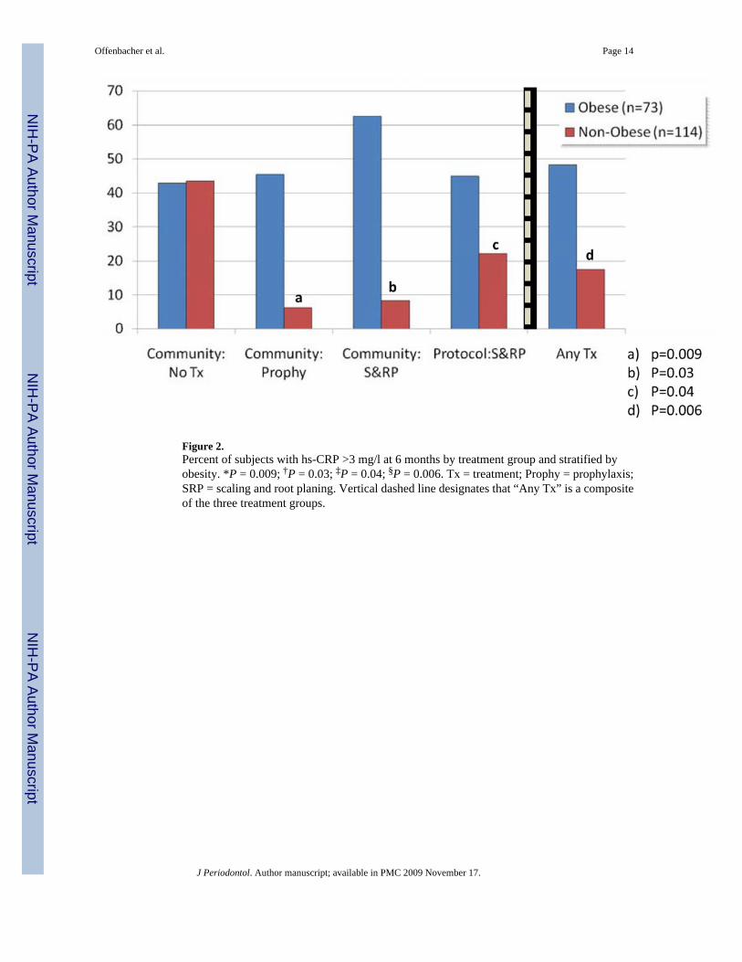
Figure 2.Percent of subjects with hs-CRP >3 mg/l at 6 months by treatment group and stratified byobesity. *P = 0.009; †P = 0.03; ‡P = 0.04; §P = 0.006. Tx = treatment; Prophy = prophylaxis;SRP = scaling and root planing. Vertical dashed line designates that “Any Tx” is a compositeof the three treatment groups.
Offenbacher et al. Page 14
J Periodontol. Author manuscript; available in PMC 2009 November 17.
NIH
-PA Author Manuscript
NIH
-PA Author Manuscript
NIH
-PA Author Manuscript
NIH
-PA Author Manuscript
NIH
-PA Author Manuscript
NIH
-PA Author Manuscript
Offenbacher et al. Page 15
Table 1
Demographic Characteristics at Baseline by Group Assignment: The PAVE Study
Demographic Characteristics* Community Control (n = 152) Protocol Treatment (n = 151)P Value
Race White 127 (49.6) 129 (50.4) Non-white 25 (53.2) 22 (46.8)0.65
Gender Male 112 (51.9) 104 (48.2) Female 40 (46.0) 47 (54.0)0.35
Hypertension Yes 106 (51.5) 100 (48.5) No 46 (48.9) 48 (51.1)0.69
Diabetes type II Yes 31 (50.8) 30 (49.2) No 118 (50.4) 116 (49.6)0.96
Smoking Current 22 (43.1) 29 (56.9) Former 87 (54.4) 73 (45.6) Never 42 (46.2) 49 (53.9)0.26
Height/weight/BMI (mean [SD]) 30.4 (5.36) 30.4 (5.81)0.94Age (mean [SD]) 59.8 (8.70) 59.5 (9.14)0.75
Education High school or less 62 (50.4) 61 (49.6) Greater than high school 90 (50.0) 90 (50.0)0.94
Married Yes 99 (48.5) 105 (51.5) No 53 (53.5) 46 (46.5)0.41
Employed Yes 83 (55.7) 66 (44.3) No 69 (44.8) 85 (55.2)0.06
Type of event that made patient eligible (yes)† Blockage ≥50% 127 (51.2) 121 (48.8)0.51 MI 56 (46.7) 64 (53.3)0.30 Angioplasty (with or without stent) 98 (49.3) 101 (50.8)0.66 CABG 42 (53.9) 36 (46.2)0.47
Medications (yes)† Antihypertensive 132 (51.4) 125 (48.6)0.32 Cholesterol lowering 147 (52.3) 134 (47.7)0.008 Infection 9 (45.0) 11 (55.0)0.63 Pain/anti-inflammatory 36 (48.0) 39 (52.0)0.67
CABG = coronary artery bypass graft.
*Values reflect n (%) unless otherwise indicated.
†Some participants reported more than one problem or medication.
J Periodontol. Author manuscript; available in PMC 2009 November 17.
NIH
-PA Author Manuscript
NIH
-PA Author Manuscript
NIH
-PA Author Manuscript
Offenbacher et al. Page 16
Table 2
Baseline Periodontal and Serum hs-CRP Measures by Treatment Group
Variable* Community Control Protocol Treatment P Value†
Extent PD ≥4 mm 20.8 (1.33) 19.9 (1.38) 0.69Mean PD 2.72 (0.05) 2.69 (0.06) 0.74N PD ≥5 mm 11.4 (1.05) 11.8 (1.50) 0.83Extent AL ≥3 mm 51.9 (2.12) 49.6 (2.15) 0.45Mean AL 2.95 (0.09) 2.79 (0.08) 0.19Extent BOP 47.3 (2.18) 48.0 (3.13) 0.83Mean GCF-IL-1β 194.4 (129.8) 201.0 (133.8) 0.12Serum hs-CRP 3.18 (0.36) 3.17 (0.39) 0.76High CRP (>3 mg/l) (n [%]) 48 (33.8) 42 (30.0) 0.98
*Values reflect means (SE) unless otherwise indicated.
†P values were computed using Student t or χ2 tests.
IL-1β levels are pg/ml; serum hs-CRP levels are mg/l.
J Periodontol. Author manuscript; available in PMC 2009 November 17.

NIH
-PA Author Manuscript
NIH
-PA Author Manuscript
NIH
-PA Author Manuscript
Offenbacher et al. Page 17
Table 3
Six-Month Follow-Up of Periodontal Assessments, GCF-IL-1β Levels, and Serum Measures of hs-CRP byTreatment Group
Variable* Community Control (n = 102) Protocol Treatment (n = 126)P Value†
Extent PD ≥4 mm 16.8 (1.56) 13.3 (1.18)0.001Mean PD 2.57 (0.07) 2.41 (0.06)0.0009N PD ≥5 mm 10.4 (1.36) 7.35 (1.10)0.0003Extent AL ≥3 mm 40.7 (2.71) 42.5 (2.22)0.02Mean AL 2.72 (0.11) 2.52 (0.08)0.052Extent BOP 42.5 (2.56) 38.3 (2.17)0.16Extent subgingival calculus 58.6 (3.95) 42.8 (3.14)0.002GCF-IL-1β 222.3 (22.9) 237.5 (35.0)0.97Serum hs-CRP 3.53 (0.45) 3.12 (0.38)0.97High CRP (>3 mg/l) (n [%]) 31 (34.8) 33 (31.4)0.62
*Values reflect means (SE) unless otherwise indicated.
†P values reflect ANCOVA adjusting for baseline values.
IL-1β levels are pg/ml; serum hs-CRP levels are mg/l.
Six-month follow-up data were only available from 228 subjects.
J Periodontol. Author manuscript; available in PMC 2009 November 17.
NIH
-PA Author Manuscript
NIH
-PA Author Manuscript
NIH
-PA Author Manuscript
Offenbacher et al. Page 18
Table 4
One-Year Follow-Up of Periodontal Assessments, GCF-IL-1β Levels, and Serum Measures of hs-CRP byTreatment Group
Variable* Community Control (n = 12) Protocol Treatment (n = 25) P Value†
Extent PD ≥4 mm 19.5 (6.12) 14.3 (3.51) 0.76Mean PD 2.77 (0.22) 2.49 (0.16) 0.55N PD ≥5 mm 9.08 (4.72) 9.48 (3.30) 0.89Extent AL ≥3 mm 46.8 (8.10) 33.9 (4.94) 0.11Mean AL 2.95 (0.46) 2.25 (0.19) 0.20Extent BOP 45.8 (9.06) 37.09 (5.00) 0.88Extent subgingival calculus 63.8 (12.10) 29.2 (6.06) 0.009GCF-IL-1β 202.6 (102.20) 163.8 (42.30) 0.74Serum hs-CRP 2.79 (0.71) 3.41 (0.78) 0.74High CRP (>3 mg/l) (n [%]) 5 (33.3) 8 (34.8) 0.93
*Values reflect means (SE) unless otherwise indicated.
†P values reflect ANCOVA adjusting for baseline values.
IL-1β levels are pg/ml; serum hs-CRP levels are mg/l.
Complete results from 1-year follow-up were only available for 37 subjects.
J Periodontol. Author manuscript; available in PMC 2009 November 17.

NIH
-PA Author Manuscript
NIH
-PA Author Manuscript
NIH
-PA Author Manuscript
Offenbacher et al. Page 19
Table 5
Dental Use During First 6 Months of the Study
Protocol Treatment Community Control P Value
Number reporting non-studydental visits (%)
54 (36.5) 93 (64.1) <0.0001
Number reporting non-studyprophylaxis (%)
6 (4.1) 54 (37.2) <0.0001
Number reporting non-studysubgingival scaling and rootplaning (%)
3 (2.0) 26 (19.9) <0.0001
J Periodontol. Author manuscript; available in PMC 2009 November 17.

NIH
-PA Author Manuscript
NIH
-PA Author Manuscript
NIH
-PA Author Manuscript
Offenbacher et al. Page 20
Table 6
OR (95% CI) for hs-CRP >3 mg/l at 6 Months
Group Non-Obese* Obese* All Subjects†
Community: no treatment Referent Referent ReferentCommunity: prophylaxis 0.9 (0.01 to 0.79)‡ 1.36 (0.4 to 7.88) 0.34 (0.09 to 1.31)Community: scaling and root planing 0.10 (0.10 to 1.01) 2.55 (0.34 to 19.14) 0.70 (0.19 to 2.56)Protocol: treatment 0.34 (0.12 to 0.98)‡ 0.84 (0.22 to 3.24) 0.39 (0.153 to 0.999)‡Any treatment 0.26 (0.09 to 0.72)‡ 1.05 (0.30 to 3.75) 0.42 (0.181 to 0.995)‡
Baseline characteristics were considered for entering into the adjusted logistic models for elevated hs-CRP (data not shown). Significant baseline
characteristics included smoking, marital status, and BMI. Gender was also included because of its known relationship24 to hs-CRP even though it didnot reach statistical significance in this dataset (P = 0.08).
*Adjusted for smoking, marital status, and gender.
†Adjusted for smoking, marital status, gender, and BMI.
‡Statistically significant.
J Periodontol. Author manuscript; available in PMC 2009 November 17.
Copyright © 2022 FDOKUMEN