Restoration of photosystem II photochemistry and carbon assimilation and related changes in...
11
Journal of Experimental Botany, Vol. 62, No. 3, pp. 895–905, 2011 doi:10.1093/jxb/erq317 Advance Access publication 18 October, 2010 This paper is available online free of all access charges (see http://jxb.oxfordjournals.org/open_access.html for further details) RESEARCH PAPER Restoration of photosystem II photochemistry and carbon assimilation and related changes in chlorophyll and protein contents during the rehydration of desiccated Xerophyta scabrida leaves P. Pe ´ rez 1, *, G. Rabnecz 2 , Z. Laufer 3 , D. Gutie ´ rrez 1 , Z. Tuba 2,3,† and R. Martı´nez-Carrasco 1 1 Institute of Natural Resources and Agrobiology of Salamanca, CSIC, Apartado 257, 37071 Salamanca, Spain 2 Institute of Botany and Ecophysiology, Faculty of Agriculture and Environmental Sciences, Szent Istva ´ n University, Pa ´ ter K. 1., 2103 Go ¨ do ¨ ll } o, Hungary 3 ‘Plant Ecology’ Departmental Research Group of the Hungarian Academy of Sciences, Szent Istva ´ n University, Pa ´ ter K. 1., 2103 Go ¨ do ¨ ll } o, Hungary y Deceased. * To whom correspondence should be addressed. E-mail: [email protected] Received 9 March 2010; Revised 17 September 2010; Accepted 21 September 2010 Abstract Recovery of photosynthesis in rehydrating desiccated leaves of the poikilochlorophyllous desiccation-tolerant plant Xerophyta scabrida was investigated. Detached leaves were remoistened under 12 h light/dark cycles for 96 h. Water, chlorophyll (Chl), and protein contents, Chl fluorescence, photosynthesis–CO 2 concentration response, and the amount and activity of Rubisco were measured at intervals during the rehydration period. Leaf relative water contents reached 87% in 12 h and full turgor in 96 h. Chl synthesis was slower before than after 24 h, and Chla:Chlb ratios changed from 0.13 to 2.6 in 48 h. The maximum quantum efficiency recovered faster during rehydration than the photosystem II operating efficiency and the efficiency factor, which is known to depend mainly on the use of the electron transport chain products. From 24 h to 96 h of rehydration, net carbon fixation was Rubisco limited, rather than electron transport limited. Total Rubisco activity increased during rehydration more than the Rubisco protein content. Desiccated leaves contained, in a close to functional state, more than half the amount of the Rubisco protein present in rehydrated leaves. The results suggest that in X. scabrida leaves Rubisco adopts a special, protective conformation and recovers its activity during rehydration through modifications in redox status. Key words: Chlorophyll fluorescence, desiccation tolerance, non-photochemical quenching, photosynthesis, poikilochlorophylly, relative water content, Rubisco, Xerophyta scabrida. Introduction Desiccation-tolerant (DT) plants can withstand the loss of up to 90–95% of the water of their vegetative tissues and revive when humidity is available, in contrast to the majority of plants (Proctor and Tuba, 2002). Desiccation tolerance entails cellular, biochemical, and molecular changes during dehydration (Vicre ´ et al., 2004), including the accumulation of carbohydrates (Whittaker et al., 2001; Toldi et al., 2009), late embryogenesis-abundant (LEA) proteins (Ingram and Bartels, 1996), and antioxidants (Kranner et al., 2002; Mowla et al., 2002; Vicre ´ et al., 2004), as well as altered expression of target genes and transcription factors (Frank et al., 1998; Ramanjulu and Bartels, 2002). Recovery from the desiccated state is much faster in homoiochlorophyllous DT (HDT) plants such as Abbreviations: Chl, chlorophyll; DT, desiccation tolerant; HDT, homoiochlorophyllous DT; PDT, poikilochlorophyllous DT; RuBP, ribulose-1,5-bisphosphate; RWC, relative water content. ª 2010 The Author(s). This is an Open Access article distributed under the terms of the Creative Commons Attribution Non-Commercial License (http://creativecommons.org/licenses/by- nc/2.5), which permits unrestricted non-commercial use, distribution, and reproduction in any medium, provided the original work is properly cited. at Centro de Información y Documentación Científica on January 18, 2011 jxb.oxfordjournals.org Downloaded from
Transcript of Restoration of photosystem II photochemistry and carbon assimilation and related changes in...
Journal of Experimental Botany, Vol. 62, No. 3, pp. 895–905, 2011doi:10.1093/jxb/erq317 Advance Access publication 18 October, 2010This paper is available online free of all access charges (see http://jxb.oxfordjournals.org/open_access.html for further details)
RESEARCH PAPER
Restoration of photosystem II photochemistry and carbonassimilation and related changes in chlorophyll and proteincontents during the rehydration of desiccated Xerophytascabrida leaves
P. Perez1,*, G. Rabnecz2, Z. Laufer3, D. Gutierrez1, Z. Tuba2,3,† and R. Martınez-Carrasco1
1 Institute of Natural Resources and Agrobiology of Salamanca, CSIC, Apartado 257, 37071 Salamanca, Spain2 Institute of Botany and Ecophysiology, Faculty of Agriculture and Environmental Sciences, Szent Istvan University, Pater K. 1., 2103Godoll}o, Hungary3 ‘Plant Ecology’ Departmental Research Group of the Hungarian Academy of Sciences, Szent Istvan University, Pater K. 1., 2103Godoll}o, Hungary
y Deceased.* To whom correspondence should be addressed. E-mail: [email protected]
Received 9 March 2010; Revised 17 September 2010; Accepted 21 September 2010
Abstract
Recovery of photosynthesis in rehydrating desiccated leaves of the poikilochlorophyllous desiccation-tolerant plant
Xerophyta scabrida was investigated. Detached leaves were remoistened under 12 h light/dark cycles for 96 h.
Water, chlorophyll (Chl), and protein contents, Chl fluorescence, photosynthesis–CO2 concentration response, and
the amount and activity of Rubisco were measured at intervals during the rehydration period. Leaf relative water
contents reached 87% in 12 h and full turgor in 96 h. Chl synthesis was slower before than after 24 h, and Chla:Chlb
ratios changed from 0.13 to 2.6 in 48 h. The maximum quantum efficiency recovered faster during rehydration thanthe photosystem II operating efficiency and the efficiency factor, which is known to depend mainly on the use of the
electron transport chain products. From 24 h to 96 h of rehydration, net carbon fixation was Rubisco limited, rather
than electron transport limited. Total Rubisco activity increased during rehydration more than the Rubisco protein
content. Desiccated leaves contained, in a close to functional state, more than half the amount of the Rubisco
protein present in rehydrated leaves. The results suggest that in X. scabrida leaves Rubisco adopts a special,
protective conformation and recovers its activity during rehydration through modifications in redox status.
Key words: Chlorophyll fluorescence, desiccation tolerance, non-photochemical quenching, photosynthesis, poikilochlorophylly,
relative water content, Rubisco, Xerophyta scabrida.
Introduction
Desiccation-tolerant (DT) plants can withstand the loss of
up to 90–95% of the water of their vegetative tissues and
revive when humidity is available, in contrast to the
majority of plants (Proctor and Tuba, 2002). Desiccationtolerance entails cellular, biochemical, and molecular
changes during dehydration (Vicre et al., 2004), including
the accumulation of carbohydrates (Whittaker et al., 2001;
Toldi et al., 2009), late embryogenesis-abundant (LEA)
proteins (Ingram and Bartels, 1996), and antioxidants
(Kranner et al., 2002; Mowla et al., 2002; Vicre et al.,
2004), as well as altered expression of target genes andtranscription factors (Frank et al., 1998; Ramanjulu and
Bartels, 2002). Recovery from the desiccated state is much
faster in homoiochlorophyllous DT (HDT) plants such as
Abbreviations: Chl, chlorophyll; DT, desiccation tolerant; HDT, homoiochlorophyllous DT; PDT, poikilochlorophyllous DT; RuBP, ribulose-1,5-bisphosphate; RWC,relative water content.ª 2010 The Author(s).
This is an Open Access article distributed under the terms of the Creative Commons Attribution Non-Commercial License (http://creativecommons.org/licenses/by-nc/2.5), which permits unrestricted non-commercial use, distribution, and reproduction in any medium, provided the original work is properly cited.
at Centro de Inform
ación y Docum
entación Científica on January 18, 2011
jxb.oxfordjournals.orgD
ownloaded from
Haberlea rhodopensis (Georgieva et al., 2005, 2007) than in
poikilochlorophyllous DT (PDT) plants such as Xerophyta
scabrida (Tuba et al., 1993a, 1994). The former retain their
chlorophyll (Chl), preserve their photosynthetic apparatus,
and undergo morphological changes during drying that
protect their tissues against oxidative stress (Vicre et al.,
2004). In contrast, the latter lose all of their Chl and
dismantle their photosynthetic apparatus during drying,and they resynthesize these molecules after rehydration
(Tuba et al., 1994, 1998a; Sherwin and Farrant, 1996).
Xerophyta scabrida preserves most of its Chl when dried in
the dark, so most of the loss seems to result from
photooxidative degradation (Tuba et al., 1997). The PDT
strategy evolved in plants that are anatomically complex
and that include the largest in size of all DT species, and it
can be seen as the younger strategy in evolutionary terms(Proctor and Tuba, 2002).
DT plants regain their water content within a time span
ranging from minutes in bryophytes and pteridophytes
(Csintalan et al., 1999) to days in angiosperms (Tuba et al.,
1994; Proctor and Tuba, 2002; Georgieva et al., 2005;
Degl’Innocenti et al., 2008). As could be expected for HDT
plants, the Chl contents, the Chla:Chlb ratio, and the
relative amounts of the Chl–protein complexes remainmostly unchanged in control, desiccated, and rehydrated
leaves (Georgieva et al., 2005, 2007). In remoistened PDT
plants, Chl resynthesis begins after 6–12 h of rehydration
(Tuba et al., 1993a, 1994) at 36% relative water contecnt
(RWC) (Degl’Innocenti et al., 2008), and is completed by
48–72 h at 84% RWC. Synthesis of the photosystem II
(PSII) reaction centre and antenna proteins correlates with
the recovery and increase in photosynthetic capacity (Ingleet al., 2008).
Non-radiative energy dissipation can play an important
protective role during both desiccation and rehydration. In
several mosses (Csintalan et al., 1999) and in Ramonda
serbica (Augusti et al., 2001; Degl’Innocenti et al., 2008),
non-photochemical quenching (NPQ) shows a transient
increase upon remoistening. Maximum quantum efficiency,
Fv/Fm, is completely recovered at 48 h (Degl’Innocentiet al., 2008) or 72 h (Tuba et al., 1994) after rewatering.
Faster increases during rehydration were recorded in Fv/Fm
than in PSII operating efficiency, Fq#/Fm# (also termed
UPSII; Csintalan et al., 1999). Nonetheless, the involvement
in the non-photochemical energy dissipation of basal, non-
radiative decays and of the regulated non-photochemical
energy loss (Baker et al., 2007; Klughammer and Schreiber,
2008) during the rehydration of PDT plants has not beeninvestigated.
Full recovery of photochemical activity in PDT plants
requires the assimilation of CO2 as an acceptor of the
products of photosynthetic electron transport. In the moss
Polytrichum formosum, carbon fixation is completely re-
stored 3 h after rewetting (Proctor et al., 2007) but is
resumed at 12 h (51% RWC), and is not fully re-established
at 48 h (84% RWC) after rehydration in higher plant DTspecies (Tuba et al., 1994; Degl’Innocenti et al., 2008).
Recently, photosynthesis–CO2 concentration responses in
hydrated HDT leaves were reported (Peeva and Cornic,
2009), but information concerning the fate of ribulose-
1,5-bisphosphate carboxylase oxygenase (Rubisco) and the
relative capacities of Rubisco carboxylation and electron
transport in rehydrating PDT plants is scarce. In earlier
studies, desiccated fronds (Harten and Eickmeier, 1986) and
leaves (Daniel and Gaff, 1980) of DT plants conserved from
40% to 62% of the control Rubisco activity. A decrease inRubisco content was observed during dehydration of the C4
DT plant Sporobolus stapfianus (Martinelli et al., 2007),
whereas Rubisco (fraction I) protein did not appear to
decrease relative to hydrated X. viscosa leaves (Daniel and
Gaff, 1980). Consistent with this, it was surmised that the
carboxylating enzymes in X. scabrida would only be
inactivated, but not degraded, during desiccation (Tuba
et al., 1998b). In contrast, Rubisco activity was undetectablebelow 51% RWC (12 h rehydration) in R. serbica leaves
(Degl’Innocenti et al., 2008). Drying-induced disruption of
the electron transport chain causes oxidative stress (Vicre
et al., 2004), which can induce aggregation and polymeriza-
tion, membrane association, and the degradation of
Rubisco (Marın-Navarro and Moreno, 2006). On the other
hand, in stressed Lemna minor fronds Rubisco was not
degraded but gradually became polymerized to inactiveaggregates, accompanied by a reduction in the number of
sulphydryl groups (Ferreira and Shaw, 1989). The Benson–
Calvin cycle enzymes have a tendency to form soluble and
membrane-bound multienzyme complexes (Sainis and Harris,
1986; Gontero et al., 1988, 1993; Sainis et al., 1989; Persson
and Johansson, 1989; Hermoso et al., 1992; Anderson et al.,
1995; Agarwal et al., 2009) with higher catalytic efficiency
and less susceptibility to auto-oxidation and proteolysis thanfree enzymes (Gontero et al., 1988, 1993).
The aim of this study was to determine to what extent the
recovery from desiccation of X. scabrida photosynthesis is
dependent on photochemical and carboxylation capacities.
The hypothesis was that restoration of Rubisco activity
limits the attainment of photosynthetic competence of
rehydrated PDT plants. To test this hypothesis, Chl
fluorescence and photosynthesis–CO2 response curves weredetermined while turgor was being regained. To assess the
carboxylation capacity, the free or aggregated state, as well
as the amount and activity of Rubisco were determined in
desiccated and rehydrating leaves.
Materials and methods
A description of X. scabrida morphology has been provided in anearlier article (Tuba et al., 1993b). Briefly, it is a 40–90 cm high,branched pseudoshrub with perennial leaves. Dry leaves areusually 24–30 cm long, 5–6 mm wide, and folded over along themidrib. In July 2004, desiccated X. scabrida (Pax) Th. Dur. etSchinz branches were collected in Tanzania (Mindu Hill, WSW ofMorogoro town, 6�50.78’S, 37�36.76’E) and were kept in paperbags at room temperature until rehydration and analysis. Centralsections of desiccated leaves having a purple-black or blackish-green coloration were selected for this study. As representative oftime zero inmediately prior to watering, triplicate desiccated leaveswere briefly immersed in water in a vacuum desiccator to saturate
896 | Perez et al. at C
entro de Información y D
ocumentación C
ientífica on January 18, 2011jxb.oxfordjournals.org
Dow
nloaded from
the intercellular air spaces with water. Subsequently the leafmaterial was blotted, weighed, frozen in liquid nitrogen, andstored at –80 �C for Chl, protein, and Rubisco activity measure-ments (see below). Previous experience (Tuba et al., 1993b) hasshown that placing whole plants with their roots in water does notresult in a recovery response in X. scabrida, because the roots aredry and unable to transport water to the leaves; only a directrewatering of the leaves by immersion in water led to regreening.Moreover, in the natural habitat, new root development and wateruptake were preceded by the rehydration and regreening of theleaves. Consequently, in the present experiments, additional leafsamples were rehydrated by submerging them in a 10.0 l glass tankfilled with tap water and aerated with a pump (Tuba et al., 1993a,1994). The container was placed in a growth chamber witha 21 �C/15 �C day/night temperature, under a 340 lmol m�2 s�1
photon flux density in a 12 h photoperiod (modified after Tubaet al., 1994). The water was changed daily.
Water contents
Triplicate samples of desiccated leaves were weighed before andafter drying in an oven for 48 h at 60 �C; the second of theserecordings was taken as the dry weight. This was preferred to theoven-dry weight after full rehydration, which may be affected bylosses during rewatering due to respiration or release of solublecompounds. Additional leaves (in triplicate) that were submergedin water were blotted and their fresh weight was determined at 12,24, 48, 72, 96, and 120 h after the start of rehydration. No furtherweight gain was recorded after 96 h and this was considered as theturgid weight. The RWCs at successive times in the rehydrationperiod were determined as (fresh weight–dry weight)3100/(turgidweight–dry weight). Chl and protein contents, and Rubiscoactivity were determined in other leaves sequentially sampledduring rehydration (see below) and were expressed on a turgidweight basis. The latter was estimated from the fresh weight ofthese leaf samples and the water contents measured in the samplesused for RWC measurements.
Chl fluorescence and gas exchange measurements
For Chl fluorescence and CO2 assimilation measurements, tripli-cate leaf samples kept in water were collected between 3 h and 8 hafter the start of the photoperiod at the times indicated above andplaced in the fluorometer leaf clip or the infrared gas analyser(IRGA) leaf chamber (see below) with both ends of the leaveswrapped in moistened filter paper. After measurements, the leafsamples were returned to the water container in the growth roomfor a 30 min adaptation period prior to harvesting for leaf analyses(see below). Chl fluorescence was measured with a modulatedfluorometer (PAM-2000, Walz, Effeltrich, Germany). Leaf sectionswere kept in the dark for 20 min with leaf clips (Gutierrez et al.,2009), after which dark-adapted state fluorescence parameters weremeasured. Fo was recorded and a saturating flash of light (;8000lmol m�2 s�1) was applied for 0.8 s to determine Fm. Fo and Fm,respectively, represent the minimal and maximal fluorescence inthe dark-adapted state, and Fv/Fm [(Fm–Fo)/Fm] represents themaximum quantum efficiency. Light-adapted leaves were illumi-nated with the red actinic light source of the fluorometer to obtainan irradiance of 1500 lmol m�2 s�1. Saturating light pulses weregiven every 20 s until steady-state Chl fluorescence parametervalues were obtained, the fluorescence values being recordedimmediately before (F#, steady-state fluorescence) and after (Fm#,maximal fluorescence in the light) each pulse. Then, the leaf wascovered with a black cloth, the actinic light was switched off, andan infrared light was switched on for 3 s to quickly reoxidize thePSII centres and measure Fo#, the minimal fluorescence with anNPQ similar to that found in the steady-state under light. Theequipment determines Fq#/Fm# [(Fm#–F#)/Fm#], which is the PSIIoperating efficiency (also termed UPSII) (Baker et al., 2007). ThePSII efficiency factor Fq#/Fv# (also termed qP) [(Fm#–F#)/ (Fm#–Fo#)]
and Fv#/Fm# [(Fm#–Fo#)/Fm#], the PSII maximum efficiency underlight, were calculated. The fraction of PSII centres in the openstate, qL, equates to (Fq#/Fv#) (Fo#/F#). The quantum yield of basal,non-radiative decays, UNO, is 1/[NPQ+1+qL(Fm/Fo–1)], whereNPQ is (Fm/Fm#)–1, and the quantum yield of non-photochemicalquenching, UNPQ, is 1–(Fq#/Fm#)–UNO (Kramer et al., 2004).
Light-saturated photosynthesis–CO2 response curves of leaveswere recorded at the same times and with the same samplingscheme as Chl fluorescence. Measurements were carried out withan air flow rate of 300 ml min�1, 1500 lmol m�2 s�1 irradiance,and a 1.660.23 kPa vapour pressure deficit, using a 1.7 cm2
window leaf chamber connected to a portable IRGA (CIRAS-2,PP Systems, Hitchin, Herts, UK) with differential operation in anopen system. Temperature was kept at 25 �C with the Peltiersystem of the IRGA. The air CO2 concentration was decreased infour steps from 34 Pa to 6 Pa and then increased from 34 Pa to180 Pa in six steps. Chloroplast CO2 concentration, Cc, themaximum carboxylation rate allowed by Rubisco, Vcmax, and therate of photosynthetic electron transport, J, were determined fromthe photosynthesis responses to CO2 with the Rubisco kineticparameters and the Excel utility of Sharkey et al. (2007).
Rubisco activity assay
Triplicate leaf samples that had been equilibrated in aerated waterin the growth chamber after Chl fluorescence and gas exchangemeasurements were blotted dry, rapidly transferred in situ to liquidnitrogen, and stored at –80 �C until analysed. Rubisco activity wasassayed on the basis of the procedure described by Lilley andWalker (1974), modified by Ward and Keys (1989) and Sharkeyet al. (1991). Aliquots (80 mg) of frozen leaves were ground ina mortar with liquid nitrogen, extracted with 4 ml of 100 mMN,N-bis(2-hydroxyethyl)glycine (Bicine)-NaOH (pH 7.8), 10 mMMgCl2, 0.5 mM dithiothreitol (DTT), 1 mM ethylenediaminetetra-acetic acid (EDTA), 1 mM ethylene glycol tetraacetic acid(EGTA), 1% (v/v) Triton X-100, 0.25% (w/v) bovine serumalbumin (BSA), 20% (v/v) glycerol, 1 mM benzamidine, 1 mMe-aminocaproic acid, 10 lM leupeptin, and 1 mM phenylmethyl-sulphonyl fluoride (PMSF), and then centrifuged at 13 000 g. Thetotal time from extraction to the assay of initial Rubisco activitywas <2.5 min. Activity was assayed by adding extract (40 ll) toa mixture of 100 mM Bicine (pH 8.2), 20 mM MgCl2, 10 mMNaHCO3, 18 mM KCl, 0.6 mM ribulose-1,5-bisphosphate(RuBP), 0.2 mM NADH, 1 mM ATP, 5 mM creatine phosphate,25 U ml�1 phosphocreatine kinase, 47U ml�1 phosphoglyceratekinase, 47 U ml�1 glyceraldehyde 3-phosphate dehydrogenase,10 mM DTT, 1 mM EDTA, 0.02% (w/v) BSA (800 ll totalvolume) and recording the decrease in absorbance at 340 nmminus 400 nm for 40–60 s, at a stoichiometry of 2:1 betweenNADH oxidation and RuBP carboxylation. The spectrophotome-ter cell compartment was thermostated with a circulating waterbath. To assay total Rubisco activity, an aliquot of the extract wasincubated with NaHCO3 and MgCl2 for 10 min at room temper-ature before the addition of coupling enzymes and NADH; thereaction was started by adding RuBP. The activation state wasestimated as initial activity, as a percentage of total activity.Activation and assays were performed either at room temperatureor at 35 �C. Commercial coupling enzymes suspended in ammo-nium sulphate were precipitated by centrifugation and dissolved in20% glycerol (Sharkey et al., 1991). With the assay bufferdescribed, the initial lag in the reaction reported by others (Wardand Keys, 1989; Sharkey et al., 1991) was not observed.
Chlorophyll and protein analysis
Total Chl, Chla, and Chlb in 80% acetone extracts of frozentriplicate subsamples were determined according to Arnon (1949),who used the extinction coefficents for Chla (16.75 l g�1 cm�1 and82.04 l g�1 cm�1 at 645 nm and 663 nm, respectively) and Chlb(45.6 l g�1 cm�1 and 9.27 l g�1 cm�1 at 645 nm and 663 nm,
Restoration of photosynthesis in rehydrating Xerophyta scabrida leaves | 897 at C
entro de Información y D
ocumentación C
ientífica on January 18, 2011jxb.oxfordjournals.org
Dow
nloaded from
respectively) given by MacKinney (1941). The soluble proteins wereextracted by grinding frozen leaf subsamples to a fine powder in50 mM N-[tri(hydroxymethyl)methyl] glycine (Tricine) buffer (pH8.0), 2 mM EDTA, 10 mM NaCl, 5 mM MgCl2, 75 mM sucrose,5 mM e-aminocaproic acid, 2 mM benzamidine, 8 mM b-mercap-toethanol (+bme), and 2 mM PMSF for 5 min on ice. This wasfollowed by centrifugation at 12 500 g at 4 �C for 30 min. Proteinconcentrations were measured in the decanted supernatant(Bradford, 1976), and 5 vols of cold acetone were added to analiquot containing 200 mg of protein, which was left overnight inthe freezer. The samples were then centrifuged at 12 000 g at 4 �Cfor 15 min. The acetone was allowed to evaporate off. Theprecipitates were dissolved in 65 mM TRIS-HCl (pH 6.8), 3 Msucrose, 0.6 M bme, 5% sodium dodecylsulphate (SDS, w/v), and0.01% bromophenol blue at 96 �C for 7 min. The samples were thencooled to room temperature and aliquots of the SDS-dissociatedextracts, containing 15 lg of protein, were loaded onto a 12.5%SDS–polyacrylamide gel (Mini-Protean 3 Cell, Bio Rad). Thisprotein amount was within the range of linear response of opticaldensity to the concentration of BSA standard (66 kDa), accordingto previous calibration measurements. The solubilized proteins wereseparated by SDS–PAGE (Laemmli, 1970) using a 0.75 mm thickgel (12.5% resolving, 4% stacking). Electrophoresis was carried outat room temperature at a constant 200 V. The gels were fixed in500:150:75 (v/v/v) water–methanol–acetic acid mixture for 75 min,stained in EZ Blue Gel Staining (Sigma) solution for 2 h, andsubsequently rinsed in water to remove excess stain. Finally, the gelswere scanned with a high-resolution scanner (Scanjet G4050,Hewlett Packard, Spain) and the amount of Rubisco subunits wasdetermined by densitometry with image analysis software (ImageQuant, Molecular Dynamics, GE Healthcare, Spain). Alternatively,when electrophoresis with non-reducing (–bme) gels was performed,the frozen leaf samples were extracted in buffer without bmecontaining 10 mM iodoacetamide to prevent the formation ofdisulphide bonds, before the addition of SDS loading buffer withoutbme and boiling. To a separate aliquot, bme was added to a finalconcentration of 0.6 M, as a control of the same samples underreducing (+bme) conditions (Marın-Navarro and Moreno, 2006).
Following electrophoresis, additional gels were blotted for75 min to PVDF membranes (Bio-Rad, Madrid, Spain) pre-wettedin methanol and equilibrated in 25 mM TRIS, 192 mM glycine,
20% methanol, and 0.1% SDS (pH 8.3) using an electrotransfer cell(Mini Trans-Blot, Bio-Rad, Madrid, Spain) at 400 mA. The blotswere blocked immediately following transfer in 2% ECL Advanceblocking reagent (GE Healthcare, Barcelona, Spain) in 20 mMTRIS, 137 mM NaCl (pH 7.6) with 0.1% (v/v) Tween-20 (TBS-T)for 1 h at room temperature with shaking. Blots were briefly rinsedtwice in TBS-T, then probed with 1:50 000 diluted polyclonalantibody specific for the large Rubisco subunit (Rubisco quantita-tion kit, Agrisera, Vannas, Sweden) for 1 h at room temperaturewith shaking. The antibody solution was decanted and the blotwas briefly rinsed twice, and then washed once for 15 min andthree times for 5 min in TBS-T at room temperature with shaking.Next, the blots were incubated in secondary antibody (anti-chickenIg Y peroxidase conjugate, Sigma, Spain) diluted at 1:160 000 in2% ECL Advance blocking solution for 1 h at room temperaturewith shaking. The blots were then washed as above and developedfor 5 min with ECL Advance detection reagent according to themanufacturer’s instructions. Images of the blots were obtainedusing a CCD imager (Fluor-S Multilmager, Bio-Rad).
Statistical analyses
One-way analyses of variance with sampling time as a factor werecarried out with the GenStat 6.2 (VSN International Ltd, HemelHempstead, UK) statistical software. From these analyses, thestandard errors of the differences (SEDs), and the least significantdifferences of means (three replicates) at P <0.05 probability werederived. The latter were used for the inspection of differencesamong values for each sampling time. The homogeneity ofvariance and the significance of the analysis were not modifiedappreciably by using arcsine-square root transformation of per-centage variables (Rubisco activation and percentage in solubleprotein). Therefore, the untransformed data were used.
Results
Water and Chl contents
Leaf RWC (Table 1) increased sharply from 3.4% to 87.0%
during the first 12 h of rehydration, and additional water
Table 1. Relative water contents (RWC), Chl contents, and Chl fluorescence parameters in leaves of Xerophyta scabrida during
rehydration
Chl fluorescence parameters in the light were measured at 1500 lmol m�2 s�1 light intensity.
Parameter Time of rehydration (h) P SED
0 12 24 48 72 96
RWC (%) 3.4 a 87 b 92 c 96 d 97 e 100 f <0.001 0.04
Chla (mg g�1 t. wt) 0.010 a 0.014 a 0.047 a 0.19 b 0.29 b 0.43 c <0.001 0.054
Chlb (mg g�1 t. wt) 0.039 a 0.049 a 0.043 a 0.070 a,b 0.11b 0.17 c <0.001 0.023
Chla:Chlb 0.13 a 0.18 a 1.1 b 2.7 c 2.6 c 2.5 c <0.001 0.24
Chl a+b (mg g�1 t. wt) 0.049 a 0.063 a 0.090 a 0.26 b 0.41 b 0.60 c <0.001 0.074
Fo 0.015 a 0.13 c 0.090 b 0.095 b 0.091 b <0.001 0.012
Fm 0.020 a 0.25 b 0.36 c 0.42 c 0.45 c <0.001 0.037
Fv/Fm 0.21 a 0.44 b 0.75 c 0.77 c 0.80 c <0.001 0.085
Fq’/Fm’ 0 a 0.047 b 0.086 c 0.099 c 0.004 0.017
Fv’/Fm’ 0.28 a 0.50 b 0.53 b 0.56 b 0.041 0.072
Fq’/Fv’ 0 a 0.10 b 0.16 b 0.18 b 0.01 0.034
qL 0 a 0.060 a 0.081 a 0.088 a 0.064 0.025
UNPQ 0.60 a 0.67 a 0.69 a 0.67 a 0.26 0.034
UNO 0.40 b 0.28 a 0.23 a 0.24 a 0.017 0.037
P, probability in the analysis of variance; SED, standard error of the difference among means (n¼3); within each row, values with the same letterare not significanty different; t. wt, turgid weight.
898 | Perez et al. at C
entro de Información y D
ocumentación C
ientífica on January 18, 2011jxb.oxfordjournals.org
Dow
nloaded from
was gained until full turgor was reached at 96 h after the
start of rehydration. Concomitant changes in specific leaf
area, with a maximum of ;0.17 cm2 mg�1 dry weight by
12 h and little further change, have been reported pre-
viously (Tuba et al., 1993b). Chl contents (Table 1) were low
in desiccated leaves, Chlb being relatively more abundant
than Chla. Chl contents rose during the rehydration period,
with a faster increase after 24 h. Chla accumulated toa greater extent than Chlb, such that the Chla:Chlb ratios,
which were initially <1, reached a value >2.5—within the
range found in other plants—by 48 h.
Chl fluorescence
The maximum quantum efficiency of PSII photochemistry
(Fv/Fm, Table 1) increased from 12 h to 48 h of rehydra-
tion and changed little thereafter. This change was
a consequence of a small increase in Fo and a large increase
in Fm. The lack of variable fluorescence in the light-
adapted state prevented the determination of the fluores-cence parameters until 24 h of rehydration. In illuminated
leaves (1500 lmol m�2 s�1), the PSII operating efficiency
and the efficiency factor (Fq#/Fm# and Fq#/Fv#, respectively;
Table 1) underwent increases from 24 h to 72 h of re-
hydration, while Fv#/Fm# rose from 24 h to 48 h with little
further change, as was the case for Fv/Fm. The pattern of
change with time in the fraction of open PSII centres (qL)
was similar to that in Fq#/Fv#, but with lower absolutevalues. The quantum yield of non-photochemical quench-
ing (UNPQ, Table 1) underwent little change during the
rehydration period, while the quantum yield of non-
radiative decay (UNO) decreased by 43% from 24 h to
72 h. By comparison, a rise in NPQ was observed in this
interval (data not shown).
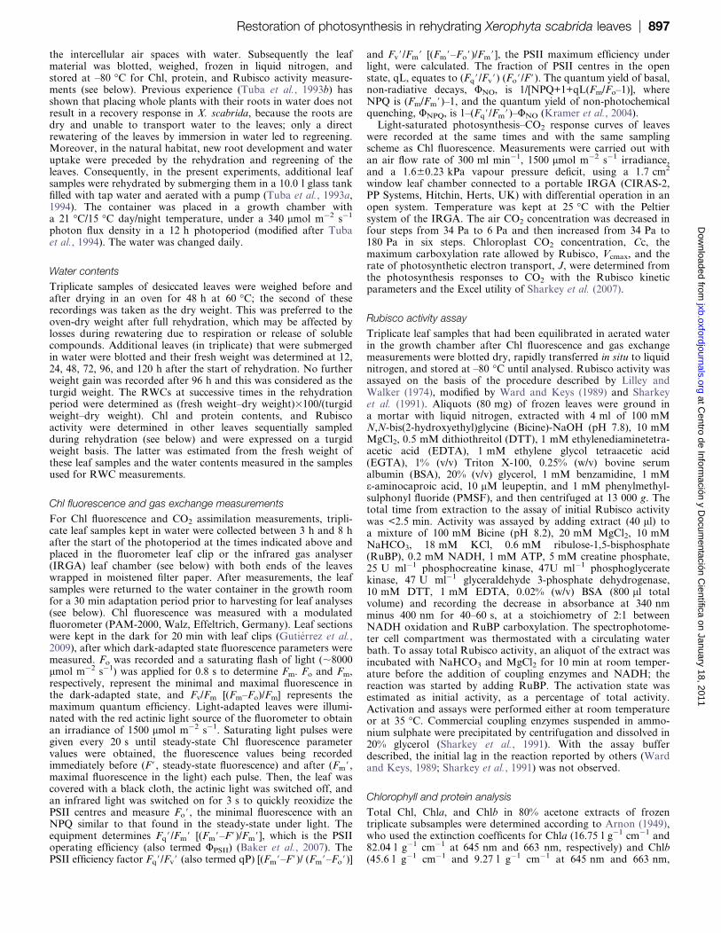
CO2 fixation
Regardless of the CO2 concentration used in measurements,
there was no CO2 uptake at 24 h of rehydration (Fig. 1A)
or before. Photosynthesis increased in the following 3 d
rehydration period and was Rubisco limited up to very highchloroplast CO2 concentrations at 48 h and 72 h (Fig. 1B,
C). The transition from Rubisco-limited to RuBP regener-
ation-limited photosynthesis decreased to 33 Pa CO2 partial
pressure at 96 h (Fig. 1D), which is still high in comparison
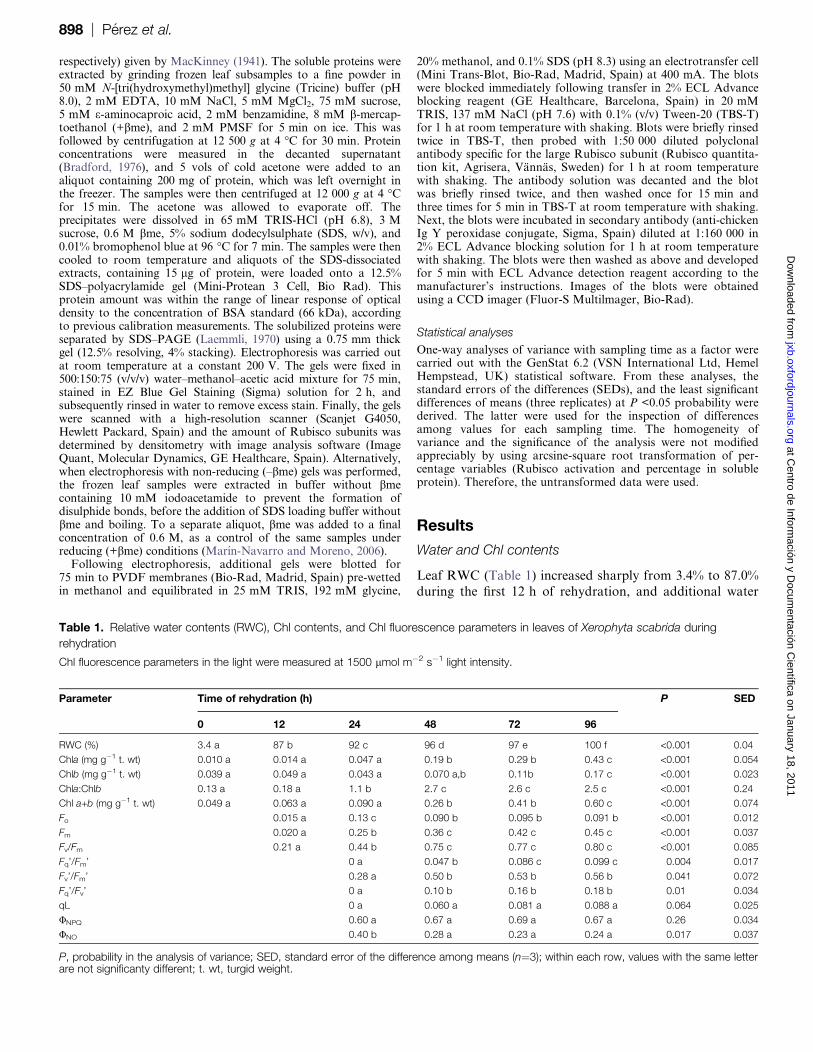
with other plants. Vcmax and J were calculated (Fig. 2) from
the photosynthesis–CO2 response curves. Except for a drop
in Vcmax at 72 h, which can be attributed to a variation
between samples, both J and Vcmax increased from 24 h to96 h, without reaching a plateau. There were relatively
higher increases in J than in Vcmax.
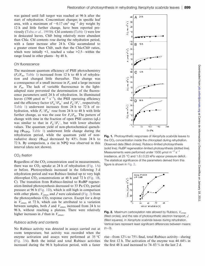
Rubisco activity and contents
No Rubisco activity was detected in assays carried out at
room temperature, but activity was recorded when the
enzyme activation and assays were performed at 35 �C(Fig. 3A). Both the initial and total Rubisco activities
increased during the 96 h hydration period, with a faster
rise—from 12% to 75% final, total Rubisco activity—during
the first 12 h. The activation of the enzyme was 44–64% inthe first 48 h and increased to 74–83 % in the last 2 d.
Fig. 1. Photosynthetic responses of Xerophyta scabrida leaves to
the CO2 concentration inside the chloroplast during rehydration.
Observed data (filled circles); Rubisco-limited photosynthesis
(solid line); RuBP regeneration-limited photosynthesis (dotted line).
Measurements were performed under 1500 lmol m�2 s�1
irradiance, at 25 �C and 1.660.23 kPa vapour pressure deficit.
The statistical significance of the parameters derived from this
figure is shown in Fig. 2.
Fig. 2. Maximum carboxylation rate allowed by Rubisco, Vcmax
(filled circles), and the rate of photosynthetic electron transport, J
(filled squares), in Xerophyta scabrida leaves during rehydration.
Vertical bars represent least significant differences between means
(n¼3).
Restoration of photosynthesis in rehydrating Xerophyta scabrida leaves | 899 at C
entro de Información y D
ocumentación C
ientífica on January 18, 2011jxb.oxfordjournals.org
Dow
nloaded from
Rubisco protein amounts (Fig. 3B) were quantified by
SDS–PAGE densitometric analysis of samples extracted
with +bme (see Materials and methods). There was little
change in the amount of Rubisco protein during the first
48 h of rehydration, although this was followed by an
increase of ;56%. Similarly, total soluble protein remained
unchanged for 48 h and then increased by ;18% (Fig. 3B).
As a fraction of soluble protein, Rubisco was relatively lowinitially and increased (from 19% to 27%) in the last 2 d of
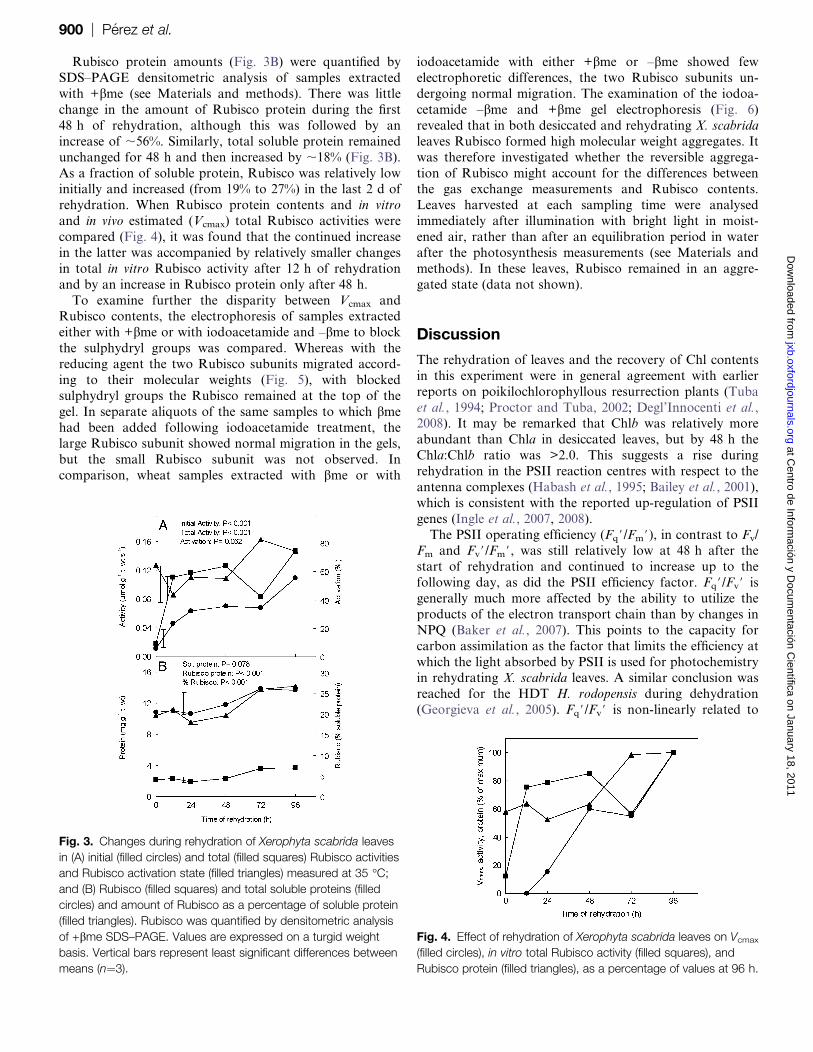
rehydration. When Rubisco protein contents and in vitro
and in vivo estimated (Vcmax) total Rubisco activities were
compared (Fig. 4), it was found that the continued increase
in the latter was accompanied by relatively smaller changes
in total in vitro Rubisco activity after 12 h of rehydration
and by an increase in Rubisco protein only after 48 h.
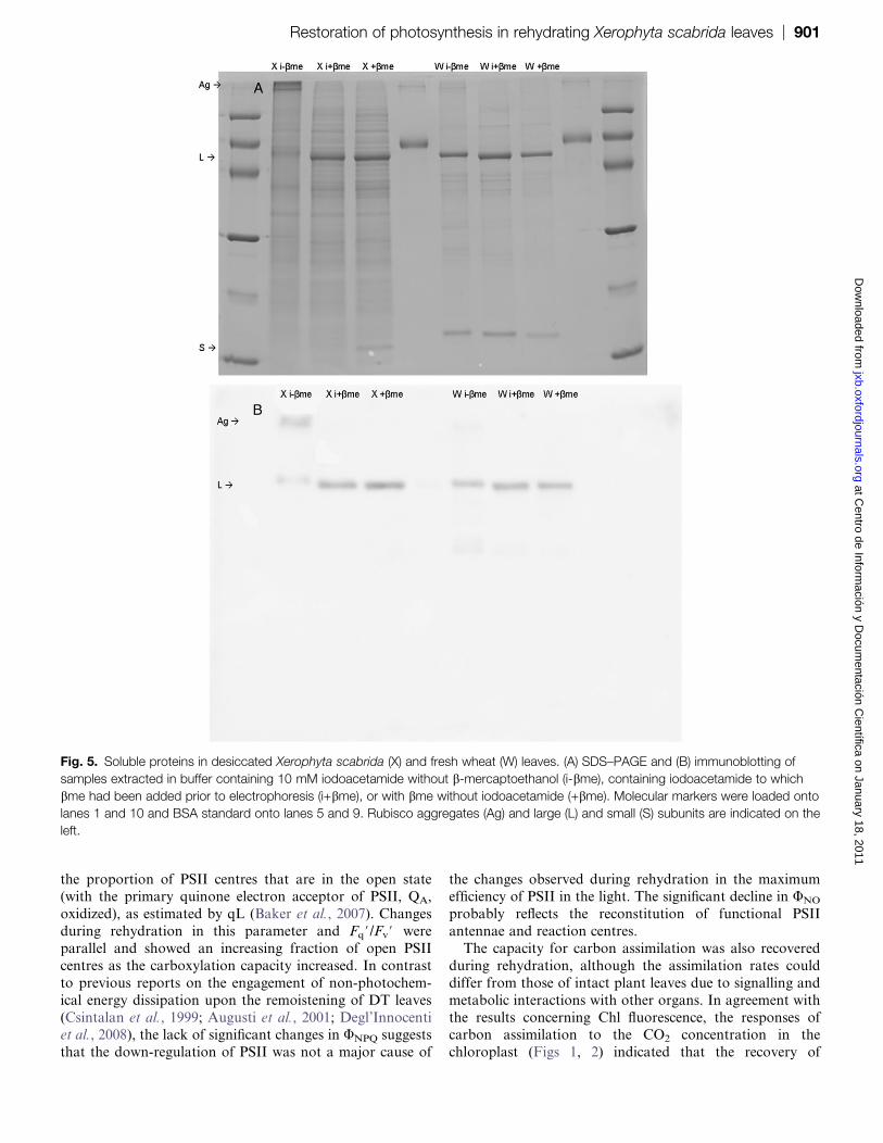
To examine further the disparity between Vcmax andRubisco contents, the electrophoresis of samples extracted
either with +bme or with iodoacetamide and –bme to block
the sulphydryl groups was compared. Whereas with the
reducing agent the two Rubisco subunits migrated accord-
ing to their molecular weights (Fig. 5), with blocked
sulphydryl groups the Rubisco remained at the top of the
gel. In separate aliquots of the same samples to which bme
had been added following iodoacetamide treatment, thelarge Rubisco subunit showed normal migration in the gels,
but the small Rubisco subunit was not observed. In
comparison, wheat samples extracted with bme or with
iodoacetamide with either +bme or –bme showed few
electrophoretic differences, the two Rubisco subunits un-
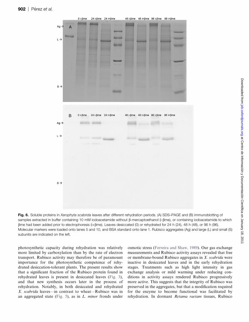
dergoing normal migration. The examination of the iodoa-
cetamide –bme and +bme gel electrophoresis (Fig. 6)
revealed that in both desiccated and rehydrating X. scabrida
leaves Rubisco formed high molecular weight aggregates. It
was therefore investigated whether the reversible aggrega-
tion of Rubisco might account for the differences betweenthe gas exchange measurements and Rubisco contents.
Leaves harvested at each sampling time were analysed
immediately after illumination with bright light in moist-
ened air, rather than after an equilibration period in water
after the photosynthesis measurements (see Materials and
methods). In these leaves, Rubisco remained in an aggre-
gated state (data not shown).
Discussion
The rehydration of leaves and the recovery of Chl contents
in this experiment were in general agreement with earlier
reports on poikilochlorophyllous resurrection plants (Tuba
et al., 1994; Proctor and Tuba, 2002; Degl’Innocenti et al.,2008). It may be remarked that Chlb was relatively more
abundant than Chla in desiccated leaves, but by 48 h the
Chla:Chlb ratio was >2.0. This suggests a rise during
rehydration in the PSII reaction centres with respect to the
antenna complexes (Habash et al., 1995; Bailey et al., 2001),
which is consistent with the reported up-regulation of PSII
genes (Ingle et al., 2007, 2008).
The PSII operating efficiency (Fq#/Fm#), in contrast to Fv/Fm and Fv#/Fm#, was still relatively low at 48 h after the
start of rehydration and continued to increase up to the
following day, as did the PSII efficiency factor. Fq#/Fv# is
generally much more affected by the ability to utilize the
products of the electron transport chain than by changes in
NPQ (Baker et al., 2007). This points to the capacity for
carbon assimilation as the factor that limits the efficiency at
which the light absorbed by PSII is used for photochemistryin rehydrating X. scabrida leaves. A similar conclusion was
reached for the HDT H. rodopensis during dehydration
(Georgieva et al., 2005). Fq#/Fv# is non-linearly related to
Fig. 3. Changes during rehydration of Xerophyta scabrida leaves
in (A) initial (filled circles) and total (filled squares) Rubisco activities
and Rubisco activation state (filled triangles) measured at 35 �C;
and (B) Rubisco (filled squares) and total soluble proteins (filled
circles) and amount of Rubisco as a percentage of soluble protein
(filled triangles). Rubisco was quantified by densitometric analysis
of +bme SDS–PAGE. Values are expressed on a turgid weight
basis. Vertical bars represent least significant differences between
means (n¼3).
Fig. 4. Effect of rehydration of Xerophyta scabrida leaves on Vcmax
(filled circles), in vitro total Rubisco activity (filled squares), and
Rubisco protein (filled triangles), as a percentage of values at 96 h.
900 | Perez et al. at C
entro de Información y D
ocumentación C
ientífica on January 18, 2011jxb.oxfordjournals.org
Dow
nloaded from
the proportion of PSII centres that are in the open state(with the primary quinone electron acceptor of PSII, QA,
oxidized), as estimated by qL (Baker et al., 2007). Changes
during rehydration in this parameter and Fq#/Fv# were
parallel and showed an increasing fraction of open PSII
centres as the carboxylation capacity increased. In contrast
to previous reports on the engagement of non-photochem-
ical energy dissipation upon the remoistening of DT leaves
(Csintalan et al., 1999; Augusti et al., 2001; Degl’Innocentiet al., 2008), the lack of significant changes in UNPQ suggests
that the down-regulation of PSII was not a major cause of
the changes observed during rehydration in the maximumefficiency of PSII in the light. The significant decline in UNO
probably reflects the reconstitution of functional PSII
antennae and reaction centres.
The capacity for carbon assimilation was also recovered
during rehydration, although the assimilation rates could
differ from those of intact plant leaves due to signalling and
metabolic interactions with other organs. In agreement with
the results concerning Chl fluorescence, the responses ofcarbon assimilation to the CO2 concentration in the
chloroplast (Figs 1, 2) indicated that the recovery of
Fig. 5. Soluble proteins in desiccated Xerophyta scabrida (X) and fresh wheat (W) leaves. (A) SDS–PAGE and (B) immunoblotting of
samples extracted in buffer containing 10 mM iodoacetamide without b-mercaptoethanol (i-bme), containing iodoacetamide to which
bme had been added prior to electrophoresis (i+bme), or with bme without iodoacetamide (+bme). Molecular markers were loaded onto
lanes 1 and 10 and BSA standard onto lanes 5 and 9. Rubisco aggregates (Ag) and large (L) and small (S) subunits are indicated on the
left.
Restoration of photosynthesis in rehydrating Xerophyta scabrida leaves | 901 at C
entro de Información y D
ocumentación C
ientífica on January 18, 2011jxb.oxfordjournals.org
Dow
nloaded from
photosynthetic capacity during rehydration was relatively
more limited by carboxylation than by the rate of electron
transport. Rubisco activity may therefore be of paramount
importance for the photosynthetic competence of rehy-
drated desiccation-tolerant plants. The present results showthat a significant fraction of the Rubisco protein found in
rehydrated leaves is present in desiccated leaves (Fig. 3),
and that new synthesis occurs later in the process of
rehydration. Notably, in both desiccated and rehydrated
X. scabrida leaves—in contrast to wheat—Rubisco was in
an aggregated state (Fig. 5), as in L. minor fronds under
osmotic stress (Ferreira and Shaw, 1989). Our gas exchange
measurements and Rubisco activity assays revealed that free
or membrane-bound Rubisco aggregates in X. scabrida were
inactive in desiccated leaves and in the early rehydration
stages. Treatments such as high light intensity in gasexchange analysis or mild warming under reducing con-
ditions in activity assays rendered Rubisco progressively
more active. This suggests that the integrity of Rubisco was
preserved in the aggregates, but that a modification required
for the enzyme to become functional was facilitated by
rehydration. In dormant Retama raetam tissues, Rubisco
Fig. 6. Soluble proteins in Xerophyta scabrida leaves after different rehydration periods. (A) SDS–PAGE and (B) immunoblotting of
samples extracted in buffer containing 10 mM iodoacetamide without b-mercaptoethanol (i-bme), or containing iodoacetamide to which
bme had been added prior to electrophoresis (i+bme). Leaves desiccated (0) or rehydrated for 24 h (24), 48 h (48), or 96 h (96).
Molecular markers were loaded onto lanes 5 and 10, and BSA standard onto lane 1. Rubisco aggregates (Ag) and large (L) and small (S)
subunits are indicated on the left.
902 | Perez et al. at C
entro de Información y D
ocumentación C
ientífica on January 18, 2011jxb.oxfordjournals.org
Dow
nloaded from
and other proteins also appear to be present as high
molecular weight complexes (Pnueli et al., 2002). These
complexes precipitated during extraction with reducing
buffers, a result that was observed for the small Rubisco
subunit only when bme was added to extracts containing
iodoacetamide. Pnueli et al. (2002) suggested that the
dilution of reducing equivalents upon rehydration releases
proteins from the aggregates into their soluble, active form.However, some of the DTT concentrations used by Pnueli
et al. (2002) in the protein extraction buffer have been
shown to cause Rubisco aggregation and precipitation (Cho
et al., 2008). Moreover, the present results suggested that
the increase in Rubisco activity during rehydration was not
associated with protein release from the aggregates. The
lower oxidation states of thiol groups (disulphide and
sulphenic acid) may easily be reverted again to thesulphydryl state by disulphide exchange with free thiols, by
DTT (in vitro) or by thioredoxins and glutaredoxins
(Marcus et al., 2003; Moreno et al., 2008). It is possible
that oxidative conditions during desiccation could induce
the formation of disulphides in the Rubisco molecule, and
that the recovery of photochemical activity could lead to an
increasingly reduced stroma, favouring the reductive activa-
tion of Rubisco. While upon desiccation of X. scabrida, andindeed of all poikilochlorophyllous plant species, Chl and
the photosynthetic apparatus are lost, it is concluded that
Rubisco is preserved in large amounts in a close to
functional state. Rubisco aggregation may be a part of the
poikilochlorophylly strategy.
Acknowledgements
This work has been funded by the Spanish Ministry of
Science and Innovation and the Hungarian Science and
Technology Office (Integrated Action HH2006-0019, ESP-
41/2006), and by the Spanish National Research and
Development Programme-European Regional DevelopmentFund ERDF (Project AGL2006-13541-C02-02/AGR). DG
was the recipient of a Junta de Castilla y Leon fellowship.
This paper is dedicated to the memory of Zoltan Tuba, who
passed away while this research was in progress.
References
Agarwal R, Ortlebb S, Sainis JK, Melzer M. 2009. Immunoelectron
microscopy for locating Calvin cycle enzymes in the thylakoids of
Synechocystis 6803. Molecular Plant 2, 32–42.
Anderson LE, Goldhaber-Gordo IM, Li D, Tang XY, Xiang M,
Prakash N. 1995. Enzyme–enzyme interaction in the chloroplast,
glyceradehyde-3-phosphate dehydrogenase, triose phosphate
isomerase and aldolase. Planta 196, 245–255.
Arnon DI. 1949. Copper enzymes in isolated chloroplasts. Polyphenol
oxidase in Beta vulgaris. Plant Physiology 24, 1–15.
Augusti A, Scartazza A, Navari-Izzo F, Sgherri CLM,
Stevanovic B, Brugnoli E. 2001. Photosystem II photochemical
efficiency, zeaxanthin and antioxidant contents in the poikilohydric
Ramonda serbica during dehydration and rehydration. Photosynthesis
Research 67, 79–88.
Bailey S, Walters RG, Jansson S, Horton P. 2001. Acclimation of
Arabidopsis thaliana to the light environment: the existence of separate
low light and high light responses. Planta 213, 794–801.
Baker NR, Harbinson J, Kramer DM. 2007. Determining the
limitations and regulation of photosynthetic energy transduction in
leaves. Plant, Cell and Environment 30, 1107–1125.
Bradford M. 1976. A rapid and sensitive method for the quantitation
of microgram quantities of protein utilizing the principle of protein–dye
binding. Analytical Biochemistry 72, 248–254.
Cho JH, Hwang H, Cho MH, Kwon YK, Jeon JS, Bhoo SH,
Hahn TR. 2008. The effect of DTT in protein preparations for
proteomic analysis: removal of a highly abundant plant enzyme,
ribulose bisphosphate carboxylase/oxygenase. Journal of Plant
Biology 51, 297–301.
Csintalan Zs, Proctor MCF, Tuba Z. 1999. Chlorophyll
fluorescence during drying and rehydration in the mosses
Rhytidiadelphus loreus (Hedw.) Warnst., Anomodon viticulosus
(Hedw.) Hook. & Tayl. and Grimmia pulvinata (Hedw.) Sm. Annals of
Botany 84, 235–244.
Daniel V, Gaff DF. 1980. Sulfhydryl and disulphide levels in protein
fractions from hydrated and dry leaves of resurrection plants. Annals of
Botany 45, 163–171.
Degl’Innocenti E, Guidi L, Stevanovic B, Navari F. 2008. CO2
fixation and chlorophyll a fluorescence in leaves of Ramonda serbica
during a dehydration–rehydration cycle. Journal of Plant Physiology
165, 723–733.
Ferreira RB, Shaw NM. 1989. Effect of osmotic stress on protein
turnover in Lemna minor fronds. Planta 179, 456–465.
Frank W, Phillips J, Salamini F, Bartels D. 1998. Two dehydration-
inducible transcripts from the resurrection plant Craterostigma
plantagineum encode interacting homeodomain leucine zipper
proteins. The Plant Journal 15, 413–421.
Georgieva K, Maslenkova L, Peeva V, Markovska Y, Stefanov D,
Tuba Z. 2005. Comparative study on the changes in photosynthetic
activity of the homoiochlorophyllous desiccation-tolerant Haberlea
rhodopensis and desiccation-sensitive spinach leaves during
desiccation and rehydration. Photosynthesis Research 85, 191–203.
Georgieva K, Szigeti Z, Sarvari E, Gaspar L, Maslenkova L,
Peeva V, Peli E, Tuba Z. 2007. Photosynthetic activity of
homoiochlorophyllous desiccation tolerant plant Haberlea rhodopensis
during dehydration and rehydration. Planta 225, 955–964.
Gontero B, Cardenas M, Ricard J. 1988. A functional five enzyme
complex of chloroplasts involved in the Calvin cycle. European Journal
of Biochemistry 173, 437–443.
Gontero B, Mulliert G, Rault M, Giudico-Orticoni MT, Ricard J.
1993. Structural and functional properties of a multi-enzyme complex
from spinach chloroplasts: modulation of the kinetic properties of
enzymes in the aggregated state. European Journal of Biochemistry
217, 1075–1082.
Gutierrez D, Gutierrez E, Perez P, Morcuende R, Verdejo AL,
Martinez-Carrasco R. 2009. Acclimation to future atmospheric CO2
increases photochemical efficiency and mitigates photochemistry
Restoration of photosynthesis in rehydrating Xerophyta scabrida leaves | 903 at C
entro de Información y D
ocumentación C
ientífica on January 18, 2011jxb.oxfordjournals.org
Dow
nloaded from
inhibition by warm temperatures in wheat under field chambers.
Physiologia Plantarum 137, 86–100.
Habash DZ, Matthew JP, Parry MAJ, Keys AJ, Lawlor DW. 1995.
Increased capacity for photosynthesis in wheat grown at elevated
CO2: the relationship between electron transport and carbon
metabolism. Planta 197, 482–489.
Harten JB, Eickmeier WG. 1986. Enzyme dynamics of the
resurrection plant Selaginella lepidophylla (Hook. & Grev.) Spring
during rehydration. Plant Physiology 82, 61–64.
Hermoso R, Fonolla J, de Felipe MR, Vivo MA, Chueca A,
Lazaro J, Lopez Gorge J. 1992. Double immunogold localization of
thioredoxin f and photosynthetic fructose-1,6-bisphosphatase in
spinach leaves. Plant Physiology and Biochemistry 30, 39–46.
Ingle RA, Collett H, Cooper K, Takahashi Y, Farrant JM, Illing N.
2008. Chloroplast biogenesis during rehydration of the resurrection
plant Xerophyta humilis: parallels to the etioplast–chloroplast transition.
Plant, Cell and Environment 31, 1813–1824.
Ingle RA, Schmidt UG, Farrant JM, Thomson JA, Mundree SG.
2007. Proteomic analysis of leaf proteins during dehydration of the
resurrection plant Xerophyta viscosa. Plant, Cell and Environment
30, 435–446.
Ingram J, Bartels D. 1996. The molecular basis of dehydration
tolerance in plants. Annual Review of Plant Physiology and Plant
Molecular Biology 47, 377–403.
Klughammer C, Schreiber U. 2008. Complementary PS II quantum
yields calculated from simple fluorescence parameters measured by
PAM fluorometry and the saturation pulse method. PAM Application
Notes 1, 27–35.
Kramer DM, Johnson G, Kiirats O, Edwards GE. 2004.
New fluorescence parameters for determination of QA redox state
and excitation energy fluxes. Photosynthesis Research
79, 209–218.
Kranner I, Beckett RP, Wornik S, Zorn M, Pfeifhofer HW. 2002.
Revival of a resurrection plant correlates with its antioxidant status.
The Plant Journal 31, 13–24.
Laemmli UK. 1970. Cleavage of structural proteins during the
assembly of the head of bacteriophage T4. Nature 227, 680–685.
Lilley R McC, Walker DA. 1974. An improved spectrophotometric
assay for ribulose bisphosphate carboxylase. Biochimica et Biophysica
Acta 358, 226–229.
MacKinney G. 1941. Absorption of light by chlorophyll solutions.
Journal of Biological Chemistry 140, 315–322.
Marcus Y, Altman-Gueta H, Finkler A, Gurevitz M. 2003. Dual role
of cysteine 172 in redox regulation of ribulose 1,5-bisphosphate
carboxylase/oxygenase activity and degradation. Journal of
Bacteriology 185, 1509–1517.
Marın-Navarro J, Moreno J. 2006. Cysteines 449 and 459 modulate
the reduction–oxidation conformational changes of ribulose
1,5-bisphosphate carboxylase/oxygenase and the translocation of the
enzyme to membranes during stress. Plant, Cell and Environment
29, 898–908.
Martinelli T, Whittaker A, Masclaux-Daubresse C, Farrant JM,
Brilli F, Loreto F, Vazzana C. 2007. Evidence for the presence of
photorespiration in desiccation-sensitive leaves of the C4‘resurrection’
plant Sporobolus stapfianus during dehydration stress. Journal of
Experimental Botany 58, 3929–3939.
Moreno J, Garcıa-Murria MJ, Marın-Navarro J. 2008. Redox
modulation of Rubisco conformation and activity through its cysteine
residues. Journal of Experimental Botany 59, 1605–1614.
Mowla SB, Thomson JA, Farrant JM, Mundree SG. 2002. A novel
stress-inducible antioxidant enzyme identified from the resurrection
plant Xerophyta viscosa Baker. Planta 215, 716–726.
Peeva V, Cornic G. 2009. Leaf photosynthesis of Haberlea
rhodopensis before and during drought. Environmental and
Experimental Botany 65, 310–318.
Persson O, Johansson G. 1989. Studies of protein–protein
interaction using countercurrent distribution in aqueous two phase
systems. Biochemical Journal 259, 863–870.
Pnueli L, Hallak-Herr E, Rozenberg M, Cohen M, Goloubinoff P,
Kaplan A, Mittler R. 2002. Molecular and biochemical mechanisms
associated with dormancy and drought tolerance in the desert legume
Retama raetam. The Plant Journal 31, 319–330.
Proctor MCF, Ligrone R, Duckett JG. 2007. Desiccation tolerance
in the moss Polytrichum formosum: physiological and fine-structural
changes during desiccation and recovery. Annals of Botany
99, 75–93.
Proctor MCF, Tuba Z. 2002. Poikilohydry and homoihydry: antithesis
or spectrum of possibilities? New Phytologist 156, 327–349.
Ramanjulu S, Bartels D. 2002. Drought- and desiccation-induced
modulation of gene expression in plants. Plant, Cell and Environment
25, 141–151.
Sainis JK, Harris GC. 1986. The association of d-ribulose-1,
5-bisphosphate carboxylase with phosphoriboisomerase and
phosphoribulokinase. Biochemical and Biophysical Research
Communications 139, 947–954.
Sainis JK, Merriam K, Harris GC. 1989. The association of
ribulose-1,5-bisphosphate carboxylase/oxygenase with
phosphoribulokinase. Plant Physiology 89, 368–374.
Sharkey TD, Bernacchi CJ, Farquhar GD, Singsaas EL. 2007.
Fitting photosynthetic carbon dioxide response curves for C3 leaves.
Plant, Cell and Environment 30, 1035–1040.
Sharkey TD, Savitch LV, Butz ND. 1991. Photometric method for
routine determination of kcat and carbamylation of rubisco.
Photosynthesis Research 28, 41–48.
Sherwin HW, Farrant JM. 1996. Differences in rehydration of three
desiccation-tolerant angiosperm species. Annals of Botany
78, 703–710.
Toldi O, Tuba Z, Scott P. 2009. Vegetative desiccation tolerance:
is it a goldmine for bioengineering crops? Plant Science
176, 187–199.
Tuba Z, Zs Csintalan, Szente K, Nagy Z, Grace J. 1998B. Carbon
gains by desiccation tolerant plants at elevated CO2. Functional
Ecology 12, 39–44.
Tuba Z, Hartmut K, Maroti I, Zs Csintalan, Pocs T. 1993b.
Regreening of dessicated leaves of the poikilochlorophyllous
Xerophyta scabrida upon rehydration. Journal of Plant Physiology
142, 103–108.
904 | Perez et al. at C
entro de Información y D
ocumentación C
ientífica on January 18, 2011jxb.oxfordjournals.org
Dow
nloaded from
Tuba Z, Lichtenthaler HK, Csintalan Z, Nagy Z, Szente K. 1994.
Reconstitution of chlorophylls and photosynthetic CO2 assimilation
upon rehydration of the desiccated poikilochlorophyllous plant
Xerophyta scabrida (Pax) Th. Dur. et Schinz. Planta 192, 414–420.
Tuba Z, Lichtenthaler HK, Maroti I, Csintalan Zs. 1993a.
Resynthesis of thylakoids and functional chloroplasts in the desiccated
leaves of the poikilochlorophyllous plant Xerophyta scabrida upon
rehydration. Journal of Plant Physiology 142, 742–748.
Tuba Z, Proctor MCF, Csintalan Zs. 1998a. Ecophysiological
responses of homoichlorophyllous and poikilochlorophyllous
desiccation tolerant plants: a comparison and an ecological
perspective. Plant Growth Regulation 24, 211–217.
Tuba Z, Smirnoff N, Zs Csintalan, Nagy Z, Szente K. 1997.
Respiration during slow desiccation of the poikilochlorophyllous
desiccation tolerant plant Xerophyta scabrida at present-day CO2
concentrations. Plant Physiology and Biochemistry 35, 381–386.
Vicre M, Farrant JM, Driouich A. 2004. Insights into the cellular
mechanisms of desiccation tolerance among angiosperm
resurrection plant species. Plant, Cell and Environment
27, 1329–1340.
Ward DA, Keys AJ. 1989. A comparison between the coupled
spectrophotometric and uncoupled radiometric assay for RuBP
carboxylase. Photosynthesis Research 22, 167–171.
Whittaker A, Bochicchio A, Vazzana C, Lindsey G, Farrant JM.
2001. Changes in leaf hexokinase activity and metabolite levels in
response to drying in the desiccation-tolerant species Sporobolus
stapfianus and Xerophyta viscosa. Journal of Experimental Botany
52, 961–969.
Restoration of photosynthesis in rehydrating Xerophyta scabrida leaves | 905 at C
entro de Información y D
ocumentación C
ientífica on January 18, 2011jxb.oxfordjournals.org
Dow
nloaded from