Restoration of Oligodendrocyte Pools in a Mouse Model of Chronic Cerebral Hypoperfusion
12
Restoration of Oligodendrocyte Pools in a Mouse Model of Chronic Cerebral Hypoperfusion Jamie McQueen 1,2 , Michell M. Reimer 1,2 , Philip R. Holland 1,2 , Yasmina Manso 1 , Mark McLaughlin 3 , Jill H. Fowler 1 , Karen Horsburgh 1,2 * 1 Centre for Neuroregeneration, University of Edinburgh, Edinburgh, United Kingdom, 2 Centre for Cognitive Ageing and Cognitive Epidemiology, University of Edinburgh, Edinburgh, United Kingdom, 3 School of Veterinary Medicine, Division of Veterinary Biosciences, University of Glasgow, Glasgow, United Kingdom Abstract Chronic cerebral hypoperfusion, a sustained modest reduction in cerebral blood flow, is associated with damage to myelinated axons and cognitive decline with ageing. Oligodendrocytes (the myelin producing cells) and their precursor cells (OPCs) may be vulnerable to the effects of hypoperfusion and in some forms of injury OPCs have the potential to respond and repair damage by increased proliferation and differentiation. Using a mouse model of cerebral hypoperfusion we have characterised the acute and long term responses of oligodendrocytes and OPCs to hypoperfusion in the corpus callosum. Following 3 days of hypoperfusion, numbers of OPCs and mature oligodendrocytes were significantly decreased compared to controls. However following 1 month of hypoperfusion, the OPC pool was restored and increased numbers of oligodendrocytes were observed. Assessment of proliferation using PCNA showed no significant differences between groups at either time point but showed reduced numbers of proliferating oligodendroglia at 3 days consistent with the loss of OPCs. Cumulative BrdU labelling experiments revealed higher numbers of proliferating cells in hypoperfused animals compared to controls and showed a proportion of these newly generated cells had differentiated into oligodendrocytes in a subset of animals. Expression of GPR17, a receptor important for the regulation of OPC differentiation following injury, was decreased following short term hypoperfusion. Despite changes to oligodendrocyte numbers there were no changes to the myelin sheath as revealed by ultrastructural assessment and fluoromyelin however axon-glial integrity was disrupted after both 3 days and 1 month hypoperfusion. Taken together, our results demonstrate the initial vulnerability of oligodendroglial pools to modest reductions in blood flow and highlight the regenerative capacity of these cells. Citation: McQueen J, Reimer MM, Holland PR, Manso Y, McLaughlin M, et al. (2014) Restoration of Oligodendrocyte Pools in a Mouse Model of Chronic Cerebral Hypoperfusion. PLoS ONE 9(2): e87227. doi:10.1371/journal.pone.0087227 Editor: Ken Arai, Massachusetts General Hospital/Harvard Medical School, United States of America Received July 16, 2013; Accepted December 25, 2013; Published February 3, 2014 Copyright: ß 2014 McQueen et al. This is an open-access article distributed under the terms of the Creative Commons Attribution License, which permits unrestricted use, distribution, and reproduction in any medium, provided the original author and source are credited. Funding: The funding of the Lifelong Health and Wellbeing Initiative (supported by the BBSRC, EPSRC, ESRC and MRC), The Disconnected Mind (supported by Age UK) and Alzheimer’s Research UK (ARUK) is gratefully acknowledged. JHF is supported by a research fellowship from the Alzheimer’s Society. Confocal imaging was performed at the IMPACT Imaging facility and at the Euan MacDonald Centre at the University of Edinburgh. The funders had no role in study design, data collection and analysis, decision to publish, or preparation of the manuscript. Competing Interests: The authors have declared that no competing interests exist. * E-mail: [email protected] Introduction Oligodendrocytes are the myelin producing cells of the CNS and are critical for maintaining and regulating the myelination of axons. Oligodendrocyte survival and the integrity of myelinated axons is essential for maintaining saltatory conduction, neuronal communication and normal cognitive function (for review see [1]). A single oligodendrocyte can myelinate up to 50 axonal segments [2] and thus damage to individual oligodendrocytes could have a major effect on myelination of axons and efficiency of the relay of information. Oligodendroglia appear to be particularly vulnerable to blood flow reductions and in animal models of cerebral ischemia and severe hypoperfusion a marked loss of oligodendrocytes occurs rapidly in response to severe reductions in blood flow [3–6]. Additionally, in vitro models of hypoxia and oxygen-glucose deprivation, common pathways in cerebral ischaemia, have demonstrated the susceptibility of oligodendrocytes to these conditions [7,8] and it is now thought that damage to oligoden- drocytes is mediated by oxidative stress, inflammation and excitotoxicity (for review see [9]). Indeed, damage to myelinated axons and oligodendrocytes is prominent in various conditions in which cerebral blood flow is compromised such as the ageing brain [10,11], Alzheimer’s disease [12,13] and stroke [4,14] and may contribute to a functional impairment. Despite the initial degeneration of oligodendrocytes following injury there is now evidence to indicate that oligodendrocyte precursor cells (OPCs) can proliferate and differentiate and as a result may serve to replenish the loss of damaged oligodendrocytes and potentially repair functional deficits. In neonatal models of hypoxic-ischaemic injury, cell proliferation is increased and new oligodendrocytes are generated up to several weeks after the initial injury [15]. In the adult brain there also appears to be an endogenous capacity to generate new oligodendrocytes in response to cerebral ischaemia. In models of focal cerebral ischaemia when either blood flow is restored with reperfusion or in the peri-infarct region where there is sufficient collateral flow, increased numbers of OPCs are detectable [6,16,17]. Interestingly, in aged human brain, increased numbers of oligodendrocytes and OPCs occur in areas adjacent to white matter disruption where blood flow is compromised [18] and increased numbers of oligodendrocytes have been demonstrated in cases of vascular cognitive impairment PLOS ONE | www.plosone.org 1 February 2014 | Volume 9 | Issue 2 | e87227
Transcript of Restoration of Oligodendrocyte Pools in a Mouse Model of Chronic Cerebral Hypoperfusion
Restoration of Oligodendrocyte Pools in a Mouse Modelof Chronic Cerebral HypoperfusionJamie McQueen1,2, Michell M. Reimer1,2, Philip R. Holland1,2, Yasmina Manso1, Mark McLaughlin3,
Jill H. Fowler1, Karen Horsburgh1,2*
1 Centre for Neuroregeneration, University of Edinburgh, Edinburgh, United Kingdom, 2 Centre for Cognitive Ageing and Cognitive Epidemiology, University of
Edinburgh, Edinburgh, United Kingdom, 3 School of Veterinary Medicine, Division of Veterinary Biosciences, University of Glasgow, Glasgow, United Kingdom
Abstract
Chronic cerebral hypoperfusion, a sustained modest reduction in cerebral blood flow, is associated with damage tomyelinated axons and cognitive decline with ageing. Oligodendrocytes (the myelin producing cells) and their precursor cells(OPCs) may be vulnerable to the effects of hypoperfusion and in some forms of injury OPCs have the potential to respondand repair damage by increased proliferation and differentiation. Using a mouse model of cerebral hypoperfusion we havecharacterised the acute and long term responses of oligodendrocytes and OPCs to hypoperfusion in the corpus callosum.Following 3 days of hypoperfusion, numbers of OPCs and mature oligodendrocytes were significantly decreased comparedto controls. However following 1 month of hypoperfusion, the OPC pool was restored and increased numbers ofoligodendrocytes were observed. Assessment of proliferation using PCNA showed no significant differences betweengroups at either time point but showed reduced numbers of proliferating oligodendroglia at 3 days consistent with the lossof OPCs. Cumulative BrdU labelling experiments revealed higher numbers of proliferating cells in hypoperfused animalscompared to controls and showed a proportion of these newly generated cells had differentiated into oligodendrocytes in asubset of animals. Expression of GPR17, a receptor important for the regulation of OPC differentiation following injury, wasdecreased following short term hypoperfusion. Despite changes to oligodendrocyte numbers there were no changes to themyelin sheath as revealed by ultrastructural assessment and fluoromyelin however axon-glial integrity was disrupted afterboth 3 days and 1 month hypoperfusion. Taken together, our results demonstrate the initial vulnerability ofoligodendroglial pools to modest reductions in blood flow and highlight the regenerative capacity of these cells.
Citation: McQueen J, Reimer MM, Holland PR, Manso Y, McLaughlin M, et al. (2014) Restoration of Oligodendrocyte Pools in a Mouse Model of Chronic CerebralHypoperfusion. PLoS ONE 9(2): e87227. doi:10.1371/journal.pone.0087227
Editor: Ken Arai, Massachusetts General Hospital/Harvard Medical School, United States of America
Received July 16, 2013; Accepted December 25, 2013; Published February 3, 2014
Copyright: � 2014 McQueen et al. This is an open-access article distributed under the terms of the Creative Commons Attribution License, which permitsunrestricted use, distribution, and reproduction in any medium, provided the original author and source are credited.
Funding: The funding of the Lifelong Health and Wellbeing Initiative (supported by the BBSRC, EPSRC, ESRC and MRC), The Disconnected Mind (supported byAge UK) and Alzheimer’s Research UK (ARUK) is gratefully acknowledged. JHF is supported by a research fellowship from the Alzheimer’s Society. Confocalimaging was performed at the IMPACT Imaging facility and at the Euan MacDonald Centre at the University of Edinburgh. The funders had no role in study design,data collection and analysis, decision to publish, or preparation of the manuscript.
Competing Interests: The authors have declared that no competing interests exist.
* E-mail: [email protected]
Introduction
Oligodendrocytes are the myelin producing cells of the CNS
and are critical for maintaining and regulating the myelination of
axons. Oligodendrocyte survival and the integrity of myelinated
axons is essential for maintaining saltatory conduction, neuronal
communication and normal cognitive function (for review see [1]).
A single oligodendrocyte can myelinate up to 50 axonal segments
[2] and thus damage to individual oligodendrocytes could have a
major effect on myelination of axons and efficiency of the relay of
information.
Oligodendroglia appear to be particularly vulnerable to blood
flow reductions and in animal models of cerebral ischemia and
severe hypoperfusion a marked loss of oligodendrocytes occurs
rapidly in response to severe reductions in blood flow [3–6].
Additionally, in vitro models of hypoxia and oxygen-glucose
deprivation, common pathways in cerebral ischaemia, have
demonstrated the susceptibility of oligodendrocytes to these
conditions [7,8] and it is now thought that damage to oligoden-
drocytes is mediated by oxidative stress, inflammation and
excitotoxicity (for review see [9]). Indeed, damage to myelinated
axons and oligodendrocytes is prominent in various conditions in
which cerebral blood flow is compromised such as the ageing brain
[10,11], Alzheimer’s disease [12,13] and stroke [4,14] and may
contribute to a functional impairment.
Despite the initial degeneration of oligodendrocytes following
injury there is now evidence to indicate that oligodendrocyte
precursor cells (OPCs) can proliferate and differentiate and as a
result may serve to replenish the loss of damaged oligodendrocytes
and potentially repair functional deficits. In neonatal models of
hypoxic-ischaemic injury, cell proliferation is increased and new
oligodendrocytes are generated up to several weeks after the initial
injury [15]. In the adult brain there also appears to be an
endogenous capacity to generate new oligodendrocytes in response
to cerebral ischaemia. In models of focal cerebral ischaemia when
either blood flow is restored with reperfusion or in the peri-infarct
region where there is sufficient collateral flow, increased numbers
of OPCs are detectable [6,16,17]. Interestingly, in aged human
brain, increased numbers of oligodendrocytes and OPCs occur in
areas adjacent to white matter disruption where blood flow is
compromised [18] and increased numbers of oligodendrocytes
have been demonstrated in cases of vascular cognitive impairment
PLOS ONE | www.plosone.org 1 February 2014 | Volume 9 | Issue 2 | e87227

[19]. Together these studies suggest that OPCs may respond to
reduced cerebral blood flow in an attempt to ameliorate white
matter damage.
There are a number of mechanisms that may regulate OPC
proliferation and differentiation. Relevant to blood flow alterations
both glutamate and ATP, the extracellular levels of which may be
increased with reduced blood flow, have been shown to be
important in regulating OPC proliferation and differentiation
[20,21]. In addition, recent studies have identified a role for a G-
coupled protein receptor, GPR17, as an important mediator of
OPC differentiation and white matter repair [22,23]. GPR17 has
been shown to be expressed by a subset of OPCs [23,24] and it is
thought that these cells may operate as an early sensor of brain
damage whereby they are activated by uracil nucleotides and
cysteinyl leukotrienes which are increased in response to cerebral
ischemia. In support of this, GPR17 positive cells are upregulated
in response to cerebral ischemia and associated with oligodendro-
cyte differentiation [23].
The present study sought to determine whether the pools of
oligodendrocytes and OPCs would be influenced by modest
reductions in blood flow more akin to those occurring in the
ageing brain. We utilised a mouse model of cerebral hypoperfu-
sion induced by permanent bilateral carotid stenosis which we
have previously shown to result in diffuse white matter pathology
[12,25,26]. Importantly, these mice also exhibit impaired spatial
working memory [12,27], providing a link between white matter
disruption and cognitive decline. More recently, microarray
analysis in mice subject to cerebral hypoperfusion has revealed
increased expression of several genes involved in cell proliferation
[25] which may underlie a potential white matter repair
mechanism. We therefore also investigated the extent of OPC
proliferation and differentiation and whether this was mediated by
GPR17. In addition, ultrastructural analysis and myelin labelling
were carried out to determine whether alterations to oligodendro-
cyte pools influenced myelin sheath thickness.
Materials and Methods
Ethics statementAll procedures were authorised under the Home Office
approved Project Licenses, ‘Pathophysiology of Alzheimer’s
disease: link to cerebrovascular disease’ (licence number 60/
3722) and ‘Pathophysiology of vascular cognitive impairment and
Alzheimer’s disease’ (licence number 60/4350) held by Prof. K.
Horsburgh. The licences were approved by the University of
Edinburgh’s Ethical Review Committee and the Home Office,
and adhered to regulations specified in the Animals (Scientific
Procedures) Act (1986).
Animals and surgeryAdult male C57Bl/6J mice (aged 3–5 months old, 25–30 g)
were obtained from Charles River Laboratories Inc, UK. Animals
were subject to chronic cerebral hypoperfusion as previously
described [25–28]. In brief, wire microcoils (0.18 mm internal
diameter, Sawane Spring Co., Japan) were applied to both
common carotid arteries under isoflurane anaesthesia (induced at
5%, and maintained at 1.2–1.6%). A 30 minute interval was left
between left and right coil application. Sham-operated animals
underwent identical procedures with the exception that coils were
not placed around the arteries. Housing of animals and all
procedures were carried out in pathogen-free animal units.
BrdU labellingAnimals from the 1 month cohort were given intraperitoneal
injections (35 mg/kg body weight) of 59-bromo-29-deoxyuridine
(BrdU; Fluka, UK) twice daily for the first 3 days following surgery
to label proliferating cells during this period by an individual
blinded to surgical group.
Laser speckle contrast imagingAn additional cohort of animals underwent measurement of
cerebral blood flow using laser speckle flowmetry. Animals were
anaesthetised with 5% isoflurane in oxygen for 1.5 minutes in an
anaesthetic chamber. Animals were then transferred to a
stereotaxic frame and their heads were fixed into position.
Anaesthesia was maintained at 2–2.5% isoflurane via a nose cone
and body temperature was monitored and regulated. An incision
was made to expose the skull and the skin overlying the skull was
reflected. The skull was moistened using saline and a small amount
of water-based gel (37uC) was spread evenly onto the skull. A
moorFLPI2 Speckle Contrast Imager (Moor Instruments, UK)
was positioned 20 cm above the head. Image sequences were
acquired at a resolution of 7526580 pixels and a frequency of 1
frames/second (20 ms/frame). Following stabilisation of perfusion
readings, a 2 minute perfusion recording was carried out.
Raw speckle contrast sequences were analysed using
moorFLPI2 Review software (v4.0). Regions of interest were
consistent between each mouse and made 1 mm to 22 mm from
Bregma with care taken to avoid any artefacts on the skull surface.
Data were measured in blood perfusion units (PU) and calculated
for each mouse as the percentage change relative to baseline
(Figure S1).
Tissue preparation and immunohistochemistryAt 3 days or 28 days post-surgery, mice were deeply
anaesthetised with 5% isoflurane and transcardially perfused with
20 ml 0.9% heparinised saline followed by 20 ml 4% paraformal-
dehyde (PFA) in 0.1% phosphate buffer (PB, pH 7.4). Following
perfusion, the brains were removed and post-fixed in 4% PFA
overnight. Brains were then transferred to PB and stored overnight
at 4uC. The brains were cut along the midline and free-floating
50 mm sagittal sections were cut using a vibrating blade microtome
(Hydrax V50, Zeiss, Germany). Sections were stored in cryopro-
tective medium (30% glycerol/30% ethylene glycol in PB) at
220uC until required. Different cohorts of animals were used due
to the sensitivity of some antibodies to tissue fixation (n = 13 sham,
12 hypoperfused and n = 9 sham, 10 hypoperfused for 3 day
studies; n = 10 sham, 11 hypoperfused and n = 9 sham, 9
hypoperfused for 1 month studies). Occasionally animals were
excluded from analysis if there was an absence of cellular staining
or the quality was deemed too poor to perform accurate analysis.
The following primary antibodies were used in this study: anti-
BrdU (1:200, AB6326, Abcam, UK), anti-CC1 (APC 1:20, OP80,
Calbiochem, USA), anti-GFAP (1:1000, Z0334, Dako UK), anti-
GPR17 (1:200, 10136, Cayman Chemical, USA) anti-Iba1 (1:100,
ab5076, Abcam UK), anti-MAG (1:100, sc-9543, Santa Cruz,
USA) anti-NG2 (1:100, AB5320, Millipore, UK), anti-Olig2
(1:500, Ab9610, and 1:100, MABN50, both Millipore, UK),
anti-PCNA (1:500 ab29, Abcam UK), anti-PDGFRa (1:100,
558774, BD Pharmingen UK), and anti-PDGFRb (1:100,
AF1042, R and D Systems, UK). Cy2, Cy3, DyLight 488 and
Alexa Fluor 488 and 647 (all 1:200) conjugated secondary
antibodies were purchased from Jackson ImmunoResearch
Laboratories Inc (USA). Alexa Fluor 488 and 546 conjugated
secondary antibodies (1:500) were purchased from Life Technol-
ogies Ltd (UK). Double labelling experiments were carried out to
Oligodendrocyte Pools after Cerebral Hypoperfusion
PLOS ONE | www.plosone.org 2 February 2014 | Volume 9 | Issue 2 | e87227
confirm the cellular specificity of antibodies used for OPC and
oligodendrocyte labelling. Non-specific labelling was blocked using
3% normal serum and sections were incubated in primary and
secondary antibodies overnight at 4uC. Sections were mounted
onto SuperFrost slides and mounted using Vectashield hard set
mounting medium containing the nuclear stain 49,6-diamidino-2-
phenylindole (DAPI) (H-1500, Vector Laboratories, USA).
For BrdU labelling experiments, an additional antigen retrieval
step was required. Following 3 washes in PBS, sections were
incubated for 30 minutes in 2M HCl at 37uC to allow
denaturation of DNA. Following this, sections were given three
5 minute washes in 0.1 M sodium borate buffer (Na2B4O7,
pH 8.5). For PCNA labelling, sections were retrieved in 10 mM
citrate buffer for 30 minutes at 85uC and blocked using 10%
normal serum and 0.5% bovine serum albumin.
Fluoromyelin staining was used to assess myelin integrity
following hypoperfusion. Free-floating sections were washed in
PBS and mounted onto slides. Following rehydration in PBS,
sections were incubated in Fluoromyelin Green (1:200, Invitrogen)
for 1 hour at room temperature. At the outset the conditions were
optimised as recommended by the manufacturer. This protocol
was determined to be optimal for studying myelin alterations in
thick vibratome sections and is a slight modification of that
suggested by the manufacturer for thin paraffin sections.
Confocal laser scanning microscopy and image analysisImmunolabelled 50 mm sections were imaged using confocal
laser scanning microscopy (Zeiss Axioskop LSM 510 or Zeiss
LSM710, Zeiss, Germany). All images were acquired using a 206objective (numerical aperture 0.75) representing an area of
4606460 mm. Images were obtained at a resolution of
102461024 pixels. Z-stacks of a minimum of 7 mm were acquired
with a step size of 1 mm. The region of the corpus callosum, in
sagittal sections, was imaged above the lateral ventricle at the
stereotactic co-ordinates, lateral 2.4060.1 mm, Bregma
21.560.1 mm.
Stereological cell counting was performed using ImageJ
software (version 1.42q) (National Institutes of Health, USA).
Cells were identified based on expression of the immunolabel(s) of
interest co-localised with the nuclear stain (DAPI). Cells were
manually identified and counted using the ImageJ Cell Counter
plugin. To prevent over-counting, cells crossing the left and top
sides of the region of interest were included, but any cells crossing
the right or bottom boundaries were not counted. Images from the
top of the Z-stack were excluded from analysis whilst counts from
the bottom image from the stack were included. Cell counts are
expressed as the percentage of sham controls or as the number of
cells/0.01 mm3.
To assess the intensity of GPR17 and fluoromyelin staining,
sections were imaged using identical gain and offset settings on the
confocal microscope which ensured a common threshold was set
in the acquisition of all images. For GPR17 staining, individual
GPR17+ cells within the corpus callosum were manually outlined
and the mean gray value measured and expressed as the average
per animal. To assess the intensity of fluoromyelin staining, the
corpus callosum was manually outlined and mean gray value
measured. All intensity measurements were carried out in
triplicate and values averaged. For analysis of MAG immuno-
staining, images were acquired using identical confocal settings
and background subtraction was applied before calculating the
percentage area of positive staining. All experiments and
subsequent analysis were carried out blind to surgical condition.
Western blot analysisIn a separate cohort of mice after one month of hypoperfusion
(n = 8) or a sham (n = 5) procedure, after decapitation, the brain
was rapidly removed, the cerebellum discarded and the remaining
brain frozen in liquid nitrogen. Myelin-enriched fractions were
prepared by sucrose density centrifugation [29] and the total
protein concentration was determined using Pierce BCA Protein
Assay Kit (Thermo Fisher Scientific, UK). Proteins were separated
by Bis-Tris 4–12% SDS-PAGE (NuPageH NovexH, Life Technol-
ogies) and transferred onto PVDF membrane (Immobilon-FL,
Millipore). Immunobloting was performed using the Odyssey
Infrared Imaging System (LiCor Biosciences, Lincoln, NE, USA).
Membranes were blocked 1 hour at room temperature in Odyssey
blocking buffer (diluted 1:1 with phosphate-buffered saline),
washed in phosphate-buffered saline–Tween (phosphate-buffered
saline with 0.1% Tween) and incubated over-night at 4uC with
MBP (1:10.000, Millipore). After gentle washing, membranes were
then incubated 1 hour at room temperature with GAPDH
(1:14.000, Sigma) which was used as a loading control.
Membranes were then incubated for 45 minutes with the
appropriate fluorescent secondaries (1:3000, LiCor Biosciences).
The western blots were analysed using the LiCOR Bioscience
Odyssey system and software. The four MBP isoforms were
analysed together and normalized to GAPDH.
Electron microscopyTo determine whether 3 days or 1 month of chronic cerebral
hypoperfusion led to alterations in myelin sheath thickness in the
corpus callosum, transmission electron microscopy was carried
out.
Animals were transcardially perfused with 5% glutaraldehyde/
4% paraformaldehyde, 3 days (n = 5 sham, 6 hypoperfused) or 28
days (n = 7 sham, 7 hypoperfused) after the onset of cerebral
hypoperfusion. The brains were then cut into 1 mm thick sections
and the corpus callosum manually dissected out. Corpus callosum
samples were fixed in 3% glutaraldehyde in 0.1M sodium
cacodylate buffer (pH 7.3) for 2 hours and then washed 3 times
in the same buffer. Samples were then post-fixed in 1% osmium
tetroxide in 0.1M sodium cacodylate. Following dehydration,
sections were embedded in araldite resin. Samples were then cut
from the midline into the corpus callosum and 60 nm ultrathin
transverse sections were cut using a Reichert OMU4 ultramicro-
tome (Leica Microsystems (UK) Ltd, Milton Keynes) and stained
in uranyl acetate and lead citrate. Sections were viewed using a
Philips CM120 transmission electron microscope (FEI UK Ltd.,
Cambridge, England) and images taken at a magnification of
35006using a Gatan CCD camera (Gatan UK, Oxon, England).
To quantify changes in myelin sheath thickness, a lined grid of
0.9560.95 mm2 was overlaid onto each image using Image J
software (v1.42q) and fibres were selected for analysis if their
myelin sheath was intersected by a grid line. Using the freehand
tool, the perimeters of each fibre and axon were manually traced
and whole fibre area and axonal area were measured. These
values were used to calculate corresponding fibre and axonal
diameters. To calculate g-ratio, axonal diameter was divided by
whole fibre diameter. For each animal a minimum of 137 fibres
were analysed with the observer blind to surgical condition.
Statistical analysisData were analysed using Student’s unpaired t-test or the
Mann-Whitney U-test depending on parametric or non-paramet-
ric distribution. Statistical analysis was performed using GraphPad
Prism 5 software (GraphPad Software, San Diego, USA). A
Oligodendrocyte Pools after Cerebral Hypoperfusion
PLOS ONE | www.plosone.org 3 February 2014 | Volume 9 | Issue 2 | e87227
probability (p) value #0.05 was considered to be statistically
significant.
Results
Decreased numbers of OPCs and matureoligodendrocytes during the early response to cerebralhypoperfusion
To assess the acute response of OPCs to chronic cerebral
hypoperfusion, NG2 labelling was carried out and numbers of
NG2+ OPCs were counted. We determined that NG2 specifically
labelled OPCs in the corpus callosum through colocalisation with
the OPC marker PDGFRa (Figure S2A). In addition, NG2+ cells
lacked PDGFRb expression, a marker of pericytes (Figure S2B).
Two distinct populations of NG2+ cells were identified: one
population showed circular immunoreactivity around the nucleus
and few processes, corresponding to ‘early’ stage OPCs, whilst the
other were more intensely stained and displayed more extensive
cellular processes, corresponding to ‘late’ stage OPCs (Figure 1A).
Cell counting of both NG2+ populations revealed that during the
acute response to hypoperfusion, numbers of early NG2+ cells
were significantly decreased compared to sham controls
(p = 0.015) (Figures 1B, S3A). Numbers of NG2+ cells displaying
late OPC morphology were unchanged after 3 days (p = 0.532)
(Figure 1C).
The early effects of cerebral hypoperfusion on mature
oligodendrocyte populations were then examined using CC1
immunolabelling (Figure 1D, Figure S3B). This revealed a
significant decrease in oligodendrocyte number in the hypoper-
fused group compared to sham controls (p = 0.027) (Figure 1E). It
has been reported that astrocytes may express the CC1 antigen
[30], but in this study CC1+/GFAP+ double labelling determined
that only 0.8% of CC1+ cells were GFAP+ in the corpus callosum
(Figure S4A). Furthermore the numbers of astrocytes were
unchanged after 1 month of cerebral hypoperfusion (Figures
S4B-C). Together these results show that rapid alterations in OPC
and oligodendrocyte populations occur in response to modest
reductions in cerebral blood flow.
Restoration of the precursor pool and increased numbersof oligodendrocytes after long term cerebralhypoperfusion
The longer term responses of the oligodendrocyte pools to
cerebral hypoperfusion were next investigated. NG2+ labelled cells
were counted and showed no differences in numbers of early
(p = 0.245) or late (p = 0.860) NG2+ cells between sham and
hypoperfused groups (Figure 2A and 2B respectively). In contrast
to the loss of mature oligodendrocytes observed after 3 days of
hypoperfusion, CC1+ labelling (Figures 2C, S3B) revealed a
significant increase (19%) in the number of mature oligodendro-
cytes in the hypoperfused animals compared to sham operated
animals (p = 0.007) (Figure 2D). Together these results indicate
that in response to longer term cerebral hypoperfusion a
replacement mechanism is acting to restore OPCs and in the
case of mature oligodendrocytes, increase numbers of cells.
Proliferation and differentiation of OPCs in response tocerebral hypoperfusion
To characterise levels of proliferation in response to hypoper-
fusion, PCNA labelling was carried out to determine numbers of
proliferating cells at 3 days and 1 month of hypoperfusion
(Figure 3A). This showed that overall numbers of proliferating cells
were not different between groups at either 3 days (p = 0.20)
(Figure 3B) or 1 month (p = 0.564) (Figure 3C). We next sought to
determine the extent of OPC proliferation and whether this
contributed to the restoration of oligodendroglial pools following
hypoperfusion. For technical reasons PCNA/NG2 double label-
ling could not be carried out therefore PCNA/Olig2 labelling
experiments were carried out in 3 day and 1 month cohorts
(Figure 3D). Whilst Olig2 is expressed throughout the oligoden-
drocyte lineage, only OPCs can proliferate and thus express
PCNA. The number of PCNA+/Olig2+ cells were significantly
decreased in 3 day hypoperfused animals compared to sham
controls (p = 0.02) (Figure 3E) while no differences in proliferating
OPCs were observed after 1 month of hypoperfusion (p = 0.323)
(Figure 3F). Together these results show that whilst overall levels of
proliferation were unchanged after 3 days of hypoperfusion,
numbers of proliferating OPCs were decreased in hypoperfused
animals compared to controls.
To further investigate the early proliferative responses to
cerebral hypoperfusion, animals from the 1 month cohort received
injections of BrdU for the first three days following surgery to label
all proliferating cells within this period (Figure 3G). BrdU+ cells
were present in 2 of the 9 (22%) of the sham control group and in
5 out of 10 (50%) of the hypoperfused group (Figure 3H) although
the difference between groups was not statistically significant
(p = 0.157). BrdU and NG2 double labelling failed to demonstrate
any proliferating OPCs (data not shown).
We next sought to determine whether BrdU+ cells generated
early in response to hypoperfusion had differentiated into mature
oligodendrocytes and carried out BrdU+/CC1+ labelling
(Figure 3I). This showed that BrdU+/CC1+ cells were present in
3 of the 10 (30%) 1 month hypoperfused mice but were completely
absent in sham-operated controls (Figure 3J). Although the
difference was not statistically significant (p = 0.095), the presence
of these double labelled cells in a proportion of hypoperfused
animals indicates that OPC differentiation has occurred in a subset
of animals in response to reduced cerebral blood flow. CBF
responses in individual mice may vary which may account for
differences in the proliferative/differentiation responses. An
indication of the individual CBF responses was investigated in
different cohorts of mice to those used to assess pathology at 3 days
and 1 month hypoperfusion (see Figure S1).
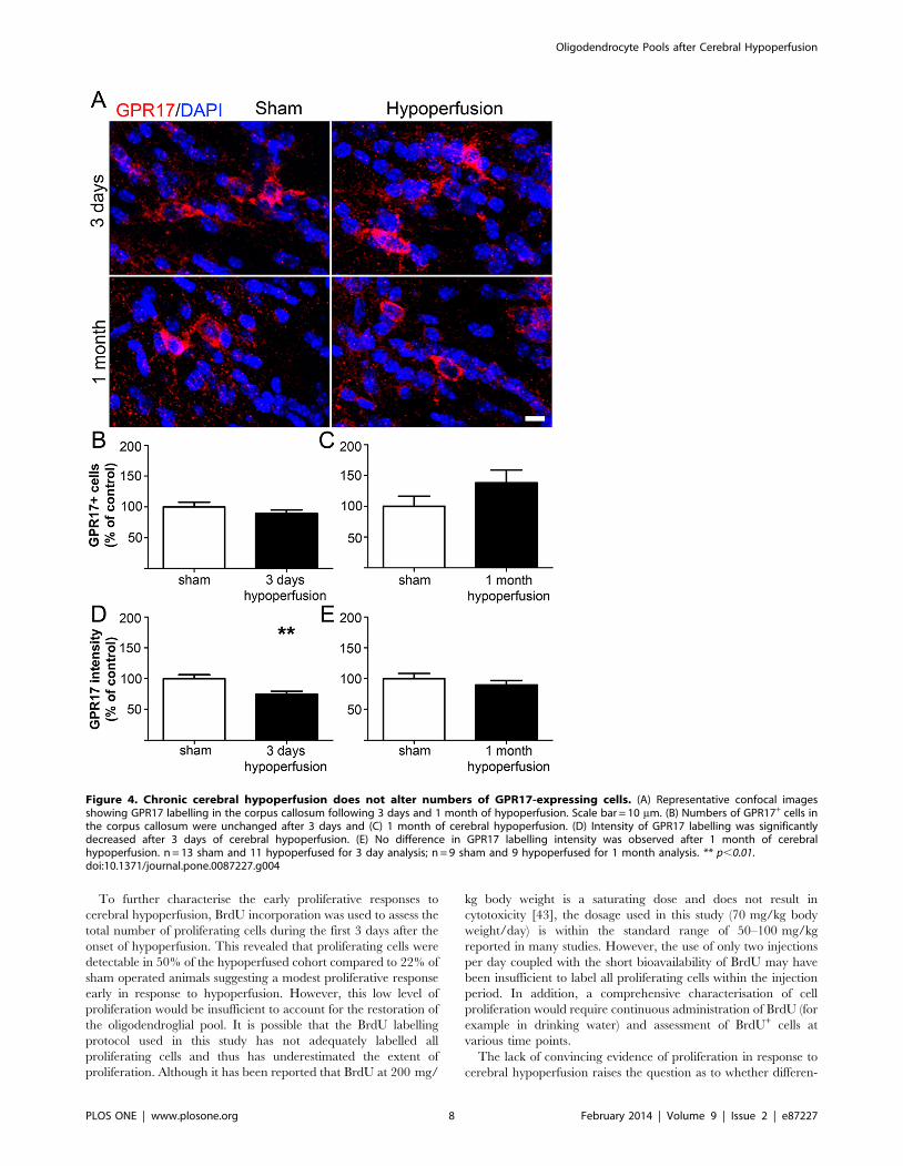
Decreased expression of GPR17 in response to cerebralhypoperfusion
To elucidate a possible mechanism involved in OPC differen-
tiation in response to cerebral hypoperfusion, we analysed the
expression of the G protein-coupled receptor GPR17 (Figures 4A,
S3D). Previous studies have shown that in the intact brain GPR17
is expressed by premyelinating oligodendrocytes and a subset of
OPCs, and receptor activation has been demonstrated to play a
permissive role in OPC differentiation [22,23]. GPR17/NG2
double labelling could not be performed due to the antibodies
being raised in the same species therefore we confirmed GPR17
expression by oligodendrocyte lineage cells using Olig2. This
showed approximately 64% of GPR17+ cells co-expressed Olig2
(Figure S5), which is in agreement with a previous report [23].
Numbers of GPR17+ cells were not significantly different between
groups at either time point examined (Figures 4B and 4C). We
next assessed expression of the receptor, as indicated by the
intensity of staining. This revealed a decrease in the GPR17
labelling intensity after 3 days in hypoperfused as compared to
shams (p = 0.007) (Figure 4D). However, after 1 month of
hypoperfusion there was no difference between groups
(p = 0.363) (Figure 4E).
Oligodendrocyte Pools after Cerebral Hypoperfusion
PLOS ONE | www.plosone.org 4 February 2014 | Volume 9 | Issue 2 | e87227

Impairment of axon-glial integrity, increased microgliaand absence of gross myelin alterations in response tocerebral hypoperfusion
To determine whether alterations in oligodendrocyte numbers
may be paralleled by alterations in myelin density following
cerebral hypoperfusion, myelin status was assessed using the
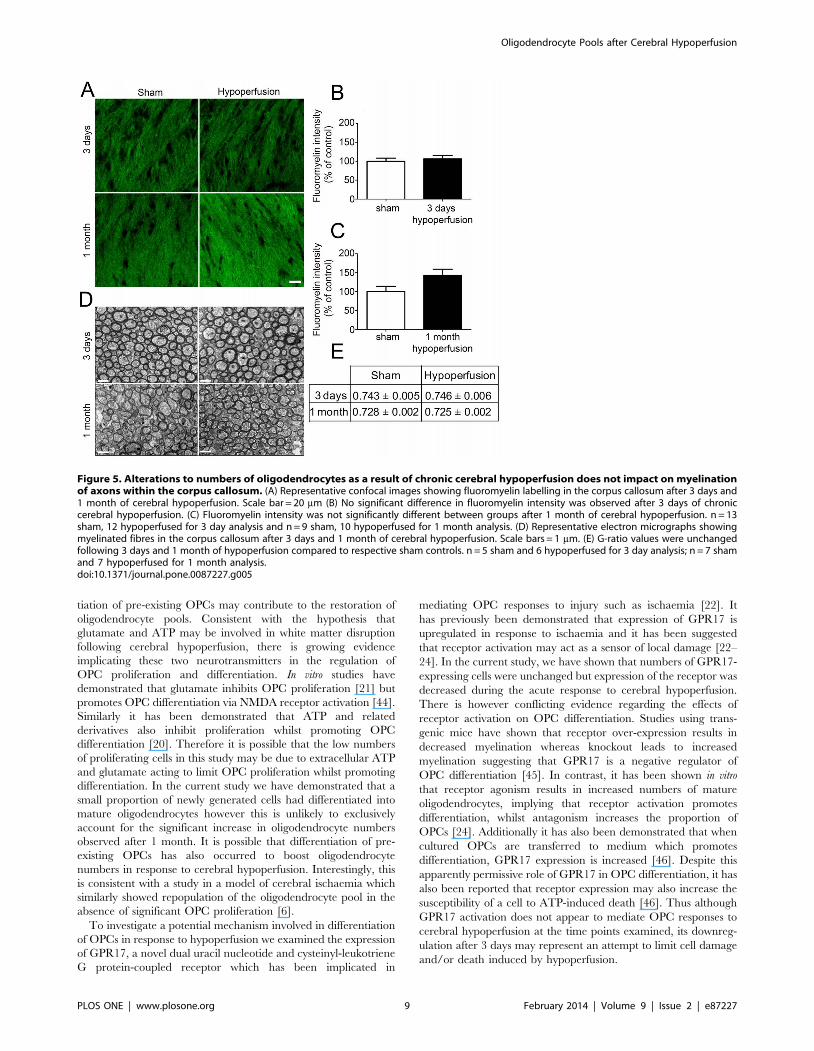
fluorescent lipophilic dye fluoromyelin (Figure 5A). However,
there were no significant differences in staining intensity following
either 3 days of cerebral hypoperfusion (p = 0.598) or after 1
month of cerebral hypoperfusion (p = 0.063) (Figure 5B & C).
Investigation of myelin-enriched extracts using Western blot
analysis additionally indicated that there was no significant
difference in MBP levels between sham and one month
hypoperfused mice (Figure S6).
To further investigate the myelin integrity, electron microscopy
was carried out and measurements of myelin sheath thickness
relative to fibre diameter, i.e. g-ratio, were conducted in separate
cohorts of 3 day and 1 month hypoperfused animals (Figure 5D).
This revealed no significant difference in g-ratio values following
both short and long term hypoperfusion compared to respective
sham-operated controls (Figure 5E). As a note since the 3 days and
1 month cohorts underwent surgery and tissue processing at
different times no statistical comparisons can be made between
sham cohorts or the hypoperfused cohorts.
Despite an absence of gross myelin alterations, in our group we
have consistently determined that cerebral hypoperfusion results in
a disruption of axon-glial integrity [12,25,26] and thus sought to
verify whether similar alterations occur in the white matter in this
study. Axon-glial integrity was assessed by myelin associated
glycoprotein (MAG), a key myelin protein involved in the
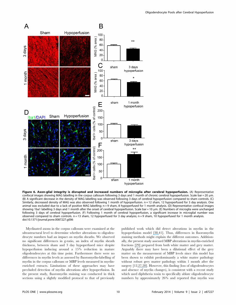
maintenance of axon-glial integrity (Figure 6A). There was a
reduction in the density of MAG staining in the corpus callosum
after 3 days (p = 0.008) and 1 month (p = 0.027) of chronic
cerebral hypoperfusion (Figures 6B and 6C).
Figure 1. Reduced numbers of OPCs and mature oligodendrocytes after 3 days of chronic cerebral hypoperfusion. (A) Representativeconfocal images showing morphology of early and late NG2+ OPCs in the corpus callosum. (B) A significant decrease in numbers of early NG2+ OPCswas found in the corpus callosum after 3 days of chronic cerebral hypoperfusion. (C) No significant differences in numbers of late NG2+ cells wereobserved after 3 days. (D) Representative confocal images of CC1+ labelling of oligodendrocyte cell bodies in the corpus callosum. (E) A significantdecrease in CC1+ oligodendrocytes was found after 3 days of hypoperfusion. n = 13 sham, 12 hypoperfused for NG2 labelling experiments; n = 9sham, 10 hypoperfused for CC1 labelling. Scale bars = 10 mm * p,0.05.doi:10.1371/journal.pone.0087227.g001
Oligodendrocyte Pools after Cerebral Hypoperfusion
PLOS ONE | www.plosone.org 5 February 2014 | Volume 9 | Issue 2 | e87227

A pronounced microglial response as determined by Iba1
immunoreactivity has also been a robust finding in our model of
hypoperfusion (Figure 6D). In support of this after 1 month of
hypoperfusion, numbers of microglia were significantly increased
compared to sham controls (p = 0.0011) (Figure 6E), whilst
numbers of microglia were unchanged after 3 days of cerebral
hypoperfusion (p = 0.425) (Figure 6F), consistent with earlier
studies using this mouse model [12,28].
Taken together these data indicate that whilst gross myelin
morphology remains intact and increased numbers of oligoden-
drocytes are observed following hypoperfusion, axon-glial integrity
is impaired supporting our previous observations [25].
Discussion
Previous studies have demonstrated the susceptibility of
oligodendrocytes to severe reductions in cerebral blood flow
(.70% to that of baseline levels) with profound oligodendrocyte
loss occurring early in response to the insult [6,17]. The present
study additionally highlights the vulnerability of these oligoden-
droglial cells to more modest reductions in blood flow comparable
to those observed in the ageing brain.
In the present study we investigated the pools of OPCs and
mature oligodendrocytes. We observed two different populations
of NG2+ cells identified by differences in morphology. One
population of NG2+ cells showed circular reactivity around the
nucleus and few processes, corresponding to ‘early’ stage OPCs,
whilst the other population of cells was more intensely stained and
more processed, corresponding to ‘late’ stage OPCs. Interestingly
only the ‘early’ stage OPCs were affected by hypoperfusion. It is
possible that these early OPCs have responded rapidly to
hypoperfusion by extending more processes and have thus been
classified as late OPCs. Mature oligodendrocytes were also
reduced in response to hypoperfusion, highlighting the vulnera-
bility of these glial cells to even modest reductions in cerebral
blood flow. Although the mechanisms underlying this early
oligodendroglial loss with hypoperfusion remain to be determined,
these may involve damage to the oligodendrocytes and OPCs as a
result of oxidative stress [31,32] or inflammation [33,34], both of
which are known inducers of oligodendroglial damage and/or
death (for review see [9,35]). In the present study, consistent with
our previous work, we demonstrated a marked microglial response
with hypoperfusion. Previously we have also shown alterations in
indices of hypoxia [25] in white matter after hypoperfusion.
Localised alterations in glutamate levels as a result of compromised
blood flow could also contribute to the loss of oligodendrocytes
and OPCs via NMDA receptor activation leading to intracellular
Ca2+-dependent injury to oligodendroglia [36,37]. However it
should be noted that whilst we propose that the loss of CC1+ and
NG2+ labelling of cells is an indicator of cell loss, a loss of cellular
antigenicity could also account for the reduction in cellular
staining.
With sustained hypoperfusion, marked alterations in oligoden-
droglial pools were observed. The OPC pool was restored and
Figure 2. Restoration of the NG2+ precursor pool and increased numbers of mature oligodendrocytes after 1 month of chroniccerebral hypoperfusion. (A) No significant differences in numbers of early or (B) late NG2+ cells were observed after 1 month of cerebralhypoperfusion. (C) Representative confocal images of CC1+ labelling of oligodendrocyte cell bodies in the corpus callosum. Scale bar = 10 mm. (D) Asignificant increase in CC1+ oligodendrocytes in the corpus callosum was observed following 1 month of chronic cerebral hypoperfusion. n = 10sham, 11 hypoperfused for NG2 and CC1 labelling. ** p,0.01.doi:10.1371/journal.pone.0087227.g002
Oligodendrocyte Pools after Cerebral Hypoperfusion
PLOS ONE | www.plosone.org 6 February 2014 | Volume 9 | Issue 2 | e87227

numbers of mature oligodendrocytes were increased when
examined after 1 month hypoperfusion. This suggests that there
is sufficient capacity with the adult brain to overcome the initial
loss of oligodendrocyte pools. Similarly, in models of focal cerebral
ischaemia when either blood flow is restored with reperfusion or in
the peri-infarct region where there is sufficient collateral flow,
increased numbers of OPCs are detectable [16]. In contrast, other
studies of mouse cerebral hypoperfusion have shown that
oligodendrocyte numbers remain reduced at one month of
hypoperfusion [38,39]. There are notable differences between
the model of cerebral hypoperfusion in our group compared to
others [28,38,39]. Importantly we do not detect demyelination but
instead a robust disruption of axon-glial integrity and a
pronounced microglial response in white matter [12,25,26]. There
may also be differences in the time course of progression of
oligodendrocyte changes between our studies and others. The level
of reduction in cerebral blood flow may be a critical factor which
influences the extent of pathology and proliferation/differentiation
response. We used laser speckle imaging and demonstrated that in
our hands the reduction in CBF was approximately 36% that of
baseline at 3 days and then restored to 22% that of shams at 1
month. These are slightly greater than the levels reported
previously by Shibata et al. 2004 [28] where the maximal levels
of CBF reduction as assessed by laser Doppler flowmetry were
30% although are consistent with more recent studies such as that
by Duan et al. [40] who have reported reductions of 37% from
that of baseline using Laser Speckle imaging. However, there are a
number of other factors that may influence the outcome and
differences in pathology in models between different laboratories
including the anaesthetic used; the background strain of mice
(influences differences in cerebrovasculature) and environment
(pathogen status, temperature). Notably however, in these studies
that report sustained reductions in oligodendrocyte numbers
[38,39,41,42] the pools can be restored by either pharmacological
intervention [39,41,42] or bone marrow cell treatment [38]
indicating that there is restorative capacity of oligodendrocytes in
the model. In a previous study we detected a marked increase in
genes associated with cell proliferation in white matter in response
to hypoperfusion [25] and as a consequence expected to observe
marked increases in cell proliferation. However, assessment using
the acute proliferation marker PCNA showed that overall levels of
proliferation were unchanged with hypoperfusion but revealed
that proliferation of Olig2+ cells was decreased in 3 day
hypoperfused animals compared to controls. It is important to
note that whilst proliferation of OPCs was reduced after 3 days,
there may be a proliferative response of other cells within the white
matter although characterisation of this was beyond the scope of
the current study.
Figure 3. Low numbers of proliferating cells are observed in response to cerebral hypoperfusion however a proportion of newlygenerated cells differentiate into mature oligodendrocytes within 1 month of cerebral hypoperfusion. (A) Representative confocalimages showing PCNA labelling of proliferating cells in the corpus callosum. (B) No significant differences in numbers of PCNA+ cells were observedafter 3 days or (C) 1 month of cerebral hypoperfusion. (D) Confocal images showing Olig2+/PCNA+ labelling of proliferating oligodendroglia in thecorpus callosum. (E) Decreased numbers of Olig2+/PCNA+ cells were observed after 3 days of cerebral hypoperfusion. (F) No significant differences innumbers of Olig2+/PCNA+ cells were found after 1 month of hypoperfusion. (G) Representative confocal images showing BrdU labelled cells in thecorpus callosum. (H) BrdU+ cells were present in 2 out of 9 (22%) sham control animals but were observed in 5 out of 10 (50%) hypoperfused animals.(I) Representative confocal images showing BrdU+/CC1+ cells in the corpus callosum. (J) CC1+/BrdU+ double labelled cells were present in 3 out of 10(30%) of the hypoperfused cohort but were completely absent from the sham control group. n = 13 sham, 11 hypoperfused for PCNA and Olig2labelling at 3 days. One animal was excluded from analysis on the basis of poor cellular staining; n = 9 sham, 9 hypoperfused for PCNA/Olig2 and CC1/BrdU labelling at 1 month. Scale bars = 10 mm. * p,0.05.doi:10.1371/journal.pone.0087227.g003
Oligodendrocyte Pools after Cerebral Hypoperfusion
PLOS ONE | www.plosone.org 7 February 2014 | Volume 9 | Issue 2 | e87227
To further characterise the early proliferative responses to
cerebral hypoperfusion, BrdU incorporation was used to assess the
total number of proliferating cells during the first 3 days after the
onset of hypoperfusion. This revealed that proliferating cells were
detectable in 50% of the hypoperfused cohort compared to 22% of
sham operated animals suggesting a modest proliferative response
early in response to hypoperfusion. However, this low level of
proliferation would be insufficient to account for the restoration of
the oligodendroglial pool. It is possible that the BrdU labelling
protocol used in this study has not adequately labelled all
proliferating cells and thus has underestimated the extent of
proliferation. Although it has been reported that BrdU at 200 mg/
kg body weight is a saturating dose and does not result in
cytotoxicity [43], the dosage used in this study (70 mg/kg body
weight/day) is within the standard range of 50–100 mg/kg
reported in many studies. However, the use of only two injections
per day coupled with the short bioavailability of BrdU may have
been insufficient to label all proliferating cells within the injection
period. In addition, a comprehensive characterisation of cell
proliferation would require continuous administration of BrdU (for
example in drinking water) and assessment of BrdU+ cells at
various time points.
The lack of convincing evidence of proliferation in response to
cerebral hypoperfusion raises the question as to whether differen-
Figure 4. Chronic cerebral hypoperfusion does not alter numbers of GPR17-expressing cells. (A) Representative confocal imagesshowing GPR17 labelling in the corpus callosum following 3 days and 1 month of hypoperfusion. Scale bar = 10 mm. (B) Numbers of GPR17+ cells inthe corpus callosum were unchanged after 3 days and (C) 1 month of cerebral hypoperfusion. (D) Intensity of GPR17 labelling was significantlydecreased after 3 days of cerebral hypoperfusion. (E) No difference in GPR17 labelling intensity was observed after 1 month of cerebralhypoperfusion. n = 13 sham and 11 hypoperfused for 3 day analysis; n = 9 sham and 9 hypoperfused for 1 month analysis. ** p,0.01.doi:10.1371/journal.pone.0087227.g004
Oligodendrocyte Pools after Cerebral Hypoperfusion
PLOS ONE | www.plosone.org 8 February 2014 | Volume 9 | Issue 2 | e87227
tiation of pre-existing OPCs may contribute to the restoration of
oligodendrocyte pools. Consistent with the hypothesis that
glutamate and ATP may be involved in white matter disruption
following cerebral hypoperfusion, there is growing evidence
implicating these two neurotransmitters in the regulation of
OPC proliferation and differentiation. In vitro studies have
demonstrated that glutamate inhibits OPC proliferation [21] but
promotes OPC differentiation via NMDA receptor activation [44].
Similarly it has been demonstrated that ATP and related
derivatives also inhibit proliferation whilst promoting OPC
differentiation [20]. Therefore it is possible that the low numbers
of proliferating cells in this study may be due to extracellular ATP
and glutamate acting to limit OPC proliferation whilst promoting
differentiation. In the current study we have demonstrated that a
small proportion of newly generated cells had differentiated into
mature oligodendrocytes however this is unlikely to exclusively
account for the significant increase in oligodendrocyte numbers
observed after 1 month. It is possible that differentiation of pre-
existing OPCs has also occurred to boost oligodendrocyte
numbers in response to cerebral hypoperfusion. Interestingly, this
is consistent with a study in a model of cerebral ischaemia which
similarly showed repopulation of the oligodendrocyte pool in the
absence of significant OPC proliferation [6].
To investigate a potential mechanism involved in differentiation
of OPCs in response to hypoperfusion we examined the expression
of GPR17, a novel dual uracil nucleotide and cysteinyl-leukotriene
G protein-coupled receptor which has been implicated in
mediating OPC responses to injury such as ischaemia [22]. It
has previously been demonstrated that expression of GPR17 is
upregulated in response to ischaemia and it has been suggested
that receptor activation may act as a sensor of local damage [22–
24]. In the current study, we have shown that numbers of GPR17-
expressing cells were unchanged but expression of the receptor was
decreased during the acute response to cerebral hypoperfusion.
There is however conflicting evidence regarding the effects of
receptor activation on OPC differentiation. Studies using trans-
genic mice have shown that receptor over-expression results in
decreased myelination whereas knockout leads to increased
myelination suggesting that GPR17 is a negative regulator of
OPC differentiation [45]. In contrast, it has been shown in vitro
that receptor agonism results in increased numbers of mature
oligodendrocytes, implying that receptor activation promotes
differentiation, whilst antagonism increases the proportion of
OPCs [24]. Additionally it has also been demonstrated that when
cultured OPCs are transferred to medium which promotes
differentiation, GPR17 expression is increased [46]. Despite this
apparently permissive role of GPR17 in OPC differentiation, it has
also been reported that receptor expression may also increase the
susceptibility of a cell to ATP-induced death [46]. Thus although
GPR17 activation does not appear to mediate OPC responses to
cerebral hypoperfusion at the time points examined, its downreg-
ulation after 3 days may represent an attempt to limit cell damage
and/or death induced by hypoperfusion.
Figure 5. Alterations to numbers of oligodendrocytes as a result of chronic cerebral hypoperfusion does not impact on myelinationof axons within the corpus callosum. (A) Representative confocal images showing fluoromyelin labelling in the corpus callosum after 3 days and1 month of cerebral hypoperfusion. Scale bar = 20 mm (B) No significant difference in fluoromyelin intensity was observed after 3 days of chroniccerebral hypoperfusion. (C) Fluoromyelin intensity was not significantly different between groups after 1 month of cerebral hypoperfusion. n = 13sham, 12 hypoperfused for 3 day analysis and n = 9 sham, 10 hypoperfused for 1 month analysis. (D) Representative electron micrographs showingmyelinated fibres in the corpus callosum after 3 days and 1 month of cerebral hypoperfusion. Scale bars = 1 mm. (E) G-ratio values were unchangedfollowing 3 days and 1 month of hypoperfusion compared to respective sham controls. n = 5 sham and 6 hypoperfused for 3 day analysis; n = 7 shamand 7 hypoperfused for 1 month analysis.doi:10.1371/journal.pone.0087227.g005
Oligodendrocyte Pools after Cerebral Hypoperfusion
PLOS ONE | www.plosone.org 9 February 2014 | Volume 9 | Issue 2 | e87227
Myelinated axons in the corpus callosum were examined at the
ultrastructural level to determine whether alterations to oligoden-
drocyte numbers had an impact on myelin sheaths. We observed
no significant differences in g-ratio, an index of myelin sheath
thickness, between sham and 3 day hypoperfused mice despite
hypoperfusion inducing around a 15% reduction in mature
oligodendrocytes at this time point. Furthermore there were no
differences in myelin levels as assessed by fluoromyelin-labelling of
myelin in the corpus callosum or MBP levels measured in myelin-
enriched extracts. Limitations of these approaches may have
precluded detection of myelin alterations after hypoperfusion. In
the present study, fluoromyelin staining was conducted in thick
sections using a slightly modified protocol to that of previously
published work which did detect alterations in myelin in the
hypoperfusion model [38,41]. Thus, differences in fluoromyelin
staining methods might explain the different outcomes. Addition-
ally, the present study assessed MBP alterations in myelin-enriched
fractions [29] prepared from both white matter and grey matter.
Arguably there may have been a dilutional effect of the grey
matter on the measurement of MBP levels since this model has
been shown to exhibit predominantly a white matter pathology
without robust grey matter pathology within 1 month after the
surgery [12,27,28]. However, this finding (loss of oligodendrocytes
and absence of myelin changes), is consistent with a recent study
which used diphtheria toxin to specifically ablate oligodendrocyte
numbers by approximately 26% and reported that myelin was
Figure 6. Axon-glial integrity is disrupted and increased numbers of microglia after cerebral hypoperfusion. (A) Representativeconfocal images showing MAG labelling in the corpus callosum following 3 days and 1 month of chronic cerebral hypoperfusion. Scale bar = 20 mm.(B) A significant decrease in the density of MAG labelling was observed following 3 days of cerebral hypoperfusion compared to sham controls. (C)Similarly, decreased density of MAG was also observed following 1 month of hypoperfusion. n = 12 sham, 12 hypoperfused for 3 day analysis. Oneanimal was excluded due to a lack of positive MAG labelling; n = 9 sham, 8 hypoperfused for 1 month analysis. (D) Representative confocal imagesshowing 1ba1 labelling 3 days and 1 month after the onset of cerebral hypoperfusion. Scale bar = 10 mm. (E) Numbers of microglia were unchangedfollowing 3 days of cerebral hypoperfusion. (F) Following 1 month of cerebral hypoperfusion, a significant increase in microglial number wasobserved compared to sham controls. n = 13 sham, 12 hypoperfused for 3 day analysis; n = 9 sham, 10 hypoperfused for 1 month analysis.doi:10.1371/journal.pone.0087227.g006
Oligodendrocyte Pools after Cerebral Hypoperfusion
PLOS ONE | www.plosone.org 10 February 2014 | Volume 9 | Issue 2 | e87227

preserved despite this extensive oligodendrocyte loss [47]. This
lack of effect on myelination despite profound alterations in
oligodendrocyte pools is also consistent with previous studies by
our group which have determined that there are no gross
alterations to the protein levels of myelin basic protein as assessed
by immunohistochemistry [25]. Instead, in this model, hypoper-
fusion results in disruption of MAG and breakdown of axon-glial
integrity associated with disruption of the paranodal septate
junctions [25]. The current study used an antibody against the
CC1 protein which labels oligodendrocyte cell bodies but not
processes, and as a result cannot reliably distinguish between
myelinating and non-myelinating oligodendrocytes. One possible
explanation for our findings is that in response to cerebral
hypoperfusion, a non-myelinating population of CC1+ oligoden-
drocytes are lost and thus no disruption to myelin has been
observed. Another possibility is that neighbouring oligodendro-
cytes may compensate for oligodendrocyte loss by extending
processes and ‘filling in’ non-myelinated areas [48]. We also
observed that g-ratio was similarly unchanged following 1 month
of hypoperfusion and so it remains to be determined whether
increased numbers of oligodendrocytes observed at this time point
are surplus to requirements. Overall these findings suggest that
even in the presence of cerebral hypoperfusion, the CNS can
tolerate significant alterations to mature oligodendrocyte pools
without any apparent detriment to myelin thickness or density.
Thus in conclusion this study provides further support that
oligodendrocytes are vulnerable to modest blood flow reductions
and evidence supporting their regenerative capacity. This turnover
of the pool of oligodendrocytes appears to occur in the absence of
changes in myelination but it would be interesting to investigate in
future studies whether there are any functional consequences of
these changes within the white matter.
Supporting Information
Figure S1 Reduced cerebral blood flow in hypoperfusedanimals. Cerebral blood flow was measured using laser speckle
flowmetry prior to surgery (baseline) and at 3 days and 1 month
following surgery to assess the extent of hypoperfusion at the times
white matter alterations were investigated. (A) Representative
images showing speckle images at baseline, 3 days and 1 month for
a sham and hypoperfused mouse. Images show the average of 100
frames. Baseline CBF was not significantly different (p,0.05)
between groups (1130636 perfusion units in shams vs 1072624
perfusion units in hypoperfused group). (B&C) After 3 days and 1
month, CBF was significantly decreased in hypoperfused com-
pared to sham animals to (p,0.001 and p,0.005 respectively).
Data are calculated for each mouse as the percentage change
relative to baseline. (B) Cerebral blood flow was decreased by
approximately 36% to that of shams following 3 days of cerebral
hypoperfusion. (C) Following 1 month of hypoperfusion, CBF
values had recovered to approximately 22% of sham levels. Data is
shown for each mouse, n = 8 sham, 6 hypoperfused.
(TIF)
Figure S2 NG2 is a specific marker of OPCs. NG2 and
PDGFRa labelling confirmed the specificity of NG2 as an OPC
marker in the corpus callosum. (A) NG2 and PDGFRa co-labelled
OPCs in the corpus callosum. Scale bar = 10 mm. (B) Confocal
images showing representative NG2 and PDGFRb labelling. No
PDGFRb+ labelling of pericytes was observed in the corpus
callosum and only occasional PDGFRb+ labelling was observed in
the cortex. No NG2+/PDGFRb+ cells were observed in either
region. Scale bar = 20 mm.
(TIF)
Figure S3 Low magnification images of NG2, CC1,Olig2 and GPR17 labelling. (A) Low magnification confocal
images showing NG2+ labelling of OPCs in the corpus callosum
(CC). (B) Confocal images showing CC1+ labelling of mature
oligodendrocytes in the corpus callosum. (C) Confocal images
showing Olig2+ labelling of oligodendroglia in the corpus
callosum. (D) Confocal images showing GPR17+ labelling in the
corpus callosum. Scale bars = 50 mm. CTx = Cortex, CC = corpus
callosum, CPu = caudate putamen.
(TIF)
Figure S4 Astrocytes are not labelled with CC1 andnumbers are unchanged following 1 month of cerebralhypoperfusion. It has been reported that a subpopulation of
astrocytes can express the CC1 (APC) antigen, therefore CC1 and
GFAP double labelling was carried out to determine numbers of
CC1+ astrocytes. (A) Representative confocal images showing
CC1+/GFAP+ double labelling in the corpus callosum (CC). Scale
bar = 50 mm, inset scale bar = 10 mm. Cell counts of numbers of
double labelled cells revealed approximately 0.8% of CC1+ cells
expressed GFAP thus confirming the high specificity of CC1 as a
marker of mature oligodendrocytes. (B) Representative confocal
images showing GFAP+ labelling of astrocytes in the corpus
callosum (CC). Scale bar = 50 mm. (C) Numbers of GFAP+
astrocytes are unchanged following 1 month of chronic cerebral
hypoperfusion. CTx = Cortex, CC = corpus callosum, CPu = cau-
date putamen.
(TIF)
Figure S5 GPR17 is expressed by oligodendroglia and asmall number of microglia. (A) Representative confocal
images showing GPR17+/Olig2+ double labelling in the corpus
callosum. Arrows indicate double labelled cells. (B) Representative
confocal images showing GPR17+/Iba1+ labelling in the corpus
callosum. (C) Cell counting revealed approximately 64.463.33%
of GPR17+ cells express Olig2 in the corpus callosum. Only
8.2960.75% of GPR17+ cells co-expressed Iba1. Scale
bars = 50 mm, inset scale bars = 10� mm. CTx = Cortex,
CC = corpus callosum, CPu = caudate putamen.
(TIF)
Figure S6 MBP levels are unchanged following 1 monthof cerebral hypoperfusion. (A) Representative Western blot
from myelin enriched extracts showing the different MBP isoforms
(25–16 kDa) and GAPDH, the later used as a loading control. (B)
Analysis of fluorescent intensity relative to GAPDH showed no
significant changes in MBP levels after 1 month of cerebral
hypoperfusion. n = 5 sham, n = 8 hypoperfused.
(TIF)
Acknowledgments
We would like to thank the Euan MacDonald Centre for the use of
confocal facilities, and Julia Edgar and Maj-lis McCulloch for their input
and advice regarding electron microscopy studies.
Author Contributions
Conceived and designed the experiments: KH JM. Performed the
experiments: JM MR JF PH YM MM. Analyzed the data: JM JF KH
YM. Wrote the paper: JM KH.
Oligodendrocyte Pools after Cerebral Hypoperfusion
PLOS ONE | www.plosone.org 11 February 2014 | Volume 9 | Issue 2 | e87227
References
1. Nave KA (2010) Myelination and support of axonal integrity by glia. Nature
468: 244–252.2. Rivers LE, Young KM, Rizzi M, Jamen F, Psachoulia K, et al. (2008)
PDGFRA/NG2 glia generate myelinating oligodendrocytes and piriformprojection neurons in adult mice. Nature Neuroscience 11: 1392–1401.
3. Tomimoto H, Ihara M, Wakita H, Ohtani R, Lin JX, et al. (2003) Chronic
cerebral hypoperfusion induces white matter lesions and loss of oligodendrogliawith DNA fragmentation in the rat. Acta Neuropathology 106: 527–534.
4. Valeriani V, Dewar D, McCulloch J (2000) Quantitative assessment of ischemicpathology in axons, oligodendrocytes, and neurons: attenuation of damage after
transient ischemia. Journal of Cerebral Blood Flow and Metabolism 20: 765–
771.5. Pantoni L, Garcia JH, Gutierrez JA (1996) Cerebral white matter is highly
vulnerable to ischemia. Stroke 27: 1641–1646; discussion 1647.6. McIver SR, Muccigrosso M, Gonzales ER, Lee JM, Roberts MS, et al. (2010)
Oligodendrocyte degeneration and recovery after focal cerebral ischemia.Neuroscience 169: 1364–1375.
7. Lyons SA, Kettenmann H (1998) Oligodendrocytes and microglia are selectively
vulnerable to combined hypoxia and hypoglycemia injury in vitro. Journal ofCerebral Blood Flow and Metabolism 18: 521–530.
8. McDonald JW, Levine JM, Qu Y (1998) Multiple classes of the oligodendrocytelineage are highly vulnerable to excitotoxicity. Neuroreport 9: 2757–2762.
9. Merrill JE, Scolding NJ (1999) Mechanisms of damage to myelin and
oligodendrocytes and their relevance to disease. Neuropathology and AppliedNeurobiology 25: 435–458.
10. Head D, Buckner RL, Shimony JS, Williams LE, Akbudak E, et al. (2004)Differential vulnerability of anterior white matter in nondemented aging with
minimal acceleration in dementia of the Alzheimer type: evidence from diffusiontensor imaging. Cerebral Cortex 14: 410–423.
11. Kohama SG, Rosene DL, Sherman LS (2011) Age-related changes in human
and non-human primate white matter: from myelination disturbances tocognitive decline. Age DOI 10.1007/s11357-011-9357-7.
12. Coltman R, Spain A, Tsenkina Y, Fowler JH, Smith J, et al. (2011) Selectivewhite matter pathology induces a specific impairment in spatial working
memory. Neurobiology of Aging 32: 2324 e2327–2312.
13. Desai MK, Mastrangelo MA, Ryan DA, Sudol KL, Narrow WC, et al. (2010)Early oligodendrocyte/myelin pathology in Alzheimer’s disease mice constitutes
a novel therapeutic target. American Journal of Pathology 177: 1422–1435.14. Aboul-Enein F, Rauschka H, Kornek B, Stadelmann C, Stefferl A, et al. (2003)
Preferential loss of myelin-associated glycoprotein reflects hypoxia-like whitematter damage in stroke and inflammatory brain diseases. Journal of
Neuropathology and Experimental Neurology 62: 25–33.
15. Zaidi AU, Bessert DA, Ong JE, Xu H, Barks JD, et al. (2004) Newoligodendrocytes are generated after neonatal hypoxic-ischemic brain injury in
rodents. Glia 46: 380–390.16. Tanaka K, Nogawa S, Ito D, Suzuki S, Dembo T, et al. (2001) Activation of
NG2-positive oligodendrocyte progenitor cells during post-ischemic reperfusion
in the rat brain. Neuroreport 12: 2169–2174.17. Tanaka K, Nogawa S, Suzuki S, Dembo T, Kosakai A (2003) Upregulation of
oligodendrocyte progenitor cells associated with restoration of matureoligodendrocytes and myelination in peri-infarct area in the rat brain. Brain
Research 989: 172–179.18. Simpson JE, Fernando MS, Clark L, Ince PG, Matthews F, et al. (2007) White
matter lesions in an unselected cohort of the elderly: astrocytic, microglial and
oligodendrocyte precursor cell responses. Neuropathology and Applied Neuro-biology 33: 410–419.
19. Back SA, Kroenke CD, Sherman LS, Lawrence G, Gong X, et al. (2011) Whitematter lesions defined by diffusion tensor imaging in older adults. Annals of
Neurology 70: 465–476.
20. Agresti C, Meomartini ME, Amadio S, Ambrosini E, Volonte C, et al. (2005)ATP regulates oligodendrocyte progenitor migration, proliferation, and
differentiation: involvement of metabotropic P2 receptors. Brain ResearchReviews 48: 157–165.
21. Yuan X, Eisen AM, McBain CJ, Gallo V (1998) A role for glutamate and its
receptors in the regulation of oligodendrocyte development in cerebellar tissueslices. Development 125: 2901–2914.
22. Ciana P, Fumagalli M, Trincavelli ML, Verderio C, Rosa P, et al. (2006) Theorphan receptor GPR17 identified as a new dual uracil nucleotides/cysteinyl-
leukotrienes receptor. The EMBO Journal 25: 4615–4627.23. Lecca D, Trincavelli ML, Gelosa P, Sironi L, Ciana P, et al. (2008) The recently
identified P2Y-like receptor GPR17 is a sensor of brain damage and a new target
for brain repair. PLoS One 3: e3579.
24. Boda E, Vigano F, Rosa P, Fumagalli M, Labat-Gest V, et al. (2011) The
GPR17 receptor in NG2 expressing cells: Focus on in vivocell maturation andparticipation in acute trauma and chronic damage. Glia 59: 1958–1973.
25. Reimer MM, McQueen J, Searcy L, Scullion G, Zonta B, et al. (2011) Rapiddisruption of axon-glial integrity in response to mild cerebral hypoperfusion.
Journal of Neuroscience 31: 18185–18194.
26. Holland PR, Bastin ME, Jansen MA, Merrifield GD, Coltman RB, et al. (2011)MRI is a sensitive marker of subtle white matter pathology in hypoperfused
mice. Neurobiology of Aging 32: 2325.e2321–e2326.27. Shibata M, Yamasaki N, Miyakawa T, Kalaria RN, Fujita Y, et al. (2007)
Selective impairment of working memory in a mouse model of chronic cerebral
hypoperfusion. Stroke 38: 2826–2832.28. Shibata M, Ohtani R, Ihara M, Tomimoto H (2004) White matter lesions and
glial activation in a novel mouse model of chronic cerebral hypoperfusion.Stroke 35: 2598–2603.
29. Yool DA, Klugmann M, McLaughlin M, Vouyiouklis DA, Dimou L, et al.(2001) Myelin proteolipid proteins promote the interaction of oligodendrocytes
and axons. J Neurosci Res 63: 151–164.
30. Bhat RV, Axt KJ, Fosnaugh JS, Smith KJ, Johnson KA, et al. (1996) Expressionof the APC tumor suppressor protein in oligodendroglia. Glia 17: 169–174.
31. Back SA, Gan X, Li Y, Rosenberg PA, Volpe JJ (1998) Maturation-dependentvulnerability of oligodendrocytes to oxidative stress-induced death caused by
glutathione depletion. Journal of Neuroscience 18: 6241–6253.
32. Husain J, Juurlink BH (1995) Oligodendroglial precursor cell susceptibility tohypoxia is related to poor ability to cope with reactive oxygen species. Brain
Research 698: 86–94.33. Mabuchi T, Kitagawa K, Ohtsuki T, Kuwabara K, Yagita Y, et al. (2000)
Contribution of microglia/macrophages to expansion of infarction and responseof oligodendrocytes after focal cerebral ischemia in rats. Stroke 31: 1735–1743.
34. Deng Y, Lu J, Sivakumar V, Ling EA, Kaur C (2008) Amoeboid microglia in the
periventricular white matter induce oligodendrocyte damage through expressionof proinflammatory cytokines via MAP kinase signaling pathway in hypoxic
neonatal rats. Brain Pathology 18: 387–400.35. McTigue DM, Tripathi RB (2008) The life, death, and replacement of
oligodendrocytes in the adult CNS. Journal of Neurochemistry 107: 1–19.
36. Alix JJ, Fern R (2009) Glutamate receptor-mediated ischemic injury ofpremyelinated central axons. Annals of Neurology 66: 682–693.
37. Stys PK, Lipton SA (2007) White matter NMDA receptors: an unexpected newtherapeutic target? Trends in Pharmacological Sciences 28: 561–566.
38. Fujita Y, Ihara M, Ushiki T, Hirai H, Kizaka-Kondoh S, et al. (2010) Earlyprotective effect of bone marrow mononuclear cells against ischemic white
matter damage through augmentation of cerebral blood flow. Stroke; a journal
of cerebral circulation 41: 2938–2943.39. Maki T, Ihara M, Fujita Y, Nambu T, Miyashita K, et al. (2011) Angiogenic and
vasoprotective effects of adrenomedullin on prevention of cognitive decline afterchronic cerebral hypoperfusion in mice. Stroke; a journal of cerebral circulation
42: 1122–1128.
40. Duan W, Ran H, Zhou Z, He Q, Zheng J (2012) Adenosine A2A receptordeficiency up-regulates cystatin F expression in white matter lesions induced by
chronic cerebral hypoperfusion. PLoS One 7: e52566.41. Miyamoto N, Maki T, Pham LD, Hayakawa K, Seo JH, et al. (2013) Oxidative
Stress Interferes With White Matter Renewal After Prolonged CerebralHypoperfusion in Mice. Stroke.
42. Miyamoto N, Pham LD, Hayakawa K, Matsuzaki T, Seo JH, et al. (2013) Age-
Related Decline in Oligodendrogenesis Retards White Matter Repair in Mice.Stroke.
43. Taupin P (2007) BrdU immunohistochemistry for studying adult neurogenesis:paradigms, pitfalls, limitations, and validation. Brain Res Rev 53: 198–214.
44. Cavaliere F, Urra O, Alberdi E, Matute C (2012) Oligodendrocyte
differentiation from adult multipotent stem cells is modulated by glutamate.Cell Death and Disease 3: e268.
45. Chen Y, Wu H, Wang S, Koito H, Li J, et al. (2009) The oligodendrocyte-specific G protein-coupled receptor GPR17 is a cell-intrinsic timer of
myelination. Nature Neuroscience 12: 1398–1406.
46. Ceruti S, Vigano F, Boda E, Ferrario S, Magni G, et al. (2011) Expression of thenew P2Y-like receptor GPR17 during oligodendrocyte precursor cell maturation
regulates sensitivity to ATP-induced death. Glia 59: 363–378.47. Oluich LJ, Stratton JA, Lulu Xing Y, Ng SW, Cate HS, et al. (2012) Targeted
ablation of oligodendrocytes induces axonal pathology independent of overtdemyelination. Journal of Neuroscience 32: 8317–8330.
48. Richardson WD, Young KM, Tripathi RB, McKenzie I (2011) NG2-glia as
multipotent neural stem cells: fact or fantasy? Neuron 70: 661–673.
Oligodendrocyte Pools after Cerebral Hypoperfusion
PLOS ONE | www.plosone.org 12 February 2014 | Volume 9 | Issue 2 | e87227