Requirement for RecFOR-mediated recombination in priA mutant: Properties of priA recFOR mutants
12
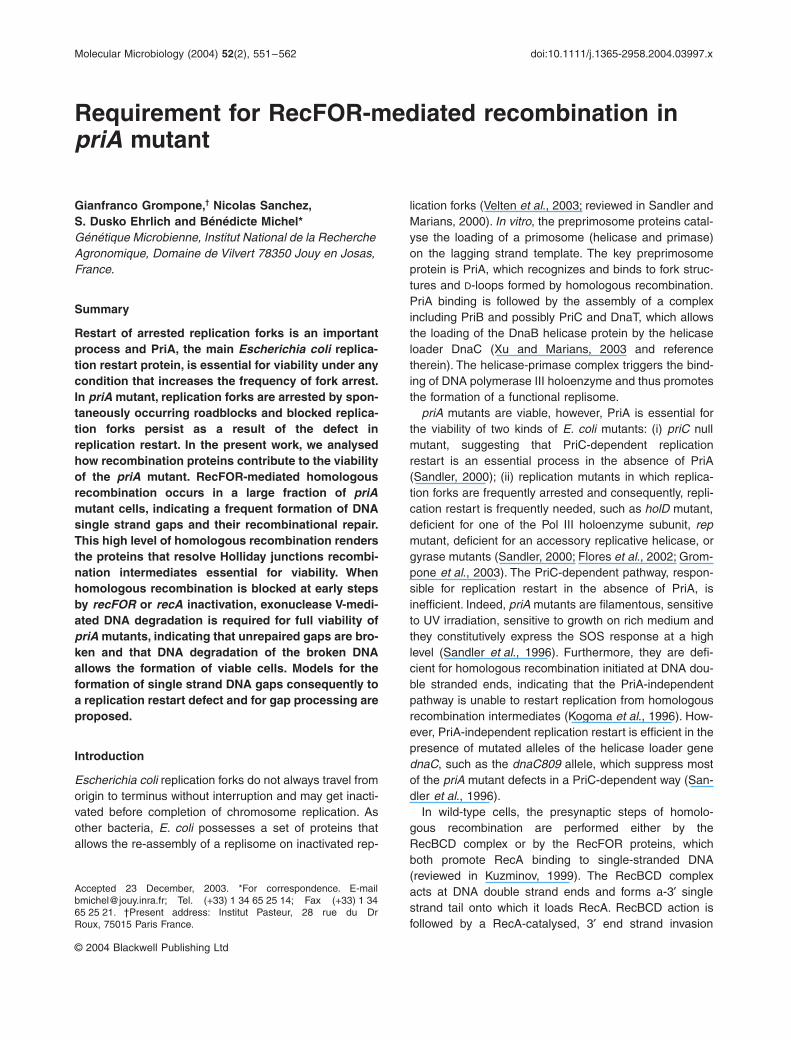
Molecular Microbiology (2004) 52(2), 551–562 doi:10.1111/j.1365-2958.2004.03997.x © 2004 Blackwell Publishing Ltd Blackwell Science, LtdOxford, UKMMIMolecular Microbiology0950-382XBlackwell Publishing Ltd, 2004 ? 2004522551562Original ArticleProperties of priA recFOR mutantsG. Grompone, N. Sanchez, S. Dusko Ehrlich and B. Michel Accepted 23 December, 2003. *For correspondence. E-mail [email protected]; Tel. (+33) 1 34 65 25 14; Fax (+33) 1 34 65 25 21. †Present address: Institut Pasteur, 28 rue du Dr Roux, 75015 Paris France. Requirement for RecFOR-mediated recombination in priA mutant Gianfranco Grompone, † Nicolas Sanchez, S. Dusko Ehrlich and Bénédicte Michel* Génétique Microbienne, Institut National de la Recherche Agronomique, Domaine de Vilvert 78350 Jouy en Josas, France. Summary Restart of arrested replication forks is an important process and PriA, the main Escherichia coli replica- tion restart protein, is essential for viability under any condition that increases the frequency of fork arrest. In priA mutant, replication forks are arrested by spon- taneously occurring roadblocks and blocked replica- tion forks persist as a result of the defect in replication restart. In the present work, we analysed how recombination proteins contribute to the viability of the priA mutant. RecFOR-mediated homologous recombination occurs in a large fraction of priA mutant cells, indicating a frequent formation of DNA single strand gaps and their recombinational repair. This high level of homologous recombination renders the proteins that resolve Holliday junctions recombi- nation intermediates essential for viability. When homologous recombination is blocked at early steps by recFOR or recA inactivation, exonuclease V-medi- ated DNA degradation is required for full viability of priA mutants, indicating that unrepaired gaps are bro- ken and that DNA degradation of the broken DNA allows the formation of viable cells. Models for the formation of single strand DNA gaps consequently to a replication restart defect and for gap processing are proposed. Introduction Escherichia coli replication forks do not always travel from origin to terminus without interruption and may get inacti- vated before completion of chromosome replication. As other bacteria, E. coli possesses a set of proteins that allows the re-assembly of a replisome on inactivated rep- lication forks (Velten et al ., 2003; reviewed in Sandler and Marians, 2000). In vitro , the preprimosome proteins catal- yse the loading of a primosome (helicase and primase) on the lagging strand template. The key preprimosome protein is PriA, which recognizes and binds to fork struc- tures and D-loops formed by homologous recombination. PriA binding is followed by the assembly of a complex including PriB and possibly PriC and DnaT, which allows the loading of the DnaB helicase protein by the helicase loader DnaC (Xu and Marians, 2003 and reference therein). The helicase-primase complex triggers the bind- ing of DNA polymerase III holoenzyme and thus promotes the formation of a functional replisome. priA mutants are viable, however, PriA is essential for the viability of two kinds of E. coli mutants: (i) priC null mutant, suggesting that PriC-dependent replication restart is an essential process in the absence of PriA (Sandler, 2000); (ii) replication mutants in which replica- tion forks are frequently arrested and consequently, repli- cation restart is frequently needed, such as holD mutant, deficient for one of the Pol III holoenzyme subunit, rep mutant, deficient for an accessory replicative helicase, or gyrase mutants (Sandler, 2000; Flores et al ., 2002; Grom- pone et al ., 2003). The PriC-dependent pathway, respon- sible for replication restart in the absence of PriA, is inefficient. Indeed, priA mutants are filamentous, sensitive to UV irradiation, sensitive to growth on rich medium and they constitutively express the SOS response at a high level (Sandler et al ., 1996). Furthermore, they are defi- cient for homologous recombination initiated at DNA dou- ble stranded ends, indicating that the PriA-independent pathway is unable to restart replication from homologous recombination intermediates (Kogoma et al ., 1996). How- ever, PriA-independent replication restart is efficient in the presence of mutated alleles of the helicase loader gene dnaC , such as the dnaC809 allele, which suppress most of the priA mutant defects in a PriC-dependent way (San- dler et al ., 1996). In wild-type cells, the presynaptic steps of homolo- gous recombination are performed either by the RecBCD complex or by the RecFOR proteins, which both promote RecA binding to single-stranded DNA (reviewed in Kuzminov, 1999). The RecBCD complex acts at DNA double strand ends and forms a-3 ¢ single strand tail onto which it loads RecA. RecBCD action is followed by a RecA-catalysed, 3 ¢ end strand invasion
-
Upload
independent -
Category
Documents
-
view
0 -
download
0
Transcript of Requirement for RecFOR-mediated recombination in priA mutant: Properties of priA recFOR mutants
Molecular Microbiology (2004)
52
(2), 551–562 doi:10.1111/j.1365-2958.2004.03997.x
© 2004 Blackwell Publishing Ltd
Blackwell Science, LtdOxford, UKMMIMolecular Microbiology0950-382XBlackwell Publishing Ltd, 2004
? 2004
52
2551562
Original Article
Properties of priA recFOR mutantsG. Grompone, N. Sanchez, S. Dusko Ehrlich and B. Michel
Accepted 23 December, 2003. *For correspondence. [email protected]; Tel. (+33) 1 34 65 25 14; Fax (+33) 1 3465 25 21. †Present address: Institut Pasteur, 28 rue du DrRoux, 75015 Paris France.
Requirement for RecFOR-mediated recombination in
priA
mutant
Gianfranco Grompone,
†
Nicolas Sanchez, S. Dusko Ehrlich and Bénédicte Michel*
Génétique Microbienne, Institut National de la Recherche Agronomique, Domaine de Vilvert 78350 Jouy en Josas, France.
Summary
Restart of arrested replication forks is an importantprocess and PriA, the main
Escherichia coli
replica-tion restart protein, is essential for viability under anycondition that increases the frequency of fork arrest.In
priA
mutant, replication forks are arrested by spon-taneously occurring roadblocks and blocked replica-tion forks persist as a result of the defect inreplication restart. In the present work, we analysedhow recombination proteins contribute to the viabilityof the
priA
mutant. RecFOR-mediated homologousrecombination occurs in a large fraction of
priA
mutant cells, indicating a frequent formation of DNAsingle strand gaps and their recombinational repair.This high level of homologous recombination rendersthe proteins that resolve Holliday junctions recombi-nation intermediates essential for viability. Whenhomologous recombination is blocked at early stepsby
recFOR
or
recA
inactivation, exonuclease V-medi-ated DNA degradation is required for full viability of
priA
mutants, indicating that unrepaired gaps are bro-ken and that DNA degradation of the broken DNAallows the formation of viable cells. Models for theformation of single strand DNA gaps consequently toa replication restart defect and for gap processing areproposed.
Introduction
Escherichia coli
replication forks do not always travel fromorigin to terminus without interruption and may get inacti-vated before completion of chromosome replication. Asother bacteria,
E. coli
possesses a set of proteins thatallows the re-assembly of a replisome on inactivated rep-
lication forks (Velten
et al
., 2003; reviewed in Sandler andMarians, 2000).
In vitro
, the preprimosome proteins catal-yse the loading of a primosome (helicase and primase)on the lagging strand template. The key preprimosomeprotein is PriA, which recognizes and binds to fork struc-tures and
D
-loops formed by homologous recombination.PriA binding is followed by the assembly of a complexincluding PriB and possibly PriC and DnaT, which allowsthe loading of the DnaB helicase protein by the helicaseloader DnaC (Xu and Marians, 2003 and referencetherein). The helicase-primase complex triggers the bind-ing of DNA polymerase III holoenzyme and thus promotesthe formation of a functional replisome.
priA
mutants are viable, however, PriA is essential forthe viability of two kinds of
E. coli
mutants: (i)
priC
nullmutant, suggesting that PriC-dependent replicationrestart is an essential process in the absence of PriA(Sandler, 2000); (ii) replication mutants in which replica-tion forks are frequently arrested and consequently, repli-cation restart is frequently needed, such as
holD
mutant,deficient for one of the Pol III holoenzyme subunit,
rep
mutant, deficient for an accessory replicative helicase, orgyrase mutants (Sandler, 2000; Flores
et al
., 2002; Grom-pone
et al
., 2003). The PriC-dependent pathway, respon-sible for replication restart in the absence of PriA, isinefficient. Indeed,
priA
mutants are filamentous, sensitiveto UV irradiation, sensitive to growth on rich medium andthey constitutively express the SOS response at a highlevel (Sandler
et al
., 1996). Furthermore, they are defi-cient for homologous recombination initiated at DNA dou-ble stranded ends, indicating that the PriA-independentpathway is unable to restart replication from homologousrecombination intermediates (Kogoma
et al
., 1996). How-ever, PriA-independent replication restart is efficient in thepresence of mutated alleles of the helicase loader gene
dnaC
, such as the
dnaC809
allele, which suppress mostof the
priA
mutant defects in a PriC-dependent way (San-dler
et al
., 1996).In wild-type cells, the presynaptic steps of homolo-
gous recombination are performed either by theRecBCD complex or by the RecFOR proteins, whichboth promote RecA binding to single-stranded DNA(reviewed in Kuzminov, 1999). The RecBCD complexacts at DNA double strand ends and forms a-3
¢
singlestrand tail onto which it loads RecA. RecBCD action isfollowed by a RecA-catalysed, 3
¢
end strand invasion
552
G. Grompone, N. Sanchez, S. Dusko Ehrlich and B. Michel
© 2004 Blackwell Publishing Ltd,
Molecular Microbiology
,
52
, 551–562
reaction that creates a target for PriA, accounting for
priA
mutant defects for DNA double strand break repair(Xu and Marians, 2003). However, not all RecBCD-initi-ated recombination events require PriA for completion,as nuclease catalysed DNA double strand breaks occur-ring in the duplicated part of the chromosome arerepaired by a RecBC-initiated recombination reactionthat does not require PriA (Cromie
et al.
, 2000). RecF,RecO and RecR proteins form RecOR and RecFRcomplexes
in vitro
and allow RecA binding to singlestranded gaps coated with single strand DNA bindingprotein (SSB) (Umezu
et al
., 1993; Webb
et al
., 1997;Morimatsu and Kowalczykowski, 2003). RecFOR havebeen proposed to act
in vivo
on DNA single strandedgaps located in the duplicated part of the chromosome(reviewed in Kuzminov, 1999), or at replication forksarrested by a UV lesion (Rangarajan
et al
., 2002 andreferences therein). In both RecBC and RecFOR path-ways, RecA-catalysed strand exchange results in theformation of 4-strands junctions (Holliday junctions),which are resolved by RuvABC or RecG. The RuvA,RuvB and RuvC proteins act in concert, as the RuvABcomplex catalyses branch migration of Holliday junctionsand RuvC resolves the junctions (reviewed in West,1997). RecG catalyses branch migration of three andfour strand structures formed by homologous recombi-nation (Whitby
et al
., 1994; Whitby and Lloyd, 1995). Incontrast with double-strand end-initiated recombination,RecFOR-initiated gap repair does not require PriA andgap repair intermediates have not been reported to pro-mote replication initiation.
With the aim of understanding the role of recombinationproteins in
priA
mutant cell, we analysed the geneticrequirements for recombination proteins in a
priA
nullmutant. In
priA
mutants, inactivation of
recA
or
recBC
does not significantly affect growth (McCool and Sandler,2001; this work). We report here the study of the role ofproteins that belong to the RecFOR recombination path-way. Single or multiple recombination mutations werecombined with
priA
inactivation. Our results indicate thatinactivation of the RecFOR recombination pathwaydecreases
priA
mutant cells viability, suggesting gap for-mation in the absence of PriA.
Results
Viability of
priA
recombination mutants
priA
null mutants grow slowly and are prone to acquiremutations that suppress the slow growth phenotype (San-dler
et al
., 1996). In order to measure the viability of
priA
mutants, while limiting cell propagation to ensure theabsence of suppressor mutations, we used pAM-
priA
, aplasmid that replicates only in the presence of IPTG and
carries the
pri
A wild-type gene. Propagation of pAM-
priA
containing cells in the presence of IPTG allows plasmidmaintenance and expression of the plasmid-encoded PriAprotein. In the absence of IPTG, the pAM-
priA
plasmidstops replicating and cell division leads to the formationof plasmidless cells (Grompone
et al
., 2003). To preventthe growth defect of
priA
mutants caused by the inductionof the division inhibitor SfiA by the SOS response, allstrains were constructed in a
sfiA
mutant background(Nurse
et al
., 1991).Propagation of
priA
[pAM-priA] cells in the absence ofIPTG allows the recovery of
priA
mutant cells. Some
priA
mutants that lack recombination functions, such as
priArecA
and
priA recB
, could be propagated in liquid culturesin a reproducible way and multiplied at a rate similar to
priA
single mutants (generation time in minimal mediumat 37
∞
C of 65–70 min, data not shown). Others, such as
priA recF
,
priA recO
and
priA recR,
multiplied too slowlyto allow a reliable determination of their growth rate bypropagation in liquid cultures (generation time of morethan 2 h). However, these poorly growing mutants stillformed colonies, allowing quantification of the viability of
priA rec
strains by colony analysis. Colonies formed onminimal medium at 37
∞
C, in 48 h, were resuspended inliquid salt medium and appropriate dilutions were plated,to measure the number of cfu per colony.
priA
mutantcolonies grown for 48 h contained in average 3.4
¥
10
7
viable cells, versus 1.1
¥
10
9
for wild-type colonies(Table 1). Measures of the number of cfu per colony forvarious
priA rec
mutants allowed to classify recombinationmutations in three groups. In a first group are mutationsthat do not highly modify colony formation of
priA
mutant:
recA, recB, recD, lexA
(Ind) (Table 2). These results indi-cate that inactivation of either RecBC-mediated homolo-gous recombination (
recA, recB
), or SOS induction [
recA,lexA
(Ind)], or exonuclease V mediated degradation (
recB,recD)
does not strongly affect
priA
mutant growth, aspreviously observed (McCool and Sandler, 2001
).
In a second group are mutations that reduce the num-ber of cfu per colony 10–100 fold. Those are
recF
,
recO
,
recR
,
recG
(Table 2). This decrease in viability is indeedcaused by the combination of these mutations with the
priA
mutation as it was not observed in PriA
+
cells(Table 1). These results indicate that inactivation ofeither presynaptic recombination proteins RecF, RecOor RecR, or the multifunctional helicase RecG affectsthe
priA
mutant growth. Finally, r
uvABC
and
ruvC
muta-tions were the most detrimental in a
priA
mutant as theyreduced the number of cfu per colony more than a 100fold (Table 2), which was not observed in PriA
+
cells(Table 1). The lethality of
priA ruvC
and the very lowviability of
priA ruvABC
confirm previously reportedobservations (McGlynn and Lloyd, 2000; McCool andSandler, 2001)
.
Properties of
priA recFOR
mutants
553
© 2004 Blackwell Publishing Ltd,
Molecular Microbiology
,
52
, 551–562
Consequences of RecFOR inactivation in the
priA
mutant
Based on the observation that
recF
point mutations, suchas
recF143,
were compatible with
priA
inactivationwhereas a
recF
deletion (
D
recF349
) was lethal, RecF wasproposed to have two distinct functions. Point mutationswould inactivate homologous recombination only andleave intact the second function of RecF, which would beessential in
priA
mutants (Sandler, 1996). Alternatively,deletion of the
recF
gene in the chromosome may affectproteins other than RecF, which are essential for survivalof
priA
null mutant. Indeed, recF is expressed from anoperon that comprises three replication genes encoding
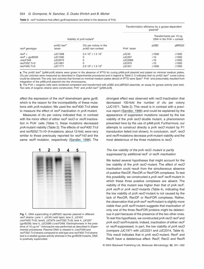
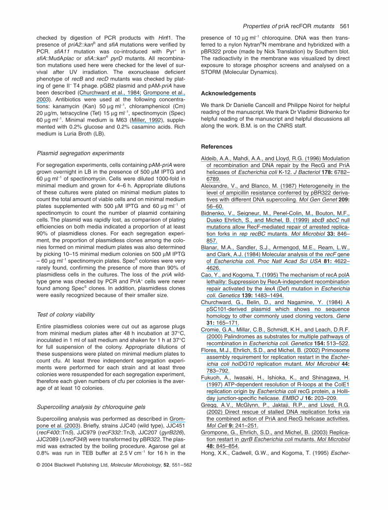
the replication initiator DnaA, the DNA polymerase III bclamp DnaN, and the gyrase subunit GyrB in the followingorder: dnaA dnaN recF gyrB (Perez Roger et al., 1991;and references therein). These three genes are essentialfor E. coli viability and, in addition, partial inactivation ofgyrB causes a requirement for PriA (Grompone et al.,2003). We tested the possibility that the DrecF349 muta-tion affects the expression of the downstream gyrB gene.To assay gyrase activity, we used the observation thatmutations that affect gyrase prevent the propagation of thepGB2 vector plasmid, presumably because the efficiencyof the partition site of pSC101 derived plasmids requiresfunctional gyrase activity (Wahle and Kornberg, 1988;Miller et al., 1990). pGB2 transformed very poorly cellscarrying the DrecF349 or a gyrB mutation whereas thepBR322 control plasmid transformed efficiently bothstrains (Table 3). Two other recF null mutations,recF332::Tn3 and recF400::Tn5 mutants were also testedfor pGB2 transformation and for viability in combinationwith priA mutation. pGB2 could be introduced in therecF400::Tn5 mutant as efficiently as in RecF+ cells butnot in the recF332::Tn3 mutant (Table 3). Similarly, priArecF plasmidless cells were readily obtained fromrecF400::Tn5 priA2::kan [pAM-priA] cultures and notfrom recF332::Tn3 priA2::kan [pAM-priA] or DrecF349priA2::kan [pAM-priA] mutants (Table 3). The observationof a strict correlation between the lack of pGB2 transfor-mants and the incompatibility with priA inactivation sug-gests that the expression of the gyrB gene is affected bythe DrecF349 and recF332::Tn3 mutations. pBR322 DNAsupercoiling levels monitored by chloroquine gels con-firmed that recF332::Tn3 and DrecF349 mutations modifyDNA supercoiling, albeit differently than a gyrB mutation,whereas recF400::Tn5 does not (Fig. 1). We concludethat recF332::Tn3 and DrecF349 mutations are likely to
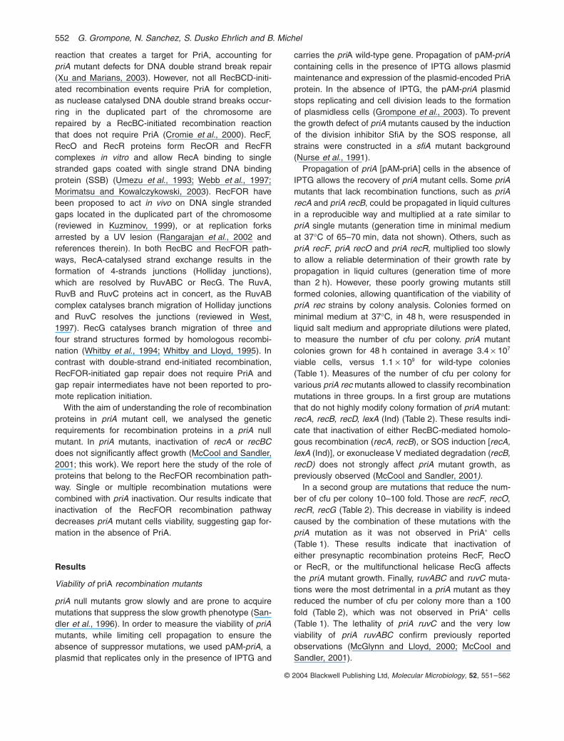
Table 1. Colony formation in PriA+ recombination mutants.
Strain Relevant genotype Cfu per colony Relative to wild type
JJC40 wild-type 1.1 109 ± 4.4 108 1JJC1398 priA2::kanR 3.4 107 ± 1.5 107 0.027JJC451 recF400::Tn5 8 108 ± 3.2 108 0.64JJC2135 recO1504::Tn5 8.9 108 ± 1.8 108 0.7JJC2142 recR252::Tn10–9 9.4 108 ± 1.4 108 0.76JJC315 recB268::Tn10 2.9 108 ± 2.3 107 0.22JJC2136 recB268::Tn10 recF400::Tn5 2.4 108 ± 1 108 0.19JJC2146 recB268::Tn10 recO1504::Tn5 2.8 108 ± 7.9 107 0.22JJC2147 recB268::Tn10 recR252::Tn10–9 2.5 108 ± 8 107 0.20JJC353 D(recA-srl)::Tn10 2.6 108 ± 1.4 108 0.21JJC432 recA938::CmR recD1901::Tn10 3.1 108 ± 8.6 107 0.25JJC1105 recA938::CmR recB268::Tn10 1.2 108 ± 3 107 0.1JJC754 DruvABC::CmR 2.9 108 ± 9.2 107 0.23JJC783 DruvC::CmR 1.1 108 ± 2.6 107 0.09JJC1782 DrecG265::CmR 6.7 108 ± 7.3 107 0.6JJC2350 DrecG265::CmR D(recA-srl)::Tn10 2.8 108 ± 9.3 107 0.25
Cells were grown to mid-logarithmic phase and plated on minimal medium plates. After 48 h of incubation at 37∞C, individual colonies wereresuspended and appropriate dilutions were plated to determine the number of cfu per colony. Averages of 6–10 determinations are presented.
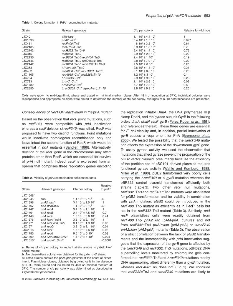
Table 2. Viability of priA recombination deficient mutants.
Strain Relevant genotype Cfu per colonyRelativeto priAa
JJC1548/JJC1945 sfiA 1.1 109 ± ¥ 108 32JJC1398 priA2::kanR. 3.4 107 ± 1.5 107 1JJC1767 priA dnaC809 1.1 109 ± ¥ 108 32JJC1447 priA recA 3.4 107 ± 1.1 107 1JJC1401 priA recB 2.4 107 ± 7.5 106 0.7JJC1448 priA recD 1.5 107 ± 5.6 106 0.44JJC1676 priA lexA (Ind)1 3.6 107 ± 2.1 107 1JJC1771 priA recF400::Tn5 3.1 106 ± 1.9 106 0.08JJC1971 priA recO 3.6 105 ± 3.1 105 0.01JJC2016 priA recR 1.6 106 ± 7.6 105 0.05JJC1783 priA recG 9.5 105 ± 5 105 0.03JJC1509 priA DruvABC::CmR 1.4 105 ± 1 105 0.004JJC1510b priA DruvC::CmR 0 <0.0001
a. Ratios of cfu per colony for mutant strain relative to priA2::kanR
single mutant.b. No plasmidless colony was obtained for the priA ruvC strain.All listed strains contain the pAM-priA plasmid at the onset of exper-iment. Plasmidless clones, obtained by growing cells in the absenceof IPTG, were plated and incubated for 48 h on minimal medium at37∞C. The number of cfu per colony was determined as described inExperimental procedures.
554 G. Grompone, N. Sanchez, S. Dusko Ehrlich and B. Michel
© 2004 Blackwell Publishing Ltd, Molecular Microbiology, 52, 551–562
affect the expression of the recF downstream gene gyrB,which is the reason for the incompatibility of these muta-tions with priA mutation. We used the recF400::Tn5 alleleto measure the effect of recF inactivation in priA mutant.
Measures of cfu per colony indicated that, in contrastwith the minor effect of either recF recO or recR inactiva-tion in PriA+ cells (Table 1), these mutations decreasedpriA mutant viability (Table 2). The effects of recF400::Tn5and recR252::Tn10–9 mutations, about 12-fold, were verysimilar to those previously reported for recF143 and thesame recR mutation, respectively (Sandler, 1996). The
strongest effect was observed with recO inactivation thatdecreased 100-fold the number of cfu per colony(JJC1971, Table 2). This result is in contrast with a previ-ous report (Sandler, 1996) and could be explained by theappearance of suppressor mutations caused by the lowviability of the priA recO double mutant, a phenomenonprevented here by the use of pAM-priA. Furthermore, ourattempts to construct directly a priA recO mutant by P1transduction failed (not shown). In conclusion, recF, recOand recR mutations decrease priA mutant viability and themost deleterious of the three mutations is recO.
The low viability of the priA recO mutant is partly suppressed by additional recF or recR inactivation
We tested several hypotheses that might account for thelow viability of the priA recO mutant. The effect of recOinactivation could result from the simultaneous absenceof putative RecOF, RecOR or RecFOR complexes. To testthis possibility, we constructed a priA recF recR mutant inwhich these three putative complexes are absent. Theviability of this mutant was higher than that of priA recF,priA recR or priA recO mutants (Table 4), indicating thatthe low viability of priA recO mutant is not caused by thelack of RecOR, RecOF or RecFOR complexes. Rather,the observation that priA recF recR mutant is slightly moreviable than priA recR mutant suggests that inactivation ofonly one of the three RecFOR proteins might be deleteri-ous in part because of the presence of the two other ones.To test this hypothesis, we constructed priA recO recF andpriA recO recR mutants. Indeed, inactivation of either recFor recR suppressed, in part, the low viability of priA recO(compare JJC1971 with JJC2221 and JJC2314, Table 4).This result indicates that in priA recO mutant, RecF andRecR have a deleterious effect. RecF, RecO and RecR
Table 3. recF mutations that affect gyrB expression are lethal in the absence of PriA.
recF genotype
Viability of priA mutanta
Transformation efficiency by a gyrase-dependent plasmidb
PriA+ strain
Transformants per 10 ng DNA in the PriA + context
priA2::kanR
.strainCfu per colony in thepriA2::kan context
pGB2 pBR322
recF + JJC1398 3.4 107 ± 1.5 107 JJC40 ≥1000 ≥1000recF + gyrB226 JJC1592 0 JJC207 <10 ≥1000DrecF349 JJC2073 0 JJC2089 <10 ≥1000recF332::Tn3 JJC1661 0 JJC979 <10 ≥1000recF400::Tn5 JJC1771 2.9 106 ± 1.9 106 JJC451 ≥1000 ≥1000
a. The priA2::kanR. [pAM-priA] strains were grown in the absence of IPTG for curing pAM-priA plasmid and plated on minimal medium plates.Cfu per colonies were measured as described in Experimental procedures and in legend to Table 2. 0 indicates that no priA2::kanR. cured colonycould be obtained. The only rare colonies that formed on minimal medium plates devoid of IPTG were SpecR. PriA+ and presumably resulted fromintegration of the pAM-priA plasmid into the chromosome.b. The PriA + isogenic cells were rendered competent and transformed with pGB2 and pBR322 plasmids, an assay for gyrase activity (see text).Two sets of isogenic strains were constructed, PriA+ and priA2::kanR. [pAM-priA].
Fig. 1. DNA supercoiling of pBR322 reporter plasmid in different recF strains. Lane 1, JJC40 (wild type); lane 2, JJC451 (recF400::Tn5); lane3, JJC979 (recF332::Tn3); lane 4, JJC207 (gyrB226); lane 5, JJC2089 (DrecF349). Electrophoresis in the pres-ence of 10 mg ml-1 chloroquine was performed as described in Exper-imental procedures. Plasmid DNA is relaxed in DrecF349 and recF332::Tn3 strains compared to wild type and recF400::Tn5 strains due to a weaker gyrase activity whereas in the gyrB226 mutants, DNA is positively supercoiled.
Properties of priA recFOR mutants 555
© 2004 Blackwell Publishing Ltd, Molecular Microbiology, 52, 551–562
have been reported to form RecFR and RecOR com-plexes in vitro (Umezu et al., 1993; Webb et al., 1997;Morimatsu and Kowalczykowski, 2003). Our results sug-gest that a RecFR complex indeed forms in vivo and hasa toxic effect in priA mutant in the absence of RecO. Weconclude from the analysis of priA recOF, priA recOR andpriA recFR mutants that the three RecFOR protein act inconcert in the priA mutant, and that their absence lowerspriA mutant viability.
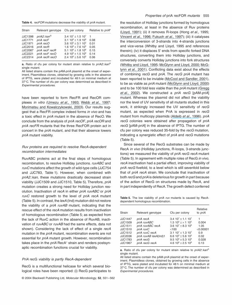
Ruv proteins are required to resolve RecA-dependent recombination intermediates
RuvABC proteins act at the final steps of homologousrecombination, to resolve Holliday junctions. ruvABC andruvC mutations affect the growth of wild-type cells (JJC754and JJC783, Table 1). However, when combined withpriA2::kan, these mutations drastically decreased strainviability (JJC1509 and JJC1510, Table 5). Therefore, priAmutation creates a strong need for Holliday junction res-olution. Inactivation of recA in either priA ruvABC or priAruvC restored growth to the level of priA recA mutant(Table 5). In contrast, the lexA (Ind) mutation did not restorethe viability of a priA ruvAB mutant, indicating that therescue effect of the recA mutation results from inactivationof homologous recombination (Table 5; as expected fromthe lack of RuvC action in the absence of RuvAB, inacti-vation of ruvABC or ruvAB had the same effects, data notshown). Considering the lack of effect of a single recAmutation in the priA mutant, recombination events are notessential for priA mutant growth. However, recombinationtakes place in the priA RecA+ strain and renders postsyn-aptic recombination functions crucial for viability.
PriA recG viability is partly RecA-dependent
RecG is a multifunctional helicase for which several bio-logical roles have been reported: (i) RecG participates to
the resolution of Holliday junctions formed by homologousrecombination, at least in the absence of Ruv proteins(Lloyd, 1991); (ii) it removes R-loops (Hong et al., 1995;Vincent et al., 1996; Fukuoh et al., 1997); (iii) it catalysesthe interconversion of 3-strands into 4-strands junctionsand vice-versa (Whitby and Lloyd, 1995 and referencestherein); (iv) it displaces 5¢ ends from specific forked DNAstructures, converting them into Holliday junctions, andconversely converts Holliday junctions into fork structures(Whitby and Lloyd, 1995; McGlynn and Lloyd, 2000; McG-lynn et al., 2001). Conflicting data exist about the effectsof combining recG and priA. The recG priA mutant hasbeen reported to be inviable (McCool and Sandler, 2001),to be as viable as priA mutant (McGlynn and Lloyd, 2000)and to be 100 fold less viable than the priA mutant (Gregget al., 2002). We constructed a priA recG [pAM-priA]mutant. Whereas the plasmid did not affect the viabilitynor the level of UV sensitivity of all mutants studied in thiswork, it strikingly increased the UV sensitivity of recGmutant, as expected when PriA is expressed in recGmutant from multicopy plasmids (Aldeib et al., 1996). priArecG colonies were obtained after propagation of priArecG [pAM-priA] in the absence of IPTG. The number ofcfu per colony was reduced 35-fold by the recG mutation,indicating a synergistic effect of priA and recG mutations(Table 5).
Since several of the RecG substrates can be made byRecA in vivo (Holliday junctions, R-loops, 3-strands junc-tions) we measured the viability of priA recG recA mutant(Table 5). In agreement with multiple roles of RecG in vivo,recA inactivation had a partial effect, improving viability ofpriA recG fivefold, to a level which is still sevenfold belowthat of priA recA strain. We conclude that inactivation ofboth recG and priA is deleterious for growth in part becauseof the action of RecG on structures made by RecA, andin part independently of RecA. The growth defect conferred
Table 4. recFOR mutations decrease the viability of priA mutant.
Strain Relevant genotype Cfu per colony Relative to priAa
JJC1398 priA2::kanR. 3.4 107 ± 1.5 107 1JJC1771 priA recF 3.1 106 ± 1.9 106 0.08JJC1971 priA recO 3.6 105 ± 3.1 105 0.01JJC2016 priA recR 1.6 106 ± 7.6 105 0.05JJC2087 priA recF recR 5.1 106 ± 1.8 106 0.15JJC2221 priA recF recO 4.9 106 ± 1.5 106 0.14JJC2314 priA recR recO 2.4 106 ± 5.6 105 0.06
a. Ratio of cfu per colony for mutant strain relative to priA2::kanR.
single mutant.All listed strains contain the pAM-priA plasmid at the onset of exper-iment. Plasmidless clones, obtained by growing cells in the absenceof IPTG, were plated and incubated for 48 h on minimal medium at37∞C. The number of cfu per colony was determined as described inExperimental procedures.
Table 5. The low viability of priA ruv mutants is caused by RecA-dependent homologous recombination.
Strain Relevant genotype Cfu per colonyRelativeto priA
JJC1447 priA recA 3.4 107 ± 1.1 107 1JJC1509 priA ruvABC 1.5 105 ± ¥ 1 105 0.004JJC1511 priA ruvABC recA 3.6 107 ± 8.3 106 1.05JJC1510 priA ruvC <100 <0.00001JJC1512 priA ruvC recA 3.1 107 ± 1.3 107 0.9JJC2036 priA ruvAB lexA(Ind)1 6.6 105 ± 5.6 105 0.02JJC1783 priA recG 9.5 105 ± 5.3 105 0.028JJC1967 priA recG recA 4.6 106 ± 2.5 106 0.13
a. Ratio of cfu per colony for mutant strain relative to priA2::kanR.
single mutant.All listed strains contain the pAM-priA plasmid at the onset of exper-iment. Plasmidless clones, obtained by growing cells in the absenceof IPTG, were plated and incubated for 48 h on minimal medium at37∞C. The number of cfu per colony was determined as described inExperimental procedures.
556 G. Grompone, N. Sanchez, S. Dusko Ehrlich and B. Michel
© 2004 Blackwell Publishing Ltd, Molecular Microbiology, 52, 551–562
by the recG mutation in a priA mutant, independently ofRecA, may result from a role of RecG in the PriA-inde-pendent restart pathway. Alternatively, priA inactivationmay affect recG mutant because of a requirement forefficient replication restart in the absence of RecG.
Exonuclease V activity is required for viability in priA cells that lack RecO or RecA
The combination of priA and recFOR mutations could bedetrimental for three different reasons: (i) PriA and Rec-FOR perform a common function; (ii) the absence of PriAleads to the formation of gaps and therefore creates aneed for RecFOR-mediated recombinational repair ofgaps (iii) the defect in gap recombinational repair of rec-FOR mutants creates a need for replication restart. Incontrast with model (i), models (ii) and (iii) predict thatinactivation of other proteins involved in gap repair shouldalso affect priA mutant viability. Arguing in favour of these
models, inactivation of ruv proteins has a dramatic effectin priA mutant. However, arguing against these models,inactivation of recA has no effect (Table 2).
These results could be reconciled if the viability of thepriA recA mutant resulted from the action of an alternativepathway of gap processing. In order to test this hypothe-sis, we inactivated the exonuclease V activity of theRecBCD complex (exo V) by either a recB or a recDmutation. The viability of priA recA mutant was stronglydecreased by inactivation of Exo V (Table 6, compareJJC1447 with JJC2088 and JJC1681). The very low via-bility of priA recA recD compared with priA recA mutantsuggests that unrepaired recombination substrates areconverted into DNA double strand breaks which rendersdegradation of the broken DNA by Exo V essential for cellviability.
If the substrates for Exo V are formed in priA recAcells by breakage of DNA single stranded gaps, unre-paired gaps may also be broken and processed byRecBCD in priA recFOR mutants. However, action ofRecBCD in priA recFOR mutants is expected to havetwo possible outcomes: either DNA degradation by exo-nuclease V activity, or RecBCD- RecA-mediated homol-ogous recombination. The role of RecBCD and RecA inpriA recFOR mutants was tested by introduction of arecA mutation (to inactivate homologous recombination),a recD mutation (to inactivate Exo V), or a recBmutation (to inactivate both).
RecA inactivation increased the number of cfu per col-ony twofold to fourfold in all priA recFOR mutants tested(Table 6; Table 7). Inactivation of homologous recombina-tion is responsible for the beneficial effect of the recAmutation as inactivation of only the SOS response by alexA (Ind) mutation did not improve the viability of priArecO and priA recO recF mutants (see JJC2283 andJJC2284, Table 7). These results indicate that RecA-catal-ysed recombination events are detrimental in priA recFORcells. In contrast with recA inactivation, recD inactivationslightly decreased the viability of priA recF and decreasedthat of priA recO mutant 50-fold, indicating a requirementfor DNA degradation initiated at DNA double strand ends
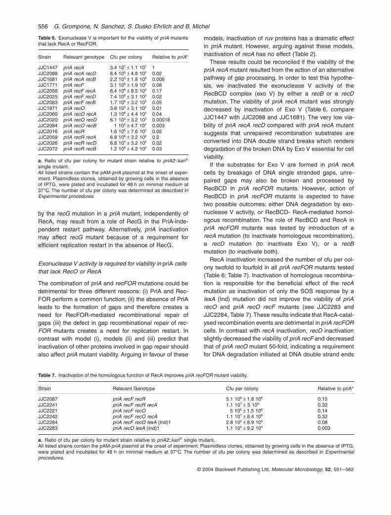
Table 6. Exonuclease V is important for the viability of priA mutantsthat lack RecA or RecFOR.
Strain Relevant genotype Cfu per colony Relative to priAa
JJC1447 priA recA 3.4 107 ± 1.1 107 1JJC2088 priA recA recD 8.4 105 ± 4.8 105 0.02JJC1681 priA recA recB 2.2 105 ± 1.8 105 0.006JJC1771 priA recF 3.1 106 ± 1.9 106 0.08JJC2058 priA recF recA 6.4 106 ± 8.5 105 0.17JJC2025 priA recF recD 7.4 105 ± 3.1 105 0.02JJC2083 priA recF recB 1.7 106 ± 3.2 105 0.05JJC1971 priA recO 3.6 105 ± 3.1 105 0.01JJC2060 priA recO recA 1.3 106 ± 4.4 105 0.04JJC2020 priA recO recD 6.1 103 ± 3.2 103 0.00018JJC2084 priA recO recB 1 105 ± 4.7 104 0.003JJC2016 priA recR 1.6 106 ± 7.6 105 0.05JJC2059 priA recR recA 6.8 106 ± 3.2 106 0.2JJC2026 priA recR recD 8.8 105 ± 3.2 105 0.02JJC2072 priA recR recB 1.2 106 ± 4.2 105 0.03
a. Ratio of cfu per colony for mutant strain relative to priA2::kanR.
single mutant.All listed strains contain the pAM-priA plasmid at the onset of exper-iment. Plasmidless clones, obtained by growing cells in the absenceof IPTG, were plated and incubated for 48 h on minimal medium at37∞C. The number of cfu per colony was determined as described inExperimental procedures.
Table 7. Inactivation of the homologous function of RecA improves priA recFOR mutant viability.
Strain Relevant Genotype Cfu per colony Relative to priAa
JJC2087 priA recF recR 5.1 106 ± 1.8 106 0.15JJC2241 priA recF recR recA 1.1 107 ± 5 106 0.32JJC2221 priA recF recO 5 106 ± 1.5 106 0.14JJC2242 priA recF recO recA 1.1 107 ± 8.4 106 0.32JJC2284 priA recF recO lexA (Ind)1 2.8 106 ± 8.9 105 0.08JJC2283 priA recO lexA (Ind)1 1.1 105 ± 9.2 104 0.003
a. Ratio of cfu per colony for mutant strain relative to priA2::kanR. single mutant.All listed strains contain the pAM-priA plasmid at the onset of experiment. Plasmidless clones, obtained by growing cells in the absence of IPTG,were plated and incubated for 48 h on minimal medium at 37∞C. The number of cfu per colony was determined as described in Experimentalprocedures.
Properties of priA recFOR mutants 557
© 2004 Blackwell Publishing Ltd, Molecular Microbiology, 52, 551–562
(Table 6). Finally, recB inactivation did not significantlyaffect the viability of the priA recR and priA recF mutants,and decreased only threefold the viability of priA recOmutant, particularly sensitive to recD inactivation(Table 6). We conclude from these experiments that linearDNA, target of RecBCD, is formed particularly in priA recOmutants. The level of viability of the priA recO mutantdepends on how this linear DNA is acted upon byRecBCD. DNA degradation, favoured by the absence ofRecA (recA mutant), leads to a higher level of viable cells,whereas DNA recombination, favoured by the absence ofRecD (recD mutant), leads to increased lethality. Theobservation that recA inactivation improves priA recFORviability suggests the occurrence of RecBC-mediatedrecombination in the absence of PriA, as previouslyobserved (Cromie et al., 2000). On the other hand, it isparadoxical that efficient DNA degradation can improveviability. Possibly, the sequences that need to be degradedare linked to an otherwise intact chromosome, which canonly yield a viable cell if the damaged part is degraded.
Altogether, these results argue in favour of a role for thehomologous recombination function of RecFOR in priAmutant viability and against a direct role for RecFOR pro-teins in PriA-independent replication restart.
The viability of priA recFOR, priA ruvABC, priA recG and priA recA mutants is restored by the dnaC809 mutation
The dnaC809 mutation restores full viability of the priAmutant, presumably by promoting efficient loading of theDnaB helicase to fork structures in the absence of PriA(Xu and Marians, 2000; Table 8). recF, recO, recR, ruv-ABC, recG and recA mutations were introduced in the priAdnaC809 [pAM-priA] strain, the resulting strains werepropagated in the absence of IPTG and plasmidless col-onies were analysed. recF, recO, recR, ruvABC, recG andrecA mutations did not affect the viability of the priAdnaC809 mutant more than that of wild-type cells (com-pare Table 8 and Table 1). We conclude that the RecFORpathway of recombination is not required for the viability
of the priA dnaC809 mutant, hence is not requiredprovided that an efficient helicase loading protein ispresent.
Discussion
In this work, we analysed the viability of priA mutantsdeficient for different recombination functions. The viabilitywas more than 50-fold lower relative to the single priAmutant in three types of recombination mutants: (i)mutants that lack RecO; (ii) mutants that lack both theRecFOR recombination pathway (recA, recFOR) and theexonuclease V activity of RecBCD, and (iii) mutants thatlack the post-synaptic proteins RuvABC.
Some recF mutations affect gyrB expression and are incompatible with priA inactivation.
The recF gene is located just upstream of gyrB, in thednaA dnaN recF gyrB operon. RecF and gyrB genesoverlap, as the recF coding sequence ends between the-35 and the -10 sequences of the gyrB promoter. In thiswork we show that the lethality caused by certain recFmutations in a priA background correlates with a modifi-cation of the level of gyrB gene expression: the two recFmutants alleles that are lethal in combination with priAprevent transformation by the pGB2 plasmid, a plasmidthat does not transform gyrB mutants, and affect DNAsupercoiling of a pBR322 reporter plasmid. Becausemutations that affect gyrB are incompatible with priA nullmutation (Grompone et al., 2003), we propose that thelethal effect of certain recF null mutations in priA mutantresults from a decrease in gyrB expression. RecF wasalso proposed to play a role in replication distinct from itsrole in recombination in a recA polA lexAdef strain, inwhich recF but not recO nor recR mutations had an effect(Cao and Kogoma, 1995). However, the recF allele usedwas the recF332::Tn3 allele that also affects the expres-sion of gyrB. The lethality of the recA polA lexAdefrecF332::Tn3 combination of mutations, previously attrib-
Table 8. dnaC809 restores the viability of priA rec mutants.
Strain Relevant genotype Cfu per colony Relative to priA dnaC809
JJC40 wild-type 1.1 109 ± 4.4 108 1.2JJC1398 priA2::kanR. 3.4 107 ± 1.5 107 0.037JJC1767/JJC2327 priA dnaC809 9.1 108 ± 3.8 108 1JJC2294 priA dnaC809 recF 1.1 109 ± 2.9 108 1.2JJC2313 priA dnaC809 recO 1.1 109 ± 2.6 108 1.2JJC2296 priA dnaC809 recR 1.1 109 ± 4.7 108 1.2JJC2034/JJC2332 priA dnaC809 ruvABC 8.9 107 ± 3.3107 0.1JJC2351 priA dnaC809 recG 3.8 108 ± 2 108 0.4JJC2322 priA dnaC809 recA 7.6 108 ± 4.5 108 0.8
All listed strains contain the pAM-priA plasmid at the onset of experiment. Plasmidless clones, obtained by growing cells in the absence of IPTG,were plated and incubated for 48 h on minimal medium at 37∞C. The number of cfu per colony was determined as described in Experimentalprocedures.
558 G. Grompone, N. Sanchez, S. Dusko Ehrlich and B. Michel
© 2004 Blackwell Publishing Ltd, Molecular Microbiology, 52, 551–562
uted to recF inactivation may also result from a decreasein gyrB expression.
The regulation of the dnaA dnaN recF gyrB operon iscomplex. This region carries several promoters and tran-scription terminators and the expression of the four genesis tightly regulated (Perez Roger et al., 1991). gyrB isthought to be expressed from its own promoter, whichoverlaps with the recF stop codon (Menzel and Gellert,1987). This promoter is intact in strains carrying theDrecF349 or the recF332::Tn3 allele, which are a deletionfrom nucleotides 61–197, and a Tn3 insertion at nucle-otide 72 of the recF coding sequence, respectively (Blanaret al., 1984; Sandler and Clark, 1993). The gyrB promoterhas been shown to be strongly dependent on the level ofDNA supercoiling and is activated by DNA relaxation(Menzel and Gellert, 1983). According to the twin domainmodel, a decrease in DNA supercoiling is induced down-stream of transcribed genes (Liu and Wang, 1987). Wepropose that in wild-type strains the gyrB promoter isactivated by the decrease in DNA supercoiling due to recFgene transcription. Both recF349 deletion andrecF332::Tn3 transposon insertion are likely to cause pre-mature transcription termination, and may thus fail to acti-vate the downstream gyrB gene. RecF alleles that arecompatible with priA inactivation are recF400::Tn5 andseveral point mutations that completely abolish RecFOR-mediated homologous recombination (Table 3; Sandler,1996). gyrB may be expressed in strains carryingrecF400::Tn5 allele from the outside-directed promotercarried by IS5. recF point mutations are not expected toaffect recF transcription, hence gyrB promoter activity.
Inactivation of only RecO protein is more deleterious than inactivation of two of the RecFOR proteins
RecF, RecO and RecR proteins have been shown to formin vitro two complexes: RecOR and RecFR. recO inacti-vation is more deleterious than recF or recR mutations inpriA mutant (100-fold versus 12– to 20-fold by our assay),and renders the priA mutant very sensitive to recD inac-tivation. The observation that the viability of priA recO issignificantly improved by the additional inactivation of recFor recR supports the idea of the existence of a RecFRcomplex in vivo, which could be deleterious in theabsence of PriA and RecO. A RecFR complex has beenobserved in vitro and binds to double stranded-singlestranded DNA junctions (Webb et al., 1997; Morimatsuand Kowalczykowski, 2003). In vivo, this complex couldblock replication progression or prevent the action of otherproteins than RecFOR at gaps. For example, if RecFRcomplexes remains stably bound to DNA gap ends in theabsence of RecO, they could prevent gap filling by a DNApolymerase, which would account for the dramatic pheno-type of priA recO mutant. A RecOR complex also forms
in vitro, it improves RecA binding to SSB-coated DNA andis counteracted by RecF (Umezu et al., 1993; Morimatsuand Kowalczykowski, 2003). However, we observed nosignificant modification of the priA recF mutant viabilityupon inactivation of recO or recR. This observation sug-gests that in contrast with the RecFR complex in theabsence of RecO, the RecOR complex is not toxic in priAmutant in the absence of RecF.
Single strand gaps form in the newly synthesized chromosomes in priA mutant
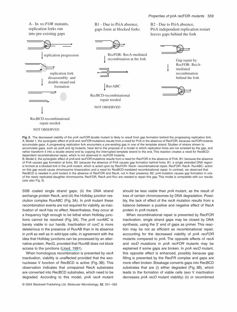
RecFOR acts at single stranded DNA regions. Formally,those could form either in the unreplicated region of theparental chromosome, or at the fork, or in one of the newlyreplicated regions of the chromosome (Fig. 2).
Gaps or nicks have been proposed to accumulate in thejust replicated part of the chromosome in several replica-tion mutants such as polA and ligase mutants, defectivefor Okazaki fragment completion, and in some DNA repairmutants (reviewed in Kuzminov, 1995). It was proposedthat a single strand interruption creates a need forRecBCD, RecA and PriA by being converted into a DNAdouble strand end upon the arrival of the next replicationfork (Fig. 2A). If recFOR mutants accumulate gaps thatare converted into double strand ends, it remains to beexplained why they do not require RecA or RecB forviability (Table 1, Table 2). Therefore, our observations aredifficult to reconcile with a model in which PriA would berequired in recFOR mutants because of the encounter ofreplication forks with pre-existing gaps.
In a second model, single strand DNA regions couldform specifically at blocked forks in the priA mutant andbe processed by RecFOR- RecA- mediated recombination(Fig. 2B). However, homologous recombination betweenleading and lagging strands at blocked forks would resultin a reversed fork structure. The observation that priA ruvmutants are poorly viable and are rescued by recA inac-tivation implies that the Holliday junction formed by Rec-FOR and RecA is resolved by Ruv proteins, which would,in the case of reversed forks render RecBCD essential forviability (Fig. 2B1). In contrast, in the priA mutant, RecBCDis required in the absence of RecFOR or RecA and not intheir presence. Consequently, this second model is alsodifficult to reconcile with our observations.
Therefore, we propose that the priA mutation causes anincreased formation of gapped DNA behind progressingreplication forks (Figs 2B2 and 3).
Gap processing in priA mutant
RecFOR-mediated recombination involves the successiveactions of (i) RecFOR, which facilitate RecA binding to
Properties of priA recFOR mutants 559
© 2004 Blackwell Publishing Ltd, Molecular Microbiology, 52, 551–562
SSB coated single strand gaps; (ii) the DNA strandexchange protein RecA, and (iii) the Holliday junction res-olution complex RuvABC (Fig. 3A). In priA mutant theserecombination events are not required for viability, as inac-tivation of recA has no effect. Nevertheless, they occur ata frequency high enough to be lethal when Holliday junc-tions cannot be resolved (Fig. 3A). The priA ruvABC isbarely viable in our hands. Inactivation of ruvC is moredeleterious in the presence of RuvAB than in its absencein priA as well as in wild-type cells, in agreement with theidea that Holliday junctions can be processed by an alter-native protein, RecG, provided that RuvAB does not blockaccess to the junctions (Lloyd, 1991).
When homologous recombination is prevented by recAinactivation, viability is unaffected provided that the exo-nuclease V function of RecBCD is active (Fig. 3B). Thisobservation indicates that unrepaired RecA substratesare converted into RecBCD substrates, which need to bedegraded. According to this model, priA recA mutant
should be less viable than priA mutant, as the result ofloss of certain chromosomes by DNA degradation. Possi-bly, the lack of effect of the recA mutation results from abalance between a positive and negative effect of RecAprotein in priA mutant.
When recombinational repair is prevented by RecFORinactivation, single strand gaps may be closed by DNAsynthesis, using the 3¢ end of gaps as primer. This reac-tion may be not as efficient as recombinational repair,accounting for the decreased viability of priA recFORmutants compared to priA. The opposite effects of recAand recD mutations in priA recFOR mutants may beexplained if some gaps are broken. In priA recO mutant,this opposite effect is enhanced, possibly because gapfilling is prevented by the RecFR complex and gaps aremore often broken. Breakage converts gaps into RecBCDsubstrates that are (i) either degraded (Fig. 3B), whichleads to the formation of viable cells (exo V inactivationdecreases priA recO mutant viability) (ii) or recombined
Fig. 2. The decreased viability of the priA recFOR double mutant is likely to result from gap formation behind the progressing replication fork.A. Model 1, the synergistic effect of priA and recFOR mutations results from a need for PriA in the absence of RecFOR, because recFOR mutants accumulate gaps. A progressing replication fork encounters a pre-existing gap in one of the template strand. Studies of strains shown to accumulate gaps, such as polA and lig mutants, have led to the proposal of a model in which replication forks are not arrested by the gap, and rather transform it into a double strand end by copying the interrupted template strand to the end. This reaction creates a need for RecBCD-dependent recombinational repair, which is not observed in recFOR mutants.B. Model 2, the synergistic effect of priA and recFOR mutations results from a need for RecFOR in the absence of PriA, B1: because the absence of PriA causes gap formation at forks, B2: because the absence of PriA causes gap formation behind forks. B1: a single stranded DNA region is formed at a blocked fork in the priA mutant, which is acted upon by RecFOR- RecA- recombinational repair. RecFOR- RecA- RuvABC- action on this gap would cause chromosome linearization and a need for RecBCD-mediated recombinational repair. In contrast, we observed that RecBCD is needed in priA mutant in the absence of RecFOR and RecA, not in their presence. B2: priA mutation causes gap formation in one of the newly replicated daughter chromosome. RecFOR, RecA and Ruv are needed to repair this gap. This model is compatible with our results (see also Fig. 3).
A - In recFOR mutants, replication forks run into pre-existing gaps
RecBCD-recombinational repair needed
B1 - Due to PriA absence, gaps form at blocked forks
RecFOR- RecA-mediated recombination at the fork
RecBCD-recombinational repair needed
B2 - Due to PriA absence, PriA independent replication restart leaves gaps behind the fork
Gap repair by RecFOR- RecA-mediated recombination behind the fork
replication progression
replication fork dissassembly and double strand end
formation RuvABC
NOT OBSERVED
NOT OBSERVED

560 G. Grompone, N. Sanchez, S. Dusko Ehrlich and B. Michel
© 2004 Blackwell Publishing Ltd, Molecular Microbiology, 52, 551–562
(Fig. 3C), which may not allow the formation of viable cells(recA inactivation increases priA recO mutant viability).Why would RecBCD-mediated recombination be detri-mental after conversion of gaps into double strandbreaks? Recombinational repair of DNA double strandbreaks occurring in the replicated part of the chromosomehas been shown to require both RecFOR and RecBC(Cromie et al., 2000). A model was proposed in whichRecFOR initiates recombination from one end of thebreak and RecBCD from the other end (Cromie et al.,2000). In the absence of RecFOR, a RecBC-mediatedrepair of only half of the break may not allow the formationof viable cells (Fig. 3C).
Our results lead us to conclude that gap processing isan essential process in priA mutant, due to gap formationin daughter chromosomes during replication. This couldimply a role for PriA in the coordination of the two strandsynthesis during replication fork progression, so that incells that lack PriA, uncoupling of the two polymeraseswould lead to gap formation. However, the need for gaprepair is prevented by the dnaC809 mutation, which sug-gests that the defect in helicase loading is responsible forgap formation in the absence of PriA. This observation isdifficult to reconcile with a direct role for PriA in replisomecoordination and rather argues in favour of gaps resultingfrom the replication restart defect. Alternative modes ofreplication restart have been postulated to operate in priA
mutant, they have not been reconstituted in vitro so farand may be prone to generate gaps.
Experimental procedures
Strains and plasmids
Strain background is JJC40, which is a hsdR Thr+ Pro+
derivative of AB1157. All strains were constructed by P1transduction, except recA recB double mutants constructedby co-conjugation of this two mutations from a recA recB[pBR-RecBCD+] Hfr strain (JJC1130). Because of theredundancy of antibiotic markers, when needed,priA2::kanR mutation (Sandler, 1996) was introduced by co-transduction with Arg+, and recF, recO and recR mutationsby co-transduction with zid::Tn10, zfh208::Tn10 andzbb3055::Tn10, respectively (Nichols et al., 1998).recF400::Tn5 allele is from W. Wackernagel. recF332::Tn3and DrecF349 have been described (Blanar et al., 1984;Sandler and Clark, 1993). Other recombination mutationsare recA938::CmR, D(recA-srl)::Tn10, recB268::Tn10,recD1901::Tn10, recO1504::Tn5, recR252::Tn10–9,ruvA60::Tn10, DruvABC::CmR, DruvC::CmR ,DrecG265::CmR (Lloyd et al., 1988; Seigneur et al., 1998;Bidnenko et al., 1999). The recG265::cmR allele is from R.G. Lloyd. The gyrB226 strain was obtained by transductionof the gyrB226 mutation from SD7 (Aleixandre and Blanco,1987). The lexA(Ind)1 mutation was co-introduced with amal::Tn9 marker and lexA(Ind)1 mutants were identified bymeasure of survival after UV irradiation. dnaC809 was co-introduced with Thr+, and the presence of the mutation was
Fig. 3. Models for the repair of DNA single stranded gaps formed in priA mutant.A. In cells proficient for gap recombinational repair, the successive action of RecFOR, RecA and RuvABC proteins leads to the formation of two intact chromosomes at each replication round. The absence of functional RuvABC proteins leads to non-disjunction of the two daughter chromosomes that have recombined, killing the replicating cell.B. In cells deficient for RecA, the unrepaired gaps are broken by nucleases and RecBCD-mediated DNA degradation destroys the chromosome of the daughter cell in which the gap has been transferred, allowing the daughter cell that contain an intact chromosome to propagate.C. In cells deficient for RecO and RecD the broken gap is partly recombined with the intact daughter chromosome, using RecBC and RecA proteins. However, this repair reaction remains partial in the absence of both RecFOR and PriA and kills the replicating cell.
RecFORRecA
A priA mutant
RuvABC(RecG)
B priA recA mutant
single strand endonuclease
RecBCD
2 viable cells 1 viable cell
C priA recO recD mutant
single strand endonuclease
RecBCRecA
No viable cell
Properties of priA recFOR mutants 561
© 2004 Blackwell Publishing Ltd, Molecular Microbiology, 52, 551–562
checked by digestion of PCR products with Hinf1. Thepresence of priA2::kanR and sfiA mutations were verified byPCR. sfiA11 mutation was co-introduced with Pyr+ insfiA::MudAplac or sfiA::kanR pyrD mutants. All recombina-tion mutations used here were checked for the level of sur-vival after UV irradiation. The exonuclease deficientphenotype of recB and recD mutants was checked by plat-ing of gene II– T4 phage. pGB2 plasmid and pAM-priA havebeen described (Churchward et al., 1984; Grompone et al.,2003). Antibiotics were used at the following concentra-tions: kanamycin (Kan) 50 mg ml-1, chloramphenicol (Cm)20 mg/m, tetracycline (Tet) 15 mg ml-1, spectinomycin (Spec)60 mg ml-1. Minimal medium is M63 (Miller, 1992), supple-mented with 0.2% glucose and 0.2% casamino acids. Richmedium is Luria Broth (LB).
Plasmid segregation experiments
For segregation experiments, cells containing pAM-priA weregrown overnight in LB in the presence of 500 mM IPTG and60 mg ml-1 of spectinomycin. Cells were diluted 1000-fold inminimal medium and grown for 4–6 h. Appropriate dilutionsof these cultures were plated on minimal medium plates tocount the total amount of viable cells and on minimal mediumplates supplemented with 500 mM IPTG and 60 mg ml-1 ofspectinomycin to count the number of plasmid containingcells. The plasmid was rapidly lost, as comparison of platingefficiencies on both media indicated a proportion of at least90% of plasmidless clones. For each segregation experi-ment, the proportion of plasmidless clones among the colo-nies formed on minimal medium plates was also determinedby picking 10–15 minimal medium colonies on 500 mM IPTG– 60 mg ml-1 spectinomycin plates. SpecR colonies were veryrarely found, confirming the presence of more than 90% ofplasmidless cells in the cultures. The loss of the priA wild-type gene was checked by PCR and PriA+ cells were neverfound among SpecS clones. In addition, plasmidless cloneswere easily recognized because of their smaller size.
Test of colony viability
Entire plasmidless colonies were cut out as agarose plugsfrom minimal medium plates after 48 h incubation at 37∞C,inoculated in 1 ml of salt medium and shaken for 1 h at 37∞Cfor full suspension of the colony. Appropriate dilutions ofthese suspensions were plated on minimal medium plates tocount cfu. At least three independent segregation experi-ments were performed for each strain and at least threecolonies were resuspended for each segregation experiment,therefore each given numbers of cfu per colonies is the aver-age of at least 10 colonies.
Supercoiling analysis by chloroquine gels
Supercoiling analysis was performed as described in Grom-pone et al. (2003). Briefly, strains JJC40 (wild type), JJC451(recF400::Tn5), JJC979 (recF332::Tn3), JJC207 (gyrB226),JJC2089 (DrecF349) were transformed by pBR322. The plas-mid was extracted by the boiling procedure. Agarose gel at0.8% was run in TEB buffer at 2.5 V cm-1 for 16 h in the
presence of 10 mg ml-1 chloroquine. DNA was then trans-ferred to a nylon Nytran®N membrane and hybridized with apBR322 probe (made by Nick Translation) by Southern blot.The radioactivity in the membrane was visualized by directexposure to storage phosphor screens and analysed on aSTORM (Molecular Dynamics).
Acknowledgements
We thank Dr Danielle Canceill and Philippe Noirot for helpfulreading of the manuscript. We thank Dr Vladimir Bidnenko forhelpful reading of the manuscript and helpful discussions allalong the work. B.M. is on the CNRS staff.
References
Aldeib, A.A., Mahdi, A.A., and Lloyd, R.G. (1996) Modulationof recombination and DNA repair by the RecG and PriAhelicases of Escherichia coli K-12. J Bacteriol 178: 6782–6789.
Aleixandre, V., and Blanco, M. (1987) Heterogeneity in thelevel of ampicillin resistance conferred by pBR322 deriva-tives with different DNA supercoiling. Mol Gen Genet 209:56–60.
Bidnenko, V., Seigneur, M., Penel-Colin, M., Bouton, M.F.,Dusko Ehrlich, S., and Michel, B. (1999) sbcB sbcC nullmutations allow RecF-mediated repair of arrested replica-tion forks in rep recBC mutants. Mol Microbiol 33: 846–857.
Blanar, M.A., Sandler, S.J., Armengod, M.E., Ream, L.W.,and Clark, A.J. (1984) Molecular analysis of the recF geneof Escherichia coli. Proc Natl Acad Sci USA 81: 4622–4626.
Cao, Y., and Kogoma, T. (1995) The mechanism of recA polAlethality: Suppression by RecA-independent recombinationrepair activated by the lexA (Def) mutation in Escherichiacoli. Genetics 139: 1483–1494.
Churchward, G., Belin, D., and Nagamine, Y. (1984) ApSC101-derived plasmid which shows no sequencehomology to other commonly used cloning vectors. Gene31: 165–171.
Cromie, G.A., Millar, C.B., Schmidt, K.H., and Leach, D.R.F.(2000) Palindromes as substrates for multiple pathways ofrecombination in Escherichia coli. Genetics 154: 513–522.
Flores, M.J., Ehrlich, S.D., and Michel, B. (2002) Primosomeassembly requirement for replication restart in the Escher-ichia coli holDG10 replication mutant. Mol Microbiol 44:783–792.
Fukuoh, A., Iwasaki, H., Ishioka, K., and Shinagawa, H.(1997) ATP-dependent resolution of R-loops at the ColE1replication origin by Escherichia coli recG protein, a Holli-day junction-specific helicase. EMBO J 16: 203–209.
Gregg, A.V., McGlynn, P., Jaktaji, R.P., and Lloyd, R.G.(2002) Direct rescue of stalled DNA replication forks viathe combined action of PriA and RecG helicase activities.Mol Cell 9: 241–251.
Grompone, G., Ehrlich, S.D., and Michel, B. (2003) Replica-tion restart in gyrB Escherichia coli mutants. Mol Microbiol48: 845–854.
Hong, X.K., Cadwell, G.W., and Kogoma, T. (1995) Escher-
562 G. Grompone, N. Sanchez, S. Dusko Ehrlich and B. Michel
© 2004 Blackwell Publishing Ltd, Molecular Microbiology, 52, 551–562
ichia coli RecG and RecA proteins in R-loop formation.EMBO J 14: 2385–2392.
Kogoma, T., Cadwell, G.W., Barnard, K.G., and Asai, T.(1996) The DNA replication priming protein, PriA, isrequired for homologous recombination and double-strandbreak repair. J Bacteriol 178: 1258–1264.
Kuzminov, A. (1995) Collapse and repair of replication forksin Escherichia coli. Mol Microbiol 16: 373–384.
Kuzminov, A. (1999) Recombinational repair of DNA damagein Escherichia coli and bacteriophage lambda. MicrobiolMol Biol Rev 63: 751–813.
Liu, L.F., and Wang, J.C. (1987) Supercoiling of the DNAtemplate during transcription. Proc Natl Acad Sci USA 84:7024–7027.
Lloyd, R.G. (1991) Conjugational recombination in resolvase-deficient ruvC mutants of Escherichia coli K-12 dependson recG. J Bacteriol 173: 5414–5418.
Lloyd, R.G., Porton, M.C., and Buckman, C. (1988) Effect ofrecF, recJ, recN, recO and ruv mutations on ultravioletsurvival and genetic recombination in a recD strain ofEscherichia coli K12. Mol Gen Genet 212: 317–324.
McCool, J.D., and Sandler, S.J. (2001) Effects of mutationsinvolving cell division, recombination, and chromosomedimer resolution on a priA2::kan mutant. Proc Nat Acad SciUSA 98: 8203–8210.
McGlynn, P., and Lloyd, R.G. (2000) Modulation of RNApolymerase by (P) ppGpp reveals a RecG-dependentmechanism for replication fork progression. Cell 101: 35–45.
McGlynn, P., Lloyd, R.G., and Marians, K.J. (2001)Formation of Holliday junctions by regression of nascentDNA in intermediates containing stalled replication forks:RecG stimulates regression even when the DNA isnegatively supercoiled. Proc Natl Acad Sci USA 98: 8235–8240.
Menzel, R., and Gellert, M. (1983) Regulation of the genesfor E. coli DNA gyrase: homeostatic control of DNA super-coiling. Cell 34: 105–113.
Menzel, R., and Gellert, M. (1987) Modulation of transcriptionby DNA supercoiling: a deletion analysis of the Escherichiacoli gyrA and gyrB promoters. Proc Natl Acad Sci USA 84:4185–4189.
Miller, C.A., Beaucage, S.L., and Cohen, S.N. (1990) Role ofDNA superhelicity in partitioning of the pSC101 plasmid.Cell 62: 127–133.
Miller, J.H. (1992) A Short Course in Bacterial Genetics. ColdSpring Harbor, New York: Cold Spring Harbor Press.
Morimatsu, K., and Kowalczykowski, S.C. (2003) RecFORproteins load RecA protein onto gapped DNA to accelerateDNA strand exchange: a universal step of recombinationalrepair. Mol Cell 11: 1337–1347.
Nichols, B.P., Shafiq, O., and Meiners, V. (1998) Sequenceanalysis of Tn10 insertion sites in a collection of E. Colistrains used for genetics mapping and strain construction.J Bacteriol 180: 6408–6411.
Nurse, P., Zavitz, K.H., and Marians, K.J. (1991) Inactivationof the Escherichia coli PriA DNA replication protein inducesthe SOS response. J Bacteriol 173: 6686–6693.
Perez Roger, I., Garcia Sogo, M., Navarro Avino, J.P., LopezAcedo, C., Macian, F., and Armengod, M.E. (1991)Positive and negative regulatory elements in the dnaA-
dnaN-recF operon of Escherichia coli. Biochimie 73: 329–334.
Rangarajan, S., Woodgate, R., and Goodman, M.F. (2002)Replication restart in UV-irradiated Escherichia coli involv-ing pols II, III, V, PriA, RecA and RecFOR proteins. MolMicrobiol 43: 617–628.
Sandler, S.J. (1996) Overlapping functions for recF and priAin cell viability and UV-inducible SOS expression are dis-tinguished by dnaC809 in Escherichia coli K-12. Mol Micro-biol 19: 871–880.
Sandler, S.J. (2000) Multiple genetic pathways for restartingDNA replication forks in Escherichia coli K-12. Genetics155: 487–497.
Sandler, S.J., and Clark, A.J. (1993) Use of high and lowlevel overexpression plasmids to test mutant alleles of therecF gene of Escherichia coli K-12 for partial activity.Genetics 135: 643–654.
Sandler, S.J., and Marians, K.J. (2000) Role of PriA in repli-cation fork reactivation in Escherichia coli. J Bacteriol 182:9–13.
Sandler, S.J., Samra, H.S., and Clark, A.J. (1996) Differentialsuppression of priA2:: kan phenotypes in Escherichia coliK-12 by mutations in priA, lexA, and dnaC. Genetics 143:5–13.
Seigneur, M., Bidnenko, V., Ehrlich, S.D., and Michel, B.(1998) RuvAB acts at arrested replication forks. Cell 95:419–430.
Umezu, K., Chi, N.W., and Kolodner, R.D. (1993) Biochemi-cal Interaction of the Escherichia coli RecF, RecO, andRecR proteins with RecA protein and single-stranded DNAbinding protein. Proc Natl Acad Sci USA 90: 3875–3879.
Velten, M., McGovern, S., Marsin, S., Ehrlich, S.D., Noirot,P., and Polard, P. (2003) A two-protein strategy for thefunctional loading of a cellular replicative DNA helicase.Mol Cell 11: 1009–1020.
Vincent, S.D., Mahdi, A.A., and Lloyd, R.G. (1996) The RecGbranch migration protein Escherichia coli dissociates R-loops. J Mol Biol 264: 713–721.
Wahle, E., and Kornberg, A. (1988) The partition locus ofplasmid pSC101 is a specific binding site for DNA gyrase.EMBO J 7: 1889–1895.
Webb, B.L., Cox, M.M., and Inman, R.B. (1997) Recombina-tional DNA repair: The RecF and RecR proteins limit theextension of RecA filaments beyond single-strand DNAgaps. Cell 91: 347–356.
West, S.C. (1997) Processing of recombination intermediatesby the RuvABC proteins. Ann Rev Genet 31: 213–244.
Whitby, M.C., and Lloyd, R.G. (1995) Branch migration ofthree-strand recombination intermediates by RecG, a pos-sible pathway for securing exchanges initiated by 3¢-tailedduplex DNA. EMBO J 14: 3302–3310.
Whitby, M.C., Vincent, S.D., and Lloyd, R.G. (1994) Branchmigration of Holliday junctions: Identification of RecG pro-tein as a junction specific: DNA helicase. EMBO J 13:5220–5228.
Xu, L.W., and Marians, K.J. (2000) Purification and charac-terization of DnaC810, a primosomal protein capable ofbypassing PriA function. J Biol Chem 275: 8196–8205.
Xu, L., and Marians, K.J. (2003) PriA mediates DNA replica-tion pathway choice at recombination intermediates. MolCell 11: 817–826.