Requirement for down-regulation of the CCAAT-binding activity of the NF-Y transcription factor...
49
1 Requirement for down-regulation of the CCAAT-binding activity of the NF-Y transcription factor during skeletal muscle differentiation. Aymone Gurtner # , Isabella Manni # , Paola Fuschi # , Roberto Mantovani°, Fiorella Guadagni + , Ada Sacchi # and Giulia Piaggio # * #Molecular Oncogenesis Laboratory, Experimental Oncology Department, Regina Elena Cancer Institute, Rome, Italy. + Laboratory of Clinical Pathology, Regina Elena Cancer Institute, Rome, Italy. °Dipartimento di Biologia Animale, Università di Modena e Reggio, Modena, Italy *Corresponding author: Giulia Piaggio: phone: 39-06-52662531; fax: 39-06- 4180526; e-mail: [email protected] Running title: NF-Y expression in adult muscle tissues Key words: transcription, adult muscle tissues, cell cycle, chromatin immunoprecipitation MBC in Press, published on April 4, 2003 as 10.1091/mbc.E02-09-0600
-
Upload
independent -
Category
Documents
-
view
1 -
download
0
Transcript of Requirement for down-regulation of the CCAAT-binding activity of the NF-Y transcription factor...
1
Requirement for down-regulation of the CCAAT-binding activity of the NF-Y
transcription factor during skeletal muscle differentiation.
Aymone Gurtner#, Isabella Manni#, Paola Fuschi#, Roberto Mantovani°, Fiorella
Guadagni+, Ada Sacchi# and Giulia Piaggio#*
#Molecular Oncogenesis Laboratory, Experimental Oncology Department,
Regina Elena Cancer Institute, Rome, Italy.
+Laboratory of Clinical Pathology, Regina Elena Cancer Institute, Rome, Italy.
°Dipartimento di Biologia Animale, Università di Modena e Reggio, Modena, Italy
*Corresponding author: Giulia Piaggio: phone: 39-06-52662531; fax: 39-06-
4180526; e-mail: [email protected]
Running title: NF-Y expression in adult muscle tissues
Key words: transcription, adult muscle tissues, cell cycle, chromatin
immunoprecipitation
MBC in Press, published on April 4, 2003 as 10.1091/mbc.E02-09-0600
2
Abstract
NF-Y is composed of three subunits, NF-YA, NF-YB, and NF-YC, all required for
DNA binding. All subunits are expressed in proliferating skeletal muscle cells,
while NF-YA alone is undetectable in terminally differentiated cells in vitro. By
immuno-histochemistry we show that the NF-YA protein is not expressed in the
nuclei of skeletal and cardiac muscle cells in vivo. By chromatin
immunoprecipitation experiments we demonstrate here that NF-Y does not bind
to the CCAAT boxes of target promoters in differentiated muscle cells. Consistent
with this, the activity of these promoters is down-regulated in differentiated
muscle cells. Finally, forced expression of the NF-YA protein in cells committed
to differentiate leads to an impairment in the down-regulation of cyclin A, cyclin
B1 and cdk1 expression and is accompanied by a delay in myogenin expression.
Thus, our results indicate that the suppression of NF-Y function is of crucial
importance for the inhibition of several cell cycle genes and the induction of the
early muscle-specific program in postmitotic muscle cells.
3
Introduction
The CCAAT-binding transcription factor NF-Y is a heteromeric protein
composed of three subunits, NF-YA, NF-YB and NF-YC, all necessary for
CCAAT-binding. NF-YB and NF-YC tight association is a prerequisite for NF-YA
binding and sequence-specific DNA interactions. NF-YA and NF-YC have Q-rich
activation domains. Both NF-YB and NF-YC contain putative histone fold motifs
(Mantovani, 1999). The CCAAT motif is present in 25% of eukaryotic promoters
and NF-Y has been shown to bind over 120 of these (CCAAT-containing)
promoters (Mantovani, 1998). In accordance with the widespread presence of
CCAAT boxes, NF-Y subunits are extremely conserved and have been identified
in several eukaryotic kingdoms (Mantovani, 1999).
The NF-Y complex supports the basal transcription of a class of regulatory genes
responsible for cell cycle progression, among which cyclin A, -B1, -B2, cdc25B,
cdc25C, and cdk1 (Farina et al., 1999; Bolognese et al., 1999; Korner et al.,
2001; Zwicher et al., 1995a; Zwicher et al., 1995b). Consistent with this, the over-
expression of a dominant negative mutant of the NF-YA subunit that inhibits DNA
binding of the endogenous NF-Y results in retardation of fibroblast growth (Hu et
al., 2000). We previously demonstrated that the cyclin B1 promoter activity is
switched off during myogenic differentiation due to the loss of a functional NF-Y
complex (Farina et al., 1999). This evidence supports the hypothesis that the
transcription of several cell cycle regulatory genes is down regulated in
differentiated muscle cells trough the loss of NF-Y activity.
4
The differential expression of NF-YA, resulting in an alteration of NF-Y CCAAT
binding activity, has been observed in several cell lines both during cell cycle
progression and under specific conditions. NF-YA expression is modulated
during the cell cycle, being high in G1, further increasing in S and then
decreasing in the G2/M phase (Bolognese et al., 1999). Reduction of NF-YA
expression has been reported in IMR-90 fibroblasts after serum-deprivation and
in human monocytes (Chang and Liu, 1994; Marziali et al., 1997). Complete
down-regulation of NF-YA protein expression occurs in terminally differentiated
C2C12 muscle cells. The loss of NF-YA expression results in the loss of a
functional NF-Y complex suggesting that, although NF-Y is a ubiquitous
transcription factor, differential expression of NF-YA subunit can occur during
growth and differentiation of different cell lines (Farina et al., 1999).
Permanent down-regulation of the NF-YA subunit could play a particularly
important role in reducing the expression of several cell cycle control genes in
differentiated muscle cells. In the present study we show that NF-YA is not
expressed in the nuclei of adult skeletal and cardiac muscle tissues, while NF-YB
and NF-YC are still expressed. Consistent with this, by chromatin immuno-
precipitation and genomic footprinting experiments, we demonstrate that in vivo
cell cycle regulatory genes are targets for the NF-Y binding activity only in
proliferating muscle cells. Interestingly, forced expression of NF-YA in cells
committed to differentiate leads to a reduction in the down-regulation of cyclin B1,
cyclin A, and cdk1 expression. Moreover, exogenous NF-YA protein interferes
5
with the induction of p21, cyclin D3 and myogenin expression occurring early
during muscle differentiation.
6
MATERIALS AND METHODS
Cell culture. The mouse myoblast C2C12 cell line (Blau et al., 1985), a clone
derived from the C2C12 cell line (Yaffe and Saxel, 1977), was cultured in
Dulbecco’s modified Eagle’s medium (DMEM) containing 10% fetal bovine serum
(P). Differentiation was induced by plating the cells into collagen-coated dishes
and switching them for 72 h (TD) to serum free (SF) medium: DMEM
supplemented with Redu-Ser (Upstate Biotechnology Incorporated, Lake Placid,
NY) to a final concentration of 5 µg/ml human insulin, 5 µg/ml human (holo)
transferrin, and 5 ng/ml sodium selenite. 50 µM cytosine arabinoside (Ara-C) was
added to the SF medium to eliminate undifferentiated cells. As previously
described (Tiainen et al., 1996), under these conditions more than 90% of the
cells become terminally differentiated (TD). For the analysis of cell cycle control
and muscle specific gene expression after over-expression of NF-YA protein (Fig.
6), the cells were induced to differentiate in the absence of Ara-C for 6hr, 12hr,
24hr, 48hr.
Total cell and tissue lysates and Western Blot analysis. Total-cell extracts
were prepared by incubating C2C12 cells in 0,15M NaCl buffer containing 50mM
Tris-HCl (pH 8), 5mM EDTA, 1% NP-40, 0.5% DOC, 10 µg/ml leupeptin, 4 µg/ml
pepstatin, 5 µg/ml aprotinin, 50mM NaF, 1mM NaOrthovanadate, 1mM PMSF.
BDF1 wild type adult mice were sacrificed by vertebral dislocation. Organs were
dissected, immediately frozen in liquid nitrogen, and stored at - 70°C until used.
7
Purified muscle and fibroblast were obtained by treatment of the tissue with
trypsin, collagenase and mechanical disruption (Porrello et al., 2000). Total-
tissue extracts were prepared by homogenization in lysis buffer (20mM Tris-HCl
pH8, 1%NP-40, 10% glycerol, 137mM NaCl, 5mM EDTA pH8, 5mM EGTA pH7,
1mM NaOrthovanadate, 50mM NaF, 5 µg/ml Aprotinin, 10 µg/ml Leupeptin,
10mM PMSF) on dry ice. 50 µg of total C2C12 lysate and 150 µg of total tissue
lysate were separated by SDS-polyacrylamide gel electrophoresis (12.5%) and
electroblotted onto nitrocellulose. After blocking with 5% non-fat dried milk, the
filters containing tissue lysates were immunoreacted with each of the following
antibodies: anti-NF-YA rabbit polyclonal antibody (0.3 µg/ml) (Rockland), anti-NF-
YB rabbit polyclonal antibody (0.3 µg/ml) (Mantovani et al., 1994), anti-NF-YC
rabbit polyclonal antibody (0.3 µg/ml) (Mantovani et al.,1994), anti-Hsp70 mouse
monoclonal antibody (0.25 µg/ml) (Stress Gen, Biotechnologies Corp., Victo-ria
Canada). The filters containing C2C12 lysates were immunoreacted with 0.3
µg/ml anti-NF-YA rabbit polyclonal antibody (Rockland), 0.1 µg/ml anti-cyclin B1
rabbit polyclonal antibody, 0.1 µg/ml anti-cyclin A rabbit polyclonal antibody, 0.1
µg/ml anti-cdk1 p34 rabbit polyclonal antibody, 1 µg/ml anti-myogenin rabbit
polyclonal antibody (Santa Cruz Biotechnology, Inc. California), 1:500 anti-mouse
p21 (kindly provided by C. Schneider), rabbit polyclonal antibody 0.25 µg/ml anti-
Hsp70 mouse monoclonal antibody (Stress Gen, Biotechnologies Corp., Victo-ria
Canada), following the manufacturer’s directions. Peroxidase activity of the
appropriate secondary antibodies was visualized by enhanced
chemiluminescence detection system (Amersham, Little Chalfont, U. K.).
8
Immunohistochemistry. BDF1 wild type adult mice were sacrificed by vertebral
dislocation, tissues were fixed in 100% MetOH at 4°C, dehydrated, and
embedded in paraffin. Five-micrometer-thick serial sections were mounted on
slides, deparaffinized in xylene and rehydrated. Sections were washed in PBS,
incubated 30 min. in 1M glycine pH 7.8 at 4°C, 10 min in 3% H2O2 and incubated
with 5µg/100µl anti-NF-YA rabbit polyclonal antibody (Rockland) and 2µg/100µl
anti-NF-YB rabbit polyclonal antibody (Mantovani et al., 1994) for 1 hr at room
temperature. Sections were washed 4 times in PBS, 1% BSA, 0.1% Triton x-100
and incubated with a peroxidase-conjugated goat anti-rabbit antibody as
secondary antibody. Sections were then washed in PBS, 0.2% BSA, 0.1% Triton
X-100 and counterstained with hematoxylin. Peroxidase activity of the secondary
antibodies was visualized by reaction with 3,3'-Diaminobenzidine (DAB) and
H2O2 in sodium phosphate buffer.
Electromobility shift assays (EMSA). Electromobility shift assays were
performed in a 25 µl DNA binding reaction which contained 150µg of murine
tissues extracts, 6 fmol of labelled duplex oligonucleotide, binding buffer (20mM
Tris-HCl pH 7.8, 60mM KCl, 0.5mM EDTA pH8, 0.1mM DTT, 3mM MgCl2), 1.5
µg of poly (dI-dC), and 10mM spermidine. The reaction was carried out on ice for
30 min and the protein-DNA complexes were subjected to native electrophoresis
on 5% polyacrylamide, 0.5X TBE gels. The following oligonucleotides were used
as probes or as autocompetitor (300 fmol) consensus sites are underlined: B1up
9
CCAAT, 5’-CCG CAG CCG CCA ATG GGA AGG GAG TGA; B1down CCAAT,
5’-GAA CAG GCC AAT AAG GAG GGA G; unrelated competitor (1500 fmol):
cyclin B1 (SP1) up, 5’-GGG CTG TGG CCC CGC CCC TCT C, cyclin B1 (SP1)
down, 5’-GGG GAG AGG GGC GGG GCC ACA G. Production and purification of
recombinant NF-YA protein were previously described (Mantovani et al., 1994).
Formaldehyde cross-linking and chromatin immunoprecipitations (ChIPs).
Formaldehyde cross-linking and chromatin immunoprecipitations were performed
as previously described (Fontemaggi et al., 2001). Formaldehyde was added
directly to proliferating and terminally differentiated C2C12 cells at a final
concentration of 1% at 22°C for 10 min. The reaction was stopped by addition of
glycine to a final concentration of 0,125M. The cells were then rinsed with cold
PBS 1X, incubated with 0,2X trypsin-EDTA in PBS 1X and scraped. Cells were
collected by centrifugation, washed in cold PBS 1X plus 0,5 mM PMSF, and
resuspended in lysis buffer (piperazine N,N bis zethone sulfonic acid pH 8 5mM,
KCl 85mM, NP40 0,5%). Nuclei were solicited in the sonication buffer (0,1 %
SDS, EDTA 10mM, Tris HCl pH 8 50 mM, 0,5% DOC) 1 time for 10 min. using
microultrasonic cell disruptor. The chromatin was sheared to an average size of
500 bp and immunoprecipitation was performed with protein-G agarose (KPL).
The chromatin solution was precleared by adding protein-G for 1 h at 4°C,
aliquoted (each aliquot corresponding to 2X10⁶ cells) and incubated with 2 µg of
affinity-purified rabbit polyclonal antibodies anti-NF-YB (gift from R. Mantovani) 2
µg of affinity-purified rabbit polyclonal antibodies anti-NF-YA (Santa Cruz), 2 µg
10
of affinity-purified rabbit polyclonal antibodies anti-E2F1 (Santa Cruz) overnight at
4°C with mild shaking. Before use protein-G was blocked with 1 µg/µl sheared
herring sperm DNA and 1 µg/µl BSA for 3 hr at 4°C and then incubated with
chromatin and antibody for 2 hr at 4°C. Immunoprecipitates were washed 10
times by adding 1 ml of immunoprecipitation buffer. The control sample
supernatant (not immuno-precipitated with antibody) was collected as input
sample, prior to washing. Immunoprecipitates were eluted from the beads with
elution buffer (1% SDS, 50mM NaHCO3, 1,5 µg/ml sonicated salmon sperm) at
37°C for 30 min. with vigorous shaking. The samples were heated at 65°C
overnight to reverse formaldehyde cross-links, and ethanol precipitated.
Recovered material was treated with 25 µl proteinase K buffer (1,25% SDS,
50mM Tris pH 7,5, 25mM EDTA pH8), 1,5 µl proteinase K (Boehringer
Mannheim), 100 µl TE 1X (10mM Tris pH 7,5, 1mM EDTA pH8), extracted with
phenol:chloroform:isoamyl alcohol (25:24:1), and ethanol precipitated. The
pellets were resuspended in 30 µl of H2O and analysed by using PCR. Total
input sample was resuspended in 100 µl of H2O and diluted 1:100 before PCR.
For PCR analysis on cyclin B2 and cyclin A the following oligonucleotides were
used: cyclin B2 up3, 5'-TGT AGA CAA GGA AAC AAC AAA GCC TGG TGG CC;
cyclin B2 down2, 5'-CAG CCA CTC CGG TCT GCG ACA; cyclin A up2, 5'-CTG
TAA GAT TCC CGT CGG GCC TTC G; cyclin A down2, 5'-GTA GAG CCC AGG
AGC CGC GAG.
11
In vivo DNA footprinting. The in vivo DNA footprinting was performed by using
LM-PCR as described (Dey et al., 1992). Proliferating (P) and terminally
differentiated (TD) cells were treated with 0.1% dimethyl sulfate (DMS) for 2 min.
After DMS treatment, cells were washed three times with cold phosphate-
buffered saline (PBS). DNA was isolated and cleaved with piperidine. Labeled
PCR products were resolved on a 6% polyacrylamide-8M urea sequencing gel. In
vitro control DNA samples consisted of purified naked DNA from HeLa cells
treated with 0.125% DMS for 2 min. and cleaved with piperidine as described
above. The following oligonucleotides were used:
For cyclin B2
first primer, TM = 45°C 5’-ATATCAGGGACTAGAATTTG
second primer, TM = 61,4°C 5’-GACTGTAGACAAGGAAACAACAAAGCCTG
third primer, TM = 65°C 5’-TGTAGACAAGGAAACAACAAAGCCTGGTGGCC
For cyclin A
first primer, TM = 54°C 5’-GCGGGAGGAGCGTAGAG
second primer, TM = 65,8°C 5'-GTAGAGCCCAGGAGCCGCGAG
third primer, TM = 70°C 5’-AAGATTCCCGTCGGGCCTTCGCTCG
DNA transfections, CAT and Luciferase assays. Stable transfections were
performed as described previously (Farina et al., 1999). CAT or Luc-reporter
constructs carrying the cyclin B1 (Piaggio et al., 1995), cdk1 (Dalton, 1992),
cyclin A (Schulze et al., 1995), cyclin B2 (Bolognese et al., 1999), cdc25A (Vigo
et al., 1999), cdc25C (Manni et al., 2001), and cyclin E (Botz et al., 1996) were
12
stably transfected by calcium-phosphate in C2C12 cells. The obtained poly-clonal
cell lines were analyzed for CAT or Luciferase activity. Transient transfections of
p21 (kindly provided by G. Blandino), and myogenin (kindly provided by P.L. Puri)
promoters were performed by calcium-phosphate precipitation in proliferating
C2C12 cells. Transient NF-YA overexpression were performed using the
FuGENETM 6 Transfection reagent (Boeringer Mannheim, Cat.N° 1814443).
1x10⁶ cells were transfected with 10 µg of the eukaryotic expression vector
containing the human wild type cDNA for the NF-YA subunit under the control of
the SV40 promoter (NF-YA-polyII), or the vector alone (polyII), using 40 µl of
FuGENETM 6 Transfection reagent. Cells were incubated with transfection
medium for sixteen hours, differentiation was induced after transfection by adding
SF medium in the absence of AraC for 6h, 12h, 24h, 48h. The transfection
efficiency (approximately 40%) was evaluated by scoring GFP positive cells in a
parallel sample transfected with GFP vector under the same experimental
conditions.
Creatine Kinase Assay. Creatine kinase activity was assayed
spectrophotometrically using a diagnostic kit (Sigma). Briefly, 0, 6, 12, 24 and 48
hours cultures of NF-YA or vector alone (polyII) transfected cells grown in
differentiation medium were washed twice with ice-cold PBS and scraped. Cells
were spun at 1000 × g and resuspended in PBS containing 0.1% Tween 20 and
incubated on ice for 15 min. The lysates were centrifuged at 10,000 × g for 15
min at 4 °C, and the protein concentration in the supernatant was determined
using the Bio-Rad protein assay reagent. Protein content in the samples was
13
then adjusted to 0.4 µg/µl with PBS containing 0.1% Tween 20, and, typically,
100 µl of sample was used for creatine kinase assay according to the
manufacturer's instructions. The activity of the enzyme was determined by
measuring the absorbance at 510 nm and expressed as units/mg protein. A unit
of creatine kinase activity was defined as (A510 units/min·1000)/6.22 (extinction
coefficient).
14
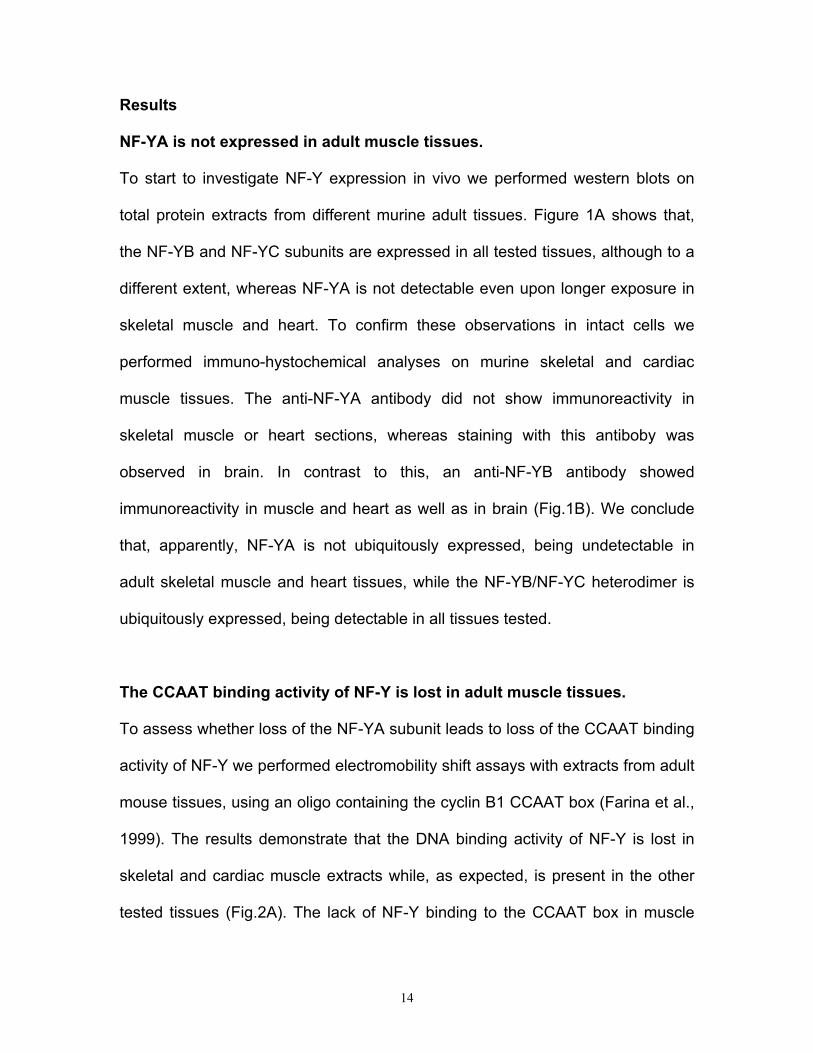
Results
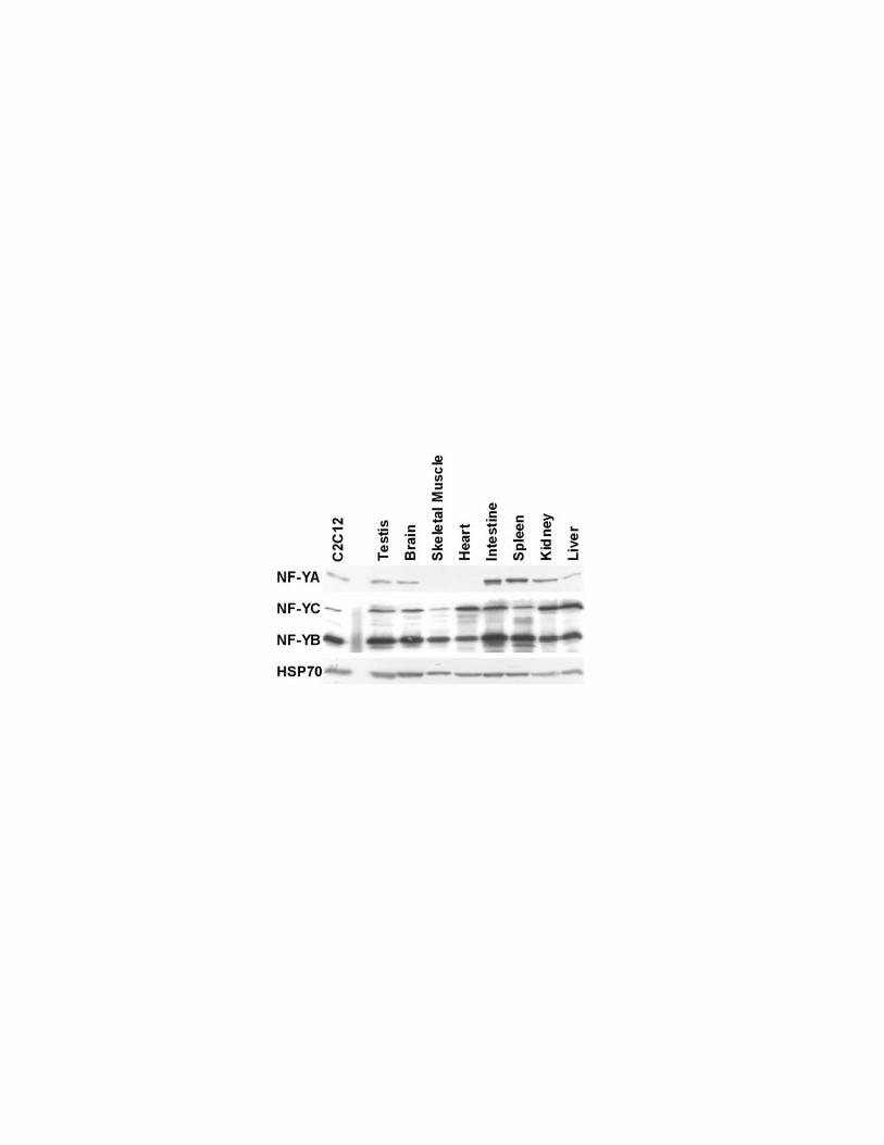
NF-YA is not expressed in adult muscle tissues.
To start to investigate NF-Y expression in vivo we performed western blots on
total protein extracts from different murine adult tissues. Figure 1A shows that,
the NF-YB and NF-YC subunits are expressed in all tested tissues, although to a
different extent, whereas NF-YA is not detectable even upon longer exposure in
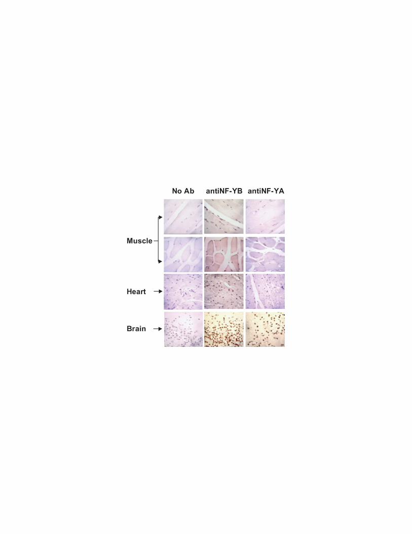
skeletal muscle and heart. To confirm these observations in intact cells we
performed immuno-hystochemical analyses on murine skeletal and cardiac
muscle tissues. The anti-NF-YA antibody did not show immunoreactivity in
skeletal muscle or heart sections, whereas staining with this antiboby was
observed in brain. In contrast to this, an anti-NF-YB antibody showed
immunoreactivity in muscle and heart as well as in brain (Fig.1B). We conclude
that, apparently, NF-YA is not ubiquitously expressed, being undetectable in
adult skeletal muscle and heart tissues, while the NF-YB/NF-YC heterodimer is
ubiquitously expressed, being detectable in all tissues tested.
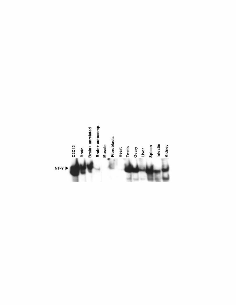
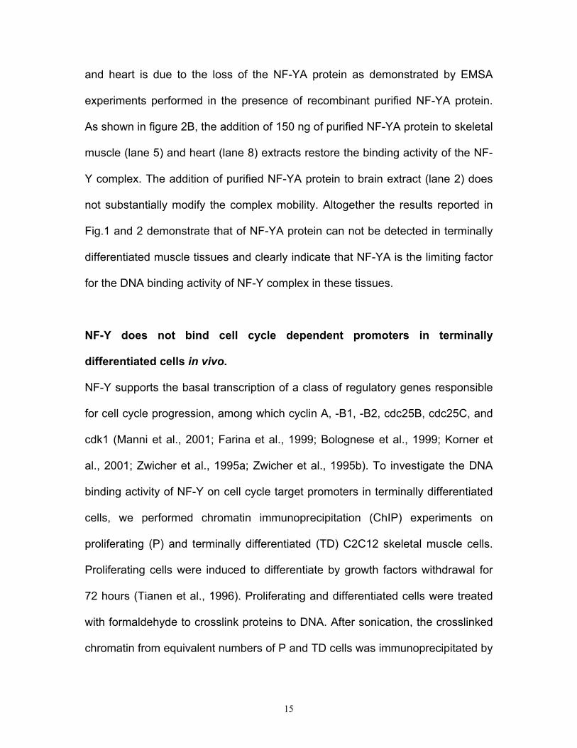
The CCAAT binding activity of NF-Y is lost in adult muscle tissues.
To assess whether loss of the NF-YA subunit leads to loss of the CCAAT binding
activity of NF-Y we performed electromobility shift assays with extracts from adult
mouse tissues, using an oligo containing the cyclin B1 CCAAT box (Farina et al.,
1999). The results demonstrate that the DNA binding activity of NF-Y is lost in
skeletal and cardiac muscle extracts while, as expected, is present in the other
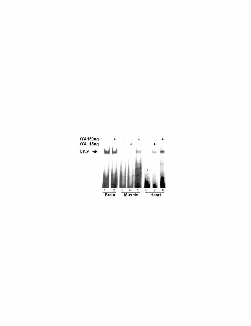
tested tissues (Fig.2A). The lack of NF-Y binding to the CCAAT box in muscle
15
and heart is due to the loss of the NF-YA protein as demonstrated by EMSA
experiments performed in the presence of recombinant purified NF-YA protein.
As shown in figure 2B, the addition of 150 ng of purified NF-YA protein to skeletal
muscle (lane 5) and heart (lane 8) extracts restore the binding activity of the NF-
Y complex. The addition of purified NF-YA protein to brain extract (lane 2) does
not substantially modify the complex mobility. Altogether the results reported in
Fig.1 and 2 demonstrate that of NF-YA protein can not be detected in terminally
differentiated muscle tissues and clearly indicate that NF-YA is the limiting factor
for the DNA binding activity of NF-Y complex in these tissues.
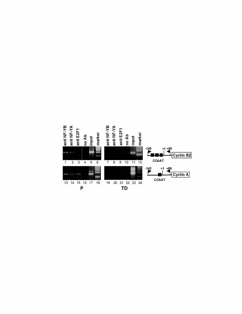
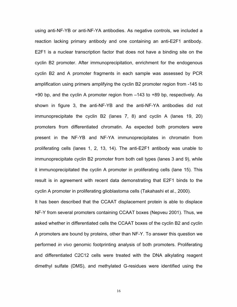
NF-Y does not bind cell cycle dependent promoters in terminally
differentiated cells in vivo.
NF-Y supports the basal transcription of a class of regulatory genes responsible
for cell cycle progression, among which cyclin A, -B1, -B2, cdc25B, cdc25C, and
cdk1 (Manni et al., 2001; Farina et al., 1999; Bolognese et al., 1999; Korner et
al., 2001; Zwicher et al., 1995a; Zwicher et al., 1995b). To investigate the DNA
binding activity of NF-Y on cell cycle target promoters in terminally differentiated
cells, we performed chromatin immunoprecipitation (ChIP) experiments on
proliferating (P) and terminally differentiated (TD) C2C12 skeletal muscle cells.
Proliferating cells were induced to differentiate by growth factors withdrawal for
72 hours (Tianen et al., 1996). Proliferating and differentiated cells were treated
with formaldehyde to crosslink proteins to DNA. After sonication, the crosslinked
chromatin from equivalent numbers of P and TD cells was immunoprecipitated by
16
using anti-NF-YB or anti-NF-YA antibodies. As negative controls, we included a
reaction lacking primary antibody and one containing an anti-E2F1 antibody.
E2F1 is a nuclear transcription factor that does not have a binding site on the
cyclin B2 promoter. After immunoprecipitation, enrichment for the endogenous
cyclin B2 and A promoter fragments in each sample was assessed by PCR
amplification using primers amplifying the cyclin B2 promoter region from -145 to
+90 bp, and the cyclin A promoter region from –143 to +89 bp, respectively. As
shown in figure 3, the anti-NF-YB and the anti-NF-YA antibodies did not
immunoprecipitate the cyclin B2 (lanes 7, 8) and cyclin A (lanes 19, 20)
promoters from differentiated chromatin. As expected both promoters were
present in the NF-YB and NF-YA immunoprecipitates in chromatin from
proliferating cells (lanes 1, 2, 13, 14). The anti-E2F1 antibody was unable to
immunoprecipitate cyclin B2 promoter from both cell types (lanes 3 and 9), while
it immunoprecipitated the cyclin A promoter in proliferating cells (lane 15). This
result is in agreement with recent data demonstrating that E2F1 binds to the
cyclin A promoter in proliferating glioblastoma cells (Takahashi et al., 2000).
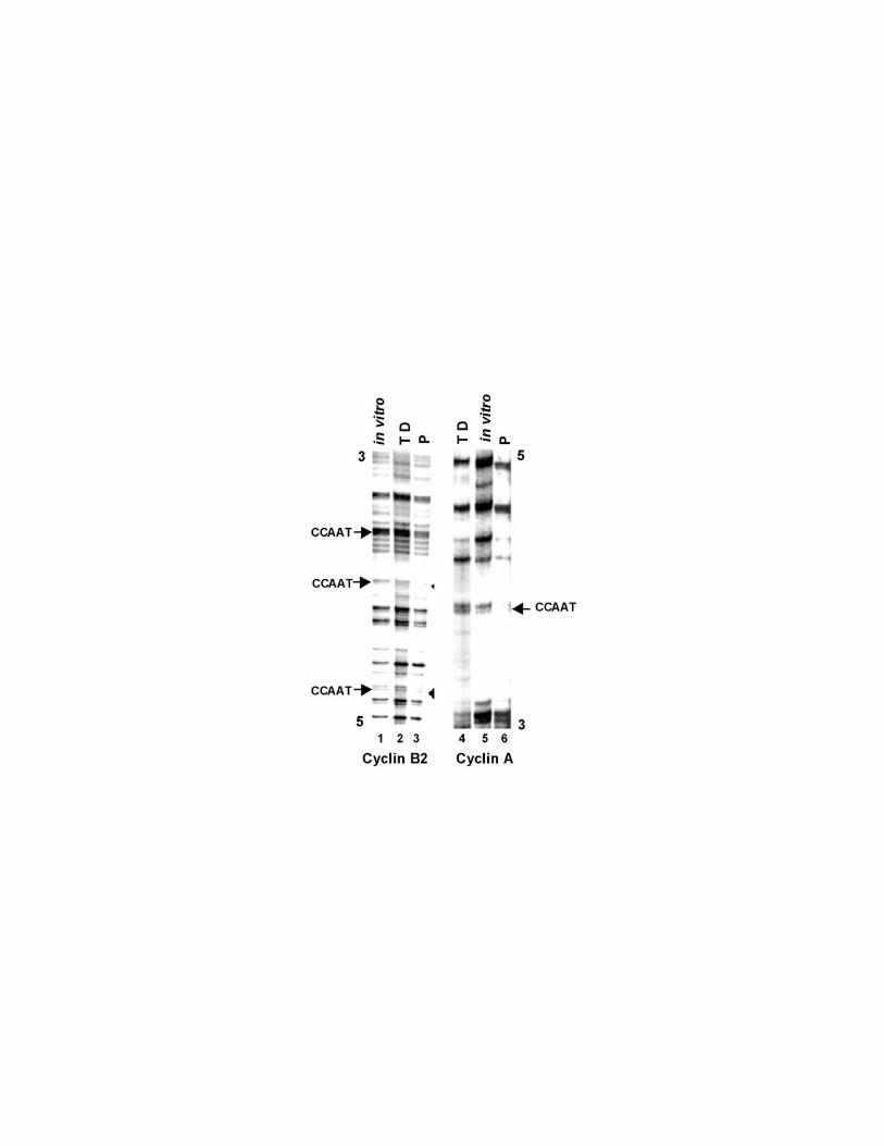
It has been described that the CCAAT displacement protein is able to displace
NF-Y from several promoters containing CCAAT boxes (Nepveu 2001). Thus, we
asked whether in differentiated cells the CCAAT boxes of the cyclin B2 and cyclin
A promoters are bound by proteins, other than NF-Y. To answer this question we
performed in vivo genomic footprinting analysis of both promoters. Proliferating
and differentiated C2C12 cells were treated with the DNA alkylating reagent
dimethyl sulfate (DMS), and methylated G-residues were identified using the
17
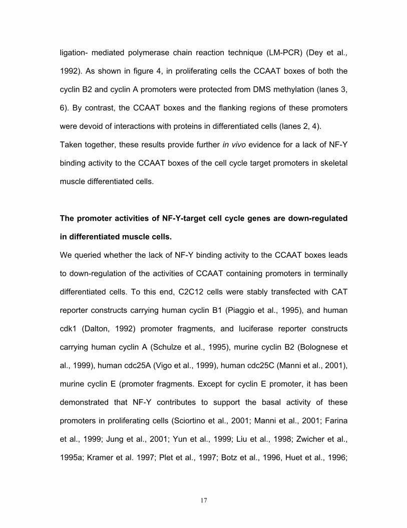
ligation- mediated polymerase chain reaction technique (LM-PCR) (Dey et al.,
1992). As shown in figure 4, in proliferating cells the CCAAT boxes of both the
cyclin B2 and cyclin A promoters were protected from DMS methylation (lanes 3,
6). By contrast, the CCAAT boxes and the flanking regions of these promoters
were devoid of interactions with proteins in differentiated cells (lanes 2, 4).
Taken together, these results provide further in vivo evidence for a lack of NF-Y
binding activity to the CCAAT boxes of the cell cycle target promoters in skeletal
muscle differentiated cells.
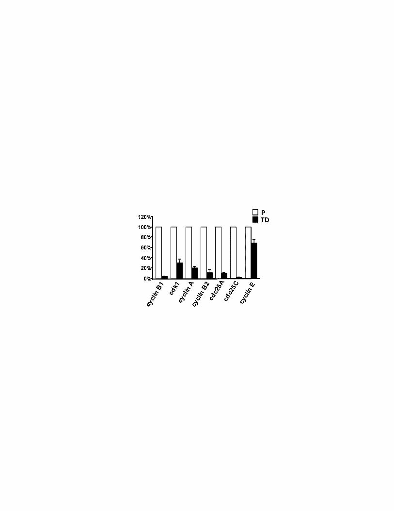
The promoter activities of NF-Y-target cell cycle genes are down-regulated
in differentiated muscle cells.
We queried whether the lack of NF-Y binding activity to the CCAAT boxes leads
to down-regulation of the activities of CCAAT containing promoters in terminally
differentiated cells. To this end, C2C12 cells were stably transfected with CAT
reporter constructs carrying human cyclin B1 (Piaggio et al., 1995), and human
cdk1 (Dalton, 1992) promoter fragments, and luciferase reporter constructs
carrying human cyclin A (Schulze et al., 1995), murine cyclin B2 (Bolognese et
al., 1999), human cdc25A (Vigo et al., 1999), human cdc25C (Manni et al., 2001),
murine cyclin E (promoter fragments. Except for cyclin E promoter, it has been
demonstrated that NF-Y contributes to support the basal activity of these
promoters in proliferating cells (Sciortino et al., 2001; Manni et al., 2001; Farina
et al., 1999; Jung et al., 2001; Yun et al., 1999; Liu et al., 1998; Zwicher et al.,
1995a; Kramer et al. 1997; Plet et al., 1997; Botz et al., 1996, Huet et al., 1996;
18
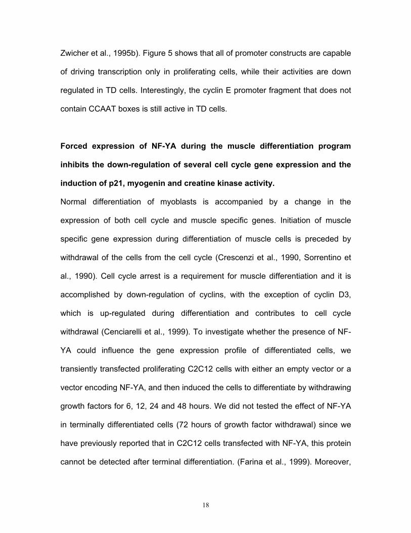
Zwicher et al., 1995b). Figure 5 shows that all of promoter constructs are capable
of driving transcription only in proliferating cells, while their activities are down
regulated in TD cells. Interestingly, the cyclin E promoter fragment that does not
contain CCAAT boxes is still active in TD cells.
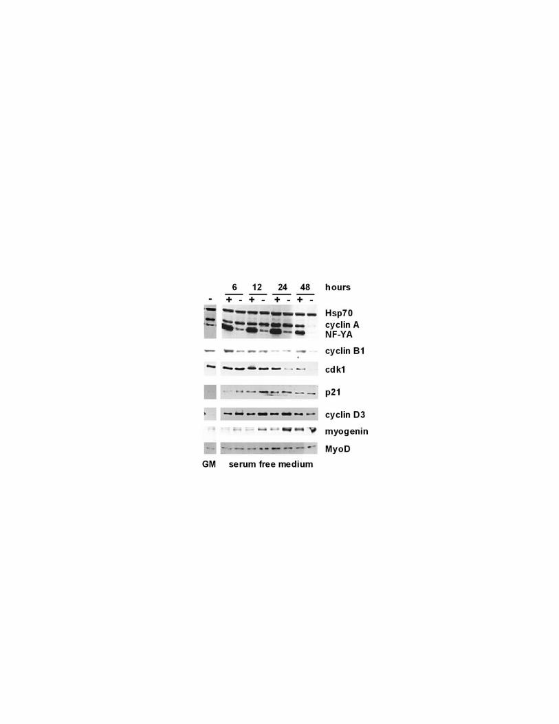
Forced expression of NF-YA during the muscle differentiation program
inhibits the down-regulation of several cell cycle gene expression and the
induction of p21, myogenin and creatine kinase activity.
Normal differentiation of myoblasts is accompanied by a change in the
expression of both cell cycle and muscle specific genes. Initiation of muscle
specific gene expression during differentiation of muscle cells is preceded by
withdrawal of the cells from the cell cycle (Crescenzi et al., 1990, Sorrentino et
al., 1990). Cell cycle arrest is a requirement for muscle differentiation and it is
accomplished by down-regulation of cyclins, with the exception of cyclin D3,
which is up-regulated during differentiation and contributes to cell cycle
withdrawal (Cenciarelli et al., 1999). To investigate whether the presence of NF-
YA could influence the gene expression profile of differentiated cells, we
transiently transfected proliferating C2C12 cells with either an empty vector or a
vector encoding NF-YA, and then induced the cells to differentiate by withdrawing
growth factors for 6, 12, 24 and 48 hours. We did not tested the effect of NF-YA
in terminally differentiated cells (72 hours of growth factor withdrawal) since we
have previously reported that in C2C12 cells transfected with NF-YA, this protein
cannot be detected after terminal differentiation. (Farina et al., 1999). Moreover,
19
since the rationale of this experiment was that the presence of NF-YA could
retain the cells in the cell cycle, the cells were induced to differentiate in the
absence of Ara-C. Indeed, in the presence of Ara-C the proliferating cells would
be killed. In this experimental system we examined the endogenous expression
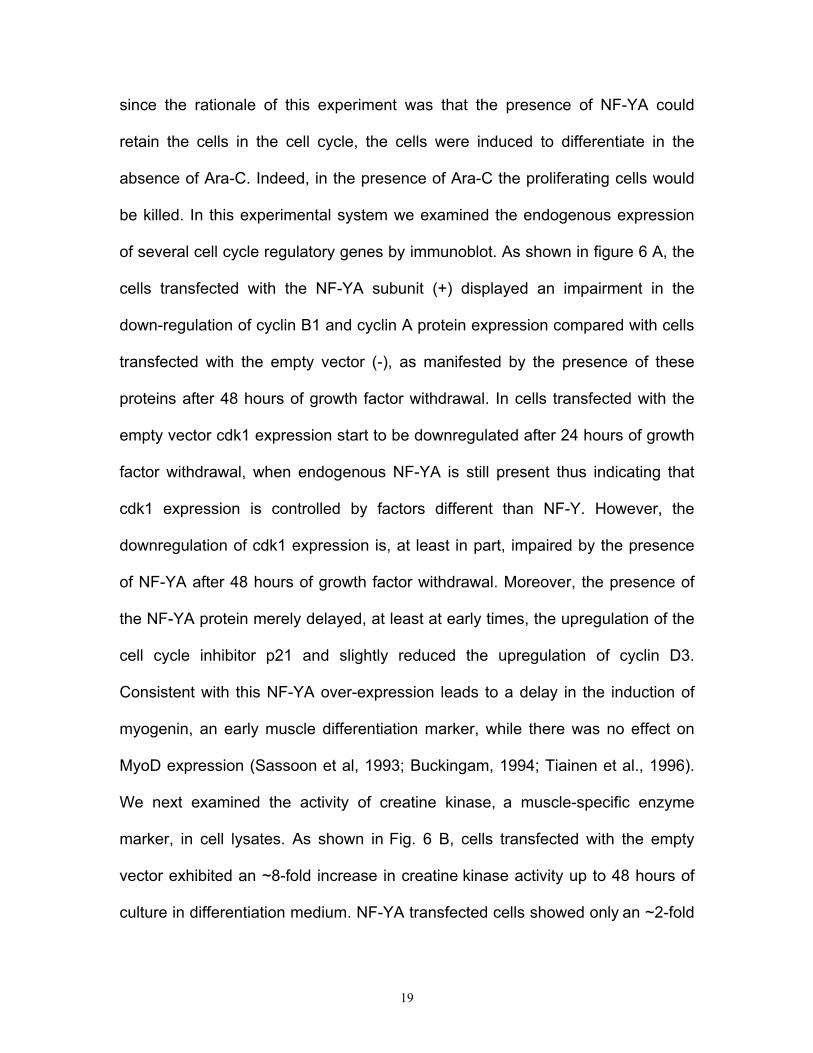
of several cell cycle regulatory genes by immunoblot. As shown in figure 6 A, the
cells transfected with the NF-YA subunit (+) displayed an impairment in the
down-regulation of cyclin B1 and cyclin A protein expression compared with cells
transfected with the empty vector (-), as manifested by the presence of these
proteins after 48 hours of growth factor withdrawal. In cells transfected with the
empty vector cdk1 expression start to be downregulated after 24 hours of growth
factor withdrawal, when endogenous NF-YA is still present thus indicating that
cdk1 expression is controlled by factors different than NF-Y. However, the
downregulation of cdk1 expression is, at least in part, impaired by the presence
of NF-YA after 48 hours of growth factor withdrawal. Moreover, the presence of
the NF-YA protein merely delayed, at least at early times, the upregulation of the
cell cycle inhibitor p21 and slightly reduced the upregulation of cyclin D3.
Consistent with this NF-YA over-expression leads to a delay in the induction of
myogenin, an early muscle differentiation marker, while there was no effect on
MyoD expression (Sassoon et al, 1993; Buckingam, 1994; Tiainen et al., 1996).
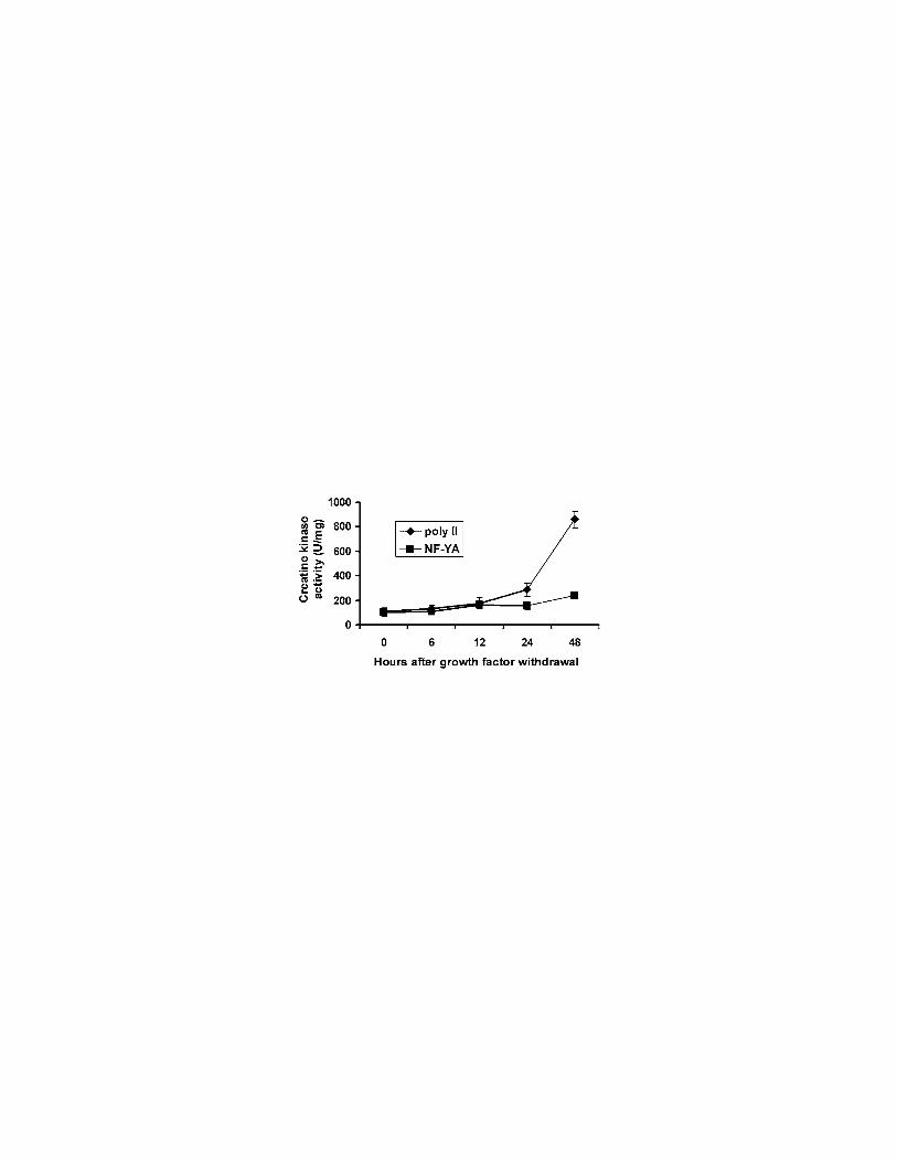
We next examined the activity of creatine kinase, a muscle-specific enzyme
marker, in cell lysates. As shown in Fig. 6 B, cells transfected with the empty
vector exhibited an ~8-fold increase in creatine kinase activity up to 48 hours of
culture in differentiation medium. NF-YA transfected cells showed only an ~2-fold
20
greater creatine kinase activity after 48 hours of growth factor withdrawal. The
results presented here indicate that the down-regulation of NF-YA expression is
strictly required for the correct modulation of the cell cycle and muscle specific
gene expression during differentiation of muscle cells. These results also suggest
that NF-YA interferes, at least in part, with the myogenic differentiation program
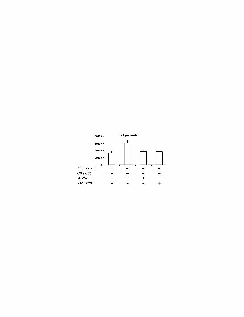
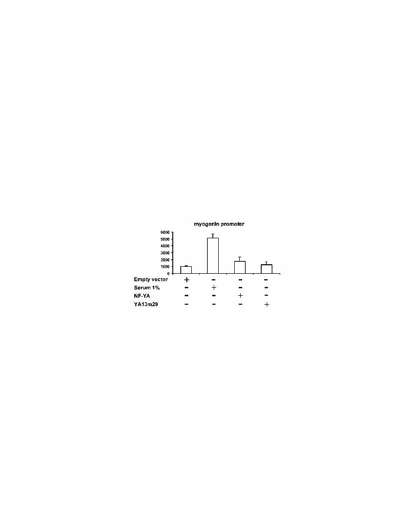
of C2C12 cells. To further investigate whether NF-YA directly regulate
transcription of genes involved in specifying myogenic fate, we employed a
dominant-negative NF-YA protein, YA13m29 (Mantovani et al., 1994). Upon
transfection in mammalian cells, this vector expresses a mutant protein
containing a triple amino acids substitution in the NF-YA DNA binding subdomain
enabling the subunit to interact with the NF-YB·NF-YC dimer. The resulting trimer
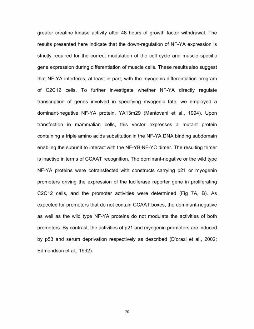
is inactive in terms of CCAAT recognition. The dominant-negative or the wild type
NF-YA proteins were cotransfected with constructs carrying p21 or myogenin
promoters driving the expression of the luciferase reporter gene in proliferating
C2C12 cells, and the promoter activities were determined (Fig 7A, B). As
expected for promoters that do not contain CCAAT boxes, the dominant-negative
as well as the wild type NF-YA proteins do not modulate the activities of both
promoters. By contrast, the activities of p21 and myogenin promoters are induced
by p53 and serum deprivation respectively as described (D’orazi et al., 2002;
Edmondson et al., 1992).
21
Discussion
The results presented in this study show that the CCAAT binding activity of the
NF-Y transcription factor is absent in adult skeletal and cardiac muscle tissues
and that loss of this activity is due to the down-regulation of the NF-YA protein in
these tissues. The relevance of this observation resides in the fact that recent
studies, in various cell types, have indicated that NF-Y could serve as a common
transcription factor for an increasing number of cell cycle control genes. For
instance, the cyclin B1, cyclin B2, cyclin A, cdc25B cdc25C, and cdk1 genes all
contain in their promoters NF-Y sites, which are required for their transcriptional
activation at S phase (Bolognese et al., 1999; Korner and Muller., 2001; Manni et
al., 2001; Zwicher et al., 1995a; Zwicher et al., 1995b). We have already
demonstrated that the loss of a functional NF-Y complex in differentiated muscle
cells leads to down-regulation of the cyclin B1 promoter (Farina et al., 1999). We
now extend this observation to the promoters of the other cell cycle control genes
that are known to be regulated by NF-Y during the cell cycle. Moreover, the
results of the in vivo chromatin immunoprecipitation experiments on the cyclin B2
and cyclin A promoters indicate that down-regulation of these genes in
differentiated cells could be due to the absence of NF-Y on their promoters.
Interestingly, the lack of NF-Y binding to cyclin B2 and cyclin A promoters in
differentiated cells does not lead to the binding of other proteins to the CCAAT
boxes as demonstrated by in vivo genomic footprinting experiments. These
results provide evidence for a model in which NF-Y serves as a common
transcription factor in proliferating muscle cells (myoblasts) for several key cell
22
cycle control genes. Permanent down-regulation of NF-YA expression in
differentiated cells (myotubes) leads to the inhibition of the expression of these
genes. A recent analysis of a large database of 1031 human promoters indicated
that the CCAAT box is present in 63% of them (Suzuki et al., 2001). It will be of
interest to investigate whether the transcription from other CCAAT-containing
promoters, in post-mitotic skeletal muscle cells, is down regulated by the same
common mechanism presently described.
In adult muscle tissues the expression of NF-YA protein is down regulated, while
NF-YB and -C are still expressed. It has been demonstrated that the NF-Y
histone-like dimer, NF-YB/C, can form complexes with H3-H4 histones for whose
formation the CCAAT box is not required. Moreover, the NF-Y histone-like dimer
associates with DNA during nucleosome formation in the absence of NF-YA
(Caretti et al., 1999). Proteins involved in histone acetylation are found
associated with NF-YB (Currie, 1998, Li et al., 1998). This evidence supports the
hypothesis that the NF-YB/C heterodimer could serve the dual function of binding
NF-YA to activate transcription of CCAAT-containing promoters and, thanks to
the histone-like domains, associating DNA during nucleosome formation. Thus,
skeletal muscle cells represent an excellent system to study whether the NF-
YB/C dimer exerts a biological role in the absence of NF-YA.
Initiation of muscle specific gene expression, i.e., myogenin, during differentiation
of muscle cells, is preceded by withdrawal of the cells from the cell cycle. Exit
from the cell cycle is accomplished by down-regulation of several cyclins and
cdks, and by upregulation of cell cycle inhibitors, such as p21, and cyclin D3
23
(Perry and Rudnick, 2000). We demonstrate here that ectopic expression of NF-
YA protein could interfere with the onset of both cell cycle withdrawal and early
muscle specific program. While NF-YA directly binds the endogenous promoters
of the analyzed cell cycle regulatory genes, cyclin A and cyclin B2, as well as
cyclin B1 (Sciortino et al., 2001), and cdk1 (A.G. personal observation), NF-YA
does not directly interfere with the muscle differentiation program. Indeed, our
results indicate that NF-YA does not modulate the transcriptional actvity of p21
and myogenin promoters. However, unrestricted expression of NF-YA interferes,
although in an indirect way, with the differentiation process as indicated by the
inhibition of creatine kinase activity occurring during differentiation in the
presence of NF-YA.
Altogether, the above results are consistent with a scenario in which NF-YA
expression inhibits the differentiation program and keeps the myoblasts in the cell
cycle. Since the induction of p21, cyclin D3, and myogenin (Cenciarelli et al,
1999; Molkentin and Olson, 1996), as well as the down-regulation of the of cell
cycle control genes (Perry and Rudnick, 2000) are important events in the
induction of muscle differentiation, our data suggest that NF-Y exerts a biological
role in the switch from proliferation to differentiation of muscle cells.
Although the presence of NF-YA in cells committed to differentiate could lead to
conflicting signals, no apparent apoptosis was seen in our experimental
conditions. It remains to be determined whether expression of NF-YA in the
presence of differentiation stimuli for periods of time longer than those tested
here, leads to apoptosis. Indeed, the level of NF-YA drops between 24 and 48
24
hours in the transfected cells and after 72 hours of growth factor withdrawal it
becomes undetectable (our unpublished results). This could explain why
overexpression of NF-YA can only delay rather than inhibit differentiation.
Our previous results indicate that the regulation of NF-YA expression depends on
a post-transcriptional level of regulation (Farina et al., 1999). Based on these
consideration we tested the effect of the specific calpain I and II and catepsin
inhibitor, LN-acetyl-leu-leu-norleucinal (LLnL), on NF-Y stability (Manni et al.,
manuscript in preparation). These preliminary experiments show that there is a
cytoplasmatic increase of NF-YA protein, suggesting that the degradation of NF-
YA involves an ubiquitine-proteasome pathway. The study of the regulation of
NF-Y expression in adult muscle tissues, in particular of the NF-YA subunit, will
be an important step towards a full understanding of cell cycle control in these
cells.
25
ACKNOWLEDGEMENTS
The authors would like to thank Kristian Helin for cdc25A promoter, Pidder
Yansen-Durr for cyclin A and cyclin E promoter, Stephen Dalton for human cdk1
promoter. Marco Crescenzi and Silvia Bacchetti for critical reading of the
manuscript and helpful discussions; Maria Pia Gentileschi and Giulio Tibursi for
technical advice; Vincenzo Giusti for computing assistance. This work has been
partially supported by grants from AIRC, Ministero della Sanita’ (N°: ICS-
120.4/RA00-90), CNR and ASI to GP. AG is a recipient of an FIRC fellowship.
26
References
Blau, H.M., Pavlath, G.K., Hardeman, E.C., Chiu, C.P., Silberstein, L., Webster,
S.G, Miller, S.C., and Webster, C. (1985). Plasticity of the differentiated state.
Science. 230, 758-766.
Bolognese, F., Wesner, M., Lange-zu Dohna, C., Gurtner, A., Ronchi, H., Muller,
A., Manni, I., Mossner, J., Piaggio, G., Mantovani, R. et al. (1999). The cyclin B2
promoter depends on NF-Y, a trimer whose CCAAT-binding activity is cell cycle
regulated. Oncogene 18, 1845-1853.
Botz, J., Zerfass-Thome, K., Spitkovsky, D., Delius, H., Vogt, B., Eilers, M.,
Hatzigeorgiou, A., and Yansen-Durr. P. (1996). Cell cycle regulation of the
murine cyclin E gene depends on an E2F binding site in the promoter. Mol Cell
Biol. 16, 3401-3409.
Buckingham, M.E. (1994). Muscle: the regulation of myogenesis.Curr Opin.
Genet. Dev. 4, 745-751. Review.
Caretti, G., Motta M.C., and Mantovani R. (1999). NF-Y associates with H3-H4
tetramers and octamers by multiple mechanisms. Mol. Cell Biol. 19, 8591-60.
27
Cenciarelli, C., De Santa, F., Puri, P.L., Mattei, E., Ricci, L., Bucci, F., Felsani, A.,
and Caruso. A.M. (1999). Critical role played by cyclin D3 in the MyoD-mediated
arrest of cell cycle during myoblast differentiation. Mol. Cell. Biol. 19, 5203-5217.
Chang, Z.F., and Liu, C.J. (1994). Human thymidine kinase CCAAT-binding
protein is NF-Y, whose A subunit expression is serum-dependent in human IMR-
90 diploid fibroblasts. J.Biol.Chem. 269, 17893-17898.
Crescenzi, M., Fleming, T.P., Lasser, A.B., Weintraub, H., and Aaronson. S.A.
(1990). MyoD induces growth arrest independent of differentiation in normal and
transformed cells. Proc. Nat. Acad .Sci. USA 87, 8442-8446.
Currie, R.A. (1998). NF-Y is associated with the histone acetyltransferases GCN5
and P/CAF. J. Biol. Chem. 273, 1430-1434.
Dalton, S. (1992). Cell cycle regulation of the human cdc2 gene. EMBO J. 11,
1797-1804.
Dey, A., Thornton, A.M., Lonergan, M., Weissman, S.M., Chamberlain, J.W., and
Ozato K. (1992). Occupancy of upstream regulatory sites in vivo coincides with
major histocompatibility complex class I gene expression in mouse tissues. Mol.
Cell. Biol. 12, 3590-3599.
28
D'Orazi, G., Cecchinelli, B., Bruno, T., Manni, I., Higashimoto, Y., Saito, S.,
Gostissa, M., Coen, S., Marchetti, A., Del Sal, G., Piaggio, G., Fanciulli, M.,
Appella, E. and Soddu, S. (2002). Homeodomain-interacting protein kinase-2
phosphorylates p53 at Ser 46 and mediates apoptosis. Nat Cell Biol 4,11-19
Edmondson, D.G., Cheng, T.C., Cserjesi, P., Chakraborty, T., and Olson, E.N.
Analysis of the myogenin promoter reveals an indirect pathway for positive
autoregulation mediated by the muscle-specific enhancer factor MEF-2. Mol Cell
Biol 12, 3665-3677.
Farina, A., Manni, I., Fontemaggi, G., Tiainen, M., Cenciarelli, C., Bellorini, M.,
Mantovani, R., Sacchi, A., and Piaggio, G. (1999). Down-regulation of cyclin B1
gene transcription in terminally differentiated skeletal muscle cells is associated
with loss of functional CCAAT-binding NF-Y complex. Oncogene 18, 2818-2827.
Fontemaggi, G., Gurtner, A., Strano, S., Higashi, Y., Sacchi, A., Piaggio, G,. and
Blandino, G. (2001). The transcriptional repressor ZEB regulates p73 expression
at the crossroad between proliferation and differentiation. Mol. Cell. Biol. 21,
8461-8470.
Hu, Q., and Maity, S.N. (2000). Stable expression of a dominant negative mutant
of CCAAT binding factor/NF-Y in mouse fibroblast cells resulting in retardation of
29
cell growth and inhibition of transcription of various cellular genes. J. Biol. Chem.
275, 4435-4444.
Huet, X., Rech, J., Plet, A., Vie, A., and Blanchard J.M. (1996). Cyclin A
expression is under negative transcriptional control during the cell cycle. Mol Cell
Biol. 16, 3789-3798.
Jung, M.S., J. Yun, H.D. Chae, J.M. Kim, S.C. Kim, T.S. Choi, and Shin, D.Y.
(2001). p53 and its homologues, p63 and p73, induce a replicative senescence
through inactivation of NF-Y transcription factor. Oncogene. 20, 5818-5825.
Korner, K., Jerome, V., Schmidt, T., and Muller, R. (2001). Cell cycle regulation
of the murine cdc25B promoter: essential role for NF-Y and a proximal repressor
element. J. Biol. Chem. 276, 9662-9669.
Kramer, A., Carstens, C.P., Wasserman, W.W., and Fahl. W.E., (1997).
CBP/cycA, a CCAAT-binding protein necessary for adhesion-dependent cyclin A
transcription, consists of NF-Y and a novel Mr 115,000 subunit. Cancer Res. 57,
5117-5121.
Li, Q., Herrler M., Landsberger N., Kaludov N., Ogryzko V.V., Nakatani Y., and
Wolffe A.P. (1998). Xenopus NF-Y pre-sets chromatin to potentiate p300 and
30
acetylation-responsive transcription from the Xenopus hsp70 promoter in vivo.
EMBO J. 17, 6300-15.
Liu, Q., Yan, H., Dawes, N.J., Lu, Y., and Zhu. H., (1998). Transcriptional
activation of the p34cdc2 gene by cdc2 promoter binding factor/nuclear factor-Y
in fetal rat ventricular myocytes. Circ. Res. 82, 251-260.
Manni, I., Mazzaro, G., Gurtner, A., Mantovani, R., Haugwitz, U., Krause, K.,
Engeland, K., Sacchi, A., Soddu, S., and Piaggio. G. (2001). NF-Y mediates the
transcriptional inhibition of the cyclin B1, cyclin B2, and cdc25C promoters upon
induced G2 arrest. J Biol Chem. 276, 5570-5576.
Mantovani, R. (1998). A survey of 178 NF-Y binding CCAAT boxes. Nucleic
Acids Res. 26, 1135-1143.
Mantovani, R. (1999). The molecular biology of the CCAAT-binding factor NF-Y.
Gene 239, 15-27.
Mantovani, R., Li, X.Y., Pessara, U., Hoof van Huijsduijnen, R., Benoist, C., and
Mathis D. (1994). Dominant negative analogs of NF-YA. J. Biol. Chem. 269,
20340-20346.
31
Marziali, G., Perrotti, E., Ilari, R., Testa, U., Coccia, E.M., and Battistini, A.
(1997). Transcriptional regulation of the ferritin heavy-chain gene: the activity of
the CCAAT binding factor NF-Y is modulated in heme-treated Friend leukemia
cells and during monocyte-to-macrophage differentiation. Mol. Cell. Biol. 17,
1387-1395.
Molkentin, J.D., and Olson, E.N. (1996). Combinatorial control of muscle
development by basic helix-loop-helix and MADS-box transcription factors. Proc
Natl Acad Sci USA 93, 9366-9373.
Nepveu, A., (2001). Role of the multifunctional CDP/Cut/Cux homeodomain
transcription factor in regulating differentiation, cell growth and development.
Gene 270, 1-15.
Perry R.L., and Rudnick M.A. (2000) Molecular mechanisms regulating myogenic
determination and differentiation.. Front. Biosci. 5,750-767
Piaggio, G., Farina, A., Perrotti, D., Manni, I., Fuschi, P., Sacchi, A., and Gaetano
C. (1995). Structure and growth-dependent regulation of the human cyclin B1
promoter. Exp. Cell. Res. 216, 396-402.
Plet, A., Huet, X., Algarte, M., Rech, J., Imbert, J., Philips, A., and Blanchard,
J.M. (1997). Relief of cyclin A gene transcriptional inhibition during activation of
32
human primary T lymphocytes via CD2 and CD28 adhesion
molecules.Oncogene. 14, 2575-2583.
Sassoon, D.A. (1993). Myogenic regulatory factors: dissecting their role and
regulation during vertebrate embryogenesis. Dev. Biol. 156, 11-23. Review.
Schulze, A., Zerfass, K., Spitkovsky, D., Middendorp, S., Berges, J., Helin, K.,
Yansen Durr, P., and Henglein. B. (1995). Cell Cycle Regulation of the Cyclin A
Gene Promoter is Mediated by a Variant E2F Site Proc. Natl. Acad. Sci. USA 92,
11264-11268.
Sciortino, S., Gurtner, A., Dey, A., Sacchi, A., Ozato, K., and Piaggio, G. (2001).
Cyclin B1 gene is actively transcribed during mitosis in HeLa cells. Embo Rep. 2,
1018-1023.
Sorrentino, V., Pepperkok, R., Davis, R.L., Ansorge W., and Philipson, L. (1990).
Cell proliferation inhibited by MyoD1 independently of myogenic differentiation.
Nature. 345, 813-815.
Suzuki, Y., Tsunoda, T., Sese, J., Taira, H., Mizushima-Sugano, J., Hata, H.,
Ota, T., Isogai, T., Tanaka, T., Nakamura, Y., et al. (2001). Identification and
characterization of the potential promoter regions of 1031 kinds of human genes.
Genome Res. 11, 677-684.
33
Takahashi, Y., Rayman, J.B., and Dynlacht B.D. (2000). Analysis of promoter
binding by the E2F and pRB families in vivo: distinct E2F proteins mediate
activation and repression. Genes Dev. 14, 804-816.
Tiainen, M., Pajalunga, D., Ferrantelli, F., Soddu, S., Salvatori, G., Sacchi, A.,
and Crescenzi, M. (1996). Terminally differentiated skeletal myotubes are not
confined to G0 but can enter G1 upon growth factor stimulation. Cell Growth Diff.
7, 1039-1050.
Vigo, E., Müller, H., Prosperini, E., Hateboer, G., Cartwright, P., Moroni, M.C.,
and Helin, K. (1999). CDC25A phosphatase is a target of E2F and is required for
efficient E2F-induced S phase. Mol. Cell. Biol. 19, 6379-6395.
Yaffe, D., and Saxel, O. (1977). Serial passaging and differentiation of myogenic
cells isolated from dystrophic mouse muscle. Nature. 270, 725-727.
Yun, J., Chae, H.D., Choy, H.E., Chung, J., Yoo, H.S., Han, M.H., and Shin. D.Y.,
(1999). p53 negatively regulates cdc2 transcription via the CCAAT-binding NF-Y
transcription factor. J. Biol. Chem. 274, 29677-82.
Zwicker, J., Gross, C., Lucibello, F.C., Truss, M., Ehlert, F., Engeland, K., and
Muller R. (1995a). Cell cycle regulation of cdc25C transcription is mediated by
34
the periodic repression of the glutamine-rich activators NF-Y and Sp1. Nucleic.
Acids Res. 23, 3822-3830.
Zwicher J., Lucibello, F.C., Wolfraim, L.A., Gross, C., Truss, M., Engeland, K.,
and Muller R. (1995b). Cell cycle regulation of cdc25C transcription is mediated
by the periodic repression of the glutamine-rich activators NF-Y and Sp1. EMBO
J. 14, 4514-4522.
35
Figure Legend
Figure 1. NF-YA is not expressed in adult muscle tissues. A) Western blot
analysis was performed on total lysates from the indicated tissues. The extracts
were probed with rabbit polyclonal antiserum raised against NF-YA, NF-YB, NF-
YC. To normalise protein’s loading, the filter was stained with a monoclonal
antibody against the HSP70 protein. B) Murine skeletal muscle, heart and brain
tissues were methanol fixed and paraffin-embedded prior to sectioning. Sections
were incubated with polyclonal rabbit antibodies against NF-YA or NF-YB as
primary antibodies and/or with peroxidase-conjugated goat anti-rabbit antibody
as secondary antibody. The peroxidase activity was detected with DAB prior to
counter-staining the sections with haematoxylin.
Figure 2. A) The CCAAT binding activity of NF-Y is lost in adult muscle tissues.
EMSA were performed with B1upCCAAT 32P-labelled oligonucleotide and 150 µg
of total extracts from the indicated tissues. The complex was specifically cross-
competed by a 200-fold molar excess of unlabelled oligonucleotide
(brain+autocomp.), but not by a 1000-fold molar excess of an unrelated
oligonucleotide (brain+unrelated). B) NF-YA subunit is the limiting factor for the
binding of NF-Y complex to the CCAAT box. EMSA were performed as above
except for the adding of recombinant purified NF-YA protein (rYA) to the extracts
from purified muscle, heart and brain.
36
Figure 3: NF-Y does not bind cell cycle dependent promoters in terminally
differentiated cells in vivo. Formaldehyde crosslinked chromatin was prepared
from the same number of proliferating (P) and terminally differentiated (TD) cells
and was incubated with antibodies to NF-Y (lanes 1,2,7,8,13,14,19,20), E2F1
(lanes 3,9,15,21) or no antibody (4,10,16,22). Immunoprecipitates from each
antibody were aliquoted and then analyzed by PCR with specific primers for the
mouse cyclin B2 or cyclin A promoters. A sample representing 0,02% of the total
input chromatin (input) was included in the PCR reactions.
Figure 4 Cyclin B2 and cyclin A promoters are devoid of sequence-specific
transcription factor interactions in differentiated cells. Genomic footprinting of the
coding strand of the mouse cyclin B2 and the non coding strand of the mouse
cyclin A promoters from proliferating (P) and terminally differentiated (TD) C2C12
cells. As a control, the DMS reactivity for DNA purified from asynchronous
C2C12 cells is shown (in vitro). The CCAAT boxes are indicated.
Figure 5 The promoter activities of NF-Y cell cycle target genes are down-
regulated in differentiated muscle cells. CAT or Luc-reporter constructs carrying
the cyclin B1, cdk1, cyclin A, cyclin B2, cdc25A, cdc25C and cyclin E promoters
were stably transfected by calcium-phosphate in C2C12 cells. The obtained poly-
clonal cell lines were analysed for CAT or Luciferase activity. The values in TD
cells are expressed, on the Y axis, as percentages of the CAT or Luciferase
37
activity obtained from proliferating cells (100%). Results represent one typical
experiment performed in triplicate.
Figure 6. Forced expression of NF-YA in differentiated muscle cells inhibits the
down-regulation of several cell cycle gene expression and the induction of p21,
myogenin and creatine kinase activity. C2C12 cells were transiently transfected
with a plasmid carrying NF-YA (+) or the empty vector (-) as indicated. After
transfection, cells were grown in the absence of growth factor for 6, 12, 24 or 48
hours and lysed to prepare WCE (A) and measured creatine kinase activity (B) .
WCEs from cells cultured in growth medium (GM) and in the absence of growth
factor were subjected to Western blot analysis using antibodies against the
indicated proteins. B) creatine kinase activity was measured on extracts prepared
from transfected and untrasfected cells. Results are the mean ± S.E. of three
independent experiments.
Figure 7. NF-YA does not directly modulate the transcriptional actvity of p21 and
myogenin promoters. 80 ng of a vector carrying p21 promoter (A) and 2 µg of a
vector carrying myogenin promoter (B) have been transiently cotransfected in
proliferating C2C12 cells together with the same amount of the empty vector or
vector expressing wild type or mutant (YA13m29) NF-YA proteins. As positive
control, vector carrying p21 promoter has been cotransfected with an eukaryotic
vector expressing p53 protein and the luciferase activity of the vector carrying
myogenin promoter has been measured in low serum (1%) growth condition. As
38
internal control of transfection efficiency all samples has been cotransfected with
the CMVβgal reporter construct. Values are the means +- standard deviations of
four independent experiments.