Starflower Image Herbarium Flowering Deciduous Shrubs and ...
Upload
danforthcenterCategory
view
1download
0
Repression of Flowering in Arabidopsis Requires Activationof FLOWERING LOCUS C Expression by the HistoneVariant H2A.Z W OA
Roger B. Deal,a Christopher N. Topp,b Elizabeth C. McKinney,a and Richard B. Meaghera,1
a Department of Genetics, University of Georgia, Athens, Georgia 30602b Department of Plant Biology, University of Georgia, Athens, Georgia 30602
The histone variant H2A.Z has been implicated in numerous chromatin-mediated processes, including transcriptional
activation, euchromatin maintenance, and heterochromatin formation. In yeast and humans, H2A.Z is deposited into
chromatin by a conserved protein complex known as SWR1 and SRCAP, respectively. Here, we show that mutations in the
Arabidopsis thaliana homologs of two components of this complex, ACTIN-RELATED PROTEIN6 (ARP6) and PHOTO-
PERIOD-INDEPENDENT EARLY FLOWERING1 (PIE1), produce similar developmental phenotypes and result in the misregu-
lation of a common set of genes. Using H2A.Z-specific antibodies, we demonstrate that ARP6 and PIE1 are required for the
deposition of H2A.Z at multiple loci, including the FLOWERING LOCUS C (FLC) gene, a central repressor of the transition to
flowering. Loss of H2A.Z from chromatin in arp6 and pie1 mutants results in reduced FLC expression and premature
flowering, indicating that this histone variant is required for high-level expression of FLC. In addition to defining a novel
mechanism for the regulation of FLC expression, these results support the existence of a SWR1-like complex in Arabidopsis
and show that H2A.Z can potentiate transcriptional activation in plants. The finding that H2A.Z remains associated with
chromatin throughout mitosis suggests that it may serve an epigenetic memory function by marking active genes and
poising silenced genes for reactivation.
INTRODUCTION
To ensure reproductive success, plants must align their transition
to flowering with favorable environmental conditions. This is
achieved through the integration of signals from multiple path-
ways that sense and respond to various environmental and
endogenous cues, such as daylength, temperature, and devel-
opmental state (Simpson and Dean, 2002). In Arabidopsis thali-
ana, several of these signaling pathways converge on the central
floral repressor gene FLOWERING LOCUS C (FLC) and either
activate or repress its expression to suppress or promote the
transition to flowering, respectively (Henderson and Dean, 2004;
Simpson, 2004; Sung and Amasino, 2004). The terminal effectors
of these pathways controlling FLC expression are now known to
represent many types of chromatin-modifying factors, including
histone acetyltransferases, histone deacetylases, histone meth-
yltransferases, polycomb-type proteins, and a putative histone
demethylase (He and Amasino, 2005; Reyes, 2006). Thus, FLC
serves as a model for how chromatin remodeling and modifica-
tion can regulate a critical developmental switch. In contrast with
histone modifications, there has been no evidence to date for
the role of histone variants in the control of FLC expression.
The fundamental repeating unit of chromatin, known as the
nucleosome, consists of ;150 bp of DNA wrapped around a
protein particle composed of two copies of each of the four core
histones: H2A, H2B, H3, and H4. These histone proteins are
encoded by multiple gene copies and are produced in large
quantities to accommodate the nascent genome during DNA
replication. In addition to these bulk histones, eukaryotic ge-
nomes also encode variant histones that are deposited inde-
pendently of DNA replication and serve to functionally specialize
or differentiate specific chromatin regions. In Saccharomyces,
Drosophila, and human, the histone variant H2A.Z is deposited
into chromatin by a conserved protein complex known as SWR1,
Tip60, and SRCAP, respectively (Krogan et al., 2003; Kobor et al.,
2004; Kusch et al., 2004; Mizuguchi et al., 2004; Ruhl et al., 2006).
The yeast H2A.Z variant plays important roles in both activating
transcription and antagonizing the spread of heterochromatin
into euchromatic regions (Santisteban et al., 2000; Adam et al.,
2001; Larochelle and Gaudreau, 2003; Meneghini et al., 2003).
Several genome-wide studies of yeast H2A.Z occupancy have
shown that this variant localizes to the promoters of thousands of
euchromatic genes, and its presence is required for the optimal
transcriptional activation of many of these genes (Guillemette
et al., 2005; Li et al., 2005; Raisner et al., 2005; Zhang et al.,
2005). In contrast with the situation in yeast, cytological and ge-
netic studies suggest that in metazoans H2A.Z may also play a role
in heterochromatin formation and/or maintenance (Rangasamy
et al., 2003; Swaminathan et al., 2005), although this variant
has been observed at the 59 ends of several active genes in
human and chicken cell lines (Bruce et al., 2005; Farris et al.,
2005). However, null mutations in mouse and Drosophila H2A.Z
1 To whom correspondence should be addressed. E-mail [email protected]; fax 706-542-1387.The author responsible for distribution of materials integral to thefindings presented in this article in accordance with the policy describedin the Instructions for Authors (www.plantcell.org) is: Richard B.Meagher ([email protected]).W Online version contains Web-only data.OA Open Access articles can be viewed online without a subscription.www.plantcell.org/cgi/doi/10.1105/tpc.106.048447
The Plant Cell, Vol. 19: 74–83, January 2007, www.plantcell.org ª 2007 American Society of Plant Biologists
result in embryonic lethality (van Daal and Elgin, 1992; Faast
et al., 2001), making it difficult to study the developmental
functions of H2A.Z in these organisms.
Previous work in Arabidopsis showed that loss-of-function
mutations in two genes encoding putative homologs of com-
ponents of the SWR1/SRCAP complex, ACTIN-RELATED
PROTEIN6 (ARP6) and the Snf2 protein PHOTOPERIOD-
INDEPENDENT EARLY FLOWERING1 (PIE1), led to premature
flowering as a result of reduced FLC expression (Noh and
Amasino, 2003; Choi et al., 2005; Deal et al., 2005; Martin-Trillo
et al., 2006). However, the mechanism by which these proteins
regulate FLC expression has not been addressed. Here, we
provide evidence for the existence of a SWR1-like complex in
plants and show that ARP6 and PIE1 are both required for the
deposition of H2A.Z into chromatin at FLC and the FLC homol-
ogous genes MADS AFFECTING FLOWERING4 (MAF4) and
MAF5. Loss of H2A.Z from chromatin in arp6 and pie1 mutants
results in reduced expression of FLC, MAF4, and MAF5, indi-
cating that H2A.Z acts to potentiate the transcriptional activation
of these genes. Thus, in addition to the host of chromatin-
modifying factors identified previously, the histone variant H2A.Z
and its deposition machinery define a novel mechanism for pro-
moting FLC expression, thereby ensuring the proper timing of the
transition from vegetative growth to flowering.
RESULTS
arp6 and pie1 Mutants Have Similar Developmental
and Molecular Phenotypes
To investigate the similarities in phenotype caused by disruption
of the ARP6 and PIE1 genes, we compared the null mutants
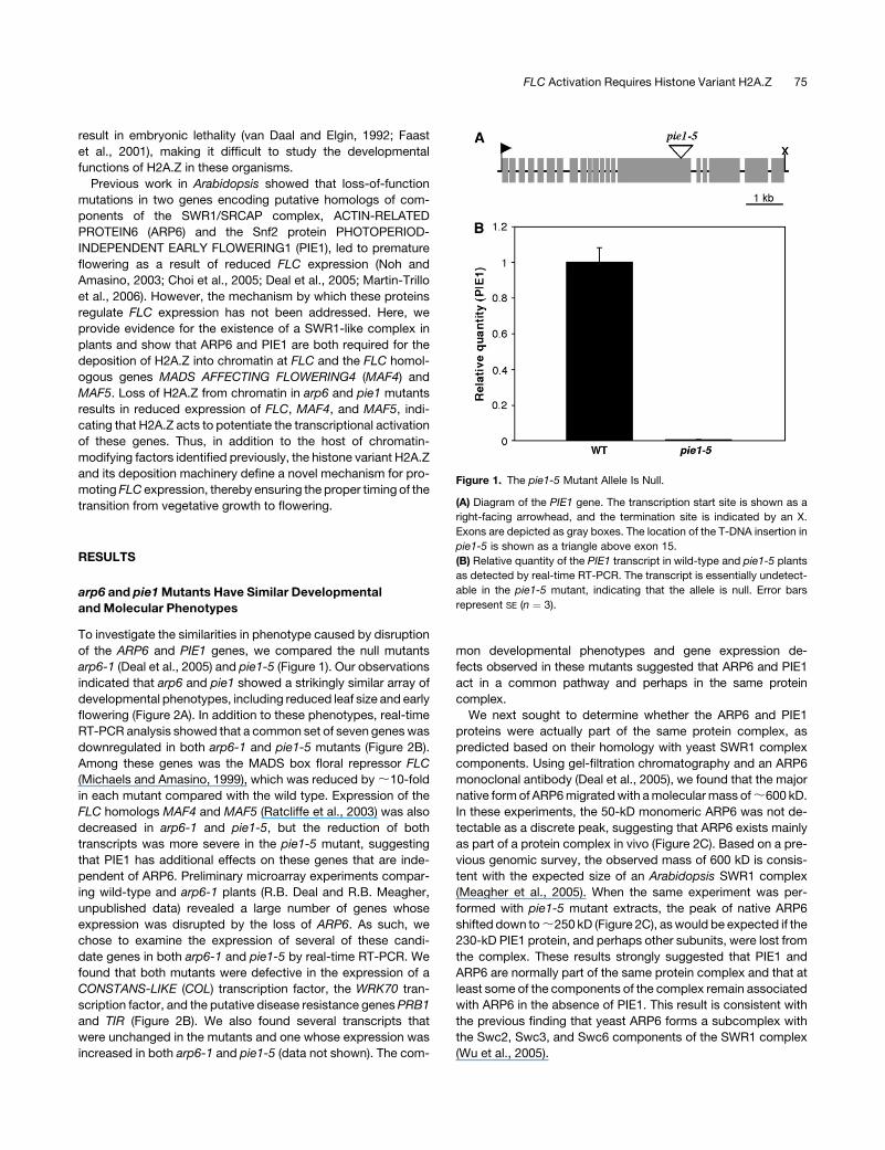
arp6-1 (Deal et al., 2005) and pie1-5 (Figure 1). Our observations
indicated that arp6 and pie1 showed a strikingly similar array of
developmental phenotypes, including reduced leaf size and early
flowering (Figure 2A). In addition to these phenotypes, real-time
RT-PCR analysis showed that a common set of seven genes was
downregulated in both arp6-1 and pie1-5 mutants (Figure 2B).
Among these genes was the MADS box floral repressor FLC
(Michaels and Amasino, 1999), which was reduced by ;10-fold
in each mutant compared with the wild type. Expression of the
FLC homologs MAF4 and MAF5 (Ratcliffe et al., 2003) was also
decreased in arp6-1 and pie1-5, but the reduction of both
transcripts was more severe in the pie1-5 mutant, suggesting
that PIE1 has additional effects on these genes that are inde-
pendent of ARP6. Preliminary microarray experiments compar-
ing wild-type and arp6-1 plants (R.B. Deal and R.B. Meagher,
unpublished data) revealed a large number of genes whose
expression was disrupted by the loss of ARP6. As such, we
chose to examine the expression of several of these candi-
date genes in both arp6-1 and pie1-5 by real-time RT-PCR. We
found that both mutants were defective in the expression of a
CONSTANS-LIKE (COL) transcription factor, the WRK70 tran-
scription factor, and the putative disease resistance genes PRB1
and TIR (Figure 2B). We also found several transcripts that
were unchanged in the mutants and one whose expression was
increased in both arp6-1 and pie1-5 (data not shown). The com-
mon developmental phenotypes and gene expression de-
fects observed in these mutants suggested that ARP6 and PIE1
act in a common pathway and perhaps in the same protein
complex.
We next sought to determine whether the ARP6 and PIE1
proteins were actually part of the same protein complex, as
predicted based on their homology with yeast SWR1 complex
components. Using gel-filtration chromatography and an ARP6
monoclonal antibody (Deal et al., 2005), we found that the major
native form of ARP6 migrated with a molecular mass of ;600 kD.
In these experiments, the 50-kD monomeric ARP6 was not de-
tectable as a discrete peak, suggesting that ARP6 exists mainly
as part of a protein complex in vivo (Figure 2C). Based on a pre-
vious genomic survey, the observed mass of 600 kD is consis-
tent with the expected size of an Arabidopsis SWR1 complex
(Meagher et al., 2005). When the same experiment was per-
formed with pie1-5 mutant extracts, the peak of native ARP6
shifted down to ;250 kD (Figure 2C), as would be expected if the
230-kD PIE1 protein, and perhaps other subunits, were lost from
the complex. These results strongly suggested that PIE1 and
ARP6 are normally part of the same protein complex and that at
least some of the components of the complex remain associated
with ARP6 in the absence of PIE1. This result is consistent with
the previous finding that yeast ARP6 forms a subcomplex with
the Swc2, Swc3, and Swc6 components of the SWR1 complex
(Wu et al., 2005).
Figure 1. The pie1-5 Mutant Allele Is Null.
(A) Diagram of the PIE1 gene. The transcription start site is shown as a
right-facing arrowhead, and the termination site is indicated by an X.
Exons are depicted as gray boxes. The location of the T-DNA insertion in
pie1-5 is shown as a triangle above exon 15.
(B) Relative quantity of the PIE1 transcript in wild-type and pie1-5 plants
as detected by real-time RT-PCR. The transcript is essentially undetect-
able in the pie1-5 mutant, indicating that the allele is null. Error bars
represent SE (n ¼ 3).
FLC Activation Requires Histone Variant H2A.Z 75

ARP6 Interacts with H2A.Z
The Arabidopsis genome encodes 13 H2A histones that fall into
four distinct phylogenetic classes, each of which is also con-
served in the monocot Oryza sativa. Of these 13 H2As, 4 are
clearly H2A.Z variants, being more closely related to yeast and
metazoan H2A.Z proteins than to other plant H2As (Figure 3A).
Using synthetic peptides as antigens, we raised polyclonal
antibodies that reacted with the relatively conserved N termini
of the Arabidopsis H2A.Z subclass proteins HTA9 and HTA11 but
not with representatives of any of the other three H2A subclasses
(Figures 3B and 3C). Immunoprecipitations performed on plant
protein extracts with the H2A.Z-specific antibodies efficiently
precipitated ARP6, whereas parallel experiments using preim-
mune rabbit serum as primary antibody failed to precipitate
ARP6 (Figure 3D). These results confirmed that ARP6 interacts
with H2A.Z, either directly or indirectly, and further support the
notion that ARP6 is involved in the deposition of H2A.Z into
chromatin.
H2A.Z Is Excluded from Chromocenters and Retained
in Mitotic Chromosomes
To examine the distribution of H2A.Z in chromatin, we performed
immunolocalization on isolated leaf and root cells with the H2A.Z
antibody. We found that H2A.Z was dispersed widely through-
out the nucleus but was conspicuously absent from the chro-
mocenters (Figures 4A to 4C), which are composed of highly
condensed heterochromatin containing centromeric and peri-
centromeric repeats (Fransz et al., 2002). This finding indicated
that H2A.Z is found mainly in euchromatic regions and is likely
responsible for regulating the expression of many genes. The
incorporation pattern observed is in contrast with that of mam-
malian and Drosophila H2A.Zs, which accumulate in both hetero-
chromatic and euchromatic chromatin (Leach et al., 2000;
Rangasamy et al., 2003). In dividing cells, we observed H2A.Z
throughout the condensed chromosomes (Figures 4D to 4F),
indicating that this variant remains in the chromatin through
mitosis and therefore may serve as a stable epigenetic mark.
From these data, we cannot yet discern whether H2A.Z is simply
inherited through DNA replication or is reincorporated after
replication, before mitosis.
ARP6 and PIE1 Are Required for H2A.Z Deposition
We next sought to examine the possibility that ARP6 and PIE1
were involved in the deposition of H2A.Z at the FLC gene,
because FLC expression was greatly reduced in both arp6-1 and
pie1-5 (Figure 2B). Chromatin immunoprecipitation (ChIP) using
the Arabidopsis H2A.Z antibody was used to examine H2A.Z
abundance across the FLC gene in wild-type and mutant plants.
In wild-type plants, H2A.Z was predominantly enriched in two
discrete domains, one at each end of the transcribed region, with
relatively low H2A.Z levels between these two domains (Figures
5A and 5E). By contrast, the arp6-1 and pie1-5 mutants showed
very little H2A.Z enrichment across the FLC gene, and the pattern
of enrichment was nearly indistinguishable between the two
mutants (Figure 5A).
ChIP experiments examining the MAF4 and MAF5 genes in
wild-type plants revealed a pattern of H2A.Z distribution similar
to that found at FLC, with the highest levels near the beginning
and end of each transcribed region (Figures 5B and 5F). In the
case of MAF4, we observed only a small decrease in H2A.Z over
the middle of the transcribed region, whereas H2A.Z levels were
Figure 2. arp6 and pie1 Mutants Exhibit Similar Phenotypes.
(A) Twenty-day-old wild-type, arp6-1, and pie1-5 plants grown under
long-day conditions.
(B) Real-time RT-PCR data showing relative expression of the indicated
genes in wild-type, arp6-1, and pie1-5 plants. Average relative quantities
6 SE (n ¼ 3) are shown for each sample.
(C) Protein gel blots of even-numbered gel filtration fractions from a
Sephacryl S-300 column run with extract from wild-type (top panel) or
pie1-5 (bottom panel) plants. Blots were probed with an ARP6 mono-
clonal antibody. The input lanes in each blot were loaded with ;25 mg of
the unfractionated protein extract from each genotype. Asterisks indi-
cate the ARP6 peak fractions, and calibrated molecular masses are given
below the blots.
76 The Plant Cell

quite low in the middle of the MAF5 gene (Figures 5B and 5F).
Again, in contrast with the wild type, there was little or no H2A.Z
accumulation across the MAF4 and MAF5 genes in arp6-1 and
pie1-5. Control ChIP experiments using a histone H2B antibody,
which should react with all nucleosomes, showed no significant
difference in overall H2B distribution between the wild type and
arp6-1 on FLC, MAF4, or MAF5 (Figures 5C and 5D). Thus, the
observed differences in H2A.Z enrichment on these genes were
not simply attributable to gross differences in nucleosome oc-
cupancy among genotypes. These results indicated that ARP6
and PIE1 were both required for the deposition of H2A.Z at
the three genes examined, supporting the existence of a SWR1/
SRCAP-like complex in Arabidopsis. In addition, H2A.Z is
normally distributed across the FLC and MAF genes with the
highest levels at the beginning and end of the transcribed
regions, and loss of H2A.Z from chromatin in the mutants is
correlated with decreased gene expression and early flowering.
Thus, Arabidopsis H2A.Z is required for high-level expression of
the FLC, MAF4, and MAF5 genes, suggesting that it plays a role
similar to yeast H2A.Z in promoting transcription.
H2A.Z Occupancy Is Inversely Correlated with FLC
Transcript Levels
To gain insight into the role of H2A.Z in promoting the high-level
expression of FLC, we examined H2A.Z distribution in plant
samples with widely different FLC transcript levels. H2A.Z ac-
cumulation on FLC was measured in wild-type cauline leaves,
10-d-old wild-type shoots, and 10-d-old shoots carrying the
strong FLC activator FRIGIDA (FRI) (Lee and Amasino, 1995).
Among these samples, the FRI-expressing line showed the
highest levels of FLC expression, the wild type was intermediate,
Figure 3. H2A Phylogeny and H2A.Z-Specific Antibodies.
(A) Neighbor-joining protein sequence phylogeny showing H2A proteins from Arabidopsis (At), rice (Os), Drosophila (Dm), human (Hs), and
Saccharomyces (Sc). Bootstrap values are shown on the branch points of the tree. The H2A.Z clade is indicated by a vertical bar at right, and asterisks
indicate proteins used in (B) and (C).
(B) Coomassie blue–stained SDS-PAGE gel showing purified recombinant Arabidopsis H2A histones. The H2A.Z variants are indicated by a horizontal
bar above the protein names. Molecular masses are shown at right.
(C) Protein gel blot of a gel loaded as in (B), probed with the H2A.Z-specific antibody. Molecular masses are shown at right. The polyclonal antibody
(pAb) recognized HTA9 and HTA11 and was predicted to react with HTA8, because it is nearly identical to HTA11 in the N-terminal region. The HTA4
protein is highly divergent from the other H2A.Zs at the N terminus and is not expected to be recognized by the antibody.
(D) Protein gel blot of immunoprecipitates probed with an ARP6 monoclonal antibody (mAb). The input sample was 5% of the total protein used in each
immunoprecipitation. The antibody used for immunoprecipitation is indicated above the blot: either the H2A.Z antibody or preimmune serum (PI) from
the same rabbit.
FLC Activation Requires Histone Variant H2A.Z 77

and cauline leaves had a 10-fold lower level of FLC than did the
FRI line (Figure 6A). Interestingly, the spatial distribution of H2A.Z
on FLC was the same in each sample, and the overall levels of
H2A.Z in each were also surprisingly similar, showing only minor
differences except at the 59 and 39 ends of the gene, where an
inverse correlation between transcript level and H2A.Z occu-
pancy was observed (Figures 6B and 6D). This correlation was
most clear at the 39 end of the gene.
Because nucleosome occupancy on FLC might have differed
among these samples, we performed parallel ChIP experiments
with an H2B antibody to quantify total nucleosome distribution,
allowing us to measure H2A.Z enrichment relative to that of H2B.
We found that the H2A.Z:H2B ratio across the FLC gene was also
nearly indistinguishable among the three samples at most sites
tested, but the H2A.Z:H2B ratio at the 59 and 39 ends of the gene
still showed an inverse correlation with FLC transcript levels
(Figures 6C and 6D). Thus, the higher the transcript level, the less
H2A.Z was present at the 59 and 39 ends of the gene, similar to the
trend observed previously for yeast H2A.Z at the 59 ends of genes
(Guillemette et al., 2005; Zhang et al., 2005). Collectively, our
results indicate that H2A.Z is required for the high-level expres-
sion of FLC but that its presence does not directly induce
transcriptional activation, suggesting that this variant poises
the gene in a state competent for activation by other factors.
DISCUSSION
Previous studies of Arabidopsis ARP6 and PIE1 revealed that
these proteins were both involved in regulating multiple devel-
opmental processes, including leaf development, inflorescence
and flower development, and repression of the transition to
flowering. In the case of flowering time control, both ARP6 and
PIE1 were shown to be required for high-level expression of the
floral repressor gene FLC (Noh and Amasino, 2003; Choi et al.,
2005; Deal et al., 2005; Martin-Trillo et al., 2006), indicating that
these proteins were likely involved in transcriptional regulation.
Concurrent with the elucidation of ARP6 and PIE1 function in
Arabidopsis, several groups discovered that in Saccharomyces
cerevisiae, ARP6 was a component of the SWR1 chromatin-
remodeling complex (Krogan et al., 2003; Kobor et al., 2004;
Mizuguchi et al., 2004). This complex was shown to have the
novel activity of replacing histone H2A with the variant H2A.Z in
particular nucleosomes, thus functionally specializing the sur-
rounding chromatin and in many cases potentiating the expres-
sion of nearby genes. Given this information, we considered the
possibility that such a complex might also exist in Arabidopsis.
A comparison of the yeast SWR1 ATPase subunit with all
Arabidopsis proteins indicated that of the 42 Swi2/Snf2 family
proteins encoded by the Arabidopsis genome, PIE1 was the
most closely related to SWR1. In addition to ARP6 and PIE1, the
Arabidopsis genome also encodes clear orthologs of most other
SWR1 complex components (Meagher et al., 2005) as well as
multiple H2A.Z isovariants (Figure 3). Could the developmental
functions of Arabidopsis ARP6 and PIE1 be attributable to their
activity within a plant SWR1-like complex? In yeast, the SWR1
complex deposits the histone H2A.Z variant into euchromatic
regions near telomeres (Krogan et al., 2003; Kobor et al., 2004;
Mizuguchi et al., 2004) and in the promoters of many euchro-
matic genes (Guillemette et al., 2005; Li et al., 2005; Raisner
et al., 2005; Zhang et al., 2005), thereby preventing the spread
of silent heterochromatin into euchromatic regions (Meneghini
et al., 2003) and promoting transcriptional activation, respec-
tively. Perhaps a plant SWR1-like complex could have an
analogous function of depositing H2A.Z into FLC chromatin,
ensuring the competence of the gene for high-level expression
and thus allowing the flowering program to be repressed and
vegetative growth to continue. In this investigation, we explored
the hypothesis that the contribution of ARP6 and PIE1 to the
developmental program in Arabidopsis was a manifestation of
their role in depositing H2A.Z into chromatin and thus regu-
lating the expression of multiple developmentally important
genes.
A direct comparison of arp6 and pie1 indicated that these
mutants shared many developmental and molecular pheno-
types. Both mutations caused aberrations in leaf development
and early flowering and resulted in the misregulation of a com-
mon set of genes, including the flowering regulators FLC, MAF4,
and MAF5. In addition, we found that ARP6 was a component of
a high-molecular-mass protein complex and that the size of this
complex was reduced dramatically in the absence of PIE1,
suggesting that ARP6 and PIE1 were indeed part of the same
protein complex (Figure 2). Furthermore, polyclonal antibodies
that reacted with at least two of the four Arabidopsis H2A.Z
proteins efficiently immunoprecipitated ARP6 from plant ex-
tracts, confirming an interaction between ARP6 and H2A.Z
(Figure 3). Collectively, these results suggested that ARP6 and
PIE1 were part of a plant SWR1-like complex.
Figure 4. H2A.Z Localizes to Euchromatic Regions but Not to Hetero-
chromatic Chromocenters.
(A) Isolated leaf cell nucleus probed with the H2A.Z antibody.
(B) The 49,6-diamidino-2-phenylindole (DAPI) channel image of the
nucleus shown in (A). Chromocenters appear as densely stained spots
throughout the nucleus.
(C) Merge of images shown in (A) and (B).
(D) Anaphase-stage root cell probed with the H2A.Z antibody.
(E) DAPI channel image of the cell shown in (D).
(F) Merge of images shown in (D) and (E).
78 The Plant Cell

ChIP was used to determine whether ARP6 and PIE1 were
involved in the deposition of H2A.Z into chromatin at the FLC and
MAF genes, because the expression of these genes was re-
duced in both arp6 and pie1 mutants. We found that H2A.Z was
normally enriched in two discrete domains on each of these
genes, one near the transcription start site and another near
the end of the gene, and that very little H2A.Z accumulated in
the chromatin of arp6 or pie1 mutants (Figure 5). These results
indicated that ARP6 and PIE1 were indeed necessary for the
deposition of H2A.Z into chromatin at the three loci examined.
However, H2A.Z was still detectable at several sites on the FLC
and MAF genes in arp6-1and pie1-5, indicating that the variant
can be incorporated into chromatin at very low levels indepen-
dently of ARP6 and PIE1. Loss of H2A.Z from chromatin in arp6-1
and pie1-5 was correlated with a reduced expression of FLC,
MAF4, and MAF5, indicating that H2A.Z is normally required to
promote the expression of each of these genes. This observation
suggested that, like yeast H2A.Z, the Arabidopsis variant can
also potentiate transcriptional activation. ChIP analysis of the
PRB1, COL, WRKY70, and TIR genes, whose transcript levels
were also reduced in the mutants (Figure 2B), indicated that even
in wild-type plants these loci did not accumulate significant
amounts of H2A.Z (data not shown); thus, these genes were likely
to be indirectly regulated by ARP6 and PIE1. This finding indi-
cated that not all Arabidopsis genes require H2A.Z for high-level
expression and that many of the transcriptional defects in arp6
Figure 5. ARP6 and PIE1 Are Required for the Deposition of H2A.Z at FLC, MAF4, and MAF5.
(A) Enrichment of H2A.Z on the FLC gene in the wild type and mutants as measured by ChIP with the H2A.Z antibody. The graph shows average fold
enrichment 6 SE as measured by real-time PCR.
(B) Enrichment of H2A.Z on the MAF4 and MAF5 genes in the wild type and mutants as measured by ChIP and real-time PCR.
(C) Enrichment of H2B on the FLC gene in the wild type and arp6-1.
(D) Enrichment of H2B on the MAF4 and MAF5 genes in the wild type and arp6-1. For each ChIP experiment ([A] to [D]), n ¼ 3.
(E) Diagram of the FLC gene with exons indicated as gray boxes. The transcription start site is shown as an arrowhead, and the termination site is shown
as an X. PCR primer sets are shown as black boxes below the diagram. Primer set numbers correspond to the numbers on the x axis of the graphs in (A)
and (C).
(F) Diagram of the MAF4 and MAF5 genes and locations of PCR primer sets, depicted as in (E). Primer set numbers correspond to the numbers on the
x axis of the graphs in (B) and (D).
FLC Activation Requires Histone Variant H2A.Z 79

and pie1 mutants were likely secondary effects resulting from the
misregulation of a smaller number of primary target genes that do
require H2A.Z for proper expression.
In contrast with the situation in yeast, in which H2A.Z is
normally found mainly in nucleosomes around the transcription
start site (Guillemette et al., 2005; Li et al., 2005; Raisner et al.,
2005; Zhang et al., 2005), Arabidopsis H2A.Z occupies regions
near both the transcription start and termination sites on the
three genes examined. Previous studies have shown a role for
H2A.Z in recruiting RNA polymerase (Adam et al., 2001) and acting
in concert with nucleosome-remodeling complexes (Santisteban
et al., 2000) to initiate transcription. However, the data presented
here suggest that in Arabidopsis this histone variant also has
a function beyond the 59 end of genes, perhaps in facilitating
transcript elongation and/or termination. The tendency of H2A.Z
to destabilize nucleosomes that contain it (Suto et al., 2000;
Abbott et al., 2001; Zhang et al., 2005) may facilitate nucleosome
remodeling and thus polymerase passage and transcript elon-
gation. In addition, multiple lines of evidence now indicate that
chromatin-remodeling enzymes regulate all phases of the tran-
scription cycle, including termination (Alen et al., 2002; Morillon
et al., 2003). Thus, Arabidopsis H2A.Z may facilitate remodeling
at both ends of the gene to effect the proper initiation and
termination of transcription. The occupancy of H2A.Z at sites
beyond the transcription initiation region has also been observed
for several other Arabidopsis genes (A.P. Smith, R.B. Deal, and
R.B. Meagher, unpublished data), suggesting that this phe-
nomenon is not specific to the MADS box transcription factor
genes.
As a means of gaining insight into the role of H2A.Z in
promoting transcriptional activation, we examined the relation-
ship between FLC transcript levels and H2A.Z abundance on the
FLC gene. H2A.Z occupancy on FLC was measured in three
different tissues with FLC expression levels spanning a 10-fold
range, from very high to very low. In each sample, we observed
essentially the same spatial distribution of H2A.Z across the
gene, with a peak at the beginning and end of the transcribed
region. However, there was an inverse correlation between FLC
expression levels and H2A.Z abundance on the gene, such that
the higher the transcript level, the less H2A.Z was present on the
Figure 6. Relationship between FLC Expression Levels and H2A.Z
Abundance on the Gene.
(A) Real-time RT-PCR results showing the relative FLC level in each
genotype or tissue. FRI indicates a Columbia line carrying the strong FRI
allele from the Sf-2 ecotype (Lee and Amasino, 1995), WT indicates
Columbia wild type, and CL indicates cauline leaf. The graph shows
average relative quantities 6 SE (n ¼ 3).
(B) Enrichment of H2A.Z on the FLC gene in the indicated samples as
measured by ChIP and real-time PCR. The graph shows average fold
enrichment 6 SE.
(C) ChIP was performed on the indicated samples using either an H2A.Z
antibody or an H2B antibody, and data are reported as H2A.Z enrich-
ment/H2B enrichment to correct the H2A.Z levels for total nucleosome
occupancy. The graph shows average fold enrichment 6 SE. For each
ChIP experiment ([B] and [C]), n ¼ 3.
(D) Diagram of the FLC gene with exons indicated as gray boxes. The
transcription start site is shown as an arrowhead, and the termination site
is shown as an X. PCR primer sets are shown as black boxes below the
diagram. Primer set numbers correspond to the numbers on the x axis of
the graphs in (B) and (C).
80 The Plant Cell

gene (Figure 6). This inverse relationship between H2A.Z occu-
pancy and gene expression may result from a shift in the balance
between ARP6/PIE1-mediated deposition of H2A.Z and loss of
H2A.Z as a result of nucleosome disruption by RNA polymerase.
During high-level transcription, the rate of transcription-induced
depletion of H2A.Z might exceed the rate of replacement by
ARP6/PIE1, resulting in an inverse correlation between transcript
level and H2A.Z occupancy. In any case, these results clearly
indicated that there was no positive correlation between FLC
expression level and H2A.Z abundance on the gene, even over a
10-fold range of FLC transcript levels. This finding suggests that
the H2A.Z variant serves to poise the gene in a state competent
for activation by other factors, rather than activating transcription
directly. This may reflect the ability of H2A.Z to facilitate nucle-
osome remodeling (Santisteban et al., 2000) and/or to recruit the
transcription machinery (Adam et al., 2001) or other activators to
allow high-level transcription under the appropriate conditions.
Thus, in the absence of H2A.Z in arp6 and pie1 mutants, FLC
levels remain low even in the presence of strong activators such
as FRI (Noh and Amasino, 2003; Choi et al., 2005; Deal et al.,
2005; Martin-Trillo et al., 2006), resulting in early flowering.
In addition to ChIP studies on the distribution of H2A.Z across
single genes, we also used our H2A.Z antibodies to examine the
nuclear localization of this histone variant during interphase and
mitosis. In contrast with mammalian and Drosophila H2A.Zs,
which accumulate in both heterochromatic and euchromatic
chromatin (Leach et al., 2000; Rangasamy et al., 2003), we found
that Arabidopsis H2A.Z was widely distributed throughout eu-
chromatin but was excluded from the heterochromatic chromo-
centers (Figure 4). This finding indicated that H2A.Z is likely
responsible for regulating the expression of many genes in
euchromatin but is not likely involved in constitutive heterochro-
matin formation or maintenance in Arabidopsis. The observation
that H2A.Z is incorporated into euchromatin during interphase
and remains in chromatin through mitosis suggests that it may
serve an epigenetic memory function by marking actively tran-
scribed genes and providing competence for the reactivation of
silenced genes. Such a function, coupled with the ability of
H2A.Z to promote transcriptional activation, is likely to be im-
portant in the establishment and maintenance of cell fate during
development.
In conclusion, it appears that ARP6 and PIE1 act together to
control multiple developmental processes, most likely by regu-
lating the expression of a large number of genes through the
incorporation of H2A.Z into chromatin. Our results support the
notion that the H2A.Z deposition machinery is conserved in
plants as it is in yeast and metazoans, requiring both ARP6 and a
Snf2 protein of the SWR1/SRCAP class. Furthermore, the loss of
H2A.Z from chromatin in arp6-1 and pie1-5 mutants correlates
with decreased expression of FLC, MAF4, and MAF5, indicating
that H2A.Z deposition is essential for the full transcriptional
activation of these genes. Thus, H2A.Z can act to potentiate the
transcriptional activation of Arabidopsis genes, similar to its role
in yeast. In terms of flowering time control, H2A.Z and its
deposition machinery now define a novel mechanism for pro-
moting FLC expression and thus repressing the transition from
vegetative to reproductive development in Arabidopsis. During
vegetative growth, H2A.Z allows high-level FLC expression and
floral repression, yet it remains associated with the inactive gene
after flowering, perhaps to poise the gene for reactivation and
reestablishment of the vegetative growth program in the next
generation.
Future work in this area will be focused on large-scale
approaches to identifying all of the genes that require H2A.Z
for proper expression and elucidating the DNA sequence deter-
minants and other factors that promote the deposition of H2A.Z
into chromatin at particular loci. Insight into the mechanism by
which H2A.Z promotes transcriptional activation could be
gleaned from protein–protein interaction studies and the identi-
fication of second-site mutations that suppress the early flower-
ing of arp6 and pie1 mutants. Because ARP6 and PIE1 act as a
hub through which the expression of many genes is controlled,
identification of the full set of genes misregulated in arp6 and pie1
mutants should allow the assignment of many currently anony-
mous genes to particular developmental pathways.
METHODS
Plants and Growth Conditions
Arabidopsis thaliana plants were of the Columbia ecotype and were
germinated by sowing on wet soil and storing at 48C for 2 d before moving
to the growth chamber. Plants were grown at 228C under 16 h of light per
day. The pie1-5 mutation is a T-DNA insertion allele from the Salk Institute
(SALK_011204). The T-DNA insertion is in exon 15 and is null based on
RNA levels (Figure 1). The arp6-1 allele was described previously (Deal
et al., 2005).
Gel Filtration Chromatography
Gel filtration was performed on a Sephacryl S-300HR column (Amersham
Biosciences) in a buffer consisting of 20 mM Tris, pH 7.5, 200 mM NaCl,
10 mM MgCl2, and 10% (v/v) glycerol. The column was calibrated with a
mixture of standard proteins ranging in size from 670 to 60 kD. Plant ex-
tracts were prepared by grinding a mixture of leaf and flower tissue in 2
volumes of the gel filtration buffer supplemented with 1 mM b-mercapto-
ethanol and Complete protease inhibitors (Roche). After grinding, sam-
ples were cleared by centrifugation and filtration. The column was run at
room temperature with a flow rate of 0.25 mL/min, and 0.5-mL fractions
were collected. Two independent biological replicates of the gel filtration
experiment were done, and both gave very similar results.
Phylogenetic Analysis
The following Arabidopsis histone H2A protein sequences were used for
phylogenetic analysis: HTA1 (At5g54640), HTA2 (At4g27230), HTA3
(At1g54690), HTA4 (At4g13570), HTA5 (At1g08880), HTA6 (At5g59870),
HTA7 (At5g27670), HTA8 (At2g38810), HTA9 (At1g52740), HTA10
(At1g51060), HTA11 (At3g54560), HTA12 (At5g02560), and HTA13
(At3g20670). Other H2A proteins used for the analysis were from Oryza
sativa: HTA701 (Os01g31800), HTA702 (Os08g33100), HTA703
(Os12g25120), HTA704 (Os03g51200), HTA705 (Os10g28230), HTA706
(Os05g38640), HTA707 (Os05g02300), HTA708 (Os07g36140), HTA709
(Os07g36130), HTA710 (Os03g17100), HTA711 (Os12g34510), HTA712
(Os03g06670), and HTA713 (Os03g53190); Homo sapiens: Hs H2A.X
(NP_002096) and Hs H2A.Z (NP_002097); Drosophila melanogaster:
Dm H2Av (NP_524519); and Saccharomyces cerevisiae: Sc Htz1
(NP_014631). Sequences were aligned with ClustalW 1.82 (Higgins and
Sharp, 1988) using default settings (protein gap open penalty ¼ 10,
FLC Activation Requires Histone Variant H2A.Z 81

protein gap extension penalty ¼ 0.2, protein matrix ¼ gonnet, protein
ENDGAP¼ �1, protein GAPDIST ¼ 4), and phylogenies were constructed
with PAUP 4.0 (Swofford, 2003) using the neighbor-joining method. Ties
were broken randomly using the initial seed function. Bootstrap support
values were based on 1000 replicates using a full heuristic search.
Antibody Preparation
Polyclonal antibodies specific to Arabidopsis H2A.Zs were produced
in rabbits by injecting peptides representing the N termini of HTA9
and HTA11. The peptide sequences N-SGKGAKGLIMGKPSGSDKDKD-
KKKPIT-C (HTA9) and N-AGKGGKGLVAAKTMAANKDKDKDKKKPIS-C
(HTA11) were synthesized as fourfold multiple antigenic peptides. The
primary injection and three subsequent boosts were done with 250 mg of
each peptide. Antibodies were affinity-purified on resin to which the
H2A.Z peptides had been coupled.
Immunoprecipitation
Plant extracts were prepared by grinding a mixture of leaf and flower
tissue in 2 volumes of immunoprecipitation buffer (50 mM Tris, pH 7.5,
150 mM NaCl, 10 mM MgCl2, 0.1% Nonidet P-40, 1 mM b-mercapto-
ethanol, and Roche Complete protease inhibitors) followed by centrifu-
gation and filtration to clear the extracts. Extracts were then divided into
aliquots in multiple tubes (900 mL each), and 40 mL of protein A–agarose
beads (Roche) was added to each tube. Tubes were rocked at 48C for 1 h
to preclear the extracts, and then the beads were removed by centrifu-
gation. Antibodies or preimmune serum were then added, and tubes were
kept on ice for 1 h to allow antibody binding. H2A.Z antibodies were used
at a 1:100 dilution, and an equivalent amount of preimmune serum was
used based on antibody concentration. Antibody–antigen complexes
were captured by adding 50 mL of protein A–agarose beads and rocking
for 1 h at 48C. Beads were collected by centrifugation and washed three
times for 20 min in immunoprecipitation buffer at 48C.
Protein Gel Blotting and Immunofluorescence Microscopy
Both of these techniques were performed as described previously (Deal
et al., 2005).
RT-PCR
RNA was isolated from 10-d-old seedlings (minus roots) using the
RNeasy plant mini kit (Qiagen). Before reverse transcription, RNA was
treated with RQ1 RNase-free DNase I (Promega) according to the
manufacturer’s instructions. Three micrograms of each RNA was
reverse-transcribed with the Superscript III first-strand synthesis kit
(Invitrogen). Real-time PCR was used to analyze the cDNA populations
using 18S RNA as an endogenous control. The genes assayed by this
method were FLC (At5g10140), MAF4 (At5g65070), MAF5 (At5g65080),
PRB1 (At2g14580), COL (At5g24930), WRKY70 (At3g56400), and TIR
(At1g72930).
ChIP
ChIP was performed as described previously (Gendrel et al., 2005). For
each experiment, 1 to 2 g of 10-d-old seedlings (minus roots) was used.
For experiments with cauline leaves, 1 to 2 g of leaves from 30-d-old
plants was used. The H2A.Z antibody was used at a dilution of 1:100, and
the H2B antibody (ab1790; Abcam) was used at a dilution of 1:150. DNA
was analyzed by real-time PCR with the ACT2 39 untranslated region
sequence as the endogenous control for all ChIP experiments. The
relative quantity value calculated by the 2�ddCt method in ChIP experi-
ments is reported as fold enrichment.
Real-Time PCR
Real-time PCR was performed on an Applied Biosystems 7500 real-time
PCR system using SYBR Green detection chemistry (Applied Biosys-
tems). The 2�ddCt method (Livak and Schmittgen, 2001) of relative
quantification was used in all experiments. The data presented for both
RT-PCR and ChIP experiments are average relative quantities from at
least two biological replicates 6 SE. All primer sequences are available
upon request.
Accession Numbers
The accession numbers for the ARP6 and PIE1 genes are At3g33520 and
At3g12810, respectively.
Supplemental Data
The following material is available in the online version of this article.
Supplemental Figure 1. Histone H2A Alignments Used for Phyloge-
netic Analysis.
ACKNOWLEDGMENTS
We thank Michael Bender and members of the Meagher laboratory for
critical reading and discussion of the manuscript. This work was
supported by funding from the National Institutes of Health (Grant
GM-36397-18) to R.B.M. and a National Institutes of Health training
grant (Grant GM-07103-29) to R.B.D. C.N.T. was supported by funding
from the National Science Foundation (Grant 0421671).
Received October 23, 2006; revised November 27, 2006; accepted
December 15, 2006; published January 12, 2007.
REFERENCES
Abbott, D.W., Ivanova, V.S., Wang, X., Bonner, W.M., and Ausio, J.
(2001). Characterization of the stability and folding of H2A.Z chromatin
particles: Implications for transcriptional activation. J. Biol. Chem.
276: 41945–41949.
Adam, M., Robert, F., Larochelle, M., and Gaudreau, L. (2001). H2A.Z
is required for global chromatin integrity and for recruitment of RNA
polymerase II under specific conditions. Mol. Cell. Biol. 21: 6270–
6279.
Alen, C., Kent, N.A., Jones, H.S., O’Sullivan, J., Aranda, A., and
Proudfoot, N.J. (2002). A role for chromatin remodeling in transcrip-
tional termination by RNA polymerase II. Mol. Cell 10: 1441–1452.
Bruce, K., Myers, F.A., Mantouvalou, E., Lefevre, P., Greaves, I.,
Bonifer, C., Tremethick, D.J., Thorne, A.W., and Crane-Robinson,
C. (2005). The replacement histone H2A.Z in a hyperacetylated form is
a feature of active genes in the chicken. Nucleic Acids Res. 33: 5633–
5639.
Choi, K., Kim, S., Kim, S.Y., Kim, M., Hyun, Y., Lee, H., Choe, S., Kim,
S.G., Michaels, S., and Lee, I. (2005). SUPPRESSOR OF FRIGIDA3
encodes a nuclear ACTIN-RELATED PROTEIN6 required for floral
repression in Arabidopsis. Plant Cell 17: 2647–2660.
Deal, R.B., Kandasamy, M.K., McKinney, E.C., and Meagher, R.B.
(2005). The nuclear actin-related protein ARP6 is a pleiotropic devel-
opmental regulator required for the maintenance of FLOWERING
LOCUS C expression and repression of flowering in Arabidopsis.
Plant Cell 17: 2633–2646.
82 The Plant Cell

Faast, R., Thonglairoam, V., Schulz, T.C., Beall, J., Wells, J.R.,
Taylor, H., Matthaei, K., Rathjen, P.D., Tremethick, D.J., and
Lyons, I. (2001). Histone variant H2A.Z is required for early mamma-
lian development. Curr. Biol. 11: 1183–1187.
Farris, S.D., Rubio, E.D., Moon, J.J., Gombert, W.M., Nelson, B.H.,
and Krumm, A. (2005). Transcription-induced chromatin remodeling
at the c-myc gene involves the local exchange of histone H2A. J. Biol.
Chem. 280: 25298–25303.
Fransz, P., De Jong, J.H., Lysak, M., Castiglione, M.R., and Schubert,
I. (2002). Interphase chromosomes in Arabidopsis are organized as
well defined chromocenters from which euchromatin loops emanate.
Proc. Natl. Acad. Sci. USA 99: 14584–14589.
Gendrel, A.V., Lippman, Z., Martienssen, R., and Colot, V. (2005).
Profiling histone modification patterns in plants using genomic tiling
microarrays. Nat. Methods 2: 213–218.
Guillemette, B., Bataille, A.R., Gevry, N., Adam, M., Blanchette, M.,
Robert, F., and Gaudreau, L. (2005). Variant histone H2A.Z is globally
localized to the promoters of inactive yeast genes and regulates
nucleosome positioning. PLoS Biol. 3: E384.
He, Y., and Amasino, R.M. (2005). Role of chromatin modification in
flowering-time control. Trends Plant Sci. 10: 30–35.
Henderson, I.R., and Dean, C. (2004). Control of Arabidopsis flowering:
The chill before the bloom. Development 131: 3829–3838.
Higgins, D.G., and Sharp, P.M. (1988). CLUSTAL: A package for
performing multiple sequence alignment on a microcomputer. Gene
73: 237–244.
Kobor, M.S., Venkatasubrahmanyam, S., Meneghini, M.D., Gin, J.W.,
Jennings, J.L., Link,A.J.,Madhani, H.D., and Rine,J. (2004). A protein
complex containing the conserved Swi2/Snf2-related ATPase Swr1p
deposits histone variant H2A.Z into euchromatin. PLoS Biol. 2: E131.
Krogan, N.J., et al. (2003). A Snf2 family ATPase complex required for
recruitment of the histone H2A variant Htz1. Mol. Cell 12: 1565–1576.
Kusch, T., Florens, L., Macdonald, W.H., Swanson, S.K., Glaser,
R.L., Yates, J.R., III, Abmayr, S.M., Washburn, M.P., and Workman,
J.L. (2004). Acetylation by Tip60 is required for selective histone vari-
ant exchange at DNA lesions. Science 306: 2084–2087.
Larochelle, M., and Gaudreau, L. (2003). H2A.Z has a function rem-
iniscent of an activator required for preferential binding to intergenic
DNA. EMBO J. 22: 4512–4522.
Leach, T.J., Mazzeo, M., Chotkowski, H.L., Madigan, J.P., Wotring,
M.G., and Glaser, R.L. (2000). Histone H2A.Z is widely but non-
randomly distributed in chromosomes of Drosophila melanogaster.
J. Biol. Chem. 275: 23267–23272.
Lee, I., and Amasino, R.M. (1995). Effect of vernalization, photoperiod,
and light quality on the flowering phenotype of Arabidopsis plants
containing the FRIGIDA gene. Plant Physiol. 108: 157–162.
Li, B., Pattenden, S.G., Lee, D., Gutierrez, J., Chen, J., Seidel, C.,
Gerton, J., and Workman, J.L. (2005). Preferential occupancy of
histone variant H2AZ at inactive promoters influences local histone
modifications and chromatin remodeling. Proc. Natl. Acad. Sci. USA
102: 18385–18390.
Livak, K.J., and Schmittgen, T.D. (2001). Analysis of relative gene
expression data using real-time quantitative PCR and the 2(-Delta
Delta C(T)). Methods 25: 402–408.
Martin-Trillo, M., Lazaro, A., Poethig, R.S., Gomez-Mena, C., Pineiro,
M.A., Martinez-Zapater, J.M., and Jarillo, J.A. (2006). EARLY IN
SHORT DAYS 1 (ESD1) encodes ACTIN-RELATED PROTEIN 6
(AtARP6), a putative component of chromatin remodelling complexes
that positively regulates FLC accumulation in Arabidopsis. Development
133: 1241–1252.
Meagher, R.B., Deal, R.B., Kandasamy, M.K., and McKinney, E.C.
(2005). Nuclear actin-related proteins as epigenetic regulators of
development. Plant Physiol. 139: 1576–1585.
Meneghini, M.D., Wu, M., and Madhani, H.D. (2003). Conserved
histone variant H2A.Z protects euchromatin from the ectopic spread
of silent heterochromatin. Cell 112: 725–736.
Michaels, S.D., and Amasino, R.M. (1999). FLOWERING LOCUS C
encodes a novel MADS domain protein that acts as a repressor of
flowering. Plant Cell 11: 949–956.
Mizuguchi, G., Shen, X., Landry, J., Wu, W.H., Sen, S., and Wu, C.
(2004). ATP-driven exchange of histone H2AZ variant catalyzed by
SWR1 chromatin remodeling complex. Science 303: 343–348.
Morillon, A., Karabetsou, N., O’Sullivan, J., Kent, N., Proudfoot, N.,
and Mellor, J. (2003). Isw1 chromatin remodeling ATPase coordi-
nates transcription elongation and termination by RNA polymerase II.
Cell 115: 425–435.
Noh, Y.S., and Amasino, R.M. (2003). PIE1, an ISWI family gene, is
required for FLC activation and floral repression in Arabidopsis. Plant
Cell 15: 1671–1682.
Raisner, R.M., Hartley, P.D., Meneghini, M.D., Bao, M.Z., Liu, C.L.,
Schreiber, S.L., Rando, O.J., and Madhani, H.D. (2005). Histone
variant H2A.Z marks the 59 ends of both active and inactive genes in
euchromatin. Cell 123: 233–248.
Rangasamy, D., Berven, L., Ridgway, P., and Tremethick, D.J.
(2003). Pericentric heterochromatin becomes enriched with H2A.Z
during early mammalian development. EMBO J. 22: 1599–1607.
Ratcliffe, O.J., Kumimoto, R.W., Wong, B.J., and Riechmann, J.L.
(2003). Analysis of the Arabidopsis MADS AFFECTING FLOWERING
gene family: MAF2 prevents vernalization by short periods of cold.
Plant Cell 15: 1159–1169.
Reyes, J.C. (2006). Chromatin modifiers that control plant development.
Curr. Opin. Plant Biol. 9: 21–27.
Ruhl, D.D., Jin, J., Cai, Y., Swanson, S., Florens, L., Washburn, M.P.,
Conaway, R.C., Conaway, J.W., and Chrivia, J.C. (2006). Purifica-
tion of a human SRCAP complex that remodels chromatin by incor-
porating the histone variant H2A.Z into nucleosomes. Biochemistry
45: 5671–5677.
Santisteban, M.S., Kalashnikova, T., and Smith, M.M. (2000). Histone
H2A.Z regulates transcription and is partially redundant with nucleo-
some remodeling complexes. Cell 103: 411–422.
Simpson, G.G. (2004). The autonomous pathway: Epigenetic and post-
transcriptional gene regulation in the control of Arabidopsis flowering
time. Curr. Opin. Plant Biol. 7: 570–574.
Simpson, G.G., and Dean, C. (2002). Arabidopsis, the Rosetta stone of
flowering time? Science 296: 285–289.
Sung, S., and Amasino, R.M. (2004). Vernalization and epigenetics:
How plants remember winter. Curr. Opin. Plant Biol. 7: 4–10.
Suto, R.K., Clarkson, M.J., Tremethick, D.J., and Luger, K. (2000).
Crystal structure of a nucleosome core particle containing the variant
histone H2A. Nat. Struct. Biol. 7: 1121–1124.
Swaminathan, J., Baxter, E.M., and Corces, V.G. (2005). The role
of histone H2Av variant replacement and histone H4 acetylation in
the establishment of Drosophila heterochromatin. Genes Dev. 19:
65–76.
Swofford, D.L. (2003). PAUP*. Phylogenetic Analysis Using Parsi-
mony (*and Other Methods), Version 4. (Sunderland, MA: Sinauer
Associates).
van Daal, A., and Elgin, S.C. (1992). A histone variant, H2AvD, is
essential in Drosophila melanogaster. Mol. Biol. Cell 3: 593–602.
Wu, W.H., Alami, S., Luk, E., Wu, C.H., Sen, S., Mizuguchi, G., Wei,
D., and Wu, C. (2005). Swc2 is a widely conserved H2AZ-binding
module essential for ATP-dependent histone exchange. Nat. Struct.
Mol. Biol. 12: 1064–1071.
Zhang, H., Roberts, D.N., and Cairns, B.R. (2005). Genome-wide
dynamics of Htz1, a histone H2A variant that poises repressed/basal
promoters for activation through histone loss. Cell 123: 219–231.
FLC Activation Requires Histone Variant H2A.Z 83
Copyright © 2022 FDOKUMEN