Repair of rabbit femur defects with organic bovine bone cancellous block or cortical granules
10
The International Journal of Oral & Maxillofacial Implants 1167 B one defects and inadequate bone volume are com- mon problems in dental and orthopedic surgery. In these situations, the use of bone grafts is indicated to promote healing and regeneration. Different bone re- placement materials have been developed for applica- tion in bone defects. The xenogeneic bone graft is an organic or inorganic material obtained from bone of different animal species, often from bovines, because of their greater availability. The use of demineralized bone matrix (DBM) to re- pair bone defects was originally reported in 1889 by Senn. 1 Following this early report, there was little clini- cal interest in DBM until 1965, when Urist discovered its osteoinductive properties. The pioneering studies of Urist, 2 Urist et al, 3 and Reddi et al 4,5 have resulted in an extensive use of allogeneic demineralized bone as a graft material in surgery practices. Organic bovine bone materials also have the advantage of an inherent osteoinductive capacity. 6,7 Many studies have reported positive results con- cerning bone repair using xenogeneic bone graft materials. Torricelli et al 8 demonstrated that xenoge- neic DBM seems to be effective in bone defect healing, since it increases mineralized tissue volume. Bovine DBM has provided satisfactory bone healing when used to fill created defects in rat mandible 9 and rabbit radius 10,11 and for maxillary sinus augmentation in rab- bits. 12 When used to fill periodontal intrabony defects in humans, it improved healing outcomes, promoting reduction of probing depths, resolution of osseous defects, and gains in clinical attachment. 13 Mai et al 14 concluded that a hydroxyapatite–bovine collagen ce- ment composite showed good resorption and bone 1 PhD in Endodontics, Bauru Dental School, University of São Paulo, Bauru, SP, Brazil. 2 Professor, Department of Endodontics, Bauru Dental School, University of São Paulo, Bauru, SP, Brazil. 3 PhD in Biologic Sciences, Bauru Dental School, University of São Paulo, Bauru, SP, Brazil. 4 Associate Professor, Department of Cell and Molecular Biology, Institute of Biology, Fluminense Federal University, Niterói, RJ, Brazil. 5 Professor, Department of Biologic Sciences, Bauru Dental School, University of São Paulo, Bauru, SP, Brazil. Correspondence to: Dr Luciana Viti Betti, Al. Octávio Pinheiro Brisola 9-75 Bauru-SP Brasil 17012-901. Fax: +55-14-3224-2788. Email: [email protected] Repair of Rabbit Femur Defects with Organic Bovine Bone Cancellous Block or Cortical Granules Luciana Viti Betti, DDS, PhD 1 /Clovis Monteiro Bramante, DDS, PhD 2 /Tânia Mary Cestari, BSc, PhD 3 / José Mauro Granjeiro, DDS, PhD 4 /Roberto Brandão Garcia, DDS, PhD 2 / Ivaldo Gomes de Moraes, DDS, PhD 2 /Norberti Bernardineli, DDS, PhD 2 /Rumio Taga, DDS, PhD 5 Purpose: To investigate the osseous repair in epiphyseal rabbit femur defects treated with organic bovine bone (OBB) cancellous block or OBB cortical granules. Materials and Methods: Forty-eight trephined defects were made bilaterally in 30 rabbits. In 18 animals, the defects were filled with OBB cancellous block (experimental group 1) or OBB cortical granules (experimental group 2), and in 12 animals (control group), the defects on both sides were filled only with blood clot. After 30, 90, and 180 days, the femur epiphyses were collected, fixed, radiographed both digitally and conventionally, and then processed for histologic analyses. The newly formed cortical bone was measured in the histologic sections using a digital image analysis system. Results: Histologically, both experimental groups showed total or partial closure of the defects with woven bone at 30 days, whereas in the control sites, a marked migration of connective tissue into the defect was apparent. At 90 and 180 days, a similar pattern of repair was observed in the experimental groups and the control group. Quantitatively, however, no significant differences between groups ( P > .05) were observed throughout the experimental period. A significant reduction in newly formed cortical bone occurred at 90 and 180 days ( P < .05) in all groups. Radiographically, no statistically significant differences were seen among the groups ( P > .05). Conclusions: The study showed minor descriptive differences at the first healing period that were not evident at later periods and that were not shown through histomorphometry and radiographic analysis. The only significant results were related to time. INT J ORAL MAXILLOFAC IMPLANTS 2011;26:1167–1175 Key words: bone repair, demineralized bone matrix, osteogenesis, rabbit, xenograft © 2011 BY QUINTESSENCE PUBLISHING CO, INC. PRINTING OF THIS DOCUMENT IS RESTRICTED TO PERSONAL USE ONLY.. NO PART OF MAY BE REPRODUCED OR TRANSMITTED IN ANY FORM WITHOUT WRITTEN PERMISSION FROM THE PUBLISHER.
-
Upload
independent -
Category
Documents
-
view
1 -
download
0
Transcript of Repair of rabbit femur defects with organic bovine bone cancellous block or cortical granules
The International Journal of Oral & Maxillofacial Implants 1167
Bone defects and inadequate bone volume are com-mon problems in dental and orthopedic surgery. In
these situations, the use of bone grafts is indicated to promote healing and regeneration. Different bone re-placement materials have been developed for applica-tion in bone defects. The xenogeneic bone graft is an organic or inorganic material obtained from bone of different animal species, often from bovines, because of their greater availability.
The use of demineralized bone matrix (DBM) to re-pair bone defects was originally reported in 1889 by Senn.1 Following this early report, there was little clini-cal interest in DBM until 1965, when Urist discovered its osteoinductive properties. The pioneering studies of Urist,2 Urist et al,3 and Reddi et al4,5 have resulted in an extensive use of allogeneic demineralized bone as a graft material in surgery practices. Organic bovine bone materials also have the advantage of an inherent osteoinductive capacity.6,7
Many studies have reported positive results con-cerning bone repair using xenogeneic bone graft materials. Torricelli et al8 demonstrated that xenoge-neic DBM seems to be effective in bone defect healing, since it increases mineralized tissue volume. Bovine DBM has provided satisfactory bone healing when used to fill created defects in rat mandible9 and rabbit radius10,11 and for maxillary sinus augmentation in rab-bits.12 When used to fill periodontal intrabony defects in humans, it improved healing outcomes, promoting reduction of probing depths, resolution of osseous defects, and gains in clinical attachment.13 Mai et al14 concluded that a hydroxyapatite–bovine collagen ce-ment composite showed good resorption and bone
1 PhD in Endodontics, Bauru Dental School, University of São Paulo, Bauru, SP, Brazil.
2 Professor, Department of Endodontics, Bauru Dental School, University of São Paulo, Bauru, SP, Brazil.
3 PhD in Biologic Sciences, Bauru Dental School, University of São Paulo, Bauru, SP, Brazil.
4 Associate Professor, Department of Cell and Molecular Biology, Institute of Biology, Fluminense Federal University, Niterói, RJ, Brazil.
5 Professor, Department of Biologic Sciences, Bauru Dental School, University of São Paulo, Bauru, SP, Brazil.
Correspondence to: Dr Luciana Viti Betti, Al. Octávio Pinheiro Brisola 9-75 Bauru-SP Brasil 17012-901. Fax: +55-14-3224-2788. Email: [email protected]
Repair of Rabbit Femur Defects with Organic Bovine Bone Cancellous Block or Cortical Granules
Luciana Viti Betti, DDS, PhD1/Clovis Monteiro Bramante, DDS, PhD2/Tânia Mary Cestari, BSc, PhD3/ José Mauro Granjeiro, DDS, PhD4/Roberto Brandão Garcia, DDS, PhD2/
Ivaldo Gomes de Moraes, DDS, PhD2/Norberti Bernardineli, DDS, PhD2/Rumio Taga, DDS, PhD5
Purpose: To investigate the osseous repair in epiphyseal rabbit femur defects treated with organic bovine bone
(OBB) cancellous block or OBB cortical granules. Materials and Methods: Forty-eight trephined defects were
made bilaterally in 30 rabbits. In 18 animals, the defects were filled with OBB cancellous block (experimental
group 1) or OBB cortical granules (experimental group 2), and in 12 animals (control group), the defects on
both sides were filled only with blood clot. After 30, 90, and 180 days, the femur epiphyses were collected,
fixed, radiographed both digitally and conventionally, and then processed for histologic analyses. The newly
formed cortical bone was measured in the histologic sections using a digital image analysis system. Results:
Histologically, both experimental groups showed total or partial closure of the defects with woven bone at 30
days, whereas in the control sites, a marked migration of connective tissue into the defect was apparent. At
90 and 180 days, a similar pattern of repair was observed in the experimental groups and the control group.
Quantitatively, however, no significant differences between groups (P > .05) were observed throughout the
experimental period. A significant reduction in newly formed cortical bone occurred at 90 and 180 days
(P < .05) in all groups. Radiographically, no statistically significant differences were seen among the groups
(P > .05). Conclusions: The study showed minor descriptive differences at the first healing period that were
not evident at later periods and that were not shown through histomorphometry and radiographic analysis.
The only significant results were related to time. Int J Oral MaxIllOfac IMplants 2011;26:1167–1175
Key words: bone repair, demineralized bone matrix, osteogenesis, rabbit, xenograft
© 2011 BY QUINTESSENCE PUBLISHING CO, INC. PRINTING OF THIS DOCUMENT IS RESTRICTED TO PERSONAL USE ONLY.. NO PART OF MAY BE REPRODUCED OR TRANSMITTED IN ANY FORM WITHOUT WRITTEN PERMISSION FROM THE PUBLISHER.
Betti et al
1168 Volume 26, Number 6, 2011
integration in minipig mandible defects. In contrast to the aforementioned studies, Oliveira et al,15 evaluating the repair of critical-size bone defects in rat calvaria treated with demineralized bovine bone, concluded that this material did not provide complete repair of the defects, with significantly less new bone formation than that seen with autogenous bone graft.
Various studies have demonstrated that the pres-ence of interconnecting porosities in grafting materi-als is very important for the process of ossification, because it allows the ingrowth of vessels into the tis-sue. Thus, the amount of vascularization in the grafted region depends to a major extent on its porosity.16 In this respect, graft materials obtained from cancellous bone showed more porosity than material obtained from cortical bone, presenting a major osteoconduc-tive property.
The effects of bovine bone graft materials on bone repair have not been fully elucidated, and the evidence is contradictory. The rabbit femur defect model17–24 has not yet been used as an experimental model to test or-ganic bovine bone matrix. Therefore, the present study sought to evaluate histologically and radiographically the healing of rabbit femoral defects treated with ei-ther organic bovine bone (OBB) cancellous block or OBB cortical granules. The positive hypothesis is that each graft material would be able to promote superior bone defect repair in terms of bone volume (morpho-metric analysis) and bone density (radiographic analy-sis) than a control defect treated only with blood clot.
MATERIALS AND METHODS
AnimalsTwenty-four adult male white New Zealand rabbits, each weighing 3.2 to 4.0 kg (average, 3.58 kg), were used. All experiments were carried out in accordance with the European Communities Council Directive of 24 November 1986 (86/609/EEC) regarding the care and use of animals for experimental procedures, and the study protocol was approved by the Ethics Com-mittee for Teaching and Research in Animals of Bauru Dental School, University of São Paulo. The animals were housed individually in standard rabbit cages, where they were fed with rabbit chow and water ad libitum.
Test MaterialThe bovine-derived organic bone grafting material (Gen-Ox-Org, Baumer SA, Ministry of Health registra-tion 103.455.00001) is an organic matrix obtained from cortical and cancellous bovine bone. After primary cleaning, the bovine bone goes through washings and chemical removal of all soft tissue and fat residues. This
material is then decalcified, neutralized, lyophilized, and sterilized with gamma radiation.
Microscopic Examination of BiomaterialsFor microstructural analysis, five samples of OBB can-cellous block and five of OBB cortical granules were mounted on aluminum stubs, sputter coated with 100- to 200-Å gold (sputter coater model MED 010, Bal-zers), and examined in a scanning electron microscope (SEM) (Carl Zeiss) at magnifications between 20× and 1,000× at acceleration potentials between 10 and 15 keV. The images were captured with DITI software (Carl Zeiss).
The materials were examined to evaluate the po-rosity pattern, which would determine conduction of the cells during the repair process. The OBB cancellous bone showed a system of interconnecting macropores with variations in size, shape, and distribution of pores, similar to natural bovine cancellous bone. The OBB cortical granules exhibited particle sizes ranging from 0.25 to 1.0 mm with a compact structure and some mi-cropores, similar to natural bovine cortical bone.
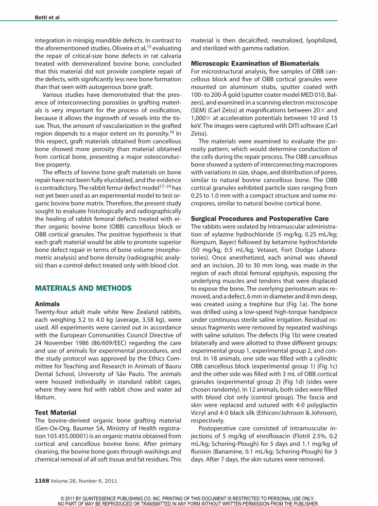
Surgical Procedures and Postoperative CareThe rabbits were sedated by intramuscular administra-tion of xylazine hydrochloride (5 mg/kg, 0.25 mL/kg; Rompum, Bayer) followed by ketamine hydrochloride (50 mg/kg, 0.5 mL/kg; Vetaset, Fort Dodge Labora-tories). Once anesthetized, each animal was shaved and an incision, 20 to 30 mm long, was made in the region of each distal femoral epiphysis, exposing the underlying muscles and tendons that were displaced to expose the bone. The overlying periosteum was re-moved, and a defect, 6 mm in diameter and 8 mm deep, was created using a trephine bur (Fig 1a). The bone was drilled using a low-speed high-torque handpiece under continuous sterile saline irrigation. Residual os-seous fragments were removed by repeated washings with saline solution. The defects (Fig 1b) were created bilaterally and were allotted to three different groups: experimental group 1, experimental group 2, and con-trol. In 18 animals, one side was filled with a cylindric OBB cancellous block (experimental group 1) (Fig 1c) and the other side was filled with 3 mL of OBB cortical granules (experimental group 2) (Fig 1d) (sides were chosen randomly). In 12 animals, both sides were filled with blood clot only (control group). The fascia and skin were replaced and sutured with 4-0 polyglactin Vicryl and 4-0 black silk (Ethicon/Johnson & Johnson), respectively.
Postoperative care consisted of intramuscular in-jections of 5 mg/kg of enrofloxacin (Flotril 2.5%, 0.2 mL/kg; Schering-Plough) for 5 days and 1.1 mg/kg of flunixin (Banamine, 0.1 mL/kg; Schering-Plough) for 3 days. After 7 days, the skin sutures were removed.
© 2011 BY QUINTESSENCE PUBLISHING CO, INC. PRINTING OF THIS DOCUMENT IS RESTRICTED TO PERSONAL USE ONLY.. NO PART OF MAY BE REPRODUCED OR TRANSMITTED IN ANY FORM WITHOUT WRITTEN PERMISSION FROM THE PUBLISHER.
Betti et al
The International Journal of Oral & Maxillofacial Implants 1169
Specimen Collection and FixationThe animals were sacrificed with overdoses of anes-thetics at 30, 90, and 180 days postsurgery. The distal femoral epiphyses were removed and fixed in 10% phosphate-buffered formalin for 1 week.
Radiographic AnalysisConventional and digital radiographs were obtained of each epiphysis. Insight periapical films (Eastman Kodak) and Digora image plates (Soredex Orion) were used and exposed for 0.6 s and 0.4 s, respectively, at 70 kVp and 15 mA (Gnatus). The source-to-object dis-tance was 40 cm. The epiphyses were placed on the film or Digora image plates, with the external face (where the defect had been created) directed upward and perpendicular to the x-ray beam. The films were processed manually by the temperature-time method. The direct digital images were stored on a personal computer. For each specimen, two image configura-tions were made in Digora: conventional (positive) and reverse (negative).
Evaluation of digital and conventional radiographs was conducted by two calibrated examiners at two different times, 1 week apart. All direct digital images were assessed directly on a computer screen in a dark-room using the software Digora for Windows (Eastman Kodak). The examiners could manipulate the bright-ness and contrast of the images as needed. Conven-tional radiographs were viewed in a darkroom using
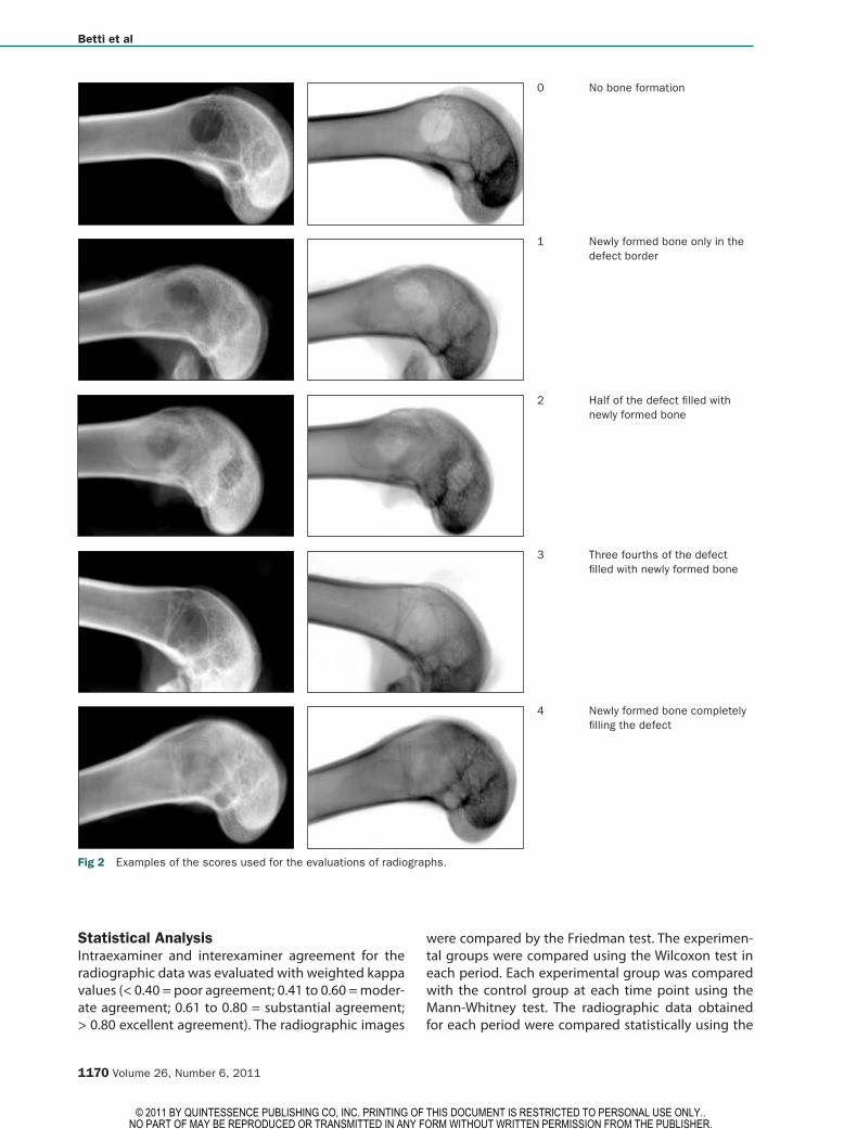
an illuminated view box and a viewing device that pro-vided 3× magnification. Each specimen was assigned a score, using a scale adapted from Lane and Shandu25 (Fig 2): 0 = no bone formation; 1 = newly formed bone only in the defect borders; 2 = half of the defect filled with newly formed bone; 3 = three fourths of the de-fect filled with newly formed bone; 4 = newly formed bone completely filling the defect.
Histologic Processing and Histomorphometric AnalysisAfter fixation, the specimens were demineralized in ethylenediaminetetraacetic acid (Merck) and then em-bedded in paraffin plus plastic resin (Histosec, Merck). Alternating 6-µm-thick sections were obtained and stained with hematoxylin and eosin (h&e) and exam-ined under a light microscope (Carl Zeiss). The area of new cortical bone (in square millimeters) was mea-sured with a digital image analysis system using a mi-croscope (Axioskop, Carl Zeiss) connected to a color video camera (Sony CCD-IRIS-RGB camera, Sony) and software (KS300, Kontron Elektronik, Image Analysis Division). The area of new cortical bone was outlined in each section, and the software calculated the area in square millimeters. The sum of the areas of each sec-tion divided by the number of sections of the speci-men resulted in a mean figure for the new cortical bone area of the specimen. The mean new cortical bone area of each group was also calculated.
Fig 1a A trephine with a delimiter is used to ensure a standard defect depth.
Fig 1b Defect in the femur head.
Fig 1c Implantation of OBB cancellous block.
Fig 1d Implantation of 3 mL of OBB corti-cal granules.
Fig 1 Surgical procedures.
a
c
b
d
© 2011 BY QUINTESSENCE PUBLISHING CO, INC. PRINTING OF THIS DOCUMENT IS RESTRICTED TO PERSONAL USE ONLY.. NO PART OF MAY BE REPRODUCED OR TRANSMITTED IN ANY FORM WITHOUT WRITTEN PERMISSION FROM THE PUBLISHER.
Betti et al
1170 Volume 26, Number 6, 2011
Statistical AnalysisIntraexaminer and interexaminer agreement for the radiographic data was evaluated with weighted kappa values (< 0.40 = poor agreement; 0.41 to 0.60 = moder-ate agreement; 0.61 to 0.80 = substantial agreement; > 0.80 excellent agreement). The radiographic images
were compared by the Friedman test. The experimen-tal groups were compared using the Wilcoxon test in each period. Each experimental group was compared with the control group at each time point using the Mann-Whitney test. The radiographic data obtained for each period were compared statistically using the
0 No bone formation
Fig 2 Examples of the scores used for the evaluations of radiographs.
1 Newly formed bone only in the defect border
2 Half of the defect filled with newly formed bone
3 Three fourths of the defect filled with newly formed bone
4 Newly formed bone completely filling the defect
© 2011 BY QUINTESSENCE PUBLISHING CO, INC. PRINTING OF THIS DOCUMENT IS RESTRICTED TO PERSONAL USE ONLY.. NO PART OF MAY BE REPRODUCED OR TRANSMITTED IN ANY FORM WITHOUT WRITTEN PERMISSION FROM THE PUBLISHER.
Betti et al
The International Journal of Oral & Maxillofacial Implants 1171
nonparametric Kruskal-Wallis test; the Dunn test was chosen as an a posteriori test to assess the statistical significance of differences among them. The morpho-metric data of the new cortical bone area obtained for each group and period showed a normal distribution; thus, the groups were analyzed by two-way analysis of variance followed by a paired Tukey multiple-com-parisons test. Each experimental group was compared with the control group at each time point using the t test. A P value less than .05 indicated statistical signifi-cance. Statistica version 7.1 (StatSoft) was used to per-form statistical analyses.
RESULTS
Clinical Outcomes The animals were mobile within 1 to 2 days postsur-gery, and all but four recovered very well. One animal in an experimental group experienced fractures in both legs and was sacrificed before the planned time point. Three animals, two in the control group and one in an experimental group, died of unknown causes. In the control group, only one defect of each animal was se-lected for analysis through random number generation using Excel (Microsoft).
Radiographic FindingsWith respect to intraexaminer and interexaminer agreement, the kappa values for conventional radio-graphs and positive and negative digital images were all greater than 0.61. Thus, the agreement was sub-stantial. Conventional radiographs, positive digital im-ages, and negative digital images were compared in each evaluation of each examiner. The agreement be-tween the digital images using kappa values was gen-erally substantial, whereas the agreement between digital and conventional radiographs was generally moderate. These evaluations were also compared by the Friedman test, which showed no significant differ-ences among conventional radiographs, positive digi-tal images, and negative digital images (P > .05).
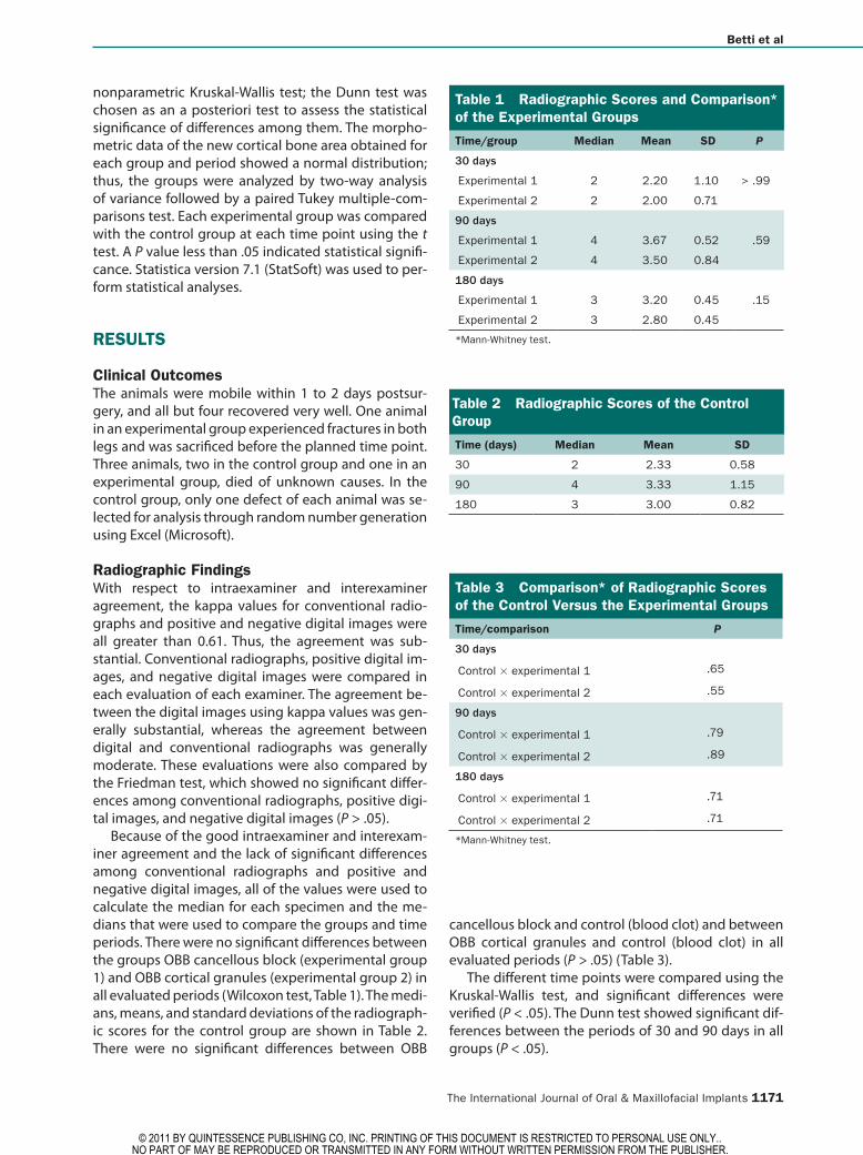
Because of the good intraexaminer and interexam-iner agreement and the lack of significant differences among conventional radiographs and positive and negative digital images, all of the values were used to calculate the median for each specimen and the me-dians that were used to compare the groups and time periods. There were no significant differences between the groups OBB cancellous block (experimental group 1) and OBB cortical granules (experimental group 2) in all evaluated periods (Wilcoxon test, Table 1). The medi-ans, means, and standard deviations of the radiograph-ic scores for the control group are shown in Table 2. There were no significant differences between OBB
cancellous block and control (blood clot) and between OBB cortical granules and control (blood clot) in all evaluated periods (P > .05) (Table 3).
The different time points were compared using the Kruskal-Wallis test, and significant differences were verified (P < .05). The Dunn test showed significant dif-ferences between the periods of 30 and 90 days in all groups (P < .05).
Table 1 Radiographic Scores and Comparison* of the Experimental Groups
Time/group Median Mean SD P
30 days
Experimental 1 2 2.20 1.10 > .99
Experimental 2 2 2.00 0.71
90 days
Experimental 1 4 3.67 0.52 .59
Experimental 2 4 3.50 0.84
180 days
Experimental 1 3 3.20 0.45 .15
Experimental 2 3 2.80 0.45
*Mann-Whitney test.
Table 2 Radiographic Scores of the Control Group
Time (days) Median Mean SD
30 2 2.33 0.58
90 4 3.33 1.15
180 3 3.00 0.82
Table 3 Comparison* of Radiographic Scores of the Control Versus the Experimental Groups
Time/comparison P
30 days
Control × experimental 1 .65
Control × experimental 2 .55
90 days
Control × experimental 1 .79
Control × experimental 2 .89
180 days
Control × experimental 1 .71
Control × experimental 2 .71
*Mann-Whitney test.
© 2011 BY QUINTESSENCE PUBLISHING CO, INC. PRINTING OF THIS DOCUMENT IS RESTRICTED TO PERSONAL USE ONLY.. NO PART OF MAY BE REPRODUCED OR TRANSMITTED IN ANY FORM WITHOUT WRITTEN PERMISSION FROM THE PUBLISHER.
Betti et al
1172 Volume 26, Number 6, 2011
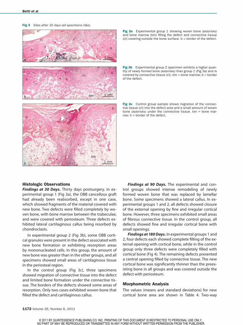
Histologic Observations Findings at 30 Days. Thirty days postsurgery, in ex-perimental group 1 (Fig 3a), the OBB cancellous graft had already been reabsorbed, except in one case, which showed fragments of the material covered with new bone. Two defects were filled completely by wo-ven bone, with bone marrow between the trabeculae, and were covered with periosteum. Three defects ex-hibited lateral cartilaginous callus being resorbed by chondroclasts.
In experimental group 2 (Fig 3b), some OBB corti-cal granules were present in the defect associated with new bone formation or exhibiting resorption areas by mononucleated cells. In this group, the amount of new bone was greater than in the other groups, and all specimens showed small areas of cartilaginous tissue in the periosteal region.
In the control group (Fig 3c), three specimens showed migration of connective tissue into the defect and limited bone formation under the connective tis-sue. The borders of the defects showed some areas of resorption. Only two cases exhibited woven bone that filled the defect and cartilaginous callus.
Findings at 90 Days. The experimental and con-trol groups showed intense remodeling of newly formed woven bone that was replaced by lamellar bone. Some specimens showed a lateral callus. In ex-perimental groups 1 and 2, all defects showed closure of the external opening by fine and irregular cortical bone. However, three specimens exhibited small areas of fibrous connective tissue. In the control group, all defects showed fine and irregular cortical bone with small openings.
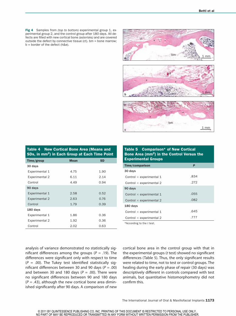
Findings at 180 Days. In experimental groups 1 and 2, four defects each showed complete filling of the ex-ternal opening with cortical bone, while in the control group only three defects were completely filled with cortical bone (Fig 4). The remaining defects presented a central opening filled by connective tissue. The new cortical bone was significantly thinner than the preex-isting bone in all groups and was covered outside the defect with periosteum.
Morphometric AnalysisThe values (means and standard deviations) for new cortical bone area are shown in Table 4. Two-way
b
ct
bm
b
**
1 mm
bct
bm
b
*
*
1 mm
b
ct
bm
b
*
*
1 mm*
Fig 3a Experimental group 1 showing woven bone (asterisks) and bone marrow (bm) filling the defect and connective tissue (ct) covering outside the bone surface. b = border of the defect.
Fig 3b Experimental group 2 specimen exhibits a higher quan-tity of newly formed bone (asterisks) than group 1 (Fig 3a) and is covered by connective tissue (ct). bm = bone marrow; b = border of the defect.
Fig 3c Control group sample shows migration of the connec-tive tissue (ct) into the defect area and a small amount of woven bone (asterisks) under the connective tissue. bm = bone mar-row; b = border of the defect.
Fig 3 Sites after 30 days (all specimens h&e).
a
c
b
© 2011 BY QUINTESSENCE PUBLISHING CO, INC. PRINTING OF THIS DOCUMENT IS RESTRICTED TO PERSONAL USE ONLY.. NO PART OF MAY BE REPRODUCED OR TRANSMITTED IN ANY FORM WITHOUT WRITTEN PERMISSION FROM THE PUBLISHER.
Betti et al
The International Journal of Oral & Maxillofacial Implants 1173
analysis of variance demonstrated no statistically sig-nificant differences among the groups (P = .19). The differences were significant only with respect to time (P = .00). The Tukey test identified statistically sig-nificant differences between 30 and 90 days (P = .00) and between 30 and 180 days (P = .00). There were no significant differences between 90 and 180 days (P = .43), although the new cortical bone area dimin-ished significantly after 90 days. A comparison of new
cortical bone area in the control group with that in the experimental groups (t test) showed no significant differences (Table 5). Thus, the only significant results were related to time, not to test or control groups. The healing during the early phase of repair (30 days) was descriptively different in controls compared with test animals, but quantitative histomorphometry did not confirm this.
Fig 4 Samples from (top to bottom) experimental group 1, ex-perimental group 2, and the control group after 180 days. All de-fects are filled with new cortical bone (asterisks) and are covered outside the defect by connective tissue (ct). bm = bone marrow; b = border of the defect (h&e).
ct
bm
*
*
1 mm
ct
bm
* *
1 mm
ct
bm
*
1 mm
*
Table 4 New Cortical Bone Area (Means and SDs, in mm2) in Each Group at Each Time Point
Time/group Mean SD
30 days
Experimental 1 4.75 1.90
Experimental 2 6.11 2.14
Control 4.49 0.94
90 days
Experimental 1 2.58 0.52
Experimental 2 2.63 0.76
Control 1.79 0.39
180 days
Experimental 1 1.86 0.36
Experimental 2 1.92 0.36
Control 2.02 0.63
Table 5 Comparison* of New Cortical Bone Area (mm2) in the Control Versus the Experimental Groups
Time/comparison P
30 days
Control × experimental 1 .834
Control × experimental 2 .272
90 days
Control × experimental 1 .055
Control × experimental 2 .082
180 days
Control × experimental 1 .645
Control × experimental 2 .777
*According to the t test.
a
c
b
© 2011 BY QUINTESSENCE PUBLISHING CO, INC. PRINTING OF THIS DOCUMENT IS RESTRICTED TO PERSONAL USE ONLY.. NO PART OF MAY BE REPRODUCED OR TRANSMITTED IN ANY FORM WITHOUT WRITTEN PERMISSION FROM THE PUBLISHER.
Betti et al
1174 Volume 26, Number 6, 2011
DISCUSSION
In this study, the osseous repair potential of two bo-vine-derived grafting materials, OBB cancellous block and OBB cortical granules, was evaluated in trephined defects, 6 mm in diameter and 8 mm in depth, cre-ated in the femoral epiphyses of rabbits. The positive hypothesis tested here was that both materials sepa-rately would be able to promote superior healing of the defects when compared to control defects treated solely with blood clot. The hypothesis was rejected, because only descriptive differences were apparent at the first healing period and were not supported by histomorphometric and radiographic data. The only significant results were related to time and not to test or control graft materials.
The size of the defects and the periods of evaluation of 30, 90, and 180 days postsurgeries used here were based on other studies17–19,21,23 that used the same model to test other bone grafting materials. According to Rimondini et al21 and Dallari et al,23 defects of 6 mm in diameter and 10 mm in depth are classified as criti-cal-size defects because the formation of new bone in untreated defects was restricted to the edges of the defects, and the largest part of the center remained free of bone up to 12 weeks. In this context, Leupold et al22 verified that their control group showed the least amount of bone ingrowth at 3 months postsurgery, and Al Ruhaimi19 showed that a femoral control de-fect, 5 mm in diameter and 8 mm in depth, presented larger empty spaces and soft tissue (96%), with normal trabecular bone only at the borders.
The SEM microstructural analysis confirmed that the OBB cancellous bone graft exhibits interconnect-ing macropores, while the particulate OBB cortical bone graft has a compact structure with fewer micro-pores. According to Kuboki et al26 and Ayers et al,16 a porous environment in the graft material favors new bone formation because it allows cell migration and proliferation and new vascularization.
The radiographic results failed to demonstrate sig-nificant differences between the test and control inter-ventions. Bighan et al,11 who tested two graft materials (bovine DBM and bovine fetal growth plate), also ob-served no significant differences in bone formation us-ing radiographic analysis. Nevertheless, in the present study, there was a significant increase in radiopacity from 30 and 90 days to 180 days at the defect area in all groups.
Histologically, at 30 days, both experimental groups presented a greater amount of newly formed—primar-ily woven—bone. In the control group, during this ini-tial phase of repair, connective tissue ingrowth into the defects occurred, which delayed new bone formation. In the experimental groups, the graft materials prevented
this ingrowth, favoring osteogenesis. On the other hand, with respect to histologic appearances of the two ex-perimental groups during this first period, the OBB cor-tical bone–grafted group showed a higher quantity of formed woven bone than the porous OBB cancellous bone–grafted group. These differences were a result of the presence of native growth factors and bone mor-phogenetic proteins in cortical bone matrix, which are not present in cancellous bone matrix, which merely showed excellent osteoconduction. It should be pointed out that, during the initial phases of repair, these sub-stances stimulate cell proliferation and differentiation of osteoblasts, respectively.27,28 In this respect, based on the results obtained following treatment of rabbit radius defects, Bigham et al10 postulated that the grafted xe-nogeneic bovine DBM encourages osteoinductive ac-tivity by releasing some bone morphogenetic proteins, similar to autogenous cortical bone.
Although descriptive differences were seen in con-trols compared to test animals in the early phase of repair, with the graft material preventing fibrous con-nective tissue ingrowth, the histomorphometric analy-sis did not confirm these differences. At 30, 90, and 180 days, there were no statistically significant differences among the groups in relation to the newly formed cor-tical bone area. In contrast, Torricelli et al,8 using xeno-geneic DBM in rat femoral condylar defects of 2 mm in diameter and 3 mm in depth, showed that the amount of repair in control defects was always less than that seen in the treated defects. These differences in the re-sults could be a result of the different defect sizes or animal species used in each experiment.
At 90 days, the two grafts used in the present study had been resorbed. Oliveira et al15 verified that par-ticles of DBM were almost completely resorbed at 21 days postoperatively, while remaining fragments of autogenous bone could be seen until 90 days postop-eratively in rat skull defects.
The significant reduction in new cortical bone area thickness from 30 and 90 days to 180 days occurred via bone remodeling, which is normal in later phases of bone repair, where the woven bone created in the ini-tial phases of healing is resorbed and replaced by more structured lamellar bone. In this respect, Bigham et al11 did not observe remodeling on the 14th and 28th days postoperative, although there was some remodeling on the 42nd day postoperative in rabbit radius defects grafted with bovine DBM and bovine fetal growth plate.
The results of this study indicate that the clinical use of OBB grafts in critical-size bone defects will prevent connective tissue ingrowth into the defect during the earlier phases of repair but may have no effect at lat-er periods. However, this study has some limitations, mainly in relation to the animal model. Further stud-ies are needed to evaluate these graft materials in a
© 2011 BY QUINTESSENCE PUBLISHING CO, INC. PRINTING OF THIS DOCUMENT IS RESTRICTED TO PERSONAL USE ONLY.. NO PART OF MAY BE REPRODUCED OR TRANSMITTED IN ANY FORM WITHOUT WRITTEN PERMISSION FROM THE PUBLISHER.
Betti et al
The International Journal of Oral & Maxillofacial Implants 1175
different animal model, especially a species with bone structure, size, and metabolic rate that are more similar to those of humans, and in various clinical situations.
CONCLUSIONS
Radiographic evaluation showed no significant differ-ences among the experimental and control groups in terms of bone density, and the results of digital and conventional methods were similar. Histomorphomet-ric analysis did not show significant differences among the groups in terms of bone volume. The only signifi-cant results of the various grafting materials used here were related to time, with a reduction of the cortical bone formed at 90 and 180 days in all groups.
ACKNOWLEDGMENTS
The authors would like to thank Danielle Ceolin for her valuable help and technical assistance in the histologic preparation, Dr José Roberto Pereira Lauris for his valuable help and assistance in the statistical analysis, and Dr Orivaldo Tavano for his valu-able comments in the elaboration of the radiographic analysis.
REFERENCES
1. Senn N. On the healing of aseptic cavities by implantation of anti-septic decalcified bone. Am J Med Sci 1889;98:219.
2. Urist MR. Bone formation by autoinduction. Science 1965;150:893–899. 3. Urist MR, DeLange RJ, Finerman GAM. Bone cell differentiation and
growth factors. Science 1983;220:680–686. 4. Reddi AH, Wientroub S, Muthukumaran N. Biologic principles of
bone induction. Orthop Clin North Am 1987;18:207–212. 5. Reddi AH, Muthukumaran N, Ma S, et al. Initiation of bone develop-
ment by osteogenin and promotion by growth factors. Connect Tissue Res 1989;20:303–312.
6. Torricelli P, Fini M, Rocca M, Giavaresi G, Giardino R. Xenogenic demineralized bone matrix: Osteoinduction and influence of as-sociated skeletal defects in heterotopic bone formation in rats. Int Orthop 1999;23:178–181.
7. Murugan R, Ramakrishna S, Rao KP. Analysis of bovine-derived de-mineralized bone extracts. J Mater Sci Mater Med 2008;19:2423–2426.
8. Torricelli P, Fini M, Giavaresi G, Rimondini L, Giardino R. Charac-terization of bone defect repair in young and aged rat femur induced by xenogenic demineralized bone matrix. J Periodontol 2002;73:1003–1009.
9. Leite FRM, Ramalho LTO. Bone regeneration after demineralized bone matrix and castor oil (Ricinus communis) polyurethane implan-tation. J Appl Oral Sci 2008;16:122–126.
10. Bigham AS, Dehghani SN, Shafiei Z, Torabi Nezhad S. Xenogenic demineralized bone matrix and fresh autogenous cortical bone effects on experimental bone healing: Radiological, histopatho-logical and biomechanical evaluation. J Orthopaed Traumatol 2008;9:73–80.
11. Bigham AS, Dehghani SN, Shafiei Z, Torabi Nezhad S. Experimental bone defect healing with xenogenic demineralized bone matrix and bovine fetal growth plate as a new xenograft: Radiological, histopathological and biomechanical evaluation. Cell Tissue Bank 2009;10:33–41.
12. Sicca CM, Corotti MV, Sgarbosa SHPV, et al. Comparative histo-morphometric and tomographic analysis of maxillary sinus floor augmentation in rabbits using autografts and xenografts. J Biomed Mater Res B Appl Biomater 2008;86:188–196.
13. Gupta R, Pandit N, Malik R, Sood S. Clinical and radiological evalua-tion of an osseous xenograft for the treatment of intrabony defects. J Canad Dent Assoc 2007;73:513.
14. Mai R, Reinstorf A, Pilling E, et al. Histologic study of incorporation and resorption of a bone cement-collagen composite: An in vivo study in the minipig. Oral Surg Oral Med Oral Pathol Oral Radiol Endod 2008;105:e9–e14.
15. Oliveira RC, Oliveira FHG, Cestari TM, Taga R, Granjeiro JM. Morpho-metric evaluation of the repair of critical-size defects using demin-eralized bovine bone and autogenous bone grafts in rat calvária. Clin Oral Implants Res 2008;19:749–754.
16. Ayers RA, Simske SJ, Nunes LM, Wolford CR. Long-term bone in-growth and residual microhardness of porous block hydroxyapatite implants in humans. J Oral Maxillofac Surg 1998;56:1297–1301.
17. Kania RE, Meunier A, Hamadouche M, Sedel L, Petite H. Addition of sealant to ceramic promotes bone repair: Long-term study in rabbit femoral defect model. J Biomed Mater Res 1998;43:38–45.
18. Oonishi H, Hench LL, Wilson J, et al. Comparative bone growth be-havior in granules of bioceramic materials of various sizes. J Biomed Mater Res 1999;44:31–43.
19. Al Ruhaimi KA. Effect of adding resorbable calcium sulfate to grafting materials on early bone regeneration in osseous defects in rabbits. Int J Oral Maxillofac Implants 2000;15:859–864.
20. Voor MJ, Arts JJC, Klein SA, Walschot LHB, Verdonschot N, Buma P. J Is hydroxyapatite cement an alternative for allograft bone chips in bone grafting procedures? A mechanical and histological study in a rabbit cancellous bone defect model. Biomed Mater Res B Appl Biomater 2004;71:398–407.
21. Rimondini L, Nicoli-Aldini N, Fini M, Guzzardella G. In vivo experi-mental study on bone regeneration in critical bone defects using an injectable PLA/PGA copolymer. Oral Surg Oral Med Oral Pathol Oral Radiol Endod 2005;99:148–154.
22. Leupold JA, Barfield WR, An YH, Hartsock LA. A comparison of ProOsteon, DBX and collagraft in a rabbit model. J Biomed Mater Res B Appl Biomater 2006;79:292–297.
23. Dallari D, Fini M, Stagni C, et al. In vivo study on the healing of bone defects treated with bone marrow stromal cell, platelet-rich plasma, and freeze-dried bone allografts, alone and in combina-tion. J Orthop Res 2006;24:877–888.
24. Tsai CH, Lin RM, Ju CP, Lin JHC. Bioresorption behavior of tetracal-cium phosphate-derived calcium phosphate cement implanted in femur of rabbits. Biomaterials 2008;29:984–993.
25. Lane JM, Shandu HS. Current approach to experimental bone graft-ing. Orthop Clin North Am 1987;18:213–225.
26. Kuboki Y, Takita H, Kobayashi D, et al. BMP-induced osteogenesis on the surface of hydroxyapatite with geometrically feasible and non-feasible structure: Topology of osteogenesis. J Biomed Mater Res 1998;39:190–199.
27. ten Dijke P. Bone morphogenetic protein signal transduction in bone. Curr Med Res Opin 2006;22(suppl 1):S7–S11.
28. Xiao YT, Xiang LX, Shao JZ. Bone morphogenetic protein. Biochem Biophys Res Commun 2007;362:550–553.
© 2011 BY QUINTESSENCE PUBLISHING CO, INC. PRINTING OF THIS DOCUMENT IS RESTRICTED TO PERSONAL USE ONLY.. NO PART OF MAY BE REPRODUCED OR TRANSMITTED IN ANY FORM WITHOUT WRITTEN PERMISSION FROM THE PUBLISHER.

Copyright of International Journal of Oral & Maxillofacial Implants is the property of Quintessence Publishing
Company Inc. and its content may not be copied or emailed to multiple sites or posted to a listserv without the
copyright holder's express written permission. However, users may print, download, or email articles for
individual use.