Repair of 104 Failed Anti-Reflux Operations
10
ORIGINAL ARTICLES Repair of 104 Failed Anti-Reflux Operations Atif Iqbal, MD,* Ziad Awad, MD, FRCSI,† Jennifer Simkins, MD,* Ricky Shah, BS,‡ Mumnoon Haider, MD,* Vanessa Salinas, MD,* Kiran Turaga, MD,* Anouki Karu, MS,‡ Sumeet K. Mittal, MD,* and Charles J. Filipi, MD, FACS* Objective: To assess whether reoperative surgery for failed Nissen fundoplication is beneficial and to classify all mechanisms of failure recognized. Summary Background Data: Antireflux surgery is often neces- sary, but a 10% failure rate is commonplace. We report results for patients undergoing reoperative surgery and present a nomenclature of mechanisms of failure. Methods: A total of 104 patients, who had a previous fundoplication for gastroesophageal reflux disease (GERD), underwent reoperative surgery. Manometry (n 86), endoscopy (n 101), pH monitoring (n 27), upright esophagram (n 90), gastric emptying (n 26), and symptom assessment (n 104) were performed prior to reop- erative surgery. Patients were also assessed before and during reoperation for mechanism of failure using a newly proposed clas- sification. The operative approach was laparoscopic in 58 patients, via open laparotomy in 12, and a thoracotomy in 34 patients. Follow-up was conducted by phone interview and was completed in 97 patients (97%; 3 were deceased) with a mean follow-up of 32 months (range, 1–146 months). Results: The conversion rate to laparotomy for laparoscopic patients was 8%. The perioperative complication rate was 32%. One patient died of respiratory insufficiency after a laparotomy. Seven patients required additional surgery for correction of persistent or recurrent symptoms. The short and long-term complication rate was similar for the different operative approachs. Symptom resolution (rare or absent) occurred in 74% of patients with dysphagia, 75% with heartburn, 85% with regurgitation, and 94% with chest pain. The overall post-reoperative patient satisfaction was 7 on a scale of 1 to 10 and 3 on a scale of 1 to 4 when patients were asked to grade the operative result. There was no difference in the symptom resolution for patients operated upon by the laparoscopic approach as com- pared with laparotomy, but those patients undergoing a Collis gastroplasty had poorer results. The preoperative accuracy of assess- ment for mechanism of failure was 78%. A nomenclature of mech- anisms of failure is included to aide reoperative assessment and new mechanisms of failure are described. Conclusion: Reoperative surgery results for GERD are satisfactory. A variety of operative approaches proved equally effective. Poorer results were observed in patients with more advanced disease. (Ann Surg 2006;244: 42–51) G astroesophageal reflux disease (GERD) is a common disorder accounting for approximately 75% of esopha- geal pathology. 1 Although medication is often effective, long-term therapy has become commonplace. 2 However, pro- longed medical therapy often includes escalating dosages, which can be an important financial burden on patients, especially as insurance coverage for pharmaceuticals contin- ues to diminish. 2,3 Surgical therapy for GERD has improved with a better understanding of the underlying pathophysiol- ogy of GERD and improved patient selection. 4,5 A controlled randomized trial of severe GERD patients demonstrated surgery to be superior to medical therapy in terms of symptom resolution. 6 Other investigators have provided evidence to favor antireflux surgery over medical therapy, 7,8 and Nissen fundoplication remains the operation of choice in the management of patients with severe GERD and its associated complications. The number of operations is increasing annually. Within the last decade, there has been almost an 8-fold procedural increase. This is attributed to the increased use of esophageal pH and motility testing, greater awareness of GERD-related quality of life issues, the advent of minimally invasive Nissen fundoplication, 9 –11 functional results of laparoscopic antireflux procedures that are equal to that of open surgery, significantly less postoperative morbidity, and a shorter hospital stay. 12–14 The surgical management of GERD, whether performed open or laparoscopically, does have a failure rate and reop- eration may be required for optimal results. Failure of open fundoplication occurs in 9% to 30% of patients, 4,15,16 whereas published failure rates of laparoscopic Nissen fun- doplication are 2% to 17%. 6,17–20 These lower published rates probably reflect shorter follow-up rather than an intrinsically better operation. Reoperations for failed or recurrent GERD are techni- cally more demanding due to adhesions from previous sur- gery, obscured anatomy, and more advanced disease. A morbidity of 4% to 40% and a mortality rate of 0% to 4.9% can be expected with reoperation, 15,21,22 and the overall From the *Department of Surgery, Creighton University School of Medicine, Omaha, NE; †Department of Surgery, University of Missouri, Columbia, MO; and ‡Creighton University School of Medicine, Omaha, NE. Reprints: Charles J. Filipi, MD, FACS, Department of Surgery, Creighton University School of Medicine, 601 N. 30th Street, Suite 3740, Omaha, NE 68131. E-mail: cjfi[email protected]. Copyright © 2006 by Lippincott Williams & Wilkins ISSN: 0003-4932/06/24401-0042 DOI: 10.1097/01.sla.0000217627.59289.eb Annals of Surgery • Volume 244, Number 1, July 2006 42
-
Upload
independent -
Category
Documents
-
view
1 -
download
0
Transcript of Repair of 104 Failed Anti-Reflux Operations

ORIGINAL ARTICLES
Repair of 104 Failed Anti-Reflux Operations
Atif Iqbal, MD,* Ziad Awad, MD, FRCSI,† Jennifer Simkins, MD,* Ricky Shah, BS,‡Mumnoon Haider, MD,* Vanessa Salinas, MD,* Kiran Turaga, MD,* Anouki Karu, MS,‡
Sumeet K. Mittal, MD,* and Charles J. Filipi, MD, FACS*
Objective: To assess whether reoperative surgery for failed Nissenfundoplication is beneficial and to classify all mechanisms of failurerecognized.Summary Background Data: Antireflux surgery is often neces-sary, but a 10% failure rate is commonplace. We report results forpatients undergoing reoperative surgery and present a nomenclatureof mechanisms of failure.Methods: A total of 104 patients, who had a previous fundoplicationfor gastroesophageal reflux disease (GERD), underwent reoperativesurgery. Manometry (n � 86), endoscopy (n � 101), pH monitoring(n � 27), upright esophagram (n � 90), gastric emptying (n � 26),and symptom assessment (n � 104) were performed prior to reop-erative surgery. Patients were also assessed before and duringreoperation for mechanism of failure using a newly proposed clas-sification. The operative approach was laparoscopic in 58 patients,via open laparotomy in 12, and a thoracotomy in 34 patients.Follow-up was conducted by phone interview and was completed in97 patients (97%; 3 were deceased) with a mean follow-up of 32months (range, 1–146 months).Results: The conversion rate to laparotomy for laparoscopic patientswas 8%. The perioperative complication rate was 32%. One patientdied of respiratory insufficiency after a laparotomy. Seven patientsrequired additional surgery for correction of persistent or recurrentsymptoms. The short and long-term complication rate was similarfor the different operative approachs. Symptom resolution (rare orabsent) occurred in 74% of patients with dysphagia, 75% withheartburn, 85% with regurgitation, and 94% with chest pain. Theoverall post-reoperative patient satisfaction was 7 on a scale of 1 to10 and 3 on a scale of 1 to 4 when patients were asked to grade theoperative result. There was no difference in the symptom resolutionfor patients operated upon by the laparoscopic approach as com-pared with laparotomy, but those patients undergoing a Collisgastroplasty had poorer results. The preoperative accuracy of assess-ment for mechanism of failure was 78%. A nomenclature of mech-anisms of failure is included to aide reoperative assessment and newmechanisms of failure are described.
Conclusion: Reoperative surgery results for GERD are satisfactory.A variety of operative approaches proved equally effective. Poorerresults were observed in patients with more advanced disease.
(Ann Surg 2006;244: 42–51)
Gastroesophageal reflux disease (GERD) is a commondisorder accounting for approximately 75% of esopha-
geal pathology.1 Although medication is often effective,long-term therapy has become commonplace.2 However, pro-longed medical therapy often includes escalating dosages,which can be an important financial burden on patients,especially as insurance coverage for pharmaceuticals contin-ues to diminish.2,3 Surgical therapy for GERD has improvedwith a better understanding of the underlying pathophysiol-ogy of GERD and improved patient selection.4,5
A controlled randomized trial of severe GERD patientsdemonstrated surgery to be superior to medical therapy interms of symptom resolution.6 Other investigators haveprovided evidence to favor antireflux surgery over medicaltherapy,7,8 and Nissen fundoplication remains the operationof choice in the management of patients with severe GERDand its associated complications. The number of operations isincreasing annually. Within the last decade, there has beenalmost an 8-fold procedural increase. This is attributed to theincreased use of esophageal pH and motility testing, greaterawareness of GERD-related quality of life issues, the adventof minimally invasive Nissen fundoplication,9–11 functionalresults of laparoscopic antireflux procedures that are equal tothat of open surgery, significantly less postoperative morbidity,and a shorter hospital stay.12–14
The surgical management of GERD, whether performedopen or laparoscopically, does have a failure rate and reop-eration may be required for optimal results. Failure of openfundoplication occurs in 9% to 30% of patients,4,15,16
whereas published failure rates of laparoscopic Nissen fun-doplication are 2% to 17%.6,17–20 These lower published ratesprobably reflect shorter follow-up rather than an intrinsicallybetter operation.
Reoperations for failed or recurrent GERD are techni-cally more demanding due to adhesions from previous sur-gery, obscured anatomy, and more advanced disease. Amorbidity of 4% to 40% and a mortality rate of 0% to 4.9%can be expected with reoperation,15,21,22 and the overall
From the *Department of Surgery, Creighton University School of Medicine,Omaha, NE; †Department of Surgery, University of Missouri, Columbia,MO; and ‡Creighton University School of Medicine, Omaha, NE.
Reprints: Charles J. Filipi, MD, FACS, Department of Surgery, CreightonUniversity School of Medicine, 601 N. 30th Street, Suite 3740, Omaha,NE 68131. E-mail: [email protected].
Copyright © 2006 by Lippincott Williams & WilkinsISSN: 0003-4932/06/24401-0042DOI: 10.1097/01.sla.0000217627.59289.eb
Annals of Surgery • Volume 244, Number 1, July 200642

symptom resolution rates as compared with those of primarysurgery are compromised.
Large case series are needed to better understand thebenefit of reoperative surgery.23–26 To accomplish this exten-sive primary surgery experience, a strong referral base and agood esophageal physiology laboratory is necessary. Thisstudy adds to accumulated knowledge for prognosis of thesecomplex patients by reviewing our results of 104 failedfundoplications. We also provide a comprehensive nomen-clature for fundoplication failure, which includes morpho-logic, functional, and therapeutic mechanisms in addition todiagnostic errors.
MATERIALS AND METHODSAn Institutional Review Board exemption for informed
consent was given as some patients had been followed annu-ally for more than 10 years. Between 1993 and 2004, 850patients underwent primary laparoscopic Nissen fundoplica-tion (LNF) at Creighton University Medical Center, 28 pa-tients of which underwent both primary and reoperativefundoplication by coauthors (C.J.F. or S.K.M.). An additional76 patients (73%) were referred for reoperation from othercenters for a total of 104 patients. The inclusion and exclu-sion criteria for this study are shown in Table 1. Patients withreoperative antireflux surgery without a fundoplication ineither the primary or reoperative procedure were not in-cluded, eg, those with a primary antireflux prosthesis, hiatalhernia repair without fundoplication and those undergoing agastrectomy for reflux control or a complete fundoplicationtakedown at reoperation.
Preoperative EvaluationPatients presenting to the esophageal center with per-
sistent or recurrent symptoms after fundoplication were eval-uated with a standardized esophageal questionnaire followedby esophagogastroduodenoscopy (n � 101), manometry (n �86), an upright barium esophagogram (n � 90), 24-hour pHmonitoring (n � 27), and/or a gastric emptying study (n �26). The questionnaire used is shown in Table 2. In addition,timing of symptom recurrence, number of dilations after reop-erative surgery, a subjective grading system, and an overallrating of the procedure were included.
EsophagramThe esophagram was performed using both single and
double contrast. After a standard upright esophagram wascompleted, an upright anterior-posterior and lateral chestx-ray was obtained with barium in the distal esophagus. Acentimeter ruler was superimposed on the anterior projectionand the size of the hiatal hernia was determined by measuringagainst the ruler from the proximal extent of the gastric tissueto the extrapolated hemi-diaphragm lines at the midline (Fig.1). A short esophagus was suspected if the gastroesophagealjunction (GEJ) was 5 or more centimeters above the hiatus.
Upper EndoscopyUpper endoscopy was performed to determine the pres-
ence or absence of cancer, Barrett esophagus by direct in-spection and biopsy, a peptic stricture, the size of a hiatalhernia, the presence of food within the stomach, the positionof the fundoplication on retroflexion, the length of gastrictissue above the fundoplication, the location of the fundopli-cation, and the tightness of the fundoplication (also deter-mined by the maximum size dilator that could be safelyused). The depth of the fundoplication anterior and posteriortucks and the apposition of tissue to the retroflexed endoscopewere used to determine the presence or absence of fundopli-cation disruption.
ManometryManometry was conducted as per standard protocol,
but in addition, the fundoplication pressure and relaxationwere assessed using 2 to 3 wet swallows at each centimeterlevel (normal fundoplication pressure is 20–35 mm Hg in ourlaboratory). Relaxation below 85% at any one level wasconsidered abnormal. Esophageal body dysmotility and waveamplitudes were also noted.
pH MonitoringTwenty-four-hour pH monitoring was performed selec-
tively on the basis of previous completed positive studies, theresponse to proton pump inhibitors (PPIs) and presence orabsence of esophagitis on endoscopy. A standard probe wasused for the majority of patients; however, the BRAVOcapsule was used later in the study period.
Gastric Emptying StudiesThis study was performed if a previous vagotomy had
been performed, there was food within the stomach at upperendoscopy after an appropriate fast, the patient gave a historyof vomiting old food, or the patient had intractable vomiting.A half-time of greater than 180 minutes was considered abnor-mal and an indication for a pyloroplasty.
Mechanism of FailureA nomenclature for mechanisms postfundoplication
failure was developed and is shown in Table 3. The preop-erative mechanism was determined by a combination of testsincluding endoscopy, pH monitoring, manometry, an uprightesophagram, and a gastric emptying study, when indicated.The crus closure failures were readily apparent at endoscopy.The stenotic crus closure was difficult to differentiate preop-eratively but was suspected if a large dilator could not easily
TABLE 1. Inclusion and Exclusion Criteria for Study
Inclusion Criteria for Study Exclusion Criteria for Study
Patients who had a previousfundoplication/Hillcardioplasty anduncontrolled or difficult tocontrol recurrent symptoms
Diffuse esophageal spasmEpiphrenic diverticulumLaparoscopic nissen fundoplication
followed by complete takedownbefore re-operation
Failed collis nissen fundoplicationFailed thoracoscopic belseyHiatal hernia recurrence onlyPrimary heller myotomy with
fundoplicationAttempted but failed primary
fundoplication at outside facility
Annals of Surgery • Volume 244, Number 1, July 2006 Reoperation for Failed Anti-Reflux Surgery
© 2006 Lippincott Williams & Wilkins 43

traverse the hiatus and manometry did not show a hyperten-sive or poorly relaxing fundoplication. The criteria for sus-pecting a short esophagus were the presence of Barrettesophagus, a peptic stricture, short manometric length, andthe GEJ being 5 or more cm above the confluence of thehemidiaphragms on upright esophagram.27 The final determi-nation of esophageal length, however, was at the time ofsurgery when after esophageal mobilization the GEJ had torest without tension 3 cm below the arch of the diaphragmatichiatus if the laparoscopic approach was used, and 2 cm foropen surgery.
Indications and Operative ApproachReoperative fundoplication was advised when the eval-
uation revealed a surgically correctable disorder that had notor could not respond to aggressive medical management.Patients with an asymptomatic small recurrent hiatal herniawere not offered operative intervention but those with chestpain were operated upon if a cardiac evaluation was negativeand the chest pain was repetitive.
An open thoracotomy was performed when more than2 cm of gastric tissue was within the mediastinum on uprightesophagram, a short esophagus was suspected, or the patienthad a previous mediastinal dissection. A laparotomy waschosen if the patient had 2 or more prior failed operations. Alaparoscopic approach was used if the patient did not meet theabove criterion. In patients with a short esophagus, a Collisgastroplasty was performed.
Reoperative Laparoscopic TechniqueThe undersurface of the lateral segment of the left lobe
of the liver was separated from adherent viscera by sharpdissection. The dissection was started posteriorly and caudadwhere the adhesions were less dense. Care was taken to stay
immediately adjacent to the liver capsule and the previousoperative note was referenced to determine if an aberrant lefthepatic artery was present. The proximal short gastric vesselswere ligated in all cases if still present and the pancreaticgastric adhesions/ligament was divided.
After separation of the anterior and posterior wings thefundoplication was dismantled. Final attachments of the rightposterior limb to the proximal stomach could be best seen byretracting the anterior fundus to the patient’s right side andusing a 45° laparoscope to view the left side of the hiatus.Complete dismantling of the fundoplication, when indicated,was confirmed by intraoperative endoscopy. Sharp dissectionwas used throughout. Perforations were closed by definingthe edges and placing interrupted 3– 0 silk sutures, beingsure to include the mucosa. Knots were tied intracorpore-ally. Intraoperative endoscopy was always performed toassure a secure closure and to determine if other perfora-tions were present.
Nissen fundoplication redundancy was tailored to man-ometric findings. Conversion to a Toupet fundoplication wasused if the patient had a too tight Nissen fundoplication andthe initial fundoplication was well formed without evidenceof twisting or a 2-compartment stomach. A Dor fundoplica-tion was constructed if the patient had no esophageal peri-stalsis. Crus closure, if necessary, was completed withinterrupted O Ethibond nonabsorbable sutures (EthiconInc., Sommerville, NJ). Mesh reinforcement was used (n �4) when crus closure sutures tore muscular tissue or thepatient was required to lift heavy objects during employment.
Reoperative Thoracotomy TechniqueAfter exploration, the esophagus was mobilized up to
the level of the left mainstem bronchus. Any hernia present
TABLE 2. The Symptom Grading System Administered to Patients
Symptoms Dysphagia Regurgitation Heart-Burn Chest Pain
Grading 0 None 0 None 0 None 0 None
1 Minimal, once a weekor less
1 Mild, after straining or largemeals
1 Minimal, episodic, notreatment required
1 Minimal, episodic2 Moderate, reason for visit
2 Moderate, �once aweek, requires dietaryadjustment
3 Severe, preventingingestion of food
2 Moderate, predictable, withposition change/lying/straining
3 Severe, constant regurgitationwith or without aspiration
2 Moderate, controlled withmedication
3 Severe, interferes with dailyactivity or not controlledwith medication
3 Severe, interferes with dailyactivity
Pulmonary Symptoms Nausea/Vomiting Abdominal Bloating Other Symptoms0 None 0 None 0 None
1 Recurrent cough 1 Occasional episodes of nausea 1 Present
2 Nocturnal cough3 Recurrent pneumonitis4 Asthma5 Change of voice
2 Frequent nausea or episodicvomiting
3 Continuous nausea or frequentvomiting
Medications Name
How often?
Duration b/wk redo surgery and reappearance of symptoms (months)
Number of times dilation done since redo surgery?
Satisfaction rate: in relation to symptoms you had the reoperation for (Scale 1–10 with one being the least satisfied and 10 the most)
Grading rate: for results of redo surgery 1, Poor 2, Fair 3, Good 4, Excellent
Iqbal et al Annals of Surgery • Volume 244, Number 1, July 2006
© 2006 Lippincott Williams & Wilkins44

was mobilized and the hernia sac entered. If the adhesions onthe peritoneal side were difficult to visualize, a diaphragmcounterincision was made 2 to 3 cm below the rib cage. Thefundoplication was dismantled and a Collis gastroplasty (4–5cm long) was performed if the skeletonized GEJ could not be
easily reduced 2 cm into the peritoneal cavity. A partialfundoplication was used only in patients with very pooresophageal motility. Diaphragm closure was performed withinterrupted 0 Ethibond sutures.
Reoperative Laparotomy TechniqueThe laparotomies were performed through an upper
midline incision. Dissection and mobilization were per-formed as described for the laparoscopic technique althoughpalpation was used liberally to identify the vagus nerves.Dismantling the fundoplication was easier as exposure to theposterior esophagus could be achieved readily. The choice offundoplication was similar to that used for the laparoscopicapproach.
FIGURE 1. An upright esophagram shows an incarceratedhiatal hernia with the gastroesophageal junction above thediaphragm. The hemidiaphragm lines are extrapolated tothe midline. From that point, the gastroesophageal junctionand the proximal extent of the gastric tissue are measured;values a and b. (“a” should not exceed 5 cm and “b”should not exceed 2 cm for the laparoscopic approach).
TABLE 3. Mechanisms of Post-Fundoplication Failure
n (%)
Hiatus closure failure 57 (55)
Crus closure disruption 52 (50)
Stenosis* 5 (4.8)
Too tight closure 0
Fundoplication failure 88 (85)
Partial disruption 36 (35)
Complete disruption 7 (6.7)
Hypertensive 24 (23)
Twisted
Two compartment stomach
Relative to esophageal dysmotility
Fibrosis of fundoplication�
Slipped† 18 (17)
Intrathoracic fundoplication
Paraesophageal component
Intraabdominal fundoplication
Paraesophageal component
Hourglass‡
Gastric body fundoplication
Too loose 2 (1.9)
Inadequate esophageal length, short§ 16 (15)
Postoperative gastroparesis 2 (1.9)
Inadvertent vagotomy
Gas bloat syndrome
Fistula formation 0
Internal
External
Incorrect diagnosis 1 (1)
Achalasia
Visceral hyperalgesia
Gastric or esophageal cancer 1 (1)
Gastric hypersecretion
Gastric outlet obstruction
Gastroparesis
Note: The percentages do not add to 100% as many patients had multiple mechanismsof failure.
*Dense fibrous scar formation.†2 cm or more gastric mucosa above fundoplication.‡Intrathoracic fundus, fundoplication in abdomen.§Gastroesophageal junction less than 3 cm below arch of crus after esophageal
mobilization during laparscopic reoperation.�Secondary to pledget erosion or suture perforation.
Annals of Surgery • Volume 244, Number 1, July 2006 Reoperation for Failed Anti-Reflux Surgery
© 2006 Lippincott Williams & Wilkins 45
Follow-upThree patients were lost to follow-up and 4 patients
died, 1 of which was a postoperative death. Ninety-sevenpatients (97% follow-up) were successfully contacted bytelephone. The symptom questionnaire (Table 2) was admin-istered to determine symptom recurrence. In addition, medi-cation use, subjective grading of results on a scale of 1 to 10,and overall satisfaction grade (excellent, good, fair, or poor)was determined. The mean length of follow-up was 32 months,and the range was 1 to 146 months. The criteria for failureafter reoperation are the following: 1) need for further sur-gery, 2) daily H2 blocker or PPI for GERD, or 3) grade 3symptoms.
Data AnalysisAn Excel (Microsoft) spreadsheet was used to store
data. Symptom results were compared with the mechanism offailure, the type of reoperative procedure performed, thepresence or absence of Barrett esophagus, a short esophagus,and complications. To determine the difference in symptomresolution between groups, the �2 with Fisher exact testmodification was used. A paired t test was used to comparepre- and postoperative symptoms. A multivariate analysiswas conducted to determine risk factors for poor patientoutcomes.
RESULTSA total of 104 patients with a mean age of 50 years
(range, 22–80 years), a mean weight of 185 pounds (range,88–262 lb), a mean BMI of 30 (range, 15–56 kg/m2) under-went reoperative fundoplication at our center during an 11-year period. The baseline patient characteristics are shown inTable 4. The mean duration between the primary procedureand reappearance of symptoms was 22 months (range, 0–120months) and the mean time elapsed between procedures was38 months (range, 0–672 months). Ninety patients (87%) hadtheir primary fundoplication performed by laparoscopy, and14 (13%) by laparotomy. None of the primary fundoplica-tions were performed transthoracically. The most commonsurgical indication was dysphagia (38.4%). Thirty-eight pa-tients were operated upon for recurrent GERD (37%), 22%chest pain, 1% for a hiatal hernia with a Cameron’s ulcer andanemia, and 1% for an intrathoracic stomach. Surgical indi-cations for those patients with recurrent GERD were positive24 hour pH monitoring (n � 13) and/or uncontrolled symp-toms on PPIs (n � 21), a slipped Nissen with concomitantdysphagia (n � 2), and irreversible esophagitis with symp-toms (n � 2). pH testing was not performed if the patient hada good response to PPI initially but required escalating dosesor if they had severe esophagitis. Of the 104 patients, 58 werereoperated laparoscopically (56%), 34 by open thoracotomy(33%), and 12 by laparotomy (11%).
Reoperative SurgeryThe laparoscopic reoperative procedures were 33 Nis-
sen fundoplications, 22 Toupet fundoplications, 1 Dor fun-doplication, 1 Thal fundoplication with Heller myotomy, and1 Collis Nissen fundoplication. Using the transthoracic ap-proach, we performed 16 Nissen fundoplications, 11 Collis
Nissen fundoplications, 3 Collis Belsey fundoplications, 2Dor fundoplications, and 2 Belsey Mark IV fundoplications.By laparotomy, 8 Nissen fundoplications, 3 Toupet fundop-lications, and 1 Collis Nissen fundoplication plus antrectomyand truncal vagotomy were performed. The mean operatingtime was 4 hours (range, 1–7 hours) and the mean hospitalstay for the entire series was 4 days (range, 1–23 days).Hospitalization for laparoscopic surgery was 3 days and 8.4days for open procedures. The average estimated blood lossduring procedures was 280 mL (range, 25–1500 mL). Intra-operative dilation was performed in 83 patients (80%) and themedian size of the dilator was 60 Fr. A 48-Fr dilator was usedto form the neo-esophagus for patients undergoing a Collisgastroplasty.
Conversion From Laparoscopy to LaparotomyFive laparoscopic patients were converted to open lap-
arotomy for a conversion rate of 7.9% (5 of 63). The causesof conversion were extensive adhesions in 2 patients, ofwhich one had been operated upon 2 times before, uncon-trollable bleeding (n � 2) and inability to form a satisfactoryfundoplication due to a funnel-shaped gastric fundus (n � 1).The mean operative time for conversion cases was 5 hours,the mean BMI was 33 kg/m2, the mean blood loss was 750mL, and the mean length of hospitalization was 10 days. Oneconversion patient developed a postoperative bile fistula sec-ondary to upper arm retractor liver folding. This problemresolved spontaneously.
TABLE 4. Patient Demographics
Mean (SD)
Age 50 (13)
Sex* �n (%)�
Male 47 (45%)
Female 57 (55%)
Weight (lbs) 185 (34)
Height (inches) 67 (5)
BMI 30 (6)
Primary procedure performed* �n (%)�
Laparoscopic nissen fundoplication 86 (83%)
Open nissen fundoplication 11 (10%)
Laparoscopic toupet fundoplication 3 (3%)
Open thal fundoplication 1 (1%)
Laparoscopic hill cardioplasty 1 (1%)
Open hill fundoplication 2 (2%)
Presenting symptom* �n (%)�
Heartburn 33 (32%)
Dysphagia 29 (28%)
Chest pain 21 (20%)
Regurgitation 8 (8%)
Pulmonary symptoms 8 (8%)
Epigastric pain 5 (4%)
Duration between operations (months) 38
All values reported as mean (standard deviation) except * which are reported asn (%).
Iqbal et al Annals of Surgery • Volume 244, Number 1, July 2006
© 2006 Lippincott Williams & Wilkins46
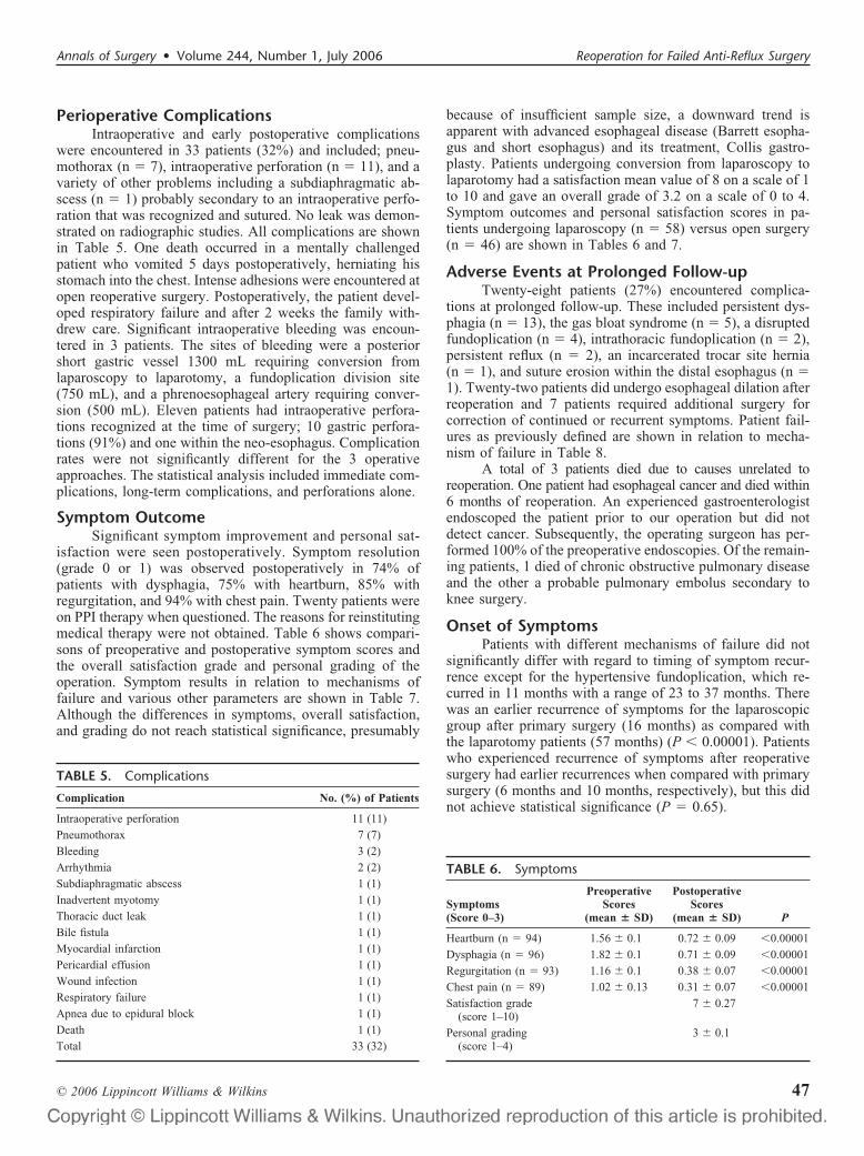
Perioperative ComplicationsIntraoperative and early postoperative complications
were encountered in 33 patients (32%) and included; pneu-mothorax (n � 7), intraoperative perforation (n � 11), and avariety of other problems including a subdiaphragmatic ab-scess (n � 1) probably secondary to an intraoperative perfo-ration that was recognized and sutured. No leak was demon-strated on radiographic studies. All complications are shownin Table 5. One death occurred in a mentally challengedpatient who vomited 5 days postoperatively, herniating hisstomach into the chest. Intense adhesions were encountered atopen reoperative surgery. Postoperatively, the patient devel-oped respiratory failure and after 2 weeks the family with-drew care. Significant intraoperative bleeding was encoun-tered in 3 patients. The sites of bleeding were a posteriorshort gastric vessel 1300 mL requiring conversion fromlaparoscopy to laparotomy, a fundoplication division site(750 mL), and a phrenoesophageal artery requiring conver-sion (500 mL). Eleven patients had intraoperative perfora-tions recognized at the time of surgery; 10 gastric perfora-tions (91%) and one within the neo-esophagus. Complicationrates were not significantly different for the 3 operativeapproaches. The statistical analysis included immediate com-plications, long-term complications, and perforations alone.
Symptom OutcomeSignificant symptom improvement and personal sat-
isfaction were seen postoperatively. Symptom resolution(grade 0 or 1) was observed postoperatively in 74% ofpatients with dysphagia, 75% with heartburn, 85% withregurgitation, and 94% with chest pain. Twenty patients wereon PPI therapy when questioned. The reasons for reinstitutingmedical therapy were not obtained. Table 6 shows compari-sons of preoperative and postoperative symptom scores andthe overall satisfaction grade and personal grading of theoperation. Symptom results in relation to mechanisms offailure and various other parameters are shown in Table 7.Although the differences in symptoms, overall satisfaction,and grading do not reach statistical significance, presumably
because of insufficient sample size, a downward trend isapparent with advanced esophageal disease (Barrett esopha-gus and short esophagus) and its treatment, Collis gastro-plasty. Patients undergoing conversion from laparoscopy tolaparotomy had a satisfaction mean value of 8 on a scale of 1to 10 and gave an overall grade of 3.2 on a scale of 0 to 4.Symptom outcomes and personal satisfaction scores in pa-tients undergoing laparoscopy (n � 58) versus open surgery(n � 46) are shown in Tables 6 and 7.
Adverse Events at Prolonged Follow-upTwenty-eight patients (27%) encountered complica-
tions at prolonged follow-up. These included persistent dys-phagia (n � 13), the gas bloat syndrome (n � 5), a disruptedfundoplication (n � 4), intrathoracic fundoplication (n � 2),persistent reflux (n � 2), an incarcerated trocar site hernia(n � 1), and suture erosion within the distal esophagus (n �1). Twenty-two patients did undergo esophageal dilation afterreoperation and 7 patients required additional surgery forcorrection of continued or recurrent symptoms. Patient fail-ures as previously defined are shown in relation to mecha-nism of failure in Table 8.
A total of 3 patients died due to causes unrelated toreoperation. One patient had esophageal cancer and died within6 months of reoperation. An experienced gastroenterologistendoscoped the patient prior to our operation but did notdetect cancer. Subsequently, the operating surgeon has per-formed 100% of the preoperative endoscopies. Of the remain-ing patients, 1 died of chronic obstructive pulmonary diseaseand the other a probable pulmonary embolus secondary toknee surgery.
Onset of SymptomsPatients with different mechanisms of failure did not
significantly differ with regard to timing of symptom recur-rence except for the hypertensive fundoplication, which re-curred in 11 months with a range of 23 to 37 months. Therewas an earlier recurrence of symptoms for the laparoscopicgroup after primary surgery (16 months) as compared withthe laparotomy patients (57 months) (P � 0.00001). Patientswho experienced recurrence of symptoms after reoperativesurgery had earlier recurrences when compared with primarysurgery (6 months and 10 months, respectively), but this didnot achieve statistical significance (P � 0.65).
TABLE 6. Symptoms
Symptoms(Score 0–3)
PreoperativeScores
(mean � SD)
PostoperativeScores
(mean � SD) P
Heartburn (n � 94) 1.56 � 0.1 0.72 � 0.09 �0.00001
Dysphagia (n � 96) 1.82 � 0.1 0.71 � 0.09 �0.00001
Regurgitation (n � 93) 1.16 � 0.1 0.38 � 0.07 �0.00001
Chest pain (n � 89) 1.02 � 0.13 0.31 � 0.07 �0.00001
Satisfaction grade(score 1–10)
7 � 0.27
Personal grading(score 1–4)
3 � 0.1
TABLE 5. Complications
Complication No. (%) of Patients
Intraoperative perforation 11 (11)
Pneumothorax 7 (7)
Bleeding 3 (2)
Arrhythmia 2 (2)
Subdiaphragmatic abscess 1 (1)
Inadvertent myotomy 1 (1)
Thoracic duct leak 1 (1)
Bile fistula 1 (1)
Myocardial infarction 1 (1)
Pericardial effusion 1 (1)
Wound infection 1 (1)
Respiratory failure 1 (1)
Apnea due to epidural block 1 (1)
Death 1 (1)
Total 33 (32)
Annals of Surgery • Volume 244, Number 1, July 2006 Reoperation for Failed Anti-Reflux Surgery
© 2006 Lippincott Williams & Wilkins 47

Mechanisms of FailureThe underlying mechanisms of failure for primary an-
tireflux surgery are shown in Table 3. The predominantcombinations included crus closure failure with partial fun-doplication disruption (n � 14), crus closure failure withcomplete fundoplication disruption (n � 3), crus closurefailure with a short esophagus (n � 3), and crus closurefailure with partial fundoplication disruption and a shortesophagus (n � 4). The accuracy of preoperative mechanismassessment was 78%. Those patients with Barrett esophagushad crus closure disruption (n � 10) 62.5% of the time, andthose with severe esophagitis (grade C or D) had crus closuredisruption (n � 6) 100% of the time. The accuracy of predictionfor each mechanism is shown in Table 9. The short esophagusproved to be most difficult to predict. Hiatal stenosis can only bedetermined at surgery.
Predictors of Failure or SuccessVarious preoperative variables including age, gender
BMI, both or either preoperative Barrett esophagus andsevere esophagitis (grade III or IV), Collis gastroplasty,laparoscopic primary surgery, open primary surgery, laparo-scopic versus open reoperative surgery, and a short esopha-gus were studied as outcome predictors by multivariate anal-ysis. Barrett esophagus alone and Barrett esophagus withesophagitis were predictive of improved regurgitation scores(P � 0.03; odds ratio, 3.6) and dysphagia control (P � 0.04;odds ratio, 0.3); however, patients after a Collis gastroplastycontinued to have significantly greater regurgitation (meanscore 0.86 vs. 0.3, P � 0.013).
DISCUSSIONWe report a large series of reoperations for failed
antireflux surgery and demonstrate that significant and sus-tained symptom improvement can be achieved. Symptomresolution ranged from 75% for dysphagia to 93% for chestpain at a mean follow-up of 38 months. Laparoscopic revi-sions are comparable to results attained when operatingthrough an open incision. Patients undergoing a laparoscopicrepair of a too tight fundoplication had better results thanother subsets of laparoscopic reoperative patients. As ex-pected, results trended downward for patients suffering withmore advanced disease. The perioperative complication ratewas 33%, including one death. The complication rate of thethoracotomies did not exceed that of patients undergoing anabdominal approach. We were able to predict the mechanismof failure, as found at surgery 78% of the time, and results asrelated to the mechanisms of failure demonstrated trendsthat may be important.
These results are encouraging and consistent with pre-vious reports.23–27 Our series does, however, include patientsrequiring reoperative surgery for more advanced disease;thus, 16 (15%) patients required an esophageal lengtheningprocedure. This operation is less than ideal in terms ofheartburn and acid regurgitation control as the newly formedneo-esophagus may secrete acid.28 Thirty-four thoracotomieswere performed because of advanced disease. The overallcomplication rate was higher primarily because intraoperativeTA
BLE
7.M
echa
nism
sof
Failu
re
Mec
hani
smof
Fai
lure
Hea
rtbu
rnD
ysph
agia
Reg
urgi
tati
onC
hest
Pai
nM
ean
Sati
sfac
tion
(Sco
re1–
10)
Mea
nP
erso
nal
Gra
ding
(Sco
re1–
4)P
reP
ost
Pre
Pos
tP
reP
ost
Pre
Pos
t
Cru
scl
osur
efa
ilur
e(n
�57
)1.
1�
0.4
0.7
�0.
32
�0.
40.
8�
0.3
0.8
�0.
30.
3�
0.1
1.2
�0.
40.
2�
0.2
7.3
3.1
Hyp
erte
nsiv
efu
ndop
lica
tion
(n�
24)
1�
0.2
0.7
�0.
22.
4�
0.2
0.8
�0.
20.
8�
0.2
0.2
�0.
11.
1�
0.3
0.4
�0.
27.
43
Sli
pped
fund
opli
cati
on(n
�18
)1.
8�
0.2
0.7
�0.
21.
8�
0.3
0.8
�0.
31.
6�
0.2
0.6
�0.
21
�0.
20.
2�
0.1
7.1
3
Fun
dopl
icat
ion
fail
ure
(n�
88)
1.6
�0.
10.
8�
0.1
1.8
�0.
10.
7�
0.1
1.2
�0.
10.
3�
0.1
1�
0.1
0.3
�0.
17.
33.
1
Sho
rtes
opha
gus
(n�
20)
2.0
�0.
90.
8�
1.0
2.0
�1.
31.
0�
0.9
1.1
�1.
10.
7�
0.9
1.2
�1.
40.
4�
0.8
6.4
2.8
Col
lis
pati
ents
(n�
16)
1.9
�1.
00.
9�
1.0
2.1
�1.
21.
1�
0.9
1.2
�1.
20.
8�
0.9
1.4
�1.
40.
5�
0.8
62.
7
Bar
rett
pati
ents
(n�
16)
2.1
�0.
21.
2�
0.3
1.8
�0.
31
�0.
31.
7�
0.2
0.2
�0.
10.
7�
0.3
0.6
�0.
36.
32.
7
Rec
urre
ntG
ER
D(n
�39
)1.
7�
1.2
1.1
�0.
91.
8�
1.1
0.7
�0.
91.
3�
10.
6�
0.8
0.8
�1.
20.
5�
0.8
6.6
2.8
Red
ola
paro
scop
icpa
tien
ts(n
�58
)1.
4�
0.1
0.7
�0.
11.
8�
0.1
0.6
�0.
11.
1�
0.1
0.3
�0.
10.
8�
0.2
0.3
�0.
17.
33.
1
Red
ola
paro
tom
ypa
tien
ts(n
�46
)1.
5�
0.4
0.5
�0.
22.
4�
0.3
0.9
�0.
31.
1�
0.3
0.1
�0.
11
�0.
40.
3�
0.1
7.2
3.1
Red
oth
orac
otom
ypa
tien
ts(n
�34
)1.
7�
10.
8�
0.9
1.6
�1.
40.
8�
11.
2�
1.1
0.6
�0.
81.
3�
1.3
0.3
�0.
66.
93.
0
Iqbal et al Annals of Surgery • Volume 244, Number 1, July 2006
© 2006 Lippincott Williams & Wilkins48

perforations were included, only one of which resulted inpostoperative sequelae. Careful attention to operative planeswill reduce the perforation rate and that was apparent as ourexperience increased. The postoperative death due to respi-ratory insufficiency was probably unavoidable, but the patientthat developed metastases from a missed adenocarcinomademonstrates the need for the surgeon to perform all preop-erative endoscopies and that if dissection is more difficultthan expected, conversion to an open operation is necessary.
Avoidable technical pitfalls at primary surgery arenumerous, but the following are highlighted. Inadequate fun-dus size; a funnel-shaped fundus is difficult to recognize atlaparoscopy. In this circumstance, the temptation is to errorby choosing a fundoplication lead point too low on the gastricbody creating a 2-compartment deformity. This results in apoorly relaxing fundoplication and unremitting dysphagia.The inadequate fundus can be recognized when calibratingthe fundoplication over a 60-Fr bougie and the antidote isthorough mobilization of the gastric fundus. The literaturedoes show this is not routinely required and we agree.However, if one is to become comfortable with short gastricvessel division, routine mobilization is helpful. If inadequatefundic size remains, a Toupet or even a Dor fundoplication isappropriate. Another pitfall is the missed short esophagus thatoften results in an intrathoracic fundoplication with fundopli-cation disruption.
The controversy concerning the short esophagus con-tinues, however, all agree that normally the distal esophaguslies within the abdominal cavity. Most agree that the point atwhich the gastric serosa meets the longitudinal muscle fibersof the esophagus is the GEJ. The left transthoracic approach
provides several advantages, including complete skeletoniza-tion of the esophagus to the level of the left main stembronchus, easier preservation of the vagal nerves, fresh dis-section planes, and accurate assessment of esophageal length.An excellent view of GEJ after fundoplication dismantlingand fat pad resection allows the surgeon to determine exactlywhere the GEJ lies in relation to the diaphragm. If the GEJwas still proximal to the diaphragm or we were unable toreduce it 2 cm into the abdominal cavity without tension, wethen performed a gastroplasty. This anatomic finding wasrepeatedly seen and has been observed by the senior coau-thors in patients at primary surgery as well.
The reoperation failure rate is 20% to 30% usuallybecause of crus closure failure and/or fundoplication disrup-tion. We must ask: is the patient collagen deficient (a skin testis needed), does the patient play a hidden role in their disease(by controlled or uncontrolled vomiting, retching, lifting), orwas the operation technically deficient? The later can occur ifthe fundus size is inadequate, as mentioned at the primaryoperation, or especially if a short esophagus is not recog-nized. At reoperation, there is often fibrosis involving theGEJ and the esophageal longitudinal smooth muscle gastricserosa interface may not be easy to distinguish laparoscopi-cally or even by direct vision. If doubt remains conversion toan open procedure is appropriate after which a lengtheningprocedure may be indicated.
A short esophagus is defined in this series as the GEJbeing less than 3 cm below the arch of the crus after laparoscopicesophageal mobilization. A 2 cm measurement was acceptablefor open surgery. This may result in crus closure failure, fundo-plication disruption, or a slipped repair. This mechanism was
TABLE 8. In Relation to Mechanism of Failure
Postoperative Mechanismof Failure
Re-Redo Patients(n � 7)
Grade 3 PostoperativeSymptom Scores
(n � 10)>1/wk Medication Use
(n � 21)Total
(n � 33)
Crus closure failure 3 6 9 18
Hiatal stenosis — 1 2 3
Partial disruption 3 3 6 12
Complete disruption 1 1 — 2
Hypertensive fundoplication 2 1 5 8
Slipped fundoplication 1 1 2 4
Too loose fundoplication — — 2 2
Short esophagus 1 1 2 4
TABLE 9. Predictability of Mechanism of Failure
CrusClosureFailure
HiatalStenosis
PartialDisruption
CompleteDisruption
HypertensiveFundoplication
SlippedFundoplication
Too LooseFundoplication
ShortEsophagus Gastroparesis
Postoperative(no. of patients)
52 5 36 7 24 18 2 16 2
Preoperative(no. of patients)
50 1 32 6 22 16 1 6 2
% predictedcorrectly
96 20 86 86 92 89 50 37 100
Annals of Surgery • Volume 244, Number 1, July 2006 Reoperation for Failed Anti-Reflux Surgery
© 2006 Lippincott Williams & Wilkins 49
most frequently overlooked preoperatively in our series, al-though we specifically get an upright esophagram to assist inits diagnosis. Further investigation into more accurate predic-tors of this important entity is needed.
Postoperative pain, the risk of pulmonary complications,inexperience, and lack of thoracic privileges have discouragedsurgeons from performing thoracotomies for recurrent benignesophageal disease. Resolution of preoperative chest painwas not compromised after thoracotomy in this series, and nothoracotomy patient required prolonged ventilation. Carefulpreoperative assessment is necessary to assure that the patientcan tolerate this approach and perioperative epidural analge-sia was routinely used. On occasion, a diaphragmatic coun-terincision is necessary if dense abdominal adhesions areencountered. Abdominal surgeons will not find this difficultand closure of the diaphragm is safe and effective. Trainingand experience with the transthoracic approach are availablefor general and thoracic surgeons alike. An esophageal fel-lowship or prolonged preceptorship will make hospital priv-ileges attainable in most circumstances.
Previous attempts have been made to classify fundo-plication failure mechanisms. These systems have used nu-merical designations, anatomic/functional designations, anddiagnostic errors in describing specific failures.22–24,29–31 Theprevious classifications emphasized anatomic malformations,but Skinner31 used 5 functional categories in describing thecause of symptoms for fundoplication failures. We attemptedto use a mechanism of failure classification but found that mostpatients had several mechanisms of failure, thus making eachpatient difficult to categorize. A consensus conference coulddevelop a classification of fundoplication failure, thereby im-proving communication and eventually prognostication.
The stenotic hiatus is a new phenomenon and has beenreported previously by our group.32 It is probably related tocautery dissection of the hiatus and, although rare, is animportant new mechanism for patients suffering with dyspha-gia. It can be easily handled at operation if recognized. The2-compartment stomach has also been recently reported26 andis an important new mechanism that affected 5 of our pa-tients. We also experienced a too loose fundoplication createdat the time of surgery. These patients never experience improve-ment in their heartburn and regurgitation. To our knowledge,this mechanism, although inherently obvious and expected, hasnever been reported previously.
Remaining questions include the role of mesh reinforce-ment, collagen metabolism and its effect on fundoplicationfailure and recurrent hiatal hernia formation, a better alternativethan the Collis gastroplasty, when esophagectomy is appropri-ate, and the true role of diaphragm stressors. Long-term random-ized mesh trials with objective hernia recurrence follow-up areunderway. Initial data for mesh reinforcement are encourag-ing.33–35 Klinge et al have shown that the collagen I to III ratiois related to ventral hernia recurrence and perhaps this is true forrecurrent hiatal hernias as well.36 The role of esophagectomy forpatients with advanced benign disease requires further study.Finally, diaphragm stress factors have been analyzed,37,38 but amore comprehensive series with a validated questionnaire isneeded.
CONCLUSIONReoperative surgery for GERD is safe and effective. The
laparoscopic approach appears to be as effective as laparotomyfor reoperative surgery, and the percentage of patients requiringa Collis gastroplasty for a short esophagus was high in thisseries. The transthoracic approach does not result in excessivepostoperative morbidities.
REFERENCES1. DeMeester TR, Stein HJ. Surgical treatment of gastroesophageal reflux
disease. In: Castell DO, ed. The Esophagus. Boston: Little, Brown;1992:579–626.
2. Sataloff DM, Pursnani K, Hoyo S, et al. An objective assessment oflaparoscopic antireflux surgery. Am J Surg. 1997;174:63–67.
3. Hetzel DJ, Dent J, Reed WD, et al. Healing and relapse of severe pepticesophagitis after treatment with omeprazole. Gastroenterology. 1988;95:903–912.
4. DeMeester TR, Bonavina L, Albertucci M. Nissen fundoplication forgastroesophageal reflux disease: evaluation of primary repair in 100consecutive patients. Ann Surg. 1986;204:9–20.
5. Donahue PE, Samelson S, Nyhus LM, et al. The floppy Nissen fundo-plication: effective long-term control of pathologic reflux. Arch Surg.1985;120:663–668.
6. Stirling MC, Orringer MB. Surgical treatment after the failed antirefluxoperation. J Thorac Cardiovasc Surg. 1986;92:667–672.
7. Cadiere GB, Himpens J, Rajan A, et al. Laparoscopic Nissen fundopli-cation: laparoscopic dissection technique and results. Hepatogastroen-terology. 1997;44:4–10.
8. Isolauri J, Luostarinen M, Viljakka M, et al. Long-term comparison ofantireflux surgery versus conservative therapy for reflux esophagitis.Ann Surg. 1997;225:295–299.
9. Dallemagne B, Weerts JM, Jehaes C, et al. Laparoscopic Nissen fundo-plication: preliminary report. Surg Laparosc Endosc. 1991;1:138–143.
10. Dallemagne B, Weerts JM, Jeahes C, et al. Results of laparoscopicNissen fundoplication. Hepatogastroenterology. 1998;45:1338–1343.
11. Hinder RA, Filipi CJ, Wetscher G, et al. Laparoscopic Nissen fundopli-cation is an effective treatment for gastroesophageal reflux disease. AnnSurg. 1994;220:472–481.
12. Frantzides CT, Carlson MA. Laparoscopic redo Nissen fundoplication.J Laparoendosc Adv Surg Tech. 1997;7:235–239.
13. Hinder RA, Perdikis G, Klinger PJ, et al. The surgical option forgastroesophageal reflux disease. Am J Med. 1997;103(suppl):144–148.
14. Laine S, Rantala A, Gullichsen R, et al. Laparoscopic vs conventionalNissen fundoplication: a prospective randomized study. Surg Endosc. 1997;11:441–444.
15. Hiebert CA, O’Mara CS. The Belsey operation for hiatal hernia: atwenty year experience. Am J Surg. 1979;137:532–535.
16. Shirazi SS, Schulze K, Soper RT. Long-term follow-up for treatment ofcomplicated chronic reflux esophagitis. Arch Surg. 1987;122:548–552.
17. Cuschieri A, Hunter J, Wolfe B, et al. Multicenter prospective evaluationof laparoscopic antireflux surgery: preliminary report. Surg Endosc.1993;7:505–510.
18. Hunter JG, Trus TL, Branum GD, et al. A physiologic approach tolaparoscopic fundoplication for gastroesophageal reflux disease. AnnSurg. 1996;223:673–685.
19. Jamieson GG, Watson DI, Britten-Jones R, et al. Laparoscopic Nissenfundoplication. Ann Surg. 1994;220:137–145.
20. Peters JH, Heimbucher J, Kauer WK, et al. Clinical and physiologiccomparison of laparoscopic and open Nissen fundoplication. J Am CollSurg. 1995;180:385–393.
21. DePaula AL, Hashiba K, Bafutto M, et al. Laparoscopic reoperationsafter failed and complicated antireflux operations. Surg Endosc. 1995;9:681–686.
22. Siewert JR, Isolauri J, Feussner H. Reoperation following failed fundo-plication. World J Surg. 1989;13:791–796.
23. Horgan S, Pohl D, Bogetti D, et al. Failed antireflux surgery: what havewe learned from reoperations? Arch Surg. 1999;134:809–815.
24. Floch NR, Hinder RA, Klingler PJ, et al. Is laparoscopic re-operation forfailed antireflux surgery feasible? Arch Surg. 1999;134:733–737.
Iqbal et al Annals of Surgery • Volume 244, Number 1, July 2006
© 2006 Lippincott Williams & Wilkins50

25. Coelho JC, Goncalves CG, Claus CM, et al. Late laparoscopic reopera-tion of failed antireflux procedures. Surg Laparosc Endosc. 2004;14:113–117.
26. Hunter JG, Smith CD, Branum GD, et al. Laparoscopic fundoplicationfailures: patterns of failure and response to fundoplication revision. AnnSurg. 1999;230:595–619.
27. Smith DC, McClusky DA, Murad AR, et al. When fundoplication failsredo? Ann Surg. 2005;241:861–871.
28. Horvath KD, Swanstrom LL, Jobe BA. The short esophagus: pathophys-iology, incidence, presentation, and treatment in the era of laparoscopicantireflux surgery. Ann Surg. 2000;232:630–640.
29. Hill LD, Ilves R, Stevenson JK, et al. Reoperation for disruption andrecurrence after Nissen fundoplication. Arch Surg. 1979;114:542–548.
30. Leonardi HK, Crozier RE, Ellis FH Jr. Reoperation for complications ofthe Nissen fundoplication. J Thorac Cardiovasc Surg. 1981;81:50–56.
31. Skinner DB. Pathophysiology of gastroesophageal reflux. Ann Surg.1985;202:546–556.
32. Selima MA, Awad ZT, Filipi CJ. Hiatal stenosis after laparoscopicNissen fundoplication: a report of 2 cases. JSLS. 2002;6:397–399.
33. Granderath FA, Schweiger UM, Kamolz T, et al. Laparoscopic Nissenfundoplication with prosthetic hiatal closure reduces postoperative in-trathoracic wrap herniation: preliminary results of a prospective random-ized functional and clinical study. Arch Surg. 2005;140:40–48.
34. Frantzides CT, Madan AK, Carlson MA, et al. A prospective, random-ized trial of laparoscopic polytetrafluoroethylene (PTFE) patch repair vssimple cruroplasty for large hiatal hernia. Arch Surg. 2002;137:649–652.
35. Targarona EM, Bendahan G, Balague C, et al. Mesh in the hiatus: acontroversial issue. Arch Surg. 2004;139:1286–1296.
36. Klinge U, Si ZY, Zheng H, et al. Abnormal collagen I to III distributionin the skin of patients with incisional hernia. Eur Surg Res. 2000;32:43–48.
37. Iqbal A, Kakarlapudi GV, Awad ZT, et al. Assessment of diaphragmaticstressors as risk factors for failure of laparoscopic Nissen fundoplicationand postoperative hiatal hernia formation �Abstract�. Submitted toSSAT.
38. Soper NJ, Dunnegan D. Anatomic fundoplication failure after laparo-scopic antireflux surgery. Ann Surg. 1999;229:669–676.
Annals of Surgery • Volume 244, Number 1, July 2006 Reoperation for Failed Anti-Reflux Surgery
© 2006 Lippincott Williams & Wilkins 51