Renal Intercalated Cells Sense and Mediate Inflammation via the P2Y14 Receptor
24
RESEARCH ARTICLE Renal Intercalated Cells Sense and Mediate Inflammation via the P2Y 14 Receptor Anie Azroyan 1,2 , Virna Cortez-Retamozo 1 , Richard Bouley 1,2 , Rachel Liberman 1,2 , Ye Chun Ruan 1,2 , Evgeny Kiselev 3 , Kenneth A. Jacobson 3 , Mikael J. Pittet 1 , Dennis Brown 1,2 , Sylvie Breton 1,2* 1 Center for Systems Biology, Massachusetts General Hospital/Harvard Medical School, Boston, Massachusetts, United States of America, 2 Program in Membrane Biology/Nephrology Division, Massachusetts General Hospital/Harvard Medical School, Boston, Massachusetts, United States of America, 3 Laboratory of Bioorganic Chemistry, National Institute of Diabetes and Digestive and Kidney Diseases, National Institutes of Health, Bethesda, Maryland, United States of America * [email protected] Abstract Uncontrolled inflammation is one of the leading causes of kidney failure. Pro-inflammatory responses can occur in the absence of infection, a process called sterile inflammation. Here we show that the purinergic receptor P2Y 14 (GPR105) is specifically and highly expressed in collecting duct intercalated cells (ICs) and mediates sterile inflammation in the kidney. P2Y 14 is activated by UDP-glucose, a damage-associated molecular pattern molecule (DAMP) released by injured cells. We found that UDP-glucose increases pro-inflammatory chemokine expression in ICs as well as MDCK-C11 cells, and UDP-glucose activates the MEK1/2-ERK1/2 pathway in MDCK-C11 cells. These effects were prevented following inhi- bition of P2Y 14 with the small molecule PPTN. Tail vein injection of mice with UDP-glucose induced the recruitment of neutrophils to the renal medulla. This study identifies ICs as novel sensors, mediators and effectors of inflammation in the kidney via P2Y 14 . Introduction Kidney failure is almost always associated with uncontrolled inflammation [1,2]. A link be- tween renal inflammation and signaling via purinergic receptors has been established, but very little is currently known about the underlying mechanisms involved and how to prevent and al- leviate the severe damage cause by inflammation. Numerous purinergic receptors are expressed in the kidney, and deregulation of purinergic signaling is associated with several pathologies, including hypertension, chronic kidney disease, acute kidney injury, diabetic nephropathy and glomerulonephritis [3,4]. Purinergic receptors are involved in the regulation of water, electro- lyte, and volume homeostasis by collecting duct principal cells [5–8]. However, there is limited knowledge on the purinergic regulation of the other major cell type of the collecting duct, the intercalated cell (IC). ICs participate in the maintenance of acid/base homeostasis via the pro- ton-pumping V-ATPase [9,10]. In the epididymis, ATP and adenosine are potent activators of V-ATPase-dependent proton secretion in clear cells, which are analogous to ICs [11]. PLOS ONE | DOI:10.1371/journal.pone.0121419 March 23, 2015 1 / 24 OPEN ACCESS Citation: Azroyan A, Cortez-Retamozo V, Bouley R, Liberman R, Ruan YC, Kiselev E, et al. (2015) Renal Intercalated Cells Sense and Mediate Inflammation via the P2Y 14 Receptor. PLoS ONE 10(3): e0121419. doi:10.1371/journal.pone.0121419 Academic Editor: Niels Olsen Saraiva Câmara, Universidade de Sao Paulo, BRAZIL Received: October 24, 2014 Accepted: February 1, 2015 Published: March 23, 2015 Copyright: © 2015 Azroyan et al. This is an open access article distributed under the terms of the Creative Commons Attribution License, which permits unrestricted use, distribution, and reproduction in any medium, provided the original author and source are credited. Data Availability Statement: All relevant data are within the paper. Funding: This work was supported by NIH grants HD040793 and DK097124 (to S.B.) and NIH grant DK042956 (to D.B.). The Microscopy Core facility of the MGH Program in Membrane Biology receives support from the Boston Area Diabetes and Endocrinology Research Center (DK57521) and the Center for the Study of Inflammatory Bowel Disease (DK43351). S.B. is a recipient of the Charles and Ann Sanders Research Scholar Award at MGH. K.A.J. is funded by the Intramural Research Program of the NIH, NIDDK. E.K. is funded by the PRAT Program,
Transcript of Renal Intercalated Cells Sense and Mediate Inflammation via the P2Y14 Receptor

RESEARCH ARTICLE
Renal Intercalated Cells Sense and MediateInflammation via the P2Y14 ReceptorAnie Azroyan1,2, Virna Cortez-Retamozo1, Richard Bouley1,2, Rachel Liberman1,2, YeChun Ruan1,2, Evgeny Kiselev3, Kenneth A. Jacobson3, Mikael J. Pittet1, Dennis Brown1,2,Sylvie Breton1,2*
1 Center for Systems Biology, Massachusetts General Hospital/Harvard Medical School, Boston,Massachusetts, United States of America, 2 Program in Membrane Biology/Nephrology Division,Massachusetts General Hospital/Harvard Medical School, Boston, Massachusetts, United States of America,3 Laboratory of Bioorganic Chemistry, National Institute of Diabetes and Digestive and Kidney Diseases,National Institutes of Health, Bethesda, Maryland, United States of America
AbstractUncontrolled inflammation is one of the leading causes of kidney failure. Pro-inflammatory
responses can occur in the absence of infection, a process called sterile inflammation. Here
we show that the purinergic receptor P2Y14 (GPR105) is specifically and highly expressed
in collecting duct intercalated cells (ICs) and mediates sterile inflammation in the kidney.
P2Y14 is activated by UDP-glucose, a damage-associated molecular pattern molecule
(DAMP) released by injured cells. We found that UDP-glucose increases pro-inflammatory
chemokine expression in ICs as well as MDCK-C11 cells, and UDP-glucose activates the
MEK1/2-ERK1/2 pathway in MDCK-C11 cells. These effects were prevented following inhi-
bition of P2Y14 with the small molecule PPTN. Tail vein injection of mice with UDP-glucose
induced the recruitment of neutrophils to the renal medulla. This study identifies ICs as
novel sensors, mediators and effectors of inflammation in the kidney via P2Y14.
IntroductionKidney failure is almost always associated with uncontrolled inflammation [1,2]. A link be-tween renal inflammation and signaling via purinergic receptors has been established, but verylittle is currently known about the underlying mechanisms involved and how to prevent and al-leviate the severe damage cause by inflammation. Numerous purinergic receptors are expressedin the kidney, and deregulation of purinergic signaling is associated with several pathologies,including hypertension, chronic kidney disease, acute kidney injury, diabetic nephropathy andglomerulonephritis [3,4]. Purinergic receptors are involved in the regulation of water, electro-lyte, and volume homeostasis by collecting duct principal cells [5–8]. However, there is limitedknowledge on the purinergic regulation of the other major cell type of the collecting duct, theintercalated cell (IC). ICs participate in the maintenance of acid/base homeostasis via the pro-ton-pumping V-ATPase [9,10]. In the epididymis, ATP and adenosine are potent activators ofV-ATPase-dependent proton secretion in clear cells, which are analogous to ICs [11].
PLOSONE | DOI:10.1371/journal.pone.0121419 March 23, 2015 1 / 24
OPEN ACCESS
Citation: Azroyan A, Cortez-Retamozo V, Bouley R,Liberman R, Ruan YC, Kiselev E, et al. (2015) RenalIntercalated Cells Sense and Mediate Inflammationvia the P2Y14 Receptor. PLoS ONE 10(3): e0121419.doi:10.1371/journal.pone.0121419
Academic Editor: Niels Olsen Saraiva Câmara,Universidade de Sao Paulo, BRAZIL
Received: October 24, 2014
Accepted: February 1, 2015
Published: March 23, 2015
Copyright: © 2015 Azroyan et al. This is an openaccess article distributed under the terms of theCreative Commons Attribution License, which permitsunrestricted use, distribution, and reproduction in anymedium, provided the original author and source arecredited.
Data Availability Statement: All relevant data arewithin the paper.
Funding: This work was supported by NIH grantsHD040793 and DK097124 (to S.B.) and NIH grantDK042956 (to D.B.). The Microscopy Core facility ofthe MGH Program in Membrane Biology receivessupport from the Boston Area Diabetes andEndocrinology Research Center (DK57521) and theCenter for the Study of Inflammatory Bowel Disease(DK43351). S.B. is a recipient of the Charles and AnnSanders Research Scholar Award at MGH. K.A.J. isfunded by the Intramural Research Program of theNIH, NIDDK. E.K. is funded by the PRAT Program,

Extracellular ATP stimulates bone resorption in osteoclasts, a process that also requires activityof the V-ATPase [12,13]. These studies suggest a role for the purinergic regulation of acid/basetransport in the kidney, but the purinergic receptor signature of ICs still remains tobe characterized.
Nucleotide-activated purinergic receptors are separated into two families, P2X receptorsthat are ligand-gated ion channels, and P2Y receptors that are G protein-coupled receptors(GPCRs) [5–7]. Based on its homology with other P2 receptors, p2y5 was initially proposed tobe a nucleotide-receptor, but it was subsequently shown to be insensitive to nucleotides [14]and to bind lysophosphatidic acid (LPA) [15]. Similarly, p2y10 is a lysophospholipid receptorthat is not activated by nucleotides [16]. The P2Y14 receptor (also known as GPR105) is themost recent addition to the P2Y receptor family [17,18]. P2Y14 is specifically activated by nu-cleotide sugars including UDP-glucose and it is insensitive to ADP/ATP and UTP [18]. WhileUDP-glucose is used in the metabolism of nucleotide sugars, it is also released by cells and actsas an autocrine activator of the P2Y14 receptor. Most nucleotides are rapidly degraded by ecto-nucleotidases after their release, but UDP-glucose resists hydrolysis by these enzymes [19].While virtually all cells release nucleotides under basal conditions [20], this release can be ac-centuated in response to stimuli leading to activation of purinergic receptors [21]. UDP-glu-cose, extracellular ATP and adenosine are emerging as immune-regulatory factors known asDAMPs (damage associated molecular pattern) molecules [22,23]. DAMPs initiate sterile in-flammatory reactions, as opposed to PAMP (pathogen associated molecular patterns), whichperpetuate infectious pro-inflammatory responses [23–26].
The role of P2Y14 as an inflammatory mediator was suggested based on its high expressionlevels in immune cells, and on the increased release of its ligand, UDP-glucose, by damagedcells [17,20,27–32]. In addition to immune cells, several tissues including the brain, the gastro-intestinal tract, the kidney and the lung express P2Y14 mRNA [18]. Patients with cystic fibrosisand asthma secrete high amounts of UDP-glucose in their lungs [27,33], and P2Y14 activationby UDP-glucose in airway epithelial cells leads to IL-8 secretion [34]. Furthermore, injection ofUDP-glucose into the mouse uterus induces the recruitment of neutrophils into the endometri-um [35]. Another indication of the inflammatory role of P2Y14 is the up-regulation of itsmRNA expression by lipopolysaccharides (LPS) [35,36]. However, while purinergic signalinghas been established in the modulation of renal inflammation, the role of P2Y14 in mediatingimmune responses in the kidney has not been described. The objective of this study was, there-fore, to examine the potential participation of this receptor in the initiation of inflammation inthe kidney.
In this study we uncover elevated expression of a restricted number of P2 receptors in ICs,most notably P2Y14. Moreover, P2Y14 expression was not detectable in other renal epithelialcells. We also provide evidence that P2Y14 activation by UDP-glucose induces a pro-inflamma-tory response in ICs that is mediated by activation of the MAPK pathway. This is followed byincreased expression of pro-inflammatory chemokines in ICs, and subsequent neutrophil infil-tration in the renal medulla. Thus, we have identified a novel inflammatory role for renal ICsvia P2Y14 signaling.
Materials and Methods
Reagents and antibodiesUridine 50-diphosphoglucose disodium salt hydrate from Saccharomyces cerevisiae (UDP-glu-cose) and the MEK inhibitor PD98059 were purchased from Sigma Aldrich (St. Louis, MO).Uridine diphospho-D-[6-3H] glucose ([3H]UDP-glucose) was purchased from Perkin Elmer(Waltham, MA). PPTN, a selective high affinity antagonist of the P2Y14 receptor has been
Immune Role of P2Y14 in Intercalated Cells
PLOS ONE | DOI:10.1371/journal.pone.0121419 March 23, 2015 2 / 24
NIGMS. The funders had no role in study design,data collection and analysis, decision to publish, orpreparation of the manuscript.
Competing Interests: The authors have declaredthat no competing interests exist.

described previously [37]. The chicken antibody against the V-ATPase B1 subunit has been de-scribed previously [38]. An affinity-purified chicken antibody against the V-ATPase A subunitwas raised against the same sequence as previously described for a rabbit anti-V-ATPase Asubunit antibody [39,40]. A rabbit anti-P2Y14 antibody and its immunizing peptide were pur-chased from Alomone labs (Jerusalem, Israel). Rabbit anti-cytoskeletal actin antibody wasfrom Bethyl Laboratory (Montgomery, TX). Rabbit anti-pendrin antibody was a kind gift fromDr. Aronson (Yale University). Rabbit monoclonal antibodies against p-p44/p42 MAPK andagainst total p44/p42 MAPK were purchased from Cell Signaling Technology (Boston, MA).Rat anti-Ly6G antibody was from Biolegend (San Diego, CA). Mouse anti-pan-actin was pur-chased from EMDMillipore (Billerica, MA). All secondary antibodies used were affinity puri-fied and purchased from Jackson Immunoresearch (West Grove, PA), except for a goat anti-ratcy3, which was purchased from Invitrogen (Grand Island, NY). Streptavidin-fluorescein waspurchased from Invitrogen. Cell culture medium was purchased from Invitrogen, and bovineserum was purchased from Atlanta Biologicals (Lawrenceville, CA).
AnimalsAdult male mice (eight to ten weeks old) were used for all experiments. Transgenic mice ex-pressing EGFP under the promoter of the V-ATPase B1 subunit (B1-EGFP) mice have beendescribed previously [41]. Wild type (C57BL/6 x CBAF1) mice were purchased from JacksonLaboratory (Bar Harbor, ME). Animals were housed under standard conditions and main-tained on a standard rodent diet. The Massachusetts General Hospital (MGH) Subcommitteeon Research Animal Care approved all animal studies, in accordance with National Institutesof Health, Department of Agriculture, and Accreditation of Laboratory Animal Care require-ments. Our Institutional Animal Care and Use Committee (IACUC) specifically approved thisstudy. For tail vein injections, animals were kept under isofluorane anesthesia (Baxter, Deer-field, IL) for several minutes to allow the injection of 200 μl of either saline solution or a salinesolution containing 100 μMUDP-glucose (200 mg/kg of body weight).
Isolation of intercalated cells from mouse kidneysMice were anesthetized using pentobarbital sodium (50 mg/kg body, ip, Nembutal, AbbottLaboratories, Abbott Park, IL). The blood was flushed out of the organs by perfusing the ani-mals with a phosphate-buffered saline (PBS) through the cardiac left ventricle at a constantflow rate of 17 ml/min. Kidneys were excised and sliced, and some kidneys were microdissectedto separate the cortex from the medulla. Tissues were then minced immediately in RPMI 1640medium (Invitrogen, Grand Island, NY) containing 1.0 mg/ml collagenase type I (Invitrogen),1.0 mg/ml collagenase type II (Sigma Aldrich) and 2 mg/ml hyaluronidase (Sigma Aldrich),and digested for 45 min at 37°C. A 40-μm-nylon mesh was used to remove undigested materialfollowing tissue digestion. Cells were then washed once with RPMI 1640 medium and oncewith a calcium-free PBS. EGFP-positive (EGFP(+)) and negative (EGFP(−)) cells were isolatedimmediately by FACS, based on their green fluorescence intensity, as we have described previ-ously [42]. Cell isolation was performed at the MGH flow cytometry core facility using a modi-fied FACS Vantage cell sorter (BD Biosciences, San Jose, CA). FACS isolated samples wereused without delay for subsequent protein and RNA isolation.
RNA isolation and RT-PCRTotal RNA was isolated from cells using RNeasy micro kit (Qiagen, Valencia, CA) and fromtissues using RNeasy Mini kit, as we have described previously [42]. The sequences of the PCRprimer sets, purchased from Invitrogen, are listed in Table 1. For end point PCRs, reaction
Immune Role of P2Y14 in Intercalated Cells
PLOS ONE | DOI:10.1371/journal.pone.0121419 March 23, 2015 3 / 24
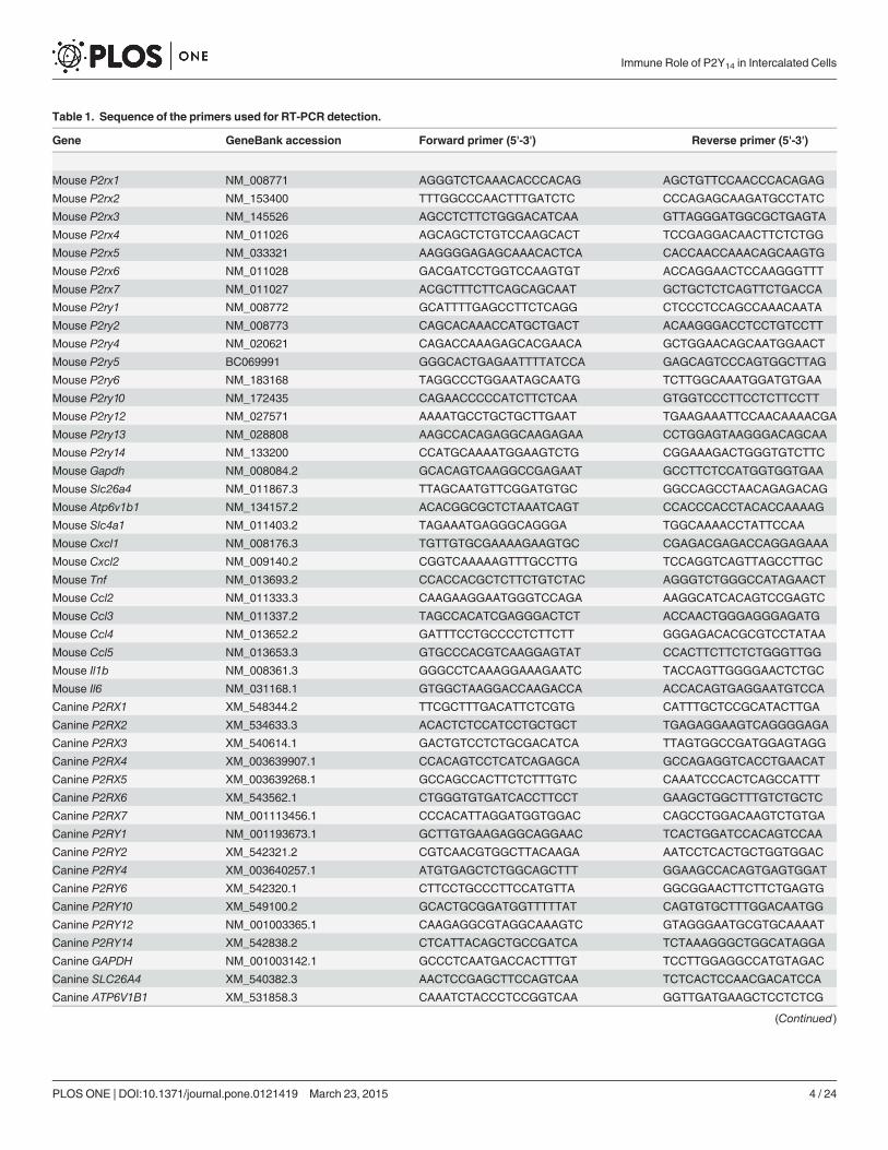
Table 1. Sequence of the primers used for RT-PCR detection.
Gene GeneBank accession Forward primer (5'-3') Reverse primer (5'-3')
Mouse P2rx1 NM_008771 AGGGTCTCAAACACCCACAG AGCTGTTCCAACCCACAGAG
Mouse P2rx2 NM_153400 TTTGGCCCAACTTTGATCTC CCCAGAGCAAGATGCCTATC
Mouse P2rx3 NM_145526 AGCCTCTTCTGGGACATCAA GTTAGGGATGGCGCTGAGTA
Mouse P2rx4 NM_011026 AGCAGCTCTGTCCAAGCACT TCCGAGGACAACTTCTCTGG
Mouse P2rx5 NM_033321 AAGGGGAGAGCAAACACTCA CACCAACCAAACAGCAAGTG
Mouse P2rx6 NM_011028 GACGATCCTGGTCCAAGTGT ACCAGGAACTCCAAGGGTTT
Mouse P2rx7 NM_011027 ACGCTTTCTTCAGCAGCAAT GCTGCTCTCAGTTCTGACCA
Mouse P2ry1 NM_008772 GCATTTTGAGCCTTCTCAGG CTCCCTCCAGCCAAACAATA
Mouse P2ry2 NM_008773 CAGCACAAACCATGCTGACT ACAAGGGACCTCCTGTCCTT
Mouse P2ry4 NM_020621 CAGACCAAAGAGCACGAACA GCTGGAACAGCAATGGAACT
Mouse P2ry5 BC069991 GGGCACTGAGAATTTTATCCA GAGCAGTCCCAGTGGCTTAG
Mouse P2ry6 NM_183168 TAGGCCCTGGAATAGCAATG TCTTGGCAAATGGATGTGAA
Mouse P2ry10 NM_172435 CAGAACCCCCATCTTCTCAA GTGGTCCCTTCCTCTTCCTT
Mouse P2ry12 NM_027571 AAAATGCCTGCTGCTTGAAT TGAAGAAATTCCAACAAAACGA
Mouse P2ry13 NM_028808 AAGCCACAGAGGCAAGAGAA CCTGGAGTAAGGGACAGCAA
Mouse P2ry14 NM_133200 CCATGCAAAATGGAAGTCTG CGGAAAGACTGGGTGTCTTC
Mouse Gapdh NM_008084.2 GCACAGTCAAGGCCGAGAAT GCCTTCTCCATGGTGGTGAA
Mouse Slc26a4 NM_011867.3 TTAGCAATGTTCGGATGTGC GGCCAGCCTAACAGAGACAG
Mouse Atp6v1b1 NM_134157.2 ACACGGCGCTCTAAATCAGT CCACCCACCTACACCAAAAG
Mouse Slc4a1 NM_011403.2 TAGAAATGAGGGCAGGGA TGGCAAAACCTATTCCAA
Mouse Cxcl1 NM_008176.3 TGTTGTGCGAAAAGAAGTGC CGAGACGAGACCAGGAGAAA
Mouse Cxcl2 NM_009140.2 CGGTCAAAAAGTTTGCCTTG TCCAGGTCAGTTAGCCTTGC
Mouse Tnf NM_013693.2 CCACCACGCTCTTCTGTCTAC AGGGTCTGGGCCATAGAACT
Mouse Ccl2 NM_011333.3 CAAGAAGGAATGGGTCCAGA AAGGCATCACAGTCCGAGTC
Mouse Ccl3 NM_011337.2 TAGCCACATCGAGGGACTCT ACCAACTGGGAGGGAGATG
Mouse Ccl4 NM_013652.2 GATTTCCTGCCCCTCTTCTT GGGAGACACGCGTCCTATAA
Mouse Ccl5 NM_013653.3 GTGCCCACGTCAAGGAGTAT CCACTTCTTCTCTGGGTTGG
Mouse Il1b NM_008361.3 GGGCCTCAAAGGAAAGAATC TACCAGTTGGGGAACTCTGC
Mouse Il6 NM_031168.1 GTGGCTAAGGACCAAGACCA ACCACAGTGAGGAATGTCCA
Canine P2RX1 XM_548344.2 TTCGCTTTGACATTCTCGTG CATTTGCTCCGCATACTTGA
Canine P2RX2 XM_534633.3 ACACTCTCCATCCTGCTGCT TGAGAGGAAGTCAGGGGAGA
Canine P2RX3 XM_540614.1 GACTGTCCTCTGCGACATCA TTAGTGGCCGATGGAGTAGG
Canine P2RX4 XM_003639907.1 CCACAGTCCTCATCAGAGCA GCCAGAGGTCACCTGAACAT
Canine P2RX5 XM_003639268.1 GCCAGCCACTTCTCTTTGTC CAAATCCCACTCAGCCATTT
Canine P2RX6 XM_543562.1 CTGGGTGTGATCACCTTCCT GAAGCTGGCTTTGTCTGCTC
Canine P2RX7 NM_001113456.1 CCCACATTAGGATGGTGGAC CAGCCTGGACAAGTCTGTGA
Canine P2RY1 NM_001193673.1 GCTTGTGAAGAGGCAGGAAC TCACTGGATCCACAGTCCAA
Canine P2RY2 XM_542321.2 CGTCAACGTGGCTTACAAGA AATCCTCACTGCTGGTGGAC
Canine P2RY4 XM_003640257.1 ATGTGAGCTCTGGCAGCTTT GGAAGCCACAGTGAGTGGAT
Canine P2RY6 XM_542320.1 CTTCCTGCCCTTCCATGTTA GGCGGAACTTCTTCTGAGTG
Canine P2RY10 XM_549100.2 GCACTGCGGATGGTTTTTAT CAGTGTGCTTTGGACAATGG
Canine P2RY12 NM_001003365.1 CAAGAGGCGTAGGCAAAGTC GTAGGGAATGCGTGCAAAAT
Canine P2RY14 XM_542838.2 CTCATTACAGCTGCCGATCA TCTAAAGGGCTGGCATAGGA
Canine GAPDH NM_001003142.1 GCCCTCAATGACCACTTTGT TCCTTGGAGGCCATGTAGAC
Canine SLC26A4 XM_540382.3 AACTCCGAGCTTCCAGTCAA TCTCACTCCAACGACATCCA
Canine ATP6V1B1 XM_531858.3 CAAATCTACCCTCCGGTCAA GGTTGATGAAGCTCCTCTCG
(Continued)
Immune Role of P2Y14 in Intercalated Cells
PLOS ONE | DOI:10.1371/journal.pone.0121419 March 23, 2015 4 / 24

mixtures consisted of a 20 μl final volume containing 2 μl template, 1.25 units AmpliTaq GoldDNA polymerase, 1× buffer II, 1.5 mMMgCl2, 1.0 mM each dNTP, and 0.5 μM forward andreverse oligonucleotide primers. The following parameters were used for PCR: 8 min at 95°C toactivate the polymerase, 35 cycles of melting for 30 s at 95°C, annealing for 30 s at 60°C, exten-sion for 30 s at 72°C, and a final extension for 10 min at 72°C. The amplification products werevisualized by electrophoresis on a 1–2% agarose gel containing GelStar stain (Lonza, Rockland,ME). Real Time PCR was performed with a 7300 Real Time PCR system (Applied Biosystems).Amplification products were detected using the Power SYBR Green PCR master mix (AppliedBiosystems), according to the manufacturer's instructions. Standard-curve relative quantifica-tions were performed and relative values of each sample were normalized to GAPDH values.Samples were analyzed in triplicates for each experiment.
Flow cytometry analysisTissues and cells were prepared as described above for FACS. Prior to flow cytometry, cell sus-pensions were stained in PBS with BSA 1% using the following antibodies purchased from BDBiosciences (San Jose, CA): PE-conjugated anti-CD90 (clone 53–2.1), PE-conjugated anti-B220(clone RA3-6B2), PE-conjugated anti-CD49b (clone DX5), PE-conjugated anti-NK1.1 (clonePK136), PE-conjugated anti-Ly-6G (clone 1A8), APC-Cy7-conjugated anti-CD11b (clone M1/70), PE-Cy7-conjugated anti-F4/80 (clone BM8), Alexa Fluor 700-conjugated anti-CD11c(clone HL3). Antibodies purchased from BD Pharmigen were also used: PE-conjugated anti-CD19 (clone 1D3), PE-Cy7-conjugated anti-B220 (clone RA3-6B2), FITC-conjugated anti-CD3e (clone 145-2C11), PE-Cy7-conjugated anti-CD4 (clone RM4-5) and PerCP-conjugatedanti-CD8a (clone 53–6.7). Single cell suspensions were labeled for 45 min at 4°C. For mono-cyte/neutrophil staining, the following PE-conjugated antibodies were used: anti-CD90,anti-B220, anti-CD19, anti-CD49b, anti-NK1.1 and anti-Ly-6G. Neutrophils were defined asLin+CD11b+ cells. B cells were defined as B220+CD19+ cells. Total T cells were defined asCD3e+ cells. CD4 T cells were defined as CD3e+CD4+ cells. CD8 T cells were defined asCD3e+CD8a+ cells. The number of neutrophils, B and T cells was defined as the total numberof cells per organ multiplied by the percentage of each cell type identified by flow cytometry(LSRII; BD Biosciences). Cell suspensions obtained from the spleen were labeled with appro-priate antibodies for staining controls. Data were analyzed with FlowJo v.8.8.7 (Tree Star, Inc.,Ashland, OR).
Table 1. (Continued)
Gene GeneBank accession Forward primer (5'-3') Reverse primer (5'-3')
Canine ATP6V0A4 XM_539895.3 CAGCCTTGTCTTCAACGTCA CTTGAGGTCGGTTCCCCTAT
Canine SLC4A1 NM_001048031.1 TCATCCTCACTGTGCCTCTG CTCTGAGGCTCACACCTTCC
Canine AQP2 XM_543678.3 GGGCTCCCTCCTCTACAACT GCAGCTCCACTGACTGTCG
Canine IL8 NM_001003200.1 TCAATTGAACCGCAATCCTA TGCTTGTCGAGTTTTTGCTC
Canine TNF NM_001003244.4 TCATCTTCTCGAACCCCAAG CTGGTTGTCTGTCAGCTCCA
Canine CCL2 NM_001003297.1 CAAGAAAAGCCAAACCCAAA GAGGGCATTTAGGGAAGGTT
Canine CCL3 NM_001005251.1 CAAGCCCGGTATTATCTTCG AGGCTTTCAGCTTCAGATCG
Canine CCL4 NM_001005250.1 CTTTGAGACCAGCAGCCTCT CAGTTCAGTTCCAGATCATCCA
Canone CCL5 NM_001003010.2 GCTCTGCAGTCAGGAAGGAG GGCTGAGAGGATAGCTGTGG
Canine IL1B NM_001037971.1 CCTGTGTGATGAAGGATGGA TATATCCTGGCCACCTCTGG
Canine IL6 NM_001003301.1 CTCGGCAAAATCTCTGCACT TGGAAGCATCCATCTTTTCC
doi:10.1371/journal.pone.0121419.t001
Immune Role of P2Y14 in Intercalated Cells
PLOS ONE | DOI:10.1371/journal.pone.0121419 March 23, 2015 5 / 24

Cell culture and protein preparationMDCK-C11 cells were cultured at 37°C in a 5% CO2–95% O2 mix in DMEM (Invitrogen) sup-plemented with 2 mM glutamine, 10% fetal bovine serum (Invitrogen), penicillin (100 U/ml),and streptomycin (100 μg/ml) (Invitrogen). Prior to any treatment, cells were serum starvedfor 24 hours. For cell surface biotinylation assays, cells were either grown to confluence on plas-tic dishes or on filters. Biotinylation was performed as described previously [43]. Proteins weresubjected to SDS-PAGE following denaturation in Laemmli buffer for 5 min at 95°C. For enzy-matic deglycosylation, 20 μg of total and 100 μg of biotinylated and avidin precipitated proteinswere treated for 1h at 37°C with either endoglycosydase H or PNGase F or control according tothe manufacturer’s protocol (New England Biolabs, Ipsxich, MA). For P2Y14 antagonist stud-ies, PPTN was dissolved in DMSO and applied to confluent MDCK-C11 cells at a final concen-tration of 10 μM (0.05% DMSO). Pretreatment with PPTN or vehicle (0.05% DMSO) was for30 minutes prior to control or UDP-glucose treatment.
Radioligand binding assays[3H]UDP-glucose binding assays were performed in MDCK-C11 cell line and FACS isolatedIC membrane preparations, as previously described [18]. Confluent MDCK-C11 cells werescrapped in ice-cold PBS, pelleted by centrifugation (500 g, 10 min) and then resuspended in1 ml ice-cold Tris-acetate 0.2 M buffer (pH 7.5) containing protease inhibitors (CompleteMini, Roche, Indianapolis, IN) using a 25G needle. Cell membranes were harvested by passingthrough a cell cracker (HGM lab equipment, Heidelberg, Germany) 10 times. The solution wasthen centrifuged 10 min at 17000 g and membrane pellets were frozen in liquid nitrogen andkept at −80°C until use. Protein concentration was determined using a nanodrop 2000(Thermo scientific).
For dose-displacement assays 15 μg of MDCK-C11 cell membrane proteins were incubatedfor 3 hours at 22°C in a medium containing 50 mM Tris/HCl pH 7.4, 1 mM EDTA, 5 mMMgCl2 and BSA (5 mg/ml), [3H]-UDP-glucose (3 nM) and selected concentrations of UDP-glucose or ATP. Incubation was terminated by the addition of ice-cold 50 mM Tris/HCl pH7.4, 1 mM EDTA, 5 mMMgCl2 and was followed immediately by filtration under vacuumthrough Gelman A/E glass filters (Pall life science, An Arbor, MI) pre-soaked in binding buffer.The filters were rinsed twice before the addition of 5 ml of scintillation fluid (OpticFluor, Gro-ninge, The Netherlands). Receptor-bound radioactivity was measured using liquid scintillationanalyzer Tricarb 2200 CA from Parckard. All assays were performed in triplicate. [3H]UDP-glucose binding assays were also performed in isolated EGFP(+) and EGFP(−) cells. Mem-branes were incubated with a saturating concentration of [3H]UDP-glucose for 3 hours at22°C. The non-specific [3H]UDP-glucose binding was determined in the presence of 10 μMunlabeled UDP-glucose. The specificity of [3H]UDP-glucose binding was demonstrated in thepresence of a saturating concentration of ATP (10 μM). Incubations were stopped by the addi-tion of ice-cold buffer and receptor-bound radioactivity was determined as described above.The equilibrium dissociation constant (Kd) and the capacity of binding in dose-displacementstudies were calculated using a scatchard plot and are expressed as the mean SD. Statisticalanalysis were performed using the unpaired Student t-test.
ImmunoblottingProteins were run on NuPAGE Novex bis/tris 4–12% gels (Invitrogen) and transferred to nitro-cellulose membranes (Bio-Rad). After blocking (5% BSA in TBS 0.1% Tween 20 for 1 h), mem-branes were incubated overnight with the primary antibody. After 3 washes in TBS 0.1%Tween 20, horseradish peroxidase-conjugated secondary antibodies diluted 1:10,000 in TBS
Immune Role of P2Y14 in Intercalated Cells
PLOS ONE | DOI:10.1371/journal.pone.0121419 March 23, 2015 6 / 24
0.1% Tween 20 were applied for 1 h at RT. Membranes were assayed with Western LightningChemiluminescence reagent (Perkin Elmer Life Sciences, Waltham, MA, USA) and Kodakimaging films.
ImmunofluorescenceMice were anesthetized using pentobarbital sodium (50 mg/kg body, ip). The left kidney wasperfused through the renal artery with PBS (0.9% NaCl in 10 mM phosphate buffer, pH 7.4),followed by paraformaldehyde-lysine-periodate fixative (PLP; 4% paraformaldehyde, 75 mMlysine-HCl, 10 mM sodium periodate, and 0.15 M sucrose, in 37.5 mM sodium phosphate) for10 min at a constant rate of 3.5 ml/min. Kidneys were further fixed by immersion in PLP for4 h at room temperature and subsequently overnight at 4°C. After extensive washes in PBS,cryo-protection was performed in PBS containing 0.9 M (30% wt/vol) sucrose overnight at4°C. Prior to cryo-sectioning, tissues were embedded in Tissue-Tek OCT compound 4583(Sakura Finetek USA, Torrance, CA) and frozen at −20°C. Sections (4–10 μm) were cut on aLeica CM3050-S cryostat (Leica Microsystems, Bannockburn, IL) and stored at 4°C until use[38,44]. Sections were rehydrated in PBS and antigen retrieval techniques were performed bymicrowave heating in alkaline solution (10 mM Tris buffer, 1 mM EDTA, pH 9.0) 3 times for 1min, with 5 min interval and then cooled down to room temperature. Sections were then treat-ed with 1% (wt/vol) SDS for 4 min [45]. After washes in PBS, and incubation for 20 min in 1%(wt/vol) BSA in PBS the sections were incubated for 60 min or overnight at 4°C with the prima-ry antibody diluted in PBS containing 1% BSA. The secondary antibody was applied for 1 h atroom temperature and slides were mounted in Vectashield H1200 medium containing 4,6-dia-midino-2-phenylindole (DAPI) (Vector Laboratories, Burlingame, CA). Digital images wereacquired using a Nikon 90i epifluorescence microscope (Nikon Instruments, Melville, NY).Images were analyzed using Volocity version 6.2.1 image-processing software (Perkin Elmer),and imported into Adobe Photoshop software as TIFF files and the levels command was ap-plied to the entire field of view to better represent the raw data visualized underthe microscope.
MDCK-C11 cells grown to confluence on filter (Corning) were biotinylated as describedabove and were then fixed for 30 min in 4% paraformaldehyde (Electron Microscopy Sciences,Hatfield, PA). Cells were washed three times with PBS and treated with 1% SDS for 4 min forantigen retrieval. After several washes in PBS and blocking of the proteins with 1% BSA for 30min, cells were incubated for 1 h with anti-P2Y14 antibody diluted 1:200 in PBS containing 1%BSA. Donkey anti-rabbit Cy3-conjugated antibody (1:800) and FITC-conjugated streptavidin(1:1000) were applied for 40 min at RT. After three washes with PBS, cells were mounted withVectashield (Vector Laboratories) and visualized with a Zeiss Radiance 2000 laser scanningconfocal microscope (Zeiss Laboratories) using LaserSharp 2000 version 4.1 software. Z-series(0.25 µm interval) were taken for X-Z side view representations.
Statistical analysisThe effects of treatments between two groups were determined by unpaired Student's t-testwhen appropriated. Comparisons between multigroups were determined by one-way ANOVAfollowed by a post-hoc t-test. All tests were two-tailed, and P< 0.05 was considered asstatistically significant.
Immune Role of P2Y14 in Intercalated Cells
PLOS ONE | DOI:10.1371/journal.pone.0121419 March 23, 2015 7 / 24

Results
P2 receptor mRNA expression in intercalated cellsWe first analyzed P2X and P2Y receptor mRNA expression by conventional RT-PCR in IC-en-riched EGFP(+) cells isolated by FACS from the kidneys of B1-EGFP mice as well as in wholekidney. In these mice, EGFP expression is driven by the promoter of the V-ATPase B1 subunit,and occurs specifically in type A intercalated cells (A-ICs), type B intercalated cells (B-ICs),and in the connecting tubules (CNT) [41]. We have previously shown that we can generate ahighly enriched EGFP(+) cell preparation that is depleted of all other cell types after FACS iso-lation [42,44]. As shown in Fig. 1, while transcripts specific for all P2 receptors tested were de-tected, except P2Y4, in whole kidney extracts (bottom panel), this number was narrowed downto 3 P2X (P2X1, P2X4 and P2X5) and 3 P2Y (P2Y2, p2y5 and P2Y14) receptors in EGFP(+) cells(Fig. 1, top panel). The full sequence of murine P2Y11 has not been fully identified when we ini-tiated the present work, and future studies will be required to characterize its expression in ICs.
We then took advantage of the regional separation of B-ICs and CNT cells, which are locat-ed exclusively in the renal cortex and to a lesser extent in the outer stripe of the outer medulla(OS), and A-ICs, which are located in all kidney regions except the tip of the papilla [46,47]. Amixed EGFP(+) cell population containing A-ICs, B-ICs and CNT cells was isolated from thekidney cortex and was compared with an A-IC enriched population isolated from the innerstripe of the outer medulla (IS) and inner medulla. Quantitative PCR showed a marked enrich-ment of mRNA transcripts specific for the V-ATPase B1 subunit (a marker of A-ICs, B-ICsand CNT cells) [48–50], AE1 (a marker of A-ICs) [51] and pendrin (a marker of B-ICs) [52,53]in EGFP(+) cells compared to EGFP(−) cells isolated from the same regions (Fig. 2A). As ex-pected, an increase in AE1 mRNA and a decrease in pendrin mRNA were detected in EGFP(+)cells isolated from the medulla versus the cortex, respectively, demonstrating the enrichmentof A-ICs in the medullary EGFP(+) cells versus cortical EGFP(+) cell populations. QuantitativePCR showed significantly higher expression levels for P2X4, P2Y2 and P2Y14 in EGFP(+) cellsisolated from the medulla compared to the cortex (Fig. 2B). P2X5 and p2y5 expression ap-peared to be similar in both regions and P2Y1 was detectable only in cortical EGFP(+) cells,suggesting that A-ICs most probably do not express P2Y1. The very high P2Y14 enrichment (bymore than 20 fold) that we measured in medullary EGFP(+) cells compared to cortical EGFP(+) cells prompted us to further characterize the role of this receptor in ICs.
Fig 1. RT-PCR detection of P2 receptors. Lower panel shows RT-PCR in whole kidney and upper panelshows RT-PCR in EGFP(+) cells isolated by FACS from B1-EGFPmouse kidneys. GAPDHwas used as apositive control. NC = no template control.
doi:10.1371/journal.pone.0121419.g001
Immune Role of P2Y14 in Intercalated Cells
PLOS ONE | DOI:10.1371/journal.pone.0121419 March 23, 2015 8 / 24

P2Y14 is exclusively expressed in intercalated cellsDouble-immunofluorescence labeling of kidney sections for P2Y14 (green) and the V-ATPaseB1 subunit (red) showed specific expression of P2Y14 in A-ICs and B-ICs in the cortex, whileno P2Y14 was detected in the distal and connecting tubules (Fig. 3A). In the medulla, P2Y14
was detected only in A-ICs (Fig. 3B). P2Y14 was expressed in the apical region of both A-ICsand B-ICs, as compared to the V-ATPase B1 subunit, which is apical in A-ICs, but basolateralor bi-polar in B-ICs [48]. Pre-incubating the P2Y14 antibody with its immunizing peptide abol-ished the P2Y14 staining in both the cortex (Fig. 3C) and medulla (Fig. 3D). P2Y14 was also
Fig 2. Quantitative PCR analysis of EGFP(+) cells isolated from the kidney cortex andmedulla. (A)Detection of IC markers (B1, AE1, pendrin) in EGFP(+) vs EGFP(−) cells isolated from renal cortex andmedulla. Values are normalized to GAPDH and represented as fold changes relative to the values obtained incortical EGFP(+) cells. (B) Relative P2 receptor mRNA levels, analyzed by quantitative PCR in medullary andcortical EGFP(+) cells. Data are normalized for GAPDH and are expressed as mean ± SEM (n = 3), *P<0.05,**P< 0.001.
doi:10.1371/journal.pone.0121419.g002
Immune Role of P2Y14 in Intercalated Cells
PLOS ONE | DOI:10.1371/journal.pone.0121419 March 23, 2015 9 / 24

detected in occasional immune cells, which remained attached to the blood vessel walls (datanot shown), consistent with its previously described localization in circulating immune cells[30–32]. Immunoblotting of EGFP(+) cell extracts with the P2Y14 antibody showed a predomi-nant 50-KDa band and a weaker 40-kDa band (Fig. 4A). We then performed a radiolabeledUDP-glucose binding assay using total membranes separated from FACS isolated EGFP(+)cells and EGFP(−) cells (Fig. 4B). The [3H]UDP-glucose binding measured in EGFP(+) cellswas displaced with a saturating concentration of cold UDP-glucose [10–5 M], but not withATP [10–5 M], showing UDP-glucose-specific binding. In contrast, in EGFP(−) cells no UDP-
Fig 3. Immunofluorescence localization of P2Y14 in mouse kidney.Cortical (A) and medullary (B) sections double-labeled for P2Y14 (green) and theV-ATPase B1 subunit (red). P2Y14 was detected in ICs identified by their positive labeling for the V-ATPase (yellow in the merge panels shown in A and B).No P2Y14 was detected in distal tubule cells, which also express the V-ATPase (red in the merge panel shown in A). The P2Y14 staining was abolished afterpre-incubation of the P2Y14 antibody with its immunizing peptide in the cortex (C) and medulla (D). Scale bars = 25 μm.
doi:10.1371/journal.pone.0121419.g003
Immune Role of P2Y14 in Intercalated Cells
PLOS ONE | DOI:10.1371/journal.pone.0121419 March 23, 2015 10 / 24

glucose-specific binding was measured. Altogether, these data show that ICs are the only renalepithelial cells that express P2Y14.
P2Y14 activation up-regulates pro-inflammatory chemokine mRNAsin intercalated cellsTo determine whether P2Y14 activation induces the upregulation of pro-inflammatory mRNAsin ICs in vivo, we injected B1-EGFP mice through the tail vein with either a saline solution(sham) or a solution containing 100 μMUDP-glucose. Kidneys were harvested 4 h later andprocessed for FACS isolation of EGFP(+) cells, mRNA extraction and real-time PCR to mea-sure pro-inflammatory chemokine and cytokine expression. To avoid contamination of bloodimmune cells, which also express P2Y14, we flushed the blood out of the kidneys by perfusingmice with PBS through the cardiac left ventricle. In addition, maximum purity (>95%) ofEGFP(+) cells was obtained by restricting the sorting parameters to isolate only the brightestEGFP(+) cells. Cytospin smears of EGFP(+) cells immunostained for CD45 (a marker of leuko-cytes) did not show any contamination of the samples with leukocytes (data not shown). Wewere thus confident that any changes in pro-inflammatory mediator expression followingP2Y14 activation with UDP-glucose were attributed to the presence of the receptor in ICsuniquely. As shown in Fig. 5, the neutrophil chemo-attractants, CXCL1 (KC) and CXCL2(MIP-2α) (both murine homologues of IL-8) had significantly higher expression levels follow-ing UDP-glucose treatment in vivo for 4 hours. A significant increase was also observed for themonocyte chemo-attractant CCL2 (MCP-1) and CCL3 (MIP-1α). No effect was observed after2, 6, or 12 hours (data not shown). In addition, no significant changes were observed for CCL4,CCL5, TNFα, IL1β, and IL6 expression at any time point. These results show that ICs producepro-inflammatory mediators in vivo following activation by a pro-inflammatory agonist, andthat this process can be efficiently promoted through UDP-glucose/P2Y14 signaling.
Fig 4. Expression of P2Y14 in EGFP(+) cells. (A) Representative immunoblot profile of P2Y14 in two EGFP(+) cell samples isolated by FACS. (B) Binding of [3H]UDP-glucose to total membranes prepared from FACSisolated EGFP(+) and EGFP(−) cells in the presence or absence of a saturating concentration (10–5 M) ofunlabeled UDP-glucose or ATP. Data are represented as fold changes compared to the binding measured inthe presence of unlabeled UDP-glucose. Each bar represent the average of 3 independent experiments eachperformed in triplicate. Values are expressed as mean ± SEM, * P<0.05.
doi:10.1371/journal.pone.0121419.g004
Immune Role of P2Y14 in Intercalated Cells
PLOS ONE | DOI:10.1371/journal.pone.0121419 March 23, 2015 11 / 24

Characterization of the P2Y14 signaling pathway in the renalepithelial cell line MDCK-C11We then used the Madin-Darby Canine Kidney (MDCK) subclone C11 (MDCK-C11) as arenal epithelial cell model for the initial identification and characterization of the P2Y14 signal-ing pathway. MDCK-C11 cells were previously shown to possess some characteristics of ICs,including Cl− and H+ secretion and the activation of H+ secretion by cAMP [54]. In agreementwith this notion, RT-PCR analysis showed expression of markers of ICs, including theV-ATPase B1 and a4 subunits in these cells (Fig. 6A). AQP2, a marker of collecting duct princi-pal cells, was not detected, supporting their non-principal cell phenotype [54]. MDCK-C11cells did not express AE1 and thus do not retain all characteristics of A-ICs. However, expres-sion in MDCK-C11 cells of the V-ATPase B1 and A subunits was confirmed at the proteinlevel by western blotting, in total cell lysates and in a biotinylated plasma membrane proteinfraction (Fig. 6B). The same membranes were re-blotted for actin. The absence of actin stainingin the plasma membrane preparation showed the absence of biotin contamination of intracel-lular proteins in this fraction. RT-PCR analysis showed transcripts specific for P2Y1, P2Y4,p2y10, P2Y12 and P2Y14 (Fig. 6C). With the exception of P2X2 all other P2X receptors werealso detected. Western blotting analysis showed expression of P2Y14 protein in total cell lysatesand at the cell surface (Fig. 6D). In agreement with a previous report in glioma C6 cells [55],
Fig 5. Quantitative PCR detection of pro-inflammatory mediators in EGFP(+) cells. EGFP(+) cells were isolated by FACS from B1-EGFPmice 4h afteran i.v. injection with saline (sham) or with saline containing 100 μMUDP-glucose (UDP-glu). All values are normalized to GAPDH. Data are represented as %changes relative to control. Values are mean ± SEM (n = 4), *P<0.05, ** P<0.001.
doi:10.1371/journal.pone.0121419.g005
Immune Role of P2Y14 in Intercalated Cells
PLOS ONE | DOI:10.1371/journal.pone.0121419 March 23, 2015 12 / 24

we detected a more diffuse band at higher molecular weight in the plasma membrane com-pared to the 50 kDa band in total cell lysates, indicating that only the glycosylated receptor can
Fig 6. P2Y14 expression in MDCK-C11 cells. (a) RT-PCR analysis of IC markers including the V-ATPase a4 subunit (V0A4), the V-ATPase B1 subunit(V1B1) and AE1, and the principal cell marker aquaporin 2 (AQP2), as well as P2Y14 in MDCK-C11 cells. (b) Representative immunoblots following plasmamembrane biotinylation showing cell surface versus total protein expression of the V-ATPase B1 and A subunits, and actin. (c) RT-PCR detection of P2receptors in MDCK-C11 cells. (d) Immunoblot profile of P2Y14 expression in MDCK-C11. Plasmamembrane (left) and total cell expression (right) arerepresented under control conditions (C) and after treatment with endoglycosydase H (H) and PNGase F (F). (e) X-Z confocal microscopy representation ofMDCK-C11 cells grown on filter, showing P2Y14 expression (red). Plasmamembrane is labeled with biotin-streptavidin FITC (green). The merge panelshows partial co-localization of P2Y14 with biotin in the apical membrane (orange/yellow) as well as sub-apical localization (red). Scale bars = 4 μm. (f)Concentration-dependent inhibition of [3H]UDP-glucose binding to MDCK-C11 membranes by unlabeled ligands. Membranes (15 μg protein) were incubatedfor 3 hours at 22C with [3H]UDP-glucose (3 nM) and increasing concentrations of UDP-glucose or ATP. Each point represents the average of 4 independentexperiments performed in triplicate. The data are expressed as values relative to the total binding observed in the absence of unlabeled ligand and arecorrected for non specific binding determined in the presence of a saturating concentration of UDP-glucose (10 μM).
doi:10.1371/journal.pone.0121419.g006
Immune Role of P2Y14 in Intercalated Cells
PLOS ONE | DOI:10.1371/journal.pone.0121419 March 23, 2015 13 / 24

reach the cell surface. Glycosylation of P2Y14 was confirmed by PNGaseF treatment, which re-sulted in the appearance of the deglycosylated form of P2Y14 at around 45KDa in total cell ly-sates and in cell surface protein extracts. Confocal microscopy showed P2Y14 localization inthe apical plasma membrane (red), which was biotinylated and labeled with biotin (green) inMDCK-C11 cells grown on filters (Fig. 6E). P2Y14 is also expressed in the sub-apical region ofthe cells. We then investigated the functionality of the receptor in MDCK-C11 cells by using aradiolabeled UDP-glucose binding assay. The specificity of UDP-glucose binding was analyzedby dose-displacement of [3H]UDP-glucose in the presence of unlabeled UDP-glucose or ATP.As shown in Fig. 6F, the addition of increasing concentrations of unlabeled UDP-glucose di-minished [3H]UDP-glucose binding in a dose-dependent manner. Concentrations of ATP ofup to 10 μM had no significant inhibitory effect on the [3H]UDP-glucose binding. Scatchardanalysis of the binding shows only one class of binding site with an affinity of 11.5 ± 1.3 nMand a maximal binding capacity of 16.0 0.2 pmol/mg of protein. These values are very similarto the values obtained in HEK-293 cells expressing P2Y14 (previously known as KIAA0001)[18].
UDP-glucose increases ERK1/2 phosphorylation through P2Y14
activation in MDCK-C11 cellsAs shown in Fig. 7 (upper panel), UDP-glucose (100 μM) induced a significant increase inERK1/2 phosphorylation in MDCK-C11 cells. This was prevented by pre-treatment with theP2Y14 antagonist, 4-((piperidin-4-yl)-phenyl)-(7-(4-(trifluoromethyl)-phenyl)-2-naphthoicacid (PPTN, 10 μM). Quantification of the ratio of p-ERK1/2 to total ERK1/2 showed a signifi-cant increase in ERK1/2 phosphorylation following UDP-glucose treatment, which was abol-ished in the presence of PPTN (Fig. 7, bottom panels). We did not detect an increase in thephosphorylation of other MAPK targets such as p38 and JNK/SAPK (data not shown), consis-tent with a previous report [56]. In addition, we found no effect of UDP-glucose on IkBα pro-tein expression in MDCK-C11 cells (not shown) suggesting that activation of P2Y14 does notstimulate the NF-kB pathway.
P2Y14 activation up-regulates pro-inflammatory chemokine mRNAsthrough ERK-phosphorylation in MDCK-C11 cellsSeveral studies have suggested a role for P2Y14 as a mediator of inflammation [34,35,37,57].We assessed here the effects of P2Y14 activation with UDP-glucose on mRNA expression ofseveral pro-inflammatory chemokines and cytokines in MDCK-C11 cells (Fig. 8). As shown inFig. 8A, MDCK-C11 cells increased IL-8 and CCL-2 mRNA expression following 4 h treatmentwith 100 μMUDP-glucose. IL-8 and CCL-2 (also known as MCP-1) are well-known chemo-at-tractants for neutrophils and monocytes, respectively. There was no detectable increase ofother pro-inflammatory mediators such as CCL4, CCL5, IL1β or TNFα. We then determinedthe contribution of ERK-phosphorylation in cytokine production by using the MEK inhibitorPD98059 [58]. As shown in Fig. 8B, PD98059 abolished the UDP-glucose induced up-regula-tion of IL-8 and CCL-2. Pretreatment of MDCK-C11 cells with the P2Y14 antagonist PPTN[37] also abolished the increase in IL8 and CCl2 mRNA expression induced by UDP-glucose(Fig. 8C).
Immune Role of P2Y14 in Intercalated Cells
PLOS ONE | DOI:10.1371/journal.pone.0121419 March 23, 2015 14 / 24

UDP-glucose activation induces neutrophil infiltration into the kidneymedullaThe pathophysiological relevance of P2Y14 expression in ICs was assessed by measuring the in-filtration of immune cells into the kidney using flow cytometry in mice that were challengedwith an injection of UDP-glucose. To address the spatial distribution of immune cells in thekidney, we separated the kidney into cortex and medulla. B1-EGFP mice were challenged witha tail-vein injection of either 100 μMUDP-glucose (treated) or saline (sham), as discussedabove. Mice were perfused 48 h later through the cardiac left ventricle with PBS to flush theblood out of the kidney vessels and hence measure only tissue infiltrated immune cells. This ap-proach identified a significant and selective accumulation of neutrophils in the kidney medulla(Fig. 9A). This observation is consistent with the upregulation of neutrophil chemo-attractantsobserved in EGFP(+) cells after UDP-glucose activation. Anti-inflammatory (Ly6C low)monocytes also slightly decreased in the medulla, but no other significant changes in other im-mune cell counts were observed. In the kidney cortex, the numbers of most cell types remainedunchanged upon UDP-glucose treatment, except for the Ly6C low monocyte population,which decreased slightly (Fig. 9B). No apparent changes in the number of infiltrated immune
Fig 7. P2Y14 activation by UDP-glucose increases ERK1/2-phosphorylation in MDCK-C11 cells.Representative immunoblots showing triplicates of ERK1/2 phosphorylation (upper lane) versus total ERK1/2(lower lane) in cells pretreated with vehicle or the P2Y14 antagonist PPTN (10 μM), in the absence (CTRL) orpresence of 100 μMUDP-glucose (UDP-glu). Quantification of the ratio of p-ERK/total ERK showed thatUDP-glucose induced a significant increase in ERK1/2 phosphorylation (lower left panel, n = 7) and thatPPTN prevented the increase in ERK1/2 phosphorylation induced by UDP-glucose (lower right panel, n = 5).Values are represented, relative to either control or PPTN alone, as means ± SEM, * p< 0.005.
doi:10.1371/journal.pone.0121419.g007
Immune Role of P2Y14 in Intercalated Cells
PLOS ONE | DOI:10.1371/journal.pone.0121419 March 23, 2015 15 / 24

Fig 8. Quantitative PCR detection of pro-inflammatory mediators in MDCK-C11 cells. (A) Detection ofmRNA transcripts specific for IL-8, CCL2, CCL4, CCL5, IL1b and TNFa under control conditions and 4h after100 μMUDP-glucose treatment. Data are represented as % changes relative to control. All values arenormalized to GAPDH and are shown as Means ± SEM (n = 5), ** P<0.001. (B) Quantification of changes inIL-8 (left) and CCL2 (right) mRNA expression in MDCK-C11 cells pretreated with the vehicle only or with theMEK inhibitor, PD98059 (50 μM) for 30 minutes in the absence (CTRL) or presence of 100 μMUDP-glucose(UDP-glu). (C) Quantification of changes in IL-8 (left) and CCL2 (right) mRNA expression in MDCK-C11cells pretreated with the vehicle only or with PPTN (10 μM) for 30 minutes, in the absence (CTRL) orpresence of 100 μMUDP-glucose (UDP-glu). Data are represented as % changes relative to control. Valuesare means ± SEM (n = 3), * P<0.05, **P<0.001.
doi:10.1371/journal.pone.0121419.g008
Immune Role of P2Y14 in Intercalated Cells
PLOS ONE | DOI:10.1371/journal.pone.0121419 March 23, 2015 16 / 24

cells were observed in the cortex and medulla at earlier time points (12 and 24 hours) followingUDP-glucose injection (data not shown).
The UDP-glucose induced neutrophil infiltration occurs primarily in therenal medullaKidney sections from control and UDP-glucose treated mice were stained for P2Y14 (green)and for Ly6G, a neutrophil marker (red). Mosaic images were captured and neutrophils identi-fied based on their positive labeling for Ly6G and their poly-nucleated phenotype (white cir-cles) (Fig. 10). More neutrophils were visible in the medulla of UDP-glucose treated mice(Fig. 10B) compared to sham-treated control mice (Fig. 10A). Panels A1-A5 and B1-B5 showthe individual neutrophils delineated in the circles shown in Panels A and B, respectively. Athigher magnification, neutrophils were sometimes seen in the proximity of intercalated cells inthe renal medulla of UDP-glucose treated mice (Figs. 10D, 10D', 10D"), while in control mice,collecting ducts were often seen with no surrounding neutrophils (Fig. 10C, 10C', 10C").
DiscussionIn this study, we characterized the P2 receptor profile of renal intercalated cells, and showedthat they express high levels of the pro-inflammatory P2Y14 receptor. In addition, our datademonstrate that P2Y14 activation by UDP-glucose in ICs induces an inflammatory responsemediated by chemokine upregulation and neutrophil recruitment into the kidney medulla.Neutrophil recruitment was demonstrated using two complementary approaches: quantitativeflow cytometry analysis, and immunofluorescence microscopic visualization of Ly6G-positiveneutrophils on kidney sections. Our study, therefore, identifies ICs as potential sensors, media-tors and effectors of sterile inflammation in the kidney. Our data suggest that the contributionof this novel pathway should now be examined in different models of kidney disease.
While we detected numerous P2 receptors in mRNA samples isolated from the entire kid-ney, we found only 6 (P2X1, P2X4, P2X5, P2Y2, p2y5 and P2Y14) receptors in ICs. The
Fig 9. Flow cytometry analysis of immune cell infiltration in the kidneys of mice 48 h after injection with saline (sham) or 100 μMUDP-glucose(UDP-glu). Changes in kidney medulla (A) or cortex (B) infiltrated immune cell counts are represented as % changes relative to control. Values are means ofpercent of each cell population ± SEM from 4–6 animals. *P<0.05.
doi:10.1371/journal.pone.0121419.g009
Immune Role of P2Y14 in Intercalated Cells
PLOS ONE | DOI:10.1371/journal.pone.0121419 March 23, 2015 17 / 24

expression of P2Y2 in these cells is in agreement with a previous pharmacological study show-ing functional P2Y2 in the apical membrane of ICs in rabbit CCDs [59]. P2Y2 is traditionallyviewed as a regulator of collecting duct principal cells, but our data further support its
Fig 10. Immunolocalization of neutrophils in kidneymedulla of mice 48 h after injection with saline (Sham) or 100 μMUDP-glucose.Mosaic imagesof kidney medulla double-labeled for P2Y14 (green) and the neutrophil marker Ly6G (red; white circles) frommice injected with saline (A) or 100 μMUDP-glucose (B). Individual neutrophils, delineated in the corresponding white circles in Panels A and B, are shown in the small panels A1-A5, and B1-B5,respectively. (C-D) High magnification images showing the presence of neutrophils (arrows) in proximity to medullary ICs after UDP-glucose injection (D, D',D"). In the sham animals (C, C', C"), the areas surrounding collecting ducts were often devoid of neutrophils. Scale bars = 200 μm in A and B, 25 μm in C-D.
doi:10.1371/journal.pone.0121419.g010
Immune Role of P2Y14 in Intercalated Cells
PLOS ONE | DOI:10.1371/journal.pone.0121419 March 23, 2015 18 / 24

participation in IC function. Activation of this receptor by UTP, ATP and guanosine activatesthe ERK1-2/MAPK pathway in astrocytes [60], and it would be interesting to determine wheth-er P2Y2 plays a similar role in ICs. Interestingly, while P2X7 and P2X6 have been described incollecting ducts [61,62], we did not detect these receptors in EGFP(+) cells, illustrating the im-portance of conducting cell-specific gene expression analysis. Among the receptors that weredetected in ICs, P2X4 is regulated by extracellular pH [63] and it will be interesting to deter-mine its role in the acidifying function of ICs. We also observed several purinergic receptors,including P2Y14, in MDCK-C11 cells, making them suitable models to characterize this recep-tor in vitro. Nonetheless, the limitations of cell culture models in general must be consideredwhen comparing in vitro studies with the in vivo situation. The presence of other purinergic re-ceptors, either in ICs or in MDCK-C11 cells, should not affect the outcome of the presentstudy, because the agonist used here, UDP-glucose, was shown to activate P2Y14 exclusively.
Infection and ischemic kidney injury stimulate a potent inflammatory response including arapid infiltration of neutrophils into the affected tissue [64]. Ascending pathogens induce renalepithelial cell damage [65–67]. The role of ICs in the defense against these pathogens has re-cently emerged by the discovery that they express the antimicrobial agent RNAse 7 and itsmodulator, the ribonuclease inhibitor (RI) [68–70]. It was proposed that ICs are involved inthe maintenance of luminal sterility in the kidney. Uropathogenic Escherichia coli can adhereto the apical surface of medullary ICs, where it activates TLR4-dependent and independent sig-naling pathways [71]. In a more recent study, ICs were shown to limit renal bacterial infectionvia TLR4 activation, followed by stimulation of the NF-kB pathway and release of the bacterio-static LCN2 (also known as NGAL) and protons into the lumen [72]. Multiple other studieshave characterized Toll-like receptors (TLR) as pathogen recognition and inflammation medi-ators in the kidney [71,73,74]. However, the molecular mechanisms that stimulate inflamma-tion secondary to "pathogen-free" kidney diseases, nephrotoxicity and renal transplantationamong others are poorly understood. Our present study provides evidence for a parallel non-TLR mediated inflammatory pathway determined by ICs. We suggest that the UDP-glucose re-leased from damaged cells activates P2Y14 in ICs to initiate an inflammatory response via theproduction of pro-inflammatory chemokines, which then recruit neutrophils to damaged and/or infected areas. We did not detect P2Y14 in neutrophils, so it is unlikely that UDP-glucoseacted directly on these cells to induce chemotaxis in an autocrine manner. We, therefore, pro-pose that P2Y14 receptors act as danger sensors in kidney ICs. Intriguingly we found thatP2Y14 mRNA expression is 20 times higher in medullary ICs compared to cortical ICs, despitethe fact that immunostaining detected abundant protein in both cortical and medullary ICs.This could reflect a higher level of mRNA expression in A-type ICs, which are enriched in theisolated medullary preparation compared to the cortical EGFP(+) cells. Alternatively, the renalmedulla is the primary site of exposure to urinary ascending pathogens, which induce renal ep-ithelial cell damage [65–67]. The large amount of P2Y14 mRNA seen in this region could be ex-plained by the necessity to synthetize a functional protein immediately following infection, inorder to induce a rapid inflammatory response. It will be interesting to test the possibility thatthe ascending bacteria themselves might release UDP-glucose into the luminal compartment,thereby initiating an inflammatory response in ICs secondary to infections.
P2Y14 activates the MAPK pathway, which regulates the stability of IL-8 mRNA as opposedto LPS, which transcriptionally activates the IL-8 gene via the NF-kB pathway [75]. In agree-ment with this notion, we show here that UDP-glucose administered both in vitro and in vivoinduces a potent inflammatory response in ICs by up-regulating pro-inflammatory chemokineexpression through MAPK activation and subsequent recruitment of neutrophils into the renalmedulla. This would suggest that P2Y14 (MAPK activation) and LPS (mainly NF-kB activation)act in parallel to increase IL-8 secretion and neutrophil recruitment. Alternatively, P2Y14
Immune Role of P2Y14 in Intercalated Cells
PLOS ONE | DOI:10.1371/journal.pone.0121419 March 23, 2015 19 / 24

receptor activation was previously shown to reduce adenylyl cyclase activity leading to reduc-tion of intracellular cAMP [24] and increase of intracellular calcium [34]. Future studies will berequired to determine the role of the cAMP/PKA pathway in mediating some of the inflamma-tory effects observed here. We have previously shown that cAMP induces the apical accumula-tion of V-ATPase in type A ICs [76]. It will be interesting to determine whether a reduction incAMP in response to P2Y14 receptor activation would "switch" the phenotype of ICs frombeing proton secreting cells to pro-inflammatory mediators.
Cytokines are important contributors to the initiation of inflammation in the injured kid-ney. Pro-inflammatory cytokines such as TNFα, IL6 and IL1β induce chemokines throughcomplementary activation, and the NF-kB and TLR related pathways. In our study we showthat the increase in neutrophil and monocyte chemo-attractants is not accompanied by an in-crease of IL1β, TNFα or IL6 expression in ICs, indicating that UDP-glucose can itself act as apro-inflammatory mediator bypassing the cytokine effects. We suggest that by their ability toproduce chemokines, ICs act as immune defense cells by creating a chemotaxic gradient favor-able to neutrophil recruitment.
The increase in the amount of infiltrated neutrophils that we observed in the kidney medullafollowing UDP-glucose administration is in quantitative agreement with previous studiesshowing a 3-fold increase in renal neutrophil content after bilateral ischemia-reperfusion [77],and a 4-fold increase in the mouse uterus after UDP-glucose administration [35]. In addition,we found significant neutrophil infiltration after 48 hours, a time course identical to the infil-tration observed in the mouse uterus [35].
In conclusion, our data suggest that ICs are DAMP sensors that mediate the recruitment ofpro-inflammatory neutrophils to the kidney via activation of P2Y14. Almost all kidney diseasestrigger a strong inflammatory response that can ultimately lead to kidney failure. Our study,therefore, identifies P2Y14 as a potential therapeutic target for the prevention or treatment ofsterile inflammation (and potentially pathogen-associated inflammation) in the kidney. In thiscontext, we are currently examining whether the UDP-glucose/P2Y14 signaling pathway is in-volved in the physiology and pathophysiology of the human kidney.
AcknowledgmentsWe would like to thank T. Kendall Harden from the University of North Carolina School ofMedicine for his constructive assessment of our study and for providing us with thePPTN inhibitor.
Author ContributionsConceived and designed the experiments: AA VCR RB RL YCRMJP DB SB. Performed the ex-periments: AA VCR RB RL YCR. Analyzed the data: AA VCR RB RL YCRMJP SB. Contribut-ed reagents/materials/analysis tools: EK KAJ MJP. Wrote the paper: AA SB. Critical revisionand final approval of manuscript: AA VCR RB RL YCR EK KAJ MJP DB SB.
References1. Anders HJ, Muruve DA. The inflammasomes in kidney disease. J Am Soc Nephrol 2011; 22: 1007–
1018. doi: 10.1681/ASN.2010080798 PMID: 21566058
2. Bonventre JV, Zuk A. Ischemic acute renal failure: an inflammatory disease? Kidney Int 2004; 66: 480–485. PMID: 15253693
3. Arulkumaran N, Turner CM, SixmaML, Singer M, Unwin R, Tam FW. Purinergic signaling in inflamma-tory renal disease. Front Physiol 2013; 4: 194. doi: 10.3389/fphys.2013.00194 PMID: 23908631
4. Burnstock G, Evans LC, Bailey MA. Purinergic signalling in the kidney in health and disease. PurinergicSignal 2013;in press.
Immune Role of P2Y14 in Intercalated Cells
PLOS ONE | DOI:10.1371/journal.pone.0121419 March 23, 2015 20 / 24

5. Praetorius HA, Leipziger J. Intrarenal purinergic signaling in the control of renal tubular transport. AnnuRev Physiol 2010; 72: 377–393. doi: 10.1146/annurev-physiol-021909-135825 PMID: 20148681
6. Rieg T, Vallon V. ATP and adenosine in the local regulation of water transport and homeostasis by thekidney. Am J Physiol Regul Integr Comp Physiol 2009; 296: R419–427. doi: 10.1152/ajpregu.90784.2008 PMID: 19020292
7. Vallon V, Stockand J, Rieg T. P2Y receptors and kidney function. Wiley Interdiscip Rev Membr TranspSignal 2012; 1: 731–742. PMID: 23145369
8. Kishore BK, Nelson RD, Miller RL, Carlson NG, Kohan DE. P2Y(2) receptors and water transport in thekidney. Purinergic Signal 2009; 5: 491–499. doi: 10.1007/s11302-009-9151-5 PMID: 19319665
9. Breton S, Brown D. Regulation of luminal acidification by the V-ATPase. Physiology (Bethesda) 2013;28: 318–329. doi: 10.1152/physiol.00007.2013 PMID: 23997191
10. Wagner CA, Finberg KE, Breton S, Marshansky V, Brown D, Geibel JP. Renal Vacuolar H+-ATPase.Physiol Rev 2004; 84: 1263–1314. PMID: 15383652
11. Belleannee C, Da Silva N, ShumWW, Brown D, Breton S. Role of purinergic signaling pathways in V-ATPase recruitment to the apical membrane of acidifying epididymal clear cells. Am J Physiol Cell Phy-siol 2010; 298: C817–830. doi: 10.1152/ajpcell.00460.2009 PMID: 20071692
12. Gallagher JA. ATP P2 receptors and regulation of bone effector cells. J Musculoskelet Neuronal Inter-act 2004; 4: 125–127. PMID: 15615109
13. Kaunitz JD, Yamaguchi DT. TNAP, TrAP, ecto-purinergic signaling, and bone remodeling. J Cell Bio-chem 2008; 105: 655–662. doi: 10.1002/jcb.21885 PMID: 18773425
14. Li Q, Schachter JB, Harden TK, Nicholas RA. The 6H1 orphan receptor, claimed to be the p2y5 recep-tor, does not mediate nucleotide-promoted second messenger responses. Biochem Biophys Res Com-mun 1997; 236: 455–460. PMID: 9240460
15. Lee CW, Rivera R, Gardell S, Dubin AE, Chun J. GPR92 as a new G12/13- and Gq-coupled lysopho-sphatidic acid receptor that increases cAMP, LPA5. J Biol Chem 2006; 281: 23589–23597. PMID:16774927
16. Murakami M, Shiraishi A, Tabata K, Fujita N. Identification of the orphan GPCR, P2Y(10) receptor asthe sphingosine-1-phosphate and lysophosphatidic acid receptor. Biochem Biophys Res Commun2008; 371: 707–712. doi: 10.1016/j.bbrc.2008.04.145 PMID: 18466763
17. Freeman K, Tsui P, Moore D, Emson PC, Vawter L, Naheed S, et al. Cloning, pharmacology, and tissuedistribution of G-protein-coupled receptor GPR105 (KIAA0001) rodent orthologs. Genomics 2001; 78:124–128. PMID: 11735218
18. Chambers JK, Macdonald LE, Sarau HM, Ames RS, Freeman K, Foley JJ, et al. A G protein-coupled re-ceptor for UDP-glucose. J Biol Chem 2000; 275: 10767–10771. PMID: 10753868
19. Zimmermann H. Extracellular metabolism of ATP and other nucleotides. Naunyn Schmiedebergs ArchPharmacol 2000; 362: 299–309. PMID: 11111825
20. Lazarowski ER, Shea DA, Boucher RC, Harden TK. Release of cellular UDP-glucose as a potential ex-tracellular signaling molecule. Mol Pharmacol 2003; 63: 1190–1197. PMID: 12695547
21. Leipziger J. Luminal nucleotides are tonic inhibitors of renal tubular transport. Curr Opin Nephrol Hyper-tens 2011; 20: 518–522. doi: 10.1097/MNH.0b013e3283487393 PMID: 21670675
22. Chen Y, Corriden R, Inoue Y, Yip L, Hashiguchi N, Zinkernagel A, et al. ATP release guides neutrophilchemotaxis via P2Y2 and A3 receptors. Science 2006; 314: 1792–1795. PMID: 17170310
23. Elliott MR, Chekeni FB, Trampont PC, Lazarowski ER, Kadl A, Walk SF, et al. Nucleotides released byapoptotic cells act as a find-me signal to promote phagocytic clearance. Nature 2009; 461: 282–286.doi: 10.1038/nature08296 PMID: 19741708
24. Harden TK, Sesma JI, Fricks IP, Lazarowski ER. Signalling and pharmacological properties of the P2Yreceptor. Acta Physiol (Oxf) 2010; 199: 149–160. doi: 10.1111/j.1748-1716.2010.02116.x PMID:20345417
25. Chen GY, Nunez G. Sterile inflammation: sensing and reacting to damage. Nat Rev Immunol 2010; 10:826–837. doi: 10.1038/nri2873 PMID: 21088683
26. Kono H, Rock KL. How dying cells alert the immune system to danger. Nat Rev Immunol 2008; 8: 279–289. doi: 10.1038/nri2215 PMID: 18340345
27. Sesma JI, Esther CR Jr., Kreda SM, Jones L, O'Neal W, Nishihara S, et al. Endoplasmic reticulum/golginucleotide sugar transporters contribute to the cellular release of UDP-sugar signaling molecules. JBiol Chem 2009; 284: 12572–12583. doi: 10.1074/jbc.M806759200 PMID: 19276090
28. Lazarowski ER, Sesma JI, Seminario L, Esther CR Jr., Kreda SM. Nucleotide release by airway epithe-lia. Subcell Biochem 2011; 55: 1–15. doi: 10.1007/978-94-007-1217-1_1 PMID: 21560042
Immune Role of P2Y14 in Intercalated Cells
PLOS ONE | DOI:10.1371/journal.pone.0121419 March 23, 2015 21 / 24

29. Dovlatova N, Wijeyeratne YD, Fox SC, Manolopoulos P, Johnson AJ, White AE, et al. Detection of P2Y(14) protein in platelets and investigation of the role of P2Y(14) in platelet function in comparison withthe EP(3) receptor. Thromb Haemost 2008; 100: 261–270. PMID: 18690346
30. Gao ZG, Ding Y, Jacobson KA. UDP-glucose acting at P2Y14 receptors is a mediator of mast cell de-granulation. Biochem Pharmacol 2010; 79: 873–879. doi: 10.1016/j.bcp.2009.10.024 PMID: 19896471
31. Scrivens M, Dickenson JM. Functional expression of the P2Y14 receptor in murine T-lymphocytes. Br JPharmacol 2005; 146: 435–444. PMID: 15997228
32. Scrivens M, Dickenson JM. Functional expression of the P2Y14 receptor in human neutrophils. Eur JPharmacol 2006; 543: 166–173. PMID: 16820147
33. Okada SF, Zhang L, Kreda SM, Abdullah LH, Davis CW, Pickles RJ, et al. Coupled nucleotide andmucin hypersecretion from goblet-cell metaplastic human airway epithelium. Am J Respir Cell Mol Biol2011; 45: 253–260. doi: 10.1165/rcmb.2010-0253OC PMID: 20935191
34. Muller T, Bayer H, Myrtek D, Ferrari D, Sorichter S, Ziegenhagen MW, et al. The P2Y14 receptor of air-way epithelial cells: coupling to intracellular Ca2+ and IL-8 secretion. Am J Respir Cell Mol Biol 2005;33: 601–609. PMID: 16109883
35. Arase T, Uchida H, Kajitani T, Ono M, Tamaki K, Oda H, et al. The UDP-glucose receptor P2RY14 trig-gers innate mucosal immunity in the female reproductive tract by inducing IL-8. J Immunol 2009; 182:7074–7084. doi: 10.4049/jimmunol.0900001 PMID: 19454705
36. Moore DJ, Murdock PR, Watson JM, Faull RL, Waldvogel HJ, Szekeres PG, et al. GPR105, a novel Gi/o-coupled UDP-glucose receptor expressed on brain glia and peripheral immune cells, is regulated byimmunologic challenge: possible role in neuroimmune function. Brain Res Mol Brain Res 2003; 118:10–23. PMID: 14559350
37. Barrett MO, Sesma JI, Ball CB, Jayasekara PS, Jacobson KA, Lazarowski ER, et al. A selective high-affinity antagonist of the P2Y14 receptor inhibits UDP-glucose-stimulated chemotaxis of human neutro-phils. Mol Pharmacol 2013; 84: 41–49. doi: 10.1124/mol.113.085654 PMID: 23592514
38. ShumWW, Da Silva N, Belleannee C, McKee M, Brown D, Breton S. Regulation of V-ATPase recyclingvia a RhoA- and ROCKII-dependent pathway in epididymal clear cells. Am J Physiol Cell Physiol 2011;301: C31–43. doi: 10.1152/ajpcell.00198.2010 PMID: 21411727
39. Hurtado-Lorenzo A, Skinner M, El Annan J, Futai M, Sun-Wada GH, Bourgoin S, et al. V-ATPase inter-acts with ARNO and Arf6 in early endosomes and regulates the protein degradative pathway. Nat CellBiol 2006; 8: 124–136. PMID: 16415858
40. Paunescu TG, Jones AC, Tyszkowski R, Brown D. V-ATPase expression in the mouse olfactory epithe-lium. Am J Physiol Cell Physiol 2008; 295: C923–930. doi: 10.1152/ajpcell.00237.2008 PMID:18667600
41. Miller RL, Zhang P, Smith M, Beaulieu V, Paunescu TG, Brown D, et al. V-ATPase B1-subunit promoterdrives expression of EGFP in intercalated cells of kidney, clear cells of epididymis and airway cells oflung in transgenic mice. Am J Physiol Cell Physiol 2005; 288: C1134–1144. PMID: 15634743
42. Da Silva N, Pisitkun T, Belleannee C, Miller LR, Nelson R, Knepper MA, et al. Proteomic analysis of V-ATPase-rich cells harvested from the kidney and epididymis by fluorescence-activated cell sorting. AmJ Physiol Cell Physiol 2010; 298: C1326–1342. doi: 10.1152/ajpcell.00552.2009 PMID: 20181927
43. Azroyan A, Morla L, Crambert G, Laghmani K, Ramakrishnan S, Edwards A, et al. Regulation of pen-drin by cAMP: possible involvement in beta-adrenergic-dependent NaCl retention. Am J Physiol RenalPhysiol 2012; 302: F1180–1187. doi: 10.1152/ajprenal.00403.2011 PMID: 22262479
44. Vedovelli L, Rothermel JT, Finberg KE, Wagner CA, Azroyan A, Hill E, et al. Altered V-ATPase expres-sion in renal intercalated cells isolated from B1-subunit deficient mice by fluorescence activated cellsorting. Am J Physiol Renal Physiol 2013; 304: F522-F532. doi: 10.1152/ajprenal.00394.2012 PMID:23269648
45. Brown D, Lydon J, McLaughlin M, Stuart-Tilley A, Tyszkowski R, Alper S. Antigen retrieval in cryostattissue sections and cultured cells by treatment with sodium dodecyl sulfate (SDS). HistochemCell Biol1996; 105: 261–267. PMID: 9072183
46. Al-Awqati Q, Gao XB. Differentiation of intercalated cells in the kidney. Physiology (Bethesda) 2011;26: 266–272. doi: 10.1152/physiol.00008.2011 PMID: 21841074
47. Brown D, Bouley R, Paunescu TG, Breton S, Lu HAJ. New insights into the dynamic regulation of waterand acid-base balance by renal epithelial cells. Am J Physiol Cell Physiol 2012; 302: C1421–1433. doi:10.1152/ajpcell.00085.2012 PMID: 22460710
48. Brown D, Hirsch S, Gluck S. An H+-ATPase in opposite plasmamembrane domains in kidney epithelialcell subpopulations. Nature 1988; 331: 622–624. PMID: 2893294
49. Brown D, Hirsch S, Gluck S. Localization of a proton-pumping ATPase in rat kidney. J Clin Invest 1988;82: 2114–2126. PMID: 2904451
Immune Role of P2Y14 in Intercalated Cells
PLOS ONE | DOI:10.1371/journal.pone.0121419 March 23, 2015 22 / 24

50. Nelson RD, Guo XL, Masood K, Brown D, Kalkbrenner M, Gluck S. Selectively amplified expression ofan isoform of the vacuolar H(+)-ATPase 56-kilodalton subunit in renal intercalated cells. Proc Natl AcadSci U S A 1992; 89: 3541–3545. PMID: 1373501
51. Alper SL, Natale J, Gluck S, Lodish HF, Brown D. Subtypes of intercalated cells in rat kidney collectingduct defined by antibodies against erythroid band 3 and renal vacuolar H+-ATPase. Proc Natl Acad SciU S A 1989; 86: 5429–5433. PMID: 2526338
52. Royaux IE, Wall SM, Karniski LP, Everett LA, Suzuki K, Knepper MA, et al. Pendrin, encoded by thePendred syndrome gene, resides in the apical region of renal intercalated cells and mediates bicarbon-ate secretion. Proc Natl Acad Sci U S A 2001; 98: 4221–4226. PMID: 11274445
53. Wall SM, Hassell KA, Royaux IE, Green ED, Chang JY, Shipley GL, et al. Localization of pendrin inmouse kidney. Am J Physiol Renal Physiol 2003; 284: F229–241. PMID: 12388426
54. Gekle M, Wunsch S, Oberleithner H, Silbernagl S. Characterization of two MDCK-cell subtypes as amodel system to study principal cell and intercalated cell properties. Pflugers Arch 1994; 428: 157–162.PMID: 7971172
55. Krzeminski P, Pomorski P, Baranska J. The P2Y14 receptor activity in glioma C6 cells. Eur J Pharma-col 2008; 594: 49–54. doi: 10.1016/j.ejphar.2008.06.092 PMID: 18638471
56. Fricks IP, Carter RL, Lazarowski ER, Harden TK. Gi-dependent cell signaling responses of the humanP2Y14 receptor in model cell systems. J Pharmacol Exp Ther 2009; 330: 162–168. doi: 10.1124/jpet.109.150730 PMID: 19339661
57. Xu J, Morinaga H, Oh D, Li P, Chen A, Talukdar S, et al. GPR105 ablation prevents inflammation andimproves insulin sensitivity in mice with diet-induced obesity. J Immunol 2012; 189: 1992–1999. doi:10.4049/jimmunol.1103207 PMID: 22778393
58. Alessi DR, Cuenda A, Cohen P, Dudley DT, Saltiel AR. PD 098059 is a specific inhibitor of the activa-tion of mitogen-activated protein kinase kinase in vitro and in vivo. J Biol Chem 1995; 270: 27489–27494. PMID: 7499206
59. Woda CB, Leite M Jr., Rohatgi R, Satlin LM. Effects of luminal flow and nucleotides on [Ca(2+)](i) in rab-bit cortical collecting duct. Am J Physiol Renal Physiol 2002; 283: F437–446. PMID: 12167594
60. Ballerini P, Di Iorio P, Caciagli F, Rathbone MP, Jiang S, Nargi E, et al. P2Y2 receptor up-regulation in-duced by guanosine or UTP in rat brain cultured astrocytes. Int J Immunopathol Pharmacol 2006; 19:293–308. PMID: 16831297
61. Hillman KA, Burnstock G, Unwin RJ. The P2X7 ATP receptor in the kidney: a matter of life or death?Nephron Exp Nephrol 2005; 101: e24–30. PMID: 15925905
62. Turner CM, Vonend O, Chan C, Burnstock G, Unwin RJ. The pattern of distribution of selected ATP-sensitive P2 receptor subtypes in normal rat kidney: an immunohistological study. Cells Tissues Or-gans 2003; 175: 105–117. PMID: 14605489
63. Holzer P. Acid sensing by visceral afferent neurones. Acta Physiol (Oxf) 2011; 201: 63–75. doi: 10.1111/j.1748-1716.2010.02143.x PMID: 20456281
64. Bolisetty S, Agarwal A. Neutrophils in acute kidney injury: not neutral any more. Kidney Int 2009; 75:674–676. doi: 10.1038/ki.2008.689 PMID: 19282858
65. Chassin C, Tourneur E, Bens M, Vandewalle A. A role for collecting duct epithelial cells in renal antibac-terial defences. Cell Microbiol 2011; 13: 1107–1113. doi: 10.1111/j.1462-5822.2011.01614.x PMID:21615666
66. de Souza RM, Olsburgh J. Urinary tract infection in the renal transplant patient. Nat Clin Pract Nephrol2008; 4: 252–264. doi: 10.1038/ncpneph0781 PMID: 18334970
67. Pelle G, Vimont S, Levy PP, Hertig A, Ouali N, Chassin C, et al. Acute pyelonephritis represents a riskfactor impairing long-term kidney graft function. Am J Transplant 2007; 7: 899–907. PMID: 17286620
68. Spencer JD, Schwaderer AL, Dirosario JD, McHugh KM, McGillivary G, Justice SS, et al. Ribonuclease7 is a potent antimicrobial peptide within the human urinary tract. Kidney Int 2011; 80: 174–180. doi: 10.1038/ki.2011.109 PMID: 21525852
69. Spencer JD, Schwaderer AL, Eichler T, Wang H, Kline J, Justice SS, et al. An endogenous ribonucle-ase inhibitor regulates the antimicrobial activity of ribonuclease 7 in the human urinary tract. Kidney Int2013.
70. Spencer JD, Schwaderer AL, Wang H, Bartz J, Kline J, Eichler T, et al. Ribonuclease 7, an antimicrobialpeptide upregulated during infection, contributes to microbial defense of the human urinary tract. Kid-ney Int 2013; 83: 615–625. doi: 10.1038/ki.2012.410 PMID: 23302724
71. Chassin C, Goujon JM, Darche S, du Merle L, Bens M, Cluzeaud F, et al. Renal collecting duct epithelialcells react to pyelonephritis-associated Escherichia coli by activating distinct TLR4-dependent and -in-dependent inflammatory pathways. J Immunol 2006; 177: 4773–4784. PMID: 16982918
Immune Role of P2Y14 in Intercalated Cells
PLOS ONE | DOI:10.1371/journal.pone.0121419 March 23, 2015 23 / 24

72. Paragas N, Kulkarni R, Werth M, Schmidt-Ott KM, Forster C, Deng R, et al. alpha-Intercalated cells de-fend the urinary system from bacterial infection. J Clin Invest 2014; 124: 2963–2976. doi: 10.1172/JCI71630 PMID: 24937428
73. Chassin C, Hornef MW, Bens M, Lotz M, Goujon JM, Vimont S, et al. Hormonal control of the renal im-mune response and antibacterial host defense by arginine vasopressin. J Exp Med 2007; 204: 2837–2852. PMID: 17967904
74. Chassin C, Goujon JM, Le Bouguenec C, Buzoni-Gatel D, Vandewalle A. A novel function for renal col-lecting duct intercalated cells: defense against uropathogenic Escherichia coli. Med Sci (Paris) 2007;23: 32–34. PMID: 17212928
75. Hoffmann E, Dittrich-Breiholz O, Holtmann H, Kracht M. Multiple control of interleukin-8 gene expres-sion. J Leukoc Biol 2002; 72: 847–855. PMID: 12429706
76. Paunescu TG, Ljubojevic M, Russo LM, Winter C, McLaughlin MM,Wagner CA, et al. cAMP stimulatesapical V-ATPase accumulation, microvillar elongation, and proton extrusion in kidney collecting duct A-intercalated cells. Am J Physiol Renal Physiol 2010; 298: F643-F654. doi: 10.1152/ajprenal.00584.2009 PMID: 20053793
77. Awad AS, Rouse M, Huang L, Vergis AL, Reutershan J, Cathro HP, et al. Compartmentalization of neu-trophils in the kidney and lung following acute ischemic kidney injury. Kidney Int 2009; 75: 689–698.doi: 10.1038/ki.2008.648 PMID: 19129795
Immune Role of P2Y14 in Intercalated Cells
PLOS ONE | DOI:10.1371/journal.pone.0121419 March 23, 2015 24 / 24