Removal of deaminated cytosines and detection of in vivo methylation in ancient DNA
12
Removal of deaminated cytosines and detection of in vivo methylation in ancient DNA Adrian W. Briggs*, Udo Stenzel, Matthias Meyer, Johannes Krause, Martin Kircher and Svante Pa ¨a ¨ bo Max-Planck-Institute for Evolutionary Anthropology, D-04103 Leipzig, Germany Received September 28, 2009; Revised October 30, 2009; Accepted November 24, 2009 ABSTRACT DNA sequences determined from ancient organisms have high error rates, primarily due to uracil bases created by cytosine deamination. We use synthetic oligonucleotides, as well as DNA extracted from mammoth and Neandertal remains, to show that treatment with uracil–DNA–glycosylase and endonuclease VIII removes uracil residues from ancient DNA and repairs most of the resulting abasic sites, leaving undamaged parts of the DNA fragments intact. Neandertal DNA sequences deter- mined with this protocol have greatly increased accuracy. In addition, our results demonstrate that Neandertal DNA retains in vivo patterns of CpG methylation, potentially allowing future studies of gene inactivation and imprinting in ancient organisms. INTRODUCTION Under favourable conditions, DNA can survive in tissue remains for several millennia and in some cases over 100 000 years (1). Over such long time periods, DNA is invariably affected by degradation and modifications. A ubiquitous feature of ancient DNA is the presence of miscoding lesions that causes incorrect nucleotides to be incorporated during DNA amplification. The identities and underlying biochemical processes of nucleotide misincorporations in ancient DNA have been the subject of some debate (2–5). However, it has now been estab- lished (4,6,7) that the vast majority of damage-derived errors in ancient DNA sequences are caused by hydrolytic deamination of cytosine to uracil or possibly hydroxyuracil, which leads to apparent C!T or G!A substitutions in the DNA sequences determined. Uracil–DNA glycosylase (UDG), which removes uracil residues from DNA to leave abasic sites (8), has been shown to reduce C/G!T/A misincorporations from ancient DNA (4). However, abasic sites prevent replica- tion by Taq polymerase used in the polymerase chain reaction (PCR) (9). Consequently, the use of UDG has not been widely adopted in ancient DNA studies since template molecules in ancient DNA extracts are often limited in number such that UDG treatment may destroy all amplifiable templates available in an extract (4). This is particularly the case when direct PCR is used to retrieve DNA sequences since comparatively long tem- plates are needed to accommodate two primers; further- more, investigators often target the longest possible templates by PCR in order to reduce work and costs. UDG treatment is then particularly likely to be detrimental since longer template molecules are especially rare in ancient DNA (10) and are more likely than short molecules to contain at least one uracil residue. High-throughput direct sequencing (11,12), which sequences DNA molecules of all lengths, has opened new possibilities to analyze ancient DNA (13–15). In this approach, the ends of ancient DNA fragments are made amenable to ligation by treatment with T4 DNA polymerase and T4 polynucleotide kinase (PNK). Subsequently, DNA adaptors are ligated to the fragment ends and then used to amplify individual molecules and initiate sequencing reactions on a highly parallelized platform. In theory, UDG treatment would be less harmful with this method than with direct PCR, since most ancient DNA molecules are very short and so are less likely to contain uracil than the few longer molecules accessible to direct PCR. However, direct sequencing of ancient DNA molecules generates many C/G!T/A errors at fragment ends (6). These misincorporations probably derive from single-stranded overhangs of a few bases in the ancient DNA (6,7), where cytosine deamination is much faster than in double-stranded DNA (16). Thus, in some extracts, up to 60% of all endogenous DNA frag- ments are estimated to contain at least one uracil (Supplementary Table S1) and would be excluded from sequencing if the template was treated with UDG. *To whom correspondence should be addressed. Tel: +49 (0) 341 3550 539; Fax: 49 (0) 341 3550 550; Email: [email protected] Published online 22 December 2009 Nucleic Acids Research, 2010, Vol. 38, No. 6 e87 doi:10.1093/nar/gkp1163 ß The Author(s) 2009. Published by Oxford University Press. This is an Open Access article distributed under the terms of the Creative Commons Attribution Non-Commercial License (http://creativecommons.org/licenses/ by-nc/2.5), which permits unrestricted non-commercial use, distribution, and reproduction in any medium, provided the original work is properly cited. by guest on July 29, 2014 http://nar.oxfordjournals.org/ Downloaded from
-
Upload
uni-tuebingen -
Category
Documents
-
view
3 -
download
0
Transcript of Removal of deaminated cytosines and detection of in vivo methylation in ancient DNA
Removal of deaminated cytosines and detectionof in vivo methylation in ancient DNAAdrian W. Briggs*, Udo Stenzel, Matthias Meyer, Johannes Krause, Martin Kircher and
Svante Paabo
Max-Planck-Institute for Evolutionary Anthropology, D-04103 Leipzig, Germany
Received September 28, 2009; Revised October 30, 2009; Accepted November 24, 2009
ABSTRACT
DNA sequences determined from ancient organismshave high error rates, primarily due to uracil basescreated by cytosine deamination. We use syntheticoligonucleotides, as well as DNA extracted frommammoth and Neandertal remains, to show thattreatment with uracil–DNA–glycosylase andendonuclease VIII removes uracil residues fromancient DNA and repairs most of the resultingabasic sites, leaving undamaged parts of the DNAfragments intact. Neandertal DNA sequences deter-mined with this protocol have greatly increasedaccuracy. In addition, our results demonstrate thatNeandertal DNA retains in vivo patterns of CpGmethylation, potentially allowing future studies ofgene inactivation and imprinting in ancientorganisms.
INTRODUCTION
Under favourable conditions, DNA can survive in tissueremains for several millennia and in some cases over100 000 years (1). Over such long time periods, DNA isinvariably affected by degradation and modifications. Aubiquitous feature of ancient DNA is the presence ofmiscoding lesions that causes incorrect nucleotides to beincorporated during DNA amplification. The identitiesand underlying biochemical processes of nucleotidemisincorporations in ancient DNA have been the subjectof some debate (2–5). However, it has now been estab-lished (4,6,7) that the vast majority of damage-derivederrors in ancient DNA sequences are caused by hydrolyticdeamination of cytosine to uracil or possiblyhydroxyuracil, which leads to apparent C!T or G!Asubstitutions in the DNA sequences determined.
Uracil–DNA glycosylase (UDG), which removes uracilresidues from DNA to leave abasic sites (8), has been
shown to reduce C/G!T/A misincorporations fromancient DNA (4). However, abasic sites prevent replica-tion by Taq polymerase used in the polymerase chainreaction (PCR) (9). Consequently, the use of UDG hasnot been widely adopted in ancient DNA studies sincetemplate molecules in ancient DNA extracts are oftenlimited in number such that UDG treatment maydestroy all amplifiable templates available in an extract(4). This is particularly the case when direct PCR is usedto retrieve DNA sequences since comparatively long tem-plates are needed to accommodate two primers; further-more, investigators often target the longest possibletemplates by PCR in order to reduce work and costs.UDG treatment is then particularly likely to bedetrimental since longer template molecules are especiallyrare in ancient DNA (10) and are more likely than shortmolecules to contain at least one uracil residue.High-throughput direct sequencing (11,12), which
sequences DNA molecules of all lengths, has openednew possibilities to analyze ancient DNA (13–15). Inthis approach, the ends of ancient DNA fragments aremade amenable to ligation by treatment with T4 DNApolymerase and T4 polynucleotide kinase (PNK).Subsequently, DNA adaptors are ligated to the fragmentends and then used to amplify individual molecules andinitiate sequencing reactions on a highly parallelizedplatform. In theory, UDG treatment would be lessharmful with this method than with direct PCR, sincemost ancient DNA molecules are very short and so areless likely to contain uracil than the few longer moleculesaccessible to direct PCR. However, direct sequencing ofancient DNA molecules generates many C/G!T/A errorsat fragment ends (6). These misincorporations probablyderive from single-stranded overhangs of a few bases inthe ancient DNA (6,7), where cytosine deamination ismuch faster than in double-stranded DNA (16). Thus, insome extracts, up to �60% of all endogenous DNA frag-ments are estimated to contain at least one uracil(Supplementary Table S1) and would be excluded fromsequencing if the template was treated with UDG.
*To whom correspondence should be addressed. Tel: +49 (0) 341 3550 539; Fax: 49 (0) 341 3550 550; Email: [email protected]
Published online 22 December 2009 Nucleic Acids Research, 2010, Vol. 38, No. 6 e87doi:10.1093/nar/gkp1163
� The Author(s) 2009. Published by Oxford University Press.This is an Open Access article distributed under the terms of the Creative Commons Attribution Non-Commercial License (http://creativecommons.org/licenses/by-nc/2.5), which permits unrestricted non-commercial use, distribution, and reproduction in any medium, provided the original work is properly cited.
by guest on July 29, 2014http://nar.oxfordjournals.org/
Dow
nloaded from
It would therefore be valuable if DNA fragments could berepaired following the removal of uracils by UDG.Here, we demonstrate that a simple modification to
high-throughput sequencing library preparation removesuracil residues from ancient DNA and subsequentlyrepairs the DNA fragments, greatly increasing theaccuracy of the DNA sequences determined while main-taining DNA sequence yield from precious DNA sources.In addition, we show that remaining C/G!T/A misincor-porations are due to in vivo methylation of cytosine inNeandertal DNA, demonstrating the survival of this epige-netic modification in DNA over 38 000 years old and thuspotentially allowing its study in ancient organisms.
MATERIALS AND METHODS
Synthetic oligonucleotides
Oligonucleotide design. Oligonucleotides designed tosimulate typical ancient DNA fragments were orderedfrom Sigma-Aldrich. Each �60 bp double-strandedoligonucleotide (A–D, Figure 2) was created by mixingthe constituent ssDNA oligonucleotides together to a con-centration of 50 mM each, heating to 95�C for 10 s andramp cooling at 0.5�C s�1 to 25�C. Oligonucleotideswere diluted to 5 mM and purified with QIAGENMinElute spin columns to remove salts left over fromoligonucleotide synthesis.
Repair. Each oligo of 500 ng was added to a 50 ml repairreaction containing 1� NE Buffer 2, 0.1mg ml�1 bovineserum albumen (BSA), 1mM ATP, 300 mM each of dATP,dCTP, dGTP and dTTP, and one of the following fourenzyme repair combinations: (i) 20U T4 PNK (NewEngland Biolabs); (ii) 20U PNK, 5U Escherichia coliUDG (New England Biolabs); (iii) 20U PNK, 3UUSER enzyme (New England Biolabs). USER enzyme isa proprietary-ratio mixture of UDG and endonucleaseVIII (endoVIII) produced by New England Biolabs.Five units of USER enzyme perform similarly in thisprotocol to a self-made mixture of 5U UDG and 20UendoVIII (Supplementary Figure S1). After an incubationperiod of 3 h at 37�C, 6U T4 DNA polymerase wereadded to every tube followed by incubation at 25�C for30min. Products were purified with QIAGEN MinElutespin columns and eluted in 14 ml buffer EB (Qiagen).
Ligation. Each purified repair product of 12 ml was mixedwith 1 ml 454 adaptors (20 mM each adaptor) (12). Themixture was added to 27 ml ligation mix, making a 40 mlreaction containing 1� Quick Ligation buffer (NewEngland Biolabs) and 1 ml Quick ligase (New EnglandBiolabs). The mixture was incubated at 25�C for 15min,after which products were purified with QIAGENMinElute columns and eluted in 14 ml buffer EB.
Adaptor fill-in. Each purified ligation product of 12 mlwas added to a 40 ml adaptor fill-in reaction containing1� Thermopol buffer (New England Biolabs), 300 mMeach of dATP, dCTP, dGTP and dTTP, and 16U BstDNA polymerase (New England Biolabs). The reactionwas incubated for 30min at 37�C. Note that unlike
previous studies (17) we performed fill-in in solution anddid not use streptavidin beads, as they are unnecessary(18) and likely to cause some loss of material.
Product visualization, quantification and sequencing.Twenty microliters of products were visualized on a2.5% agarose gel (Figure 2). In order to quantifyligation success and to measure the extent to whichuracils had been successfully removed from the templates,all products were quantified by quantitative (q) PCR using454 adaptor primers, in the presence or absence of 1UUDG. Apart from the addition of UDG and a 30min,37�C incubation step at the start of the qPCR, conditionsfor the qPCR were exactly as described (19). Two of theproducts (Figure 2 and Supplementary Figure S2) weresubjected to emulsion PCR and sequenced on a 16thlane of the 454/Roche FLX platform according to themanufacturer’s instructions.
Mammoth DNA
DNA was extracted from 280mg of a �43 000-year-oldmammoth bone from Siberia (20) as described (21). TheDNA extract was prepared for 454 adaptor ligation andsequencing under different conditions as follows.
Standard blunt end repair. To reproduce the currentlypublished library preparation protocol, 1 ml mammothDNA extract (out of the 100 ml total) was incubated in a50 ml reaction containing 1� NEBuffer 2, 1mM ATP,0.1mgml�1 BSA, 300 uM each of dATP, dCTP, dGTPand dTTP, 20U PNK and 6U T4 DNA polymerase.Two replicate reactions were made up, and incubatedfor 30min at 25�C. Products were purified withQIAGEN MinElute columns, eluting in 14 ml buffer EB.
Damage repair, no CIP treatment. To test the new damagerepair protocol, 50 ml reactions were set up each contain-ing: 1 ml of the mammoth DNA extract, 1� NE Buffer 2,1mM ATP, 0.1mgml�1 BSA, 300 uM each of dATP,dCTP, dGTP and dTTP, 20U ml�1 PNK and one of thefollowing three repair enzyme conditions: (i) no repairenzyme; (ii) 5U UDG; (iii) 3U USER enzyme (equivalentto 5U UDG and 20U endoVIII; see SupplementaryFigure S1). Two replicates were performed for each con-dition. Samples were incubated for 3 h at 37�C, then 6UT4DNA polymerase were added to each tube, followed by30min incubation at 25�C. Products were purified withQIAGEN MinElute spin columns and eluted in 14 mlbuffer EB.
Damage repair, CIP-treatment. Ten microliter of themammoth DNA extract was included in a 100 mlreaction containing 1� NE Buffer 3, 0.1mgml�1 BSAand 20U CIP(New England Biolabs), incubating for 30minu at 37�C. The product was purified in one QIAGENMinElute column and eluted in 30 ml EB. Thedephosphorylated product was split equally into six 50 mlreactions, each containing 1� NE Buffer 2, 1mM ATP,0.1mgml�1 BSA, 300 uM each of dATP, dCTP, dGTPand dTTP, and one of the following three repair enzymeconditions: (i) no repair enzyme; (ii) 5U UDG; (iii) 3U
e87 Nucleic Acids Research, 2010, Vol. 38, No. 6 PAGE 2 OF 12
by guest on July 29, 2014http://nar.oxfordjournals.org/
Dow
nloaded from
USER enzyme. Two replicates were performed per condi-tion. Samples were incubated for 3 h at 37�C, then 6U T4DNA polymerase were added to each tube, followed by30-min incubation at 25�C. Products were purified withQIAGEN MinElute spin columns and eluted in 14 mlbuffer EB.
Ligation. All purified products of the standard blunt endrepair, no-CIP-treatment damage repair and CIP-treatment damage repair reactions were subjected to thesame 454 adaptor ligation and fill-in procedure as follows:12 ml of each purified repair product was mixed with 1 ml454A and B adaptor mix (12), diluted 1:10 relative to themanufacturer’s instructions due to the low amounts oftemplate DNA in this experiment. The mixture wasadded to 27 ml ligation mix, making a 40 ml reaction con-taining 1� NEB Quick Ligation buffer and 1 ml NEBQuick ligase. The mixture was incubated at 25�C for15min, after which products were purified withQIAGEN MinElute columns and eluted in 15 mlbuffer EB.
Adaptor fill-in. Each purified ligation product of 12 ml wasadded to a 40 ml adaptor fill-in reaction containing 1�NEB Thermopol buffer, 300 mM each of dATP, dCTP,dGTP and dTTP, and 16U Bst DNA polymerase (NewEngland Biolabs). The reaction was incubated for 30minat 37�C followed by 20min at 80�C to inactivate the Bstpolymerase. As with the synthetic oligonucleotide experi-ment described above, streptavidin beads were not used inthis step.
Quantification and sequencing. Each product of 1 ml wasquantified directly using qPCR as described (19). Productswere then subjected to emPCR at 2 copies per bead, andeach sequenced on one 16th lane of the 454/Roche FLXplatform. Sequences were aligned to the draft elephantgenome (Loxafr2.0, AAGU00000000.2/GI:202071911)using a custom mapper, ANFO (22) (freely available fordownload at http://bioinf.eva.mpg.de/anfo/).
Neandertal DNA
DNA was extracted from 150mg of a �38 000-year-oldNeandertal bone from Vindija cave, Croatia (23) asdescribed (21). Thirty-two microliters of the 100ml totalextract was split equally into four 50 ml reactions contain-ing 1� NE Buffer 2, 1mM ATP, 0.1mg ml�1 BSA,250 uM each of dATP, dCTP, dGTP and dTTP, 20UPNK, and either (1) no repair enzyme or (2) 5U UDGand 20U endoVIII. Reactions were incubated for 3 h at37�C, before 6U T4 DNA polymerase was added,followed by 30min at 25�C. Products were purified withQIAGEN MinElute columns and eluted in 14ml EB.Ligation and fill-in were performed as described in (17),including use of a modified 454 adaptor containing aNeandertal project-specific key (6). Note that in this exper-iment, streptavidin beads were used in the fill-in step unlikein the synthetic oligonucleotide and mammoth DNA exper-iments; this was because we had not switched at that pointto the more efficient no-beads protocol (18). To make use ofthe greater parallel sequencing capacity of the Illumina
Genome Analyzers (GA) platform relative to 454/Roche,the library was first universally amplified for 5 PCR cyclesusing the protocol described in (24). The amplifiedproduct was subsequently converted into anIllumina-amenable library by a 7 cycle re-amplificationunder similar conditions except that tailed PCR primerswere used that attach the Illumina P5 and P7 graftingsequences outside the 454 adaptor sequences (sequencesavailable on request). This allowed subsequent bridgeamplification and 2� 51 bp paired end sequencing runsto be performed on the GAII platform, following to themanufacturer’s instructions except that sequencingprimers were used that anneal to the 454 adaptorsequences. The sequencing run was analyzed startingfrom raw images using the Illumina GA pipeline 1.3.2.To overcome issues introduced by identical key sequencesat the beginning of the first read, for cluster identificationthe first five sequencing cycles were used. The Ibisbasecalling program was used (25). Raw sequences forthe two paired end reads of each sequencing cluster weremerged by checking for a minimum 11nt overlap betweenthe first and the second read. For bases in the overlappingsequence, a consensus was called by considering the basewith the higher quality score or, in case of agreement,summing up the quality scores observed. For furtheranalysis, only successfully merged sequences were consid-ered and aligned to the human genome (NCBI 36.1/hg18),all CpG islands annotated by UCSC (http://genome.ucsc.edu/cgi-bin/hgTables; table: cpgIslandExt) and themtDNA sequence of this individual (AM948965) using acustom mapper, ANFO (22). Custom scripts were used tocount alignment mismatch frequencies.
RESULTS
A UDG/endoVIII repair scheme
In library preparation protocols for high-throughputsequencing of ancient DNA, PNK and T4 DNApolymerase are used to generate ends amenable to DNAligation (17). This is achieved by phosphorylation ofnon-phosphorylated 50-ends by PNK, and removal of30-overhangs and fill-in of 50-overhangs by T4 polymerase,producing blunt ended, 50-phosphorylated molecules. Alarge proportion of nucleotide misincorporationsgenerated from ancient DNA libraries are caused byuracils present in short 50-overhangs in ancient DNA frag-ments, which are filled in during this end-repairreaction (6). We reasoned that if DNA is incubated withUDG and endoVIII prior to T4 DNA polymerase treat-ment, this would result in the removal of uracil residuesfrom DNA by UDG, and cleavage on the 50- and 30-sidesof the resulting abasic sites by endoVIII. PNK and T4polymerase would then remove 30-phosphate groups andgenerate blunt ends (Figure 1), thus avoiding the loss ofthese molecules from the library.
Repair of oligonucleotides
In order to test the ability of UDG and endoVIII to repairancient DNA, we designed four double-strandedoligonucleotides that carry short overhanging ends
PAGE 3 OF 12 Nucleic Acids Research, 2010, Vol. 38, No. 6 e87
by guest on July 29, 2014http://nar.oxfordjournals.org/
Dow
nloaded from
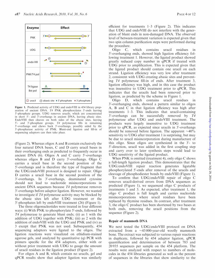
(Figure 2). Whereas oligos A and B contain exclusively thefour natural DNA bases, C and D carry uracil bases intheir overhanging ends as predicted to frequently occur inancient DNA (6). Oligos A and C carry 50-overhangswhereas oligos B and D carry 30-overhangs. Oligo Ccarries a uracil base in the second position of the50-overhangs and is therefore the type of fragment thatthe UDG/endoVIII protocol is designed to repair. OligoD carries a uracil base in the second position of the30-overhangs. In 30-overhangs, deaminated cytosineshould not lead to nucleotide misincorporations inancient DNA sequences because T4 polymerase removes30-overhangs before adaptor ligation. However, we wantedto investigate if T4 polymerase activity may be blocked bythe abasic sites left after UDG treatment or the30-phosphates left by endoVIII treatment (26) (Figure 1).The three oligonucleotides were treated in four different
ways: (i) With PNK to phosphorylate 50-ends, followed byT4 polymerase to generate blunt ends; (ii) as 1 with theaddition of UDG together with PNK; (iii) as 2 with theaddition of endoVIII with the UDG and PNK and (iv) as3 except that PNK was not used. Subsequently, 454sequencing adaptors were ligated to the oligos. Theligation reactions were visualized on ethidium-stainedagarose gels, and analyzed by quantitative (q) PCR withprimers specific for the 454 adaptors, either with orwithout prior treatment with UDG to gauge the amountof uracil residues in the ligation product (Figure 2).For oligos A and B, which contain no uracils, gel and
qPCR results show that adaptor ligation was similarly
efficient for treatments 1–3 (Figure 2). This indicatesthat UDG and endoVIII do not interfere with the gener-ation of blunt ends in non-damaged DNA. The observedlevel of between-treatment variation is expected given thattwo spin column purification steps were performed duringthe procedure.
Oligo C, which contains uracil residues in50-overhanging ends, showed high ligation efficiency fol-lowing treatment 1. However, the ligated product showedgreatly reduced copy number in qPCR if treated withUDG prior to amplification. This is expected given thatthe ligated product should contain one uracil on eachstrand. Ligation efficiency was very low after treatment2, consistent with UDG creating abasic sites and prevent-ing T4 polymerase fill-in of ends. After treatment 3,ligation efficiency was high, and in this case the productwas insensitive to UDG treatment prior to qPCR. Thisindicates that the uracils had been removed prior toligation, as predicted by the scheme in Figure 1.
Oligo D, which contains uracil residues in30-overhanging ends, showed a pattern similar to oligosA, B and C in that ligation efficiency was high aftertreatments 1–3. This indicates that uracil-containing30-overhangs can be successfully removed by T4polymerase after UDG and endoVIII treatment. Theproducts were largely insensitive to UDG treatmentprior to qPCR, as expected since uracils in 30-overhangsshould be removed before ligation. The apparent �40%sensitivity to UDG after treatment 1 is surprising, but maybe due to uracil misincorporation during manufacture ofthis oligo. Since oligos are synthesized in the 30- to50-direction, uracil was added in the first coupling stepand carry over to later synthesis steps would lead toUDG sensitivity of the ligated product.
When PNK is omitted (treatment 4), only oligo C showsa full-length ligation product. This demonstrates that theUDG/endoVIII repair reaction generates ligatablephosphorylated 50-ends after removal of the uracils andcleavage of phosphodiester bonds by endoVIII (Figure 1).
To confirm that UDG/endoVIII repair of oligo Cremoves uracil-derived errors from DNA sequences aspredicted (Figure 1), we sequenced oligo C products oftreatments 1 and 3. As expected, after treatment 1, theoligo C product is full length and carries nucleotidemisincorporations where uracil residues have beenreplaced by thymine residues. In contrast, after treatment3, the oligo C product has been shortened by two bases atboth ends, removing the uracil positions from thesequence (Figure 2).
Repair of mammoth DNA
We next tested the UDG/endoVIII protocol on DNAextracted from a �43 000-year-old woolly mammothbone. The extract was subjected to various repair reactionsin duplicate, followed by 454 adaptor ligation, qPCRquantification and determination of between 783 and20 935 sequences per sample on the 454 platform. Theresults were analyzed with respect to numbers of mole-cules in the 454 libraries generated as well as the percentof sequences in the libraries that show similarity to the
5’ 3’UPNK
UDG
endo VIII
PNK/T4 pol
T4 ligase
Bst pol
Effect
A B
U
APAP
AP
AP
U APuracil abasic site
Enzyme
5’ phosphate 3’ phosphate
Figure 1. Predicted activity of UDG and endoVIII in 454 library prep-aration of ancient DNA. T4 PNK phosphorylates 50-ends leaving50-phosphate groups. UDG removes uracils, which are concentratedin short 50- and 30-overhangs in ancient DNA, leaving abasic sites.EndoVIII then cleaves on both sides of the abasic sites, leaving50- and 30-phosphate groups. T4 polymerase fills in remaining50-overhangs and chews back 30-overhangs, possibly aided by the30-phosphatase activity of PNK. Blunt-end ligation and fill-in ofsequencing adaptors can then take place.
e87 Nucleic Acids Research, 2010, Vol. 38, No. 6 PAGE 4 OF 12
by guest on July 29, 2014http://nar.oxfordjournals.org/
Dow
nloaded from

elephant genome and are thus assumed to be of mammothorigin. In most ancient remains, only a small proportionof sequences retrieved are from the organism under study.For this extract, it is �3%, the rest presumably stemmingfrom microorganisms that have colonized the sample afterthe death of the individual. If the endogenous mammothDNA carried more DNA modifications than the environ-mental DNA, the relative amount of mammoth DNAmight be changed by the repair reactions. Other relevantaspects such as the length of the mammoth sequences andthe frequency and type of nucleotide misincorporationswere also analyzed.
One issue that potentially complicates the application ofthe UDG/endoVIII protocol to ancient DNA extracts isthat DNA modifications other than deaminated cytosines
may be present (27,28). Therefore, we first tested ifmammoth DNA retrieval was affected by the longer incu-bation (3 h) at a higher temperature (37�C) that differsfrom the standard library preparation (30min at 25�C)and thus could conceivably cause DNA degradation byaffecting some unknown modifications. Neither in termsof numbers of molecules in the resultant 454 library, thepercent of mammoth sequences, the length of mammothsequences or patterns of misincorporations did theincubation conditions strongly influence the results(Supplementary Figure S3).We next tested the three treatments previously per-
formed on the oligonucleotides, i.e. (i) PNK+T4polymerase; (ii) PNK+T4 polymerase+UDG; (iii)PNK+T4 polymerase+UDG+endoVIII. In a second
a GTAGGTGAAAACCTCTGACACATGCAGCTCCCGGAGACGGTCACAGCTTGTCTGTAAGCGGATGCACGCATb --AGGTGAAAACCTCTGACACATGCAGCTCCCGGAGACGGTCACAGCTTGTCTGTAAGCGGATGCACGC--
200bp150bp100bp
50bp
Oligo A
Oligo B
Oligo CGCAGGTGAAAACCTCTGACACATGCAGCTCCCGGAGACGGTCACAGCTTGTCTGTAAGCGGATGCACG ||||||||||||||||||||||||||||||||||||||||||||||||||||||||||||||||| CCACTTTTGGAGACTGTGTACGTCGAGGGCCTCTGCCAGTGTCGAACAGACATTCGCCTACGTGCGCA
GUAGGTGAAAACCTCTGACACATGCAGCTCCCGGAGACGGTCACAGCTTGTCTGTAAGCGGATGCACG ||||||||||||||||||||||||||||||||||||||||||||||||||||||||||||||||| CCACTTTTGGAGACTGTGTACGTCGAGGGCCTCTGCCAGTGTCGAACAGACATTCGCCTACGTGCGUA
GCAGGTGAAAACCTCTGACACATGCAGCTCCCGGAGACGGTCACAGCTTGTCTGTAAGCGGATGCAUG |||||||||||||||||||||||||||||||||||||||||||||||||||||||||||||||||AUGCGTCCACTTTTGGAGACTGTGTACGTCGAGGGCCTCTGCCAGTGTCGAACAGACATTCGCCTACG
GCAGGTGAAAACCTCTGACACATGCAGCTCCCGGAGACGGTCACAGCTTGTCTGTAAGCGGATGCACCG |||||||||||||||||||||||||||||||||||||||||||||||||||||||||||||||||ACGCGTCCACTTTTGGAGACTGTGTACGTCGAGGGCCTCTGCCAGTGTCGAACAGACATTCGCCTACG
Oligo D
160140120100
80604020
0rela
tive
lib
ray
yiel
d /
%
Oligo A
a b
Oligo B Oligo C Oligo D
+ + + - + + + - + + + - + + + -PNKUDG
endo VIIItreatment
+ +- ++- +-
+ +- ++- +-
+ +- ++- +-
+ +- ++- +-
1 32 41 32 4 1 32 4 1 32 4
Figure 2. Demonstration of UDG/endoVIII repair on synthetic oligonucleotides. 1 mg of each of four synthetic double-stranded oligos A-D (top)was subjected to 454 adaptor ligation after enzymatic repair in four conditions: (1) Incubation with PNK followed by addition of T4 polymerase; (2)PNK and UDG followed by T4 polymerase; (3) PNK, UDG and endoVIII followed by T4 polymerase; (4) UDG and endoVIII followed by T4polymerase (i.e. no PNK). Products were first visualized on agarose gels (middle). The first lane on each gel after the ladder is the untreated oligo.Major bands in the other lanes correspond to the unligated oligos (62–67 bp), the oligos plus one 44-base adaptor and the oligos plus two 44-baseadaptors. For some products higher weight bands are visible that probably indicate end-to-end chimeras of the oligos and adaptors. The cause of thefaint, diffuse bands seen between 150 and 200 bp in the untreated oligos are unknown but may be artifacts of oligo synthesis. Ligated products werealso quantified by qPCR without (dark brown) or with (light brown) prior incubation with UDG. The products marked a and b were sequenceddirectly on the 454 platform (bottom).
PAGE 5 OF 12 Nucleic Acids Research, 2010, Vol. 38, No. 6 e87
by guest on July 29, 2014http://nar.oxfordjournals.org/
Dow
nloaded from
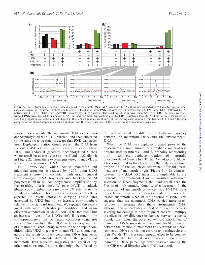
series of experiments, the mammoth DNA extract wasdephosphorylated with CIP, purified, and then subjectedto the same three treatments except that PNK was neverused. Dephosphorylation should prevent the DNA fromsuccessful 454 adaptor ligation except in cases whereUDG and endoVIII generates phosphorylated 50-endswhere uracil bases exist close to the 50-ends (c.f. oligo Bin Figure 2). Thus, these experiments tested if endoVIII isactive on the mammoth DNA.Total library yield, which includes mammoth and
microbial sequences, is reduced by �30% upon UDGtreatment (Figure 3a), consistent with uracil removalfrom damaged DNA fragments and blockage of T4polymerase fill-in or Taq polymerase amplification bythe resulting abasic sites. When endoVIII is added,library copy numbers increase by �40% relative to thestandard condition. This is unexpected since endoVIII isexpected to rescue molecules carrying abasic sitesgenerated by UDG but not to increase copy numbersrelative to the standard treatment. We repeated this exper-iment with more replicates per condition, and againobserved a reduction in yield upon UDG treatment butan increase in yield after UDG/endoVIII treatment onlyto approximately the no repair condition (data notshown). We conclude that UDG reduces copy numberof a mammoth DNA library relative to the no repair con-dition, while UDG together with endoVIII does not, sug-gesting the repair of uracil-containing DNA fragments.None of the treatments differed in the percent ofmammoth DNA sequence, suggesting that uracil or anyother unknown modifications that might be affected by
the treatments did not differ substantially in frequencybetween the mammoth DNA and the environmentalDNA.
When the DNA was dephosphorylated prior to theexperiments, a small amount of amplifiable material waspresent after treatments 1 and 2, probably representingboth incomplete dephosphorylation of naturallyphosphorylated 50-ends by CIP and 454 adaptor artifacts.This is supported by the observation that only a very smallproportion of the sequences determined after this treat-ment are of mammoth origin (Figure 3b). In contrast,treatment 3 yielded �3.5 times more amplifiable librarymolecules than treatments 1 and 2, consistent with directselection of DNA fragments that had uracil near the50-ends of both strands. Notably, after treatment 3, theproportion of mammoth sequences was 10–12%, fourtimes higher than in the libraries made from non-CIPtreated mammoth DNA in the presence of PNK. Thissuggests that the mammoth DNA carried more uracilresidues on average than the environmental DNA,although this is probably a modest difference becauseselecting for damage at both fragment ends will multiplythe effect of any difference in damage between sequencepopulations. Thus, the observed �4-fold enrichment ofmammoth DNA suggests a maximum 2-fold differencebetween the fraction of mammoth DNA strands and envi-ronmental DNA strands that carry uracil residues close totheir 50-ends. This is a small enough effect to be compat-ible with the fact that no obvious differences inmammoth DNA percentage were observed among thenon-CIP-treated libraries where PNK was used.
9876543210lib
rary
cp
/ul (
mill
ion
s) 70605040302010
0libra
ry c
p/u
l (th
ou
san
ds)
14%
12%
10%
8%
6%
4%
2%
0%frac
tio
n e
lep
han
t h
its
(a) (b)
(c)
0.00
0.20
0.40
0.60
0.80
–10 –5 0 5 10
1.00
0.00
0.20
0.40
0.60
0.80
–10 –5 0 5 10
1.00
0.00
0.20
0.40
0.60
0.80
–10 –5 0 5 10
1.00
0.00
0.20
0.40
0.60
0.80
–10 –5 0 5 10
bas
e fr
eque
ncy
1.00
ACGT
Mammoth seqs5’
Mammoth seqs5’
Mammoth seqs5’
Mammoth seqs5’
+ + +CIPPNKUDGendo VIIItreatment
+ +–+– –
1 32
– – – + + +
+ +–+– –
4 65
– – – + + ++ +–
+– –1 32
– – – + + +
+ +–+– –
4 65
– – –
Figure 3. The UDG/endoVIII repair protocol applied to mammoth DNA. (a) A mammoth DNA extract was subjected to 454 adaptor ligation afterenzymatic repair in replicates in three conditions: (1) Incubation with PNK followed by T4 polymerase; (2) PNK and UDG followed by T4polymerase; (3) PNK, UDG and endoVIII followed by T4 polymerase. The resulting libraries were quantified by qPCR. The same reactionswithout PNK were applied to mammoth DNA that had first been dephosphorylated by CIP (treatments 4–6). (b) All libraries were sequenced on454. The proportions of sequences that aligned to the elephant genome are shown. (c) For the sequences resulting from treatments 1–3 and 6 the basecomposition of aligned elephant sequences is shown for 10 bases either side of the 50-start point of mammoth sequences.
e87 Nucleic Acids Research, 2010, Vol. 38, No. 6 PAGE 6 OF 12
by guest on July 29, 2014http://nar.oxfordjournals.org/
Dow
nloaded from
The 50-start points of the mammoth sequences representthe terminal bases of the ancient DNA fragments at thepoint of adaptor ligation (6). We plotted the base compo-sition of the elephant reference sequence aligned to themammoth sequences for 10 bases on either side of themammoth 50-ends. UDG/endoVIII treatment shouldlead to an increase in cytosine frequency at the baseposition immediately preceding 50-ends (i.e. the –1position in Figure 3c), as DNA fragments containingdeaminated cytosine will be truncated to the 30-side ofsuch positions (Figure 1). Consistent with this prediction,we observed in the non-CIP-treated libraries an increasedfrequency of cytosine at position –1 in the UDG/endoVIIIcondition relative to the no-repair and UDG-treated con-ditions. This effect was much stronger in the CIP-treated,UDG/endoVIII repaired sample, where 84% of 50-endsare preceded by cytosines. This is predicted since thislibrary should predominantly contain fragments whereligatable ends were generated by excision and repair atpositions of deaminated cytosine.
The average length of mammoth sequences is around70–80 nt (Supplementary Figure S4), as is typical ofancient DNA (29). After UDG treatment, fragments are�10 nt shorter. This is expected since longer fragments aremore likely to contain uracil and thus to be left with anabasic site after UDG treatment. When endoVIII isadded, length seems to be intermediate, perhaps represent-ing rescue of some longer fragments by endoVIII treat-ment. If the DNA is dephosphorylated before UDG andendoVIII treatment, length is �10 nt shorter than afterUDG and endoVIII treatment without CIP-treatment,as expected if all of the ligated fragments in this conditionhave been truncated at both ends due to UDG/endoVIIIrepair, as opposed to the non-CIP-treated libraries whereonly a fraction of the fragments will have been truncated.
We investigated the effect of the repair protocol onnucleotide misincorporations, by plotting for eachlibrary the frequency of each of the 12 possible mis-matches observed in the mammoth-to-elephant align-ments. Ancient DNA sequences generally show an excessof C!T and G!A substitutions due to uracil-derivednucleotide misincorporations (4,6,7). As expected, thispattern is seen when the sample is not subjected toUDG treatment (Supplementary Figure S4). By takingthe excess of C/G!T/A substitutions relative toT/A!C/G substitutions as an estimate of the amount ofuracil-derived misincorporations in each sample, we foundthat uracil-derived misincorporations were reduced 4- to10-fold in samples where UDG was used (SupplementaryFigure S4).
In summary, UDG–endoVIII treatment allows moremammoth DNA sequences to be retrieved than UDGtreatment alone, while generating much lower rates ofnucleotide misincorporations than if UDG is not used.
Repair of Neandertal DNA
Mammoth and African elephant DNA diverged from eachother more than �7 million years ago (30,31). Thereforegenuine sequence differences will dominate in pairwisealignments, reducing the ability to study patterns of
nucleotide misincorporations. In contrast, Neandertalsand humans diverged much more recently. Furthermore,in contrast to the elephant genome the human genome isof finished quality allowing better resolution of nucleotidemisincorporations. We therefore applied the UDG/endoVIII repair protocol to DNA extracted from a38 000-year-old Neandertal bone from Croatia and per-formed deep sequencing of the libraries. An additionaladvantage of this specimen is that its completemitochondrial (mt) sequence is known (23), allowingdetailed analysis of sequence errors in mtDNA. Thismay be particularly interesting as mtDNA does notcarry 5-methylcytosine (50-m-C), a naturally occurringmodification in vertebrate nuclear DNA which whendeaminated is converted to thymine (32). Since UDGwill not remove thymine from DNA, deamination-derivedmisincorporations may still occur at sites of cytosinemethylation after UDG treatment of the ancient DNA.Between 10 and 11 million raw DNA sequence reads
were generated on the Illumina GAII platform from fourNeandertal DNA libraries prepared in the standard con-dition or with the UDG/endoVIII protocol (each in tworeplicates). In order to improve sequencing accuracy,which decreases substantially toward the 30-end ofIllumina reads (25), we sequenced into both ends of eachmolecules and merged the paired reads, requiring overlapsof at least 11 bases, and discarded all sequences that couldnot be merged to their paired read. Since the single readlength was 48 bases, this creates a maximum read length of85 bp. However, previous work has shown that themajority of Neandertal fragments in this bone areshorter than this (33).Sequences were aligned to the previously published
complete mtDNA sequence of this Neandertal (23), andto the human nuclear genome (hg18) using a custommapping program (ANFO) (22). In each library,0.007–0.009% of sequences aligned well to the mtDNAand 2.0–2.2% to the nuclear genome. Variation in theseproportions was as high within as between treatments.Data from replicates were then pooled for each treatmentand patterns of nucleotide mismatches between thesequences and the references were analyzed.When analyzing Neandertal DNA sequences, contami-
nation of experiments with contemporary human DNA isa potential problem (10,34). However, the level of suchcontamination in a Neandertal DNA library can beassessed by counting the ratio of Neandertal versus con-taminant fragments at nucleotide positions whereNeandertals differ from all or almost all present-dayhumans (33). The mtDNA of this Neandertal carries 133such diagnostic positions (23). The ‘no repair’ datasetyielded 139 mtDNA fragments that overlapped such posi-tions; 138 carried the Neandertal base while one matchedmodern human mtDNA. The UDG/endoVIII treateddataset yielded 128 informative fragments, of which allwere the Neandertal type. Thus, the mtDNA in alllibraries was almost completely free of contamination bymodern human mtDNA, even after treatment with UDGand endoVIII. Since the ratio of mitochondrial to nuclearDNA may differ between the contaminating and theNeandertal DNA, this estimate is strictly applicable only
PAGE 7 OF 12 Nucleic Acids Research, 2010, Vol. 38, No. 6 e87
by guest on July 29, 2014http://nar.oxfordjournals.org/
Dow
nloaded from

to the mtDNA (33). However, the estimate of mtDNAcontamination in these libraries is low enough thatwithin even a few-fold variation in mtDNA:nuclearDNA ratios between the Neandertal and contaminatingDNA, sequences aligning to the human nuclear genomewill be predominantly of Neandertal origin.To analyze patterns of nucleotide misincorporations, we
plotted the frequency of each of the 12 possible nucleotidedifferences in alignments as a function of distance from theends of DNA fragments for each dataset (Figure 4, toppanels). In the ‘no repair’ results, the transitions C!T andG!A are drastically elevated toward the 50- and 30-endsends of fragments, respectively, with up to 40–50% errorfrequencies at the fragment ends. This occurs in bothmtDNA and nuclear DNA fragments, as shownpreviously (6,23). The G!A substitutions at 30-ends offragments originate during the fill-in of 50-overhangs con-taining deaminated cytosines (6,7). After UDG/endoVIIItreatment C/G!T/A differences were drastically reduced,consistent with the removal of uracils by UDG. InmtDNA this removal seems to be almost complete,
whereas a small amount remains in nuclear DNA (seebelow).
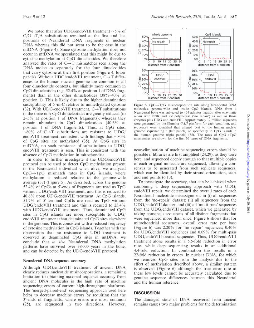
Neandertal CpG methylation
In vertebrates, DNA methylation occurs at the 50-positionof cytosine residues at �80% of CpG dinucleotides insomatic tissues (35). When 50-m-C is deaminated itbecomes a thymine residue that is not recognized as anunnatural base by UDG in vertebrate cells. As a conse-quence, CpG dinucleotides are preferentially lost fromvertebrate genomes with the result that the frequencywith which CpG dinucleotides occur genome wide is�1%, while 4–5% would be expected from the base com-position of the genome (36). In some regions of thegenome, CpG content is less reduced and can be 60% ormore of the statistically expected frequency. Such ‘CpGislands’ represent areas where methylation is reduced orabsent. About half of CpG islands overlap transcriptionalstart sites where their methylation in a tissue is positivelycorrelated with suppression of transcription.
-30 -20 -10 0
50%
40%
30%
20%
10%
0%
0 10 20 30
nt from 5’ end nt from 3’ end nt from 5’ end nt from 3’ end
mis
mat
ch r
ate
nt from 5’ end nt from 3’ end nt from 3’ endnt from 5’ end
mis
mat
ch r
ate
Nuclear DNA
50%
40%
30%
20%
10%
0%
50%
40%
30%
20%
10%
0%
50%
40%
30%
20%
10%
0%
Mitochondrial DNA
-30 -20 -10 00 10 20 30
0 10 20 30 0 10 20 300%
10%20%30%40%50%
0 10 20 30 0 10 20 30
0 10 20 30 0 10 20 300%
10%20%30%40%50%
0 10 20 30 0 10 20 30
mis
matc
h r
ate
0 10 20 30 0 10 20 300%
10%20%30%40%50%
0 10 20 30 0 10 20 30
0 10 20 30 0 10 20 300%
10%20%30%40%50%
0 10 20 30 0 10 20 30
-30 -20 -10 00 10 20 30
-30 -20 -10 00 10 20 30
C>T G>A T>C G>A Other
CA>TA CT>TT CC>TC CG>TG CA>TA CT>TT CC>TC CG>TG
no repair
no repair
UDG/endoVIII
UDG/endoVIII
no repair
UDG/endoVIII
UDG/endoVIII
no repair
(a)
(b)
nt from 5’ end nt from 5’ end
AM948965
Neandertal fragments
hg18
Neandertal fragments
Figure 4. Effect of UDG/endoVIII treatment on all nucleotide misincorporation rates and on C!T rates at CpN dinucleotides along NeandertalDNA sequences. DNA from a Neandertal bone was subjected to 454 adaptor ligation after enzymatic repair with PNK and T4 polymerase (‘norepair’) as well as those enzymes plus UDG and endoVIII. Approximately 12 million sequences were generated on the Illumina GAII platform foreach condition, and sequences were identified that aligned best to the complete mtDNA sequence of this bone AM948969 (947 and 1084 sequencesfor the no repair and UDG/endoVIII conditions, respectively, left panels) and to the human nuclear genome sequence hg18 (242 070 and 286 737sequences, right panels). (a) All 12-nt mismatch frequencies are plotted as a function of position along the aligned fragments for the no repair andUDG/endoVIII conditions. (b) The rate of C!T misincorporations along DNA fragments is shown separately for the four dinucleotides thatcontain cytosine in the 50-position.
e87 Nucleic Acids Research, 2010, Vol. 38, No. 6 PAGE 8 OF 12
by guest on July 29, 2014http://nar.oxfordjournals.org/
Dow
nloaded from
We noted that after UDG/endoVIII treatment �5% ofC/G!T/A substitutions remained at the first and lastpositions of Neandertal DNA fragments in nuclearDNA whereas this did not seem to be the case in themtDNA (Figure 4). Since cytosine methylation does notoccur in mtDNA we speculated that this might be due tocytosine methylation at CpG dinucleotides. We thereforeanalyzed the rates of C!T mismatches seen along theDNA molecules separately for the four dinucleotidesthat carry cytosine at their first position (Figure 4, lowerpanels). Without UDG/endoVIII treatment, C!T differ-ences to the human nuclear genome are common in allfour dinucleotide contexts, but slightly more common inCpG dinucleotides (e.g. 52.4% at position 1 of DNA frag-ments) than in the other dinucleotides (38%–40% atposition 1). This is likely due to the higher deaminationsusceptibility of 50-m-C relative to unmethylated cytosine(32). With UDG/endoVIII treatment, C!T substitutionsin the three non-CpG dinucleotides are greatly reduced (to2–5% at position 1 of DNA fragments), whereas theyremain abundant in CpG dinucleotides (40.6% atposition 1 of DNA fragments). Thus, at CpG sites,�80% of C!T substitutions are resistant to UDG/endoVIII treatment, consistent with findings that �80%of CpG sites are methylated (35). At CpG sites inmtDNA, no such resistance of substitutions to UDG/endoVIII treatment is seen. This is consistent with theabsence of CpG methylation in mitochondria.
In order to further investigate if the UDG/endoVIIIprotocol can be used to detect CpG methylation presentin the Neandertal individual when alive, we analyzedCpG!TpG mismatch rates in CpG islands, wheremethylation is reduced relative to the genome-wideaverage (37) (Figure 5). As described, across the genome52.4% of CpGs at 50-ends of fragments are read as TpGwithout UDG/endoVIII treatment, and this is reduced to40.6% upon UDG/endoVIII treatment. At CpG islands,51.7% of 50-terminal CpGs are read as TpG withoutUDG/endoVIII treatment and this is reduced to 23.4%with UDG/endoVIII treatment. Thus, deaminated CpGsites in CpG islands are more susceptible to UDG/endoVIII treatment than deaminated CpG sites elsewherein the genome. This is consistent with a reduced frequencyof cytosine methylation in CpG islands. Together with theobservation that no resistance to UDG treatment isobserved at deaminated CpG sites in mtDNA, weconclude that in vivo Neandertal DNA methylationpatterns have survived over 38 000 years in the bone,and can be detected by the UDG/endoVIII protocol.
Neandertal DNA sequence accuracy
Although UDG/endoVIII treatment of ancient DNAclearly reduces nucleotide misincorporations, a remaininglimitation to obtaining maximal sequence accuracy fromancient DNA molecules is the high rate of machinesequencing errors of current high-throughput platforms.The ‘merged-paired-end’ sequencing approach used herehelps to decrease machine errors by requiring that the30-ends of fragments, where errors are most common(25), are sequenced in two directions. However,
near-elimination of machine sequencing errors should bepossible if libraries are first amplified (24,29), as they werehere, and sequenced deeply enough so that multiple copiesof each original molecule are sequenced, allowing a con-sensus to be generated from such replicate sequences,which can be identified by their strand orientation, startand end points (6,13).To investigate the accuracy, that can be achieved when
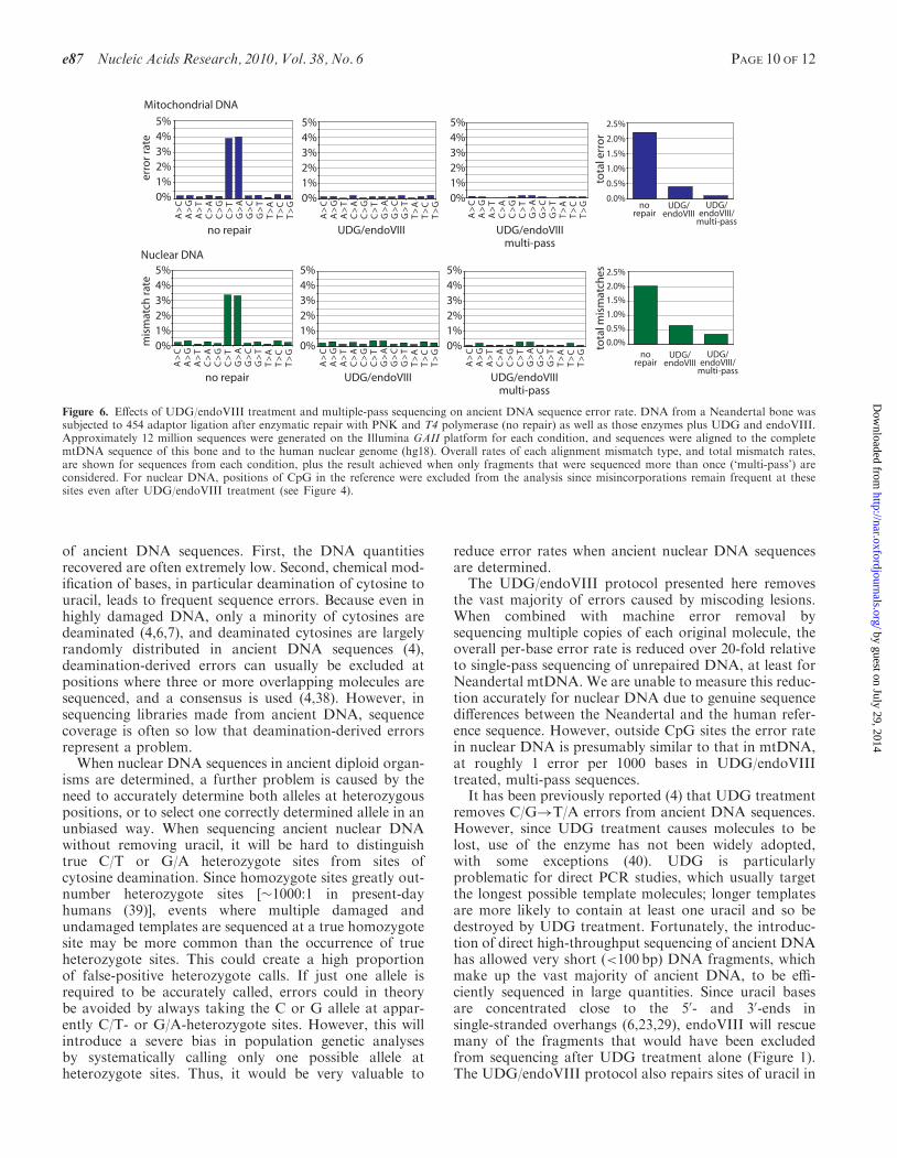
combining a deep sequencing approach with UDG/endoVIII repair, we determined the overall rates of eachNeandertal nucleotide misassignment in (i) all sequencesfrom the ‘no-repair’ dataset; (ii) all sequences from theUDG/endoVIII dataset; and (iii) all ‘multi-pass’ sequencesfrom the UDG/endoVIII dataset, which we generated bytaking consensus sequences of all distinct fragments thatwere sequenced more than once. Figure 6 shows that formitochondrial sequences, overall error rate per base(Figure 6) was 2.20% for ‘no repair’ sequences; 0.40%for UDG/endoVIII sequences and 0.09% for multi-passUDG/endoVIII-treated sequences. Thus, UDG/endoVIIItreatment alone results in a 5.5-fold reduction in errorrates while deep sequencing results in an additional4.4-fold reduction. In combination this results in a22-fold reduction in errors. In nuclear DNA, for whichwe removed CpG sites from the analysis due to theeffect of methylation described above, a similar patternis observed (Figure 6) although the true error rate atthese low levels cannot be accurately calculated due togenuine sequence differences between this Neandertaland the human reference.
DISCUSSION
The damaged state of DNA recovered from ancientremains causes two major problems for the determination
0%
10%
20%
30%
40%
50%
0 5 10 15 20 25 30
0%
10%
20%
30%
40%
50%
0 5 10 15 20 25 30
C>
T ra
te
whole genome CpG islands
No repair
UDG/endoVIII
0%
10%
20%
30%
40%
50%
0 5 10 15 20 25 30
0%
10%
20%
30%
40%
50%
0 5 10 15 20 25 30
UDG/endoVIII
No repair
C>
T ra
te
distance from 5’ end (nt)
distance from 5’ end (nt)
distance from 5’ end (nt)
distance from 5’ end (nt)
C>
T ra
teC
>T
rate
Figure 5. CpG!TpG misincorporation rate along Neandertal DNAmolecules, genome-wide and inside CpG islands. DNA from aNeandertal bone was subjected to 454 adaptor ligation after enzymaticrepair with PNK and T4 polymerase (‘no repair’) as well as thoseenzymes plus UDG and endoVIII. Approximately 12 million sequenceswere generated on the Illumina GAII platform for each condition, andsequences were identified that aligned best to the human nucleargenome sequence hg18 (left panels) or specifically to CpG islands inthe human genome (right panels) (35). The rates of CpG!TpGmismatches are shown for the first 30 bases of DNA fragments.
PAGE 9 OF 12 Nucleic Acids Research, 2010, Vol. 38, No. 6 e87
by guest on July 29, 2014http://nar.oxfordjournals.org/
Dow
nloaded from
of ancient DNA sequences. First, the DNA quantitiesrecovered are often extremely low. Second, chemical mod-ification of bases, in particular deamination of cytosine touracil, leads to frequent sequence errors. Because even inhighly damaged DNA, only a minority of cytosines aredeaminated (4,6,7), and deaminated cytosines are largelyrandomly distributed in ancient DNA sequences (4),deamination-derived errors can usually be excluded atpositions where three or more overlapping molecules aresequenced, and a consensus is used (4,38). However, insequencing libraries made from ancient DNA, sequencecoverage is often so low that deamination-derived errorsrepresent a problem.When nuclear DNA sequences in ancient diploid organ-
isms are determined, a further problem is caused by theneed to accurately determine both alleles at heterozygouspositions, or to select one correctly determined allele in anunbiased way. When sequencing ancient nuclear DNAwithout removing uracil, it will be hard to distinguishtrue C/T or G/A heterozygote sites from sites ofcytosine deamination. Since homozygote sites greatly out-number heterozygote sites [�1000:1 in present-dayhumans (39)], events where multiple damaged andundamaged templates are sequenced at a true homozygotesite may be more common than the occurrence of trueheterozygote sites. This could create a high proportionof false-positive heterozygote calls. If just one allele isrequired to be accurately called, errors could in theorybe avoided by always taking the C or G allele at appar-ently C/T- or G/A-heterozygote sites. However, this willintroduce a severe bias in population genetic analysesby systematically calling only one possible allele atheterozygote sites. Thus, it would be very valuable to
reduce error rates when ancient nuclear DNA sequencesare determined.
The UDG/endoVIII protocol presented here removesthe vast majority of errors caused by miscoding lesions.When combined with machine error removal bysequencing multiple copies of each original molecule, theoverall per-base error rate is reduced over 20-fold relativeto single-pass sequencing of unrepaired DNA, at least forNeandertal mtDNA. We are unable to measure this reduc-tion accurately for nuclear DNA due to genuine sequencedifferences between the Neandertal and the human refer-ence sequence. However, outside CpG sites the error ratein nuclear DNA is presumably similar to that in mtDNA,at roughly 1 error per 1000 bases in UDG/endoVIIItreated, multi-pass sequences.
It has been previously reported (4) that UDG treatmentremoves C/G!T/A errors from ancient DNA sequences.However, since UDG treatment causes molecules to belost, use of the enzyme has not been widely adopted,with some exceptions (40). UDG is particularlyproblematic for direct PCR studies, which usually targetthe longest possible template molecules; longer templatesare more likely to contain at least one uracil and so bedestroyed by UDG treatment. Fortunately, the introduc-tion of direct high-throughput sequencing of ancient DNAhas allowed very short (<100 bp) DNA fragments, whichmake up the vast majority of ancient DNA, to be effi-ciently sequenced in large quantities. Since uracil basesare concentrated close to the 50- and 30-ends insingle-stranded overhangs (6,23,29), endoVIII will rescuemany of the fragments that would have been excludedfrom sequencing after UDG treatment alone (Figure 1).The UDG/endoVIII protocol also repairs sites of uracil in
5%4%3%2%1%0%
A>
CA
>G
A>
TC
>A
C>
GC
>T
G>
AG
>C
G>
TT
>A
T>
CT
>G
A>
CA
>G
A>
TC
>A
C>
GC
>T
G>
AG
>C
G>
TT
>A
T>
CT
>G
A>
CA
>G
A>
TC
>A
C>
GC
>T
G>
AG
>C
G>
TT
>A
T>
CT
>G
no repair UDG/endoVIII UDG/endoVIIImulti-pass
5%4%3%2%1%0%
5%4%3%2%1%0%
5%4%3%2%1%0%
A>
CA
>G
A>
TC
>A
C>
GC
>T
G>
AG
>C
G>
TT
>A
T>
CT
>G
no repair
erro
r rat
e
A>
CA
>G
A>
TC
>A
C>
GC
>T
G>
AG
>C
G>
TT
>A
T>
CT
>G
5%4%3%2%1%0%
A>
CA
>G
A>
TC
>A
C>
GC
>T
G>
AG
>C
G>
TT
>A
T>
CT
>G
5%4%3%2%1%0%
Mitochondrial DNA
Nuclear DNA
UDG/endoVIII UDG/endoVIIImulti-pass
2.5%
2.0%
1.5%
1.0%
0.5%
0.0%
mis
mat
ch ra
te
norepair
UDG/endoVIII
UDG/endoVIII/
multi-pass
tota
l err
or
2.5%
2.0%
1.5%
1.0%
0.5%
0.0%no
repairUDG/
endoVIIIUDG/
endoVIII/multi-pass
tota
l mis
mat
ches
Figure 6. Effects of UDG/endoVIII treatment and multiple-pass sequencing on ancient DNA sequence error rate. DNA from a Neandertal bone wassubjected to 454 adaptor ligation after enzymatic repair with PNK and T4 polymerase (no repair) as well as those enzymes plus UDG and endoVIII.Approximately 12 million sequences were generated on the Illumina GAII platform for each condition, and sequences were aligned to the completemtDNA sequence of this bone and to the human nuclear genome (hg18). Overall rates of each alignment mismatch type, and total mismatch rates,are shown for sequences from each condition, plus the result achieved when only fragments that were sequenced more than once (‘multi-pass’) areconsidered. For nuclear DNA, positions of CpG in the reference were excluded from the analysis since misincorporations remain frequent at thesesites even after UDG/endoVIII treatment (see Figure 4).
e87 Nucleic Acids Research, 2010, Vol. 38, No. 6 PAGE 10 OF 12
by guest on July 29, 2014http://nar.oxfordjournals.org/
Dow
nloaded from

double-stranded parts of molecules, although in our handsthis repair is only �20% efficient (data not shown).Further work exploring the effects of other repairenzymes on ancient DNA library yield and sequenceaccuracy may be useful. However, it will be desirable forfuture repair approaches to avoid any extra purificationsteps, as these inevitably cause loss of material.
The UDG/endoVIII protocol has the additional advan-tage that any remaining C/G!T/A misincorporations canbe attributed to deamination of methylated cytosineresidues (provided that UDG treatment is complete).The observation that C!T substitutions in CpGdinucleotides, but not in other dinucleotides, are resistantto repair by UDG and that this pattern does not occur inmtDNA but in nuclear DNA where it is reduced in CpGislands, strongly suggests that the signal stems from in vivoNeandertal methylation as opposed to postmortem DNAmodifications. In order to determine the methylationstatus of any particular CpG site by this approach, veryhigh sequence coverage would obviously be needed.However, the methylation status of a region such as aparticular gene or CpG island will be measurable withmore moderate sequence coverage. Furthermore,emerging techniques such as bisulphite treatment ofsmall DNA quantities (e.g. Genetic Signatures’MethylEasy Xceed kit) or single-molecule sequencingtechnologies that can distinguish 50-m-C from C (41)may soon allow high-resolution analysis of methylationin ancient DNA. It might therefore become possible toinvestigate the activity of genes and phenomena such asX chromosomal inactivation and genetic imprinting inextinct species, provided that the signals are manifestedin cells present in bones.
SUPPLEMENTARY DATA
Supplementary Data are available at NAR Online.
ACKNOWLEDGEMENTS
We thank Hernan Burbano, Paul Czechowski, Ed Green,Gregory Hannon, Emily Hodges, Tomislav Maricic,Philip Johnson, Pavao Rudan, Dejana Brajkovic, ZeljkoKucan and Ivan Gusic and the Croatian Academy ofSciences and Arts for collaboration and help.
FUNDING
We thank the Presidential Innovation Fund of the MaxPlanck Society for funding. Funding for open accesscharge: Max Planck Society.
Conflict of interest statement. None declared.
REFERENCES
1. Orlando,L., Darlu,P., Toussaint,M., Bonjean,D., Otte,M. andHanni,C. (2006) Revisiting Neandertal diversity with a 100,000year old mtDNA sequence. Curr Biol., 16, R400–R402.
2. Gilbert,M.T., Binladen,J., Miller,W., Wiuf,C., Willerslev,E.,Poinar,H., Carlson,J.E., Leebens-Mack,J.H. and Schuster,S.C.
(2007) Recharacterization of ancient DNA miscoding lesions:insights in the era of sequencing-by-synthesis. Nucleic Acids Res.,35, 1–10.
3. Gilbert,M.T., Hansen,A.J., Willerslev,E., Rudbeck,L., Barnes,I.,Lynnerup,N. and Cooper,A. (2003) Characterization of geneticmiscoding lesions caused by postmortem damage. Am. J. Hum.Genet., 72, 48–61.
4. Hofreiter,M., Jaenicke,V., Serre,D., Haeseler Av,A. and Paabo,S.(2001) DNA sequences from multiple amplifications revealartifacts induced by cytosine deamination in ancient DNA.Nucleic Acids Res., 29, 4793–4799.
5. Stiller,M., Green,R.E., Ronan,M., Simons,J.F., Du,L., He,W.,Egholm,M., Rothberg,J.M., Keates,S.G., Ovodov,N.D. et al.(2006) Patterns of nucleotide misincorporations during enzymaticamplification and direct large-scale sequencing of ancient DNA.Proc Natl Acad. Sci. USA, 103, 13578–13584.
6. Briggs,A.W., Stenzel,U., Johnson,P.L., Green,R.E., Kelso,J.,Prufer,K., Meyer,M., Krause,J., Ronan,M.T., Lachmann,M. et al.(2007) Patterns of damage in genomic DNA sequences from aNeandertal. Proc. Natl Acad. Sci. USA, 104, 14616–14621.
7. Brotherton,P., Endicott,P., Sanchez,J.J., Beaumont,M., Barnett,R.,Austin,J. and Cooper,A. (2007) Novel high-resolutioncharacterization of ancient DNA reveals C > U-type basemodification events as the sole cause of post mortem miscodinglesions. Nucleic Acids Res., 35, 5717–5728.
8. Lindahl,T., Ljungquist,S., Siegert,W., Nyberg,B. and Sperens,B.(1977) DNA N-glycosidases: properties of uracil-DNA glycosidasefrom Escherichia coli. J. Biol. Chem., 252, 3286–3294.
9. McDonald,J.P., Hall,A., Gasparutto,D., Cadet,J., Ballantyne,J.and Woodgate,R. (2006) Novel thermostable Y-familypolymerases: applications for the PCR amplification of damagedor ancient DNAs. Nucleic Acids Res., 34, 1102–1111.
10. Paabo,S., Poinar,H., Serre,D., Jaenicke-Despres,V., Hebler,J.,Rohland,N., Kuch,M., Krause,J., Vigilant,L. and Hofreiter,M.(2004) Genetic analyses from ancient DNA. Annu. Rev. Genet.,38, 645–679.
11. Bennett,S. (2004) Solexa Ltd. Pharmacogenomics, 5, 433–438.12. Margulies,M., Egholm,M., Altman,W.E., Attiya,S., Bader,J.S.,
Bemben,L.A., Berka,J., Braverman,M.S., Chen,Y.J., Chen,Z.et al. (2005) Genome sequencing in microfabricated high-densitypicolitre reactors. Nature, 437, 376–380.
13. Green,R.E., Krause,J., Ptak,S.E., Briggs,A.W., Ronan,M.T.,Simons,J.F., Du,L., Egholm,M., Rothberg,J.M., Paunovic,M.et al. (2006) Analysis of one million base pairs of NeanderthalDNA. Nature, 444, 330–336.
14. Noonan,J.P., Coop,G., Kudaravalli,S., Smith,D., Krause,J.,Alessi,J., Chen,F., Platt,D., Paabo,S., Pritchard,J.K. et al. (2006)Sequencing and analysis of Neanderthal genomic DNA. Science,314, 1113–1118.
15. Poinar,H.N., Schwarz,C., Qi,J., Shapiro,B., Macphee,R.D.,Buigues,B., Tikhonov,A., Huson,D.H., Tomsho,L.P., Auch,A.et al. (2006) Metagenomics to paleogenomics: large-scalesequencing of mammoth DNA. Science, 311, 392–394.
16. Lindahl,T. and Nyberg,B. (1974) Heat-induced deamination ofcytosine residues in deoxyribonucleic acid. Biochemistry, 13,3405–3410.
17. Maricic,T. and Paabo,S. (2009) Optimization of 454 sequencinglibrary preparation from small amounts of DNA permitssequence determination of both DNA strands. Biotechniques, 46,51–57.
18. Wiley,G., Macmil,S., Qu,C., Wang,P., Xing,Y., White,D., Li,J.,White,J.D., Domingo,A. and Roe,B.A. (2009) Methods forgenerating shotgun and mixed shotgun/paired-end libraries for the454 DNA sequencer. Curr. Protoc. Hum. Genet., Chapter 18,Unit18 11.
19. Meyer,M., Briggs,A.W., Maricic,T., Hober,B., Hoffner,B.,Krause,J., Weihmann,A., Paabo,S. and Hofreiter,M. (2007) Frommicrograms to picograms: quantitative PCR reduces the materialdemands of high-throughput sequencing. Nucleic Acids Res., 36, e5.
20. Rompler,H., Rohland,N., Lalueza-Fox,C., Willerslev,E.,Kuznetsova,T., Rabeder,G., Bertranpetit,J., Schoneberg,T. andHofreiter,M. (2006) Nuclear gene indicates coat-colorpolymorphism in mammoths. Science, 313, 62.
PAGE 11 OF 12 Nucleic Acids Research, 2010, Vol. 38, No. 6 e87
by guest on July 29, 2014http://nar.oxfordjournals.org/
Dow
nloaded from
21. Rohland,N. and Hofreiter,M. (2007) Ancient DNA extractionfrom bones and teeth. Nat Protoc, 2, 1756–1762.
22. Stenzel,U. (2009) German Conference on Bioinformatics. Halle,Germany.
23. Green,R.E., Malaspinas,A.S., Krause,J., Briggs,A.W.,Johnson,P.L., Uhler,C., Meyer,M., Good,J.M., Maricic,T.,Stenzel,U. et al. (2008) A complete neandertal mitochondrialgenome sequence determined by high-throughput sequencing. Cell,134, 416–426.
24. Briggs,A.W., Good,J.M., Green,R.E., Krause,J., Maricic,T.,Stenzel,U. and Paabo,S. (2009) Primer extension capture: targetedsequence retrieval from heavily degraded DNA sources. J. Vis.Exp., 1573.
25. Kircher,M., Stenzel,U. and Kelso,J. (2009) Improved base callingfor the Illumina Genome Analyzer using machine learningstrategies. Genome. Biol., 10, R83.
26. Jiang,D., Hatahet,Z., Melamede,R.J., Kow,Y.W. and Wallace,S.S.(1997) Characterization of Escherichia coli endonuclease VIII.J. Biol. Chem., 272, 32230–32239.
27. Hoss,M., Jaruga,P., Zastawny,T.H., Dizdaroglu,M. and Paabo,S.(1996) DNA damage and DNA sequence retrieval from ancienttissues. Nucleic Acids Res., 24, 1304–1307.
28. Paabo,S. (1989) Ancient DNA: extraction, characterization,molecular cloning, and enzymatic amplification. Proc. Natl Acad.Sci. USA, 86, 1939–1943.
29. Briggs,A.W., Good,J.M., Green,R.E., Krause,J., Maricic,T.,Stenzel,U., Lalueza-Fox,C., Rudan,P., Brajkovic,D., Kucan,Z.et al. (2009) Targeted retrieval and analysis of five NeandertalmtDNA genomes. Science, 325, 318–321.
30. Rohland,N., Malaspinas,A.S., Pollack,J.L., Slatkin,M.,Matheus,P. and Hofreiter,M. (2007) Proboscidean mitogenomics:chronology and mode of elephant evolution using mastodon asoutgroup. PLoS Biol., 5, e207.
31. Krause,J., Dear,P.H., Pollack,J.L., Slatkin,M., Spriggs,H.,Barnes,I., Lister,A.M., Ebersberger,I., Paabo,S. and Hofreiter,M.
(2006) Multiplex amplification of the mammoth mitochondrialgenome and the evolution of Elephantidae. Nature, 439, 724–727.
32. Shen,J.C., Rideout,W.M. 3rd and Jones,P.A. (1994) The rate ofhydrolytic deamination of 5-methylcytosine in double-strandedDNA. Nucleic Acids Res., 22, 972–976.
33. Green,R.E., Briggs,A.W., Krause,J., Prufer,K., Burbano,H.A.,Siebauer,M., Lachmann,M. and Paabo,S. (2009)The Neandertal genome and ancient DNA authenticity.EMBO J., 28, 2494–2502.
34. Wall,J.D. and Kim,S.K. (2007) Inconsistencies in Neanderthalgenomic DNA sequences. PLoS Genet., 3, 1862–1866.
35. Eckhardt,F., Lewin,J., Cortese,R., Rakyan,V.K., Attwood,J.,Burger,M., Burton,J., Cox,T.V., Davies,R., Down,T.A. et al.(2006) DNA methylation profiling of human chromosomes 6,20 and 22. Nat. Genet., 38, 1378–1385.
36. Gardiner-Garden,M. and Frommer,M. (1987) CpG islands invertebrate genomes. J. Mol. Biol., 196, 261–282.
37. Illingworth,R., Kerr,A., Desousa,D., Jorgensen,H., Ellis,P.,Stalker,J., Jackson,D., Clee,C., Plumb,R., Rogers,J. et al. (2008)A novel CpG island set identifies tissue-specific methylation atdevelopmental gene loci. PLoS Biol., 6, e22.
38. Gilbert,M.T., Tomsho,L.P., Rendulic,S., Packard,M., Drautz,D.I.,Sher,A., Tikhonov,A., Dalen,L., Kuznetsova,T., Kosintsev,P.et al. (2007) Whole-genome shotgun sequencing of mitochondriafrom ancient hair shafts. Science, 317, 1927–1930.
39. Nachman,M.W. (2001) Single nucleotide polymorphisms andrecombination rate in humans. Trends Genet., 17, 481–485.
40. Pruvost,M., Grange,T. and Geigl,E.M. (2005) Minimizing DNAcontamination by using UNG-coupled quantitative real-time PCRon degraded DNA samples: application to ancient DNA studies.Biotechniques, 38, 569–575.
41. Clarke,J., Wu,H.C., Jayasinghe,L., Patel,A., Reid,S. andBayley,H. (2009) Continuous base identification forsingle-molecule nanopore DNA sequencing. Nat. Nanotechnol., 4,265–270.
e87 Nucleic Acids Research, 2010, Vol. 38, No. 6 PAGE 12 OF 12
by guest on July 29, 2014http://nar.oxfordjournals.org/
Dow
nloaded from