Regulation of Interleukin 8 Expression in Human Malignant Melanoma Cells
7
(CANCER RESEARCH 58. 1532-1537. April 1. 19981 Regulation of Interleukin 8 Expression in Human Malignant Melanoma Cells Rakesh K. Singh1 and Michelle L. Varney Department efPa&ulogy und Microbiology. The University of Nebraska Medical Center, Omaha. Nebraska 68198-5660 ABSTRACT Here, we report the molecular regulation of interleukin (IL)-8 expres sion in human melanoma cells. The inflammatory cytokines ll.-l/f and tumor necrosis factor-« (TNF-a) up-regulated IL-8 expression, in a time- and concentration-dependent manner, in three metastatic melanoma vari ants, SBC-2 (nonmetastatic), A375P (low metastatic), and A375SM (high metastatic), by increased transcription of the IL-8 gene, leading to in creased levels of IL-8 mRNA and protein production. Furthermore, we report that IFN-a and UN-/! did not inhibit steady-state IL-8 production. However, IFN-a and IFN-ß inhibited IL-l/i or TNF-a-mediated up- regulation of IL-8 mRNA. In addition, IIN-/! demonstrated a more potent inhibitory effect at a lower concentration than did IFN-a. Both pretreat ment and simultaneous treatment of melanoma cells with IFN-a or IIN-/! inhibited the Il.-l/i and TNF-a up-regulation of IL-8 mRNA levels. This inhibition was at the transcriptional levels and was unaffected by a protein synthesis inhibitor, suggesting that this did not require de novo protein synthesis. Further, modulation of IL-8 levels by II.-I/.Õ,alone or in com bination with UN-/!, affected the proliferation of melanoma cells. In summary, our data suggest that the up-regulation of IL-8 expression in melanoma cells is regulated at the transcriptional level and is rapidly and specifically inhibited by IFN-a or l KV/t, independent of de novo protein synthesis, perhaps due to a transient modification of a preexisting fac- tor(s). INTRODUCTION Tumor cell growth and metastasis depends on the intrinsic proper ties of tumor cells and their interaction with the host microenviron- ment (1-3). Organ-specific cytokines can influence the ability of tumor cells to grow and metastasize in an organ-specific microenvi- ronment by modulating the expression of cytokines/growth factors (1-9). Expression of IL-8,2 an autocrine growth factor for human melanoma cells, directly correlates with their metastatic potential in nude mice (10). Moreover, the expression of IL-8 by melanoma cells depends on the organ microenvironment; for example, melanoma cells growing in the subcutis of nude mice expressed higher levels of IL-8 than did those growing in the liver (6). Similar results were observed in vitro when melanoma cells that were cocultured with keratinocytes expressed high levels of IL-8, whereas melanoma cells that were cocultured with hepatocytes did not (6). Recent reports from our laboratory suggest that IFN-ßinhibits the up-regulation of IL-8 ex pression by IL-1/3 in human melanoma cells (11); however, the mechanism remains unclear. Here, we investigated the regulatory mechanism(s) of IL-8 expres sion in human melanoma cells. Our data indicate that the treatment of human melanoma cells with IL-Ißor TNF-a enhanced IL-8 mRNA levels by increasing transcription, as determined by nuclear run-on analysis, leading to the IL-8 protein secretion by human melanoma cells. Further, IFN-a or IFN-ßinhibited IL-Iß- or TNF-a-induced Received 7/28/97; accepted 2/2/98. The costs of publication of this article were defrayed in pan by the payment of page charges. This article must therefore be hereby marked advertisement in accordance with 18 U.S.C. Section 1734 solely to indicate this fact. 1To whom requests for reprints should be addressed, at Department of Pathology and Microbiology. The University of Nebraska Medical Center. 600 South 42nd Street. Omaha. NE 68198-5660. Phone: (402| 559-9949; Fax: (402)559-4990; E-mail: [email protected]. 2 The abbreviations used are: IL, interleukin; TNF. tumor necrosis factor; GAPDH, glyceraldehyde-3-phosphate dehydrogenase; Ah, antibody; MTT, 3-(4,5-dimethylthiazol- 2-yl)-2.5-diphenyltetra/olium bromide. IL-8 mRNA transcription. The mechanism responsible for this action appears to be specific for IL-8 because the inhibitory effect of IFN-a or IFN-ßwas not observed with the housekeeping gene GAPDH. In addition, modulation of IL-8 expression by IL-Iß,alone or in com bination with IFN-ß.affects proliferation of melanoma cells. MATERIALS AND METHODS Cell Lines and Reagents. A375P (low metastatic). A375SM (high meta static), and SBC-2 (nonmetastatic) human melanoma cell lines were used (12). The A375SM metastatic line was established in culture from lung métastases produced by the A375P cells growing s.c. in nude mice (13, 14). The cell lines were maintained in culture as an adherent monolayer in MEM supplemented with 10% fetal bovine serum, sodium pyruvate, nonessential amino acids, L-glutamine, 2-fold vitamin solution, and penicillin-streptomycin (Life Tech nologies. Inc., Gaithersburg, MD). All cultures were free of mycoplasma and pathogenic murine viruses. Cultures were maintained for no longer than 4 weeks after recovery from frozen stocks. Human recombinant IL-Ißand human recombinant TNF-a were obtained from Boehringer Mannheim (Indianapolis. IN). Human IFN-a2a (specific activity, 6 X IO6 units/mg protein) was purchased from Hoffman-LaRoche (Nutley. NJ). Human IFN-/3 (Betaseron: specific activity, 32 X IO6 units/mg protein) was a gift from Berlex Biosciences (Richmond, CA). All reagents were free of endotoxin contamination, as determined by Limulus amebocyte lysate assay. ELISA for Human IL-8. IL-8 levels in culture supernatants were deter mined using an ELISA kit paired Ab purchased from Endogen Inc. (Woburn. MA). This assay is a quantitative "sandwich" enzyme immunoassay. One hundred fil of the primary monoclonal Ab against IL-8 (2 ¿¿g/ml) were coated in Immulon plates in each well. After l h of incubation at 37°C.the plates were washed and blocked for l h with blocking buffer (4% BSA in PBS). After the plates were washed four times, 50 ¡JL\ of culture supernatants or standards at different concentrations (recombinant IL-8 protein; Endogen Inc.) and 50 ¿¿1 of biotinylated IL-8 Ab were added to each well. After 2 h of incubation, the plates were washed, and the immunoreactivity was determined using the avidin-horseradish peroxidase-3,3'.5.5'-tetramethyl benzidine detection sys tem (DAKO A/S. Glostrup, Denmark). A curve of the absorbance versus the concentration of IL-8 in the standard wells was plotted. The reactions were stopped by addition of 50 /xl of 0.18 N H2SO4. and absorbance was determined using an ELISA microtiter plate reader (Bio-Tek Instruments Inc., Winooski, VT) at 450 nm. By comparing the absorbance of the samples to the standard curve, we determined the concentration of IL-8 in the unknown samples. mRNA Analysis. Total RNA was extracted from treated or untreated cultures using Trizol reagents according to the manufacturer's instructions (Life Technologies. Inc.. Gaithersburg. MD). RNA was electrophoresed on a 1% denaturing formaldehyde-agarose gel, electrotransferred at 0.6 A to GeneScreen nylon membrane (DuPont Co., Boston, MA), and UV cross-linked with 120.000 ¿U/cm2using a UV Stratalinker 1800 (Stratagene, La Jolla, CA). Hybridizations were performed as described previously (10). The cDNA probes used in these analysis were a 1.3-kb Pstl cDNA fragment corresponding to rat GAPDH (15) and a 0.5-kb EcoRI cDNA fragment corresponding to human IL-8 (a gift of Dr. K. Matsushima, Kanazawa University. Kanazawa, Japan; Ref. 16). Each cDNA fragment was purified by agarose gel electro- phoresis, recovered using GeneClean (BIO 101, Inc., La Jolla, CA), and radiolabeled using the random primer technique with [a-32P]dNTPs. IL-8 mRNA expression was quantitated in the linear range by a Phosphorlmager using the ImageQuant software program (Molecular Dynamics. Sunnyvale. CA). For each sample, the ratio between the areas of the 1.8-kb IL-8-specific mRNA transcript and the 1.3-kb GAPDH transcript was calculated. Nuclear Run-on Analysis. Nuclear run-on analysis was performed as described previously (17). Briefly, treated or untreated control melanoma cells 1532 Research. on February 15, 2015. © 1998 American Association for Cancer cancerres.aacrjournals.org Downloaded from
-
Upload
independent -
Category
Documents
-
view
0 -
download
0
Transcript of Regulation of Interleukin 8 Expression in Human Malignant Melanoma Cells
(CANCER RESEARCH 58. 1532-1537. April 1. 19981
Regulation of Interleukin 8 Expression in Human Malignant Melanoma Cells
Rakesh K. Singh1 and Michelle L. Varney
Department efPa&ulogy und Microbiology. The University of Nebraska Medical Center, Omaha. Nebraska 68198-5660
ABSTRACT
Here, we report the molecular regulation of interleukin (IL)-8 expression in human melanoma cells. The inflammatory cytokines ll.-l/f andtumor necrosis factor-« (TNF-a) up-regulated IL-8 expression, in a time-and concentration-dependent manner, in three metastatic melanoma variants, SBC-2 (nonmetastatic), A375P (low metastatic), and A375SM (highmetastatic), by increased transcription of the IL-8 gene, leading to increased levels of IL-8 mRNA and protein production. Furthermore, wereport that IFN-a and UN-/! did not inhibit steady-state IL-8 production.However, IFN-a and IFN-ß inhibited IL-l/i or TNF-a-mediated up-regulation of IL-8 mRNA. In addition, IIN-/! demonstrated a more potentinhibitory effect at a lower concentration than did IFN-a. Both pretreatment and simultaneous treatment of melanoma cells with IFN-a or IIN-/!inhibited the Il.-l/i and TNF-a up-regulation of IL-8 mRNA levels. This
inhibition was at the transcriptional levels and was unaffected by a proteinsynthesis inhibitor, suggesting that this did not require de novo proteinsynthesis. Further, modulation of IL-8 levels by II.-I/.Õ,alone or in combination with UN-/!, affected the proliferation of melanoma cells. Insummary, our data suggest that the up-regulation of IL-8 expression in
melanoma cells is regulated at the transcriptional level and is rapidly andspecifically inhibited by IFN-a or l KV/t, independent of de novo proteinsynthesis, perhaps due to a transient modification of a preexisting fac-
tor(s).
INTRODUCTION
Tumor cell growth and metastasis depends on the intrinsic properties of tumor cells and their interaction with the host microenviron-ment (1-3). Organ-specific cytokines can influence the ability oftumor cells to grow and metastasize in an organ-specific microenvi-
ronment by modulating the expression of cytokines/growth factors(1-9). Expression of IL-8,2 an autocrine growth factor for human
melanoma cells, directly correlates with their metastatic potential innude mice (10). Moreover, the expression of IL-8 by melanoma cells
depends on the organ microenvironment; for example, melanoma cellsgrowing in the subcutis of nude mice expressed higher levels of IL-8
than did those growing in the liver (6). Similar results were observedin vitro when melanoma cells that were cocultured with keratinocytesexpressed high levels of IL-8, whereas melanoma cells that were
cocultured with hepatocytes did not (6). Recent reports from ourlaboratory suggest that IFN-ßinhibits the up-regulation of IL-8 expression by IL-1/3 in human melanoma cells (11); however, the
mechanism remains unclear.Here, we investigated the regulatory mechanism(s) of IL-8 expres
sion in human melanoma cells. Our data indicate that the treatment ofhuman melanoma cells with IL-Ißor TNF-a enhanced IL-8 mRNAlevels by increasing transcription, as determined by nuclear run-onanalysis, leading to the IL-8 protein secretion by human melanomacells. Further, IFN-a or IFN-ßinhibited IL-Iß-or TNF-a-induced
Received 7/28/97; accepted 2/2/98.The costs of publication of this article were defrayed in pan by the payment of page
charges. This article must therefore be hereby marked advertisement in accordance with18 U.S.C. Section 1734 solely to indicate this fact.
1To whom requests for reprints should be addressed, at Department of Pathology and
Microbiology. The University of Nebraska Medical Center. 600 South 42nd Street.Omaha. NE 68198-5660. Phone: (402| 559-9949; Fax: (402)559-4990; E-mail:[email protected].
2 The abbreviations used are: IL, interleukin; TNF. tumor necrosis factor; GAPDH,
glyceraldehyde-3-phosphate dehydrogenase; Ah, antibody; MTT, 3-(4,5-dimethylthiazol-2-yl)-2.5-diphenyltetra/olium bromide.
IL-8 mRNA transcription. The mechanism responsible for this actionappears to be specific for IL-8 because the inhibitory effect of IFN-aor IFN-ßwas not observed with the housekeeping gene GAPDH. Inaddition, modulation of IL-8 expression by IL-Iß,alone or in combination with IFN-ß.affects proliferation of melanoma cells.
MATERIALS AND METHODS
Cell Lines and Reagents. A375P (low metastatic). A375SM (high metastatic), and SBC-2 (nonmetastatic) human melanoma cell lines were used (12).
The A375SM metastatic line was established in culture from lung métastasesproduced by the A375P cells growing s.c. in nude mice (13, 14). The cell lineswere maintained in culture as an adherent monolayer in MEM supplementedwith 10% fetal bovine serum, sodium pyruvate, nonessential amino acids,L-glutamine, 2-fold vitamin solution, and penicillin-streptomycin (Life Tech
nologies. Inc., Gaithersburg, MD). All cultures were free of mycoplasma andpathogenic murine viruses. Cultures were maintained for no longer than 4weeks after recovery from frozen stocks.
Human recombinant IL-Ißand human recombinant TNF-a were obtainedfrom Boehringer Mannheim (Indianapolis. IN). Human IFN-a2a (specificactivity, 6 X IO6 units/mg protein) was purchased from Hoffman-LaRoche(Nutley. NJ). Human IFN-/3 (Betaseron: specific activity, 32 X IO6 units/mg
protein) was a gift from Berlex Biosciences (Richmond, CA). All reagentswere free of endotoxin contamination, as determined by Limulus amebocytelysate assay.
ELISA for Human IL-8. IL-8 levels in culture supernatants were deter
mined using an ELISA kit paired Ab purchased from Endogen Inc. (Woburn.MA). This assay is a quantitative "sandwich" enzyme immunoassay. One
hundred fil of the primary monoclonal Ab against IL-8 (2 ¿¿g/ml)were coatedin Immulon plates in each well. After l h of incubation at 37°C.the plates were
washed and blocked for l h with blocking buffer (4% BSA in PBS). After theplates were washed four times, 50 ¡JL\of culture supernatants or standards atdifferent concentrations (recombinant IL-8 protein; Endogen Inc.) and 50 ¿¿1ofbiotinylated IL-8 Ab were added to each well. After 2 h of incubation, the
plates were washed, and the immunoreactivity was determined using theavidin-horseradish peroxidase-3,3'.5.5'-tetramethyl benzidine detection sys
tem (DAKO A/S. Glostrup, Denmark). A curve of the absorbance versus theconcentration of IL-8 in the standard wells was plotted. The reactions were
stopped by addition of 50 /xl of 0.18 N H2SO4. and absorbance was determinedusing an ELISA microtiter plate reader (Bio-Tek Instruments Inc., Winooski,
VT) at 450 nm. By comparing the absorbance of the samples to the standardcurve, we determined the concentration of IL-8 in the unknown samples.
mRNA Analysis. Total RNA was extracted from treated or untreatedcultures using Trizol reagents according to the manufacturer's instructions
(Life Technologies. Inc.. Gaithersburg. MD). RNA was electrophoresed on a1% denaturing formaldehyde-agarose gel, electrotransferred at 0.6 A toGeneScreen nylon membrane (DuPont Co., Boston, MA), and UV cross-linkedwith 120.000 ¿U/cm2using a UV Stratalinker 1800 (Stratagene, La Jolla, CA).
Hybridizations were performed as described previously (10). The cDNAprobes used in these analysis were a 1.3-kb Pstl cDNA fragment correspondingto rat GAPDH (15) and a 0.5-kb EcoRI cDNA fragment corresponding tohuman IL-8 (a gift of Dr. K. Matsushima, Kanazawa University. Kanazawa,
Japan; Ref. 16). Each cDNA fragment was purified by agarose gel electro-
phoresis, recovered using GeneClean (BIO 101, Inc., La Jolla, CA), andradiolabeled using the random primer technique with [a-32P]dNTPs. IL-8
mRNA expression was quantitated in the linear range by a Phosphorlmagerusing the ImageQuant software program (Molecular Dynamics. Sunnyvale.CA). For each sample, the ratio between the areas of the 1.8-kb IL-8-specificmRNA transcript and the 1.3-kb GAPDH transcript was calculated.
Nuclear Run-on Analysis. Nuclear run-on analysis was performed as
described previously (17). Briefly, treated or untreated control melanoma cells
1532
Research. on February 15, 2015. © 1998 American Association for Cancercancerres.aacrjournals.org Downloaded from

REGULATION OF 1L-8 I:.\1'R1:SSI()N IN MELANOMA
250002000010000^
8000
"3l 6000
- 4000
2000
1 MediumIZZ3 IL-1-ß(10U/ml)
TNF-a(10U/ml)
S8C-2 A375P A375SMFig. I. Up-regulation of IL-8 production in human melanoma cells. SBC-2. A375P. and
A375SM cells (5 X IO3 cells/well) were incubated with medium alone or medium
containing IL-Iß( 10 units/ml) or TNF-a ( 10 units/ml) for 24 h. Culture supernatants werecollected and analyzed for IL-8 protein by ELISA. Column*, means of triplicate cultures:bars, SE. Data represent two experiments done in triplicate. *, significantly different fromcells cultured in medium alone (P < 0.05).
(5 X IO7 cells for each treatment) were washed three limes with ice-cold PBS.
scraped off the dish in ice-cold PBS. and centrifuged at 500 x g for 5 min. Thecells were then resuspended in 3.5 ml of hypotonie buffer [20 mM Tris-HCI
(pH 8.0), 4 mM MgCI,, 6 mM CaCU, and 0.5 mM DTT] and incubated on icefor 5 min. After addition of an equal volume of lysis buffer (600 mM sucrose,0.2% Nonidet P-40, and 0.5 mM DTT), the cells were disrupted with Dounce
homogenizers by five strokes. Nuclei were pelleted by centrifugation at500 X # for 5 min and resuspended in 100 /j.1of glycerol freezing buffer [50mM Tris-HCI (pH 8.0), 40% glycerol. 5 mM MgCU and 0.1 mM EDTA|.snap-frozen in liquid nitrogen, and stored in liquid nitrogen until use. Thenuclear run-on analysis was performed by mixing 100 /il of the nuclei and 100fil of 2 X reaction buffer [10 mM Tris-HCI (pH 8.0), 5 mM MgCI,. 0.3 M KC1,
and 5 mM DTT] containing I mM ATP, CTP, and GTP and 150 fxCi of[a-32P]UTP (3000 Ci/mmol: Amersham, Arlington Heights, IL), and theelongation reaction was allowed to proceed for 30 min at 30°C.The templates
were digested with 200 units of RNase-free DNase I (Life Technologies. Inc.),
and labeled RNA was extracted with the Trizol reagent (Life Technologies,Inc.) according to the manufacturer's protocol. 32P-labeled RNA was normal
ized to the lowest sample to achieve I X IO5 cpm/ml hybridization buffer.
Plasmids containing IL-8 cDNA and rat GAPDH cDNA insert were alkali-
denatured and immobilized on a nylon membrane (DuPont. Boston. MA).After prehybridization for 2-3 h in hybridization buffer ( 10 mM Tris (pH 7.4),2X Denhardt's solution, 400 mM NaCI, 4 mM EDTA, and 0.1% SDS with 200
/j.g/ml Escherichia coli tRNA], the filters were hybridized overnight with thelabeled nRNA probes at 65°C.After two washes at room temperature for 15min in 2X SSC-0.1% SDS and one wash in 0.2 X SSC-0.1% SDS at 65°C,the
filters were analyzed using a Phosphorlmager (Molecular Dynamics). Therelative rate of transcription was determined as a ratio of the average area ofhuman IL-8-specific signal and GAPDH-specific signal.
In Vitro Proliferation/Cytostasis Assay. Melanoma cells (5 X IO3) were
seeded into 38-mtrr wells of 96-well flat-bottomed plates in quadruplicate and
allowed to adhere overnight. The cultures were then washed and reted withmedium alone (control) or medium containing different treatments for theduration of incubation. After treatment, antiproliferative activity was determined by the MTT assay (18) using a microtiter plate reader (Bio-Tek
Instruments Inc.) at 570 nm. Growth inhibition was calculated from theformula: cytostasis (%) = [1 - (A/B)[ X 100. where /4 is the absorbance of
treated cells and B is the absorbance in untreated control cells.For analysis of melanoma cell proliferation in response to the modulated
expression of IL-8, melanoma cells were treated with IL-1/3. alone or in
combination with IFN-ß,for 4 h, washed three times with PBS. and incubated
for 96 h. The proliferation was determined using a MTT assay (18) as
described previously.Statistical Analysis. The significance of the data was determined by the
Student's t test (two-tailed) using SPSS software (SPSS Inc., Chicago, IL). PS
of <0.05 were deemed significant.
RESULTS
Up-Regulation of IL-8 Expression in Human Melanoma Cellsby IL-lßand TNF-a. In the first set of experiments, we analyzedthe induction of IL-8 production in SBC-2 (nonmetastatic), A375P
(low metastatic), and A375SM (high metastatic) melanoma cell lines.Cells were incubated in medium alone or medium containing 10units/ml recombinant human IL-lßor TNF-a for 24 h, and culturesupernatants were analyzed for IL-8 protein by ELISA. Similar to ourprevious findings (10). high metastatic A375SM cells expressed 10.3-and 71.8-fold higher levels of IL-8 protein than did A375P cells andSBC-2 cells, respectively (Fig. 1). IL-lßsignificantly enhanced theIL-8 production in SBC-2 (3.13-fold), A375P (7.10-fold), andA375SM cells (3.0-fold) as compared to cells incubated with mediumalone (Fig. 1). Similarly, TNF-a also up-regulated the production ofIL-8 in SBC-2 (2.8-fold), A375P (5.9-fold), and A375SM (2.65-fold)
cells, as compared to the untreated cells (Fig. 1).Next, we analyzed the kinetics and concentration dependence of
IL-8 induction by IL-lßor TNF-a in melanoma cells. A375P cellswere incubated with different concentrations of IL-lßor TNF-a (1
and 10 units/ml) for different time periods (2, 8, or 24 h), and culturesupernatants were analyzed for IL-8 protein by ELISA. Treatment ofcells with IL-lßat 1 unit/ml significantly enhanced the IL-8 production (4-fold increase) after 24 h of treatment, as compared to untreatedcells (Fig. 2): however, similar treatment did not up-regulate IL-8
production after 2 or 8 h of treatment. In contrast, treatment of cellswith 10 units/ml IL-lßsignificantly enhanced (8.9-, 2.8-, and 4.8-foldincreases) IL-8 production, as compared to untreated cells after 2, 8,
A.
10000
!S
ia.
?
8000
6000
4000
2000
0
8000
6000
4000
2000
I medium aloneI IL-1 (1 U/ml)I IL-1 (10 U/ml)
2 h 8 h 24 h
Duration of treatment
l 1 medium aloner^r-i TNF (1 U/ml)
IT7T7 TNF (10 U/ml)
2h 8h 24 h
Duration of treatment
Fig. 2. Time- and concentration-dependenl production of IL-8 in human melanomacells. A375P cells (I X IO4 cells/well) were treated with I and It) units/ml of lL-l/3 U)and TNF-a (ß)for 2, 8. or 24 h. Culture supernalants were collected and analy/.ed for IL-8production. OJ//I/M».V.means of two experiments done in triplicate; burs, SE. *. signili-
cantly different from cells cultured in medium alone (P < 0.05).
1533
Research. on February 15, 2015. © 1998 American Association for Cancercancerres.aacrjournals.org Downloaded from
REGULATION OF IL-8 EXPRESSION IN MELANOMA
A.
60—¿�
I so
¡40
«P
e 30
l 20
i mediumÃIL-1P(1 U/ml)i IL-1ß(10U/ml)
J
B.
2h 8h 24 h
Duration of treatment
60
50
40
=¡ 30
20
i 1 mediumP-7H TNF-a (1 U/ml)pr^n TNF-a (10 U/ml)
2h 8h 24 h
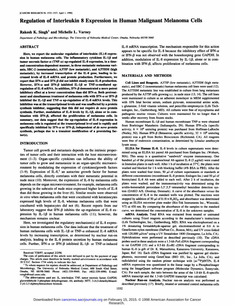
Duration of treatmentFig. 3. Up-regulation of IL-8 rtiRNA expression in melanoma cells by IL-l/3 (A) and
TNF-a (B). A375P melanoma cells were incubated with medium alone or mediumcontaining I and 10 units/ml of IL-1 ßor TNF-a for 2. 8, or 24 h. Total RNA was isolated,and expression of IL-8 mRNA was analyzed by Northern blot analysis. Data represent two
experiments.
or 24 h of treatment, respectively (Fig. 2). Similar results wereobtained after treatment with TNF-a (Fig. 2). These data suggest thatthe induction of IL-8 production by IL-Ißand TNF-a is time and
concentration dependent. Furthermore, the treatment of cells withdifferent concentrations of IL-1 ßand TNF-a for 2,4, 8, and 24 h were
noncytotoxic, as determined by MTT assay (data not shown).Stimulatory Effect of II.-I/Õand TNF-a on IL-8 mRNA Accu
mulation. We next determined whether the increased level of IL-8protein secretion was due to increased levels of IL-8 mRNA transcript. A375P cells were treated with I or 10 units/ml IL-1 ßor TNF-a
for 2, 8, or 24 h, and total RNA was isolated and analyzed for theexpression of IL-8-specific mRNA transcripts by Northern blot analysis. Similar to the protein production, IL-Ißand TNF-a up-regulatedIL-8 mRNA levels in a time- and concentration-dependent manner.However, treatment of cells with IL-lßat I unit/ml for 2, 8, and 24 hsignificantly increased the levels of IL-8 mRNA (2.8-, 14-, and8.2-fold increases, respectively, as compared to untreated cells). Sim
ilar results were obtained with the treatment of cells with lowerconcentrations of TNF-a, which enhanced the levels of IL-8 mRNA(3.5-, 11-, or 17-fold, as compared to controls following 2, 8, or 24 h
of treatment, respectively; Fig. 3). Furthermore, at a higher concentration (10 units/ml) of IL-lß and TNF-a, we observed a time-dependent increase in the levels of IL-8 mRNA (17-, 47-, or 56-foldincreases with IL-ßand 9-, 34-, or 57-fold increases with TNF-a, as
compared to controls, following 2, 8, or 24 h of treatment, respectively; Fig. 3). These data suggest that both IL-lßand TNF-a induceda concentration-dependent increase in the levels of IL-8 mRNA. Incontrast to the protein secretion, lower concentrations of IL-lßand
TNF-a significantly increased the levels of IL-8 mRNA in a time-
dependent manner (Fig. 3).Inhibitory Effects of IFN-a and IFN-ßon IL-8 mRNA Levels
Induced by IL-lß. The treatment of A375SM cells expressinghigher levels of steady-state IL-8, with different concentrations ofIFN-a or IFN-ßalone, did not inhibit IL-8 expression (data notshown). To determine whether IFN-a or IFN-ßdown-regulates IL-
lß-induced IL-8 expression, A375P cells expressing lower levels ofIL-8, were treated with different concentrations of IFN-a and IFN-ß(10, 100, or 1000 units/ml), alone or in combination with IL-lß(10
units/ml), for 2 h. Culture supernatants were collected and used forIL-8 ELISA, and total RNA was isolated for Northern blot analysis.Treatment of A375P cells with IL-lßand IFN-a inhibited the IL-lß-induced increase in the levels of IL-8 mRNA. This inhibition wasconcentration dependent, as a 35, 66, or 78% inhibition of IL-8
mRNA levels was observed in cells treated with 10, 100, and 1000units/ml of IFN-a and IL-lß,as compared to cells treated with IL-ßalone. Similarly, IFN-a also inhibited the IL-lß-induced increase inthe IL-8 mRNA levels in a concentration-dependent manner (Fig. 4).Inhibitions of 66, 76, and 88% of IL-lß-induced IL-8 mRNA wereobserved at 10, 100, and 1000 units/ml IFN-ß(Fig. 4). The inhibitoryeffect of >60% was observed with IFN-ßat 10 units/ml (16.8 nin) orIFN-a at 100 units/ml (88.7 nM), suggesting a more potent inhibitoryeffect of IFN-ßas compared to IFN-a. Similar to the inhibitionobserved at mRNA levels, both IFN-a and IFN-ßinhibited the IL-lß-induced IL-8 protein production (Table 1). However, the inhibition in IL-8 protein levels (50%) was not as high as that observed with
mRNA levels (76%).Nature of the Inhibitory Effect of IFN-ßon IL-8 mRNA Ex
pression. Most actions of IFNs are due to the stimulated expressionof selected genes in target cells (19, 20). Our earlier data suggested
100
80
60
40
5
1 20 J
0
IFN-ßIFN-a
control 10U/ml 100 U/ml 1000U/ml
IFN-a or -ßconcentrations
Fig. 4. IFN-a and IFN-ßinhibit the accumulation of IL-8 mRNA induced by IL-l |3 in
human melanoma cells. A375P melanoma cells were cultured with medium containing 10units/ml IL-lß.alone or in combination with different concentrations of IFN-a or IFN-ß.for 2 h. Total RNA was isolated and analyzed for IL-8 mRNA expression by Northern blot
analysis. Data represent two experiments.
Table I Effect of IL-lßand IFN-ßtreatment mi IL-8 productionA375P cells (1 X IO4 cells/well) were incubated with IL-ß(10 units/ml), alone or in
combination with IFN-ß(100 units/ml), for 4 h. Culture supernatant* were collected andanalyzed for IL-8 production. Values represent means ±SE. The data are one representative of two experiments done in triplicate.
TreatmentsMedium
IL-lß(10 units/ml)IFN-ß(100 units/ml)IL-lß + IFN-ßIL-8
(pg/ml)563
±523876 ±128
589 ±761954 ±165
1534
Research. on February 15, 2015. © 1998 American Association for Cancercancerres.aacrjournals.org Downloaded from
RI-Xifl.ATlON O! II.-8 I-.XPRKSSION IN MI I.ANOMA
2 4 6 8 10 12 14 16
Relative levels of IL-8 mRNA
IFN-a
IFN + IL-1
IL-1 -> IFN
O 2 4 6 8 10 12 14 16 18
Relative levels of IL-8 mRNA
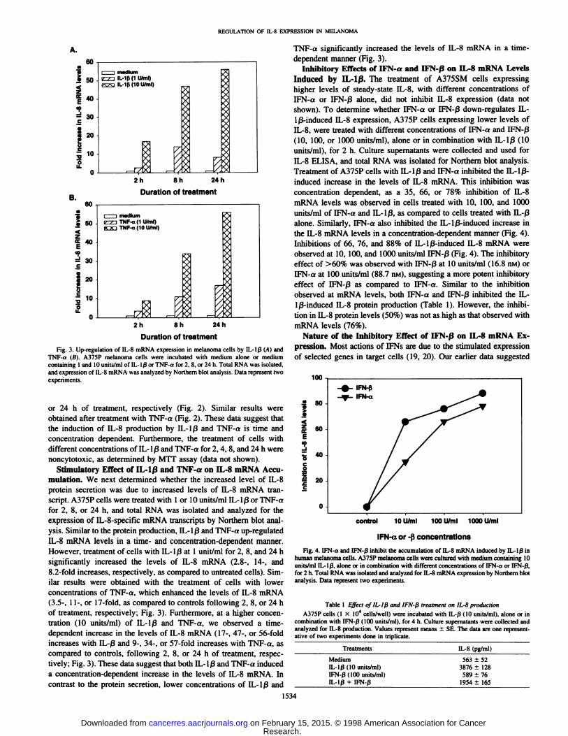
Fig. 5. Effect of sequential treatment of melanoma cells with IFN-a or IFN-ßon theaccumulation of IL-8 mRNA induced by IL-Ißin human melanoma cells. A375P cellswere incubated with medium alone (controll or medium containing IL-lß(IO units/ml)and IFN-a or IFN-ß(100 units/ml) in the order shown. Total RNA was isolated andanalyzed for IL-8 mRNA expression by Northern blot analysis. Data represent three
experiments.
that the pretreatment or simultaneous treatment of melanoma cellswith IFN-ßinhibits the induction of IL-8 mRNA and protein by IL-Ißor TNF-a. In these studies, we observed that this was also due to thedecrease in IL-8 mRNA in the IFN-a- or IFN-ß-treated cells. Three
treatment schedules were used: A375P cells were simultaneouslytreated with IL-1/3 (10 units/ml) and IFN-a or IFN-ß(100 units/ml)for 2 h; A375P cells were first pretreated with IFN-a or IFN-ß(100units/ml) for 2 h, washed, and subsequently treated with IL-Iß(10units/ml) for 2 h; or A375P cells were first pretreated with IL-lß( 10units/ml) for 2 h, washed, and subsequently incubated with IFN-a orIFN-ß( 100 units/ml). Simultaneous treatment of melanoma cells witheither IFN-a or IFN-ßand IL-1 ßinhibited IL-8 mRNA levels (52and 66%, respectively as compared to IL-lßtreated cultures; Fig. 5).Similarly, pretreatment of cells with IFN-a or IFN-ß,followed byIL-lß,also inhibited the IL-lß-induced IL-8 mRNA levels (66 and87.4% inhibition of IL-lß-induced IL-8 levels; Fig. 5). However, noinhibition of IL-8 mRNA was observed in the cells first treated withIL-lß,followed by IFN-a or IFN-ß(Fig. 5).
In next set of experiments, we examined whether the inhibition ofIL-8 mRNA in IL-lß-treated cells by IFN-ßrequires de novo protein
synthesis. The results shown in Fig. 6 demonstrate that the inhibitoryeffect of IFN-ßon IL-lß-induced IL-8 mRNA was not blocked in the
presence of cyclohexamide, suggesting no requirement for de novoprotein synthesis (Fig. 6).
IL-lß, TNF-a (Stimulator), and IFN-0 (Inhibitor) Modulati-thà R̈ate of IL-8 Gene Transcription. In the next set of experiments,we carried out a nuclear run-on analysis to determine whether IL-8
expression in melanoma cells is due to the differential activation oftranscription of IL-8 gene. A375P cells were incubated with mediumalone or medium containing 10 units/ml IL-lßor 10 units/ml TNF-a,alone or in combination with IFN-ß( 100 units/ml), for 2 h. Treatmentof cells with IL-lß or TNF-a alone showed increased (8.5- and5.9-fold, respectively) IL-8 gene transcription, as compared to cells
incubated with medium alone (Fig. 7). Treatment of melanoma cells
0 2 4 6 8 10 12 14
Fold increase in IL-8 mRNA levels
Fig. 6. Inhibition of IL-lß-induced IL-8 mRNA levels by IFN-ßin human melanomacells is unaffected by inhibitors of protein synthesis. A375P cells were treated with IL-lß(IO units/ml»,alone or in combination with IFN-ß(100 units/ml), in either presence orabsence of cyclohexamide ( 10 ^ig/ml). for 2 h. Total RNA was isolated and analy/ed forIL-8 mRNA expression by Northern blot analysis. Data represent two experiments.
GAPDH IL-8
medium
IL-lß
TNF-oc
IFN-ß
IL-lß+ IFN-ß
IL-lß+ IFN-ß+ CHX
Fig. 7. Effect of IFN-ßon IL-lß- or TNF-a-induced IL-8 gene transcription, asdetermined by nuclear run-on analysis. A375P cells were incubated in medium alone ormedium containing IL-lß (IO units/mil. TNF-a (10 units/ml), IFN-ß(KX) units/mil.IL-lß(10 units/ml) plus IFN-ß(100 units/ml), or IL-lßplus IFN-ß(1W units/ml) andcyclohexamide (10 fig/ml). Nuclei were isolated, and nuclear run-on analysis was performed. The hybridized blots were analyzed by digital autoradiography using a I'hos-
phorlmager (Molecular Dynamics).
1535
Research. on February 15, 2015. © 1998 American Association for Cancercancerres.aacrjournals.org Downloaded from
REGULATION OF IL-8 EXPRESSION IN MELANOMA
0.10 0.15 0.20 0.25
Absorbance (00540)
0.30
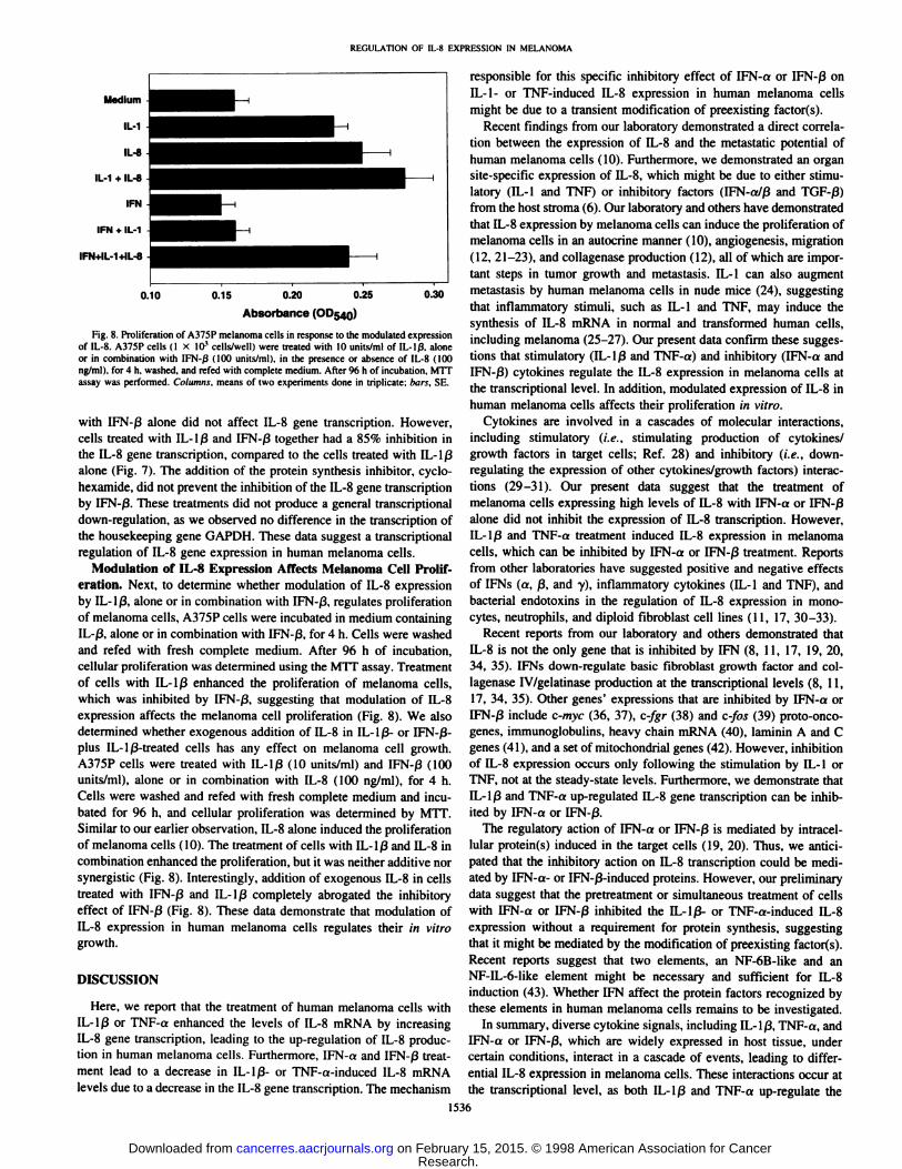
Fig. 8. Proliferation of A375P melanoma cells in response to the modulated expressionof IL-8. A375P cells (1 X 10' cells/well) were treated with 10 unils/ml of IL-lß.alone
or in combination with IFN-/3 (100 units/ml), in the presence or absence of IL-8 (100rig/ml ). for 4 h, washed, and refed with complete medium. After 96 h of incubation. MTTassay was performed. Columns, means of two experiments done in triplicate; hors, SE.
with IFN-ßalone did not affect IL-8 gene transcription. However,cells treated with IL-lßand IFN-ßtogether had a 85% inhibition inthe IL-8 gene transcription, compared to the cells treated with IL-lßalone (Fig. 7). The addition of the protein synthesis inhibitor, cyclo-hexamide, did not prevent the inhibition of the IL-8 gene transcriptionby IFN-ß.These treatments did not produce a general transcriptionaldown-regulation, as we observed no difference in the transcription of
the housekeeping gene GAPDH. These data suggest a transcriptionalregulation of IL-8 gene expression in human melanoma cells.
Modulation of IL-8 Expression Affects Melanoma Cell Proliferation. Next, to determine whether modulation of IL-8 expressionby IL-lß,alone or in combination with IFN-ß.regulates proliferation
of melanoma cells, A375P cells were incubated in medium containingIL-ß,alone or in combination with IFN-ß,for 4 h. Cells were washed
and refed with fresh complete medium. After 96 h of incubation,cellular proliferation was determined using the MTT assay. Treatmentof cells with IL-lß enhanced the proliferation of melanoma cells,which was inhibited by IFN-ß,suggesting that modulation of IL-8
expression affects the melanoma cell proliferation (Fig. 8). We alsodetermined whether exogenous addition of IL-8 in IL-lß-or IFN-ß-plus IL-lß-treated cells has any effect on melanoma cell growth.A375P cells were treated with IL-lß(10 units/ml) and IFN-ß(100units/ml), alone or in combination with IL-8 (100 ng/ml). for 4 h.
Cells were washed and refed with fresh complete medium and incubated for 96 h, and cellular proliferation was determined by MTT.Similar to our earlier observation, IL-8 alone induced the proliferationof melanoma cells (10). The treatment of cells with IL-lßand IL-8 in
combination enhanced the proliferation, but it was neither additive norsynergistic (Fig. 8). Interestingly, addition of exogenous IL-8 in cellstreated with IFN-ßand IL-lß completely abrogated the inhibitoryeffect of IFN-ß(Fig. 8). These data demonstrate that modulation ofIL-8 expression in human melanoma cells regulates their in vitro
growth.
DISCUSSION
Here, we report that the treatment of human melanoma cells withIL-lßor TNF-a enhanced the levels of IL-8 mRNA by increasingIL-8 gene transcription, leading to the up-regulation of IL-8 production in human melanoma cells. Furthermore, IFN-a and IFN-ßtreatment lead to a decrease in IL-lß- or TNF-a-induced IL-8 mRNAlevels due to a decrease in the IL-8 gene transcription. The mechanism
responsible for this specific inhibitory effect of IFN-a or IFN-ßonIL-1- or TNF-induced IL-8 expression in human melanoma cells
might be due to a transient modification of preexisting factor(s).Recent findings from our laboratory demonstrated a direct correla
tion between the expression of IL-8 and the metastatic potential of
human melanoma cells (10). Furthermore, we demonstrated an organsite-specific expression of IL-8, which might be due to either stimulatory (IL-1 and TNF) or inhibitory factors (IFN-a/ß and TGF-ß)
from the host stroma (6). Our laboratory and others have demonstratedthat IL-8 expression by melanoma cells can induce the proliferation of
melanoma cells in an autocrine manner (10). angiogenesis, migration(12, 21-23), and collagenase production (12), all of which are important steps in tumor growth and metastasis. IL-1 can also augment
metastasis by human melanoma cells in nude mice (24), suggestingthat inflammatory stimuli, such as IL-1 and TNF, may induce thesynthesis of IL-8 mRNA in normal and transformed human cells,including melanoma (25-27). Our present data confirm these suggestions that stimulatory (IL-lßand TNF-a) and inhibitory (IFN-a andIFN-ß)cytokines regulate the IL-8 expression in melanoma cells atthe transcriptional level. In addition, modulated expression of IL-8 in
human melanoma cells affects their proliferation in vitro.Cytokines are involved in a cascades of molecular interactions,
including stimulatory (i.e., stimulating production of cytokines/growth factors in target cells; Ref. 28) and inhibitory (i.e., down-
regulating the expression of other cytokines/growth factors) interactions (29-31). Our present data suggest that the treatment ofmelanoma cells expressing high levels of IL-8 with IFN-a or IFN-ßalone did not inhibit the expression of IL-8 transcription. However,IL-lßand TNF-a treatment induced IL-8 expression in melanomacells, which can be inhibited by IFN-a or IFN-ßtreatment. Reports
from other laboratories have suggested positive and negative effectsof IFNs (a, ß,and y), inflammatory cytokines (IL-1 and TNF), andbacterial endotoxins in the regulation of IL-8 expression in mono-cytes, neutrophils, and diploid fibroblast cell lines (11, 17, 30-33).
Recent reports from our laboratory and others demonstrated thatIL-8 is not the only gene that is inhibited by IFN (8, 11, 17, 19, 20,34, 35). IFNs down-regulate basic fibroblast growth factor and col
lagenase IV/gelatinase production at the transcriptional levels (8, 11,17, 34, 35). Other genes' expressions that are inhibited by IFN-a or
IFN-ßinclude c-myc (36, 37), c-fgr (38) and c-fos (39) proto-onco-
genes. immunoglobulins, heavy chain mRNA (40), laminin A and Cgenes (41 ). and a set of mitochondria! genes (42). However, inhibitionof IL-8 expression occurs only following the stimulation by IL-1 orTNF. not at the steady-state levels. Furthermore, we demonstrate thatIL-1 ßand TNF-a up-regulated IL-8 gene transcription can be inhibited by IFN-a or IFN-ß.
The regulatory action of IFN-a or IFN-ßis mediated by intracel-
lular protein(s) induced in the target cells (19, 20). Thus, we anticipated that the inhibitory action on IL-8 transcription could be mediated by IFN-a- or IFN-ß-induced proteins. However, our preliminary
data suggest that the pretreatment or simultaneous treatment of cellswith IFN-a or IFN-ßinhibited the IL-lß- or TNF-a-induced IL-8
expression without a requirement for protein synthesis, suggestingthat it might be mediated by the modification of preexisting factor(s).Recent reports suggest that two elements, an NF-6B-like and anNF-IL-6-like element might be necessary and sufficient for IL-8
induction (43). Whether IFN affect the protein factors recognized bythese elements in human melanoma cells remains to be investigated.
In summary, diverse cytokine signals, including IL-lß,TNF-a, andIFN-a or IFN-ß,which are widely expressed in host tissue, under
certain conditions, interact in a cascade of events, leading to differential IL-8 expression in melanoma cells. These interactions occur atthe transcriptional level, as both IL-lßand TNF-a up-regulate the
1536
Research. on February 15, 2015. © 1998 American Association for Cancercancerres.aacrjournals.org Downloaded from
REGULATION OF IL-8 EXPRESSION IN MELANOMA
transcription of IL-8 gene in vitro. IFN-a or IFN-/3 inhibit IL-1 ß-andTNF-a-induced IL-8 gene transcription.
REFERENCES
1. Fidler, I. J. Critical factors in the biology of human cancer metastasis: Twenty-eighthG. H. A. Clowes Memorial Award Lecture. Cancer Res., JO: 6130-6138. 1990.
2. Hart, I. R., and Fidler, I. J. Role of organ selectivity in the determination of metastaticpatterns of B16 melanoma. Cancer Res., 40: 2281-2287, 1980.
3. Fidler, I. J. Orthotopic implantation of human colon carcinomas into nude miceprovides a valuable model for the biology and therapy of cancer metastasis. CancerMetastasis Rev., 10: 229-243, 1990.
4. Morikawa, K., Walker. S. M., Nakajima, M., Pathak, S., Jessup, J. M., and Fidler, I. J.The influence of organ environment on the growth, selection and metastasis of humancolon cancer cells in nude mice. Cancer Res., 48: 6863-6871, 1988.
5. Naito, S., von Eschenbach, A. C, and Fidler, I. J. Different growth patterns andbiologic behavior of human renal cell carcinoma implanted into different organs ofnude mice. J. Nati. Cancer Inst. (Bethesda), 78: 377-385, 1987.
6. Gutman, M., Singh, R. K., Xie, K., Bucana, C. D., and Fidler, I. J. Regulation ofinterleukin-8 expression in human melanoma cells by the organ environment. CancerRes., 55: 2470-2474, 1995.
7. Singh, R. K., Bucana, C. D., Gutman, M., Fan, D., Wilson, M., and Fidler, I. J.Organ-site dependent expression of basic fibroblast growth factor in human renal cellcarcinoma cells. Am. J. Pathol., 145: 365-374, 1994.
8. Fabra, A., Nakajima, M., Bucana, C. D., and Fidler, I. J. Modulation of the invasivephenotype of human colon carcinoma cells by organ-specific fibroblasts of nudemice. Differentiation, 52: 101-110, 1992.
9. Wilmanns, C., Fan, D., O'Brian, C. A., Bucana, C. D., and Fidler. I. J. Orthotopic and
ectopie organ environments differentially influence the sensitivity of murine coloncarcinoma cells to doxorubicin and 5-fluorouracil. Int. J. Cancer, 52: 98-104. 1992.
10. Singh, R. K., Gutman. M., Radinsky, R., Bucana, C. D., and Fidler. I. J. Expressionof interleukin-8 correlates with the metastatic potential of human melanoma cells innude mice. Cancer Res, 54: 3242-3247, 1994.
11. Singh, R. K., Gutman, M., Llansa, N., and Fidler, I. J. Imerferon-ß prevents theupregulation of interleukin-8 expression in human melanoma cells. J. InterferonCytokine Res., 16: 577-584, 1996.
12. Singh, R. K.. Gutman, M., Reich, R., and Bar-Eli, M. Ultraviolet-B irradiation
promotes tumorigenic and metastatic properties in primary cutaneous melanoma viainduction of interleukin-8. Cancer Res., 55: 3669-3674, 1995.
13. Kozlowski, J. M., Hart, I. R., Fidler, I. J., and Hanna, N. A human melanoma lineheterogeneous with respect to metastatic capacity in athymic nude mice. J. Nati.Cancer Inst. (Bethesda), 72: 913-917, 1984.
14. Verschraegen, C. E., Giovanella, B. C., Mendoza, J. T.. Kozielsky, A. I., and Stehlin,J. S., Jr. Specific organ metastasis of human melanoma cells injected into arterialcirculation of nude mice. Anti-Cancer Res., //: 529-536, 1991.
15. Fort, P.. Marty, L., Piechaczyk, M.. Sabrouty, S. E., Dani, C., Jeanteur, P., andBlanchard. J. M. Various rat adult tissues express only one major mRNA species fromthe glyceraldehyde 3-phosphate-dehydrogenase multigenic family. Nucleic AcidsRes., 13: 1431-1442, 1985.
16. Matsushima, K.. Morishita, K., Yochimura. T., Lavu. S.. Kobayashi, Y.. Lew. W..Appella, E., Rung, H. F., Leonard, E. J.. and Oppenheim. J. J. Molecular cloning ofa human monocyte-derived neutrophil chemotactic factor (MDNCF) and the induction of MDNCF by interleukin-1 and tumor necrosis factor. J. Exp. Med., 767:1883-1893, 1988.
17. Singh, R. K., Bucana. C. D.. Llansa. N., Sanchez, R., and Fidler, I. J. Cell densitydependent modulation of basic fibroblast growth factor expression by human inter-feron |3. Int. J. Oncol., 8. 649-656, 1996 .
18. Alley, M. C., Scudiere, D. A.. Monks, A., Hersey, M. L., Czerwinski, M. J., Fine,D. L., Abbot, B. J., Mayo, J. G., Shoemaker, R. H., and Boyd. M. R. Feasibility ofdrug screening with panels of human tumor cell line using a microculture tetrazoliumassay. Cancer Res., 48: 589-602, 1988.
19. De Maeyer, E., and De Maeyer-Guignard, J. Interferons and Other Regulatory
Cytokines. New York: John Wiley & Sons, Inc., 1988.20. Pfeffer, L. A. Cellular effects of interferons. In: M. B. Sporn and A. B. Roberts (eds.).
Mechanisms of Interferon Action, Vol. 2, pp. 2-18. Boca Raton, FL: CRC Press,
1987.
21. Koch, A. E.. Polverini. P. J.. Kunkel. S. L.. Harlow, L. A., DiPietro, L. A., Einer,V. M., Einer, S. G., and Strieter, R. M. Interleukin-8 as a macrophage-derivedmediator of angiogenesis. Science (Washington DO, 258: 1798-1801. 1992.
22. Strieter, R. M.. Kunkel, S. L.. Einer, V. M., Martonyi. C. L.. Koch, A. E., Polverini,P. !.. and Einer. S. G. lnterleukin-8: a corneal factor that induces neovascularizalion.Am. J. Pathol., ¡41: 1279-1284, 1992.
23. Wang, J. M., Taraboletti, G., Matsushima, K., Damme. J. V., and Mantovani, A.Induction of hepatotactic migration of melanoma cells by neutrophil activatingprotein/IL-8. Biochem. Biophys. Res. Commun., 769: 165-170, 1990.
24. Giavazzi, R., Garafalo, A., Bani, M. R.. Abbate, M., Chezzi. P.. Boraschi, D..Montavani, A., and Jana, E. Interleukin-1-induced augmentation of experimentalmetastasis from a human melanoma in nude mice. Cancer Res., 50: 4771-4776, 1990.
25. Matsushima, K., and Oppenheim. J. lnterleukin-8 and MCAF: novel inflammatorycytokines inducible by IL-1 and TNF. Cytokine. /: 2-12. 1989.
26. Murphy. P.. Alexander, P., Senior. P. V.. Flemming, J.. Kirkham. N., and Taylor, I.Mechanisms of organ selective tumor growth by bloodborne cancer cells. Br. J.Cancer, 57: 19-31. 1988.
27. Zachariae. C. O. C., Thestrup-Pedersen, K.. and Matsushima. K. Expression andsecretion of leukocyte chemotactic cytokines by normal human melanocytes andmelanoma cells. J. Invest. Dermatol., 97: 593-599, 1991.
28. Thomson. A. W. The Cytokine Handbook. London: Academic Press, 1991.29. Lee, T. H., Lee, G. W., Ziff, E. B., and Vilcek, J. Isolation and characterization of
eight tumor necrosis factor-induced gene sequences from human fibroblasts. Mol.Cell Biol., 10: 1982-1988. 1990.
30. Gusella. G. L, Musso, T.. Bosco. M. C., Espinoza-Delgado, I., Matsushima, K., andVeresio, L. IL-2 upregulated but IFN-y suppressed IL-8 expression in human mono-cytes. J. Immunol., 151: 2725-2732, 1993.
31. Schnyder-Candrian. S.. Strieter. R. M.. Kunkel, S. L., and Walz, A. Interferon-a andinterferon--y downregulate the production of interleukin-8 and ENA-78 in humanmonocytes. J. Leukocyte Biol., 57: 929-935. 1995.
32. Oliveira, I. C., Sciavalino. P. J.. Lee, T. H., and Vilcek, J. Downregulation ofinterleukin-8 gene expression in human fibroblasts: unique mechanism of transcrip-tional inhibition by interferon. Proc. Nati. Acad. Sci. USA. 89: 9049-9053. 1992.
33. Hamilton. T. A., and Ohmori. Y. IFN-y selectively inhibits lipopolysaccharide-inducible JE/monocyte chemotactic protein-1 and kc/gro/melanoma growth stimulat
ing activity gene expression in mouse peritoneal macrophages. J. Immunol., 153:2204-2212, 1994.
34. Singh. R. K., Gutman. M., Bucana, C. D., Sanchez, R.. Llansa, N., and Fidler. I. J.Interferon-a and -ßdownregulate the expression of basic fibroblast growth factor inhuman carcinomas. Proc. Nati. Acad. Sci. USA. 92: 4562-4566, 1995.
35. Gohji, K.. Fidler, I. J., Tsan, R.. Radinsky. R., von Eschenbach. A. C., Tsuruo. T., andNakajima. M. Human recombinant interferon beta and gamma decrease gelatinaseproduction and invasion by human KG-2 renal carcinoma cells. Int. J. Cancer. 58:380-384, 1994.
36. Jonak, G.. Friedland. B. K., Anton, E. D.. and Knight. Jr.. E. Regulation of c-mvcmRNA and its proteins in Daudi cells by interferon-beta. J. Interferon Res.. 7: 41-52,
1987.37. Einat. M.. Resnitzky, D., and Kimchi. A. Close link between reduction of c-mvr
expression by interferon and GO/G1 arrest. Nature (Lond.), 313: 597-600, 1985.38. Sharp, N. A., Luscombe, M. J.. and Clemens. M. J. Regulation of cgf protooncogene
expression in Burkitt's lymphoma cells: effect of interferon treatment and relationship
to EBU stales and c-myc mRNA levels. Oncogene. 4: 1043-1046. 1989.39. Radzioch. D., and Varesio, L. c-fos mRNA expression in macrophages is downregu-
lated by interferon gamma at post-transcriptional levels. Mol. Cell. Biol.. //: 2718-
2722, 1991.40. Meurs, E., and Hovanessian, A. G. Alpha interferon inhibits the expression of heavy
chain Mu mRNA in Daudi cells. EMBO J.. 7: 1689-1696, 1988.41. Alldridgo, L. C., O'Farrell, M. K., and Dealtry, G. B. Downregulation of laminin A
and C by murine interferon beta. Exp. Cell Res., 195: 546-550, 1991.42. Shan, B., Vazquez, E., and Lewis, J. A. Interferon selectively inhibits the expression
of mitochondrial genes: a novel pathway for interferon mediated response. EMBO J..9.. 4307-4314, 1990.
43. Yasumoto, K., Okamoto, S., Mukaida, N., Murakami, S., Mai, M.. and Matsushima.K. Tumor necrosis factor-a and interferon-? synergistically induce ¡nterleukin-8production in human gastric cancer cell lines through acting concurrently on AP-1 andNF-6B-like binding sites of the interleukin-8 gene. J. Biol. Chem.. 267: 22506-
22511. 1992.
1537
Research. on February 15, 2015. © 1998 American Association for Cancercancerres.aacrjournals.org Downloaded from
1998;58:1532-1537. Cancer Res Rakesh K. Singh and Michelle L. Varney Melanoma CellsRegulation of Interleukin 8 Expression in Human Malignant
Updated version
http://cancerres.aacrjournals.org/content/58/7/1532
Access the most recent version of this article at:
E-mail alerts related to this article or journal.Sign up to receive free email-alerts
Subscriptions
Reprints and
To order reprints of this article or to subscribe to the journal, contact the AACR Publications
Permissions
To request permission to re-use all or part of this article, contact the AACR Publications
Research. on February 15, 2015. © 1998 American Association for Cancercancerres.aacrjournals.org Downloaded from