Expression and possible role of hPTTG1/securin in cutaneous malignant melanoma
11
Expression and possible role of hPTTG1/ securin in cutaneous malignant melanoma Ve ´ronique Winnepenninckx 1 , Maria Debiec-Rychter 2 , Jeroen AM Belie ¨n 3 , Pierre Fiten 4 , Stefan Michiels 5 , Vladimir Lazar 6 , Ghislain Opdenakker 4 , Gerrit A Meijer 3 , Alain Spatz 7 and Joost J van den Oord 1 1 Department of Morphology and Molecular Pathology, University Hospitals, Katholieke Universiteit Leuven (KUL), Leuven, Belgium; 2 Department of Human Genetics, University Hospitals, Katholieke Universiteit Leuven (KUL), Leuven, Belgium; 3 Department of Pathology, VU University Medical Center, Amsterdam, The Netherlands; 4 Department of Immunobiology, Rega Institute for Medical Research, Katholieke Universiteit Leuven (KUL), Leuven, Belgium; 5 Division of Biostatistics and Epidemiology, Gustave-Roussy Institute, Villejuif, France; 6 Division of Functional Genomics, Gustave-Roussy Institute, Villejuif, France and 7 Division of Pathology, Gustave-Roussy Institute, Villejuif, France Human pituitary tumour-transforming gene 1 or hPTTG1 is a proto-oncogene that codes for securin, a protein involved in sister chromatid separation. Based on previous microarray data, we studied the expression of hPTTG1/securin in melanocytic lesions. In contrast to nevi and radial growth phase melanomas, securin was expressed by scattered cells in the vertical growth phase, suggesting a role in tumour progression. In a series of 29 nodular and 29 superficial spreading melanomas, matched for all histological prognostic parameters, securin expression was significantly correlated with the nodular subtype (P ¼ 0.018) and not related to thickness. In other cancers, hPTTG1 is involved in various oncogenic pathways, including induction of neovascularisation and aneuploidy, and inhibition of p53 activity. We found coexpression of securin with wild- type p53 in the same neoplastic cells in a minority of melanomas. Expression of securin was significantly correlated with the extent of aneuploidy but not with basic fibroblast growth factor immunoreactivity or microvessel density. DNA cytometry revealed that nuclei-overexpressing securin frequently showed tetraploidy or aneuploidy. Our data show that hPTTG1 is frequently overexpressed in nodular melanoma, and suggest that hPTTG1 may act as an oncogene in the vertical growth phase, either by inhibiting anaphase, thereby causing aneuploidy and genomic instability, or by modulating the function of p53, thereby impairing apoptosis. Modern Pathology (2006) 19, 1170–1180. doi:10.1038/modpathol.3800627; published online 23 June 2006 Keywords: hPTTG1; securin; melanoma; aneuploidy; p53 Pituitary tumour-transforming gene (PTTG) was originally cloned from a rat pituitary tumour 1 and the function of the human homologue hPTTG1 (human pituitary tumour-transforming gene 1) was subsequently elucidated in foetal liver and testis. 2,3 hPTTG1 is a proto-oncogene encoding for securin, a protein involved in several metabolic reactions, cell- cycle progression, appropriate cell division and chromosome stability; upon overexpression, securin is involved in malignant transformation and tumor- igenesis. 4 In normal tissues, securin expression is limited, in contrast to many human tumours, including pitui- tary adenomas, 5 lung and breast cancer, 5–7 colorectal cancer, 8 oesophageal cancer 9 and some lymphoid neoplasms. 10 The oncogenic mechanisms of hPTTG1 are still barely known, but there is accumulating evidence that the oncoprotein activity of securin is exerted at different levels, that is, by induction of angiogenesis through basic fibroblast growth factor (bFGF) and vascular endothelial growth factor (VEGF), 11 by prevention of separation of sister chromatids resulting in aneuploidy, 12 by activation of c-myc 13 and/or by specific interaction with p53, thereby blocking its transcriptional activ- ity and inhibiting the ability of p53 to induce cell death. 14 In accordance with its oncogenic activities, hPTTG1 expression has been shown to have prog- nostic value. In tumours of the thyroid, securin is a Received 25 January 2006; revised and accepted 31 March 2006; published online 23 June 2006 Correspondence: Dr JJ van den Oord, MD, PhD, Department of Morphology and Molecular Pathology, University Hospitals, Katholieke Universiteit Leuven (KUL), Minderbroedersstraat 12, B-3000 Leuven, Belgium. E-mail: [email protected] Modern Pathology (2006) 19, 1170–1180 & 2006 USCAP, Inc All rights reserved 0893-3952/06 $30.00 www.modernpathology.org
Transcript of Expression and possible role of hPTTG1/securin in cutaneous malignant melanoma
Expression and possible role of hPTTG1/securin in cutaneous malignant melanoma
Veronique Winnepenninckx1, Maria Debiec-Rychter2, Jeroen AM Belien3, Pierre Fiten4,Stefan Michiels5, Vladimir Lazar6, Ghislain Opdenakker4, Gerrit A Meijer3, Alain Spatz7
and Joost J van den Oord1
1Department of Morphology and Molecular Pathology, University Hospitals, Katholieke Universiteit Leuven(KUL), Leuven, Belgium; 2Department of Human Genetics, University Hospitals, Katholieke UniversiteitLeuven (KUL), Leuven, Belgium; 3Department of Pathology, VU University Medical Center, Amsterdam, TheNetherlands; 4Department of Immunobiology, Rega Institute for Medical Research, Katholieke UniversiteitLeuven (KUL), Leuven, Belgium; 5Division of Biostatistics and Epidemiology, Gustave-Roussy Institute,Villejuif, France; 6Division of Functional Genomics, Gustave-Roussy Institute, Villejuif, France and7Division of Pathology, Gustave-Roussy Institute, Villejuif, France
Human pituitary tumour-transforming gene 1 or hPTTG1 is a proto-oncogene that codes for securin, a proteininvolved in sister chromatid separation. Based on previous microarray data, we studied the expression ofhPTTG1/securin in melanocytic lesions. In contrast to nevi and radial growth phase melanomas, securin wasexpressed by scattered cells in the vertical growth phase, suggesting a role in tumour progression. In a seriesof 29 nodular and 29 superficial spreading melanomas, matched for all histological prognostic parameters,securin expression was significantly correlated with the nodular subtype (P¼ 0.018) and not related tothickness. In other cancers, hPTTG1 is involved in various oncogenic pathways, including induction ofneovascularisation and aneuploidy, and inhibition of p53 activity. We found coexpression of securin with wild-type p53 in the same neoplastic cells in a minority of melanomas. Expression of securin was significantlycorrelated with the extent of aneuploidy but not with basic fibroblast growth factor immunoreactivity ormicrovessel density. DNA cytometry revealed that nuclei-overexpressing securin frequently showed tetraploidyor aneuploidy. Our data show that hPTTG1 is frequently overexpressed in nodular melanoma, and suggest thathPTTG1 may act as an oncogene in the vertical growth phase, either by inhibiting anaphase, thereby causinganeuploidy and genomic instability, or by modulating the function of p53, thereby impairing apoptosis.Modern Pathology (2006) 19, 1170–1180. doi:10.1038/modpathol.3800627; published online 23 June 2006
Keywords: hPTTG1; securin; melanoma; aneuploidy; p53
Pituitary tumour-transforming gene (PTTG) wasoriginally cloned from a rat pituitary tumour1 andthe function of the human homologue hPTTG1(human pituitary tumour-transforming gene 1) wassubsequently elucidated in foetal liver and testis.2,3
hPTTG1 is a proto-oncogene encoding for securin, aprotein involved in several metabolic reactions, cell-cycle progression, appropriate cell division andchromosome stability; upon overexpression, securinis involved in malignant transformation and tumor-igenesis.4
In normal tissues, securin expression is limited, incontrast to many human tumours, including pitui-tary adenomas,5 lung and breast cancer,5–7 colorectalcancer,8 oesophageal cancer9 and some lymphoidneoplasms.10 The oncogenic mechanisms ofhPTTG1 are still barely known, but there isaccumulating evidence that the oncoprotein activityof securin is exerted at different levels, that is, byinduction of angiogenesis through basic fibroblastgrowth factor (bFGF) and vascular endothelialgrowth factor (VEGF),11 by prevention of separationof sister chromatids resulting in aneuploidy,12 byactivation of c-myc13 and/or by specific interactionwith p53, thereby blocking its transcriptional activ-ity and inhibiting the ability of p53 to induce celldeath.14
In accordance with its oncogenic activities,hPTTG1 expression has been shown to have prog-nostic value. In tumours of the thyroid, securin is a
Received 25 January 2006; revised and accepted 31 March 2006;published online 23 June 2006
Correspondence: Dr JJ van den Oord, MD, PhD, Department ofMorphology and Molecular Pathology, University Hospitals,Katholieke Universiteit Leuven (KUL), Minderbroedersstraat 12,B-3000 Leuven, Belgium.E-mail: [email protected]
Modern Pathology (2006) 19, 1170–1180& 2006 USCAP, Inc All rights reserved 0893-3952/06 $30.00
www.modernpathology.org
prognostic marker for recurrence,15 and in breastcancer, the extent of securin expression is associatedwith the presence of metastatic spread and lymphnode invasion.6,16 In cancer of the oesophagus andcolorectum, high levels of hPTTG1 expressioncorrelate with tumour invasiveness.9 Recently,hPTTG1 has been identified as a key signaturegene, with high expression predicting metastases inmultiple tumour types.17
In a recent microarray study of primary malignantmelanoma,18 we found hPTTG1 to be listed amongthe genes that were significantly negatively corre-lated with outcome since patients showing over-expression of hPTTG1 had shortened distantmetastasis-free survival (DMFS) at 4 years. Thisprompted us to investigate in detail the expressionof securin in primary malignant melanoma, and tostudy the various pathways by which securinmay function as an oncoprotein in melanoma.Our results show that securin is particularly over-expressed in the nodular subtype of malignantmelanoma and that aneuploidy as well as inter-ference with p53 appears to be the main oncogenicmechanisms.
Materials and methods
Immunohistochemistry
To study the pattern of securin expression inpigment cell lesions, we used tissue microarrays,comprising a total of 570 cores of 50 mm width, takenfrom representative areas in 54 melanomas, 11metastases and 14 nevi (with a mean of 7.1 coresper lesion). From the primary melanomas, only thevertical growth phase was sampled. The pertinentclinical and histological data are listed in Table 1.As the analysis of the oligonucleotide arrayssuggested that overexpression of hPTTG1/securinoccurred predominantly in the nodular subtype ofmelanoma, we studied securin expression in thevertical growth phase of 29 pairs of superficialspreading melanoma and nodular melanoma,matched for thickness, ulceration, number of mitoticfigures, type of host response by tumour-infiltratinglymphocytes (ie brisk, nonbrisk or absent) andregression. In these 58 melanomas, all histologicalprognostic variables were assessed according tostandard criteria. To assess the tumour progressionphase in which securin immunoreactivity occurred,10 additional cases of in situ malignant melanomawere also studied.
In all cases, endogenous peroxidase was inacti-vated, and heat-induced epitope retrieval wasperformed in Tris-ethylenediaminetetraacetic acid,pH 9.0. Since preliminary experiments on normalhuman testis showed that a mixture of mousemonoclonal and rabbit polyclonal anti-securin anti-bodies yielded better immunohistochemical resultsthan either antibody alone (data not shown), allstainings were performed using a 1:1 mixture of
mouse monoclonal (clone DCS-280.2; NovocastraLabs. Ltd, Newcastle-upon-Tyne, UK) and rabbitpolyclonal (antibody Z23.YU; InVitrogen, Carlsbad,CA, USA) anti-securin antibodies, both diluted1:100 in phosphate-buffered saline. The second stepconsisted of a mixture of anti-mouse and anti-rabbitimmunoglobulins, labelled with peroxidase-conju-gated dextran polymers (Envisiont, Dakocytoma-tion, Haasrode, Belgium) and enzyme activity wasdetected using 3-amino-9-ethylcarbazole as sub-strate, revealing a brightly red reaction productthat contrasted well with the brown melanin. Thenumbers of securin-positive melanoma cells in thetissue microarrays as well as in the 29 matched pairsof melanomas were counted in a maximum of fivehigh power fields (HPF) in the areas of the verticalgrowth phase with the most abundant immuno-reactivity.
Semiserial sections of 44 out of 58 melanomaswere immunohistochemically stained for beta-cate-nin (clone 14; BD Transduction Labs., FranklinLakes, NJ, USA), bFGF (polyclonal rabbit antibodysc79; Santa Cruz Biotechnology Inc., Santa Cruz,CA, USA) and p53 (clone DO-7; Dakocytomation).To study the coexpression of securin and p53, asequential double staining was performed in 10cases with large numbers of securin-expressing cellsusing peroxidase-conjugated and alkaline phospha-tase-conjugated Envisiont reagents in the first andsecond staining; substrates for enzyme activity wereamino-ethylcarbazole and BCIP, respectively, yield-ing contrasting red and blue reaction products.Immunohistochemical controls, which were consis-tently negative, consisted of omission of primaryantibody and use of chromogen alone; in the doublestaining experiments, dye swapping was carried outby a reversal of the sequence of primary antibodies.
To assess the relationship between securin ex-pression and angiogenesis, serial sections of thetissue microarrays were stained for securin andfor CD31 (clone JC7OA; Dakocytomation) in orderto label microvessels. Then, the total number ofsecurin-immunoreactive cells as well as the micro-vessel density was assessed over the whole surfaceof each core according to the standard criteria.19
To study the relationship between securin expres-sion and aneuploidy, the number of immunoreactivemelanoma cells was counted in the vertical growthphase of 27 melanocytic tumours (seven primarymelanomas, 20 metastases) that had been karyo-typed previously.
DNA Cytometry
To study the relationship between securin expres-sion and DNA ploidy, a single 8-mm-thick section ofa melanoma that had an abnormal karyotype wasstained for securin using the alkaline phosphatase-conjugated Envisiont method without counterstain-ing. This slide was then stained by the Feulgen
hPTTG/securin in melanomaV Winnepenninckx et al
1171
Modern Pathology (2006) 19, 1170–1180
method. Briefly, the slide was placed in 5 N HCl at271C for 30 min, washed 3� 1–5 min in distilledwater, stained with fresh Schiff reagent for 45 min,washed in running tap water for 15 min and washedfor 1 min in aqua dest. From the same melanoma
sample, three 50-mm-thick sections were cut andprocessed for standard DNA flow cytometry as wellas DNA image cytometry. For the last method, acytospin specimen was prepared and stained by theFeulgen method as described above.
Table 1 Pertinent clinical and histological features of the 54 primary cutaneous melanomas included in the tissue microarray
Case no. Sex Age Status Type T Thickness (mm) Ulceration Regression Mitotic rate TILS Mean securin
1 f 78 AwRD 1 2A 1.34 � + 2 2 242 f 65 DOD 3 3A 3.74 � � 2 1 03 f 54 AwMD 2 4B 5.44 + � 2 1 3.24 f 57 DOD 1 1B 0.98 + � 2 2 75 f 75 AwRD 2 4B 7.58 + � 2 1 636 f 70 DOC 1 1A 0.82 � + 1 1 107 f 66 NED 1 3A 3.32 � � 1 1 148 f 67 DOD 3 4A 6.40 � � 2 1 109 m 72 DOD 2 3B 3.52 + � 2 1 13.3
10 f 89 DOD 3 4B 7.04 + � 2 0 1411 f 77 DOC 1 3A 2.52 � � 2 0 012 f 40 DOD 2 4B 10.88 + � 2 0 35.513 f 76 DOD 2 4B 14.56 + � 2 0 1.214 f 54 NED 2 3B 3.65 + � 1 2 11.515 m 63 NED 1 3B 3.68 + � 2 0 016 f 57 NED 1 1A 0.96 � � 1 1 417 m 76 DOD 1 3B 2.21 + � 1 1 018 f 74 NED 1 2A 1.59 � + 2 2 019 f 92 NED 2 4B 4.70 + � 2 1 0.820 f 93 NED 1 4B 6.72 + � 2 0 2.221 f 65 DOD 1 2A 1.19 � + 1 0 022 f 58 NED 1 2A 1.92 � � 0 2 023 m 71 NED 2 4B 9.28 + � 1 0 124 f 34 NED 1 1A 0.94 � + 1 1 025 f 56 NED 2 3A 2.08 � � 2 1 43.826 f 56 DOD 2 4B 8.16 + � 2 0 1227 f 37 DOD 1 2A 1.20 � + 1 1 028 m 58 NED 1 3A 2.42 � � 1 0 029 m 63 NED 4 4A 4.16 � + 2 0 7.830 m 47 LOF 1 1A 0.66 � � 0 0 031 m 38 DOD 1 4A 5.02 � � 2 0 0.232 f 9 NED 4 4B 4.70 + � 2 0 033 m 78 DOD 3 4B 14.18 + � 2 0 4.834 m 74 AwRD 1 4A 5.18 � � 1 1 0.435 f 68 DOD 1 3A 2.75 � � 1 1 0.536 f 64 NED 2 4B 17.12 + � 2 2 29.237 f 79 AwRD 2 4B 7.52 + � 1 1 31.538 m 78 DOD 1 3B 3.04 + � 1 2 16.639 f 1 DOD 2 4A 12.41 � � 2 0 22740 f 92 DOC 1 4B 6.81 + � 1 2 3941 f 47 NED 1 4A 4.61 � + 1 2 042 m 63 DOC 1 4A 5.95 � + 2 1 643 f 46 LOF 2 3A 2.08 � � 1 2 044 m 33 NED 1 2A 1.54 � � 1 1 16.345 f 55 DOD 3 3A 2.30 � � 2 0 046 m 22 DOD 4 3A 3.36 � � 2 1 1747 f 64 NED 1 2A 1.44 � + 1 2 048 f 72 DOD 2 4B 7.97 + � 2 1 049 f 73 DOD 3 3B 3.36 + � 2 1 050 m 85 DOD 4 4A 15.64 � � 2 2 051 f 67 NED 1 3A 2.05 � + 2 1 352 m 64 LOF 1 2A 1.40 � + 1 1 353 f 53 NED 1 2A 1.02 � � 1 0 2.654 f 56 DOD 2 4B 18.56 + � 2 0 0
Sex: f¼ female; m¼male. Status: AwRD¼ alive with regional disease; AwMD¼ alive with metastatic disease; DOD¼dead of disease; DOC¼deadof other cause; NED¼no evidence of disease; LOF¼ lost for follow-up. Type: histogenetic type; 1¼ superficial spreading type; 2¼nodular type;3¼ acrolentiginous type; 4¼ other types. T¼ tumour stage. Mitotic rate: 0¼no mitoses; 1¼o6/mm2; 2¼46/mm2; TILS¼ tumour-infiltratinglymphocytes; 0¼no TILS; 1¼nonbrisk infiltrate of TILS; 2¼brisk infiltrate of TILS. Mean PTTG: mean number of cells with nuclear orcytoplasmic PTTG-staining, counted in the different cores of each melanoma.
hPTTG/securin in melanomaV Winnepenninckx et al
1172
Modern Pathology (2006) 19, 1170–1180
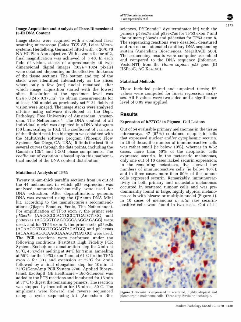
Image Acquisition and Analysis of Three-Dimensional(3-D) DNA Content
Image stacks were acquired with a confocal laserscanning microscope (Leica TCS SP, Leica Micro-systems, Heidelberg, Germany) fitted with � 20/0.70NA HC Plan Apo objective. With a zoom factor of 2,final magnification was achieved of � 40. In eachfield of vision, stacks of approximately 40 two-dimensional digital images (1024� 1024 pixels)were obtained, depending on the effective thicknessof the tissue sections. The bottom and top of thestack were identified interactively as the sliceswhere only a few (cut) nuclei remained, afterwhich image acquisition started with the lowestslice. Resolution at the specimen level was0.24� 0.24� 0.37 mm3. To obtain measurements forat least 300 nuclei as previously set,20 24 fields ofvision were imaged. The image stacks were analysedoff-line using software developed at the Dept.Pathology, Free University of Amsterdam, Amster-dam, The Netherlands.21 The DNA content of allindividual nuclei was depicted in a DNA histogram(50 bins, scaling to 10c). The coefficient of variationof the diploid peak in a histogram was obtained withthe MultiCycle software program (Phoenix FlowSystems, San Diego, CA, USA). It finds the best fit ofseveral curves through the data points, including theGaussian G0/1 and G2/M phase components. Thecoefficient of variation is based upon this mathema-tical model of the DNA content distribution.
Mutational Analysis of TP53
Twenty 10-mm-thick paraffin sections from 34 out ofthe 44 melanomas, in which p53 expression wasanalysed immunohistochemically, were used forDNA extraction. After deparaffination, genomicDNA was extracted using the QIAamp DNA Minikit, according to the manufacturer’s recommend-ations (Qiagen Benelux, Venlo, The Netherlands).For amplification of TP53 exon 7, the primer setsp53ex7s (AAGGCGCACTGGCCTCATCTTGG) andp53ex7as (AGGGGTCAGCGGCAAGCAGAGG) wereused, and for TP53 exon 8, the primer sets p53ex8s(ACAAGGGTGGTTGGAGTAGATGG) and p53ex8as(ACAAAGAGGCAAGGAAAGGTGATGG) were used.The PCR reactions were performed under thefollowing conditions (FastStart High Fidelity PCRSystem, Roche): one denaturation step for 2 min at951C, 45 cycles melting at 941C for 1 min, annealingat 661C for the TP53 exon 7 and at 611C for the TP53exon 8 for 30 s and extension at 721C for 2 minfollowed by a final elongation step for 10 min at721C (GeneAmp PCR System 2700, Applied Biosys-tems). ExoSapIt (GE Healthcare – Bio-Sciences) wasadded to the PCR reactions and incubated for 15 minat 371C to digest the remaining primers. The reactionwas stopped by incubation for 15 min at 801C. Theamplicons were forward and reverse sequencedusing a cycle sequencing kit (Amersham Bio-
sciences, DYEnamict dye terminator kit) with theprimers p53ex7s and p53ex7as for TP53 exon 7 andthe primers p53ex8s and p53ex8as for TP53 exon 8.The sequencing reactions were desalted, denaturedand run on an automated capillary DNA sequencingsystem (Amersham Biosciences, MegaBACE 500).The sequencing results were computer assembledand compared to the DNA sequence (Informax,VectorNTI) from the Homo sapiens p53 gene (IDHSP53G, AC X54156).
Statistical Methods
These included paired and unpaired t-tests; R2-values were computed for linear regression analy-ses. All P-values were two-sided and a significancelevel of 0.05 was applied.
Results
Expression of hPTTG1 in Pigment Cell Lesions
Out of 54 evaluable primary melanomas in the tissuemicroarrays, 47 (87%) contained neoplastic cellsthat expressed nuclear and/or cytoplasmic securin.In 26 of these, the number of immunoreactive cellswas rather small (ie below 10%), whereas in 8/52cases, more than 50% of the neoplastic cellsexpressed securin. In the metastatic melanomas,only one out of 10 cases lacked securin expression;in the remaining metastases, five showed lownumbers of immunoreactive cells (ie below 10%),and in three cases, more than 50% of the tumourcells expressed securin. Remarkably, immunoreac-tivity in both primary and metastatic melanomasoccurred in scattered tumour cells and was pre-dominantly found in large, highly atypical melano-ma cells with bizarre or multiple nuclei (Figure 1).In 10 cases of melanoma in situ, rare securin-positive cells were found in two cases. Out of 11
Figure 1 Securin is expressed in scattered, highly atypical andpleomorphic melanoma cells. Three-step Envision technique.
hPTTG/securin in melanomaV Winnepenninckx et al
1173
Modern Pathology (2006) 19, 1170–1180
evaluable nevi, six contained rare (ie below 1%)securin-expressing cells.
A previous supervised analysis of the differencesin gene expression between 49 superficial spreadingmelanomas and 15 nodular melanomas had revealed
that PTTG was the most significantly differentiallyexpressed gene (unpublished results). This wasconfirmed in the present study by counting thenumber of immunoreactive cells in the 54 melano-mas in the tissue microarray sections, revealing
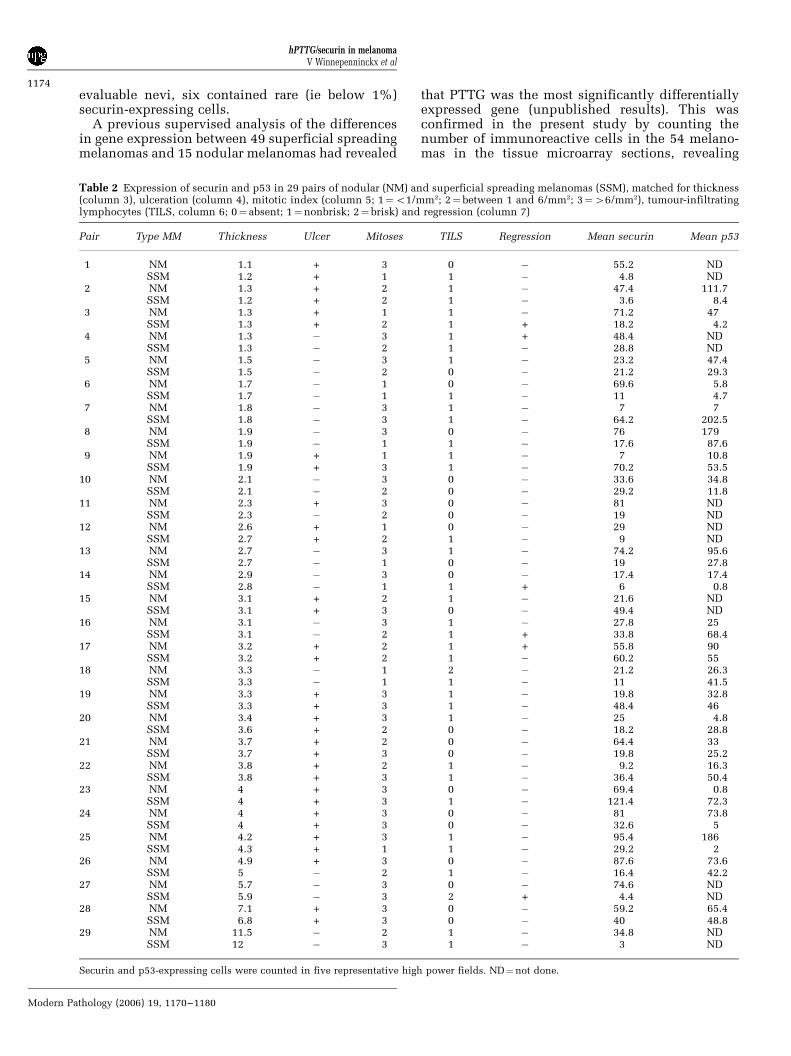
Table 2 Expression of securin and p53 in 29 pairs of nodular (NM) and superficial spreading melanomas (SSM), matched for thickness(column 3), ulceration (column 4), mitotic index (column 5; 1¼o1/mm2; 2¼between 1 and 6/mm2; 3¼46/mm2), tumour-infiltratinglymphocytes (TILS, column 6; 0¼ absent; 1¼nonbrisk; 2¼ brisk) and regression (column 7)
Pair Type MM Thickness Ulcer Mitoses TILS Regression Mean securin Mean p53
1 NM 1.1 + 3 0 � 55.2 NDSSM 1.2 + 1 1 � 4.8 ND
2 NM 1.3 + 2 1 � 47.4 111.7SSM 1.2 + 2 1 � 3.6 8.4
3 NM 1.3 + 1 1 � 71.2 47SSM 1.3 + 2 1 + 18.2 4.2
4 NM 1.3 � 3 1 + 48.4 NDSSM 1.3 � 2 1 � 28.8 ND
5 NM 1.5 � 3 1 � 23.2 47.4SSM 1.5 � 2 0 � 21.2 29.3
6 NM 1.7 � 1 0 � 69.6 5.8SSM 1.7 � 1 1 � 11 4.7
7 NM 1.8 � 3 1 � 7 7SSM 1.8 � 3 1 � 64.2 202.5
8 NM 1.9 � 3 0 � 76 179SSM 1.9 � 1 1 � 17.6 87.6
9 NM 1.9 + 1 1 � 7 10.8SSM 1.9 + 3 1 � 70.2 53.5
10 NM 2.1 � 3 0 � 33.6 34.8SSM 2.1 � 2 0 � 29.2 11.8
11 NM 2.3 + 3 0 � 81 NDSSM 2.3 � 2 0 � 19 ND
12 NM 2.6 + 1 0 � 29 NDSSM 2.7 + 2 1 � 9 ND
13 NM 2.7 � 3 1 � 74.2 95.6SSM 2.7 � 1 0 � 19 27.8
14 NM 2.9 � 3 0 � 17.4 17.4SSM 2.8 � 1 1 + 6 0.8
15 NM 3.1 + 2 1 � 21.6 NDSSM 3.1 + 3 0 � 49.4 ND
16 NM 3.1 � 3 1 � 27.8 25SSM 3.1 � 2 1 + 33.8 68.4
17 NM 3.2 + 2 1 + 55.8 90SSM 3.2 + 2 1 � 60.2 55
18 NM 3.3 � 1 2 � 21.2 26.3SSM 3.3 � 1 1 � 11 41.5
19 NM 3.3 + 3 1 � 19.8 32.8SSM 3.3 + 3 1 � 48.4 46
20 NM 3.4 + 3 1 � 25 4.8SSM 3.6 + 2 0 � 18.2 28.8
21 NM 3.7 + 2 0 � 64.4 33SSM 3.7 + 3 0 � 19.8 25.2
22 NM 3.8 + 2 1 � 9.2 16.3SSM 3.8 + 3 1 � 36.4 50.4
23 NM 4 + 3 0 � 69.4 0.8SSM 4 + 3 1 � 121.4 72.3
24 NM 4 + 3 0 � 81 73.8SSM 4 + 3 0 � 32.6 5
25 NM 4.2 + 3 1 � 95.4 186SSM 4.3 + 1 1 � 29.2 2
26 NM 4.9 + 3 0 � 87.6 73.6SSM 5 � 2 1 � 16.4 42.2
27 NM 5.7 � 3 0 � 74.6 NDSSM 5.9 � 3 2 + 4.4 ND
28 NM 7.1 + 3 0 � 59.2 65.4SSM 6.8 + 3 0 � 40 48.8
29 NM 11.5 � 2 1 � 34.8 NDSSM 12 � 3 1 � 3 ND
Securin and p53-expressing cells were counted in five representative high power fields. ND¼not done.
hPTTG/securin in melanomaV Winnepenninckx et al
1174
Modern Pathology (2006) 19, 1170–1180
significant (P¼ 0.02) differences between nodular(average number of immunoreactive cells per coresection¼ 31.5) and superficial spreading melanomas(average number¼ 5.5) (Table 1). However, sincethe nodular melanomas in the tissue microarrayseries had greater mean thickness than superficialspreading melanomas, we studied securin expres-sion in a separate series of 29 pairs of nodular andsuperficial spreading melanomas, matched for allhistological prognostic markers including tumourthickness (Table 2). Paired t-tests revealed signifi-cant (P¼ 0.018) differences in securin expressionbetween nodular and superficial spreading melano-mas (mean number of immunoreactive cells infive HPF: nodular melanoma¼ 48.1; superficialspreading melanoma¼ 29.2), whereas there was nocorrelation between securin expression and tumourthickness.
hPTTG1 and Angiogenesis in Melanoma
Ten malignant melanomas stained on semiserialsections for securin and bFGF showed no spatialrelationship in immunoreactivity for either anti-body. In addition, no correlation was found betweenthe number of CD31-immunoreactive microvesselsand the number of securin-expressing cells.
hPTTG1 and Beta-Catenin Expression in Melanoma
Zhou et al22 detected cytoplasmic accumulation ofbeta-catenin in oesophageal carcinomas that over-expressed PTTG1 and suggested that overexpressionof PTTG1 in these tumours was likely due to theactivation of beta-catenin/WNT signalling. However,in 10 melanomas overexpressing securin, the pat-terns of expression of beta-catenin varied consider-ably (ie membranous staining in 6/10; nuclearstaining in 2/10; nuclear and cytoplasmic stainingin 2/10).
hPTTG1 and Aneuploidy in Melanoma
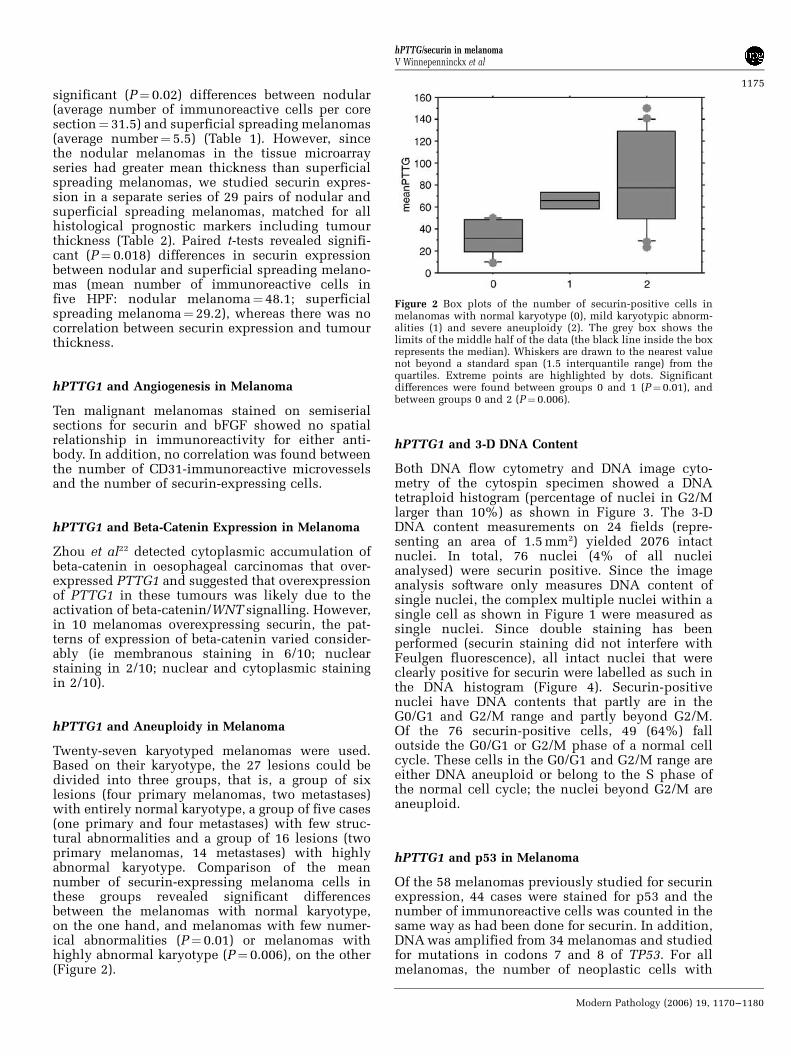
Twenty-seven karyotyped melanomas were used.Based on their karyotype, the 27 lesions could bedivided into three groups, that is, a group of sixlesions (four primary melanomas, two metastases)with entirely normal karyotype, a group of five cases(one primary and four metastases) with few struc-tural abnormalities and a group of 16 lesions (twoprimary melanomas, 14 metastases) with highlyabnormal karyotype. Comparison of the meannumber of securin-expressing melanoma cells inthese groups revealed significant differencesbetween the melanomas with normal karyotype,on the one hand, and melanomas with few numer-ical abnormalities (P¼ 0.01) or melanomas withhighly abnormal karyotype (P¼ 0.006), on the other(Figure 2).
hPTTG1 and 3-D DNA Content
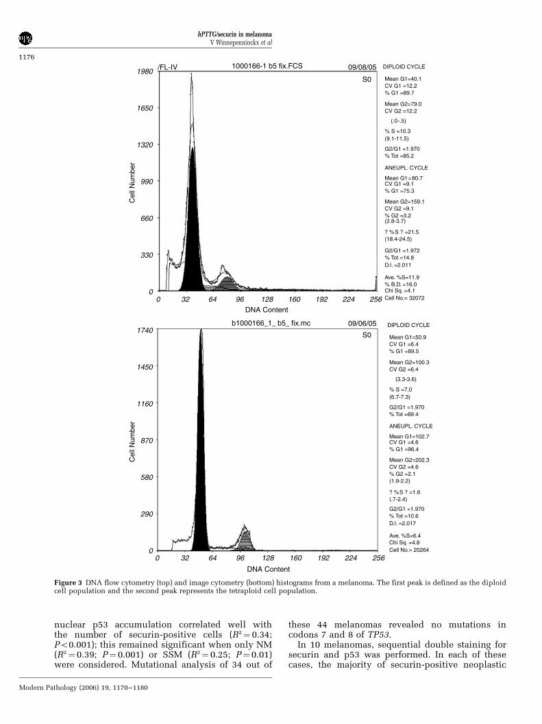
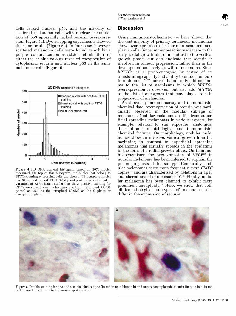
Both DNA flow cytometry and DNA image cyto-metry of the cytospin specimen showed a DNAtetraploid histogram (percentage of nuclei in G2/Mlarger than 10%) as shown in Figure 3. The 3-DDNA content measurements on 24 fields (repre-senting an area of 1.5 mm2) yielded 2076 intactnuclei. In total, 76 nuclei (4% of all nucleianalysed) were securin positive. Since the imageanalysis software only measures DNA content ofsingle nuclei, the complex multiple nuclei within asingle cell as shown in Figure 1 were measured assingle nuclei. Since double staining has beenperformed (securin staining did not interfere withFeulgen fluorescence), all intact nuclei that wereclearly positive for securin were labelled as such inthe DNA histogram (Figure 4). Securin-positivenuclei have DNA contents that partly are in theG0/G1 and G2/M range and partly beyond G2/M.Of the 76 securin-positive cells, 49 (64%) falloutside the G0/G1 or G2/M phase of a normal cellcycle. These cells in the G0/G1 and G2/M range areeither DNA aneuploid or belong to the S phase ofthe normal cell cycle; the nuclei beyond G2/M areaneuploid.
hPTTG1 and p53 in Melanoma
Of the 58 melanomas previously studied for securinexpression, 44 cases were stained for p53 and thenumber of immunoreactive cells was counted in thesame way as had been done for securin. In addition,DNA was amplified from 34 melanomas and studiedfor mutations in codons 7 and 8 of TP53. For allmelanomas, the number of neoplastic cells with
Figure 2 Box plots of the number of securin-positive cells inmelanomas with normal karyotype (0), mild karyotypic abnorm-alities (1) and severe aneuploidy (2). The grey box shows thelimits of the middle half of the data (the black line inside the boxrepresents the median). Whiskers are drawn to the nearest valuenot beyond a standard span (1.5 interquantile range) from thequartiles. Extreme points are highlighted by dots. Significantdifferences were found between groups 0 and 1 (P¼0.01), andbetween groups 0 and 2 (P¼ 0.006).
hPTTG/securin in melanomaV Winnepenninckx et al
1175
Modern Pathology (2006) 19, 1170–1180
nuclear p53 accumulation correlated well withthe number of securin-positive cells (R2¼ 0.34;Po0.001); this remained significant when only NM(R2¼ 0.39; P¼ 0.001) or SSM (R2¼ 0.25; P¼ 0.01)were considered. Mutational analysis of 34 out of
these 44 melanomas revealed no mutations incodons 7 and 8 of TP53.
In 10 melanomas, sequential double staining forsecurin and p53 was performed. In each of thesecases, the majority of securin-positive neoplastic
19801000166-1 b5 fix.FCS
b1000166_1_ b5_ fix.mc
S0
09/08/05
09/06/05
/FL-IV
S0
DIPLOID CYCLE
ANEUPL. CYCLE
Mean G1=40.1CV G1 =12.2% G1 =89.7
Mean G1=80.7CV G1 =9.1% G1 =75.3
Mean G2=159.1CV G2 =9.1% G2 =3.2
? %S ? =21.5(18.4-24.5)
(2.8-3.7)
G2/G1 =1.970% Tot =85.2
G2/G1 =1.972% Tot =14.8D.I. =2.011
Ave. %S=11.9% B.D. =16.0Chi Sq. =4.1Cell No.= 32072
Mean G2=79.0CV G2 =12.2
(.0-.5)
% S =10.3(9.1-11.5)
DIPLOID CYCLE
ANEUPL. CYCLE
Mean G1=50.9CV G1 =6.4% G1 =89.5
Mean G1=102.7CV G1 =4.6% G1 =96.4
Mean G2=202.3CV G2 =4.6% G2 =2.1
? %S ? =1.6(.7-2.4)
(1.9-2.2)
G2/G1 =1.970% Tot =89.4
G2/G1 =1.970% Tot =10.6D.I. =2.017
Ave. %S=6.4Chi Sq. =4.8Cell No.= 20264
Mean G2=100.3CV G2 =6.4
(3.3-3.6)
% S =7.0(6.7-7.3)
Cel
l Num
ber
1650
1320
990
660
330
0
1740
Cel
l Num
ber
1450
1160
870
580
290
0
0 32 64 96 128DNA Content
DNA Content
160 192 224 256
0 32 64 96 128 160 192 224 256
Figure 3 DNA flow cytometry (top) and image cytometry (bottom) histograms from a melanoma. The first peak is defined as the diploidcell population and the second peak represents the tetraploid cell population.
hPTTG/securin in melanomaV Winnepenninckx et al
1176
Modern Pathology (2006) 19, 1170–1180
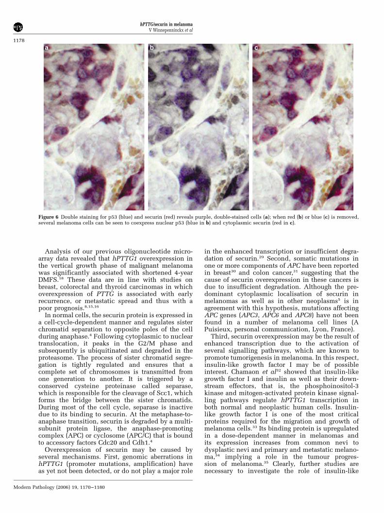
cells lacked nuclear p53, and the majority ofscattered melanoma cells with nuclear accumula-tion of p53 apparently lacked securin overexpres-sion (Figure 5a). Dye-swapping experiments showedthe same results (Figure 5b). In four cases however,scattered melanoma cells were found to exhibit apurple colour; computer-assisted elimination ofeither red or blue colours revealed coexpression ofcytoplasmic securin and nuclear p53 in the samemelanoma cells (Figure 6).
Discussion
Using immunohistochemisty, we have shown thatthe vast majority of primary cutaneous melanomasshow overexpression of securin in scattered neo-plastic cells. Since immunoreactivity was rare in theearly, radial growth phase in contrast to the verticalgrowth phase, our data indicate that securin isinvolved in tumour progression, rather than in thedevelopment and early growth of melanoma. SincehPTTG1 is a proto-oncogene by virtue of itstransforming capacity and ability to induce tumoursin nude mice,23,24 our results not only add melano-ma to the list of neoplasms in which hPTTG1overexpression is observed, but also add hPTTG1to the list of oncogenes that may play a role inprogression of melanoma.
As shown by our microarray and immunohisto-chemical data, overexpression of securin was parti-cularly observed in the nodular subtype ofmelanoma. Nodular melanomas differ from super-ficial spreading melanomas in various aspects, forexample, relation to sun exposure, anatomicaldistribution and histological and immunohisto-chemical features. On morphology, nodular mela-nomas show an invasive, vertical growth from thebeginning in contrast to superficial spreadingmelanomas that initially spreads in the epidermisin the form of a radial growth phase. On immuno-histochemistry, the overexpression of VEGF25 innodular melanoma has been inferred to explain thepoorer prognosis of this subtype. Genetically, nod-ular melanomas carry more frequently extra CMYCcopies26 and are characterised by deletions in 1p36and aberrations of chromosome 10.27 Finally, nodu-lar melanoma has been claimed to exhibit moreprominent aneuploidy.28 Here, we show that bothclinicopathological subtypes of melanoma alsodiffer in the expression of securin.
Figure 4 3-D DNA content histogram based on 2076 nucleimeasured. On top of this histogram, the nuclei that belong toPTTG/securing expressing cells are shown (76 complete nucleiand 37 capped nuclei). The DNA diploid peak has a coefficient ofvariation of 8.5%. Intact nuclei that show positive staining forPTTG are spread over the histogram, within the diploid (G0/G1phase) as well as the tetraploid (G2/M) as the S phase oraneuploid region.
Figure 5 Double staining for p53 and securin. Nuclear p53 (in red in a; in blue in b) and nuclear/cytoplasmic securin (in blue in a; in redin b) were found in distinct, nonoverlapping cells.
hPTTG/securin in melanomaV Winnepenninckx et al
1177
Modern Pathology (2006) 19, 1170–1180
Analysis of our previous oligonucleotide micro-array data revealed that hPTTG1 overexpression inthe vertical growth phase of malignant melanomawas significantly associated with shortened 4-yearDMFS.18 These data are in line with studies onbreast, colorectal and thyroid carcinomas in whichoverexpression of PTTG is associated with earlyrecurrence, or metastatic spread and thus with apoor prognosis.8,15,16
In normal cells, the securin protein is expressed ina cell-cycle-dependent manner and regulates sisterchromatid separation to opposite poles of the cellduring anaphase.4 Following cytoplasmic to nucleartranslocation, it peaks in the G2/M phase andsubsequently is ubiquitinated and degraded in theproteasome. The process of sister chromatid segre-gation is tightly regulated and ensures that acomplete set of chromosomes is transmitted fromone generation to another. It is triggered by aconserved cysteine proteinase called separase,which is responsible for the cleavage of Scc1, whichforms the bridge between the sister chromatids.During most of the cell cycle, separase is inactivedue to its binding to securin. At the metaphase-to-anaphase transition, securin is degraded by a multi-subunit protein ligase, the anaphase-promotingcomplex (APC) or cyclosome (APC/C) that is boundto accessory factors Cdc20 and Cdh1.4
Overexpression of securin may be caused byseveral mechanisms. First, genomic aberrations inhPTTG1 (promoter mutations, amplification) haveas yet not been detected, or do not play a major role
in the enhanced transcription or insufficient degra-dation of securin.29 Second, somatic mutations inone or more components of APC have been reportedin breast30 and colon cancer,31 suggesting that thecause of securin overexpression in these cancers isdue to insufficient degradation. Although the pre-dominant cytoplasmic localisation of securin inmelanomas as well as in other neoplasms5 is inagreement with this hypothesis, mutations affectingAPC genes (APC3, APC6 and APC8) have not beenfound in a number of melanoma cell lines (APuisieux, personal communication, Lyon, France).
Third, securin overexpression may be the result ofenhanced transcription due to the activation ofseveral signalling pathways, which are known topromote tumorigenesis in melanoma. In this respect,insulin-like growth factor I may be of possibleinterest. Chamaon et al32 showed that insulin-likegrowth factor I and insulin as well as their down-stream effectors, that is, the phosphoinositol-3kinase and mitogen-activated protein kinase signal-ling pathways regulate hPTTG1 transcription inboth normal and neoplastic human cells. Insulin-like growth factor I is one of the most criticalproteins required for the migration and growth ofmelanoma cells.33 Its binding protein is upregulatedin a dose-dependent manner in melanomas andits expression increases from common nevi todysplastic nevi and primary and metastatic melano-ma,34 implying a role in the tumour progres-sion of melanoma.35 Clearly, further studies arenecessary to investigate the role of insulin-like
Figure 6 Double staining for p53 (blue) and securin (red) reveals purple, double-stained cells (a); when red (b) or blue (c) is removed,several melanoma cells can be seen to coexpress nuclear p53 (blue in b) and cytoplasmic securin (red in c).
hPTTG/securin in melanomaV Winnepenninckx et al
1178
Modern Pathology (2006) 19, 1170–1180
growth factor I in the hPTTG1 overexpression inmelanoma.
Irrespective of its cause, overexpressed securin iscapable of acting as an oncoprotein. Recent mole-cular studies have suggested different mechanismsby which hPTTG1 overexpression promotes tumor-igenesis. By inhibiting chromosome seggregationand thus disruption of mitosis, hPTTG1 has beenshown to generate aneuploidy, a ubiquitous featureof human solid tumours that causes genetic in-stability and also promotes further aneuploidy.12,36
Aneuploidy is a common feature of tumours over-expressing PTTG, and in breast cancer, the largestnumber of securin-positive cells was found in thetumours with the highest degree of pleomorph-ism.6,16 We also found securin expression preferen-tially in pleomorphic melanoma cells with highlyatypical or multiple nuclei. We studied the ploidystatus in relation to hPTTG1 overexpression using aseries of melanomas that had been karyotyped, andobserved a significantly higher number of securin-positive cells in aneuploid cases. Moreover, analysisof the DNA content in securin-positive nuclei in onecase showed a substantial number of these to residewithin the aneuploid subset, or to show tetraploidy,which has been shown to precede aneuploidy.37
In addition to aneuploidy, many studies havesuggested an important role for hPTTG1 in angio-genesis through the induction of bFGF.11 Forcedoverexpression of hPTTG1 in human embryonickidney cells or NIH3T3 fibroblasts leads to anincreased expression of angiogenic factors such asbFGF, VEGF and interleukin 8.4,11,38 These factorsmay act as effectors for securin-driven angiogenesissince they contain a proline-rich SH3 domain thatbinds to the C-terminal double PXXP motif ofsecurin, a putative SH3-interacting domain.39 Ishi-kawa et al11 showed that hPTTG1 induces anangiogenic phenotype in both in vitro and in vivoangiogenesis models. Highly vascularised colorectalcancers express high levels of securin,8 and in breastcarcinoma, securin-mediated angiogenesis may con-tribute to an invasive breast tumour phenotype.6 Wecould not confirm these results as we did not find acorrelation between securin expression, on the onehand, and bFGF immunoreactivity or microvesseldensity, on the other. Therefore, our results suggestthat mechanisms other than angiogenesis are oper-ating in hPTTG1-overexpressing melanomas.
Using phage display screening, Bernal et al14
identified interaction between securin and p53 invitro and in vivo. In breast cancer cells, securinblocks the specific binding of p53 on DNA, therebyinhibiting its transcriptional activity and the abilityto induce cell death. In a lung tumour cell line,overexpression of both hPTTG1 and TP53 resultedin the downregulation of p53-regulated apoptosistogether with the downregulation of the p53-in-duced expression of downstream genes, includingBax, suggesting that in these cells, hPTTG1 inhibitsthe function of p53 rather than its expression. In our
series of melanomas, nuclear accumulation of p53was a common finding, although bidirectional DNAsequencing revealed no mutations in codons 7 and 8of TP53. These findings are in line with thosereported in the literature.40 Our results showed thatthe number of securin- and p53-positive cells aresignificantly correlated, and that in some melano-mas, overexpression of both proteins occurred in thesame neoplastic cells. Although we have no firm invitro data, we can speculate that, in analogy withother cancers, hPTTG1 overexpression in melanomamodulates the transcriptional activity of p53, result-ing in impairment of apoptosis. This oncogenicpathway of p53 inhibition may explain the commonfinding of nuclear accumulation of p53 withoutconcomitant mutation in malignant melanoma.
In conclusion, we have reported the overexpres-sion of PTTG in the vertical growth phase ofmalignant melanoma, and suggest a role of thisoncogene in the progression of nodular melanoma,either through induction of aneuploidy or throughmodulation of the function of p53.
Acknowledgement
We thank Bernadette Smets for skilful technicalassistance.
References
1 Pei L, Melmed S. Isolation and characterization of apituitary transforming gene (PTTG). Mol Endocrinol1997;11:433–441.
2 Zhang X, Horwitz GA, Prezant TR, et al. Sructure,expression and function of human pituitary tumortransforming gene (PTTG). Mol Endocrinol 1999;13:156–166.
3 Kakar SS, Jennes L. Molecular cloning and character-ization of the tumor transforming gene (TUTR1), anovel gene in human tumorigenesis. Cytogenet CellGenet 1999;84:211–216.
4 Yu R, Mehmed S. Pituitary transforming gene: anupdate. Front Horm Res 2004;32:175–185.
5 Saez C, Japon MA, Ramos-Morales F, et al. hpttg isover-expressed in pituitary adenomas and otherprimary epithelial neoplasias. Oncogene 1999;18:5473–5476.
6 Ogbagabriel S, Fernando M, Waldman FM, et al.Securin is overexpressed in breast cancer. Mod Pathol2005;18:985–990.
7 Honda S, Hayashi M, Kobayashi Y, et al. A role for thepituitary tumor-transforming gene in the genesis andprogression of non-small cell lung carcinomas. Anti-cancer Res 2003;23:3775–3782.
8 Heaney AP, Singson R, Mc Cabe CJ, et al. Expressionof pituitary-tumour transforming gene in colorectaltumours. Lancet 2000;355:716–719.
9 Shibata Y, Haruki N, Kuwabara Y, et al. Expression ofPTTG (pituitary tumor transforming gene) in esopha-geal cancer. Jpn J Clin Oncol 2002;32:233–237.
hPTTG/securin in melanomaV Winnepenninckx et al
1179
Modern Pathology (2006) 19, 1170–1180
10 Saez C, Pereda T, Borrero JJ, et al. Expression of hpttgproto-oncogene in lymphoid neoplasias. Oncogene2002;21:8173–8177.
11 Ishikawa H, Heaney AP, Yu R, et al. Human pituitarytumor transforming gene induces angiogenesis. J ClinEndocrinol Metab 2001;86:867–874.
12 Yu R, Lu W, Chen J, et al. Overexpressed pituitarytumor-transforming gene causes aneuploidy in livehuman cells. Endocrinology 2003;144:4991–4998.
13 Pei L. Identification of c-myc as a downstream targetfor pituitary tumor-transforming gene. J Biol Chem2001;276:8484–8491.
14 Bernal JA, Luna R, Espina A, et al. Human securininteracts with p53 and modulates p53-mediatedtranscriptional activity and apoptosis. Nat Genet 2002;32:306–311.
15 Boelaert K, McCabe CJ, Tannahill LA, et al. Pituitarytumor transforming gene and fibroblast growth factor-2expression: potential prognostic indicators in differ-entiated thyroid cancer. J Clin Endocrinol Metab 2003;88:2341–2347.
16 Solbach C, Roller M, Fellbaum C, et al. PTTG mRNAexpression in primary breast cancer: a prognosticmarker for lymph node invasion and tumor recurrence.Breast 2004;13:80–81.
17 Ramaswamy S, Ross KN, Lander ES, et al. A molecularsignature of metastasis in primary solid tumors. NatGenet 2003;33:49–54.
18 Winnepenninckx V, Lazar V, Michiels S, et al. Geneexpression profiling of primary cutaneous melanomaand clinical outcome. J Natl Cancer Inst 2006;98:472–482.
19 Sharma S, Sharma MC, Sarkar C. Morphology ofangiogenesis in human cancer: a conceptual overview,histoprognostic perspective and significance of neoan-giogenesis. Histopathology 2005;46:481–489.
20 Ploeger LS, Belien JAM, Poulin NM, et al. Confocal 3DDNA cytometry: assessment of required coefficient ofvariation by computer simulation. Cell Oncol 2004;26:93–99.
21 Belien JAM, van Ginkel AH, Tekola P, et al. ConfocalDNA cytometry: a contour-based segmentation algo-rithm for automated three-dimensional image segmen-tation. Cytometry 2002;49:12–21.
22 Zhou C, Liu S, Zhou X, et al. Overexpression of humanpituitary tumor transforming gene (hPTTG), is regu-lated by beta-catenin/TCF pathway in human esopha-geal squamous cell carcinoma. Int J Cancer 2005;113:891–898.
23 Hamid T, Malik MT, Kakar SS. Ectopic expression ofPTTG1/securin promotes tumorigenesis in humanembryonic kidney cells. Mol Cancer 2005;4:3.
24 Zou H, Mc Garry TJ, Bernal T, et al. Identification of avertebrate sister-chromatid separation inhibitor in-volved in transformation and tumorigenesis. Science1999;285:418–422.
25 Giatromanolaki A, Sivridis E, Kouskoukis C, et al.Hypoxia-inducible factors 1alpha and 2alpha arerelated to vascular endothelial growth factor expres-sion and a poorer prognosis in nodular malignant
melanomas of the skin. Melanoma Res 2003;13:493–501.
26 Treszl A, Adany R, Rakosy Z, et al. Extra copies of c-myc are more pronounced in nodular melanomas thanin superficial spreading melanomas as revealed byfluorescence in situ hybridisation. Cytometry B 2004;60:37–46.
27 Poetsch M, Dittberner T, Woenckhaus C. Can differentgenetic changes characterize histogenetic subtypes andbiologic behavior in sporadic malignant melanoma ofthe skin? Cell Mol Life Sci 2003;60:1923–1932.
28 De Wit PE, Kerstens HM, Poddighe PJ, et al. DNA insitu hybridization as a diagnostic tool in the discrimi-nation of melanoma and Spitz naevus. J Pathol 1994;173:227–233.
29 Kanakis D, Kirches E, Mawrin C, et al. Promotormutations are no major cause of PTTG overexpressionin pituitary adenomas. Clin Endocrinol 2003;58:151–155.
30 Park KH, Choi SE, Eom M, et al. Downregulation of theanaphase-promoting complex (APC) 7 in invasiveductal carcinomas of the breast and its clinico-pathologic relationships. Breast Cancer Res 2005;7:238–247.
31 Wang Q, Moyret-Lalle C, Couzon F, et al. Alterations ofanaphase-promoting complex genes in human coloncancer cells. Oncogene 2003;22:1486–1490.
32 Chamaon K, Kirches E, Kanakis D, et al. Regulation ofthe pituitary tumor transforming gene by insulin-like-growth factor I and insulin differs between malignantand non-neoplastic astrocytes. Biochem Biophys ResComm 2005;331:86–92.
33 Satyamoorthy K, Li G, Vaidya B, et al. Insulin-likegrowth factor-1 induces survival and growth ofbiologically early melanoma cells through both themitogen-activated protein kinase and beta-cateninpathways. Cancer Res 2001;61:7318–7324.
34 Maloney EK, McLaughlin JL, Dagdigian NE, et al. Ananti-insulin-like growth factor I receptor antibody thatis a potent inhibitor of cancer cell proliferation. CancerRes 2003;63:5073–5083.
35 Wang H, Shen SS, Wang H, et al. Expression of IGFBP2in melanocytic lesions. J Cutaneous Pathol 2003;30:599–605.
36 Kim D, Pemberton H, Stratford AL, et al.Pituitary tumour transforming gene (PTTG) inducesgenetic instability in thyroid cells. Oncogene 2005;24:4861–4866.
37 Margolis RL. Tetraploidy and tumor development.Cancer Cell 2005;8:353–354.
38 Mc Cabe CJ, Boelaert K, Tannahill LA, et al. Vascularendothelial growth factor, its receptor KDR/Flk-1 andpituitary tumor transforming gene in pituitary tumors.J Clin Endocrinol Metab 2002;87:4238–4244.
39 Boelaert K, Yu R, Tannahill LA, et al. PTTG’s C-terminal PXXP motifs modulate critical cellular pro-cesses in vitro. J Mol Endocrinol 2004;33:663–677.
40 Hussein MR. The TP53 tumor suppressor gene andmelanoma tumorigenesis: is there a relationship?Tumour Biol 2004;25:200–207.
hPTTG/securin in melanomaV Winnepenninckx et al
1180
Modern Pathology (2006) 19, 1170–1180