Regulation of ABCG2 Expression at the 3' Untranslated Region of Its mRNA through Modulation of...
15
MOLECULAR AND CELLULAR BIOLOGY, Sept. 2008, p. 5147–5161 Vol. 28, No. 17 0270-7306/08/$08.000 doi:10.1128/MCB.00331-08 Copyright © 2008, American Society for Microbiology. All Rights Reserved. Regulation of ABCG2 Expression at the 3 Untranslated Region of Its mRNA through Modulation of Transcript Stability and Protein Translation by a Putative MicroRNA in the S1 Colon Cancer Cell Line † Kenneth K. W. To, 1 * Zhirong Zhan, 1 Thomas Litman, 2 and Susan E. Bates 1 Molecular Therapeutics Section, Medical Oncology Branch, Center for Cancer Research, National Cancer Institute, National Institutes of Health, Bethesda, Maryland 20892, 1 and Exiqon A/S, Bygstubben 9, DK-2950 Vedbœk, Denmark 2 Received 27 February 2008/Returned for modification 10 April 2008/Accepted 11 June 2008 ABCG2 is recognized as an important efflux transporter in clinical pharmacology and is potentially impor- tant in resistance to chemotherapeutic drugs. To identify epigenetic mechanisms regulating ABCG2 mRNA expression at its 3 untranslated region (3UTR), we performed 3 rapid amplification of cDNA ends with the S1 parental colon cancer cell line and its drug-resistant ABCG2-overexpressing counterpart. We found that the 3UTR is >1,500 bp longer in parental cells and, using the miRBase TARGETs database, identified a putative microRNA (miRNA) binding site, distinct from the recently reported hsa-miR520h site, in the portion of the 3UTR missing from ABCG2 mRNA in the resistant cells. We hypothesized that the binding of a putative miRNA at the 3UTR of ABCG2 suppresses the expression of ABCG2. In resistant S1MI80 cells, the miRNA cannot bind to ABCG2 mRNA because of the shorter 3UTR, and thus, mRNA degradation and/or repression on protein translation is relieved, contributing to overexpression of ABCG2. This hypothesis was rigorously tested by reporter gene assays, mutational analysis at the miRNA binding sites, and forced expression of miRNA inhibitors or mimics. The removal of this epigenetic regulation by miRNA could be involved in the overexpression of ABCG2 in drug-resistant cancer cells. ABCG2, a ubiquitous ATP-binding cassette (ABC) trans- porter, besides playing a significant role in absorption, distri- bution, and elimination of its substrate drugs, may also confer multidrug resistance in cancer cells (1). ABCG2 overexpression is frequently observed in human cancer cell lines selected with various anticancer drugs (12, 34, 38, 44, 52). The molecular mechanisms regulating ABCG2 expression are not well understood. Like most TATA-less gene promot- ers, the ABCG2 promoter contains numerous Sp1, AP1, and AP2 sites and a CCAAT box. ABCG2 also has a putative CpG island located upstream of the gene (50). To date, most studies examining the regulation of ABCG2 are focused at the tran- scriptional level. Two functional cis elements in the ABCG2 promoter, namely, the estrogen (15) and hypoxia (28) response elements, and a peroxisome proliferator-activated receptor re- sponse element upstream of the ABCG2 gene (48) have been reported. An aryl hydrocarbon receptor response element has been proposed, but the exact sequence has not been identified (13). In drug-resistant MCF-7 cells, alternative promoter use due to differential expression of splice variants at the 5 un- translated region (5UTR) of ABCG2 mRNA has been ob- served (39). We recently reported that DNA methylation and histone modifications play important roles in the regulation of human ABCG2 in renal carcinoma cell lines (50) and in some drug-selected resistant cell lines (49), respectively. Less is known about the 3UTR of ABCG2. In eukaryotic mRNAs, the 3UTR plays an important role in regulating gene expression at the posttranscriptional level by modulating nu- cleocytoplasmic mRNA transport, polyadenylation status, sub- cellular targeting, translation efficiency, stability, and rates of degradation (11, 40, 45, 47, 54). The 3UTR of ABCG2 mRNA reported in GenBank (accession no. NM_004827) and in the UTR database (UTR database entry UTR:3HSA117529) is about 2 kb in length, which is considerably longer than the average 770 bp observed for human mRNAs (24, 30), suggest- ing that it may have one or more important roles in the regu- lation of gene expression. MicroRNAs (miRNAs) represent a large class of gene regulatory molecules that control fundamental cellular pro- cesses in animals and plants (2, 16, 21, 36). Gene regulation by miRNAs is typically mediated by the formation of imper- fect hybrids with the 3UTR sequences of target mRNAs, inducing translational repression and/or mRNA degrada- tion (10, 29, 42). In this study, we demonstrate that ABCG2 mRNA adopts a longer 3UTR in the parental S1 colon cancer cell line than in its drug-resistant counterpart and that a miRNA (hsa-miR- 519c) decreases endogenous ABCG2 mRNA and protein lev- els by acting through a putative hsa-miR-519c binding site located within the longer 3UTR region found only in parental cells. These findings suggest that escape from miRNA-medi- ated translational repression and mRNA degradation could * Corresponding author. Mailing address: Molecular Therapeutics Section, Medical Oncology Branch, Center for Cancer Research, Na- tional Cancer Institute, National Institutes of Health, Bldg. 10, Room 13N220, 10 Center Drive, Bethesda, MD 20892-4255. Phone: (301) 496-0795. Fax: (301) 402-1608. E-mail: [email protected]. † Supplemental material for this article may be found at http://mcb .asm.org/. Published ahead of print on 23 June 2008. 5147 on December 18, 2014 by guest http://mcb.asm.org/ Downloaded from
-
Upload
independent -
Category
Documents
-
view
0 -
download
0
Transcript of Regulation of ABCG2 Expression at the 3' Untranslated Region of Its mRNA through Modulation of...
MOLECULAR AND CELLULAR BIOLOGY, Sept. 2008, p. 5147–5161 Vol. 28, No. 170270-7306/08/$08.00�0 doi:10.1128/MCB.00331-08Copyright © 2008, American Society for Microbiology. All Rights Reserved.
Regulation of ABCG2 Expression at the 3� Untranslated Region of ItsmRNA through Modulation of Transcript Stability and Protein
Translation by a Putative MicroRNA in the S1 ColonCancer Cell Line�†
Kenneth K. W. To,1* Zhirong Zhan,1 Thomas Litman,2 and Susan E. Bates1
Molecular Therapeutics Section, Medical Oncology Branch, Center for Cancer Research, National Cancer Institute,National Institutes of Health, Bethesda, Maryland 20892,1 and Exiqon A/S, Bygstubben 9,
DK-2950 Vedbœk, Denmark2
Received 27 February 2008/Returned for modification 10 April 2008/Accepted 11 June 2008
ABCG2 is recognized as an important efflux transporter in clinical pharmacology and is potentially impor-tant in resistance to chemotherapeutic drugs. To identify epigenetic mechanisms regulating ABCG2 mRNAexpression at its 3� untranslated region (3�UTR), we performed 3� rapid amplification of cDNA ends withthe S1 parental colon cancer cell line and its drug-resistant ABCG2-overexpressing counterpart. We found thatthe 3�UTR is >1,500 bp longer in parental cells and, using the miRBase TARGETs database, identified aputative microRNA (miRNA) binding site, distinct from the recently reported hsa-miR520h site, in the portionof the 3�UTR missing from ABCG2 mRNA in the resistant cells. We hypothesized that the binding of a putativemiRNA at the 3�UTR of ABCG2 suppresses the expression of ABCG2. In resistant S1MI80 cells, the miRNAcannot bind to ABCG2 mRNA because of the shorter 3�UTR, and thus, mRNA degradation and/or repressionon protein translation is relieved, contributing to overexpression of ABCG2. This hypothesis was rigorouslytested by reporter gene assays, mutational analysis at the miRNA binding sites, and forced expression ofmiRNA inhibitors or mimics. The removal of this epigenetic regulation by miRNA could be involved in theoverexpression of ABCG2 in drug-resistant cancer cells.
ABCG2, a ubiquitous ATP-binding cassette (ABC) trans-porter, besides playing a significant role in absorption, distri-bution, and elimination of its substrate drugs, may also confermultidrug resistance in cancer cells (1). ABCG2 overexpressionis frequently observed in human cancer cell lines selected withvarious anticancer drugs (12, 34, 38, 44, 52).
The molecular mechanisms regulating ABCG2 expressionare not well understood. Like most TATA-less gene promot-ers, the ABCG2 promoter contains numerous Sp1, AP1, andAP2 sites and a CCAAT box. ABCG2 also has a putative CpGisland located upstream of the gene (50). To date, most studiesexamining the regulation of ABCG2 are focused at the tran-scriptional level. Two functional cis elements in the ABCG2promoter, namely, the estrogen (15) and hypoxia (28) responseelements, and a peroxisome proliferator-activated receptor re-sponse element upstream of the ABCG2 gene (48) have beenreported. An aryl hydrocarbon receptor response element hasbeen proposed, but the exact sequence has not been identified(13). In drug-resistant MCF-7 cells, alternative promoter usedue to differential expression of splice variants at the 5� un-translated region (5�UTR) of ABCG2 mRNA has been ob-served (39). We recently reported that DNA methylation and
histone modifications play important roles in the regulation ofhuman ABCG2 in renal carcinoma cell lines (50) and in somedrug-selected resistant cell lines (49), respectively.
Less is known about the 3�UTR of ABCG2. In eukaryoticmRNAs, the 3�UTR plays an important role in regulating geneexpression at the posttranscriptional level by modulating nu-cleocytoplasmic mRNA transport, polyadenylation status, sub-cellular targeting, translation efficiency, stability, and rates ofdegradation (11, 40, 45, 47, 54). The 3�UTR of ABCG2 mRNAreported in GenBank (accession no. NM_004827) and in theUTR database (UTR database entry UTR:3HSA117529) isabout 2 kb in length, which is considerably longer than theaverage 770 bp observed for human mRNAs (24, 30), suggest-ing that it may have one or more important roles in the regu-lation of gene expression.
MicroRNAs (miRNAs) represent a large class of generegulatory molecules that control fundamental cellular pro-cesses in animals and plants (2, 16, 21, 36). Gene regulationby miRNAs is typically mediated by the formation of imper-fect hybrids with the 3�UTR sequences of target mRNAs,inducing translational repression and/or mRNA degrada-tion (10, 29, 42).
In this study, we demonstrate that ABCG2 mRNA adopts alonger 3�UTR in the parental S1 colon cancer cell line than inits drug-resistant counterpart and that a miRNA (hsa-miR-519c) decreases endogenous ABCG2 mRNA and protein lev-els by acting through a putative hsa-miR-519c binding sitelocated within the longer 3�UTR region found only in parentalcells. These findings suggest that escape from miRNA-medi-ated translational repression and mRNA degradation could
* Corresponding author. Mailing address: Molecular TherapeuticsSection, Medical Oncology Branch, Center for Cancer Research, Na-tional Cancer Institute, National Institutes of Health, Bldg. 10, Room13N220, 10 Center Drive, Bethesda, MD 20892-4255. Phone: (301)496-0795. Fax: (301) 402-1608. E-mail: [email protected].
† Supplemental material for this article may be found at http://mcb.asm.org/.
� Published ahead of print on 23 June 2008.
5147
on Decem
ber 18, 2014 by guesthttp://m
cb.asm.org/
Dow
nloaded from
lead to overexpression of ABCG2 in drug-resistant cancercells.
MATERIALS AND METHODS
Tissue culture. The human colon cancer cell line S1 and its resistant sublineS1MI80 have been described previously (38). SW620, MCF-7, H460, and SF295cells were chosen from the NCI Tumor Drug Screen for this study. The cell lineswere maintained in Iscove modified Eagle medium (S1, S1MI80, and MCF-7cells) or RPMI medium (SW620, SF295, and H460 cells) supplemented with10% fetal bovine serum, 100 units/ml streptomycin sulfate, and 100 units/mlpenicillin G sulfate and incubated at 37°C in 5% CO2.
RNA degradation analysis. Parental S1 and resistant S1MI80 cells in 10-cmtissue culture dishes were grown to subconfluence, and then actinomycin D(Sigma, St. Louis, MO) was added to a final concentration of 5 �g/ml to arrestde novo RNA synthesis. Four, 8, and 16 h after actinomycin D treatment, thecells were harvested and ABCG2 mRNA was quantified by semiquantitativereverse transcription-PCR (RT-PCR) as described below. c-myc and glycer-aldeyde-3-phosphate (GAPDH) mRNA levels were also monitored as controls.For HEK293 cells transfected with pcDNA3.1-based vectors, actinomycin D wasadded 48 h after transfection, and the cells were harvested at the same timepoints thereafter. The expression level of cotransfected green fluorescent protein(GFP) (pEGFP-C1; BD Bioscience Clontech) was evaluated for normalizationpurposes. The value recorded was the percentage of mRNA remaining comparedwith the amount before the addition of actinomycin D. All experiments wererepeated three times.
Semiquantitative RT-PCR. Total RNA was isolated using Trizol reagent (In-vitrogen, Carlsbad, CA). RNA (1 �g) was reverse transcribed using a commer-cially available cDNA synthesis kit (Bioline, Randolph, MA). Amplification ofcDNA was done using primers specific for ABCG2 (5�-CAATGGGATCATGAAACCTG-3� [forward] and 5�-GAGGCTGATGAATGGAGAA-3� [reverse])and GAPDH (5�ACCACAGTCCATGCCATCAC-3� [forward] and 5�TCCACCACCCTGTTGCTGTA-3� [reverse]). The primers were designed in such a waythat they span introns 3 to 6 in ABCG2 and intron 8 in GAPDH. Amplificationof GAPDH cDNA served as an internal control. PCR amplification was per-formed at an annealing temperature of 55°C for 28 cycles (ABCG2) or 25 cycles(GAPDH), to yield a 584- or 330-bp product, respectively. No PCR product wasobtained when reverse transcriptase was omitted from cDNA synthesis mixtures.The PCR products were resolved in 2% agarose gels and stained with ethidiumbromide. Gel images were captured, and band intensities were quantitated byusing the APP_Collage PPC4.0 analysis software program.
Real-time PCR. For the mRNA degradation assay, measurement of ABCG2mRNA levels was repeated by a real-time quantitative PCR method, using aLightCycler Fast Start DNA Master Taqman kit (Universal ProbeLibrary; RocheMolecular Biochemicals, Indianapolis, IN) with a LightCycler 2.0 system accord-ing to the manufacturer’s recommendations. Primers were designed for ABCG2and GAPDH (for normalization), using the Roche UPL custom design service,and the appropriate fluorescent probes were chosen (probes 56 and 60 forABCG2 and GAPDH, respectively). The sequences of the primers used for theanalysis of ABCG2 and GAPDH expression were as follows: ABCG2 forward,5�-TGGCTTAGACTCAAGCACAGC-3�; ABCG2 reverse, 5�-TCGTCCCTGCTTAGACATCC-3�; GAPDH forward, 5�-AGCCACATCGCTCAGACAC-3�;and GAPDH reverse, 5�-GCCCAATACGACCAAATCC-3�. The relativeABCG2 and GADPH expression levels were determined by reading from astandard curve generated by five fourfold serial dilutions of cDNA from MCF-7cells, which plotted the threshold cycle against the log input amount of template.
Analysis of the relative stability of the ABCG2 3�UTR was repeated by quan-titative real-time RT-PCR, using a LightCycler Fast Start DNA Master Plus Sybrgreen I kit (Roche Applied Science). Quantitative PCR was performed using aLightCycler system for 35 cycles at 90°C for 10 s, the primer-specific annealingtemperature for 10 s, and 72°C for 18 s. After each cycle, the temperature wasraised to the primer’s respective acquisition temperature, and the fluorescence ofSybr green bound to double-stranded DNA was measured at 530 nm (LightCycler fluorescence channel F1). The cycle number at which the fluorescence ofthe sample exceeded that of the background was determined by the LightCyclersoftware (version 3.5), using the second derivative method. A melting curveanalysis was performed after the amplification phase to eliminate the possibilityof nonspecific amplification or primer dimer formation.
3�RACE assay. Total RNAs were extracted from parental S1 and resistantS1MI80 cells by use of Trizol reagent (Invitrogen). First-strand cDNA synthesiswas performed with a 3� rapid amplification of cDNA ends (3�RACE) cDNAamplification kit (Bioline) according to the manufacturer’s protocol. 3�RACEuses an adapter primer (AP) (Table 1; see Fig. S3A in the supplemental mate-
rial), an oligo(dT) primer with a defined (non-dT) sequence at its 5� end, toprime first-strand cDNA synthesis. The oligo(dT) portion of AP hybridizes to themRNA poly(A) tail to prime an RT reaction. The defined sequence at the 5� endof AP is thus incorporated into all cDNA molecules synthesized. PCR was thencarried out on the first-strand cDNA with Bio-X-ACT Long DNA polymerase(Bioline). An outer reverse primer (Table 1; see Fig. S3A in the supplementalmaterial) complementary to the defined sequence in AP can then be used inconjunction with an upstream primer which binds to a known ABCG2 sequenceto specifically amplify from the upstream primer to sites of mRNA polyadeny-lation. Primer ABCG2 ex16 (Table 1; see Fig. S3A in the supplemental material)was used as an upstream primer for the 3�RACE reactions reported in our study.To increase the specificity of the first PCR, a nested PCR (PCR 1) was per-formed by using a nested N1 forward primer, the outer reverse primer, and thefirst PCR mixture as the DNA template. DNA from the PCR was subcloned asa mixture into a pCR2.1 vector (Invitrogen). Multiple clones were then se-quenced by the DNA sequencing core facility at the Laboratory of ExperimentalCarcinogenesis, NCI. All sequences were aligned against the reported mRNAsequence for ABCG2 (GenBank accession no. NM_004827). To confirm theRACE findings for clones with a long 3�UTR, nested PCR 2 was performed,using the nested N1 forward primer (ABCG2 nucleotides [nt] 2401 to 2429) andan ABCG2-specific nested N2 reverse primer annealing to near the 3� end of thereported mRNA sequence (nt 4240 to 4263) (Table 1; see Fig. S3A in thesupplemental material). For S1 cells, 3�RACE analysis was repeated by usingtotal RNA harvested from cells after transfection with an hsa-miR519c-specificinhibitor (Dharmacon).
Bioinformatics. miRNA target site prediction of hsa-miR-519c was performedby using miRBase TARGETS (19) and RNAHybrid (43) software. The ABCG23�UTR was analyzed using the UTRScan program (http://bighost.area.ba.cnr.it/BIG/UTRHome/) to search for the presence of functional elements (41).
Analysis of RNA secondary structure. RNA secondary structure analysis wasperformed using the mfold program (56) (available at http://www.bioinfo.rpi.edu/applications/mfold). The folding temperature was fixed at 37°C. The miR-519cbinding sequence, with or without the neighboring nucleotides � 100 bp, wasused for analysis. Sequences were folded with mfold in a locally automatedmanner. The most thermodynamically stable structure is shown in Fig. S8 in thesupplemental material.
Cell transfection with miRNA inhibitor or mimic. A miRNA inhibitor andmimic were purchased from Dharmacon. Cells were transfected with up to 120nM of the specific hsa-miR519c inhibitor or mimic in a total volume of 10 ml byusing Oligofectamine (Invitrogen). The mimic is a double-stranded RNA thatcontains the mature miRNA sequence. When the mimic is transfected into thecell, the result is similar to adding an exogenous, processed miRNA that func-tions comparably to endogenous miRNA. The inhibitor contains a sequence thatis complementary to the mature miRNA sequence and acts as a decoy. Theendogenous miRNA binds tightly to the inhibitor and is sequestered, so there isless available for binding of endogenous targets. The expression of miRNAs wasassessed by stem-loop RT-PCR as described below.
Stem-loop RT-PCR for miRNAs. Stem-loop RT for mature hsa-miR-519c andhsa-miR-520h was performed as previously described (6, 46). All reagents formiRNA stem-loop RT were obtained from Applied Biosystems (Foster City,CA), using an hsa-miR-519c (P/N:4373251)- or hsa-miR-520h (P/N:4373258)-specific stem-loop RT primer. The RT reaction mixture was assembled andincubated for 10 min at 25°C, 60 min at 42°C, and 5 min at 70°C and then heldat 4°C. PCR products were then amplified with the following primers: miR-519cforward, 5�-GCGGCAAAGTGCATCTTTTTAGAGGAT-3�; miR-519c reverse,5�-GTGCAGGGTCCGAGGT-3�; miR-520h forward, 5�-GCGGCACAAAGTGCTTCCCTTTAGAGT-3�; miR-520h reverse, 5�-GTGCAGGGTCCGAGGT-3�; U6 forward, 5�-CTCGCTTCGGCAGCACA-3�; and U6 reverse, 5�-AACGCTTCACGAATTTGCGT-3�. PCR products were analyzed in 2% agarose gels.U6 small nuclear RNA was used as an internal control.
Luciferase reporter assays. Luciferase reporter constructs were prepared us-ing a pGL3-control vector (Promega, Madison, WI) carrying a simian virus 40(SV40) promoter-luciferase expression unit. A 5�UTR fragment of ABCG2 (nt�1289 to �396) was amplified by PCR from a previously described promoterconstruct (50), and a 3�UTR (nt �2462 to �4430) fragment was generated byRT-PCR from H460 cell total RNA. They were inserted downstream of thefirefly luciferase gene, into an XbaI site located between the luciferase codingsequence and the poly(A) signal in the pGL3-control vector. Proper orientationof the constructs was verified by restriction mapping and sequence analysis. Fromthe full-length (FL) 3�UTR construct (bases �2462 to � 4430), a series of 3�deletion constructs were made, with the 3� ends terminating at bases �4060,�3028, and �2761. The numbering of bases was designated relative to nucleo-tide 1, defined in GenBank accession no. NM_004827. Another construct
5148 TO ET AL. MOL. CELL. BIOL.
on Decem
ber 18, 2014 by guesthttp://m
cb.asm.org/
Dow
nloaded from
(3�UTR FL �3820/3841) was prepared by deleting a putative hsa-miR519c bind-ing site from the FL construct. A short sequence corresponding to the putativehsa-miR519c binding site was also cloned downstream of the luciferase gene,resulting in the 3�UTR (nt 3820 to 3841) construct. As a control, a vector with ascrambled sequence from nt 3820 to 3841 was prepared similarly (see Fig. 4A;also see Fig. S9 in the supplemental material).
The firefly luciferase constructs (200 ng) were transfected into S1 cells in a12-well plate, using Fugene 6 transfection reagent (Roche) following the man-ufacturer’s protocol. To monitor transfection efficiency, cells were cotransfectedwith 50 ng of the phRG-Basic plasmid (Promega), encoding Renilla luciferase.Luminescence was measured 48 h after transfection, using a dual-luciferasereporter assay system (Promega). All transfections were performed in duplicate,and data were analyzed by normalizing firefly luciferase activity to Renilla lucif-erase activity for each sample. Each construct was tested in three independenttransfections. For those experiments testing the effect of miRNA inhibitor ormimic on the reporter constructs, the DharmacFECT Duo transfection reagent(Dharmacon) was used for transfection. Results are expressed as means �standard deviations (SD) for three independent experiments. Data sets werecompared by unpaired Student’s t test, and significant differences were consid-ered to be those with P values of �0.05.
Western blot analysis. Whole-cell lysates prepared from transfected HEK293or A549 cells were separated by sodium dodecyl sulfate-polyacrylamide gelelectrophoresis and subjected to immunoblot analysis with the respective anti-bodies for ABCG2 (Kamiya Biomedical Company, Seattle, WA), GAPDH(American Research Products, Belmont, MA), and GFP (Cell Signaling Tech-nology, Danvers, MA). The blot was analyzed with an Odyssey (Li-Cor, Licolin,NE) infrared imaging system after incubation with a 1:10,000 dilution of goatanti-mouse (IRDye800CW) or anti-rabbit (IRDye680) secondary antibody(Li-Cor).
RESULTS
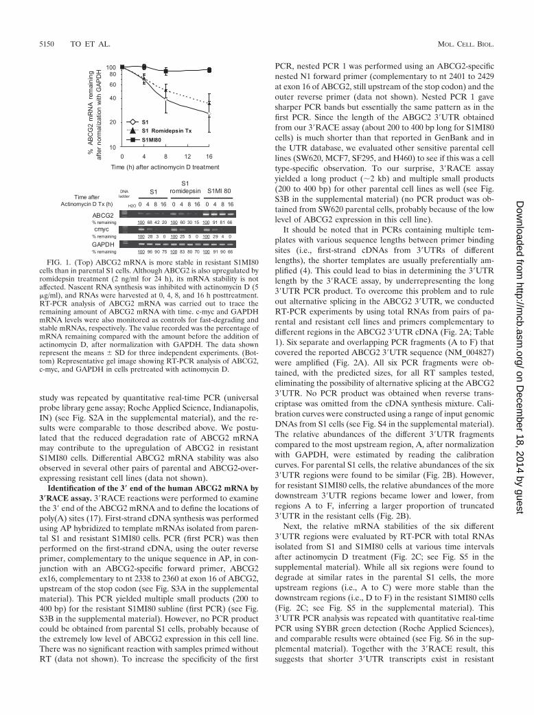
Stability of ABCG2 mRNA in parental S1 and resistantS1MI80 cell lines. The half-life of ABCG2 mRNA in the pa-rental S1 colon cancer cell line and its drug-selected resistantsubline S1MI80 was measured after the cells were treated withactinomycin D to inhibit RNA synthesis. Total RNA was iso-lated at various time intervals after actinomycin D treatmentand analyzed for the ABCG2 mRNA level by RT-PCR. Re-sults are presented as the percentages of ABCG2 mRNA re-maining after normalization with GAPDH (Fig. 1). WhileABCG2 mRNA in S1MI80 cells was fairly stable for over 16 hafter actinomycin D treatment, ABCG2 mRNA in parental S1cells had a half-life of about 6 h. Upregulation of ABCG2 byromidepsin in parental S1 cells (49) did not alter its mRNAstability (Fig. 1). To ensure that transcriptional shutoff by ac-tinomycin D was efficient in our experiments, RT-PCR wascarried out to detect c-myc and GAPDH mRNAs as controlsfor short (15 to 40 min) and long (more than 24 h) half-lives,respectively (see Fig. S1 in the supplemental material). Impor-tantly, the degradation profiles for GAPDH and c-mycmRNAs in untreated and romidepsin-treated S1 and S1MI80cells were similar (see Fig. S1 in the supplemental material).Analysis of ABCG2 and GAPDH levels in the mRNA stability
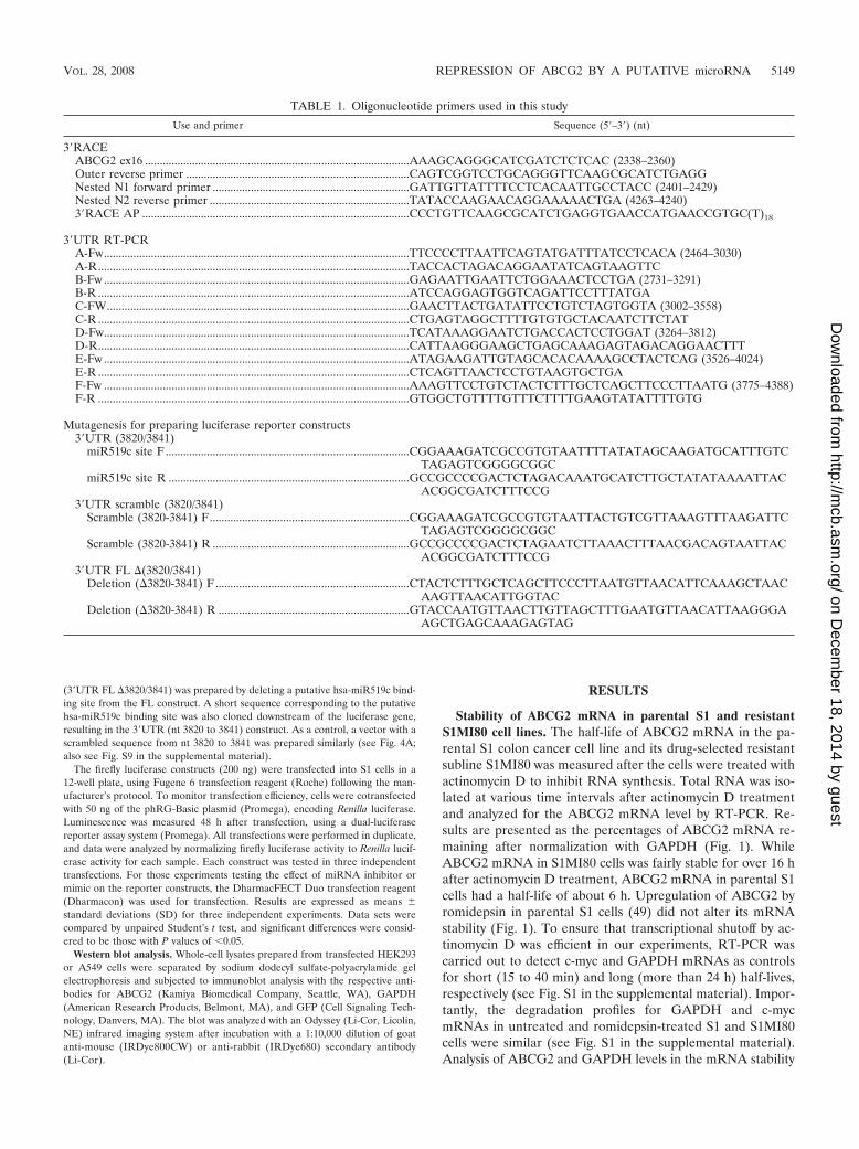
TABLE 1. Oligonucleotide primers used in this study
Use and primer Sequence (5�–3�) (nt)
3�RACEABCG2 ex16 ..........................................................................................AAAGCAGGGCATCGATCTCTCAC (2338–2360)Outer reverse primer ............................................................................CAGTCGGTCCTGCAGGGTTCAAGCGCATCTGAGGNested N1 forward primer ...................................................................GATTGTTATTTTCCTCACAATTGCCTACC (2401–2429)Nested N2 reverse primer ....................................................................TATACCAAGAACAGGAAAAACTGA (4263–4240)3�RACE AP ...........................................................................................CCCTGTTCAAGCGCATCTGAGGTGAACCATGAACCGTGC(T)18
3�UTR RT-PCRA-Fw........................................................................................................TTCCCCTTAATTCAGTATGATTTATCCTCACA (2464–3030)A-R..........................................................................................................TACCACTAGACAGGAATATCAGTAAGTTCB-Fw ........................................................................................................GAGAATTGAATTCTGGAAACTCCTGA (2731–3291)B-R ..........................................................................................................ATCCAGGAGTGGTCAGATTCCTTTATGAC-FW.......................................................................................................GAACTTACTGATATTCCTGTCTAGTGGTA (3002–3558)C-R ..........................................................................................................CTGAGTAGGCTTTTGTGTGCTACAATCTTCTATD-Fw........................................................................................................TCATAAAGGAATCTGACCACTCCTGGAT (3264–3812)D-R..........................................................................................................CATTAAGGGAAGCTGAGCAAAGAGTAGACAGGAACTTTE-Fw........................................................................................................ATAGAAGATTGTAGCACACAAAAGCCTACTCAG (3526–4024)E-R ..........................................................................................................CTCAGTTAACTCCTGTAAGTGCTGAF-Fw ........................................................................................................AAAGTTCCTGTCTACTCTTTGCTCAGCTTCCCTTAATG (3775–4388)F-R ..........................................................................................................GTGGCTGTTTTGTTTCTTTTGAAGTATATTTTGTG
Mutagenesis for preparing luciferase reporter constructs3�UTR (3820/3841)
miR519c site F ...................................................................................CGGAAAGATCGCCGTGTAATTTTATATAGCAAGATGCATTTGTCTAGAGTCGGGGCGGC
miR519c site R ..................................................................................GCCGCCCCGACTCTAGACAAATGCATCTTGCTATATAAAATTACACGGCGATCTTTCCG
3�UTR scramble (3820/3841)Scramble (3820-3841) F....................................................................CGGAAAGATCGCCGTGTAATTACTGTCGTTAAAGTTTAAGATTC
TAGAGTCGGGGCGGCScramble (3820-3841) R ...................................................................GCCGCCCCGACTCTAGAATCTTAAACTTTAACGACAGTAATTAC
ACGGCGATCTTTCCG3�UTR FL �(3820/3841)
Deletion (�3820-3841) F ..................................................................CTACTCTTTGCTCAGCTTCCCTTAATGTTAACATTCAAAGCTAACAAGTTAACATTGGTAC
Deletion (�3820-3841) R .................................................................GTACCAATGTTAACTTGTTAGCTTTGAATGTTAACATTAAGGGAAGCTGAGCAAAGAGTAG
VOL. 28, 2008 REPRESSION OF ABCG2 BY A PUTATIVE microRNA 5149
on Decem
ber 18, 2014 by guesthttp://m
cb.asm.org/
Dow
nloaded from
study was repeated by quantitative real-time PCR (universalprobe library gene assay; Roche Applied Science, Indianapolis,IN) (see Fig. S2A in the supplemental material), and the re-sults were comparable to those described above. We postu-lated that the reduced degradation rate of ABCG2 mRNAmay contribute to the upregulation of ABCG2 in resistantS1MI80 cells. Differential ABCG2 mRNA stability was alsoobserved in several other pairs of parental and ABCG2-over-expressing resistant cell lines (data not shown).
Identification of the 3� end of the human ABCG2 mRNA by3�RACE assay. 3�RACE reactions were performed to examinethe 3� end of the ABCG2 mRNA and to define the locations ofpoly(A) sites (17). First-strand cDNA synthesis was performedusing AP hybridized to template mRNAs isolated from paren-tal S1 and resistant S1MI80 cells. PCR (first PCR) was thenperformed on the first-strand cDNA, using the outer reverseprimer, complementary to the unique sequence in AP, in con-junction with an ABCG2-specific forward primer, ABCG2ex16, complementary to nt 2338 to 2360 at exon 16 of ABCG2,upstream of the stop codon (see Fig. S3A in the supplementalmaterial). This PCR yielded multiple small products (200 to400 bp) for the resistant S1MI80 subline (first PCR) (see Fig.S3B in the supplemental material). However, no PCR productcould be obtained from parental S1 cells, probably because ofthe extremely low level of ABCG2 expression in this cell line.There was no significant reaction with samples primed withoutRT (data not shown). To increase the specificity of the first
PCR, nested PCR 1 was performed using an ABCG2-specificnested N1 forward primer (complementary to nt 2401 to 2429at exon 16 of ABCG2, still upstream of the stop codon) and theouter reverse primer (data not shown). Nested PCR 1 gavesharper PCR bands but essentially the same pattern as in thefirst PCR. Since the length of the ABGC2 3�UTR obtainedfrom our 3�RACE assay (about 200 to 400 bp long for S1MI80cells) is much shorter than that reported in GenBank and inthe UTR database, we evaluated other sensitive parental celllines (SW620, MCF7, SF295, and H460) to see if this was a celltype-specific observation. To our surprise, 3�RACE assayyielded a long product (�2 kb) and multiple small products(200 to 400 bp) for other parental cell lines as well (see Fig.S3B in the supplemental material) (no PCR product was ob-tained from SW620 parental cells, probably because of the lowlevel of ABCG2 expression in this cell line).
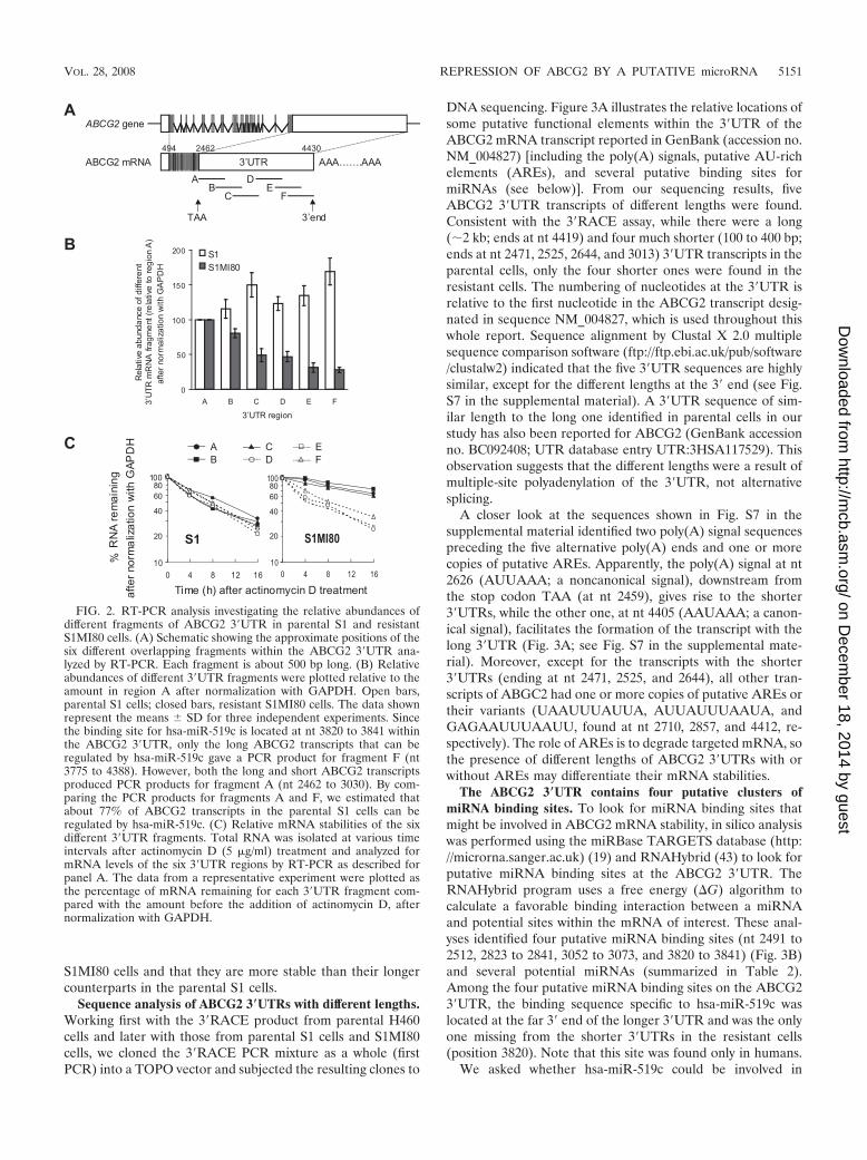
It should be noted that in PCRs containing multiple tem-plates with various sequence lengths between primer bindingsites (i.e., first-strand cDNAs from 3�UTRs of differentlengths), the shorter templates are usually preferentially am-plified (4). This could lead to bias in determining the 3�UTRlength by the 3�RACE assay, by underrepresenting the long3�UTR PCR product. To overcome this problem and to ruleout alternative splicing in the ABCG2 3�UTR, we conductedRT-PCR experiments by using total RNAs from pairs of pa-rental and resistant cell lines and primers complementary todifferent regions in the ABCG2 3�UTR cDNA (Fig. 2A; Table1). Six separate and overlapping PCR fragments (A to F) thatcovered the reported ABCG2 3�UTR sequence (NM_004827)were amplified (Fig. 2A). All six PCR fragments were ob-tained, with the predicted sizes, for all RT samples tested,eliminating the possibility of alternative splicing at the ABCG23�UTR. No PCR product was obtained when reverse trans-criptase was omitted from the cDNA synthesis mixture. Cali-bration curves were constructed using a range of input genomicDNAs from S1 cells (see Fig. S4 in the supplemental material).The relative abundances of the different 3�UTR fragmentscompared to the most upstream region, A, after normalizationwith GAPDH, were estimated by reading the calibrationcurves. For parental S1 cells, the relative abundances of the six3�UTR regions were found to be similar (Fig. 2B). However,for resistant S1MI80 cells, the relative abundances of the moredownstream 3�UTR regions became lower and lower, fromregions A to F, inferring a larger proportion of truncated3�UTR in the resistant cells (Fig. 2B).
Next, the relative mRNA stabilities of the six different3�UTR regions were evaluated by RT-PCR with total RNAsisolated from S1 and S1MI80 cells at various time intervalsafter actinomycin D treatment (Fig. 2C; see Fig. S5 in thesupplemental material). While all six regions were found todegrade at similar rates in the parental S1 cells, the moreupstream regions (i.e., A to C) were more stable than thedownstream regions (i.e., D to F) in the resistant S1MI80 cells(Fig. 2C; see Fig. S5 in the supplemental material). This3�UTR PCR analysis was repeated with quantitative real-timePCR using SYBR green detection (Roche Applied Sciences),and comparable results were obtained (see Fig. S6 in the sup-plemental material). Together with the 3�RACE result, thissuggests that shorter 3�UTR transcripts exist in resistant
FIG. 1. (Top) ABCG2 mRNA is more stable in resistant S1MI80cells than in parental S1 cells. Although ABCG2 is also upregulated byromidepsin treatment (2 ng/ml for 24 h), its mRNA stability is notaffected. Nascent RNA synthesis was inhibited with actinomycin D (5�g/ml), and RNAs were harvested at 0, 4, 8, and 16 h posttreatment.RT-PCR analysis of ABCG2 mRNA was carried out to trace theremaining amount of ABCG2 mRNA with time. c-myc and GAPDHmRNA levels were also monitored as controls for fast-degrading andstable mRNAs, respectively. The value recorded was the percentage ofmRNA remaining compared with the amount before the addition ofactinomycin D, after normalization with GAPDH. The data shownrepresent the means � SD for three independent experiments. (Bot-tom) Representative gel image showing RT-PCR analysis of ABCG2,c-myc, and GAPDH in cells pretreated with actinomycin D.
5150 TO ET AL. MOL. CELL. BIOL.
on Decem
ber 18, 2014 by guesthttp://m
cb.asm.org/
Dow
nloaded from
S1MI80 cells and that they are more stable than their longercounterparts in the parental S1 cells.
Sequence analysis of ABCG2 3�UTRs with different lengths.Working first with the 3�RACE product from parental H460cells and later with those from parental S1 cells and S1MI80cells, we cloned the 3�RACE PCR mixture as a whole (firstPCR) into a TOPO vector and subjected the resulting clones to
DNA sequencing. Figure 3A illustrates the relative locations ofsome putative functional elements within the 3�UTR of theABCG2 mRNA transcript reported in GenBank (accession no.NM_004827) [including the poly(A) signals, putative AU-richelements (AREs), and several putative binding sites formiRNAs (see below)]. From our sequencing results, fiveABCG2 3�UTR transcripts of different lengths were found.Consistent with the 3�RACE assay, while there were a long(�2 kb; ends at nt 4419) and four much shorter (100 to 400 bp;ends at nt 2471, 2525, 2644, and 3013) 3�UTR transcripts in theparental cells, only the four shorter ones were found in theresistant cells. The numbering of nucleotides at the 3�UTR isrelative to the first nucleotide in the ABCG2 transcript desig-nated in sequence NM_004827, which is used throughout thiswhole report. Sequence alignment by Clustal X 2.0 multiplesequence comparison software (ftp://ftp.ebi.ac.uk/pub/software/clustalw2) indicated that the five 3�UTR sequences are highlysimilar, except for the different lengths at the 3� end (see Fig.S7 in the supplemental material). A 3�UTR sequence of sim-ilar length to the long one identified in parental cells in ourstudy has also been reported for ABCG2 (GenBank accessionno. BC092408; UTR database entry UTR:3HSA117529). Thisobservation suggests that the different lengths were a result ofmultiple-site polyadenylation of the 3�UTR, not alternativesplicing.
A closer look at the sequences shown in Fig. S7 in thesupplemental material identified two poly(A) signal sequencespreceding the five alternative poly(A) ends and one or morecopies of putative AREs. Apparently, the poly(A) signal at nt2626 (AUUAAA; a noncanonical signal), downstream fromthe stop codon TAA (at nt 2459), gives rise to the shorter3�UTRs, while the other one, at nt 4405 (AAUAAA; a canon-ical signal), facilitates the formation of the transcript with thelong 3�UTR (Fig. 3A; see Fig. S7 in the supplemental mate-rial). Moreover, except for the transcripts with the shorter3�UTRs (ending at nt 2471, 2525, and 2644), all other tran-scripts of ABGC2 had one or more copies of putative AREs ortheir variants (UAAUUUAUUA, AUUAUUUAAUA, andGAGAAUUUAAUU, found at nt 2710, 2857, and 4412, re-spectively). The role of AREs is to degrade targeted mRNA, sothe presence of different lengths of ABCG2 3�UTRs with orwithout AREs may differentiate their mRNA stabilities.
The ABCG2 3�UTR contains four putative clusters ofmiRNA binding sites. To look for miRNA binding sites thatmight be involved in ABCG2 mRNA stability, in silico analysiswas performed using the miRBase TARGETS database (http://microrna.sanger.ac.uk) (19) and RNAHybrid (43) to look forputative miRNA binding sites at the ABCG2 3�UTR. TheRNAHybrid program uses a free energy (�G) algorithm tocalculate a favorable binding interaction between a miRNAand potential sites within the mRNA of interest. These anal-yses identified four putative miRNA binding sites (nt 2491 to2512, 2823 to 2841, 3052 to 3073, and 3820 to 3841) (Fig. 3B)and several potential miRNAs (summarized in Table 2).Among the four putative miRNA binding sites on the ABCG23�UTR, the binding sequence specific to hsa-miR-519c waslocated at the far 3� end of the longer 3�UTR and was the onlyone missing from the shorter 3�UTRs in the resistant cells(position 3820). Note that this site was found only in humans.
We asked whether hsa-miR-519c could be involved in
FIG. 2. RT-PCR analysis investigating the relative abundances ofdifferent fragments of ABCG2 3�UTR in parental S1 and resistantS1MI80 cells. (A) Schematic showing the approximate positions of thesix different overlapping fragments within the ABCG2 3�UTR ana-lyzed by RT-PCR. Each fragment is about 500 bp long. (B) Relativeabundances of different 3�UTR fragments were plotted relative to theamount in region A after normalization with GAPDH. Open bars,parental S1 cells; closed bars, resistant S1MI80 cells. The data shownrepresent the means � SD for three independent experiments. Sincethe binding site for hsa-miR-519c is located at nt 3820 to 3841 withinthe ABCG2 3�UTR, only the long ABCG2 transcripts that can beregulated by hsa-miR-519c gave a PCR product for fragment F (nt3775 to 4388). However, both the long and short ABCG2 transcriptsproduced PCR products for fragment A (nt 2462 to 3030). By com-paring the PCR products for fragments A and F, we estimated thatabout 77% of ABCG2 transcripts in the parental S1 cells can beregulated by hsa-miR-519c. (C) Relative mRNA stabilities of the sixdifferent 3�UTR fragments. Total RNA was isolated at various timeintervals after actinomycin D (5 �g/ml) treatment and analyzed formRNA levels of the six 3�UTR regions by RT-PCR as described forpanel A. The data from a representative experiment were plotted asthe percentage of mRNA remaining for each 3�UTR fragment com-pared with the amount before the addition of actinomycin D, afternormalization with GAPDH.
VOL. 28, 2008 REPRESSION OF ABCG2 BY A PUTATIVE microRNA 5151
on Decem
ber 18, 2014 by guesthttp://m
cb.asm.org/
Dow
nloaded from
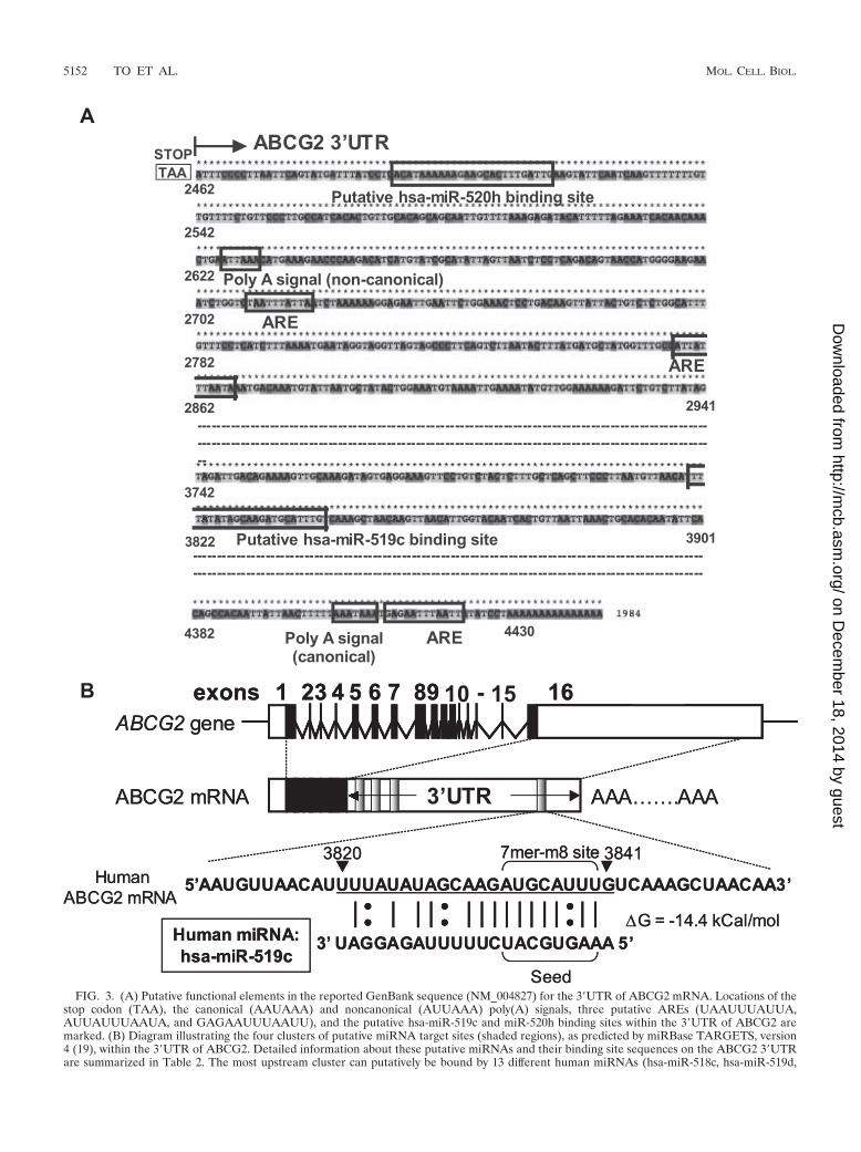
FIG. 3. (A) Putative functional elements in the reported GenBank sequence (NM_004827) for the 3�UTR of ABCG2 mRNA. Locations of thestop codon (TAA), the canonical (AAUAAA) and noncanonical (AUUAAA) poly(A) signals, three putative AREs (UAAUUUAUUA,AUUAUUUAAUA, and GAGAAUUUAAUU), and the putative hsa-miR-519c and miR-520h binding sites within the 3�UTR of ABCG2 aremarked. (B) Diagram illustrating the four clusters of putative miRNA target sites (shaded regions), as predicted by miRBase TARGETS, version4 (19), within the 3�UTR of ABCG2. Detailed information about these putative miRNAs and their binding site sequences on the ABCG2 3�UTRare summarized in Table 2. The most upstream cluster can putatively be bound by 13 different human miRNAs (hsa-miR-518c, hsa-miR-519d,
5152 TO ET AL. MOL. CELL. BIOL.
on Decem
ber 18, 2014 by guesthttp://m
cb.asm.org/
Dow
nloaded from
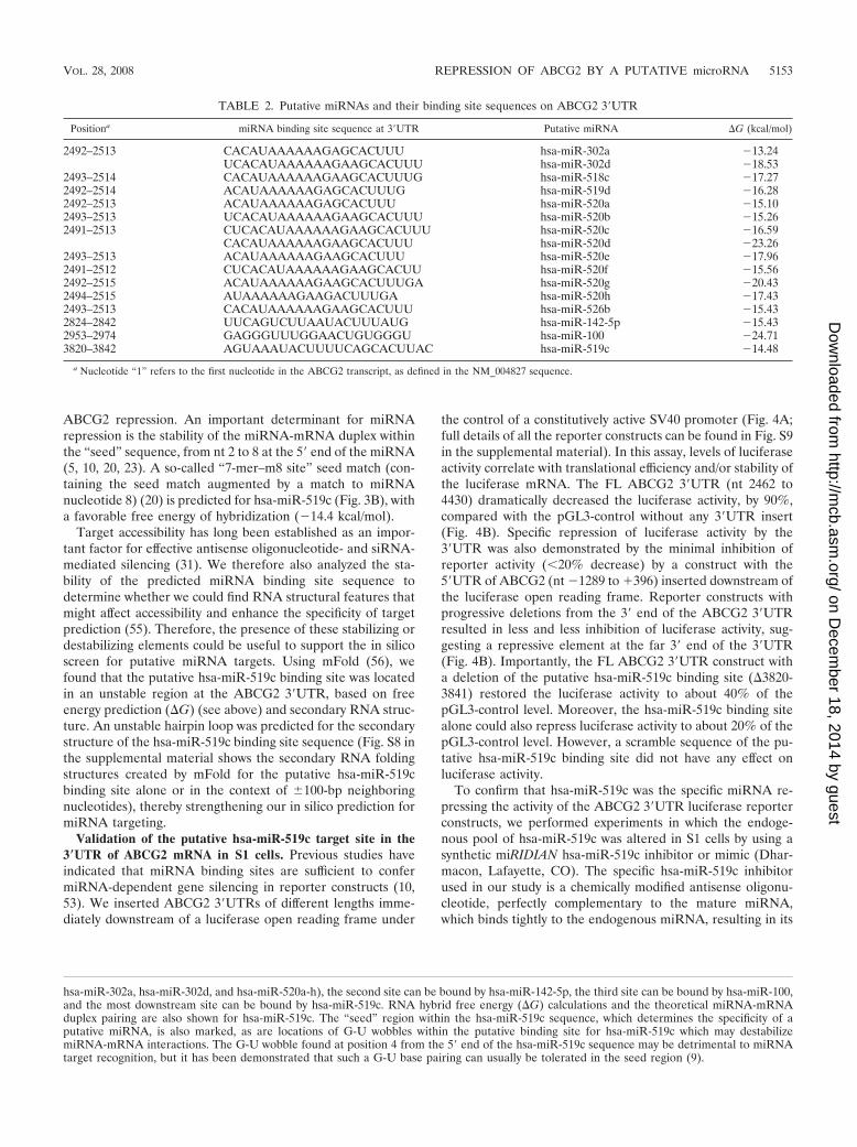
ABCG2 repression. An important determinant for miRNArepression is the stability of the miRNA-mRNA duplex withinthe “seed” sequence, from nt 2 to 8 at the 5� end of the miRNA(5, 10, 20, 23). A so-called “7-mer–m8 site” seed match (con-taining the seed match augmented by a match to miRNAnucleotide 8) (20) is predicted for hsa-miR-519c (Fig. 3B), witha favorable free energy of hybridization (�14.4 kcal/mol).
Target accessibility has long been established as an impor-tant factor for effective antisense oligonucleotide- and siRNA-mediated silencing (31). We therefore also analyzed the sta-bility of the predicted miRNA binding site sequence todetermine whether we could find RNA structural features thatmight affect accessibility and enhance the specificity of targetprediction (55). Therefore, the presence of these stabilizing ordestabilizing elements could be useful to support the in silicoscreen for putative miRNA targets. Using mFold (56), wefound that the putative hsa-miR-519c binding site was locatedin an unstable region at the ABCG2 3�UTR, based on freeenergy prediction (�G) (see above) and secondary RNA struc-ture. An unstable hairpin loop was predicted for the secondarystructure of the hsa-miR-519c binding site sequence (Fig. S8 inthe supplemental material shows the secondary RNA foldingstructures created by mFold for the putative hsa-miR-519cbinding site alone or in the context of �100-bp neighboringnucleotides), thereby strengthening our in silico prediction formiRNA targeting.
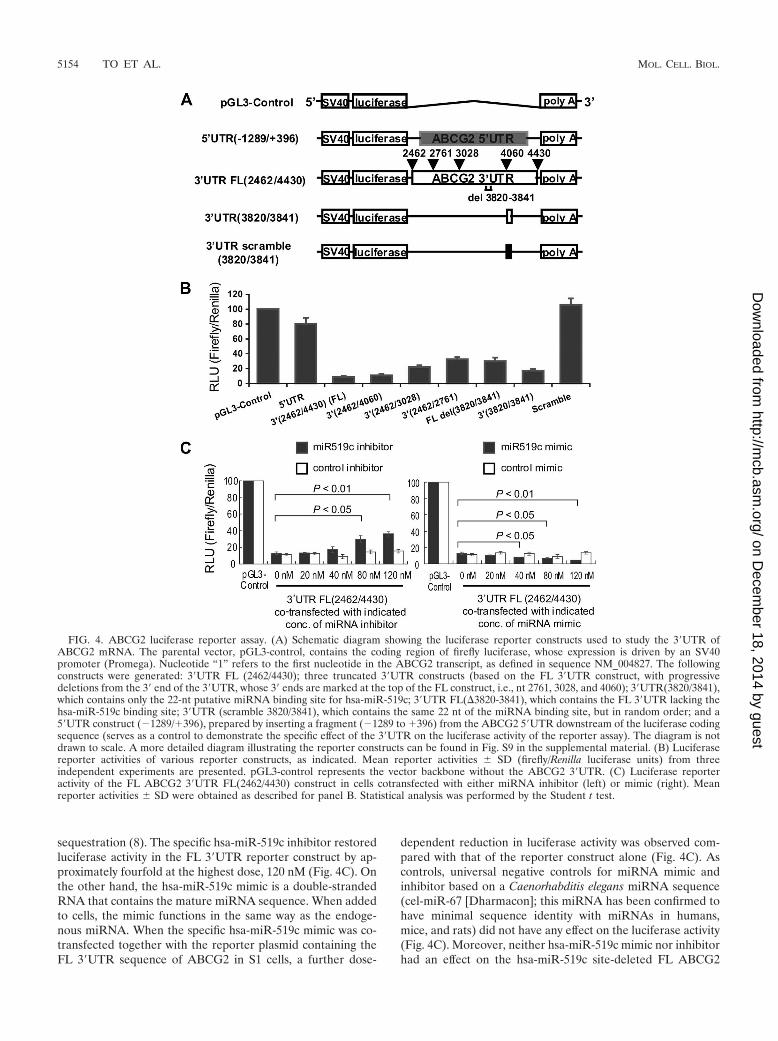
Validation of the putative hsa-miR-519c target site in the3�UTR of ABCG2 mRNA in S1 cells. Previous studies haveindicated that miRNA binding sites are sufficient to confermiRNA-dependent gene silencing in reporter constructs (10,53). We inserted ABCG2 3�UTRs of different lengths imme-diately downstream of a luciferase open reading frame under
the control of a constitutively active SV40 promoter (Fig. 4A;full details of all the reporter constructs can be found in Fig. S9in the supplemental material). In this assay, levels of luciferaseactivity correlate with translational efficiency and/or stability ofthe luciferase mRNA. The FL ABCG2 3�UTR (nt 2462 to4430) dramatically decreased the luciferase activity, by 90%,compared with the pGL3-control without any 3�UTR insert(Fig. 4B). Specific repression of luciferase activity by the3�UTR was also demonstrated by the minimal inhibition ofreporter activity (�20% decrease) by a construct with the5�UTR of ABCG2 (nt �1289 to �396) inserted downstream ofthe luciferase open reading frame. Reporter constructs withprogressive deletions from the 3� end of the ABCG2 3�UTRresulted in less and less inhibition of luciferase activity, sug-gesting a repressive element at the far 3� end of the 3�UTR(Fig. 4B). Importantly, the FL ABCG2 3�UTR construct witha deletion of the putative hsa-miR-519c binding site (�3820-3841) restored the luciferase activity to about 40% of thepGL3-control level. Moreover, the hsa-miR-519c binding sitealone could also repress luciferase activity to about 20% of thepGL3-control level. However, a scramble sequence of the pu-tative hsa-miR-519c binding site did not have any effect onluciferase activity.
To confirm that hsa-miR-519c was the specific miRNA re-pressing the activity of the ABCG2 3�UTR luciferase reporterconstructs, we performed experiments in which the endoge-nous pool of hsa-miR-519c was altered in S1 cells by using asynthetic miRIDIAN hsa-miR-519c inhibitor or mimic (Dhar-macon, Lafayette, CO). The specific hsa-miR-519c inhibitorused in our study is a chemically modified antisense oligonu-cleotide, perfectly complementary to the mature miRNA,which binds tightly to the endogenous miRNA, resulting in its
hsa-miR-302a, hsa-miR-302d, and hsa-miR-520a-h), the second site can be bound by hsa-miR-142-5p, the third site can be bound by hsa-miR-100,and the most downstream site can be bound by hsa-miR-519c. RNA hybrid free energy (�G) calculations and the theoretical miRNA-mRNAduplex pairing are also shown for hsa-miR-519c. The “seed” region within the hsa-miR-519c sequence, which determines the specificity of aputative miRNA, is also marked, as are locations of G-U wobbles within the putative binding site for hsa-miR-519c which may destabilizemiRNA-mRNA interactions. The G-U wobble found at position 4 from the 5� end of the hsa-miR-519c sequence may be detrimental to miRNAtarget recognition, but it has been demonstrated that such a G-U base pairing can usually be tolerated in the seed region (9).
TABLE 2. Putative miRNAs and their binding site sequences on ABCG2 3�UTR
Positiona miRNA binding site sequence at 3�UTR Putative miRNA �G (kcal/mol)
2492–2513 CACAUAAAAAAGAGCACUUU hsa-miR-302a �13.24UCACAUAAAAAAGAAGCACUUU hsa-miR-302d �18.53
2493–2514 CACAUAAAAAAGAAGCACUUUG hsa-miR-518c �17.272492–2514 ACAUAAAAAAGAGCACUUUG hsa-miR-519d �16.282492–2513 ACAUAAAAAAGAGCACUUU hsa-miR-520a �15.102493–2513 UCACAUAAAAAAGAAGCACUUU hsa-miR-520b �15.262491–2513 CUCACAUAAAAAAGAAGCACUUU hsa-miR-520c �16.59
CACAUAAAAAAGAAGCACUUU hsa-miR-520d �23.262493–2513 ACAUAAAAAAGAAGCACUUU hsa-miR-520e �17.962491–2512 CUCACAUAAAAAAGAAGCACUU hsa-miR-520f �15.562492–2515 ACAUAAAAAAGAAGCACUUUGA hsa-miR-520g �20.432494–2515 AUAAAAAAGAAGACUUUGA hsa-miR-520h �17.432493–2513 CACAUAAAAAAGAAGCACUUU hsa-miR-526b �15.432824–2842 UUCAGUCUUAAUACUUUAUG hsa-miR-142-5p �15.432953–2974 GAGGGUUUGGAACUGUGGGU hsa-miR-100 �24.713820–3842 AGUAAAUACUUUUCAGCACUUAC hsa-miR-519c �14.48
a Nucleotide “1” refers to the first nucleotide in the ABCG2 transcript, as defined in the NM_004827 sequence.
VOL. 28, 2008 REPRESSION OF ABCG2 BY A PUTATIVE microRNA 5153
on Decem
ber 18, 2014 by guesthttp://m
cb.asm.org/
Dow
nloaded from
sequestration (8). The specific hsa-miR-519c inhibitor restoredluciferase activity in the FL 3�UTR reporter construct by ap-proximately fourfold at the highest dose, 120 nM (Fig. 4C). Onthe other hand, the hsa-miR-519c mimic is a double-strandedRNA that contains the mature miRNA sequence. When addedto cells, the mimic functions in the same way as the endoge-nous miRNA. When the specific hsa-miR-519c mimic was co-transfected together with the reporter plasmid containing theFL 3�UTR sequence of ABCG2 in S1 cells, a further dose-
dependent reduction in luciferase activity was observed com-pared with that of the reporter construct alone (Fig. 4C). Ascontrols, universal negative controls for miRNA mimic andinhibitor based on a Caenorhabditis elegans miRNA sequence(cel-miR-67 [Dharmacon]; this miRNA has been confirmed tohave minimal sequence identity with miRNAs in humans,mice, and rats) did not have any effect on the luciferase activity(Fig. 4C). Moreover, neither hsa-miR-519c mimic nor inhibitorhad an effect on the hsa-miR-519c site-deleted FL ABCG2
FIG. 4. ABCG2 luciferase reporter assay. (A) Schematic diagram showing the luciferase reporter constructs used to study the 3�UTR ofABCG2 mRNA. The parental vector, pGL3-control, contains the coding region of firefly luciferase, whose expression is driven by an SV40promoter (Promega). Nucleotide “1” refers to the first nucleotide in the ABCG2 transcript, as defined in sequence NM_004827. The followingconstructs were generated: 3�UTR FL (2462/4430); three truncated 3�UTR constructs (based on the FL 3�UTR construct, with progressivedeletions from the 3� end of the 3�UTR, whose 3� ends are marked at the top of the FL construct, i.e., nt 2761, 3028, and 4060); 3�UTR(3820/3841),which contains only the 22-nt putative miRNA binding site for hsa-miR-519c; 3�UTR FL(�3820-3841), which contains the FL 3�UTR lacking thehsa-miR-519c binding site; 3�UTR (scramble 3820/3841), which contains the same 22 nt of the miRNA binding site, but in random order; and a5�UTR construct (�1289/�396), prepared by inserting a fragment (�1289 to �396) from the ABCG2 5�UTR downstream of the luciferase codingsequence (serves as a control to demonstrate the specific effect of the 3�UTR on the luciferase activity of the reporter assay). The diagram is notdrawn to scale. A more detailed diagram illustrating the reporter constructs can be found in Fig. S9 in the supplemental material. (B) Luciferasereporter activities of various reporter constructs, as indicated. Mean reporter activities � SD (firefly/Renilla luciferase units) from threeindependent experiments are presented. pGL3-control represents the vector backbone without the ABCG2 3�UTR. (C) Luciferase reporteractivity of the FL ABCG2 3�UTR FL(2462/4430) construct in cells cotransfected with either miRNA inhibitor (left) or mimic (right). Meanreporter activities � SD were obtained as described for panel B. Statistical analysis was performed by the Student t test.
5154 TO ET AL. MOL. CELL. BIOL.
on Decem
ber 18, 2014 by guesthttp://m
cb.asm.org/
Dow
nloaded from
3�UTR reporter construct (see Fig. S10 in the supplementalmaterial). These results indicate that hsa-miR-519c specificallyinteracts with the target binding site identified in the humanABCG2 3�UTR, which in turn represses the ABCG2 mRNAand protein expression.
Since the putative hsa-miR-519c site has been validated forABCG2 3�UTR in S1 cells, we repeated the 3�RACE assay,using S1 cells transfected with the specific hsa-miR-519c inhib-itor. We used the specific hsa-miR-519c inhibitor to increasethe level of ABCG2 3�UTR in S1 cells (see Fig. S11A in thesupplemental material) and subsequently confirmed the longsequence in S1 cells and its absence in S1MI80 cells with anested PCR 1 and nested PCR 2 (see Fig. S11B in the supple-mental material).
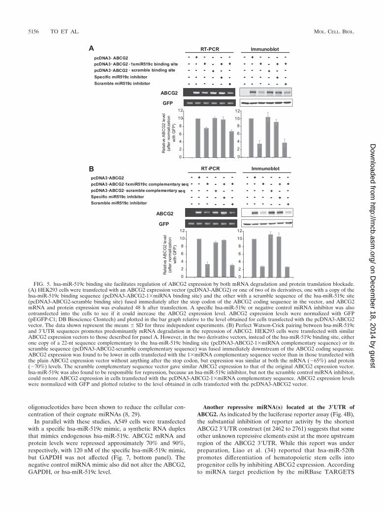
hsa-miR-519c regulates ABCG2 expression by both transla-tion repression and mRNA degradation. Regulation bymiRNAs can be due to translation repression and/or mRNAdegradation. To assess whether hsa-miR-519c has a functionalrole in the downregulation of ABCG2 expression by either ofthese mechanisms, an ABCG2 expression vector (pcDNA3-ABCG2) and two of its derivatives, one with a tandem repeatof three copies of hsa-miR-519c binding site sequence(pcDNA3-ABCG2-3miRNA binding site) and the other witha tandem repeat of three copies of a scramble sequence of thehsa-miR-519c site (pcDNA3-ABCG2-3xscramble bindingsite) fused immediately after the stop codon of the ABCG2coding sequence in the vector, were transfected into HEK293cells and ABCG2 expression levels measured. As an internalcontrol, the level of cotransfected GFP (pEGFP-C1; BD Bio-science Clontech, Mountain View, CA) was assessed as welland was used to normalize the ABCG2 mRNA levels. ABCG2expression was found to be lower in cells transfected with the3xmiRNA binding site vector than in those with the originalABCG2 expression vector without any 3�UTR insert, at boththe mRNA (�24%) and protein (even lower, at �49%) levels(see Fig. S12A in the supplemental material). The scramblebinding site vector gave similar ABCG2 expression to that ofthe plain ABCG2 expression vector. An hsa-miR-519c inhibi-tor was cotransfected with the pcDNA3-ABCG2-3miR519cbinding site vector to verify whether hsa-miR-519c inhibitsABCG2 expression specifically. Both ABCG2 mRNA and pro-tein expression levels were restored to the levels in cells trans-fected with the plain pcDNA3-ABCG2 vector by the specifichsa-miR-519c inhibitor but not by a universal negative con-trol miRNA inhibitor (see Fig. S12A in the supplementalmaterial).
The differential effect on ABCG2 mRNA and protein ex-pression by the hsa-miR-519c inhibitor was quite reproducibleand led us to postulate that protein translation blockade mayplay a more important role than mRNA degradation in therepression of ABCG2 by hsa-miR-519c. The degree ofmiRNA-mRNA pairing has been proposed to affect the mech-anism of repression, with perfect pairing leading to mRNAcleavage but imperfect pairing leading to protein translationblock (23). We prepared an ABCG2 expression vector as de-scribed above, but immediately following the stop codon weplaced either a tandem repeat of three copies of a 22-ntsequence perfectly complementary to the miR519c sequence(pcDNA3-ABCG2-3miRNA complementary sequence) ora tandem repeat of three copies of scramble sequence
(pcDNA3-ABCG2-3scramble complementary sequence).ABCG2 expression was then examined in HEK293 cellstransfected with these new vectors. As with the previousvectors, ABCG2 expression was found to be lower in cellstransfected with the 3miRNA complementary sequencevector than in cells with the plain ABCG2 expression vectorwithout anything after the stop codon, but similarly at boththe mRNA (�61%) and protein (�70%) levels (see Fig.S12B in the supplemental material), suggesting that whileaffecting both levels, the effect of hsa-miR-519c on proteintranslation predominates in the wild-type 3�UTR. The3scramble complementary sequence vector gave similarABCG2 expression to that with the original ABCG2 expres-sion vector. Interestingly, similar observations were ob-tained when the vectors contained only a single copy ofeither the hsa-miR-519c binding site or complementary se-quence (Fig. 5A and B).
ABCG2 mRNA stability was also measured in HEK293 cellstransfected with the various vectors (having a single copy ofeither the hsa-miR-519c binding site or complementary se-quence). Actinomycin D was added at 48 h posttransfection toblock transcription, and total RNA was isolated at various timeintervals and analyzed for ABCG2 mRNA levels by using RT-PCR. As an internal control, the level of cotransfected GFPwas measured for normalization. As shown in Fig. 6, ABCG2mRNA expressed from the vector with the hsa-miR-519c com-plementary sequence downstream of the ABCG2 coding se-quence was the least stable (half-life [t1/2], �6 h), followed bythe vector with the wild-type hsa-miR-519c binding site (t1/2,16 h). In contrast, the ABCG2 without any 3�UTR was fairlystable for up to 24 h after actinomycin D treatment. The assaywas repeated by real-time PCR, using the Universal Probe-Library gene assay, and similar results were obtained. Theseresults underscore the destabilizing effects in vivo of the hsa-miR-519c binding site in the ABCG2 3�UTR (nt 3820 to 3841),but only partially by promoting mRNA degradation. Interest-ingly, the perfect pairing of hsa-miR-519c with the hsa-miR-519c complementary sequence led to the most substantial deg-radation of ABCG2 mRNA.
hsa-miR-519c regulates endogenous ABCG2 mRNA andprotein expression. Finally, we evaluated whether hsa-miR-519c has a functional role in the downregulation of endoge-nous ABCG2 expression. A549 cells were chosen for our studybecause endogenous ABCG2 protein expression is readily de-tectable in these cells by Western blot analysis, and similar toother parental cell lines used in our study, A549 cells have bothlong and short ABCG2 3�UTRs, as determined by 3�RACEassay (data not shown). Transfection of A549 cells with variousconcentrations of hsa-miR-519c inhibitor, a single-strandedmodified RNA that has a complementary sequence to themature miRNA, led to a dose-dependent induction of ABCG2at both the mRNA and, albeit more pronounced, protein lev-els. ABCG2 mRNA and protein were upregulated over two-fold and sixfold, respectively, with 120 nM of miR519c inhib-itor, while no change was noted in GAPDH (Fig. 7, top panel).The negative control miRNA inhibitor did not alter theABCG2, GAPDH, or hsa-miR-519c level. Although miRNAinhibitors principally function by sequestering endogenousmiRNAs, the level of the latter was also decreased at a higherdose of miRNA inhibitor (Fig. 7, top panel). In fact, antisense
VOL. 28, 2008 REPRESSION OF ABCG2 BY A PUTATIVE microRNA 5155
on Decem
ber 18, 2014 by guesthttp://m
cb.asm.org/
Dow
nloaded from
oligonucleotides have been shown to reduce the cellular con-centration of their cognate miRNAs (8, 29).
In parallel with these studies, A549 cells were transfectedwith a specific hsa-miR-519c mimic, a synthetic RNA duplexthat mimics endogenous hsa-miR-519c. ABCG2 mRNA andprotein levels were repressed approximately 70% and 90%,respectively, with 120 nM of the specific hsa-miR-519c mimic,but GAPDH was not affected (Fig. 7, bottom panel). Thenegative control miRNA mimic also did not alter the ABCG2,GAPDH, or hsa-miR-519c level.
Another repressive miRNA(s) located at the 3�UTR ofABCG2. As indicated by the luciferase reporter assay (Fig. 4B),the substantial inhibition of reporter activity by the shortestABCG2 3�UTR construct (nt 2462 to 2761) suggests that someother unknown repressive elements exist at the more upstreamregion of the ABCG2 3�UTR. While this report was underpreparation, Liao et al. (34) reported that hsa-miR-520hpromotes differentiation of hematopoietic stem cells intoprogenitor cells by inhibiting ABCG2 expression. Accordingto miRNA target prediction by the miRBase TARGETS
FIG. 5. hsa-miR-519c binding site facilitates regulation of ABCG2 expression by both mRNA degradation and protein translation blockade.(A) HEK293 cells were transfected with an ABCG2 expression vector (pcDNA3-ABCG2) or one of two of its derivatives, one with a copy of thehsa-miR-519c binding sequence (pcDNA3-ABCG2-1miRNA binding site) and the other with a scramble sequence of the hsa-miR-519c site(pcDNA3-ABCG2-scramble binding site) fused immediately after the stop codon of the ABCG2 coding sequence in the vector, and ABCG2mRNA and protein expression was evaluated 48 h after transfection. A specific hsa-miR-519c or negative control miRNA inhibitor was alsocotransfected into the cells to see if it could increase the ABCG2 expression level. ABCG2 expression levels were normalized with GFP(pEGFP-C1; DB Bioscience Clontech) and plotted in the bar graph relative to the level obtained for cells transfected with the pcDNA3-ABCG2vector. The data shown represent the means � SD for three independent experiments. (B) Perfect Watson-Crick pairing between hsa-miR-519cand 3�UTR sequences promotes predominantly mRNA degradation in the repression of ABCG2. HEK293 cells were transfected with similarABCG2 expression vectors to those described for panel A. However, in the two derivative vectors, instead of the hsa-miR-519c binding site, eitherone copy of a 22-nt sequence complementary to the hsa-miR-519c binding site (pcDNA3-ABCG2-1miRNA complementary sequence) or itsscramble sequence (pcDNA3-ABCG2-scramble complementary sequence) was fused immediately downstream of the ABCG2 coding sequence.ABCG2 expression was found to be lower in cells transfected with the 1miRNA complementary sequence vector than in those transfected withthe plain ABCG2 expression vector without anything after the stop codon, but expression was similar at both the mRNA (�65%) and protein(�70%) levels. The scramble complementary sequence vector gave similar ABCG2 expression to that of the original ABCG2 expression vector.hsa-miR-519c was also found to be responsible for repression, because an hsa-miR-519c inhibitor, but not the scramble control miRNA inhibitor,could restore ABCG2 expression in cells transfected with the pcDNA3-ABCG2-1miRNA complementary sequence. ABCG2 expression levelswere normalized with GFP and plotted relative to the level obtained in cells transfected with the pcDNA3-ABCG2 vector.
5156 TO ET AL. MOL. CELL. BIOL.
on Decem
ber 18, 2014 by guesthttp://m
cb.asm.org/
Dow
nloaded from
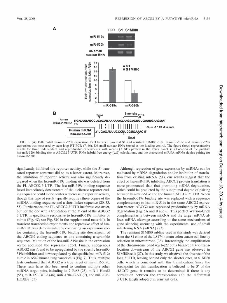
database (19) and RNAHybrid (43), the putative bindingsequence for hsa-miR-520h is located much more upstream(nt 2494 to 2514, relative to the first nucleotide defined insequence NM_004827) (Fig. 3A; see Fig. 9) at the ABCG23�UTR than that for hsa-miR-519c. More importantly, hsa-miR-520h binds to both long and short ABCG2 3�UTRs inparental S1 and resistant S1MI80 cells. We therefore exam-ined the expression level of hsa-miR-520h by stem-loopRT-PCR with S1 and S1MI80 cells to try to elucidate its rolein regulating ABCG2. Interestingly, the hsa-miR-520h levelin S1MI80 was found to be only about half of that in S1 cells(43% � 13%) (Fig. 8A).
DISCUSSION
Overexpression of the ABCG2 gene is frequently observedin cancer cell lines selected with a number of chemotherapeu-tic drugs (1, 12, 35, 38, 44, 52). However, little is known aboutthe mechanisms underlying its upregulation. Gene amplifica-tion and chromosome translocation have been found to play arole in the increased expression of the ABCG2 gene in somedrug-resistant cell lines (27). The use of alternative 5� promot-
ers at the ABCG2 gene in drug-selected cells may offer anothernovel mechanism of ABCG2 upregulation (39), a finding sim-ilar to observations for MDR-1, where rearrangement of the 5�region of MDR-1 resulted in capture of that gene by otherpromoters (22, 26, 37). Recently, we reported that a set ofpermissive histone modifications and a chromatin remodelingfactor, Brg-1, associated with the ABCG2 proximal promoterare required for the upregulation of ABCG2 in resistant cells(49). In an effort to further understand the regulation ofABCG2 in resistant cancer cells, we found that the ABCG2mRNA is less stable in a number of sensitive parental cancercell lines than in their drug-selected and ABCG2-overexpress-ing resistant counterparts. We also observed that parental cellsproduce ABCG2 mRNA with both long and short 3�UTRs,whereas resistant cells produce only the shorter ones.
Since the 3�UTR of eukaryotic mRNA contains regulatoryelements affecting mRNA stability, translation, and transport,it is logical to suspect that the absence of some regulatoryelements in the short 3�UTR compared with the long 3�UTRmay lead to differential regulation of ABCG2 in sensitive andresistant cells. We hypothesized that a putative miRNA bindsto the 3�UTR of ABCG2 in parental cells, thereby suppressing
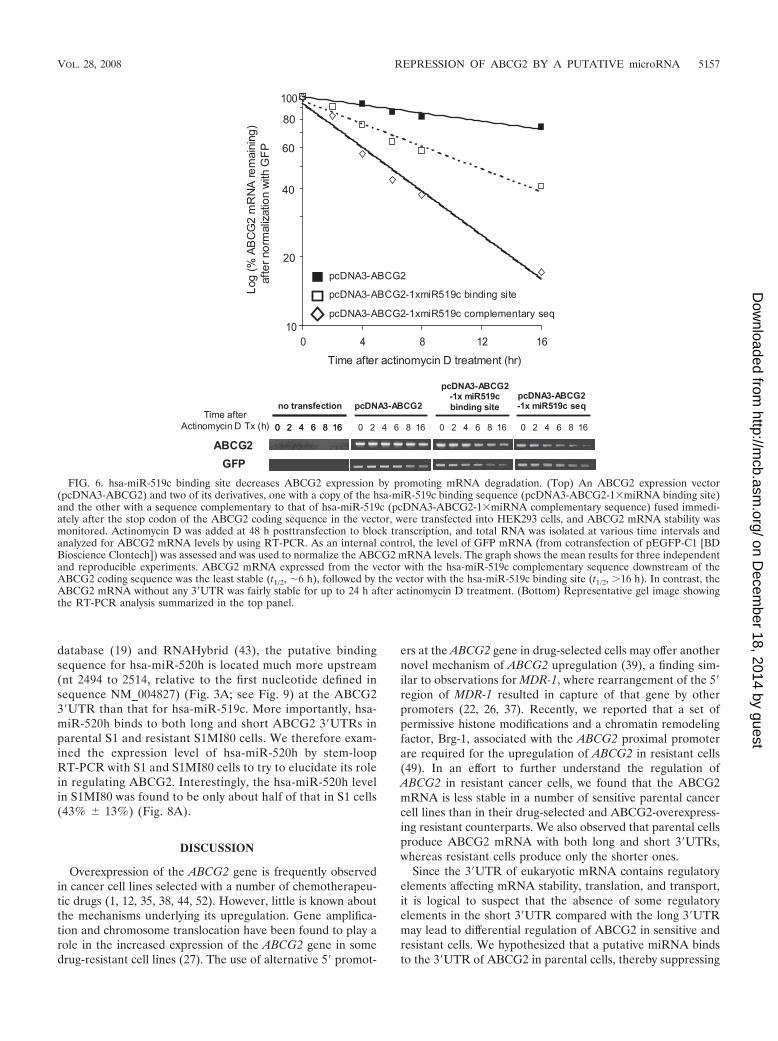
FIG. 6. hsa-miR-519c binding site decreases ABCG2 expression by promoting mRNA degradation. (Top) An ABCG2 expression vector(pcDNA3-ABCG2) and two of its derivatives, one with a copy of the hsa-miR-519c binding sequence (pcDNA3-ABCG2-1miRNA binding site)and the other with a sequence complementary to that of hsa-miR-519c (pcDNA3-ABCG2-1miRNA complementary sequence) fused immedi-ately after the stop codon of the ABCG2 coding sequence in the vector, were transfected into HEK293 cells, and ABCG2 mRNA stability wasmonitored. Actinomycin D was added at 48 h posttransfection to block transcription, and total RNA was isolated at various time intervals andanalyzed for ABCG2 mRNA levels by using RT-PCR. As an internal control, the level of GFP mRNA (from cotransfection of pEGFP-C1 [BDBioscience Clontech]) was assessed and was used to normalize the ABCG2 mRNA levels. The graph shows the mean results for three independentand reproducible experiments. ABCG2 mRNA expressed from the vector with the hsa-miR-519c complementary sequence downstream of theABCG2 coding sequence was the least stable (t1/2, �6 h), followed by the vector with the hsa-miR-519c binding site (t1/2, 16 h). In contrast, theABCG2 mRNA without any 3�UTR was fairly stable for up to 24 h after actinomycin D treatment. (Bottom) Representative gel image showingthe RT-PCR analysis summarized in the top panel.
VOL. 28, 2008 REPRESSION OF ABCG2 BY A PUTATIVE microRNA 5157
on Decem
ber 18, 2014 by guesthttp://m
cb.asm.org/
Dow
nloaded from
ABCG2 expression, but that the binding site for this miRNA islost in the shorter 3�UTR in the resistant cells. miRNAs havea direct role in regulating the processes of development anddifferentiation, and more than 4,000 have been identified inplant and animal species (miRBase Registry [http://microrna.sanger.ac.uk/sequences]). miRNA dysregulation has been cor-related with the progression and aggressiveness of severalforms of cancer (3, 25). In silico analysis revealed a putativetarget site for hsa-miR-519c at the far 3� end of the longABCG2 3�UTR transcript identified in sensitive cells.
In fact, the differential repression of alternative transcriptswith 3�UTRs of different lengths by miRNA was proposedrecently (32). When a miRNA target is located between twovariant sites of polyadenylation, the shorter transcript will betarget-free and can escape miRNA-mediated inhibition, whilelonger transcripts will be inhibited. ABCG2 mRNA containsboth a canonical AAUAAA poly(A) signal and its noncanoni-cal variant AUUAAA. The canonical and noncanonicalpoly(A) signals are usually processed differently. This provides
a means for mRNAs with multiple poly(A) sites to be regu-lated by synthesis of specific mRNA forms, thereby regulatinggene expression (14, 18). Based on our 3�RACE assay and thesubsequent DNA sequencing of ABCG2, it seems that thenoncanonical AUUAAA poly(A) signal is preferentially usedin resistant cells, giving only the short 3�UTR transcripts. Inparental cells, both the canonical AAUAAA and the nonca-nonical AUUAAA signals are used (Fig. 3; see Fig. S7 in thesupplemental material), so that long and short 3�UTR tran-scripts are generated.
Our hypothesis that ABCG2 is a true target for hsa-miR-519c was rigorously tested. Based on a current prediction al-gorithm (19), the targeting is likely because hsa-miR-519cbinds to the 3�UTR of ABCG2 mRNA with a “7-mer–m8 site”complementary to the hsa-miR-519c seed sequence (Fig. 3B).The presence of a destabilizing element (hairpin loop) in thetarget ABCG2 3�UTR region further strengthens the likeli-hood that it is a direct target (see Fig. S8 in the supplementalmaterial). Using reporter gene assays, the FL ABCG2 3�UTR
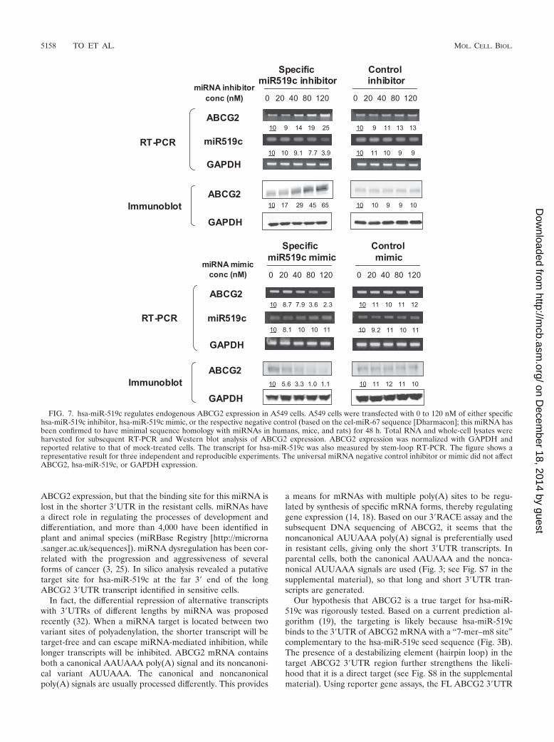
FIG. 7. hsa-miR-519c regulates endogenous ABCG2 expression in A549 cells. A549 cells were transfected with 0 to 120 nM of either specifichsa-miR-519c inhibitor, hsa-miR-519c mimic, or the respective negative control (based on the cel-miR-67 sequence [Dharmacon]; this miRNA hasbeen confirmed to have minimal sequence homology with miRNAs in humans, mice, and rats) for 48 h. Total RNA and whole-cell lysates wereharvested for subsequent RT-PCR and Western blot analysis of ABCG2 expression. ABCG2 expression was normalized with GAPDH andreported relative to that of mock-treated cells. The transcript for hsa-miR-519c was also measured by stem-loop RT-PCR. The figure shows arepresentative result for three independent and reproducible experiments. The universal miRNA negative control inhibitor or mimic did not affectABCG2, hsa-miR-519c, or GAPDH expression.
5158 TO ET AL. MOL. CELL. BIOL.
on Decem
ber 18, 2014 by guesthttp://m
cb.asm.org/
Dow
nloaded from
significantly inhibited the reporter activity, while the 3�-trun-cated reporter construct did so to a lesser extent. Moreover,the inhibition of reporter activity was also significantly de-creased when the hsa-miR-519c binding site was deleted fromthe FL ABCG2 3�UTR. The hsa-miR-519c binding sequencefused immediately downstream of the luciferase reporter cod-ing sequence could alone confer a decrease in reporter activity,though this type of result typically requires three copies of themiRNA binding sequence and a short linker sequence (20, 33,55). Furthermore, the FL ABCG2 3�UTR luciferase construct,but not the one with a truncation at the 3� end of the ABCG23�UTR, is specifically responsive to hsa-miR-519c inhibitor ormimic (Fig. 4C; see Fig. S10 in the supplemental material). Intransient transfection experiments, the repressive effect of hsa-miR-519c was demonstrated by comparing an expression vec-tor containing the hsa-miR-519c binding site downstream ofthe ABCG2 coding sequence to one containing a scramblesequence. Mutation of the hsa-miR-519c site in the expressionvector abolished the repressive effect. Finally, endogenousABCG2 was found to be upregulated by the specific hsa-miR-519c inhibitor and downregulated by the specific hsa-miR-519cmimic in A549 human lung cancer cells (Fig. 7). Thus, multipletests confirmed that ABCG2 is a true target of hsa-miR-519c.These tests have also been used to confirm multiple othermiRNA-target pairs, including let-7–RAS (25), miR-1–Hand2(55), miR-127–BCL6 (46), miR-130a–GAX (7), and miR-196–HOXB8 (53).
Although repression of gene expression by miRNAs can bemediated by mRNA degradation and/or inhibition of transla-tion from existing mRNA (51), our results suggest that theeffect of hsa-miR-519c inhibiting ABCG2 protein translation ismore pronounced than that promoting mRNA degradation,which could be predicted by the suboptimal degree of pairingbetween hsa-miR-519c and the human ABCG2 3�UTR. Whenthe hsa-miR-519c binding site was replaced with a sequencecomplementary to hsa-miR-519c in the same ABCG2 expres-sion vector, ABCG2 was repressed predominantly by mRNAdegradation (Fig. 5A and B and 6). This perfect Watson-Crickcomplementarity between miRNA and the target mRNA al-lows mRNA cleavage according to the same mechanisms ofgene silencing occurring with the experimental use of smallinterfering RNA (siRNA) (23).
The resistant S1MI80 subline used in this study was derivedfrom the S1 clone of the LS174 human colon cancer cell line byselection in mitoxantrone (38). Interestingly, no amplificationof the chromosome band 4q21-q22 but a balanced t(4,7) trans-location downstream of the ABCG2 gene was observed inS1MI80 cells (27). In this study, we observed the absence of thelong 3�UTR, leaving behind only the shorter ones, in S1MI80cells, which is coincident with this translocation. Since thebreakpoint for this translocation is believed to be 3� of theABCG2 gene, it remains to be determined if there is anycorrelation between the translocation and the differential3�UTR length adopted in resistant cells.
FIG. 8. (A) Differential hsa-miR-520h expression level between parental S1 and resistant S1MI80 cells. hsa-miR-519c and hsa-miR-520hexpression was measured by stem-loop RT-PCR (7, 46). U6 small nuclear RNA served as the loading control. The figure shows representativeresults for three independent and reproducible experiments, with means (� SD) plotted in the lower panel. (B) Location of the putativehsa-miR-520h binding site at ABCG2 3�UTR, RNA hybrid free energy (�G) calculations, and the theoretical miRNA-mRNA duplex pairing forhsa-miR-520h.
VOL. 28, 2008 REPRESSION OF ABCG2 BY A PUTATIVE microRNA 5159
on Decem
ber 18, 2014 by guesthttp://m
cb.asm.org/
Dow
nloaded from
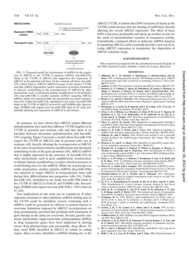
In summary, we have shown that ABCG2 adopts differentpolyadenylation sites (and thus different 3�UTR lengths) at its3�UTR in parental and resistant cells and that there is aninterplay between alternative polyadenylation and hsa-miR-519c targeting. Figure 9 presents a model where hsa-miR-519ctargets the 3�UTR of ABCG2 only in parental cells, not inresistant cells, thereby allowing the overexpression of ABCG2in the latter by permissive histone modifications and chromatinremodeling events at the gene promoter (49). ABCG2 mRNAthat is highly expressed in the presence of hsa-miR-519c byother mechanisms, such as gene amplification, translocation,or distinct histone modifications, is under selection pressure toavoid binding sites for this miRNA. While the manuscript wasunder preparation, another putative miRNA (hsa-miR-520h)was reported to target ABCG2 in hematopoietic stem cellsduring their differentiation into progenitor cells (34). Unlikehsa-miR-519c, identified in our study, hsa-miR-520h binds tothe 3�UTR of ABCG2 in both S1 and S1MI80 cells. Interest-ingly, S1MI80 cells express less hsa-miR-520h (�40%) than doS1 cells.
Two implications of this work can be considered. If otherrepressive elements or other inhibitory miRNA binding sites inthe 3�UTR could be identified, vectors containing such amiRNA could be generated for delivery to patient tumors toovercome limitations imposed by ABCG2 overexpression ondrug accumulation, provided that problems with translation ofgene therapy to the clinic are overcome. Second, genetic vari-ations (particularly single-nucleotide polymorphisms [SNPs])in drug transporter genes have been implicated in alteringcancer drug pharmacology and, presumably, drug effects. Todate, most SNPs identified in ABCG2 lie within its codingregion. Since we have identified a miRNA binding site at the
ABCG2 3�UTR, it follows that SNP variations (if found at the3�UTR) could interact with the binding of miRNA(s), therebyaffecting the overall ABCG2 expression. The effect of theseSNPs is therefore predictable and opens up another avenue forthe study of interindividual variation in transporter activity.Undoubtedly, continued efforts to delineate miRNA functionin regulating ABCG2 could eventually provide a new way of al-tering ABCG2 expression to manipulate the disposition ofABCG2 substrate drugs.
ACKNOWLEDGMENT
This research was supported by the intramural research program ofthe NIH, National Cancer Institute, Center for Cancer Research.
REFERENCES
1. Allikmets, R., L. M. Schriml, A. Hutchinson, V. Romano-Spica, and M.Dean. 1998. A human placenta-specific ATP-binding cassette gene (ABCP)on chromosome 4q22 that is involved in multidrug resistance. Cancer Res.58:5337–5339.
2. Ambros, V. 2004. The functions of animal microRNAs. Nature 431:350–355.3. Bandres, E., E. Cubedo, X. Agirre, R. Malumbres, R. Zarate, N. Ramirez, A.
Abajo, A. Navarro, I. Moreno, M. Monzo, and J. Garcia-Foncillas. 2006.Identification by real-time PCR of 13 mature microRNAs differentially ex-pressed in colorectal cancer and non-tumoral tissues. Mol. Cancer 5:29.
4. Basler, C. F., and M. S. Horwitz. 1996. Subgroup B adenovirus type 35 earlyregion 3 mRNAs differ from those of the subgroup C adenoviruses. Virology215:165–177.
5. Brennecke, J., A. Stark, R. B. Russell, and S. M. Cohen. 2005. Principles ofmicroRNA-target recognition. PLoS Biol. 3:e85.
6. Chen, C., D. A. Ridzon, A. J. Broomer, Z. Zhou, D. H. Lee, J. T. Nguyen, M.Barbisin, N. L. Xu, V. R. Mahuvakar, M. R. Andersen, K. Q. Lao, K. J.Livak, and K. J. Guegler. 2005. Real-time quantification of microRNAs bystem-loop RT-PCR. Nucleic Acids Res. 33:e179.
7. Chen, Y., and D. H. Gorski. 2008. Regulation of angiogenesis through amicroRNA (miR-130a) that down-regulates antiangiogenic homeobox genesGAX and HOXA5. Blood 111:1217–1226.
8. Davis, S., B. Lollo, S. Freier, and C. Esau. 2006. Improved targeting ofmiRNA with antisense oligonucleotides. Nucleic Acids Res. 34:2294–2304.
9. Didiano, D., and O. Hobert. 2006. Perfect seed pairing is not a generallyreliable predictor for miRNA-target interactions. Nat. Struct. Mol. Biol.13:849–851.
10. Doench, J. G., and P. A. Sharp. 2004. Specificity of microRNA target selec-tion in translation repression. Genes Dev. 18:504–511.
11. Donnini, M., A. Lapucci, L. Papucci, E. Witort, A. Jacquier, G. Brewer, A.Nicolin, S. Capaccioli, and N. Schiavone. 2004. Identification of TINO: anew evolutionarily conserved BCL-2 AU-rich element RNA-binding protein.J. Biol. Chem. 279:20154–20166.
12. Doyle, L. A., W. Yang, L. V. Abruzzo, T. Krogmann, Y. Gao, A. K. Rishi, andD. D. Ross. 1998. A multidrug resistance transporter from human MCF-7breast cancer cells. Proc. Natl. Acad. Sci. USA 95:15665–15670.
13. Ebert, B., A. Seidel, and A. Lampen. 2005. Identification of BCRP as trans-porter of benz[a]pyrene conjugates metabolically formed in Caco-2 cells andits induction by Ah-receptor agonists. Carcinogenesis 26:1754–1763.
14. Edwalds-Gilbert, G., K. L. Veraldi, and C. Milcarek. 1997. Alternativepoly(A) site selection in complex transcription units: mean to an end? Nu-cleic Acids Res. 25:2547–2561.
15. Ee, P. L., S. Kamalakaran, D. Tonetti, X. He, D. D. Ross, and W. T. Beck.2004. Identification of a novel estrogen response element in the breastcancer resistance protein (ABCG2) gene. Cancer Res. 64:1247–1251.
16. Farh, K. K., A. Grimson, C. Jan, B. P. Lewis, W. K. Johnston, L. P. Lim,C. B. Burge, and D. P. Bartel. 2005. The widespread impact of mammalianmicroRNAs on mRNA repression and evolution. Science 310:1817–1821.
17. Frohman, M. A. 1990. RACE: rapid amplification of cDNA ends, p.28–38. In M. A. Inns, D. H. Gelfand, J. J. Sninsky, and T. J. White (ed.),PCR protocols: a guide to methods and applications. Academic Press,San Diego, CA.
18. Graber, J. H., C. R. Cantor, S. C. Mohr, and T. F. Smith. 1999. In silicodetection of control signals: mRNA 3�-end-processing sequences in diversespecies. Proc. Natl. Acad. Sci. USA 96:14055–14060.
19. Griffiths-Jones, S. 2006. miRBase: the microRNA sequence database. Meth-ods Mol. Biol. 342:129–138.
20. Grimson, A., K. K. Farh, W. K. Johnston, P. Garrett-Engele, L. P. Lim, andD. P. Bartel. 2007. MicroRNA targeting specificity in mammals: determi-nants beyond seed pairing. Mol. Cell 27:91–105.
21. He, L., and G. J. Hannon. 2004. MicroRNAs: small RNAs with a big role ingene regulation. Nat. Rev. Genet. 5:522–531.
22. Huff, L. M., J. S. Lee, R. W. Robey, and T. Fojo. 2006. Characterization of
FIG. 9. Proposed model for involvement of miRNA in the regula-tion of ABCG2 at the 3�UTR. A putative miRNA, hsa-miR-519c,binds to the 3�UTR of ABCG2 and suppresses the expression ofABCG2 in the parental cell lines. In the resistant cell lines, hsa-miR-519c cannot bind to ABCG2 mRNA because of the shorter 3�UTR,and thus mRNA degradation and/or repression of protein translationis relieved, contributing to the overexpression of ABCG2 by othermechanisms, such as permissive histone modifications at its promoter(49). hsa-miR-520h is another putative miRNA targeting ABCG2mRNA, which was reported while the manuscript was under prepara-tion (34). Unlike hsa-miR-519c, identified in our study, hsa-miR-520hbinds to the 3�UTR of ABCG2 in both S1 and S1MI80 cells. Interest-ingly, S1MI80 cells express less hsa-miR-520h than do S1 cells, pre-sumably allowing less repression of ABCG2 in the former.
5160 TO ET AL. MOL. CELL. BIOL.
on Decem
ber 18, 2014 by guesthttp://m
cb.asm.org/
Dow
nloaded from
gene rearrangements leading to activation of MDR-1. J. Biol. Chem. 281:36501–36509.
23. Hutvagner, G., and P. D. Zamore. 2002. A microRNA in a multiple-turnoverRNAi enzyme complex. Science 297:2056–2060.
24. Jensen, L. E., and A. S. Whitehead. 2004. The 3�untranslated region of themembrane-bound IL-1R accessory protein mRNA confers tissue-specificdestabilization. J. Immunol. 173:6248–6258.
25. Johnson, S. M., H. Grosshans, J. Shingara, M. Byrom, R. Jarvis, A. Cheng,E. Labourier, K. L. Reinert, D. Brown, and F. J. Slack. 2005. RAS isregulated by the let-7 microRNA family. Cell 120:635–647.
26. Knutsen, T., L. A. Mickley, T. Ried, E. D. Green, S. du Manoir, E. Schrock,M. Macville, Y. Ming, R. Robey, M. Polymeropoulous, R. Torres, and T.Fojo. 1998. Cytogenetic and molecular characterization of random chromo-somal rearrangements activating the drug resistance gene, MDR1/P-glyco-protein in drug-selected cell lines and patients with drug refractory ALL.Genes Chromosomes Cancer 23:44–54.
27. Knutsen, T., V. K. Rao, T. Ried, L. Mickey, E. Schneider, K. Miyake, B. M.Ghadimi, H. Padilla-Nash, S. Pack, L. Greenberger, K. Cowan, M. Dean, T.Fojo, and S. Bates. 2000. Amplification of 4q21-q22 and the MXR gene inindependently derived mitoxantrone-resistant cell lines. Genes Chromo-somes Cancer 27:110–116.
28. Krishnamurthy, P., D. D. Ross, T. Nakanishi, K. Bailey-Dell, S. Zhou, K. E.Mercer, B. Sarkadi, B. P. Sorrentino, and J. D. Schuetz. 2004. The stem cellmarker Bcrp/ABCG2 enhances hypoxic cell survival through interactionswith heme. J. Biol. Chem. 279:24218–24225.
29. Krutzfeldt, J., N. Rajewky, R. Braich, K. G. Rajeev, T. Tuschl, M. Manoha-ran, and M. Stoffel. 2005. Silencing of microRNAs in vivo with ‘antagomirs’.Nature 438:685–689.
30. Lander, E. S., L. M. Linton, B. Birren, et al. 2001. Initial sequencing andanalysis of the human genome. Nature 409:860–921.
31. Lee, N. S., T. Dihjima, G. Bauer, H. Li, M. J. Li, A. Ehsani, P. Salvaterra,and J. Rossi. 2002. Expression of small interfering RNAs targeted againstHIV-1 rev transcripts in human cells. Nat. Biotechnol. 20:500–505.
32. Legendre, M., W. Ritchie, F. Lopez, and D. Gautheret. 2006. Differentialrepression of alternative transcripts: a screen for miRNA targets. PLoSComput. Biol. 2:e43.
33. Lewis, B. P., C. B. Burge, and D. P. Bartel. 2005. Conserved seed pairing,often flanked by adenosines, indicates that thousands of human genes aremicroRNA targets. Cell 120:15–20.
34. Liao, R., J. Sun, L. Zhang, G. Lou, M. Chen, D. Zhou, Z. Chen, and S.Zhang. 2008. MicroRNAs play a role in the development of human hema-topoietic stem cells. J. Cell Biochem. 104:805–817.
35. Maliepaard, M., M. A. van Gastelen, L. A. de Jang, D. Pluim, R. C. vanWaardenburg, M. C. Ruevekamp-Helmers, B. G. Floot, and J. H. Schellens.1999. Overexpression of the BCRP/MXR/ABCP gene in a topotecan-se-lected tumor cell line. Cancer Res. 59:4559–4563.
36. Meister, G., M. Landthaler, Y. Dorsett, and T. Tuschi. 2004. Sequence-specific inhibition of microRNA- and siRNA-induced RNA silencing. RNA10:544–550.
37. Mickley, L. A., B. A. Spengler, T. A. Knutsen, J. L. Biedler, and T. Fojo. 1997.Gene rearrangement: a novel mechanism for MDR-1 gene activation.J. Clin. Investig. 99:1947–1957.
38. Miyake, K., L. Mickley, T. Litman, Z. Zhan, R. Robey, B. Cristensen, M.Brangi, L. Greenburger, M. Dean, T. Fojo, and S. E. Bates. 1999. Molecularcloning of cDNAs which are highly overexpressed in mitoxantrone-resistancecells: demonstration of homology to ABC transport genes. Cancer Res.59:8–13.
39. Nakanishi, T., K. J. Bailer-Dell, B. A. Hassel, K. Shiozawa, D. M. Sullivan,J. Turner, and D. D. Ross. 2006. Novel 5� untranslated region variants of
BCRP mRNA are differentially expressed in drug-selected cancer cells andin normal human tissues: implications for drug resistance, tissue-specificexpression, and alternative promoter usage. Cancer Res. 66:5007–5011.
40. Nishimori, T., H. Inoue, and Y. Hirata. 2004. Involvement of the 3�-untrans-lated region of cyclooxygenase-2 gene in its post-transcriptional regulationthrough the glucocorticoid receptor. Life Sci. 74:2505–2513.
41. Pesole, G., and S. Liuni. 1999. Internet resources for the functional analysisof 5� and 3� untranslated regions of eukaryotic mRNA. Trends Genet.15:378.
42. Pillai, R. S. 2005. MicroRNA function: multiple mechanisms for a tinyRNA? RNA 11:1753–1761.
43. Rehmsmeier, M., P. Steffen, M. Hochsmann, and R. Giegerich. 2004. Fastand effective prediction of microRNA/target duplexes. RNA 10:1507–1517.
44. Robey, R., W. Y. Medina-Perez, K. Nishiyama, T. Lahusen, K. Miyake, T.Litman, A. M. Senderowicz, D. D. Ross, and S. E. Bates. 2001. Overexpres-sion of the ATP-binding cassette half transporter, ABCG2 (Mxr/BCrp/ABCP1) in flavopiridol-resistant human breast cancer cells. Clin. CancerRes. 7:145–152.
45. Roe, D. F., G. L. Craviso, and J. C. Waymire. 2004. Nicotinic stimulationmodulates tyrosine hydroxylase mRNA half-life and protein binding to the3�UTR in a manner that requires transcription. Brain Res. Mol. Brain Res.120:91–102.
46. Saito, Y., G. Liang, G. Egger, J. M. Friedman, J. C. Chuang, G. A. Coetzee,and P. A. Jones. 2006. Specific activation of microRNA-127 with downregu-lation of the proto-oncogene BCL6 by chromatin-modifying drugs in humancancer cells. Cancer Cell 8:435–443.
47. Schiavone, N., P. Rosini, A. Quattrone, M. Donnini, A. Lapucci, L. Citti, A.Bevilacqua, A. Nicolin, and S. Capaccioli. 2000. A conserved AU-rich ele-ment in the 3� untranslated region of bcl-2 mRNA is endowed with adestabilizing function that is involved in bcl-2 down-regulation during apop-tosis. FASEB J. 14:174–184.
48. Szatmari, I., G. Vamosi, P. Brazda, B. L. Balint, S. Benko, L. Szeles, V.Jeney, C. Ozvegy-Laczka, A. Szanto, E. Barta, J. Balla, B. Sarkadi, and L.Nagy. 2006. Peroxisome proliferator-activated receptor gamma-regulatedABCG2 expression confers cytoprotection to human dendritic cells. J. Biol.Chem. 281:23812–23823.
49. To, K. K. W., O. Polgar, L. Huff, M. Kuniaki, and S. E. Bates. 2008. Histonemodifications at the ABCG2 promoter following treatment with HDACinhibitor mirror those in multidrug-resistant cells. Mol. Cancer Res. 6:151–164.
50. To, K. K. W., Z. Zhan, and S. E. Bates. 2006. Aberrant promoter methylationof the ABCG2 gene in renal carcinoma. Mol. Cell. Biol. 26:8572–8585.
51. Valencia-Sanchez, M. A., J. Liu, G. J. Hannon, and R. Parker. 2006. Controlof translation and mRNA degradation by miRNAs and siRNAs. Genes Dev.20:515–524.
52. Volk, E. L., K. M. Farley, Y. Wu, F. Li, R. W. Robey, and E. Schneider. 2002.Overexpression of wild-type breast cancer resistance protein mediates meth-otrexate resistance. Cancer Res. 62:5035–5040.
53. Yekta, S., I. H. Shih, and D. P. Bartel. 2004. MicroRNA-directed cleavage ofHOXB8 mRNA. Science 304:594–596.
54. Yu, H., S. Stasinopoulos, P. Leedman, and R. L. Medcalf. 2003. Inherentinstability of plasminogen activator inhibitor type 2 mRNA is regulated bytristetraprolin. J. Biol. Chem. 278:13912–13918.
55. Zhao, Y., E. Samal, and D. Srivastava. 2005. Serum response factor regulatesa muscle-specific microRNA that targets Hand2 during cardiogenesis. Na-ture 436:214–220.
56. Zuker, M. 2003. Mfold web server for nucleic acid folding and hybridizationprediction. Nucleic Acids Res. 31:3406–3415.
VOL. 28, 2008 REPRESSION OF ABCG2 BY A PUTATIVE microRNA 5161
on Decem
ber 18, 2014 by guesthttp://m
cb.asm.org/
Dow
nloaded from