Reduced creatine kinase as a central and peripheral biomarker in Huntington's disease
33
Reduced creatine kinase as a central and peripheral biomarker in Huntington’s disease Jinho Kim, Daniel J. Amante, Jennifer P. Moody, Christina K. Edgerly, Olivia L. Bordiuk, Karen Smith, Samantha A. Matson, Wayne R. Matson, Clemens R. Scherzer, H. Diana Rosas, Steven M. Hersch, Robert J. Ferrante PII: S0925-4439(10)00089-X DOI: doi: 10.1016/j.bbadis.2010.05.001 Reference: BBADIS 63097 To appear in: BBA - Molecular Basis of Disease Received date: 1 March 2010 Revised date: 13 April 2010 Accepted date: 3 May 2010 Please cite this article as: Jinho Kim, Daniel J. Amante, Jennifer P. Moody, Christina K. Edgerly, Olivia L. Bordiuk, Karen Smith, Samantha A. Matson, Wayne R. Matson, Clemens R. Scherzer, H. Diana Rosas, Steven M. Hersch, Robert J. Ferrante, Reduced creatine kinase as a central and peripheral biomarker in Huntington’s disease, BBA - Molecular Basis of Disease (2010), doi: 10.1016/j.bbadis.2010.05.001 This is a PDF file of an unedited manuscript that has been accepted for publication. As a service to our customers we are providing this early version of the manuscript. The manuscript will undergo copyediting, typesetting, and review of the resulting proof before it is published in its final form. Please note that during the production process errors may be discovered which could affect the content, and all legal disclaimers that apply to the journal pertain.
-
Upload
independent -
Category
Documents
-
view
0 -
download
0
Transcript of Reduced creatine kinase as a central and peripheral biomarker in Huntington's disease

�������� ����� ��
Reduced creatine kinase as a central and peripheral biomarker in Huntington’sdisease
Jinho Kim, Daniel J. Amante, Jennifer P. Moody, Christina K. Edgerly,Olivia L. Bordiuk, Karen Smith, Samantha A. Matson, Wayne R. Matson,Clemens R. Scherzer, H. Diana Rosas, Steven M. Hersch, Robert J. Ferrante
PII: S0925-4439(10)00089-XDOI: doi: 10.1016/j.bbadis.2010.05.001Reference: BBADIS 63097
To appear in: BBA - Molecular Basis of Disease
Received date: 1 March 2010Revised date: 13 April 2010Accepted date: 3 May 2010
Please cite this article as: Jinho Kim, Daniel J. Amante, Jennifer P. Moody, ChristinaK. Edgerly, Olivia L. Bordiuk, Karen Smith, Samantha A. Matson, Wayne R. Matson,Clemens R. Scherzer, H. Diana Rosas, Steven M. Hersch, Robert J. Ferrante, Reducedcreatine kinase as a central and peripheral biomarker in Huntington’s disease, BBA -Molecular Basis of Disease (2010), doi: 10.1016/j.bbadis.2010.05.001
This is a PDF file of an unedited manuscript that has been accepted for publication.As a service to our customers we are providing this early version of the manuscript.The manuscript will undergo copyediting, typesetting, and review of the resulting proofbefore it is published in its final form. Please note that during the production processerrors may be discovered which could affect the content, and all legal disclaimers thatapply to the journal pertain.

ACC
EPTE
D M
ANU
SCR
IPT
ACCEPTED MANUSCRIPT
1
Reduced creatine kinase as a central and peripheral biomarker in Huntington’s
disease.
Jinho Kim,1,2 Daniel J. Amante,1,2 Jennifer P. Moody,1 Christina K. Edgerly,1 Olivia L.
Bordiuk,1 Karen Smith,1,2 Samantha A. Matson,1 Wayne R. Matson,1 Clemens R.
Scherzer,3 H. Diana Rosas,4,5 Steven M. Hersch,4,5 Robert J. Ferrante1,2
1 Geriatric Research Education Clinical Center, New England Veterans Administration
VISN 1, Bedford, MA 01730
2 Neurology, Laboratory Medicine and Pathology, and Psychiatry Departments, Boston
University School of Medicine, Boston, MA 02118
3 Center for Neurologic Diseases, Brigham & Women's Hospital and Harvard Medical
School, Cambridge, MA 02139
4 Neurology Service, Massachusetts General Hospital and Harvard Medical School,
Boston, MA 02114
5 MassGeneral Institute for Neurodegenerative Disease, Massachusetts General
Hospital, Charlestown, MA 02129
Address correspondence to: Robert J. Ferrante, Ph.D., M.Sc., GRECC Unit 182B,
Bedford VA Medical Center, 200 Springs Road, Bedford, MA 01730; Telephone: 781
687-2908; Fax: 781 687-3515; Email: [email protected]
Keywords : biomarker, creatine kinase, Huntington’s disease, energetic defects, blood
buffy coat
ACC
EPTE
D M
ANU
SCR
IPT
ACCEPTED MANUSCRIPT
2
Abstract:
A major goal of current clinical research in Huntington’s disease (HD) has been to
identify preclinical and manifest disease biomarkers, as these may improve both
diagnosis and the power for therapeutic trials. Although the underlying biochemical
alterations and the mechanisms of neuronal degeneration remain unknown, energy
metabolism defects in HD have been chronicled for many years. We report that the
brain isoenzyme of creatine kinase (CK-BB), an enzyme important in buffering energy
stores, was significantly reduced in presymptomatic and manifest disease in brain and
blood buffy coat specimens in HD mice and HD patients. Brain CK-BB levels were
significantly reduced in R6/2 mice by ∼18% to ∼68% from 21-91 days of age, while
blood CK-BB levels were decreased by ∼14% to ∼44% during the same disease
duration. Similar findings in CK-BB levels were observed in the 140 CAG mice from 4-
12 months of age, but not at the earliest time point, 2 months of age. Consistent with the
HD mice, there was a grade-dependent loss of brain CK-BB that worsened with disease
severity in HD patients from ∼28% to ∼63%, as compared to non-diseased control
patients. In addition, CK-BB blood buffy coat levels were significantly reduced in both
premanifest and symptomatic HD patients by ∼23% and ∼39%, respectively. The
correlation of CK-BB as a disease biomarker in both CNS and peripheral tissues from
HD mice and HD patients may provide a powerful means to assess disease progression
and to predict the potential magnitude of therapeutic benefit in this disorder.
Introduction:
Huntington's disease (HD) is an autosomal dominant and fatal neurological disorder
caused by an expanded trinucleotide CAG repeat in the gene coding for the protein,
huntingtin. No proven treatment to prevent the onset or to delay the progression of HD
currently exists. A major goal of current clinical research in HD is to develop biomarkers
that detect and monitor progression of preclinical and early manifest disease in order to
help facilitate clinical trials.
Despite great progress, a direct causative pathway from the HD gene mutation to
neuronal dysfunction and death has not yet been established. There is strong evidence

ACC
EPTE
D M
ANU
SCR
IPT
ACCEPTED MANUSCRIPT
3
from human and animal studies, however, suggesting that one mechanism by which
mutant huntingtin and its fragments trigger both damaging and compensatory molecular
processes is via mitochondrial damage and energy depletion, ultimately leading to
increasingly fragile neurons susceptible to more generic stresses and neuronal death
[1]. Energetic defects in HD subjects have been chronicled for many years and include
primary alterations of electron transport chain complexes [2-8]. A secondary
consequence of the gene defect causing impaired energy metabolism is mitochondrial
dysfunction. N-terminal huntingtin fragments may directly impair mitochondrial function,
leading to increased oxidative damage [7]. Strong evidence also exists for early
metabolic deficits and energy depletion in HD subjects, as early weight loss prior to the
onset of chorea [9], reduced glucose utilization and hypometabolism in both
presymptomatic and symptomatic HD patients prior to striatal atrophy [10, 11], and
magnetic resonance spectroscopy showing a significant decrease in the
phosphocreatine to inorganic phosphate ratio in resting muscle and increased lactate
concentrations in the cerebral cortex [12]. It is of interest to note that in patients with
other trinucleotide repeat diseases, such as spinocerebellar ataxias, there is a common
mechanism linking energy deficiency to the polyglutamine gene mutation [13, 14]. There
is also substantial evidence in experimental models of HD, suggesting an important
interplay between energy metabolism defects and aberrant mitochondrial function in the
pathogenesis of HD [15].
Creatine kinase (CK), an enzyme that rapidly catalyses the conversion of creatine and
consumes adenosine triphosphate to create phosphocreatine and adenosine
diphosphate, is an important enzyme in producing and buffering energy stores [16].
There are two subunits, B (brain type) and M (muscle type), with three different
circulating isoenzymes, CK-MM, CK-BB and CK-MB [16, 17]. In addition, there are two
mitochondrial creatine kinase isoenzymes, the ubiquitous (uMT-CK) and sarcomeric
form. ATP, the key energetic molecule, is tightly coupled to phosphocreatine
metabolism via the CK enzyme system [16]. As such, CK plays a central role in energy
transfer in cells with high and fluctuating energy requirements and is critically vital in
energy homeostasis. CK isoenzymes are highly susceptible to oxidative stress [18], an

ACC
EPTE
D M
ANU
SCR
IPT
ACCEPTED MANUSCRIPT
4
important pathophysiological mechanism associated with HD [19]. Of note, CK-BB is
specific to inhibitory neurons in the brain [20], those that selectively degenerate in HD
[21].
While CK activity has been reported in moderate-late stage R6/2 mice [22], there are no
studies that examine the early and progressive loss of CK in central nervous system
and peripheral blood tissue specimens from HD patients, with correlation in HD mice.
We investigated CK-BB in premanifest and symptomatic human HD patients with
parallel studies in the fragment R6/2 and the full-length 140 CAG knock-in models of HD
mice. We hypothesize that decreased brain energetics in HD may be the result of
reduced activity of the CK system and that early loss of CK-BB may be an important
unrecognized biomarker of disease.
Methods:
R6/2 and 140 CAG mouse samples. Male transgenic fragment R6/2 and heterozygous
full-length 140 CAG knock-in mice were obtained from established colonies at the
Bedford VA Medical Center and were backcrossed with B6CBA females from Jackson
Laboratory (Bar Harbor, ME). The offspring were genotyped using a PCR assay on tail
DNA. Cohort homogeneity is essential in testing potential hypotheses in murine models
of disease. Minimizing measurement variability increases the power to detect
differences. Mice were randomized from approximately 50 litters all within 4 days of the
same age from the same 'f' generation of R6/2 and 140 CAG mice. Any mice that had
altered CAG repeats outside of the expected range from both R6/2 mice (148 –153
CAG repeats) and 140 CAG mice (138-143 CAG repeats) were excluded from the
study, since increased or reduced CAG repeats outside of the expected range may
result in increased variability in disease severity [23]. Mice were equally distributed
according to weight and parentage within each cohort (n=10) of R6/2 and 140 CAG
mice and littermate age-matched wild type control mice. Since others and we have not
observed gender differences in the R6/2 and 140 CAG HD mice, female mice were
used in the experimental paradigms. The mice were housed five in each cage under
standard conditions with ad libitum access to food and water. Mice were identified by a
ACC
EPTE
D M
ANU
SCR
IPT
ACCEPTED MANUSCRIPT
5
randomly assigned code so that the studies were performed blind as to the genetic
identity of the mice. The mice were handled under the same conditions by one
investigator. Groups (n=10) of mice were euthanized by decapitation at 21, 30, 63, and
91 days of age from R6/2 mice and at 2, 4, 8, and 12 months of age from 140 CAG
mice. This allows for an analysis across the clinical spectrum of disease severity in each
HD mouse model from clinically premanifest time points, disease onset, mid stage
disease, and late stage disease. Fresh blood (0.3-0.5 ml) was collected in Eppendorf
tubes containing 0.05 ml heparin, immediately centrifuged to separate blood
components, frozen in liquid nitrogen (-80°C), and stored in a -80°C freezer for
subsequent analysis. Brains were rapidly dissected, quartered, placed in Eppendorf
tubes, flash frozen in liquid nitrogen (-80°C), and stored at -80°C. From decapitation to
freezing of brain tissue and blood specimens took no more than 70 seconds with a team
of four investigators. Our experience has been that longer dissection times result in
increasing variability in the data that precludes significance.
Groups (n=10) of R6/2 and 140 CAG mice and littermate wild-type control mice from the
late stage time points were deeply anesthetized and transcardially perfused with 2%
buffered paraformaldehyde (100 ml), with care to avoid the introduction of any perfusion
artifact. Brains were removed, cryoprotected, and serially sectioned (50 µm). Serial cut
mouse tissue sections were subsequently immunostained for CK-BB. All of the
experiments were performed in accordance with the National Institutes of Health Guide
for the Care and Use of Laboratory Animals and were approved by both the Veterans
Administration and Boston University Animal Care Committees.
Human samples. Postmortem striatal tissue specimens from 22 adult-onset HD
patients (five Grade 2 cases, nine Grade 3 cases, and eight Grade 4 cases; mean age
of death, 67.1 years; range, 59–70 years) and eight age-matched patients without any
known neurological sequela (mean age, 68.9 years; range 60–78 years) were
dissected fresh and rapidly quenched in liquid nitrogen (-80°C). Brain tissue specimens
were collected at the Bedford Veterans Administration Medical Center Brain Tissue
Archive and the Boston University Alzheimer’s Disease Center. The postmortem

ACC
EPTE
D M
ANU
SCR
IPT
ACCEPTED MANUSCRIPT
6
intervals did not exceed 18 h (mean time, 12.2 h; range, 4–14 h) and were similar for
controls and HD patients. CAG repeat length analysis was performed on the HD
specimens (mean number of CAG repeats, 44.2). The range of CAG repeats in the
adult-onset HD patients was 41–46. Each HD patient had been clinically diagnosed
based on known family history and phenotypic symptoms of HD. The diagnosis of HD
was confirmed by neuropathological examination and graded by severity [24]. Blood
was collected into heparin tubes and processed to obtain buffy coats from 30 HD
subjects and 20 controls and flash frozen in situ undisturbed by pipetting. The blood
samples were collected for the REVEAL-HD biomarker project at the Massachusetts
General Hospital Huntington’s Disease Center (HDR and SMH) under an IRB
approved protocol. Subjects included presymptomatic individuals known to possess
the genetic mutation causing HD, individuals with symptomatic HD, and spousal
controls.
As with the mouse samples, paraformaldehyde–lysine–periodate fixed striatal tissue
blocks from 10 Grade 3 and 8 age-matched non-neurological controls were rinsed in 0.1
M sodium phosphate buffer, and placed in cold cryoprotectant in increasing
concentrations of 10% and 20% glycerol, 2% DMSO solution for 24–36 h. Frozen serial
sections of the striatal tissue blocks from the anterior commissure to the rostral extent of
the globus pallidus were cut at 50 µm intervals in the coronal plane and placed within a
six-well collection container. The cut sections were stored in 0.1 M sodium phosphate
buffer with 0.08% sodium azide at 4°C for subsequen t immunocytochemistry using a
CK-BB antibody (1:500, Abcam).
Western Blot Analysis, Brain: Brain lysates from both human patients and HD mice
were obtained by fractionating striatal tissue samples in 100 mM Tris (pH 7.4) buffer
containing 1% Triton-X 100, 150 mM NaCl, 1 mM sodium orthovanadate, 5 mM sodium
fluoride, 3 mM PMSF, 3 mM DTT, 0.5 µg/ml leupeptin, and 10 µg/ml aprotinin. Thirty
micrograms of protein from tissue lysates from the medial caudate nucleus in patients
and the left frontal neostriatum in HD mice was electrophoresed under reducing
conditions on 8% polyacrylamide gels. Proteins were transferred to a nitrocellulose

ACC
EPTE
D M
ANU
SCR
IPT
ACCEPTED MANUSCRIPT
7
membrane (Bio-Rad, Hercules, CA). Nonspecific binding was inhibited by incubation in
Tris-buffered saline (TBST; 50 mM Tris HCl, pH 8.0, 0.9% NaCl, and 0.1% Tween 20)
containing 5% nonfat dried milk for 0.5 hr. Primary antibodies against CK-BB isoenzyme
(1:1000, Abcam, USA) were diluted at 1:1000 in TBST with 1% milk and exposed to
membranes overnight at 4°C. The membrane was washed twice in TBS-T 10 mins
each, incubated in secondary antibodies (1:3000 goat anti rabbit for CK-B) and 1:5000
goat anti mouse for alpha tubulin), and washed three times in TBS-T. Immunoreactive
proteins were detected according to the enhanced chemiluminescence protocol (Pierce
Biotechnology, Rockford, IL). Results were standardized to alpha tubulin and analyzed
using NIH Image.
Dot Blot Analysis, Buffy Coat: Buffy coat samples from both patients and mice were
analyzed for CK-BB. The dissection of blood buffy coats is critical to the success of the
method and must be precise. Introducing serum or RBCs within the sample alters the
results, causing increased variability and reduced significance. Tissue samples were
placed in 300 µl Tris lysis buffer (pH 7.4) containing 3.5 mM NaCl, 0.5% EDTA, 2.5 mM
NP40, 10 µg/ml NaVO4, and 200 mM PMSF. Proteins were transferred to a
nitrocellulose membrane (Bio-Rad, Hercules, CA), with light vacuum applied to the
membrane for 1 hr in a dot blot apparatus. The membrane was rinsed in TBS-T for 10
mins, blocked with 5% milk in TBS-T and 1:1000 sodium azide for 2 hrs at room
temperature and then incubated with primary antibody overnight at 4°C. The membrane
was washed twice in TBS-T 10 mins each, incubated in secondary antibodies (1:3000
goat anti rabbit for CK-BB and 1:5000 goat anti mouse for alpha tubulin) ,and washed
three times in TBS-T, incubated in chemiluminescent reagent 5 mins, blotted, exposed
to film, and developed. The optical density of immunoblots was measured using a
computer-based image analysis system (NIH Image) and standardized to alpha tubulin.
Densitometric analysis was performed with the experimenter (JK and RJF) blinded to
disease conditions on multiple rendered images. Of interest to note is that hospital lab
testing for CK is not specific for isoenzyme type and measures total enzyme activity.
ACC
EPTE
D M
ANU
SCR
IPT
ACCEPTED MANUSCRIPT
8
Behavioral Testing (Open Field Testing): Behavioral testing for 140 CAG mice was
performed during the light phase of the diurnal cycle since these mice are sufficiently
active during that time. Measurements were made for 30 minutes after 15 minutes of
acclimation to the box (Opto-Varimex Unit, Columbus Instruments, Columbus, OH,
USA). Counts of horizontal and vertical motion activity were monitored and quantitative
analysis of locomotor activity and rearing were assessed. The open field box was
cleaned before testing each mouse. Each 30 minutes of testing were analyzed as three
periods of 10 minute intervals to study the influence of novelty and measured behavior.
The position of the cage in the array was kept constant throughout testing. Any beam
interruption was recorded and processed by the Auto-Track System (ATS) software.
The ATS software recorded four separate measures: total ambulatory counts; total
resting time, ambulatory time, and total distance traveled. Mice were coded and
investigators were blinded to the genotype and analysis.
Immunocytochemistry: Immunohistochemical localization of the CK-BB antibody (1:500,
Abcam, USA) was performed by using a conjugated second antibody method. Tissue
sections from human and mouse striata were preincubated in an absolute methanol and
0.3% hydrogen peroxide solution for 30 min, washed (three times) in PBS (pH 7.4) for
10 min each, placed in 10% normal goat serum (GIBCO) for 1 h, incubated free-floating
in primary antiserum at room temperature for 12–18 h (all dilutions of primary antisera
above included 0.08% Triton X-100 and 2% normal goat serum), washed (three times)
in PBS for 10 min each, placed in horse radish peroxidase-conjugated goat anti-rabbit
IgG (1:300 in PBS, Boehringer Mannheim, Indianapolis) or goat anti-mouse IgG (1:300
in PBS, Boehringer Mannheim), washed (three times) in PBS for 10 min each, and
reacted with 3,3'-diaminobenzidine HCl (1 mg/ml) in TrisHCl buffer with 0.005%
hydrogen peroxide. Specificity for the antisera used in this study was examined in each
immunochemical experiment to assist with interpretation of the results. This examination
was accomplished by omission of the primary and secondary antibodies to determine
the amount of background generated from the detection assay.
Fluorescent immunocytochemistry: Combined immunofluorescence staining for GFAP

ACC
EPTE
D M
ANU
SCR
IPT
ACCEPTED MANUSCRIPT
9
and CK-BB was performed on human striatal HD and normal control tissue specimens.
Striatal sections were incubated with rabbit anti-CK-BB antibody (1:500, Abcam, USA)
and mouse ant-GFAP antibody (1:500, Chemicon, USA) in Tris-HCl buffer containing
0.3% Triton X-100 for 24–72 h at 4°C. Sections were then rinsed 3 times in PBS,
incubated in the dark with goat anti-rabbit Cy3 conjugate (1:200, Jackson Labs, USA)
and goat anti-mouse FITC conjugate (1:200, Vector, USA) for 2 h at 20°C. After rinsing
three times in PBS, sections were wet-mounted and coverslipped with 50% glycerol.
Identical microscopic fields were immediately photographed with a Nikon Eclipse E800
fluorescent microscope, delineating the location of GFAP and CK-BB
immunoreactivities within the same striatal section. The fields were merged and
colocalization was analyzed.
Analysis: Multiple data sets were generated for each premanisfest and manifest CK-BB
biomarker profile in mice and human subjects. Interval scale data involving multiple
groups were analyzed using ANOVA and repeated measures of ANOVA, with multiple
comparisons performed using Fishers least significant difference test. This data was
correlated and explored together from both the human and mouse profiles. Statistical
analyses of biomarker data focused on the differences between groups and changes in
the values of the biomarkers over time with clinical measures of disease progression.
Results:
An analysis of R6/2 and 140 CAG HD mice showed a significant reduction in CK-BB
levels in both brain and blood buffy coat samples that were disease severity dependent
in each mouse model. In R6/2 HD mice, brain levels of CK-BB were significantly
reduced in premanifest diseased mice at 21 days by 18.5% (Table 1, Figure 1). There
was an increasing loss of CK-BB levels through early, moderate, and severe disease
stages through 91 days of age, reaching a 68.5% loss of brain CK-BB levels, as
compared to littermate wild type control mice (Table 1, Figure 1). Consistent with a
disease dependent loss of brain CK-BB, significant reductions in CK-BB were observed
in blood buffy coat samples in presymptomatic mice and throughout disease
progression (Table 1, Figure1). The loss of CK-BB in blood samples was not as great as
ACC
EPTE
D M
ANU
SCR
IPT
ACCEPTED MANUSCRIPT
10
that observed in brain samples and ranged from 14.4% in premanifest diseased R6/2
mice to 43.9% in severe stage disease at 91 days. Immunohistological studies of cut
brain tissue sections at the level of the neostriatum showed parallel changes in CK-BB
loss that was most apparent at 91 days, with reduced immunoreactivity in both the
neuropil and cytoplasm of neurons (Figure 2).
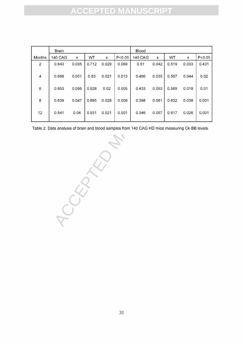
In contrast, CK-BB levels in both brain and blood buffy coat tissue samples were not
significantly reduced at the earliest time point (2 months) in 140 CAG full-length HD
mice, as compared to littermate control mice. Significant losses of brain CK-BB levels,
however, were present at 4, 6, 8, and 12 month time points in a severity of disease
dependent manner, ranging from 18.9% to 41.9% (Table 2, Figure 3). Congruous with
brain CK-BB levels, CK-BB levels in blood buffy coat samples were also significantly
reduced with increased disease severity and similar percentile levels of loss from 17.8%
to 41.4% (Table 2, Figure 3). While previous hyperkinetic motor activity has been
observed at 1 month of age and hypoactivity at 4 months [25], our open field analyses in
140 CAG mice of distance traveled, ambulatory counts, resting time, and ambulatory
time, showed significant differences from only the 3 month time point onward (p<0.5), in
comparison to littermate control mice (Figure 4), suggesting that the 2 month time point
may be a clinically premanifest time point. Immunostaining of 140 CAG mice tissue
sections using CK-BB antisera confirmed CK-BB loss in the brain, showed reduced
gross CK-BB immunoreactivity and a loss of CK-BB within the neuropil and in the
cytoplasm of neurons (Figure 5).
We evaluated CK-BB in human HD brain and blood buffy coat specimens by using well
defined HD stages of disease. CK-BB was reduced in a grade-dependent manner in
caudate nucleus specimens of all grades of severity (Grades 2-4) from HD patients, as
compared to non-diseased control tissue specimens (Figure 6). Densitometric analysis
showed significantly reduced differences between CK-BB levels with increasing grade
of severity [24] (Control vs G2: 27.6%, p<0.04; Control vs. G3; 53.4%, p<0.001; Control
vs. G4: 63.4%, p<0.0001) (Table 3, Figure 6). In addition, there were significant
differences between each HD grade (G2 vs G3; 35.6%, p<0.001; G3 vs G4: 21.5%,
ACC
EPTE
D M
ANU
SCR
IPT
ACCEPTED MANUSCRIPT
11
p<0.01) (Table 3, Figure 6). Blood buffy coat specimens showed a significant loss of
CK-BB levels in both premanifest (22.6%, p<0.01) and manifest (38.5%, p<0.001) HD
patients, with a greater loss in manifest disease samples (Table 3, Figure 6). CK-BB
immunostaining in 50 µm thick tissue sections confirmed the findings of reduced CK-BB
levels in HD striatal specimens (Figure 7). There was reduced CK-BB immunoreactivity
in the HD neostriatum. While cytosolic CK-BB immunoreactivity was markedly reduced
in neurons, intense CK-BB immunostaining was present in astrocytes in HD tissue
sections (Figure 7B and D). Combined immunofluorescence for GFAP and CK-BB
immunoreactivities confirmed this observation (Figure 7E, F and G). The latter is most
likely the consequence of increased reactive astrogliosis, a hallmark pathological
phenomenon in the neostriatum of HD subjects [24]. Of note, the expression of CK-BB
in mice is predominantly found in astrocytes and cortical inhibitory neurons in mice [20].
The expression of CK correlates with L- arginine:glycine amidinotransferase (AGAT)
and guanidinoacetate methyltransferase (GAMT), two enzymes responsible for creatine
biosynthesis [20]. As such, any alteration in these enzymes in HD may impact disease
expression. In patients with GAMT and AGAT mutations, clinical symptoms include
mental and motor retardation, extra-pyramidal symptoms, and seizures [26, 27].
Braissant and colleagues have recently suggested that there is a dissociation between
GAMT and AGAT activities in neurons, with only GAMT expression in the neostriatum
[28]. A GAMT deficiency may result in reduced CK and creatine deficiencies in HD, with
subsequent impaired energetics and neurodegeneration.
Discussion:
Creatine kinase (CK) catalyzes the reversible transfer of a phosphoryl group from
phopsphocreatine PCr to adenosine diphosphate (ADP), forming adenosine
triphosphate (ATP). Thus, creatine kinase offsets energy depletion by forming PCr,
providing a spatial energy buffer to re-phosphorylate ADP to ATP at cellular sites of
energy consumption and, in the reversible reaction, forming PCr and ADP from creatine
and ATP at cellular sites of high-energy phosphate production (PCr shuttle hypothesis)
[29-32]. As such, CK is critically vital to cellular energy homeostasis and its loss may
result in reduced energy stores and subsequent neuronal dysfunction and death in HD.

ACC
EPTE
D M
ANU
SCR
IPT
ACCEPTED MANUSCRIPT
12
It is of interest to note that CK-BB knockout mice present with pathological sequelae of
reduced body and brain weight, atrophied hippocampi, hyperventricular enlargement,
and impaired spatial learning [33, 34], all consistent with neurodegenerative effects
found in both HD mouse models and HD patients [1, 15, 35]. Our results provide
supporting evidence of mitochondrial damage and impaired energy metabolism in HD
[36]. The fact that CK-BB levels are significantly reduced in both brain and peripheral
blood buffy coat samples from HD mice and HD patients at premanifest and manifest
disease stages, are of additional significance, especially since CK-BB may be a
potential peripheral biomarker for this neurological disorder.
While it is to be expected that the brain is altered in a neurodegenerative disorder, it
may not be as obvious that biomarkers of HD could be found in peripheral tissue
samples such as blood, as it is a central nervous system disorder. For a peripheral
biomarker to represent the ‘state’ of HD, it would either have to be derived from the
brain or represent pathology occurring in the periphery. The mutant huntingin protein is
expressed ubiquitously throughout the body, including blood, and may cause
detectable, but clinically silent changes in gene expression and biochemistry anywhere
[35]. It is important to understand what relation potential peripheral biomarkers would
have to neurodegeneration in the brain. By using HD mice in these studies, we
examined complementary processes in the brain and periphery with much greater
precision and much greater control of post-mortem and other technical factors
associated with human tissue sampling. The present findings suggest that CK-BB levels
could serve as a biomarker of disease progression in premanifest and in manifest
disease HD patients. We are currently further investigating CK-BB levels in blood
samples from human clinical trials in HD patients to assess its potential as a
pharmacodynamic marker.
Although tremendous efforts have been made in recent years to identify early genetic,
clinical, and biochemical biomarkers that indicate the presence of disease prior to the
onset of clinical expression in neurodegenerative disorders, specific biomarkers for
premanifest HD are very limited at the present time [37]. We have, however,

ACC
EPTE
D M
ANU
SCR
IPT
ACCEPTED MANUSCRIPT
13
characterized one biomarker, 8-hydroxy-2'-deoxyguanosine (a marker of DNA
oxidation), in urine, blood, and brain in manifest HD patients with correlation in HD mice
that acts as a marker of disease progression and therapeutic efficacy [38-40]. Elevated
central and peripheral levels of 8-hydroxy-2'-deoxyguanosine are not specific to HD.
Others and we have found elevated levels of 8-hydroxy-2'-deoxyguanosine in other
neurodegenerative disorders and experimental models of neurological diseases, such
as amyotrophic lateral sclerosis, Parkinson’s disease, and Alzheimer’s disease [38, 39,
41-45]. Similarly, reduced levels of CK-BB are not specific to HD alone. We have novel
preliminary evidence that CK-BB is also significantly reduced in spinal cord samples
and in blood buffy coat samples from sporadic amyotrophic lateral sclerosis patients.
Biomarkers provide a diagnostic tool to improve early detection of disease in at-risk
individuals and provide greater diagnostic accuracy. Since there may be a prolonged
period of time in which neurons become dysfunctional before clinical expression of
disease, preclinical detection of biomarkers offers the promise of administering disease-
modifying medications during the premanifest period, further delaying or ameliorating
disease symptoms than treatment after disease onset. CK-BB may act as a ‘predictor’ in
defining surrogate endpoints that substitute for a true clinical outcome endpoint for the
purpose of comparing specific interventions or treatments in HD clinical trials. CK-BB
may greatly facilitate the accurate evaluation of the effectiveness of new therapies and
improve the safety and efficiency of clinical trials. While CK, also known as creatine
phosphokinase (CPK), is often determined routinely in patients, this blood test is not
specific for the type or isoenzyme of CK and measures total enzyme activity. Because
CK enzymatic activity is measured under strict conditions of temperature, pH, substrate
concentrations and activators, we are currently investigating direct assay of CK-BB in
blood from HD patients to confirm the Western analyses.
A major advance in studying HD has been the development of both transgenic and full-
length knock-in mouse models that exploit the mutation shown to underlie HD and
replicate the clinical and neuropathological phenomena observed in HD patients [15, 35,
46]. One complementary research strategy has been to perform parallel correlative

ACC
EPTE
D M
ANU
SCR
IPT
ACCEPTED MANUSCRIPT
14
studies in human and animal models of disease in order to take advantage of advances
being made in animal models and to best understand the most effective therapeutic
strategies. Although logical and attractive, the validity of this approach remains to be
proven. The HD mice have, however, identified a growing number of potential therapies
that are in early phase human trials [1, 47]. Genetic animal models of inherited
neurological diseases provide an experimentally accurate system to identify the basis of
molecular pathogenesis and provide an opportunity to test potential treatments and
explore their promise for translation to humans experiencing HD. While both the
transgenic fragment models and full-length knock-in models of HD share features with
human HD [15], the degree of similarity to human HD increases the closer the model
reproduces the exact genetic conditions for HD. As such, the full-length knock-in HD
mice may provide the best possible molecular genetic comparability to human HD,
especially prior to disease onset. The fulminant and early expression of disease in the
R6/2 mice may not clearly estimate the degree of premanifest and early symptomatic
identification of biomarkers, although others also report very early clinical phenomena in
the full-length 140 CAG HD mice [25]. It is of interest to note, however, while the CK-BB
levels were not significantly reduced in the 140 CAG mice at 2 months, there was a
significant loss of CK-BB levels in premanifest HD patients.
Strong evidence supports the importance of the creatine kinase system and specific
isoenzymes in neurodegenerative diseases [48, 49] and the neuroprotective effect of
creatine supplementation in studies of HD, amyotrophic lateral sclerosis, Parkinsonism,
brain ischemia, and other neurological conditions [50]. Creatine is involved in regulating
the octameric form of creatine kinase and decreases mitochondrial swelling when
inhibitors of creatine kinase octamer-dimer transition are present [49, 51]. Reduced
creatine stores in HD may precipitate altered creatine kinase levels [50]. Energy is
critical to the biological and molecular regulation of multiple cellular functions. As such,
reduced energy levels, as a consequence of creatine kinase dysfunction or loss,
threaten cellular homeostasis and integrity, resulting in subsequent neuronal death [48].
The present findings support the involvement of the creatine kinase system in HD. We
have evidence that chronic high-dose creatine supplementation in both R6/2 and 140
ACC
EPTE
D M
ANU
SCR
IPT
ACCEPTED MANUSCRIPT
15
CAG HD mice normalizes the reduced levels of CK-BB in both brain and blood buffy
coat specimens in manifest disease (unpublished data). The present findings justify the
ongoing clinical trials using high-dose creatine in HD patients. Interestingly, the
neuroprotective effects of creatine may be creatine kinase isoenzyme-dependent, since
creatine administration does not inhibit the mitochondrial transition pore in brain
mitochondria with native mitochondrial creatine kinase [51, 52].
Correlative analyses in brain and blood in both mice and man provide a powerful
strategy for identifying potential peripheral biomarkers for HD. The present findings
support the hypothesis of impaired energy metabolism in HD, identify a specific
mechanism of energy compromise in the reduction of CK-BB, establish CK-BB as a
potential biomarker of both central and peripheral premanifest and manifest disease,
and provide further rationale for creatine as a potential neuroprotective treatment for
HD.
Acknowledgements: We wish to thank Dr. Ann McKee for donating brain tissue
specimens from the Boston University Alzheimer’s Disease Brain Bank (NIA
P30AG13846). This work was supported by National Institutes of Health Grants
NS045806 (RJF and SMH), U01AT000613 (SMH) and NS058793 (SMH, HDR, CRS,
WRM, RJF), the Veterans Administration VISN 1 (RJF and WRM), the New England
HDSA Center of Excellence (SMH and HDR), and the CHDI Foundation (RJF).
References:
[1] E.C. Stack, R.J. Ferrante, Huntington's disease: progress and potential in the field,
Expert Opin Investig Drugs, 16 (2007) 1933-1953.
[2] W.A. Brennan, Jr., E.D. Bird, J.R. Aprille, Regional mitochondrial respiratory activity
in Huntington's disease brain, J Neurochem, 44 (1985) 1948-1950.
[3] W.D. Parker, Jr., S.J. Boyson, A.S. Luder, J.K. Parks, Evidence for a defect in
NADH: ubiquinone oxidoreductase (complex I) in Huntington's disease, Neurology, 40
(1990) 1231-1234.

ACC
EPTE
D M
ANU
SCR
IPT
ACCEPTED MANUSCRIPT
16
[4] V.M. Mann, J.M. Cooper, F. Javoy-Agid, Y. Agid, P. Jenner, A.H. Schapira,
Mitochondrial function and parental sex effect in Huntington's disease, Lancet, 336
(1990) 749.
[5] S.E. Browne, M.F. Beal, The energetics of Huntington's disease, Neurochem Res,
29 (2004) 531-546.
[6] M. Gu, M.T. Gash, V.M. Mann, F. Javoy-Agid, J.M. Cooper, A.H. Schapira,
Mitochondrial defect in Huntington's disease caudate nucleus, Ann Neurol, 39 (1996)
385-389.
[7] A.V. Panov, C.A. Gutekunst, B.R. Leavitt, M.R. Hayden, J.R. Burke, W.J.
Strittmatter, J.T. Greenamyre, Early mitochondrial calcium defects in Huntington's
disease are a direct effect of polyglutamines, Nat Neurosci, 5 (2002) 731-736.
[8] R.L. Albin, J.T. Greenamyre, Alternative excitotoxic hypotheses, Neurology, 42
(1992) 733-738.
[9] L. Djousse, B. Knowlton, L.A. Cupples, K. Marder, I. Shoulson, R.H. Myers, Weight
loss in early stage of Huntington's disease, Neurology, 59 (2002) 1325-1330.
[10] D.E. Kuhl, C.H. Markham, E.J. Metter, W.H. Riege, M.E. Phelps, J.C. Mazziotta,
Local cerebral glucose utilization in symptomatic and presymptomatic Huntington's
disease, Res Publ Assoc Res Nerv Ment Dis, 63 (1985) 199-209.
[11] T. Kuwert, H.W. Lange, K.J. Langen, H. Herzog, A. Aulich, L.E. Feinendegen,
Cortical and subcortical glucose consumption measured by PET in patients with
Huntington's disease, Brain, 113 ( Pt 5) (1990) 1405-1423.
[12] W.J. Koroshetz, B.G. Jenkins, B.R. Rosen, M.F. Beal, Energy metabolism defects
in Huntington's disease and effects of coenzyme Q10, Ann Neurol, 41 (1997) 160-165.
[13] F. Mastrogiacomo, J. LaMarche, S. Dozic, G. Lindsay, L. Bettendorff, Y. Robitaille,
L. Schut, S.J. Kish, Immunoreactive levels of alpha-ketoglutarate dehydrogenase
subunits in Friedreich's ataxia and spinocerebellar ataxia type 1, Neurodegeneration, 5
(1996) 27-33.
[14] T. Matsuishi, T. Sakai, E. Naito, S. Nagamitsu, Y. Kuroda, H. Iwashita, H. Kato,
Elevated cerebrospinal fluid lactate/pyruvate ratio in Machado-Joseph disease, Acta
Neurol Scand, 93 (1996) 72-75.

ACC
EPTE
D M
ANU
SCR
IPT
ACCEPTED MANUSCRIPT
17
[15] R.J. Ferrante, Mouse models of Huntington's disease and methodological
considerations for therapeutic trials, Biochim Biophys Acta, 1792 (2009) 506-520.
[16] W.R. Ellington, T. Suzuki, Early evolution of the creatine kinase gene family and the
capacity for creatine biosynthesis and membrane transport, Subcell Biochem, 46 (2007)
17-26.
[17] D.M. Dawson, H.M. Eppenberger, N.O. Kaplan, Creatine kinase: evidence for a
dimeric structure, Biochem Biophys Res Commun, 21 (1965) 346-353.
[18] P. Venkataraman, G. Krishnamoorthy, K. Selvakumar, J. Arunakaran, Oxidative
stress alters creatine kinase system in serum and brain regions of polychlorinated
biphenyl (Aroclor 1254)-exposed rats: protective role of melatonin, Basic Clin
Pharmacol Toxicol, 105 (2009) 92-97.
[19] E.C. Stack, W.R. Matson, R.J. Ferrante, Evidence of oxidant damage in
Huntington's disease: translational strategies using antioxidants, Ann N Y Acad Sci,
1147 (2008) 79-92.
[20] M. Tachikawa, M. Fukaya, T. Terasaki, S. Ohtsuki, M. Watanabe, Distinct cellular
expressions of creatine synthetic enzyme GAMT and creatine kinases uCK-Mi and CK-
B suggest a novel neuron-glial relationship for brain energy homeostasis, Eur J
Neurosci, 20 (2004) 144-160.
[21] N.W. Kowall, R.J. Ferrante, J.B. Martin, Patterns of cell loss in Huntington's
disease, Trends in Neurosciences, 10 (1987) 24-29.
[22] M. Perluigi, H.F. Poon, W. Maragos, W.M. Pierce, J.B. Klein, V. Calabrese, C. Cini,
C. De Marco, D.A. Butterfield, Proteomic analysis of protein expression and oxidative
modification in r6/2 transgenic mice: a model of Huntington disease, Mol Cell
Proteomics, 4 (2005) 1849-1861.
[23] E.C. Stack, J.K. Kubilus, K. Smith, K. Cormier, S.J. Del Signore, E. Guelin, H. Ryu,
S.M. Hersch, R.J. Ferrante, Chronology of behavioral symptoms and neuropathological
sequela in R6/2 Huntington's disease transgenic mice, J Comp Neurol, 490 (2005) 354-
370.
[24] J.P. Vonsattel, R.H. Myers, T.J. Stevens, R.J. Ferrante, E.D. Bird, E.P. Richardson,
Jr., Neuropathological classification of Huntington's disease, J Neuropathol Exp Neurol,
44 (1985) 559-577.

ACC
EPTE
D M
ANU
SCR
IPT
ACCEPTED MANUSCRIPT
18
[25] L.B. Menalled, J.D. Sison, I. Dragatsis, S. Zeitlin, M.F. Chesselet, Time course of
early motor and neuropathological anomalies in a knock-in mouse model of
Huntington's disease with 140 CAG repeats, J Comp Neurol, 465 (2003) 11-26.
[26] O. Braissant, C. Bachmann, H. Henry, Expression and function of AGAT, GAMT
and CT1 in the mammalian brain, Subcell Biochem, 46 (2007) 67-81.
[27] S. Stockler, P.W. Schutz, G.S. Salomons, Cerebral creatine deficiency syndromes:
clinical aspects, treatment and pathophysiology, Subcell Biochem, 46 (2007) 149-166.
[28] O. Braissant, E. Beard, C. Torrent, H. Henry, Dissociation of AGAT, GAMT and
SLC6A8 in CNS: relevance to creatine deficiency syndromes, Neurobiol Dis, 37 (2010)
423-433.
[29] S.P. Bessman, C.L. Carpenter, The creatine-creatine phosphate energy shuttle,
Annu Rev Biochem, 54 (1985) 831-862.
[30] R.A. Meyer, H.L. Sweeney, M.J. Kushmerick, A simple analysis of the
"phosphocreatine shuttle", Am J Physiol, 246 (1984) C365-377.
[31] R.M. Tombes, B.M. Shapiro, Metabolite channeling: a phosphorylcreatine shuttle to
mediate high energy phosphate transport between sperm mitochondrion and tail, Cell,
41 (1985) 325-334.
[32] E. Van Brussel, J.J. Yang, M.W. Seraydarian, Isozymes of creatine kinase in
mammalian cell cultures, J Cell Physiol, 116 (1983) 221-226.
[33] F. Streijger, F. Oerlemans, B.A. Ellenbroek, C.R. Jost, B. Wieringa, C.E. Van der
Zee, Structural and behavioural consequences of double deficiency for creatine kinases
BCK and UbCKmit, Behav Brain Res, 157 (2005) 219-234.
[34] H.J. in 't Zandt, W.K. Renema, F. Streijger, C. Jost, D.W. Klomp, F. Oerlemans,
C.E. Van der Zee, B. Wieringa, A. Heerschap, Cerebral creatine kinase deficiency
influences metabolite levels and morphology in the mouse brain: a quantitative in vivo
1H and 31P magnetic resonance study, J Neurochem, 90 (2004) 1321-1330.
[35] S.M. Hersch, H.D. Rosas, R.J. Ferrante, Neuropathology and Pathophysiology of
Huntington's Disease, in: R.L. Watts, W. Koller (Eds.) Movement Disorders:
Neurological Principles and Practice, McGraw-Hill Professional, 2004, pp. 603-629.
[36] M. Damiano, L. Galvan, N. Deglon, E. Brouillet, Mitochondria in Huntington's
disease, Biochim Biophys Acta, 1802 (2010) 52-61.
ACC
EPTE
D M
ANU
SCR
IPT
ACCEPTED MANUSCRIPT
19
[37] S.M. Henley, G.P. Bates, S.J. Tabrizi, Biomarkers for neurodegenerative diseases,
Curr Opin Neurol, 18 (2005) 698-705.
[38] S.M. Hersch, S. Gevorkian, K. Marder, C. Moskowitz, A. Feigin, M. Cox, P. Como,
C. Zimmerman, M. Lin, L. Zhang, A.M. Ulug, M.F. Beal, W. Matson, M. Bogdanov, E.
Ebbel, A. Zaleta, Y. Kaneko, B. Jenkins, N. Hevelone, H. Zhang, H. Yu, D. Schoenfeld,
R. Ferrante, H.D. Rosas, Creatine in Huntington disease is safe, tolerable, bioavailable
in brain and reduces serum 8OH2'dG, Neurology, 66 (2006) 250-252.
[39] K.M. Smith, S. Matson, W.R. Matson, K. Cormier, S.J. Del Signore, S.W. Hagerty,
E.C. Stack, H. Ryu, R.J. Ferrante, Dose ranging and efficacy study of high-dose
coenzyme Q10 formulations in Huntington's disease mice, Biochim Biophys Acta, 1762
(2006) 616-626.
[40] M.B. Bogdanov, O.A. Andreassen, A. Dedeoglu, R.J. Ferrante, M.F. Beal,
Increased oxidative damage to DNA in a transgenic mouse model of Huntington's
disease, J Neurochem, 79 (2001) 1246-1249.
[41] R.J. Ferrante, S.E. Browne, L.A. Shinobu, A.C. Bowling, M.J. Baik, U. MacGarvey,
N.W. Kowall, R.H. Brown, Jr., M.F. Beal, Evidence of increased oxidative damage in
both sporadic and familial amyotrophic lateral sclerosis, J Neurochem, 69 (1997) 2064-
2074.
[42] M. Bogdanov, W.R. Matson, L. Wang, T. Matson, R. Saunders-Pullman, S.S.
Bressman, M. Flint Beal, Metabolomic profiling to develop blood biomarkers for
Parkinson's disease, Brain, 131 (2008) 389-396.
[43] M. Bogdanov, R.H. Brown, W. Matson, R. Smart, D. Hayden, H. O'Donnell, M. Flint
Beal, M. Cudkowicz, Increased oxidative damage to DNA in ALS patients, Free Radic
Biol Med, 29 (2000) 652-658.
[44] N. Aguirre, M.F. Beal, W.R. Matson, M.B. Bogdanov, Increased oxidative damage
to DNA in an animal model of amyotrophic lateral sclerosis, Free Radic Res, 39 (2005)
383-388.
[45] C. Isobe, T. Abe, Y. Terayama, Levels of reduced and oxidized coenzyme Q-10
and 8-hydroxy-2'-deoxyguanosine in the CSF of patients with Alzheimer's disease
demonstrate that mitochondrial oxidative damage and/or oxidative DNA damage
contributes to the neurodegenerative process, J Neurol, 257 (2010) 399-404.
ACC
EPTE
D M
ANU
SCR
IPT
ACCEPTED MANUSCRIPT
20
[46] M.Y. Heng, P.J. Detloff, R.L. Albin, Rodent genetic models of Huntington disease,
Neurobiol Dis, 32 (2008) 1-9.
[47] S.M. Hersch, R.J. Ferrante, Translating therapies for Huntington's disease from
genetic animal models to clinical trials, NeuroRx, 1 (2004) 298-306.
[48] M. Wyss, O. Braissant, I. Pischel, G.S. Salomons, A. Schulze, S. Stockler, T.
Wallimann, Creatine and creatine kinase in health and disease--a bright future ahead?,
Subcell Biochem, 46 (2007) 309-334.
[49] E. O'Gorman, G. Beutner, M. Dolder, A.P. Koretsky, D. Brdiczka, T. Wallimann, The
role of creatine kinase in inhibition of mitochondrial permeability transition, FEBS Lett,
414 (1997) 253-257.
[50] A.M. Klein, R.J. Ferrante, The neuroprotective role of creatine, Subcell Biochem, 46
(2007) 205-243.
[51] N. Brustovetsky, T. Brustovetsky, J.M. Dubinsky, On the mechanisms of
neuroprotection by creatine and phosphocreatine, J Neurochem, 76 (2001) 425-434.
[52] P. Klivenyi, N.Y. Calingasan, A. Starkov, I.G. Stavrovskaya, B.S. Kristal, L. Yang,
B. Wieringa, M.F. Beal, Neuroprotective mechanisms of creatine occur in the absence
of mitochondrial creatine kinase, Neurobiol Dis, 15 (2004) 610-617.
Figure Legends:
Figure 1. Western and dot blot analyses of brain and blood from R6/2 HD mice through
the spectrum of disease at a premanifest stage (21 day), onset (30 day), and though
early (42 day), moderate (63 day), and end stage disease (91 day). There were
significant differences between mutant and littermate control mice at each disease
stage, to include premanifest disease (Table 1). Blots are shown under each time point
with alpha tubulin controls.
Figure 2. CK-BB immunohistochemistry of brain sections through anterior neostriatum
at the level of the anterior commissure in R6/2 mice at 91 days. Gross CK-BB
immunoreactivity is reduced in the R6/2 mutant mouse (B), in comparison to the wild
type littermate control (A). Higher magnification of the neostriatum from each tissue

ACC
EPTE
D M
ANU
SCR
IPT
ACCEPTED MANUSCRIPT
21
section shows a marked reduction in CK-BB immunoreactivity in the mutant R6/2
mouse (D) within both the neuropil and the cytoplasm of striatal neurons (arrows), as
compared to the wild type littermate control mouse (C). Magnification bar in A is 2 mm.
Magnification bar in D is 100 µm.
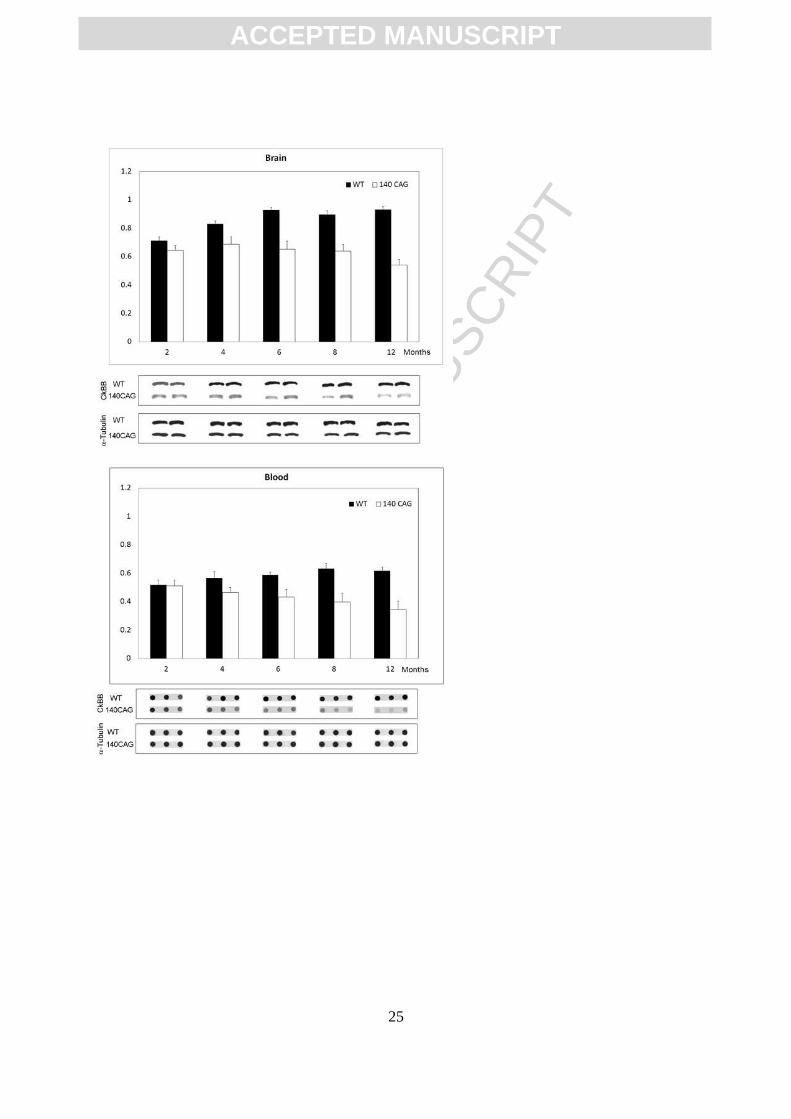
Figure 3. Western and dot blot analyses of brain and blood from 140 CAG HD mice
through the spectrum of disease at a premanifest stage (2 months), onset (4 months),
though early (6 months), moderate (8 months), and severe disease (12 months). There
were significant differences between mutant and littermate control mice at each disease
stage starting a 4 months of age (Table 2). Blots are shown under each time point with
alpha tubulin controls.
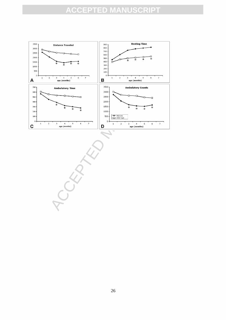
Figure 4. Open field analysis of 140 CAG full-length mutant HD mice from 1-6 months.
There are significant differences in distance traveled (A), resting time (B), ambulatory
time (C), and ambulatory counts (D) starting at 3 months of age. Significance was not
obtained at 1 and 2 months. * p<0.05
Figure 5. CK-BB immunohistochemistry of brain sections through the anterior
neostriatum at the level of the anterior commissure in 140 CAG HD mice at 8 months.
Gross CK-BB immunoreactivity is reduced in the 140 CAG mutant mouse (B), in
comparison to the wild type littermate control mouse (A). Higher magnification within the
neostriatum from each tissue section shows a reduction in CK-BB immunoreactivity in
the mutant 140 CAG mouse (D) within both the neuropil and the cytoplasm of striatal
neurons (arrows), as compared to the wild type littermate control mouse (C).
Magnification bar in A is 2 mm. Magnification bar in D is 100 µm.
Figure 6. Western analysis from the medial segment of the caudate nucleus (brain) of
age-matched control patients and Grade 2, Grade 3, and Grade 4 HD patients. There
was a significant grade-dependent reduction in CK-BB, with the greatest loss in Grade
4 HD (Table 3). Blood buffy coat CK-BB analysis from age-matched patient caregivers
and premanifest and manifest HD patients showed a significant loss of CK-BB in both
ACC
EPTE
D M
ANU
SCR
IPT
ACCEPTED MANUSCRIPT
22
premanifest disease and manifest diseased HD patients, as compared to the controls
(Table 3). Blots are shown under each time point with alpha tubulin controls.
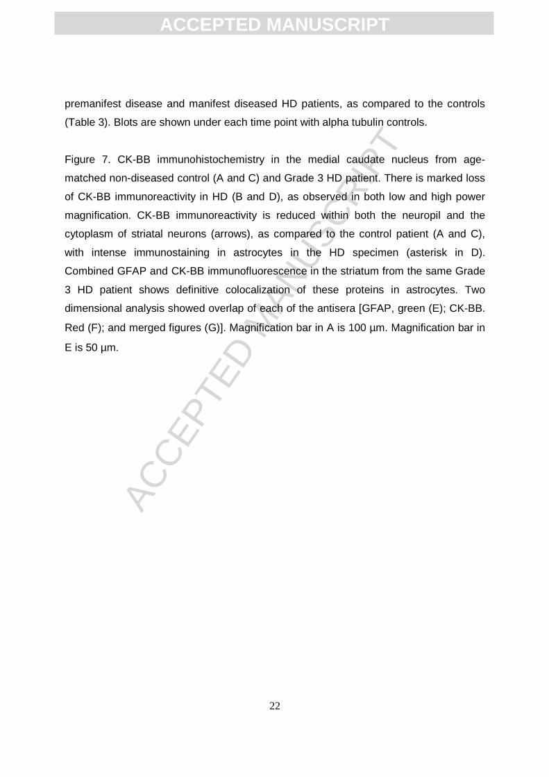
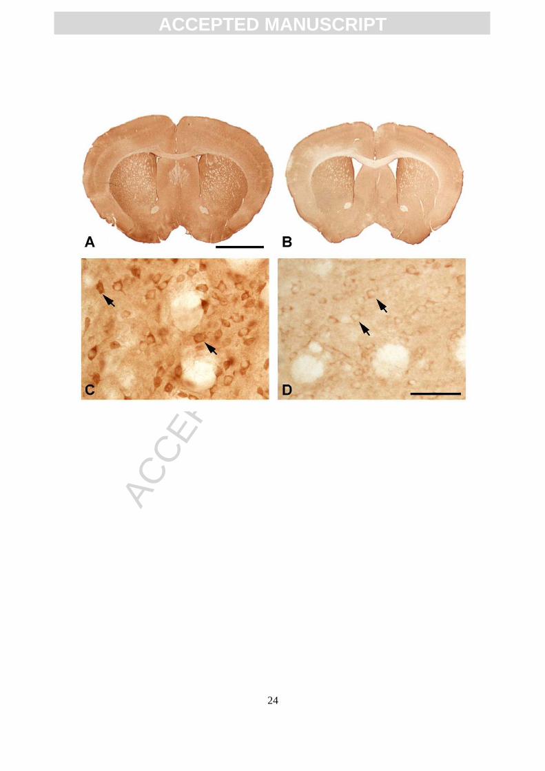
Figure 7. CK-BB immunohistochemistry in the medial caudate nucleus from age-
matched non-diseased control (A and C) and Grade 3 HD patient. There is marked loss
of CK-BB immunoreactivity in HD (B and D), as observed in both low and high power
magnification. CK-BB immunoreactivity is reduced within both the neuropil and the
cytoplasm of striatal neurons (arrows), as compared to the control patient (A and C),
with intense immunostaining in astrocytes in the HD specimen (asterisk in D).
Combined GFAP and CK-BB immunofluorescence in the striatum from the same Grade
3 HD patient shows definitive colocalization of these proteins in astrocytes. Two
dimensional analysis showed overlap of each of the antisera [GFAP, green (E); CK-BB.
Red (F); and merged figures (G)]. Magnification bar in A is 100 µm. Magnification bar in
E is 50 µm.

ACC
EPTE
D M
ANU
SCR
IPT
ACCEPTED MANUSCRIPT
23
ACC
EPTE
D M
ANU
SCR
IPT
ACCEPTED MANUSCRIPT
24
ACC
EPTE
D M
ANU
SCR
IPT
ACCEPTED MANUSCRIPT
25
ACC
EPTE
D M
ANU
SCR
IPT
ACCEPTED MANUSCRIPT
26

ACC
EPTE
D M
ANU
SCR
IPT
ACCEPTED MANUSCRIPT
27

ACC
EPTE
D M
ANU
SCR
IPT
ACCEPTED MANUSCRIPT
28

ACC
EPTE
D M
ANU
SCR
IPT
ACCEPTED MANUSCRIPT
29

ACC
EPTE
D M
ANU
SCR
IPT
ACCEPTED MANUSCRIPT
30
ACC
EPTE
D M
ANU
SCR
IPT
ACCEPTED MANUSCRIPT
31

ACC
EPTE
D M
ANU
SCR
IPT
ACCEPTED MANUSCRIPT
32