
Reading in a deep orthography: neuromagnetic evidence for dual-mechanisms
23
Reading in a Deep Orthography: Neuromagnetic Evidence for Dual-Mechanisms Tony W. Wilson 1 , Arthur C. Leuthold 2,CA , John E. Moran 3 , Patricia J. Pardo 2 , Scott M. Lewis 2 , and Apostolos P. Georgopoulos 2 1 Neuromagnetic Imaging Center, Department of Psychiatry, University of Colorado Health Sciences Center, Denver, CO 2 Brain Sciences Center, Veterans Affairs Medical Center, Minneapolis, MN 3 Department of Neurology, Henry Ford Hospital, Detroit, MI Abstract Despite substantial efforts to connect cognitive-linguistic models with appropriate anatomical correlates, the question of which cognitive model best accounts for the neuropsychological and functional neuroimaging evidence remains open. The two most popular models are grounded in conceptually different bases and thus make quasi-distinct predictions in regard to the patterns of activation that should be observed in imaging investigations of linguistic processing. Dual- mechanism models propose that high-frequency regular and irregular words are processed through a lexicon-based word code, which facilitates their processing and pronunciation latencies relative to pseudowords. In contrast, single-mechanism models suggest the same behavioral effects can be explained through semantic mediation without the existence of a lexicon. In most previous studies, words and pronounceable pseudowords were presented in lexical-decision or word reading paradigms, and hemodynamic techniques were utilized to distinguish involved anatomical areas. The results typically indicated that both word classes activated largely congruent tissues, with a magnitude advantage for pseudowords in most or all activated regions. However, since the dual-mechanism model predicts both word types utilize the entire linguistic network, but that certain operations are merely obligatorily involved, these results do not sharply refute nor clearly support the model’s main tenets. In the current study, we approach the dual- versus single mechanism question differently by focusing on the temporal dynamics of MEG imaged neuronal activity, during performance of an oddball version of continuous lexical-decision, to determine whether the onset latency of any cortical language region shows effects of word class that are indicative of preferential versus obligatory processing pathways. The most remarkable aspect of our results indicated that both words and pseudowords initially activate the left posterior fusiform region, but that the spatiotemporal dynamics clearly distinguish the two word classes thereafter. For words, this left fusiform activation was followed by engagement of the left posterior inferior temporal, and subsequently activation reached the left posterior superior temporal region. For pseudowords, this sequential order of left temporal area activations was reversed, as activity proceeded from the left fusiform to the left superior temporal and then the left inferior temporal region. For both classes, this dynamic sequential spread manifested within the first 300 ms of stimulus processing. We contend these results provide strong support for the existence of dual-mechanisms underlying reading in a deep orthographic language (i.e., English). Mailing Address: Arthur C. Leuthold, Ph.D., Brain Sciences Center (4S), Veterans Affairs Medical Center (11B), One Veterans Drive, Minneapolis, MN 55417, Office: (612) 467-5546, Fax: (612) 725-2291, Email: E-mail: [email protected]. NIH Public Access Author Manuscript Exp Brain Res. Author manuscript; available in PMC 2009 July 27. Published in final edited form as: Exp Brain Res. 2007 June ; 180(2): 247–262. doi:10.1007/s00221-007-0852-0. NIH-PA Author Manuscript NIH-PA Author Manuscript NIH-PA Author Manuscript
Transcript of Reading in a deep orthography: neuromagnetic evidence for dual-mechanisms
Reading in a Deep Orthography: Neuromagnetic Evidence forDual-Mechanisms
Tony W. Wilson1, Arthur C. Leuthold2,CA, John E. Moran3, Patricia J. Pardo2, Scott M.Lewis2, and Apostolos P. Georgopoulos21Neuromagnetic Imaging Center, Department of Psychiatry, University of Colorado Health SciencesCenter, Denver, CO2Brain Sciences Center, Veterans Affairs Medical Center, Minneapolis, MN3Department of Neurology, Henry Ford Hospital, Detroit, MI
AbstractDespite substantial efforts to connect cognitive-linguistic models with appropriate anatomicalcorrelates, the question of which cognitive model best accounts for the neuropsychological andfunctional neuroimaging evidence remains open. The two most popular models are grounded inconceptually different bases and thus make quasi-distinct predictions in regard to the patterns ofactivation that should be observed in imaging investigations of linguistic processing. Dual-mechanism models propose that high-frequency regular and irregular words are processed througha lexicon-based word code, which facilitates their processing and pronunciation latencies relative topseudowords. In contrast, single-mechanism models suggest the same behavioral effects can beexplained through semantic mediation without the existence of a lexicon. In most previous studies,words and pronounceable pseudowords were presented in lexical-decision or word readingparadigms, and hemodynamic techniques were utilized to distinguish involved anatomical areas. Theresults typically indicated that both word classes activated largely congruent tissues, with a magnitudeadvantage for pseudowords in most or all activated regions. However, since the dual-mechanismmodel predicts both word types utilize the entire linguistic network, but that certain operations aremerely obligatorily involved, these results do not sharply refute nor clearly support the model’s maintenets. In the current study, we approach the dual- versus single mechanism question differently byfocusing on the temporal dynamics of MEG imaged neuronal activity, during performance of anoddball version of continuous lexical-decision, to determine whether the onset latency of any corticallanguage region shows effects of word class that are indicative of preferential versus obligatoryprocessing pathways. The most remarkable aspect of our results indicated that both words andpseudowords initially activate the left posterior fusiform region, but that the spatiotemporal dynamicsclearly distinguish the two word classes thereafter. For words, this left fusiform activation wasfollowed by engagement of the left posterior inferior temporal, and subsequently activation reachedthe left posterior superior temporal region. For pseudowords, this sequential order of left temporalarea activations was reversed, as activity proceeded from the left fusiform to the left superior temporaland then the left inferior temporal region. For both classes, this dynamic sequential spread manifestedwithin the first 300 ms of stimulus processing. We contend these results provide strong support forthe existence of dual-mechanisms underlying reading in a deep orthographic language (i.e., English).
Mailing Address: Arthur C. Leuthold, Ph.D., Brain Sciences Center (4S), Veterans Affairs Medical Center (11B), One Veterans Drive,Minneapolis, MN 55417, Office: (612) 467-5546, Fax: (612) 725-2291, Email: E-mail: [email protected].
NIH Public AccessAuthor ManuscriptExp Brain Res. Author manuscript; available in PMC 2009 July 27.
Published in final edited form as:Exp Brain Res. 2007 June ; 180(2): 247–262. doi:10.1007/s00221-007-0852-0.
NIH
-PA Author Manuscript
NIH
-PA Author Manuscript
NIH
-PA Author Manuscript
Keywordsword; MEG; temporal; fusiform; language
IntroductionThis paper focuses on the neural implementation of reading in a quasi-regular or deeporthographic language, namely English. Reading in deep orthographies is complicated bymappings between graphemes, phonemes, and whole word sounds being inherentlyambiguous. By comparison, languages with regular orthographies (e.g., German, Italian) aremore transparent, as mappings between graphemes and phonemes are virtually one-to-one.Such straightforward grapheme-to-phoneme correspondence rules allow young readers tomore rapidly acquire proficiency in reading (Frith et al. 1998; Landerl et al. 1997), and areassociated with other benefits, like reduced pronunciation latencies, that extend well intoadulthood (Paulesu et al. 2000).
Two different classes of cognitive models successfully account for the observed phenomenaof reading complex orthography. These models are largely based on the behavioral andneuropsychological evidence that has accumulated over the last several decades. Both classesof models recognize at least two distinct reading strategies are necessary, as skilled Englishreaders are able to pronounce words with regular spelling-to-sound relationships (e.g., kernel),as well as those with irregular relationships (e.g., colonel). However, the two classes differ inhow these strategies are implemented in the model. Briefly, dual-mechanism models (Coltheartet al. 1993, 2001; Coltheart and Rastle 1994) assume orthography-to-phonology transformationcan be achieved through two distinct mechanisms; the first is a lexical or whole-word procedureand the second a sublexical or assembled procedure. The postulated lexical method operatesin parallel across the entire input string and would be engaged during processing of high-frequency words, especially those with irregular print-to-sound relationships. In contrast, thesublexical method is thought to operate serially from left-to-right, more or less assembling theword’s phonological representation in piecemeal fashion. The sublexical procedure isnecessary for novel or low-frequency words, as well as pronounceable pseudowords. Although,in the most recent formulation of this model, all words (e.g., high/low frequency, regular, andirregular) are processed in parallel through both mechanisms (Coltheart et al. 1993, 2001). Theprimary alternative to dual-mechanism accounts is a triangle-type interactive connectionistmodel (Plaut et al. 1996; Seidenberg and McClelland 1989). In this single-mechanismformulation, groups of simple units code orthographic, phonological, or semantic elements ofthe relevant corpus, and a word’s pronunciation and meaning are represented as distributedpatterns of activation across all units. Thus, words sharing phonological and/or semanticfeatures would be represented by similar patterns of activation across the array of simple units,with particular print-to-sound correspondences being resolved through competitive andcooperative interactive processing amongst units in the network. Essentially, the triangle modelincludes one direct mechanism or route for orthography-phonology conversion, and a secondindirect pathway implemented through semantics (Plaut et al. 1996). In addition, analogous tothe dual-mechanism position, the single-mechanism account also postulates that all words areprocessed in parallel through both procedures, with irregular words being more reliant thanregular on semantic mediation for successful processing. In short, these single- and dual-mechanism formulations share many common features, the primary distinction being whetherthe facilitation of word pronunciation is achieved through a semantic code (triangle-model) ora lexicon-based word code (dual-route model; for further discussion of these issues, see Binderet al. 2005).
Wilson et al. Page 2
Exp Brain Res. Author manuscript; available in PMC 2009 July 27.
NIH
-PA Author Manuscript
NIH
-PA Author Manuscript
NIH
-PA Author Manuscript
Evidence for the neurological validity of these models arises from the neuropsychologicalliterature, in particular lesion-based studies of acquired dyslexia. Most importantly, theexistence of both phonological and surface dyslexia is congruent with distinct neuralmechanisms subserving different processes for pseudo- and irregular word reading. In surfacedyslexia, patients are able to read pronounceable pseudowords and unable to read irregularwords, which suggests neural areas underlying lexical or semantic reading procedures havebeen selectively damaged. Conversely, patients with phonological dyslexia are able to readmost high-frequency words (i.e., regular and irregular), but unable to process even simplepseudowords. Thus, in contrast to surface dyslexics, patients with phonological dyslexia seemto have selectively damaged neural areas serving sublexical conversion mechanisms. Overall,the existence of both disorders indicates at least partially distinct neural regions underlie thestrategies for successful reading in deep orthographic languages. This idea of distinct neuralsystems tends to support models postulating partially discrete mechanisms for regular versusirregular words, and hence is congruent with both the dual-route and triangle-type formulations.
Another means of establishing such neurological validity is through the use of functionalneuroimaging techniques in healthy readers. A benefit of using healthy volunteers to demarcateneural areas dedicated to reading is that it circumvents concerns of compensatory strategy use,as well as neural reorganization following neurological infarcts. On the other hand, thesetechniques only illuminate the neural tissues active, but not necessarily critical for any givencognitive process. Thus, it is possible that nodes recognized via functional neuroimaging arenot absolutely necessary for successful performance. Word reading was one of the firstcognitive feats to be investigated with functional neuroimaging (Petersen et al. 1988), and morethan 15 years later this area remains one of the most active in cognitive neuroscience research.Yet, despite large amounts of empirical data, the neural mechanisms underling word and non-word reading remain an area of contention, especially for languages with deep orthographies.Many early studies used the seemingly straightforward word-pseudoword reading contrast,and reported largely non-replicable findings in regards to neural areas more active during wordprocessing (for reviews, see Binder et al. 2003; Mechelli et al. 2003). This inconsistency maybe reflective of the sufficient versus necessary confound acknowledged above; in short, theinability to reliably segregate neuronal activity in critical substrates from that in neural regionsinessential to task performance (e.g., activation reflecting sublexical procedures duringirregular word processing). Mechelli et al. (2002, 2003) have provided substantial evidencefor this position by showing the same cortical areas are active during word and pseudowordprocessing, and that the major inconsistencies emerge when differences in activationmagnitude are sought (i.e., when word- and pseudoword-induced activations are subtracted).Given this, the inconsistent findings reported in early studies of word versus pseudowordprocessing may have been attributable to very subtle differences in experimental design (e.g.,stimuli, presentation parameters, etc.), functional organization (i.e., heterogeneity acrosssubjects), and/or relatively small sample sizes (see Mechelli et al. 2003, for further discussion).
Regardless, more recent investigations of single and dual-mechanism models have been muchmore reliable, as numerous reading studies have found greater activation for pseudowords inboth left occipitotemporal and inferior frontal regions (Binder et al. 2005a; Kronbichler et al.2004; Mechelli et al. 2003; Paulesu et al. 2000; Xu et al. 2001). In addition, several studiesusing the lexical-decision task have reported greater activation for words in leftoccipitotemporal cortices, along with stronger or equivalent activation to pseudowords in leftinferior frontal regions (Binder et al. 2003, 2005b; Fiebach et al. 2002; Ischebeck et al. 2004;Rissman et al. 2003). This pattern of results highlights the important issue of task-specificeffects on neural areas more active during word or pseudoword processing. Essentially,multiple studies have collectively shown that only certain neural areas (e.g., leftoccipitotemporal) display strong task-specific responses to words and/or pseudowords, whichpotentially could provide useful information for discerning their particular role(s) in language
Wilson et al. Page 3
Exp Brain Res. Author manuscript; available in PMC 2009 July 27.
NIH
-PA Author Manuscript
NIH
-PA Author Manuscript
NIH
-PA Author Manuscript
processing. Binder et al. (2003) noted that brain regions more active for words in the lexical-decision task closely match those normally detected in tasks probing semantic processing,which they interpreted as evidence supporting the single-mechanism triangle model of wordprocessing (Plaut et al. 1996). Furthermore, by manipulating orthographic neighborhooddensity of words and pseudowords in the context of lexical decision, they demonstrated lexicalcandidates with high-density did not activate any brain area more than candidates with lowneighborhood density (Binder et al. 2003). This latter result strongly suggests task performanceis enabled by semantic units and not word-level units, as lexical candidates with highneighborhood densities should have elicited more activation than those with low-densities ifperformance were mediated by a non-semantic word-like code (i.e., a lexicon, as currentlyformulated in dual-route models; Coltheart et al. 1993, 2001). However, the existence of alexicon-like neural word-form system remains a contentious matter, as other studies haveshown word-frequency effects in regions corresponding to or near left occipitotemporal areas,which suggests word-level codes may indeed be involved in word reading (Keller et al.2001; Kuo et al. 2003). In fact, Kronbichler et al. (2004) employed a sensitive parametricvariation of word-frequency, and found strongly indicative evidence of a non-semantic word-like code mediating visual word recognition in left occipitotemporal cortex.
In our previous investigations, we reported that left perisylvian regions exhibit significantlyearlier activation for words relative to pseudowords (Wilson et al. 2005a). Thismagnetoencephalography (MEG) study used the single-moving-dipole model for sourcelocalization, which is a good method for determining the location of compact sources. Ofimportance, a limitation associated with the single-dipole method involves its preference forfocal versus highly-distributed sources. Basically, the method shows greater spatial precision,as well as fewer false negatives (i.e., failures to capture all ‘real’ sources) when the underlyingactivations are more focal in nature (Jerbi et al. 2004). This is a concerning factor in word/pseudoword comparisons, as intuitively one might expect pseudowords to elicit more diffuseactivation in left-hemispheric cortices. Presumably, pseudowords induce a search process inwhich the linguistic system attempts to match the input to a possible lexical candidate, andsuch processing could elicit more distributed activation within neural regions mediatinglexicality. Thus, it could be argued that pseudoword-induced sources are more commonlymissed in single-dipole analyses. In the present work, we use a cortically-constraineddistributed-source imaging technique to provide continuous and precise time courses for focaland diffuse neural activations, which is a substantial enhancement to our previous single-dipoleinvestigations (e.g., Wilson et al. 2005a, Wilson et al. 2005b). After imaging cortical activitywith this technique, we calculate the time series of local current-density amplitude within theregions-of-interest (ROI) most often acknowledged as critical to language processing.Extensive prior neuroimaging work has shown a small group of left hemispheric neural regionsare reliably involved in word and pseudoword processing (for reviews, see Jobard et al.2003; Mechelli et al. 2002, 2003; Price 2000; Price et al. 2003), and in the current analyses wechose to focus uniquely on these eloquent language cortices.
We hypothesized that differences in the time course of activated neural regions woulddistinguish the processing of words and pronounceable pseudowords. In other words, bothtypes of lexical candidates will likely activate each region of interest (i.e., due to obligatoryprocesses), but the relative timing of these activations should differ between the two classesof stimuli beyond what could be attributed to word-frequency effects. Essentially, ifparticipants utilize a lexicon-like word-based code in processing real words, than preferentialprocessing (i.e., shorter latencies) along this pathway should be discerned for words relativeto pseudowords during the lexical-decision task. We believe detecting such temporaldifferences would provide strong evidence for the existence of dual-mechanisms or pathwaysfor word processing (Coltheart et al. 1993, 2001), and that a lack of such differences wouldmore support triangle-type formulations of word processing (Platt et al. 1996). To investigate
Wilson et al. Page 4
Exp Brain Res. Author manuscript; available in PMC 2009 July 27.
NIH
-PA Author Manuscript
NIH
-PA Author Manuscript
NIH
-PA Author Manuscript
this, we conducted a MEG imaging study of regional brain activation during an oddball versionof the lexical-decision paradigm. Our primary aim was to quantify temporal activationdifferences, between high-frequency words and pronounceable pseudowords, in each of themost recognized cortical regions subserving language processing. To illuminate the corticaldynamics, we also transformed spatiotemporal current-density estimations into statisticalparametric maps (dSPM) reflecting region-specific time courses of neuronal activation.
Materials and MethodsParticipants
Eleven native English speakers age 18–41 years (mean: 28 years) were paid to participate inthis experiment (8 males and 3 females). One male subject’s data was discarded due toexcessive blinking. All subjects were strongly right-handed as assessed by the EdinburghHandedness Inventory (range: 75–100; Oldfield 1971). All subjects had normal or correctedto normal vision, and denied any history of neurological or psychiatric disease (including drug/alcohol abuse). Each subject provided informed consent to a protocol approved by theInstitutional Review Boards of the University of Minnesota and the Veterans Affairs MedicalCenter, Minneapolis, Minnesota.
Experimental ParadigmSubjects performed an oddball version of the lexical-decision task while supine in a dimly lit,magnetically shielded room (MSR). The stimulus set included 120 concrete nouns, 100pronounceable pseudowords, and 80 consonant strings. From these stimuli, 40 concrete nounsand 20 pseudowords were randomly selected to serve as targets. The two-syllable concretenouns were of high-frequency (range: 1.12 – 1.84 log; mean: 1.49 log; Kucera and Francis1967), and all followed regular orthography-to-phonology conversion principles (i.e., irregularor exception words were not permitted). To create pseudowords, we shuffled the phonemes ofthe concrete nouns; thus, phonemic units present in the corpus of words were preserved in thepseudowords, and the two types of stimuli did not significantly differ in mean positional bigramfrequency (mean(SD); words: 1722.6(867.9), pseudowords: 1635.5(809.4)), neighborhooddensity (words: 5.4(4.4), pseudowords: 5.1(3.3)), or total number of characters (words: 4.5(0.6), pseudowords: 4.5(0.6); Balota et al. 2002). Particular care was also taken to ensurepseudowords resembled real English words in all respects, with the exception of lexical andsemantic status (i.e., we screened for pseudohomophones and other ‘special’ pseudowords).We included consonant strings only for comparison to earlier experiments, thus these data willbe reported separately. The experiment consisted of 6 blocks, each lasting approximately 60seconds with a 15-second inter-block interval. Thus, overall recording time was ~8 minutes.In each block, participants viewed (duration = 600 ms; stimulus-onset-asynchrony = 1200 ms)40 non-targets and 10 targets in a pseudo-randomized order. The 10 targets were all words infour blocks and all pseudowords in the remaining blocks. In blocks where words served astargets, participants saw, on average, the same number of non-target pseudowords andconsonant strings within each block. In the other two blocks, participants saw only the 10pseudoword targets and the 40 non-target real words (i.e., no consonant strings). For eachparticipant, the sequential order of blocks was re-randomized, as was the order of stimuli withineach block. All stimuli were 4–6 letters long and presented only once in white 36-point Courierfont on a black background. Stimulus presentation alternated with a white fixation cross.Subjects responded with a button press when a target was observed, and did not respond toother stimuli (i.e., go/no-go task). Before MEG acquisition, subjects were asked to limitblinking during stimulus presentation. However, during the inter-block intervals, subjects weretold via visual display to blink freely, and were also notified of which word-type would functionas the target stimuli in the ensuing block. All stimuli were presented using the STIM software
Wilson et al. Page 5
Exp Brain Res. Author manuscript; available in PMC 2009 July 27.
NIH
-PA Author Manuscript
NIH
-PA Author Manuscript
NIH
-PA Author Manuscript

(Compumedics Neuroscan, El Paso, TX). An LCD projector outside the MSR projected allstimuli and instructional messages onto a screen positioned ~60 cm above the subject.
MEG Acquisition and Data Processing ProceduresWith an acquisition bandwidth of 0.1–200 Hz, neuromagnetic signals were sampledcontinuously at 508 Hz using a Magnes 3600 WH equipped with 248 axial-gradiometers (4-D Neuroimaging Inc., San Diego, CA). Each axial-gradiometer is coupled to a SQUID sensor,which acts as a low-noise magnetic flux-to-voltage converter. Along with the magnetic data,a photodiode signal (to ensure precise timing in stimulus delivery) and an electrooculogram(EOG) were also recorded. The continuous MEG data stream was exported from the acquisitionenvironment, and cardio-corrected in its continuous native format. All cardiac artifacts wereremoved from each subject’s MEG data series using the event-synchronous subtraction method(Leuthold 2003). This method of cardio-artifact removal uses a computer algorithm, along withuser input, to identify each QRS complex of the cardiac artifact waveform infiltrating the mostsusceptible MEG sensors. Each P through T heartbeat is then averaged within each sensorindividually throughout the acquisition period. In turn, this averaged heartbeat artifact,particular to a given sensor, is subtracted from each heartbeat within the respective sensor’stime series. Next, all MEG data were digitally filtered using a 30 Hz low-pass butterworth filterand split into 1 s epochs (including a 200 ms pre-stimulus baseline), with zero defined as thetime-point of stimulus onset. We used a fixed threshold method (EOG > 100 uV or MEG level> 1.25 pT), supplemented with visual inspection, to identify and reject artifactual epochs.Furthermore, epochs in which the subject behaviorally responded were also rejected (i.e.,correct button presses to targets and incorrect to non-targets). For each word type, at least 60trials (out of 80 possible) remained and an average response was calculated for each. Averageresponses were baseline corrected by subtracting the signal amplitude of the 200 ms pre-stimulus time interval. With the exception of cardio-correction, MEG data were preprocessedusing Brain Electrical Source Analysis Software (BESA, Version 5.0.4; MEGIS, Germany).
MRI Acquisition and Coregistration with MEGAnatomic images of the brain were acquired on a General Electric Signa Horizons LX 1.5TMR scanner using a neuro-vascular head coil. Sagittal scout images were taken to determinethe number and placement of subsequent axial slices. The volume covered extended from thetop of the head to the bottom of the cerebellum, including the external auditory meati bilaterally(thus, all MEG fiducial points were within coverage). T1-weighted axial images were thenacquired using a 3-dimensional SPGR sequence with the following parameters: TE = minfull,TR = 20 ms, Flip angle = 30 deg., FOV = 240 × 240 mm, matrix = 256×256, slice thickness/gap = 1.5/0, NEX = 1. The resulting voxel resolution was 0.94 × 0.94 × 1.5 mm.
Prior to MEG measurement, five small coils were attached to the participant’s head and thelocations of these coils, together with the three fiducial points and scalp surface, weredetermined with a 3-D digitizer (Fastrak 3SF0002, Polhemus Navigator Sciences, Colchester,VT). Once the participant was positioned inside the helmet of the neuromagnetometer, a smallelectric current was fed to the coils, inducing a measurable magnetic field. This allowed thecoils to be located in reference to the sensors. Since coil locations were also known in headcoordinates, all MEG data could be transformed into a common coordinate system. Moreover,since the head coordinate system could be mapped onto the participant’s MRI, individual MEGresponses could also be mapped onto the structural MRI. Coregistration of MEG and MRI datawas performed with the MEG Tools Imaging and Visualization Software (Moran andTepley, Oakland University – Henry Ford Hospital Neuromagnetism Laboratory, Detroit, MI)implemented in MATLAB (Version 7.0, R14; The MathWorks, Inc., Natick, MA).
Wilson et al. Page 6
Exp Brain Res. Author manuscript; available in PMC 2009 July 27.
NIH
-PA Author Manuscript
NIH
-PA Author Manuscript
NIH
-PA Author Manuscript
MEG Current-Density Imaging ProceduresConstruction of head model and solution space—In contrast to EEG imaging, thelocations of electrical sources can be accurately calculated for MEG data using a simplespherical volume conductor model of the head (Hämäläinen and Sarvas 1989). However,accuracy can be enhanced through a multi-sphere approach, in which several overlappingspherical models are each fit to the local curvature of a different brain region (Leahy et al.1998). In this study, we approximated the brain as an overlapping series of six miniaturespherical models, each optimally fit to the local curvature of a distinct brain region as discernedthrough MRI. To additionally enhance localization accuracy, all possible sources wereconfined to areas occupied by cortical gray matter. This constraint on source locations wasimplemented by converting the subject’s MRI slice sequence into a 3D volume, and segmentingthe skin/scalp region, skull region, CSF boundary, gray matter region, and white matter regionbased on the MRI pixel amplitude differences and boundary gradients between tissue types.Once the brain had been fully segmented, the gray matter was subdivided into ~3000 patchesof equal volume and a discrete X,Y, and Z oriented dipole source was positioned at the centerof each cortical gray matter patch. Thus, the cortical model did not constrain the orientation ofprimary source currents.
Calculation of current-density images—Multi-Resolution FOCUSS (MR-FOCUSS;Bowyer et al. 2004; Moran et al. 2001, 2005) is a whole brain current-density imaging techniquedesigned to avoid imaging the noise component of MEG data. An initial estimate of corticalactivity is recursively refined until the estimated residual (residual = recorded MEG signals –forward calculated field of estimated source activity) is mostly uncorrelated with brain activityat any source location in the cortical model. During each recursive step of the imagingalgorithm, sources are ranked according to their importance in improving the estimatedsolution. Subsequently, source rank is used to assign how each source contributes to a set of24 multi-resolution source basis structures. Transformation of the source model to a multi-resolution wavelet source basis enables MR-FOCUSS to control the sequence of both focaland extended changes that are made to the initial estimate of the source structure amplitudes.Focal source imaging characteristics of MR-FOCUSS are fine-tuned by specifying theexponent of an exponential cumulative distribution template, which is used to construct themulti-resolution wavelet source basis. In this study, we used an exponent of 0.8 (l0.8 norm forsource solution amplitudes) because the most significant sources contributing to the MEG datawere expected to be relatively small and compact. However, using this exponent does notexclude the imaging of more extended source structures provided the activity is of sufficientamplitude; rather, the tradeoff is that weak activity is simply not imaged.
Using MR-FOCUSS, the time series of whole-brain current-density images was constructedfor each event-related field (ERF) from each participant. To enhance statistical robustness ofimaged activity against initialization bias, a set of 20 MR-FOCUSS solutions were created andaveraged with each solution utilizing different initial estimate of source activity. For eachcortical location, the x, y, and z initialization amplitudes were set equal to the goodness-of-fitbetween the MEG data and the corresponding source forward model. Next, these sourceamplitudes were multiplied by a set of random numbers (mean of zero and standard deviationof 1) generated by the MATLAB function, randn. Adding variability to the set of initializationstructures improves the visibility of lower amplitude sources in the final output images (Moranet al. 2001, 2005). Thus, the final average estimates of brain activity were only partly influencedby the local ECD-metric goodness-of-fit to the MEG data. The MR-FOCUSS current-densityimaging protocol is part of the standard MEG Tools software package, and in the present studywas implemented in MATLAB (Version 7.0, R14). Further details concerning the MR-FOCUSS technique are available (Moran et al. 2005).
Wilson et al. Page 7
Exp Brain Res. Author manuscript; available in PMC 2009 July 27.
NIH
-PA Author Manuscript
NIH
-PA Author Manuscript
NIH
-PA Author Manuscript
Current-density image processing—The output of the MR-FOCUSS imaging techniqueprovided source amplitudes for X, Y, and Z orientations of each source at ~3000 corticallocations across 1000 ms of neural activity contained in the original ERF. X, Y, and Z sourceamplitudes were summed in quadrature yielding a single time series (per condition, per subject)indexing the total current-density amplitude at each source location. This eliminatedinformation on the orientation of the underlying currents, which was not required for ourexperimental goals.
Constructing Regions of Interest and Noise-Normalizing Amplitude EstimatesThe paramount goal of this research involved elucidating the time course of core brain areasserving language processing. Thus, while it is highly probable these core brain areas arecomprised of further specialized sub-regions, our analysis minimizes this effect and focuseson distinct brain regions as a whole. To that end, four ROIs including qualitatively the sameneural structures were defined on each participant’s MRI. These four left-hemispheric brainregions were chosen because extensive neuroimaging work has repeatedly identified them asthe core network underlying language processing (for reviews, see Jobard et al. 2003; Mechelliet al. 2002, 2003; Price 2000; Price et al. 2003). The spatial extent of each ROI was defined inaccordance with Duvernoy et al. (1999) and included the following neural structures in allparticipants: (1) the posterior third of the complete left fusiform gyrus (FUSIp), (2) the posteriorthird of the left inferior temporal sulcus, extending ventrally to include the corresponding gyrusin its entirety (ITG-Sp), (3) the posterior third of the left superior temporal gyrus and sulcus,extending dorsally to include the inferior aspects of the supramarginal gyrus (STG-Sp), andfinally (4) the left inferior frontal gyrus from the anterior border of the pars orbitalis to theposterior border of the pars opercularis, including all of the pars triangularis (IFG).
The mean current-density amplitude per time slice was calculated by summing the amplitudesof all sources within each ROI, and dividing this number by the total number of sourcescomprising the ROI. Since each source represented an equal volume of gray matter, the meanROI current-density amplitude was a preferable measure of brain activation as it could bereliably compared across ROIs that differed in absolute size. Thus, a single mean current-density amplitude time series was calculated for each ROI, which yielded four ROI-specificwaveforms per subject and condition.
Once the mean current-density per time slice was known for each ROI, all activity estimateswere noise-normalized by dividing the mean current-density per time slice by the predictedstandard error of the estimate due to additive noise. This was done separately for each subject,by utilizing the ROI-specific baseline periods of the mean current-density calculations for bothconditions. These noise-normalized estimates are approximately t-distributed under the nullhypothesis of no cortical activity, and since a large number of data points are used to estimatethe noise level inherent to each ROI, the t-distribution will generally approach a unit-normaldistribution (i.e., a z-score; Dale et al. 2000). These noise-normalized activity estimates canbe averaged across a sample and plotted to show the statistical reliability of cortical activity ineach ROI as a function of time. Doing so offers an alternative vantage into the temporaldynamics of each cortical region, and also provides a medium for recognizing interesting timeperiods, which can then be further explored for effects related to the experimentalmanipulations. In this study, activation for a single time slice, in a given ROI, had to be at least3 standard deviations above the noise level to be recognized as significant (i.e., z-score = 3; p< 0.001). Obviously, this is a somewhat arbitrary threshold, but the goal was not to say X regionwas more active than Y region. On the contrary, the goal was simply to recognize periods ofsignificant signal, and investigate how these temporal windows differ as a function of word-type.
Wilson et al. Page 8
Exp Brain Res. Author manuscript; available in PMC 2009 July 27.
NIH
-PA Author Manuscript
NIH
-PA Author Manuscript
NIH
-PA Author Manuscript
To further explore effects related to the experimental manipulations, difference waveformswere also constructed for each participant by subtracting the time series corresponding to thepseudoword condition from that of the word condition within each ROI. Following subtraction,the RMS (root-mean-square) amplitude differences across the 10 participants (per data point)were calculated and plotted for each ROI, which enabled identification of latency periods wherethe conditions seem to elicit reliably differential activation in a given ROI across our entiresample. Potential latency effects were then further investigated by using the original timecourse per subject and condition (i.e., the non-differenced data) in a series of ROI-specificrepeated-measures analysis-of-variance (RM-ANOVA); to examine interaction effects, weused two-tailed, paired-samples t-tests. RM-ANOVA’s account for the variation within a givensample and are virtually equivalent to the random-effects analyses more commonly used infMRI. Basically, since these statistical procedures explicate the within-sample variance, theyprovide a venue for generalizing experimental results to the population. All statistical analysesused the Standard Version of SPSS for Windows (Release 11.0.1).
ResultsBehavioral Data
Error rates for the lexical-decision task were too low for further analyses (mean: 2.53%).Subjects recognized real word stimuli faster than they rejected pseudoword stimuli (mean RT:words = 592 ms, pseudowords = 665 ms), and this difference was significant (paired t-test, t(9) = 3.25, p < 0.01). Thus, behavioral results attained through this oddball-variant of thelexical-decision task were consistent with typical observations, as words were acceptedsignificantly faster than pseudowords were rejected.
Neural DataThe whole-brain MR-FOCUSS images indicated global activation patterns were onlyminimally affected by word-type, as activation for both words and pseudowords commencedin bilateral posterior occipital cortices ~80 ms after stimulus onset, and was typically sustaineduntil ~135 ms in this brain region. Moreover, the spatial extent and time course of the initialanterior spread of activation did not indicate pronounced differences between the twoconditions; although only potent effects would have been discernable from the whole-brainMR-FOCUSS images (i.e., brain activation movies). In addition to viewing as movies, wenoise-normalized, averaged, and plotted current-density estimates to illuminate the temporaldynamics of significant activation during word and pseudoword processing (see Figure 1). Asdescribed in the Methods, the noise-normalized estimate for a single time slice, in a given ROI,had to be at least 3 standard deviations above the noise level to be recognized as significant(i.e., p < 0.001).
As shown in Figure 1, the time course of FUSIp cortices indicated words evoked an earlyoscillation peaking at ~61 ms post-stimulus onset. This activity was brief (duration: 18 ms)and had subsided by ~70 ms into the epoch. However, roughly ~25 ms later (onset: 98 ms;peak: 124 ms), a second oscillation of greater amplitude and duration (i.e., 65 ms) occurred inthe FUSIp area, and this coincided with a burst of activity in the ITG-Sp (duration: 20 ms). By~165 ms post-stimulus, activity in both regions had resolved and FUSIp cortices stayed belowthreshold for the remainder of word condition time series (see Figure 1). Words did not evokesignificant activation again until 238 ms into the time course, when a significant fluctuation(duration: 43 ms; peak: 263 ms) in STG-Sp was detected. Following the offset of STG-Spcortices, activation emerged at 352 ms in ITG-Sp and was shadowed by the first significantactivity detected in the IFG (onset: 362 ms; duration: 39 ms). Activation dissipated in bothregions shortly after 400 ms post-stimulus, but returned in parallel to these cortical areas at~420 ms. For both regions, this second period of activation peaked at ~450 ms post-stimulus
Wilson et al. Page 9
Exp Brain Res. Author manuscript; available in PMC 2009 July 27.
NIH
-PA Author Manuscript
NIH
-PA Author Manuscript
NIH
-PA Author Manuscript
(ITG-Sp peak: 450 ms; IFG peak: 448 ms); however, activity in ITG-Sp cortices remainedsignificant for a much longer duration (ITG-Sp: 134 ms; IFG: 73 ms). Activity in IFG declinedbelow significance after ~485 ms, and these cortices were idle for the remaining time course.In fact, only temporal regions showed activity persisting after ~500 ms. For the ITG-Sp,significant activity again emerged at 590 ms and was sustained until almost 730 ms.Meanwhile, activation culminated in the STG-Sp from 637–717 ms (Figure 1).
In contrast to words, pseudowords did not evoke the early ~60 ms oscillation in FUSIp cortices;instead, pseudoword-induced activation commenced with the larger second oscillation (onset:92 ms), which followed a temporal envelope very similar to that observed in the word conditionalthough with a much protracted duration (duration difference = 59 ms; see Figure 1). Withinthis same time window (i.e., 92–222 ms post-stimulus), two distinct oscillations were alsoelicited by pseudowords in STG-Sp. The first significant fluctuation in STG-Sp cortices peaked112 ms, while the second emerged at 191 ms and peaked at 204 ms. The time course of allROIs was devoid of significant activation from 220 ms until 260 ms, but shortly thereafteractivity reemerged in the STG-Sp area (onset: 261 ms). This neural activation was followedby activity in FUSIp cortices (onset: 273 ms) and the IFG region (onset: 283 ms; peak: 297ms). These ROI-specific fluctuations occurred in more-less temporal synchrony, or with shortlags (Figure 1); thus, by ~275 ms into the time series, highly interactive processes may havebeen well underway. Activation in FUSIp and IFG rescinded below threshold after ~300 ms,but reemerged in both regions around ~330 ms, and the ITG-Sp time course indicated theinitiation of processing shortly thereafter (i.e., ITG-Sp onset: 389 ms). Following thedissipation of FUSI-p activity, an extended period of activation ended in the STG-Sp (duration:154 ms), which was followed by cessation of activity in IFG and finally ITG-Sp. For theremainder of the time series, IFG cortices reflected idle processing that never approachedsignificance; however, a single oscillation was detected in both ITG-Sp (470–556 ms) andFUSIp (684–714 ms). Meanwhile, a series of four fluctuations culminated in STG-Sp cortices(464–554 ms, 584–657 ms, 670–731 ms, and 755–786 ms; see Figure 1).
While these average time courses provide substantial insight into the dynamics of languageprocessing, they are governed by the pitfalls affecting all“mean” calculations. In short, extremevalues in a subset of participants can tremendously determine the overall impression of a meanscore. Thus, we further investigated these data to find spatiotemporal activation patterns thatclearly coexisted in our group of participants. To achieve this, we constructed differencewaveforms for each ROI and used these results to identify latency periods showing pronounceddifferences. These latency periods were then further tested using ROI-specific RM-ANOVAson the original single-subject time courses. Below, we report the results attained through thisprocedure, starting with the most posterior brain region (i.e., FUSIp) and finishing with theIFG.
Regional AnalysesConditional Dynamics of FUSIp Cortices—Difference waveforms revealed fourpotential latency bins of differential activation elicited during word or pseudoword processing;these periods were 40–75 ms, 185–355 ms, 535–585 ms, and 680–720 ms. After calculatingwithin-subject means for these time periods, we performed a RM-ANOVA with condition (2factors) and latency bin (4 factors) as within-subject variables, and the mean noise-normalizedcurrent-density estimate as the dependent measure.
The current data set violated the assumption of sphericity, so Huynh-Feldt Epsilon adjusteddegrees of freedom were used to compute significance levels. The main effect of conditionwas significant F(1,9) = 29.318 (p < 0.0001), indicating more overall activation in thepseudoword condition. The effect of latency bin did not approach significance F(1.9,16.8) =0.967 (p > 0.35), and the condition-by-latency bin interaction effect suggested only marginal
Wilson et al. Page 10
Exp Brain Res. Author manuscript; available in PMC 2009 July 27.
NIH
-PA Author Manuscript
NIH
-PA Author Manuscript
NIH
-PA Author Manuscript
effects F(1.3,11.9) = 2.39 (p = 0.144). Nevertheless, we explored this interaction effect todiscern whether greater activation to pseudowords was restricted to specific latency bins. Asshown in Figure 2, paired-samples t-tests indicated significantly more pseudoword-elicitedactivation during the 185–355 ms latency bin t(9) = −2.599 (p < 0.03), and 680–720 ms latencyperiod t(9) = −3.185 (p < 0.01). Significant effects were not detected for the other bins. Thus,FUSIp activation favored pseudowords overall, but only reliably discerned stimuli from 185–355 ms and 680–720 ms (Figure 2).
Conditional Dynamics of the ITG-Sp Region—Difference waveforms indicated fivepotential latency bins for differential activation between word classes; these time periods were110–150 ms, 400–430 ms, 435–465 ms, 525–555ms, and 585–725 ms. To investigate, we againperformed RM-ANOVA with condition and latency bin as within-subject variables, and meannoise-normalized current-density as dependent measure.
Mauchly’s test of sphericity indicated the assumption of sphericity to be valid, thus all reportedvalues assume sphericity. The effect of condition was not significant F(1,9) = 2.567 (p > 0.14),indicating overall activation in ITG-Sp cortices did not differ between word classes whencollapsed across latency periods. However, the main effect of latency bin was significant F(4,36) = 3.402 (p < 0.02), and pairwise comparisons revealed greater activation during the 435–465 ms latency bin relative to 110–150 ms bin (p < 0.025) and that none of the other latencyperiods significantly differed from each other. The interaction effect was also significant F(4,36) = 18.055 (p < 0.0001), and within-subject contrasts indicated only the fourth ordercomponent was significant F(1,9) = 46.403 (p < 0.0001). Consistent with the previous analysis,we used two-tailed paired-samples t-tests to explore the interaction effect. As shown in Figure3, this set of analyses indicated significantly more word-elicited activation from 110–150 mst(9) = 2.568 (p < 0.03), and 435–465 ms t(9) = 6.50 (p < 0.001). Conversely, pseudowordstimuli evoked greater activation from 400–430 ms t(9) = −4.676 (p < 0.002), and 525–555 mst(9) = −2.832 (p < 0.025). Lastly, the final latency bin did not show significant effects. Insummary, ITG-Sp activation was more robust for words during the earliest (110–150 ms) andmiddle latency periods (435–465 ms) and stronger for pseudowords during the second (400–430 ms) and fourth (525–555 ms) latency bins (Figure 3).
Conditional Dynamics of STG-Sp Cortex—The difference waveforms approach yieldedfour time windows of interest, including 95–215 ms, 295–405 ms, 505–545 ms, and 585–645ms. To explore this latency periods, we used a RM-ANOVA with condition (2 factors) andlatency bin (4 factors) as within-subject variables, and mean noise-normalized current-densityas dependent measure.
The assumption of sphericity held in these data, and all reported values assume sphericity. Themain effect of condition was significant F(1,9) = 9.356 (p < 0.015), showing greater STG-Spactivation for pseudowords. The latency bin effect approached significance F(3,27) = 2.804 (p< 0.06), so we performed pairwise comparisons to illuminate more activation during the third(505–545 ms; p < 0.05) and fourth (585–645 ms; p < 0.01) latency windows relative to theearliest period (95–215 ms). The remaining pairwise comparisons did not show significanteffects. The condition-by-latency bin interaction effect failed to reach significance F(3,27) =2.08 (p < 0.13), but within-subject contrasts showed the linear component to be informative F(1,9) = 6.805 (p < 0.03). As shown in Figure 4, follow-up analyses (paired-samples t-tests)indicated significantly more robust activation to pseudoword stimuli during all latency bins(all p values < 0.05).
Conditional Dynamics of IFG Cortices—Difference waveforms indicated only threepotential latency windows (i.e., 275–355 ms, 385–415 ms, and 425–485 ms) for differentialactivation between word classes. Consistent with prior regional analyses, we performed RM-
Wilson et al. Page 11
Exp Brain Res. Author manuscript; available in PMC 2009 July 27.
NIH
-PA Author Manuscript
NIH
-PA Author Manuscript
NIH
-PA Author Manuscript

ANOVA with condition (2 factors) and latency bin (3 factors) as within-subject variables, andmean noise-normalized current-density as dependent measure.
Mauchly’s test of sphericity indicated the assumption of sphericity held in these data, and allvalues reported here assume sphericity. The main effect of condition was not significant F(1,9)= 4.314 (p > 0.065), and neither was the latency bin effect F(2,18) = 1.542 (p > 0.24). However,the interaction effect was significant F(2,18) = 20.507 (p < 0.0001), and within-subjectcontrasts showed linear F(1,9) = 27.13 (p < 0.002) and quadratic components F(1,9) = 13.626(p < 0.005) to be informative. As Figure 5 shows, pseudowords elicited reliably greater activityfrom 385–415 ms t(9) = −2.408 (p < 0.04), but the reverse effect of greater word-inducedactivity was observed in the 425–485 ms temporal period t(9) = 4.364 (p < 0.002). The earliestbin showed no reliable differences.
DiscussionThe lexical-decision task has been used extensively in functional neuroimaging research onlanguage processing, and in the present study we chose this same paradigm so that finalanalyses could be focused, with some degree of confidence, on only neural regions routinelyidentified as crucial to the cognitive processes. In doing so, we were able to perform an in-depth analysis of the neural dynamics underlying task performance, which in-turn provided avenue for deciphering whether a dual or single-mechanism model best accounted for activationpatterns elicited by reading in a deep orthography.
First, when collapsed across time, significant activation was found in all four ROIs for bothword classes in all subjects. This corresponds closely to research utilizing alternative imagingmodalities such as fMRI and PET (Jobard et al. 2003; Mechelli et al. 2002, 2003; Price2000; Price et al. 2003). Additionally, the cortical dynamics observed in our whole-brain MR-FOCUSS images were congruent with previous MEG language mapping studies that usedsimilar methodological approaches (Dale et al. 2000; Dhond et al. 2001, Dhond et al. 2003;Marinkovic et al. 2003), as well as those employing spatial filtering and dipole modelingtechniques (Cornelissen et al. 2003; Pammer et al. 2004; Simos et al. 2002). For example, ourdata corroborate past studies showing posterior fusiform cortices are active before 150 msregardless of the stimuli’s lexical status (Cornelissen et al. 2003; Dale et al. 2000; Marinkovicet al. 2003; Pammer et al. 2004; Simos et al. 2002). The earlier activation we detected to wordsin the left inferior temporal region has also been previously reported, as Pammer et al.(2004) detected an analogous effect by contrasting word and anagram stimuli during lexical-decision. However, this latency difference was not found in another study that compared thereading of exception words, pseudowords, and pseudohomophones (Simos et al. 2002), whichmay indicate the time course of left inferior temporal activation is task-dependent.Interestingly, the temporal behavior of left superior temporal activation may also be contingenton particular task requirements, as contrary to our results Simos et al. (2002) did not report asignificant lexicality effect on activation latency in this neural region (i.e., earlier activationfor pseudowords). Although in their study, a significant correlation was detected between thepronunciation latency of pseudowords and pseudohomophones, but not words, and the onsetof activation in left superior temporal cortices (Simos et al. 2002), which suggests this area iscritically involved in processing pseudowords and provides some credence to the idea that taskvariations may partially explain activation differences found across these studies. Overall, suchMEG investigations have indicated substantial temporal complexity underlies the activationclusters commonly found through hemodynamic investigations. Below, we focus on the resultsof our regional analyses as they speak most directly to the impetus of the current experiment.
The neurological model of reading proposed by Price 2000) provides a clear framework forinterpreting the spatiotemporal dynamics evoked by each condition, particularly in regard to
Wilson et al. Page 12
Exp Brain Res. Author manuscript; available in PMC 2009 July 27.
NIH
-PA Author Manuscript
NIH
-PA Author Manuscript
NIH
-PA Author Manuscript
single- versus dual-mechanism accounts. In this model, processing of both words andpseudowords proceeds in parallel along two different paths. The left posterior fusiform gyrusserves as the origin for both pathways and potentially stores pre-lexical visual word-forminformation, such as the bigram regularities of a language (Cohen and Dehaene 2004; Dehaeneet al. 2005). However, a related account has suggested these visual word-form representationsare likely at the lexical-level (i.e., whole words), as word-frequency effects have beendemonstrated in both left posterior and middle fusiform cortices (Kronbichler et al. 2004).Although at present, there are actually several alternative interpretations of left posteriorfusiform function which have not been decisively refuted or accepted given, at least partially,the limited spatial resolution of current neuroimaging instrumentation. For example, Devlin etal. (2004) has showed sensitivity to orthographic but not semantic priming effects in this region,and argued the area is merely an orthographic processing center that likely serves as aperceptual interface for generating phonological representations from visual input (Devlin etal. 2006; Price and Devlin 2004; Price and Mechelli 2005). Nevertheless, for the currentdiscussion, the important feature of Price’s model is the proposal that posterior fusiform outputscode in parallel to both inferior and superior temporal gyri. Based on functional neuroimagingand neuropsychological evidence, the inferior temporal gyrus is considered a semantic areawhile superior temporal gyrus is recognized for phonological functions. Thus, posteriorfusiform to inferior temporal gyrus connections are thought to constitute more of a semanticpathway, where words would receive preferential processing and perhaps top-downmodulation from anterior IFG regions (Price 2000; Price et al. 2003). Conversely, posteriorfusiform to superior temporal gyrus connections are understood as more of a non-semanticroute, where low-frequency words and pseudowords would be phonologically decoded priorto engagement of posterior IFG regions (Price 2000; Price et al. 2003). The posterior IFG (i.e.,pars opercularis) and the superior temporal gyrus have been repeatedly linked to phonologicalfunctions (e.g., IFG: McDermott et al. 2003; Poldrack et al. 1999; STG: Simos et al. 2000,2002), but their distinct contributions are not understood. Analogously, the inferior temporalgyrus and anterior IFG are both recognized as semantic areas (Devlin et al. 2003; McDermottet al. 2003; Poldrack et al. 1999), but their unique roles in semantic processing have not beencharacterized.
In the current study, activation in the FUSIp region did not reliably differ between conditionsin the earliest latency bin. However, slightly later, our analyses indicated greater activation inITG-Sp for real words (110–150 ms), along with a significant effect favoring pseudowords inthe STG-Sp (95–215 ms). These spatiotemporal dynamics could indicate neural processes inposterior cortices had already discerned the word types, and consequently engaged distinctpathways or mechanisms specialized for different linguistic functions. For words, thistranslates into early activation of the ITG-Sp, where potentially lexical items can be associatedwith semantic correlates. Conversely, for pseudowords the preferential pathway transversesSTG-Sp initially, where ostensibly stimuli are phonologically decoded before association withsemantic correlates is attempted. It is worth noting that FUSIp activation was significantlystronger for pseudowords 185–355 ms into the time course, which could indicate these corticeswork in conjunction with the STG-Sp in phonological decoding processes. It may be that FUSIpmaintains a memory trace corresponding to graphemic composition of the input stimulus, andtransmits such information serially to STG-Sp which then computes appropriate phonemictranslations. Such an interpretation of left posterior fusiform function would be consistent withthe perceptual interface proposal noted above (Devlin et al. 2006; Price and Devlin 2004; Priceand Mechelli 2005). Presumably, the activation decline in FUSIp at ~355 ms indicatesgrapheme-to-phoneme translation has been completed, which then cues initial attempts atresolving lexical and semantic status of the input stimulus. We favor this interpretation becausethe offset of FUSIp activation in the pseudoword condition was immediately followed byengagement of IFG cortices (385–415 ms) as well as the ITG-Sp (400–430 ms). Perhaps theSTG-Sp transmits a complete phonological representation to posterior IFG, which is known
Wilson et al. Page 13
Exp Brain Res. Author manuscript; available in PMC 2009 July 27.
NIH
-PA Author Manuscript
NIH
-PA Author Manuscript
NIH
-PA Author Manuscript
to contribute to phonological processing (McDermott et al. 2003; Poldrack et al. 1999). Witha complete phonological representation, IFG may then interact with ITG-Sp cortices to probeavailability of semantic correlates, in order to ultimately resolve lexical status. Lastly, it isimportant to realize that throughout this time period (until ~430 ms), and the remaining timecourse, activation in STG-Sp was significantly stronger for pseudowords. Thus, with theexception of early ITG-Sp activation (words > pseudowords), neural resources in all ROIs weremore strongly recruited by pseudoword stimuli until ~430 ms post-stimulus onset. This more-or-less global pattern may signify pseudowords place tremendously more burdensomeprocessing demands on the entire linguistic system, a position which is consonant with intuitionand also supported by the behavioral data. Moreover, such an overall pattern would be expectedif the processing of words but not pseudowords could proceed through a lexicon-like word-based code (i.e., Coltheart et al. 1993, 2001); accordingly, we propose that the relatively limiteddynamics amongst word-activated neural regions during this temporal period (i.e., before ~400ms) supports the notion that lexical processing can utilize word-based codes, as suggested incurrent dual-mechanism formulations.
However, after ~430 ms, significant effects favoring word stimuli remerged, as more robustactivation was detected in two neural regions (i.e., ITG-Sp and IFG). For the ITG-Sp, thisreflected the second period of activation during word processing, whereas for the IFG it wasthe first sign of word-induced activity. Analogous to the pseudoword condition, initialactivation of IFG cortices (i.e., 425 ms) was shadowed by engagement of the ITG-Sp regionat 435 ms. Although in contrast to pseudowords, we believe the significance of this activitydiffers in regard to the linguistic processes transpiring. This is the second wave of word-inducedactivity for ITG-Sp cortex, and presumably the system’s current processing status isconceptually different. It is well appreciated that anterior and posterior IFG regions arespecialized for distinct linguistic functions; the anterior portion being more involved insemantics and the posterior region playing a larger role in phonological processing (Devlin etal. 2003; McDermott et al. 2003;Poldrack et al. 1999). Perhaps this word-specific activity ismore semantic in nature than the analogous pattern culminating in the pseudoword conditionsome ~30 ms earlier. We contend the linguistic system has likely resolved lexical status priorto this period of activation (i.e., during the earlier period of ITG-Sp activation), and that thecurrent activity reflects selection of the precise semantic correlate of the input stimulus.Furthermore, once a semantic code has been established, the ITG-Sp may go offline leavingthe IFG to signal motor cortices to withhold the behavior response (i.e., accepting the candidateas a word meant a no-go response in this task). This contention is supported by ITG-Sp activity(435–465 ms) terminating slightly before that of IFG (425–485 ms). Obviously, the capacityto distinguish anterior from posterior IFG activation would have been beneficial to ourargument of fundamentally different processes transpiring in these two regions between thetwo word classes. However, we decided before the analysis to lump the anterior and posteriorIFG into a single ROI, as it was questionable whether anterior and posterior activity could beaccurately discerned given the spatial precision of MEG. Thus, we admit the currentinterpretation is limited due to this uncertainty, but we also argue the different temporalbehavior of ITG-Sp and IFG mandates that we consider the possibility of distinct componentoperations between the different word classes.
By ~485 ms the greater activation to words had dissipated in the IFG, and for the remainingepoch no regions showed significant activity favoring words. This pattern may indicate thatlinguistic processing had been more-or-less completed, leaving only the planning andexecution aspects of the behavioral response some ~100 later. In contrast, for pseudowordsactivity reemerged in the STG-Sp region at 505–545 ms, and this significantly greateractivation coexisted with similar dynamics in the ITG-Sp area (525–555ms). At this latency,the linguistic system may attempt a final pass at resolving lexical status. Essentially, for thesystem to conclude the input stimulus was a pseudoword, it presumably has to make some form
Wilson et al. Page 14
Exp Brain Res. Author manuscript; available in PMC 2009 July 27.
NIH
-PA Author Manuscript
NIH
-PA Author Manuscript
NIH
-PA Author Manuscript

of comparison, most likely using phonological representations, to all lexical items coded in thememory of the reader. The co-activation of the STG-Sp region, a well-established phonologicalprocessing center (Simos et al. 2000, 2002), along with the largely semantic ITG-Sp area maybe indicative of such a final pass before the search process is terminated and the lexicalcandidate is ultimately rejected as a pseudoword. Our behavioral data were consistent withprevious lexical-decision experiments showing that pseudowords are rejected significantlyslower than words are accepted. We believe the co-activation of these two neural regions, atsuch a delayed latency, may be indicative of the differential processes necessarily underlingthese observed behavioral effects. Of course, other interpretations are possible and futureresearch should investigate whether significant effects are present in other neural regionsduring this same latency range.
Finally, it is important to recognize the limitations of this work including the small samplesize, limited number of neural regions investigated, and the unconventional nature of ourlexical-decision paradigm, all of which limit the generalizability of the findings. In the currentstudy, we chose to focus on the network of brain regions most commonly accepted as centralto language processing (Jobard et al. 2003; Mechelli et al. 2002; Price 2000; Price et al.2003), so that an in-depth analysis of the dynamics would be more feasible. However, futurestudies could certainly expand our findings by including more ROI’s and a greater number ofsubjects. Another concern is whether our observations would have differed had a standardlexical-decision paradigm been used. In our task, the block-wise distribution of stimuli wasnon-conventional; such that, in blocks where pseudowords were targets only words were non-targets, and in blocks where words were targets both pseudowords and consonant strings werenon-targets. We also focused all MEG analyses on the non-target stimuli, whereas typicallyall stimuli are targets and all are analyzed. In developing this experiment, we did not believesuch task differences would significantly change the MEG results, and thus we utilized thisoddball variant of lexical-decision to garner the benefits of constant behavioral responsesacross conditions (i.e., no response in either condition) and reduced overall MEG recordingtime. Although we cannot conclude these task differences did not affect the results, ourbehavioral data were congruent with other studies that used conventional lexical-decision,suggesting it is at least unlikely that these paradigm modifications significantly impacted ourneural data. Lastly, one cannot rule-out the possibility that our activation time series findingsmerely reflect simple word-frequency differences between words and pseudowords. However,while word-frequency could have mediated the effects observed early in the FUSIp, it is clearlya much less likely explanation for our primary finding of differences in sequential order ofactivated neural regions between words and pseudowords.
In conclusion, the neural dynamics observed in the current study lend strong support to theneurological model of reading proposed by Price (2000; Price et al. 2003). In this model, wordsand pseudowords are processed in parallel along two distinct pathways. The semantic pathwaypreferentially processes real words, and is thought to rely mainly on the left posterior fusiformgyrus, the left inferior temporal gyrus, and the anterior portion of the left inferior frontal gyrus.In contrast, the non-semantic pathway provides crucial contributions to the reading of low-frequency words and pseudowords, and relies more strongly on left posterior fusiform gyrus,left superior temporal gyrus, and the posterior portion of the left inferior frontal gyrus. Thecurrent data supports the main tenets of this model, but also extends it by providing temporalstructure to the neural correlates. Mainly, processing in both pathways commenced at ~100 msin the FUSIp region. Activity specific to a certain pathway emerges ~15–20 ms later, andappears in the ITG-Sp for the semantic pathway and in STG-Sp for the non-semantic pathway.Although speculative, the pathway that is preferentially engaged (i.e., possesses the shortestactivation latency) is likely contingent on the outcome of initial processes culminating in theFUSIp area. According to the pertinent model, the initial engagement of the preferentialpathway is followed by activation of the opposing pathway. This was clearly discernable in
Wilson et al. Page 15
Exp Brain Res. Author manuscript; available in PMC 2009 July 27.
NIH
-PA Author Manuscript
NIH
-PA Author Manuscript
NIH
-PA Author Manuscript

the fixed effects analyses of the current data (see Figure 1), as words first induced ITG-Spactivity which was briefly followed by STG-Sp activation; the opposite sequential order wasobserved to pseudowords in these two neural regions. Activation along both pathways thenengages IFG cortices, with a slightly earlier latency for the non-semantic pathway. Althoughnot spatially discernable in the current data, the temporal structure suggests the IFG node ofthe semantic pathway (anterior IFG) was engaged significantly later than the IFG node of thenon-semantic pathway (posterior IFG). During and following IFG activation, the non-semanticpathway relied heavily on STG-Sp cortices for phonological decoding operations, whereas thesemantic pathway utilized ITG-Sp cortices for the majority of necessary linguistic processes.In summary, the current data provide support for a dual-mechanism account of reading(Coltheart et al. 1993, 2001) that utilizes predominantly the neural structures recognized inPrice’s model (2000; Price et al. 2003). Our most remarkable finding was clearly the reversedorder of sequential activations in temporal lobe regions between words and pseudowords.Finally, the current data provide initial evidence for the time course of crucial nodes withineach distinct pathway, and for the contention that both pathways are eventually activated byall legal graphemic stimuli (i.e., words and pronounceable pseudowords).
AcknowledgementsFunding for TWW was provided by an Eva O. Miller Endowed Fellowship through the University of Minnesota. ACLand PJP were funded by the Mental Illness and Neuroscience Discovery (MIND) Institute. JEM was funded by NIH/NINDS Grant RO1 NS30914. This work was also supported by the U.S. Department of Veterans Affairs, the AmericanLegion Auxiliary, and the American Legion Brain Sciences Chair.
ReferencesBalota, DA.; Cortese, MJ.; Hutchison, KA.; Neely, JH.; Nelson, D.; Simpson, GB., et al. The English
Lexicon Project: A web-based repository of descriptive and behavioral measures for 40,481 Englishwords and nonwords. Washington University; 2002. http://elexicon.wustl.edu/
Binder JR, McKiernan KA, Parsons ME, Westbury CF, Possing ET, Kaufman, JN, et al. Neural correlatesof lexical access during visual word recognition. J Cogn Neurosci 2003;15:372–393. [PubMed:12729490]
Binder JR, Medler DA, Desai R, Conant LL, Liebenthal E. Some neurophysiological constraints onmodels of word naming. Neuroimage 2005a;27:677–693. [PubMed: 15921937]
Binder JR, Westbury CF, McKiernan KA, Possing ET, Medler DA. Distinct brain systems for processingconcrete and abstract concepts. J Cogn Neurosci 2005b;17:905–917. [PubMed: 16021798]
Bowyer SM, Moran JE, Mason KM, Constantinou JE, Smith BJ, Barkley GL, et al. MEG localization oflanguage-specific cortex utilizing MR-FOCUSS. Neurology 2004;62:2247–2255. [PubMed:15210890]
Cohen L, Dehaene S. Specialization within the ventral stream: The case for the visual word form area.Neuroimage 2004;22:466–476. [PubMed: 15110040]
Coltheart M, Curtis B, Atkins P, Haller M. Models of reading aloud: Dual-route and parallel-distributed-processing approaches. Psychol Rev 1993;100:589–608.
Coltheart M, Rastle K. Serial processing in reading aloud: Evidence for dual-route models of reading. JExp Psychol Hum Percept Perform 1994;20:1197–1211.
Coltheart M, Rastle K, Perry C, Langdon R, Ziegler J. DRC: A dual-route cascaded model of visual wordrecognition and reading aloud. Psychol Rev 2001;108:204–256. [PubMed: 11212628]
Cornelissen PL, Tarkiainen A, Helenius P, Salmelin R. Cortical effects of shifting letter position in letterstrings of varying length. J Cogn Neurosci 2003;15:731–746. [PubMed: 12965046]
Dale AM, Liu AK, Fischl BR, Buckner RL, Belliveau JW, Lewine JD, Halgren E. Dynamic statisticalparametric mapping: Combining fMRI and MEG for high-resolution imaging of cortical activity.Neuron 2000;26:55–67. [PubMed: 10798392]
Dehaene S, Cohen L, Sigman M, Vinckier F. The neural code for written words: A proposal. TrendsCogn Sci 2005;9:335–341. [PubMed: 15951224]
Wilson et al. Page 16
Exp Brain Res. Author manuscript; available in PMC 2009 July 27.
NIH
-PA Author Manuscript
NIH
-PA Author Manuscript
NIH
-PA Author Manuscript
Devlin JT, Jamison HL, Gonnerman LM, Matthews PM. The role of the left fusiform gyrus in reading.J Cogn Neurosci 2006;18:911–922. [PubMed: 16839299]
Devlin JT, Jamison HL, Matthews PM, Gonnerman LM. Morphology and the internal structure of words.Proc Natl Acad Sci USA 2004;101:14984–14988. [PubMed: 15358857]
Devlin JT, Matthews PM, Rushworth MS. Semantic processing in the left inferior prefrontal cortex: Acombined functional magnetic resonance imaging and transcranial magnetic stimulation study. JCogn Neurosci 2003;15:71–84. [PubMed: 12590844]
Dhond RP, Buckner RL, Dale AM, Marinkovic K, Halgren E. Spatiotemporal maps of brain activityunderlying word generation and their modification during repetition priming. J Neurosci2001;21:3564–3571. [PubMed: 11331385]
Dhond RP, Marinkovic K, Dale AM, Witzel T, Halgren E. Spatiotemporal maps of past tense verbinflection. Neuroimage 2003;19:91–100. [PubMed: 12781729]
Duvernoy, HM.; Bourgouin, P.; Cabanis, EA.; Cattin, F.; Guyot, F.; Iba-Zizen, MT., et al., editors. Vol.2nd ed.. New York: Springer; 1999. The human brain: Surface, blood supply, and three-dimensionalsectional anatomy.
Fiebach CJ, Friederici AD, Muller K, von Cramon DY. fMRI evidence for dual routes to the mentallexicon in visual word recognition. J Cogn Neurosci 2002;14:11–23. [PubMed: 11798383]
Frith U, Wimmer H, Landerl K. Differences in phonological recoding in German- and English-speakingchildren. Scientific Study of Reading 1998;2:31–54.
Hämäläinen MS, Sarvas J. Realistic conductivity geometry model of the human head for interpretationsof neuromagnetic data. IEEE Trans Biomed Eng 1989;36:165–171. [PubMed: 2917762]
Ischebeck A, Indefrey P, Usui N, Nose I, Hellwig F, Taira M. Reading in a regular orthography: An fMRIstudy investigating the role of visual familiarity. J Cogn Neurosci 2004;16:727–741. [PubMed:15200701]
Jerbi K, Baillet S, Mosher JC, Nolte G, Garnero L, Leahy RM. Localization of realistic cortical activityin MEG using current multipoles. Neuroimage 2004;22:779–793. [PubMed: 15193607]
Jobard G, Crivello F, Tzourio-Mazoyer N. Evaluation of the dual route theory of reading: A metanalysisof 35 neuroimaging studies. Neuroimage 2003;20:693–712. [PubMed: 14568445]
Keller TA, Carpenter PA, Just MA. The neural basis of sentence comprehension: A fMRI examinationof syntactic and lexical processing. Cereb Cortex 2001;11:223–237. [PubMed: 11230094]
Kucera, H.; Francis, WN. Providence: Brown University Press; 1967. Computational analysis of present-day American English.
Kronbichler M, Hutzler F, Wimmer H, Mair A, Staffen W, Ladurner G. The visual word form area andthe frequency with which words are encountered: Evidence from a parametric fMRI study.Neuroimage 2004;21:946–953. [PubMed: 15006661]
Kuo WJ, Yeh TC, Lee CY, Wu YT, Chou CC, Ho LT, et al. Frequency effects of Chinese characterprocessing in the brain: An event related fMRI study. Neuroimage 2003;18:720–730. [PubMed:12667849]
Landerl K, Wimmer H, Frith U. The impact of orthographic consistency on dyslexia: A German-Englishcomparison. Cognition 1997;63:315–334. [PubMed: 9265873]
Leahy RM, Mosher JC, Spencer ME, Huang MX, Lewine JD. A study of dipole localization accuracyfor MEG and EEG using a human skull phantom. Clin Neurophysiol 1998;107:159–173.
Leuthold AC. Subtraction of heart artifact from MEG data: The matched filter revisited. Soc NeurosciAbstracts 2003;15:863.
Marinkovic K, Dhond RP, Dale AM, Glessner M, Carr V, Halgren E. Spatiotemporal dynamics ofmodality-specific and supramodal word processing. Neuron 2003;38:487–497. [PubMed: 12741994]
McDermott KB, Peterson SE, Watson JM, Ojemann JG. A procedure for identifying regions preferentiallyactivated by attention to semantic and phonological relations using functional magnetic resonanceimaging. Neuropsychologia 2003;41:293–303. [PubMed: 12457755]
Mechelli A, Gorno-Tempini ML, Price CJ. Neuroimaging studies of word and pseudoword reading:Consistencies, inconsistencies, and limitations. J Cogn Neurosci 2003;15:260–271. [PubMed:12676063]
Wilson et al. Page 17
Exp Brain Res. Author manuscript; available in PMC 2009 July 27.
NIH
-PA Author Manuscript
NIH
-PA Author Manuscript
NIH
-PA Author Manuscript

Mechelli A, Penny WD, Price CJ, Gitelman DR, Friston KJ. Effective connectivity and intersubjectvariability: Using a multisubject network to test differences and commonalities. NeuroImage2002;17:1459–1469. [PubMed: 12414285]
Moran, JE.; Tepley, N. MEG Tools: Imaging and visualization software. http://www.megimaging.com.Moran JE, Bowyer SM, Tepley N. Multi-resolution FOCUSS source imaging of MEG data. Biomed Tech
(Berl) 2001;46:112–114.Moran JE, Bowyer SM, Tepley N. Multi-resolution FOCUSS: A source imaging technique applied to
MEG data. Brain Topogr 2005;18:1–17. [PubMed: 16193262]Oldfield RC. The assessment and analysis of handedness: The Edinburgh inventory. Neuropsychologia
1971;9:97–113. [PubMed: 5146491]Pammer K, Hansen PC, Kringelbach ML, Holliday I, Barnes G, Hillebrand A, et al. Visual word
recognition: The first half second. Neuroimage 2004;22:1819–1825. [PubMed: 15275938]Paulesu E, McCrory E, Fazio F, Menoncello L, Brunswick N, Cappa SF, et al. A cultural effect on brain
function. Nat Neurosci 2000;3:91–96. [PubMed: 10607401]Petersen SE, Fox PT, Posner MI, Mintun M, Raichle ME. Positron emission tomographic studies of the
cortical anatomy of single-word processing. Nature 1988;331:585–589. [PubMed: 3277066]Plaut DC, McClelland JL, Seidenberg MS, Patterson KE. Understanding normal and impaired word
reading: Computational principles in quasi-regular domains. Psychol Rev 1996;103:56–115.[PubMed: 8650300]
Poldrack RA, Wagner AD, Prull MW, Desmond JE, Glover GH, Gabrieli JDE. Functional specializationfor semantic and phonological processing in the left inferior prefrontal cortex. NeuroImage1999;10:15–35. [PubMed: 10385578]
Price CJ. The anatomy of language: Contributions from functional neuroimaging. J Anat 2000;197:335–359. [PubMed: 11117622]
Price CJ, Devlin JT. The pro and cons of labeling a left occipitotemporal region: “The visual word formarea.”. Neuroimage 2004;22:477–479. [PubMed: 15110041]
Price CJ, Gorno-Tempini ML, Graham KS, Biggio N, Mechelli A, Patterson K, Noppeney U. Normaland pathological reading: Converging data from lesion and imaging studies. Neuroimage2003;20:S30–S41. [PubMed: 14597294]
Price CJ, Mechelli A. Reading and reading disturbance. Curr Opin Neurobiol 2005;15:231–238.[PubMed: 15831408]
Rissman J, Eliassen JC, Blumstein SE. An event-related fMRI investigation of implicit semantic priming.J Cogn Neurosci 2003;15:1160–1175. [PubMed: 14709234]
Seidenberg MS, McClelland JL. Distributed, developmental model of word recognition and naming.Psychol Rev 1989;96:523–568. [PubMed: 2798649]
Simos PG, Breier JI, Wheless JW, Maggio WM, Fletcher JM, Castillo EM, Papanicolaou AC. Brainmechanisms for reading: The role of the superior temporal gyrus in word and pseudoword naming.Neuroreport 2000;11:2443–2447. [PubMed: 10943701]
Simos PG, Breier JI, Fletcher JM, Foorman BR, Castillo EM, Papanicolaou AC. Brain mechanisms forreading words and pseudowords: An integrated approach. Cereb Cortex 2002;12:297–305. [PubMed:11839603]
Wilson TW, Leuthold AC, Lewis SM, Georgopoulos AP, Pardo PJ. The time and space of lexicality: Aneuromagnetic view. Exp Brain Res 2005a;162:1–13. [PubMed: 15517213]
Wilson TW, Leuthold AC, Lewis SM, Georgopoulos AP, Pardo PJ. Cognitive dimensions of orthographicstimuli affect occipitotemporal dynamics. Exp Brain Res 2005b;167:141–147. [PubMed: 16096785]
Xu B, Grafman J, Gaillard WD, Ishii K, Vega-Bermudez F, Pietrini P, et al. Conjoint and extended neuralnetworks for the computation of speech codes: The neural basis of selective impairment in readingwords and pseudowords. Cereb Cortex 2001;11:267–277. [PubMed: 11230098]
Wilson et al. Page 18
Exp Brain Res. Author manuscript; available in PMC 2009 July 27.
NIH
-PA Author Manuscript
NIH
-PA Author Manuscript
NIH
-PA Author Manuscript
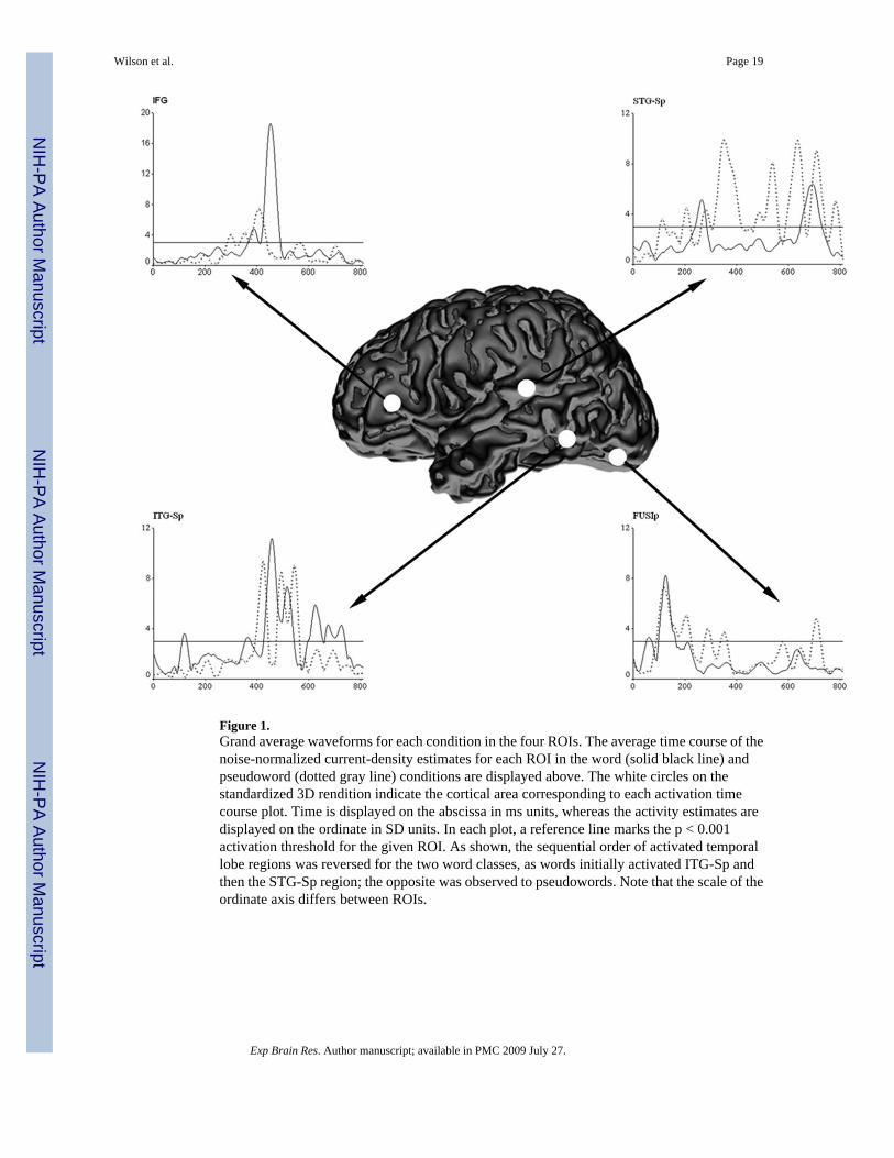
Figure 1.Grand average waveforms for each condition in the four ROIs. The average time course of thenoise-normalized current-density estimates for each ROI in the word (solid black line) andpseudoword (dotted gray line) conditions are displayed above. The white circles on thestandardized 3D rendition indicate the cortical area corresponding to each activation timecourse plot. Time is displayed on the abscissa in ms units, whereas the activity estimates aredisplayed on the ordinate in SD units. In each plot, a reference line marks the p < 0.001activation threshold for the given ROI. As shown, the sequential order of activated temporallobe regions was reversed for the two word classes, as words initially activated ITG-Sp andthen the STG-Sp region; the opposite was observed to pseudowords. Note that the scale of theordinate axis differs between ROIs.
Wilson et al. Page 19
Exp Brain Res. Author manuscript; available in PMC 2009 July 27.
NIH
-PA Author Manuscript
NIH
-PA Author Manuscript
NIH
-PA Author Manuscript
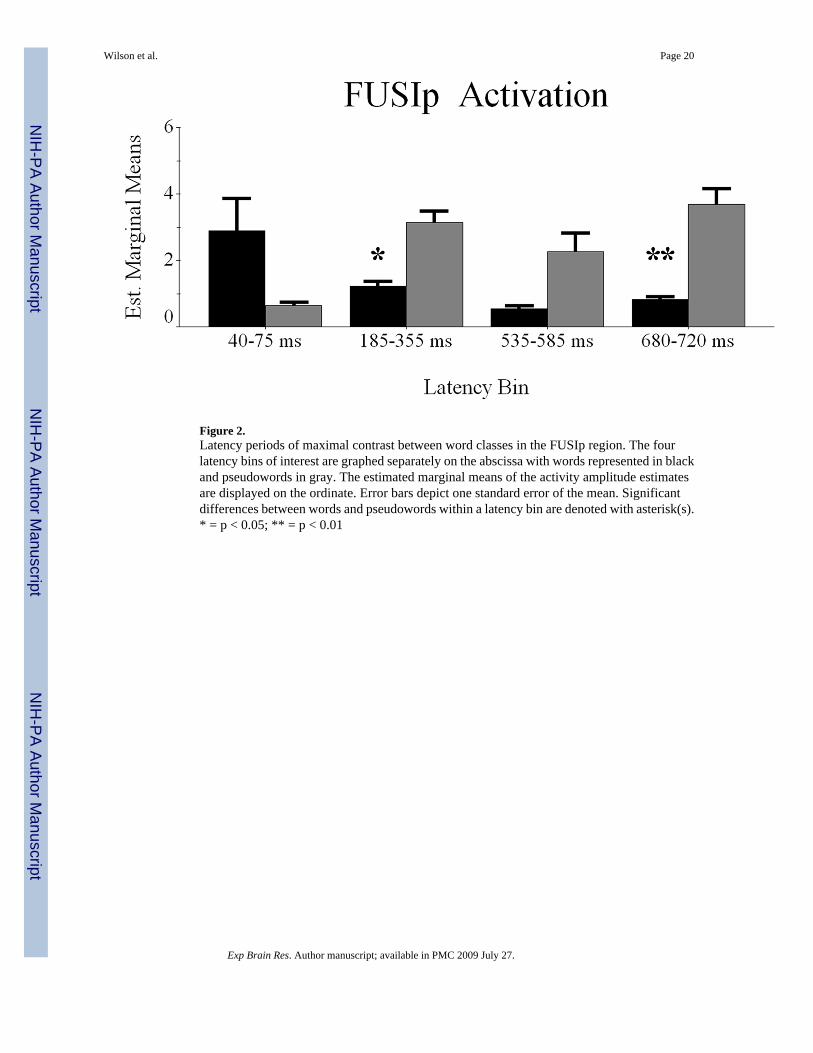
Figure 2.Latency periods of maximal contrast between word classes in the FUSIp region. The fourlatency bins of interest are graphed separately on the abscissa with words represented in blackand pseudowords in gray. The estimated marginal means of the activity amplitude estimatesare displayed on the ordinate. Error bars depict one standard error of the mean. Significantdifferences between words and pseudowords within a latency bin are denoted with asterisk(s).* = p < 0.05; ** = p < 0.01
Wilson et al. Page 20
Exp Brain Res. Author manuscript; available in PMC 2009 July 27.
NIH
-PA Author Manuscript
NIH
-PA Author Manuscript
NIH
-PA Author Manuscript
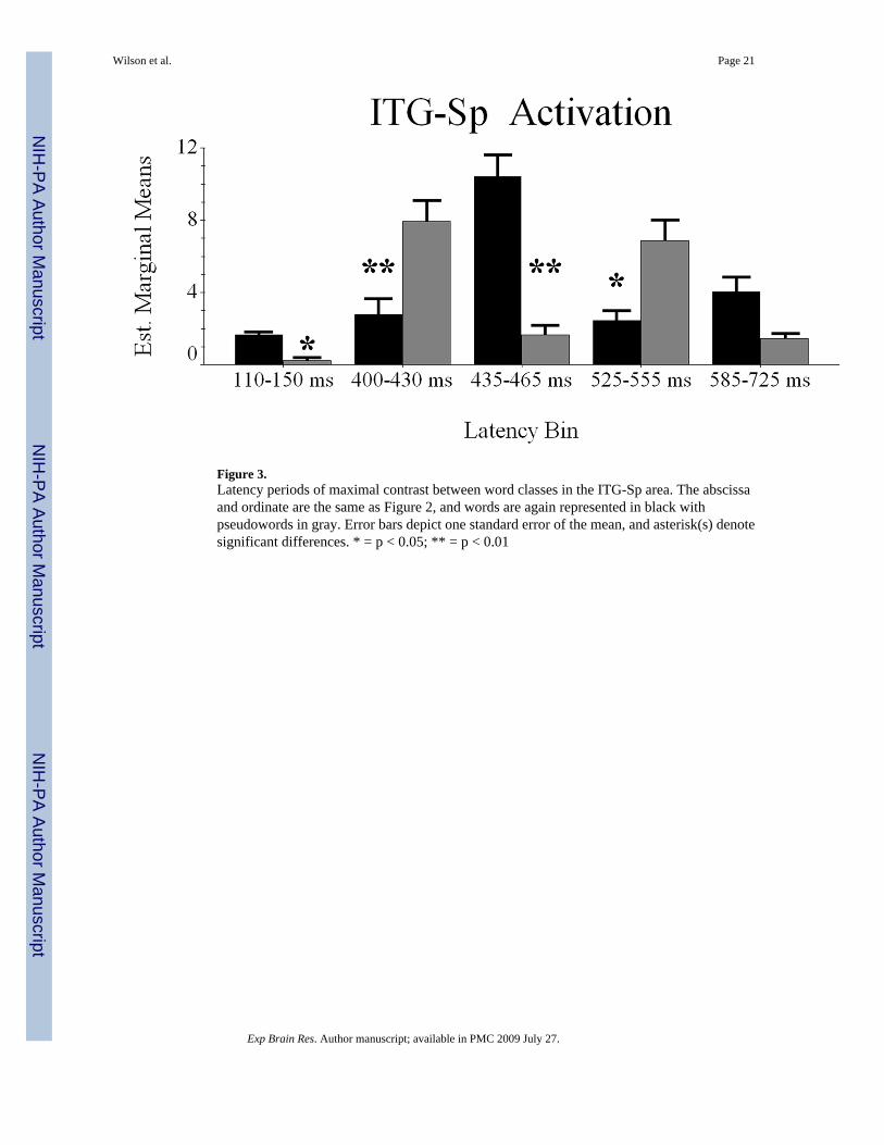
Figure 3.Latency periods of maximal contrast between word classes in the ITG-Sp area. The abscissaand ordinate are the same as Figure 2, and words are again represented in black withpseudowords in gray. Error bars depict one standard error of the mean, and asterisk(s) denotesignificant differences. * = p < 0.05; ** = p < 0.01
Wilson et al. Page 21
Exp Brain Res. Author manuscript; available in PMC 2009 July 27.
NIH
-PA Author Manuscript
NIH
-PA Author Manuscript
NIH
-PA Author Manuscript
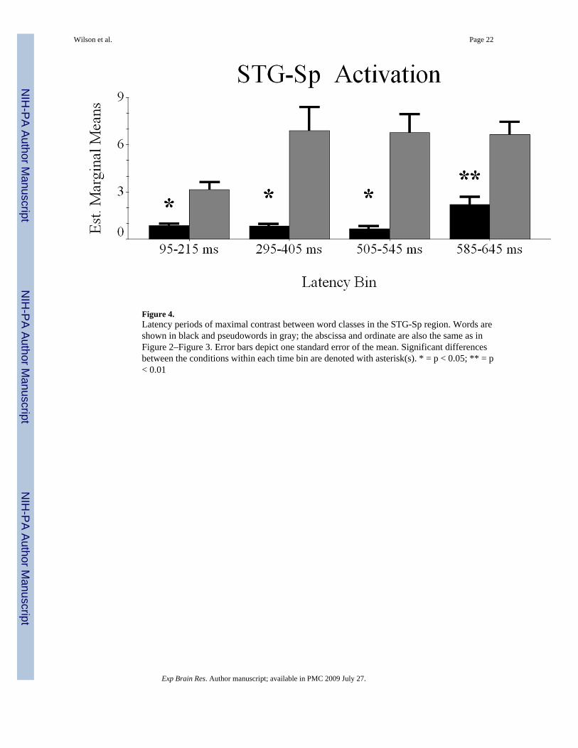
Figure 4.Latency periods of maximal contrast between word classes in the STG-Sp region. Words areshown in black and pseudowords in gray; the abscissa and ordinate are also the same as inFigure 2–Figure 3. Error bars depict one standard error of the mean. Significant differencesbetween the conditions within each time bin are denoted with asterisk(s). * = p < 0.05; ** = p< 0.01
Wilson et al. Page 22
Exp Brain Res. Author manuscript; available in PMC 2009 July 27.
NIH
-PA Author Manuscript
NIH
-PA Author Manuscript
NIH
-PA Author Manuscript

Figure 5.Latency periods of maximal contrast between word classes in IFG cortices. The abscissa andordinate are the same as Figure 2 – Figure 4, with words represented in black with pseudowordsin gray. The error bars depict one standard error of the mean, and asterisk(s) denote significantdifferences between words and pseudowords within each latency period. * = p < 0.05; ** = p< 0.01
Wilson et al. Page 23
Exp Brain Res. Author manuscript; available in PMC 2009 July 27.
NIH
-PA Author Manuscript
NIH
-PA Author Manuscript
NIH
-PA Author Manuscript