Reaction mechanism of HIV-1 protease by hybrid Car-Parrinello/classical MD simulations
11
Reaction Mechanism of HIV-1 Protease by Hybrid Car-Parrinello/Classical MD Simulations Stefano Piana, ² Denis Bucher, ‡ Paolo Carloni, § and Ursula Rothlisberger* ,‡ Scuola Internazionale Superiore di Studi AVanzati and INFM - DEMOCRITOS National Simulation Center, Via Beirut 2-4, 34014 Trieste, Italy, Laboratory of Computational Chemistry and Biochemistry, Federal Institute of Technology - EPFL, 1015 Lausanne, Switzerland, and Nanochemistry Research Institute, Curtin UniVersity of Technology, P.O. Box U 1987, Perth WA 6845, Australia ReceiVed: NoVember 30, 2003; In Final Form: March 21, 2004 We present a QM/MM ab initio molecular dynamics study of the peptide hydrolysis reaction catalyzed by HIV-1 protease. The QM/MM calculations are based on previous extensive classical MD simulations on the protein in complex with a model substrate (Piana, S.; Carloni, P.; Rothlisberger, U. Protein Sci. 2002, 11, 2393-2402). Gradient-corrected BLYP density functional theory (DFT) describes the reactive part of the active site, and the AMBER force field describes the rest of the protein, the solvent, and the counterions. An unbiased enhanced sampling of the QM/MM free-energy surface is performed to identify a plausible reaction coordinate for the second step of the reaction. The enzymatic reaction is characterized by two reaction free- energy barriers of ∼18 and ∼21 kcal mol -1 separated by a metastable gem-diol intermediate. In both steps, a proton transfer that involves the substrate and the two catalytic Asp molecules is observed. The orientation and the flexibility of the reactants, governed by the surrounding protein frame, are the key factors in determining the activation barrier. The calculated value for the barrier of the second step is slightly larger than the value expected from experimental data (∼16 kcal mol -1 ). An extensive comparison with calculations on gas-phase model systems at the Hartree-Fock, DFT-BP, DFT-BLYP, DFT-B3LYP, MP2, CCD, and QM/MM DFT- BLYP levels of theory suggests that the DFT-BLYP functional has the tendency to underestimate the energy of the gem-diol intermediate by ∼5-7 kcal mol -1 . The aspartyl protease from human immunodeficiency virus type 1 (HIV-1 PR) targets the AIDS epidemic. The enzyme is essential for viral metabolism 1 because it cleaves the long polypeptide chain that is expressed in infected host cells in specific positions to generate the active proteins that are required for viral maturation. HIV-1 PR is a homodimer with the active site located at the interface between the two subunits. The cleavage site is an Asp dyad (Asp25 and Asp25′, Figure 1), located inside a large active- site pocket that allows the enzyme to recognize and cleave sequences of six amino acids selectively. 2 Several aspects of the enzymatic reaction mechanism have been the focus of a variety of computational techniques, including molecular mechanics, 3-5 tight-binding, 6 semiempirical, 7,8 and ab initio 9-15 methods. This theoretical work has been complemented by kinetic, thermodynamic, and structural data. 8,16-29 The picture emerging from these studies can be summarized as follows. The free form of the enzyme (E) is stabilized by a low-barrier H bond (LBHB) 30 locking the Asp dyad in an almost coplanar conformation. In a first physical step, E binds to substrate SUB to form the enzyme-substrate complex ESUB, which might (ESUB(a), Figure 1) or might not (ESUB(b), Figure 1) maintain the LBHB. 2 H and 15 N kinetic isotope effect measurements 17,18,28 have established that in HIV-1 PR (i) a hydrated intermediate is reversibly formed and (ii) protonation of the peptide bond nitrogen occurs before an irreversible step. To form the hydrated intermediate, a water molecule (WAT) must attack the carbonyl carbon of SUB 15,17,18,21,31 (chemical step 1). This process can be assisted by the Asp dyad, which acts as a proton donor- acceptor group. 7,8,14,24,32 The possibility of formation of an oxyanion intermediate stabilized by tunneling 6,17,18,21 (as opposed to a gem-diol intermediate) has been proposed. 29 Quantum chemical calculations 7,8,14,15 suggest that the oxyanion might be unstable in the active site of HIV-1 PR. This is in contrast to other hydrolases, such as serine proteases, where an oxyanion cavity assists in the formation of the anion. Direct nucleophilic attack of the Asp dyad with the formation of a covalently bound intermediate has also been proposed 12,33 but is in contrast to isotope exchange experiments. 17,18 Also, the protonation of the nitrogen atom before the nucleophilic attack has been suggested. 9 In one or more subsequent steps, the C-N bond of INT breaks heterolytically (chemical step 2). Also, this process is assisted by the Asp dyad: Asp 25′ donates a proton to the INT amide group, and simultaneously 7,8,17 or subsequently 14,28 Asp 25 accepts a proton from one of the INT hydroxyl groups (TS2). This double proton transfer can be facilitated by an anti-gauche * To whom correspondence should be addressed. E-mail: ursula.roeth- [email protected]. Phone: +41-21-6930321. Fax: +41-21-6930320. ² Curtin University of Technology. ‡ Federal Institute of Technology - EPFL. § Scuola Internazionale Superiore di Studi Avanzati and INFM - DEMOCRITOS National Simulation Center. Figure 1. ESUB protonation patterns investigated. In ESUB(a), the low-barrier hydrogen bond between the two aspartic acids is still present after substrate binding. 11139 J. Phys. Chem. B 2004, 108, 11139-11149 10.1021/jp037651c CCC: $27.50 © 2004 American Chemical Society Published on Web 06/25/2004
-
Upload
independent -
Category
Documents
-
view
0 -
download
0
Transcript of Reaction mechanism of HIV-1 protease by hybrid Car-Parrinello/classical MD simulations
Reaction Mechanism of HIV-1 Protease by Hybrid Car-Parrinello/Classical MD Simulations
Stefano Piana,† Denis Bucher,‡ Paolo Carloni,§ and Ursula Rothlisberger*,‡
Scuola Internazionale Superiore di Studi AVanzati and INFM - DEMOCRITOS National Simulation Center,Via Beirut 2-4, 34014 Trieste, Italy, Laboratory of Computational Chemistry and Biochemistry, FederalInstitute of Technology - EPFL, 1015 Lausanne, Switzerland, and Nanochemistry Research Institute,Curtin UniVersity of Technology, P.O. Box U 1987, Perth WA 6845, Australia
ReceiVed: NoVember 30, 2003; In Final Form: March 21, 2004
We present a QM/MM ab initio molecular dynamics study of the peptide hydrolysis reaction catalyzed byHIV-1 protease. The QM/MM calculations are based on previous extensive classical MD simulations on theprotein in complex with a model substrate (Piana, S.; Carloni, P.; Rothlisberger, U.Protein Sci.2002, 11,2393-2402). Gradient-corrected BLYP density functional theory (DFT) describes the reactive part of theactive site, and the AMBER force field describes the rest of the protein, the solvent, and the counterions. Anunbiased enhanced sampling of the QM/MM free-energy surface is performed to identify a plausible reactioncoordinate for the second step of the reaction. The enzymatic reaction is characterized by two reaction free-energy barriers of∼18 and∼21 kcal mol-1 separated by a metastable gem-diol intermediate. In both steps,a proton transfer that involves the substrate and the two catalytic Asp molecules is observed. The orientationand the flexibility of the reactants, governed by the surrounding protein frame, are the key factors in determiningthe activation barrier. The calculated value for the barrier of the second step is slightly larger than the valueexpected from experimental data (∼16 kcal mol-1). An extensive comparison with calculations on gas-phasemodel systems at the Hartree-Fock, DFT-BP, DFT-BLYP, DFT-B3LYP, MP2, CCD, and QM/MM DFT-BLYP levels of theory suggests that the DFT-BLYP functional has the tendency to underestimate the energyof the gem-diol intermediate by∼5-7 kcal mol-1.
The aspartyl protease from human immunodeficiency virustype 1 (HIV-1 PR) targets the AIDS epidemic. The enzyme isessential for viral metabolism1 because it cleaves the longpolypeptide chain that is expressed in infected host cells inspecific positions to generate the active proteins that are requiredfor viral maturation.
HIV-1 PR is a homodimer with the active site located at theinterface between the two subunits. The cleavage site is an Aspdyad (Asp25 and Asp25′, Figure 1), located inside a large active-site pocket that allows the enzyme to recognize and cleavesequences of six amino acids selectively.2 Several aspects ofthe enzymatic reaction mechanism have been the focus of avariety of computational techniques, including molecularmechanics,3-5 tight-binding,6 semiempirical,7,8 and ab initio9-15
methods. This theoretical work has been complemented bykinetic, thermodynamic, and structural data.8,16-29 The pictureemerging from these studies can be summarized as follows. Thefree form of the enzyme (E) is stabilized by a low-barrier Hbond (LBHB)30 locking the Asp dyad in an almost coplanarconformation. In a first physical step, E binds to substrateSUBto form the enzyme-substrate complexESUB, which might(ESUB(a), Figure 1) or might not (ESUB(b), Figure 1) maintainthe LBHB.2H and15N kinetic isotope effect measurements17,18,28
have established that in HIV-1 PR (i) a hydrated intermediateis reversibly formed and (ii) protonation of the peptide bondnitrogen occurs before an irreversible step. To form the hydrated
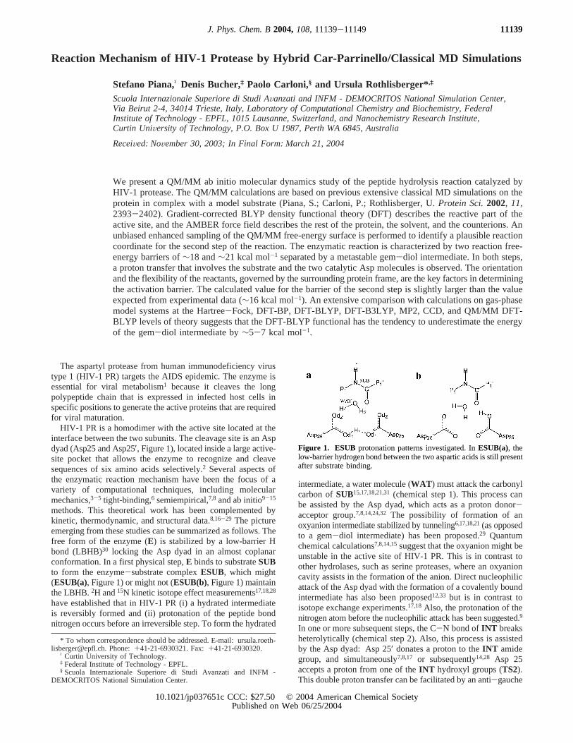
intermediate, a water molecule (WAT ) must attack the carbonylcarbon ofSUB15,17,18,21,31(chemical step 1). This process canbe assisted by the Asp dyad, which acts as a proton donor-acceptor group.7,8,14,24,32 The possibility of formation of anoxyanion intermediate stabilized by tunneling6,17,18,21(as opposedto a gem-diol intermediate) has been proposed.29 Quantumchemical calculations7,8,14,15suggest that the oxyanion might beunstable in the active site of HIV-1 PR. This is in contrast toother hydrolases, such as serine proteases, where an oxyanioncavity assists in the formation of the anion. Direct nucleophilicattack of the Asp dyad with the formation of a covalently boundintermediate has also been proposed12,33 but is in contrast toisotope exchange experiments.17,18Also, the protonation of thenitrogen atom before the nucleophilic attack has been suggested.9
In one or more subsequent steps, the C-N bond ofINT breaksheterolytically (chemical step 2). Also, this process is assistedby the Asp dyad: Asp 25′ donates a proton to theINT amidegroup, and simultaneously7,8,17 or subsequently14,28 Asp 25accepts a proton from one of theINT hydroxyl groups (TS2).This double proton transfer can be facilitated by an anti-gauche
* To whom correspondence should be addressed. E-mail: [email protected]. Phone:+41-21-6930321. Fax:+41-21-6930320.
† Curtin University of Technology.‡ Federal Institute of Technology - EPFL.§ Scuola Internazionale Superiore di Studi Avanzati and INFM -
DEMOCRITOS National Simulation Center.
Figure 1. ESUB protonation patterns investigated. InESUB(a), thelow-barrier hydrogen bond between the two aspartic acids is still presentafter substrate binding.
11139J. Phys. Chem. B2004,108,11139-11149
10.1021/jp037651c CCC: $27.50 © 2004 American Chemical SocietyPublished on Web 06/25/2004
transition in the gem-diol.8 TS2 decomposes (perhaps passingthrough a second intermediate14,28) to give the reaction products(EPROD), namely, an amine and a carboxylic acid. Finally,the products are released,29,34,35 and free formE is restored.The rate-determining step depends on the type of substrate, andit may be any of the chemical or physical steps mentionedabove.17,26,27,29,36
The present work will be aimed at characterizing the chemicalsteps that lead from the enzyme substrate complex (ESUB) tothe hydrated intermediate (EINT ) to the reaction products(EPROD). Previous studies on the conformation and reactivityof the active site of HIV-1 PR have suggested that (i) theflexibility of the protein frame is a fundamental ingredient forthe reaction;15,32 (ii) the groups interacting with the Asp dyad(in particular, the dipole moment of the Thr26(26′)-Gly27(27′)peptide bonds) are important for the conformation and stabilityof the active site8,10,11,30and therefore should be included inthe calculations in some form; and (iii) the use of an accuratefirst principles method and a relatively large basis set isrecommended to obtain reliable structural and energeticproperties.11-13 Here we take explicit account of the ingredients(i.e., i-iii). Toward this aim, we perform hybrid Car-Parrinello/molecular mechanics (QM/MM) simulations37 with the BLYPexchange-correlation functional,38 which has already been usedto describe the HIV-1 PR cleavage site,15,30,39and a plane wavebasis set (PW).40 In this approach, the flexibility of the proteinand the interaction of the active site with the rest of the proteinare fully taken into account. The calculations are based onextensive classical MD simulations on theESUB and EINTprotein complexes (Figure 2).32
The activation free energies of the reaction steps are calculatedby the thermodynamic integration of the force acting along aconstrained reaction coordinate.41 For the formation of thehydrated intermediate, this coordinate has been repeatedlyrecognized as the C(SUB)-O(WAT) distance.7,15 An appropriatechoice of constraint coordinate for chemical step 2 is lessobvious. During this step, one bond (N(INT)-H(Asp25′)) is formed,and two bonds (C(INT)-N(INT) and O(INT)-H(INT)) are broken,thus any of these distances or a combination of the three couldbe taken as a reaction coordinate. We therefore perform an initialunbiased enhanced sampling of the free-energy surface using
the CAFES formalism.42 This procedure suggests that theN(INT)-H(Asp25′) distance is a suitable constraint coordinate forchemical step 2.
The quality of the calculations is established by an extensivecomparison between the structural and energetic featuresobtained in our first principles calculations with those obtainedwith other ab initio methods.
Results and Discussion
Protonation State of ESUB.The starting structure forESUBwas taken from a classical MD simulation of the HIV-1PR/substrate complex.32 The structure obtained after 3.27 ns of MDsimulation was selected because, on the basis of previousinvestigations,15 it is expected to be a “reactive” conformation.The active-site form of HIV-1 PR that is catalytically competentis monoprotonated.17 The proton can be located either on Oδ1,with the two Asp groups connected by a low-barrier hydrogenbond (ESUB(a), Figure 1a) or on Oδ2 (in this case, no low-barrier hydrogen bond is present (ESUB(b), Figure 1b)). Thefree-energy difference between the two protonation patterns hasbeen calculated with the method of constraints. (See Methods.)It turns out thatESUB(a) is 1.5(1) kcal mol-1 more stable thanESUB(b). This small free-energy difference is in line withprevious ab initio studies on models of the active site of HIV-1PR,15,30 pepsin and endothiapepsin,11 and suggests that bothprotonation patterns are significantly populated inESUB.Interconversion between the two patterns occurs through aproton transfer from Asp25′ Oδ1 to WAT followed by a protontransfer from WAT to Asp25 Oδ2. The activation energy forthis process is 2.0(1) kcal mol-1.
QM/MM Simulation of the Formation of the HydratedReaction Intermediate.QM/MM simulations were performedstarting fromESUB(a)andESUB(b). In our simulation of theformation of the hydrated intermediate, the C(SUB)-O(WAT)
distance was constrained, and the constraint was shortened from3.7 to 1.5 Å in∼6 ps of QM/MM MD simulation. The reactionfree energy along the C(SUB)-O(WAT) reaction coordinate wascalculated as the integral of the average constraint force.41
In the simulation starting fromESUB(a)(Figure 3), the forceon the constraint increases as the water oxygen approaches thecarbonyl carbon. When the C(SUB)-O(WAT) bond is shorter than2.1 Å, the peptide bond loses its planarity, and at a C(SUB)-O(WAT) distance of 1.8 Å, a proton is shared between the watermolecule and Asp25′, forming an LBHB. At the same time,the formation of an oxyanion species (EINT(a) , Figure 3) isobserved with a negative accumulated charge on O(SUB). Thefree-energy difference betweenESUB(a) and EINT(a) is36(1.5) kcal mol-1 (Figure 4). In this simulation, the oxyanionis not stable toward the backward formation ofESUB(a), asindicated by the free-energy profile (Figure 4). Notice that inour simulations the proton dynamics is treated at the classicallevel, thus it is not possible to establish the proposed relevanceof tunneling for stabilizing an oxyanion reaction intermediate.29
A typical O‚‚‚H tunneling distance is∼1.6 Å. During the QM/MM MD simulation, this distance is never approached by anyproton located on the Asp dyad; however, this event might occuron longer time scales than these presently investigated.
In the simulation starting fromESUB(b) (Figure 3), after afew picoseconds a hydrogen bond is formed between Oδ2(Asp25)
and O(SUB). At a C(SUB)-O(WAT) distance of 1.88 Å, a concerteddouble proton transfer from Oδ2(Asp25)to O(SUB) and from O(WAT)
to Oδ2(Asp25′) is observed (TS1Figure 3). At the same time, theforce on the constraint becomes negative, indicating a transition-state crossing (Figure 4). Subsequently, the gem-diol interme-
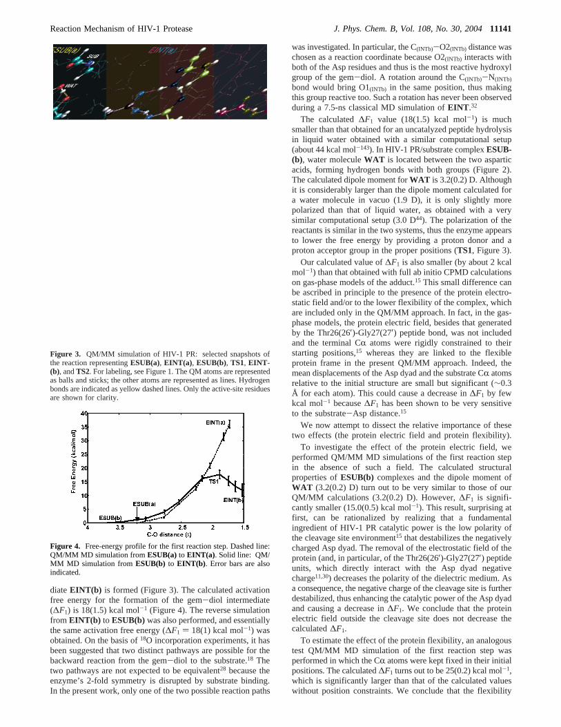
Figure 2. Structure of the HIV-1 PR/INT complex used in the QM/MM simulations. The protein is immersed in a 66.8× 55.2× 43.0 Å3
box containing 4170 water molecules. The QM atoms are representedin a ball-and-stick model. Hydrogen atoms are not shown for the sakeof clarity.
11140 J. Phys. Chem. B, Vol. 108, No. 30, 2004 Piana et al.
diateEINT(b) is formed (Figure 3). The calculated activationfree energy for the formation of the gem-diol intermediate(∆F1) is 18(1.5) kcal mol-1 (Figure 4). The reverse simulationfrom EINT(b) to ESUB(b)was also performed, and essentiallythe same activation free energy (∆F1 ) 18(1) kcal mol-1) wasobtained. On the basis of18O incorporation experiments, it hasbeen suggested that two distinct pathways are possible for thebackward reaction from the gem-diol to the substrate.18 Thetwo pathways are not expected to be equivalent28 because theenzyme’s 2-fold symmetry is disrupted by substrate binding.In the present work, only one of the two possible reaction paths
was investigated. In particular, the C(INTb)-O2(INTb) distance waschosen as a reaction coordinate because O2(INTb) interacts withboth of the Asp residues and thus is the most reactive hydroxylgroup of the gem-diol. A rotation around the C(INTb)-N(INTb)
bond would bring O1(INTb) in the same position, thus makingthis group reactive too. Such a rotation has never been observedduring a 7.5-ns classical MD simulation ofEINT .32
The calculated∆F1 value (18(1.5) kcal mol-1) is muchsmaller than that obtained for an uncatalyzed peptide hydrolysisin liquid water obtained with a similar computational setup(about 44 kcal mol-143). In HIV-1 PR/substrate complexESUB-(b), water moleculeWAT is located between the two asparticacids, forming hydrogen bonds with both groups (Figure 2).The calculated dipole moment forWAT is 3.2(0.2) D. Althoughit is considerably larger than the dipole moment calculated fora water molecule in vacuo (1.9 D), it is only slightly morepolarized than that of liquid water, as obtained with a verysimilar computational setup (3.0 D44). The polarization of thereactants is similar in the two systems, thus the enzyme appearsto lower the free energy by providing a proton donor and aproton acceptor group in the proper positions (TS1, Figure 3).
Our calculated value of∆F1 is also smaller (by about 2 kcalmol-1) than that obtained with full ab initio CPMD calculationson gas-phase models of the adduct.15 This small difference canbe ascribed in principle to the presence of the protein electro-static field and/or to the lower flexibility of the complex, whichare included only in the QM/MM approach. In fact, in the gas-phase models, the protein electric field, besides that generatedby the Thr26(26′)-Gly27(27′) peptide bond, was not includedand the terminal CR atoms were rigidly constrained to theirstarting positions,15 whereas they are linked to the flexibleprotein frame in the present QM/MM approach. Indeed, themean displacements of the Asp dyad and the substrate CR atomsrelative to the initial structure are small but significant (∼0.3Å for each atom). This could cause a decrease in∆F1 by fewkcal mol-1 because∆F1 has been shown to be very sensitiveto the substrate-Asp distance.15
We now attempt to dissect the relative importance of thesetwo effects (the protein electric field and protein flexibility).
To investigate the effect of the protein electric field, weperformed QM/MM MD simulations of the first reaction stepin the absence of such a field. The calculated structuralproperties ofESUB(b) complexes and the dipole moment ofWAT (3.2(0.2) D) turn out to be very similar to those of ourQM/MM calculations (3.2(0.2) D). However,∆F1 is signifi-cantly smaller (15.0(0.5) kcal mol-1). This result, surprising atfirst, can be rationalized by realizing that a fundamentalingredient of HIV-1 PR catalytic power is the low polarity ofthe cleavage site environment15 that destabilizes the negativelycharged Asp dyad. The removal of the electrostatic field of theprotein (and, in particular, of the Thr26(26′)-Gly27(27′) peptideunits, which directly interact with the Asp dyad negativecharge11,30) decreases the polarity of the dielectric medium. Asa consequence, the negative charge of the cleavage site is furtherdestabilized, thus enhancing the catalytic power of the Asp dyadand causing a decrease in∆F1. We conclude that the proteinelectric field outside the cleavage site does not decrease thecalculated∆F1.
To estimate the effect of the protein flexibility, an analogoustest QM/MM MD simulation of the first reaction step wasperformed in which the CR atoms were kept fixed in their initialpositions. The calculated∆F1 turns out to be 25(0.2) kcal mol-1,which is significantly larger than that of the calculated valueswithout position constraints. We conclude that the flexibility
Figure 3. QM/MM simulation of HIV-1 PR: selected snapshots ofthe reaction representingESUB(a), EINT(a) , ESUB(b), TS1, EINT-(b), andTS2. For labeling, see Figure 1. The QM atoms are representedas balls and sticks; the other atoms are represented as lines. Hydrogenbonds are indicated as yellow dashed lines. Only the active-site residuesare shown for clarity.
Figure 4. Free-energy profile for the first reaction step. Dashed line:QM/MM MD simulation fromESUB(a) to EINT(a) . Solid line: QM/MM MD simulation from ESUB(b) to EINT(b) . Error bars are alsoindicated.
Reaction Mechanism of HIV-1 Protease J. Phys. Chem. B, Vol. 108, No. 30, 200411141
of the protein frame is very important for determining thereaction free energy.
The calculated free energy for gem-diol intermediateEINT-(b) is 12(2) kcal mol-1 (Figure 3); a slightly larger value (13-(2) kcal mol-1) was obtained for the reverse simulation.
Influence of the Choice of QM and MM Regions.In a QM/MM calculation, it is important to select carefully the region ofthe system to be treated at the ab initio level. The residues thatwere treated at the QM level in the present calculation werethe two carboxylic acids (up to the Câ) and the gem-diol (upto the CR) (Figures 2 and 3). Previous ab initio calculations30
have shown that the interaction between the Asp dyad and theThr26(26′)-Gly27(27′) peptide bond dipole moment is importantin order to maintain Asp dyad coplanarity. Other studies havealso suggested that this interaction is important for the active-site energetics.8,10,11To investigate the influence of the level oftheory (QM or MM) in treating these moieties, we performedQM/MM calculations of the energy difference betweenESUB-(b) and EINT(b) in two identical systems characterized bydifferent quantum regions. In the first system (large QM), theThr26(26′)-Gly27(27′) peptide bond was included in the QMregion; in the second (small QM), it was not. A free-energycalculation with the method of constraints on the large QMsystem is computationally too demanding to be performed atthe present stage. For this reason, total energy differencesbetween geometry-optimized structures were calculated. Single-point calculations were also performed with the BP45,46and thePBE47 exchange correlation functionals using the geometriesoptimized at the BLYP level of theory. It turns out that thedifferences in relative energies between the large QM and smallQM systems are only 0.9 (QM/MM BLYP) and 0.1 (QM/MMBP and QM/MM PBE) kcal mol-1. Moreover, the rmsd,calculated for the QM atoms, between the two systems is lessthan 0.1 Å. These results indicate that the MM representationof the Thr26(26′)-Gly27(27′) peptide bonds is sufficient andthat their interaction with the Asp dyad is correctly describedby the QM/MM approach; for this reason, the Thr26(26′)-Gly27-(27′) peptide bonds were not treated at the QM level in thepresent work.
Reaction Coordinate Identification for the Decompositionof the Hydrated Intermediate. An appropriate approximatereaction coordinate for the decomposition of the hydratedintermediate has not been established yet. Therefore, a total of3 ps of QM/MM canonical adiabatic free-energy sampling(CAFES)42 was performed to identify a suitable approximatereaction coordinate. In this approach, a small group of “reactive”
atoms are heated to high temperature (i.e., 3000 K). These atomscross free-energy barriers more easily than the others, samplinga larger part of the free-energy surface in an unbiased way,whereas the rest of the system is kept at room temperature. Toavoid heat transfer between the two subsystems, they aredynamically decoupled by assigning to the reactive atomsmasses 100 times larger than those of the rest.
The QM/MM-CAFES calculations were performed on thecomplex between the protein andINT(b) (Figure 3), as obtainedfrom previous classical MD calculations.32 The reactive atomswere the oxygen and the polar hydrogen atoms of the Asp dyadand of the gem-diol as well as the gem-diol nitrogen atom(Figure 5c). In the QM/MM-CAFES simulation, two differentevents are observed: (i) Proton transfer from the gem-dioloxygen belonging toINT(b) (O2(INT)) to Asp25 (Figure 5a).This event occurs twice in the simulation and leads to theformation of a short-lived oxyanion species. However, it is notfollowed by reactive events leading to the formation of thereactants (i.e., the weakening of the C(INT)-O1(INT) bond andthe double proton-transfer event stabilizingTS1, Figure 3) orproducts, namely, the weakening of the C(INT)-N(INT) bond ofthe gem-diol. Thus, our QM/MM-CAFES calculation supportsthe proposal18 that the gem-diol is a stable reaction intermediaterather than a short-lived transient state. Furthermore, it indicatesthat, in the active site of HIV-1 PR, the gem-diol is more stablethan the oxyanion. (ii) Proton transfer from Asp25′ to the gem-diol intermediate nitrogen atom, followed by a reactive event(i.e., the reaction toward the products, namely, the weakeningand consequently the elongation of the C(INT)-N(INT) bond,Figure 5b). This proton transfer is not correlated with theweakening of other bonds involvingINT(b) such as the C(INT)-O1(INT) or C(INT)-O2(INT) bond (Figure 5a and b). Although inthe CAFES simulation the transition state for the decompositionof the hydrated intermediate is not reached, because nodecomposition is observed, the N(INT)-H(Asp25′) distance appearsto be a suitable independent reaction coordinate to approachTS2. This finding is also consistent with a previous ab initiostudy.14
Decomposition of Hydrated Intermediate EINT(b). Thesecond step investigated here is the decomposition of the gem-diol and the formation of the reaction products (fromEINT(b)to EPROD). Here, the N(INT)-H(Asp25′) distance was assumedto be the reaction coordinate based on the CAFES calculationsreported above. This distance was constrained and progressivelyshortened from 3.17 to 1.05 Å during 10 ps of QM/MMsimulation. It has been proposed that the gem-diol intermediate
Figure 5. CAFES QM/MM simulation of the HIV-1 PR/INT complex. (a, b) Selected distances (Å) are plotted as a function of simulated time(ps). (a) O2(INT)-H(INT)‚‚‚Oδ1(Asp25)hydrogen bond distance (red), O2(INT)-H(INT) bond distance (green), and C(INT)-O1(INT) bond distance (blue). (b)Oδ1-H(Asp25′)‚‚‚N(INT) hydrogen bond distance (red), Oδ1-H(Asp25′) bond distance (green), and C(INT)-N(INT) bond distance (blue). (c) Structure ofHIV-1 PR/INTb active site. The atoms held at 3000 K in the CAFES simulation are red.
11142 J. Phys. Chem. B, Vol. 108, No. 30, 2004 Piana et al.
is characterized by an anti and a gauche conformation of similarenergy and that the gauche conformation is the conformationrelevant to catalysis.8 Our results are consistent with thisproposal. In the classical MD simulation of the gem-diolintermediate,32 both conformations are populated by the anticonformation about four times more frequently than by thegauche form. In the early steps of the QM/MM MD simulation(d(N-H) ≈ 3-2.5 Å), the gem-diol is in an anti conformation,and H(Asp25′) forms a strong, short hydrogen bond with O2(INT)
(Figure 3). As the reaction proceeds, this hydrogen bond isbroken to form a weaker hydrogen bond between Asp25′ andthe gem-diol nitrogen N(INT). At the same time, an anti-gauchetransition is observed (d(N-H) ≈ 2.5-2 Å). The QM/MMcalculation indicates that the overall process requires 4 to 6 kcalmol-1 to occur. This value is consistent with the spontaneousformation of a short-lived N(INT)...H(Asp25′) hydrogen bond in an11-ns classical MD simulation ofEINT(b) .32
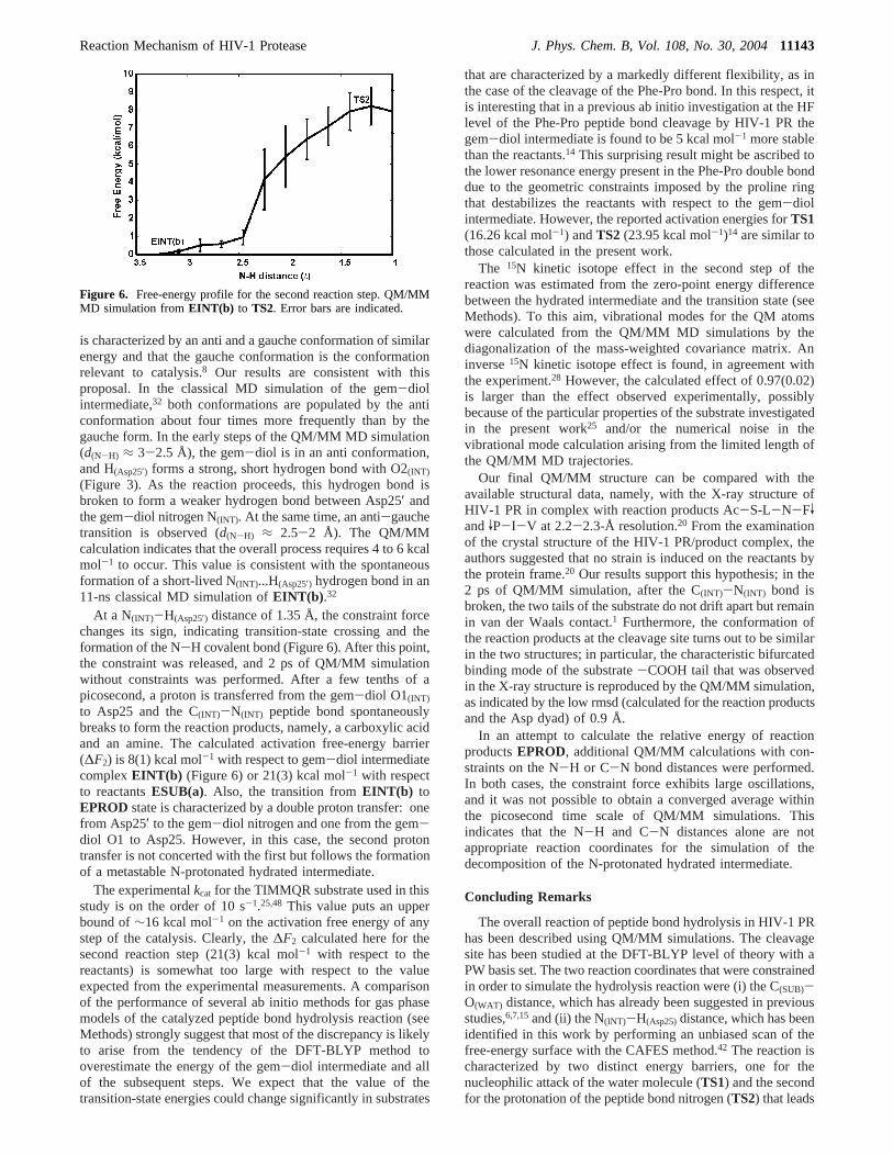
At a N(INT)-H(Asp25′) distance of 1.35 Å, the constraint forcechanges its sign, indicating transition-state crossing and theformation of the N-H covalent bond (Figure 6). After this point,the constraint was released, and 2 ps of QM/MM simulationwithout constraints was performed. After a few tenths of apicosecond, a proton is transferred from the gem-diol O1(INT)
to Asp25 and the C(INT)-N(INT) peptide bond spontaneouslybreaks to form the reaction products, namely, a carboxylic acidand an amine. The calculated activation free-energy barrier(∆F2) is 8(1) kcal mol-1 with respect to gem-diol intermediatecomplexEINT(b) (Figure 6) or 21(3) kcal mol-1 with respectto reactantsESUB(a). Also, the transition fromEINT(b) toEPROD state is characterized by a double proton transfer: onefrom Asp25′ to the gem-diol nitrogen and one from the gem-diol O1 to Asp25. However, in this case, the second protontransfer is not concerted with the first but follows the formationof a metastable N-protonated hydrated intermediate.
The experimentalkcat for the TIMMQR substrate used in thisstudy is on the order of 10 s-1.25,48 This value puts an upperbound of∼16 kcal mol-1 on the activation free energy of anystep of the catalysis. Clearly, the∆F2 calculated here for thesecond reaction step (21(3) kcal mol-1 with respect to thereactants) is somewhat too large with respect to the valueexpected from the experimental measurements. A comparisonof the performance of several ab initio methods for gas phasemodels of the catalyzed peptide bond hydrolysis reaction (seeMethods) strongly suggest that most of the discrepancy is likelyto arise from the tendency of the DFT-BLYP method tooverestimate the energy of the gem-diol intermediate and allof the subsequent steps. We expect that the value of thetransition-state energies could change significantly in substrates
that are characterized by a markedly different flexibility, as inthe case of the cleavage of the Phe-Pro bond. In this respect, itis interesting that in a previous ab initio investigation at the HFlevel of the Phe-Pro peptide bond cleavage by HIV-1 PR thegem-diol intermediate is found to be 5 kcal mol-1 more stablethan the reactants.14 This surprising result might be ascribed tothe lower resonance energy present in the Phe-Pro double bonddue to the geometric constraints imposed by the proline ringthat destabilizes the reactants with respect to the gem-diolintermediate. However, the reported activation energies forTS1(16.26 kcal mol-1) andTS2 (23.95 kcal mol-1)14 are similar tothose calculated in the present work.
The 15N kinetic isotope effect in the second step of thereaction was estimated from the zero-point energy differencebetween the hydrated intermediate and the transition state (seeMethods). To this aim, vibrational modes for the QM atomswere calculated from the QM/MM MD simulations by thediagonalization of the mass-weighted covariance matrix. Aninverse15N kinetic isotope effect is found, in agreement withthe experiment.28 However, the calculated effect of 0.97(0.02)is larger than the effect observed experimentally, possiblybecause of the particular properties of the substrate investigatedin the present work25 and/or the numerical noise in thevibrational mode calculation arising from the limited length ofthe QM/MM MD trajectories.
Our final QM/MM structure can be compared with theavailable structural data, namely, with the X-ray structure ofHIV-1 PR in complex with reaction products Ac-S-L-N-FVandVP-I-V at 2.2-2.3-Å resolution.20 From the examinationof the crystal structure of the HIV-1 PR/product complex, theauthors suggested that no strain is induced on the reactants bythe protein frame.20 Our results support this hypothesis; in the2 ps of QM/MM simulation, after the C(INT)-N(INT) bond isbroken, the two tails of the substrate do not drift apart but remainin van der Waals contact.1 Furthermore, the conformation ofthe reaction products at the cleavage site turns out to be similarin the two structures; in particular, the characteristic bifurcatedbinding mode of the substrate-COOH tail that was observedin the X-ray structure is reproduced by the QM/MM simulation,as indicated by the low rmsd (calculated for the reaction productsand the Asp dyad) of 0.9 Å.
In an attempt to calculate the relative energy of reactionproductsEPROD, additional QM/MM calculations with con-straints on the N-H or C-N bond distances were performed.In both cases, the constraint force exhibits large oscillations,and it was not possible to obtain a converged average withinthe picosecond time scale of QM/MM simulations. Thisindicates that the N-H and C-N distances alone are notappropriate reaction coordinates for the simulation of thedecomposition of the N-protonated hydrated intermediate.
Concluding Remarks
The overall reaction of peptide bond hydrolysis in HIV-1 PRhas been described using QM/MM simulations. The cleavagesite has been studied at the DFT-BLYP level of theory with aPW basis set. The two reaction coordinates that were constrainedin order to simulate the hydrolysis reaction were (i) the C(SUB)-O(WAT) distance, which has already been suggested in previousstudies,6,7,15and (ii) the N(INT)-H(Asp25)distance, which has beenidentified in this work by performing an unbiased scan of thefree-energy surface with the CAFES method.42 The reaction ischaracterized by two distinct energy barriers, one for thenucleophilic attack of the water molecule (TS1) and the secondfor the protonation of the peptide bond nitrogen (TS2) that leads
Figure 6. Free-energy profile for the second reaction step. QM/MMMD simulation fromEINT(b) to TS2. Error bars are indicated.
Reaction Mechanism of HIV-1 Protease J. Phys. Chem. B, Vol. 108, No. 30, 200411143
to the decomposition of a hydrated amide intermediate. Thecalculated reaction barriers are 18(1) and 21(3) kcal mol-1,respectively.
A comparison of our QM/MM results with previous studiesand with the analogous QM/MM simulations in which theprotein electric field is switched off indicates that, at least forthe first reaction step, the electrostatic field generated by theresidues surrounding the cleavage site plays a fundamental rolein stabilizing the Asp dyad. However, the net effect of this fieldon the reaction free energy is an increase of 3 kcal mol-1.Moreover, QM/MM calculations, in which constraints areapplied to the Asp dyad and the substrate CR atoms, establishthat the local flexibility of the cleavage site is a key factor forthe energetics of the reaction barrier, consistent with ourprevious proposals.15 This finding highlights the importance ofa computational method that explicitly takes into account theconstraints and flexibility imposed by the protein frame in thestudies of the reaction mechanism of HIV-1 PR.
The accuracy of the method used here has been investigatedby comparison to a variety of ab initio (HF, DFT, MP2, andCCD, Tables 1 and 3) methods and the QM/MM approach ongas-phase models1 and2 (Figures 8 and 9). The conclusionsare that (i) the convergence with respect to the basis set sizewithin 1 kcal mol-1 can be obtained using a PW basis set witha cutoff of 70 Ry or more and (ii) the DFT methods tested(BLYP, B3LYP, BP86) and the QM/MM method predictreasonable transition-state energies (within 2 kcal mol-1),whereas they severely underestimate the stability of the gem-diol intermediate relative to MP2 and CCD calculations.
Methods
Structural Models. The structural model of HIV-1 PRcomplexed with the substrate and the gem-diol reactionintermediate (ESUB andEINT , SUB) Thr-Ile-Met-Met-Gln-Arg) were built from the X-ray structure of HIV-1 PRcomplexed with MVT10118,25 (4HVP49). Protein residues be-longing to subunit 1 were numbered from 1 to 99, and thosebelonging to subunit 2, from 1′ to 99′. Substrate residues bindingto subunit 1 (2) were numbered from P1 (P1′) to P3 (P3′):50
Thr (P3)-Ile (P2)-Met (P1)-Met (P1′)-Gln (P2′)-Arg (P3′). Thepeptide bond to be cleaved by the enzyme belongs to residuesP1 and P1′ (Met-Met). In the simulation of the reactionintermediate, the carbonyl group of Met P1′ was substituted bya gem-diol. The total system was composed of 15 749 atoms(Figure 2).
Starting structures were taken from 7.5-ns classical moleculardynamics (MD) ofESUBandEINT .32 ForESUB, the structureobtained after 3270 ps of classical MD simulation was chosenbecause it is expected to be the most reactive conformationsampled.15 For EINT , the structure obtained after 478 ps ofclassical MD simulation was chosen as a starting structurebecause it closely resembled the average MD structure (CRatoms’ rmsd) 1.0 Å).
TABLE 1: Energetics of Gas-Phase Model 1a
6-31G6-31G
(d)6-31G(d,p)
6-31+G(df,2p)
6-31++G(3df,3p)
HF 16.3 18.6 18.7 20.1 19.3BP86 20.3 21.6 19.9 21.2 20.7BLYP 22.6 23.1 22.9 24.6 24.8BLYP (PW) 23.2§50 26.4§60 26.4§70 25.9§110 25.8§150
B3LYP 19.2 19.9 19.8 21.3 21.5MP2 17.7 16.8 17.1 17.1b
MP4 14.5CCD 14.4
a Energy differences (kcal mol-1) between 1b and 1a for model 1(∆E in the text) calculated with a variety of ab initio methods andwith different basis sets (see Methods). All structures were fullygeometry optimized.b This number has been obtained with the6-31+G(2df,2p) basis set. For BLYP(PW) calculations, DFT energieswere calculated with the program CPMD,54 the BLYP exchange-correlation functional, and a plane wave basis set. The size of the basisset is determined by the energy cutoff:§50 cutoff 50 Ry;§60 cutoff 60Ry; §70 cutoff 70 Ry;§110 cutoff 110 Ry;§150 cutoff 150 Ry. BLYP andB3LYP DFT energies were calculated with Gaussian 9864 and the BLYPand B3LYP exchange correlation functionals; MP2 post-Hartree-Fockenergies were calculated with Gaussian 9864 and Møller-Plesset second-order perturbation theory. MP4 post-Hartree-Fock energies werecalculated with Gaussian 9864 and Møller-Plesset fourth-order per-turbation theory. CCD post-Hartree-Fock energies were calculated withGaussian 9864 and the coupled-cluster doubles method.
Figure 7. Chemical structure of model1. (a) Peptide bond with ahydrogen-bonded water molecule. (b) gem-diol peptide bond hydrate.
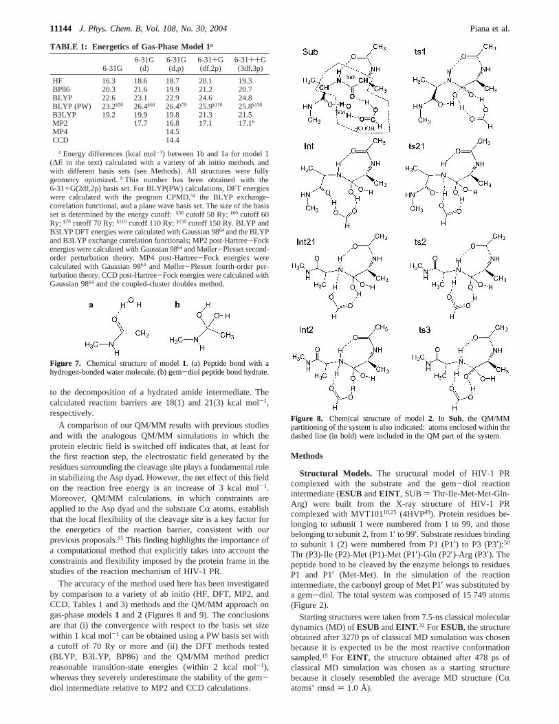
Figure 8. Chemical structure of model2. In Sub, the QM/MMpartitioning of the system is also indicated: atoms enclosed within thedashed line (in bold) were included in the QM part of the system.
11144 J. Phys. Chem. B, Vol. 108, No. 30, 2004 Piana et al.

Complexes1 and2 were built starting from the structure ofEINT . In complex1, only the atoms belonging to the P1-P1′gem-diol moiety were retained (Figure 7). In complex2, P1and P1′ were included up to the Câ, and the dangling peptidebonds were terminated with acetyl andN-methyl on the C andN termini, respectively. The carboxyl group of Asp25 was alsoincluded. All of the remaining dangling bonds were capped withhydrogen atoms. The final system was composed of 40 atoms(Figure 8).
QM/MM Calculations. These calculations were carried outwith the QM/MM scheme developed in ref 37.
The quantum part included the side chains of Asp 25 andAsp 25′, the hydrated peptide bond, and a water molecule(Figure 3). Dangling bonds in the quantum part were terminatedwith hydrogen atoms. The small unbalanced charge (0.1 e)resulting from the choice of the QM/MM partitioning wasredistributed over the 500 MM atoms located within 7.5 Å ofthe Asp dyad.
The quantum problem was solved using density functionaltheory (DFT). Exchange and correlation functionals were thoseof Becke38 and Lee, Yang, and Parr40 (BLYP), respectively.The Kohn-Sham orbitals were expanded in plane waves up to70 Ry. Martins-Troullier51 pseudopotentials were used todescribe the interactions between the ionic cores and the valenceelectrons. A 14.0× 14.0 × 10.5 Å3 quantum cell was used.The systems were treated as if they were isolated, as in Barnettand Landman.52 DFT-based MD simulations were performedaccording to the Car-Parrinello approach53 using the CPMDprogram.54 A time step of 5 au and a fictitious electron mass of600 au were used. Constant temperature was achieved bycoupling the systems to a Nose´-Hoover thermostat55 of 500-cm-1 frequency. The GROMOS9656 program combined withthe Amber94 force field57 was used to treat the classical system.A cutoff of 12.0 Å was used for nonbonded interactions, andthe P3M method58 was used to describe long-range electrostatics.
QM/MM geometry optimizations were performed on largeQM and small QM systems with a conjugate gradient algorithmup to a convergence of 5× 10⁻³ hartree Å-1 on the forces and2 × 10-6 hartree step-1 on the energies. In the small QM
systems, the Asp dyad and the hydrated peptide bond weretreated at the QM level. In the large QM systems, the Thr26-(26′)-Gly27(27′) peptide bond was also treated at the QM level.During the geometry optimizations, only the Asp dyad, thehydrated peptide bond, and the Thr26(26′)-Gly27(27′) peptidebonds were allowed to move.
Free energies were calculated with the method of con-straints.41,59-61 The activation free energies were calculated asan integral of the average forcefs acting on the constraint alongthe reaction coordinateQ.62
The selected constraint does not necessarily have to cor-respond to the entire reaction coordinate. However, it turns outthat the time required to obtain a converged MD average of theforce for each constraint point depends critically on how closethe chosen constraint matches the slowest part of the reactioncoordinate. For this reason, it is important to evaluate whichgeometric parameter most closely matches the reaction coor-dinate for each step of the reaction. To calculate the structureand relative free energy ofESUB(a)andESUB(b), theESUB-(a) complex was first equilibrated with 0.5 ps of QM/MM MD.Subsequently, the Oδ2(Asp25)-H1(WAT) distance was constrainedand reduced from 3.7 to 1.07 Å in 4 ps of constrained QM/MM MD simulation. The obtainedESUB(a) and ESUB(b)structures were used for the simulation of the first reaction step,namely, the formation of the hydrated intermediate.
The first step involves the nucleophilic attack of a watermolecule on the carbonyl carbon of the peptide bond of thesubstrate. Previous studies have established thatêCO ) d(C-(SUB)-O(WAT)) is an appropriate reaction coordinate for describ-ing this part of the reaction.6,7,15
For chemical step 2, several reaction coordinates are possible.For this reason, 3.0 ps of QM/MM-CAFES42 simulation wereperformed to obtain qualitative information about the possiblereaction coordinates. In the CAFES simulation, the temperatureof the QM oxygen and nitrogen atoms and polar protons (Figure3c) was set to 3000 K via coupling to a Nose´-Hooverthermostat55 with T ) 3000 K and a coupling frequency of 500cm-1. The masses of these atoms were increased by a factor of100 to obtain decoupling between the “hot” and “cold” partsof the system. As a result of these calculations, the N(INT)-H(Asp25) distance (êNH) turned out to be the most plausiblereaction coordinate.
Each point alongêCO and êNH was sampled until the MD-averaged forces calculated from the first and second parts ofthe trajectory differed by less than 10%. About 1.2 ps of abinitio MD was required for most of the points. About 2.0 pswas required for the points closer to the transition states. Theinitial 0.3 ps was always discarded. Overall,∼15 and 10 pswere sampled for chemical step 1 and chemical step 2,respectively.
A reverse simulation of chemical step 1 was also performedstarting fromEINT(b) ; this control simulation gave essentiallythe same results in terms of transition-state structure and energyas the direct reaction. Two additional QM/MM MD simulationsof chemical step 1 were also performed with the samecomputational scheme outlined above but without electrostaticinteractions between the classical and the quantum system orwith position constraints on the Asp dyad and gem-diol CRatoms. The error in the calculated free energy is reported inparentheses in the text and was estimated from the differencebetween the values calculated for the first and second halves ofthe simulation.
The dipole moment of the catalytic water molecule in theactive site of HIV-1 PR was calculated as an average over 40
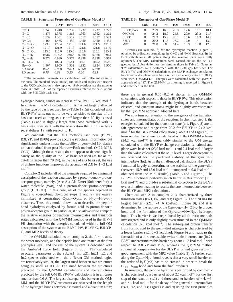
Figure 9. Energetics of gas-phase model2. Labeling is the same asin Figure 8 and Tables 1 and 3.
Reaction Mechanism of HIV-1 Protease J. Phys. Chem. B, Vol. 108, No. 30, 200411145

structures selected at regular intervals from the simulation ofthe ESUB(b) complex (Figure 3).
Kinetic isotope effects (KIE) were calculated as in ref 63
where MMI is the contribution due to the change in the momentsof inertia
ZPE is the zero-point energy contribution63
and EXC is the excited-state contribution
whereN is the number of atoms in the system.14M is the massof the 14N system, and14Ix is the moment of inertia of the14Nsystem with respect to thex axis. The number of vibrationalmodes is 3N - 6 for the reactants and 3N - 7 for the TS as, inthis approach, one degree of freedom is constrained.14ui is theith vibrational mode for the14N system, and15ui is the ithvibrational mode for the15N system:
νi is the frequency of theith vibrational mode. Vibrational modeswere calculated for the QM atoms only from diagonalizationof the mass-weighted covariance matrix of the QM atoms:
M is the diagonal matrix of atomic masses. A comparisonbetween formamide and protonated formamide ZPEs calculatedwith this method and with Gaussian 9864 indicates that about0.5 ps of ab initio MD trajectory is required to obtain ZPEsconverged to within 10%.
Gas-Phase Calculations.All of the calculations with local-ized basis sets were carried out with the Gaussian 9864 programon two models,1 and2 (Figures 7 and 8).
1 was energy minimized until the largest component of theforce was smaller than 2.5 10-3 a.u. and the largest displacementwas smaller than 1.0 10-2 Å. Basis sets of increasing size,ranging from 6-31G to 6-31++G(3df,3p), were used. DFTcalculations were performed with the BLYP, BP86, and B3LYPexchange-correlation functionals. Post-Hartree-Fock calcula-
tions were performed according to Møller-Plesset second andfourth order perturbation theory (MP2 and MP4) and with thecoupled-cluster doubles method (CCD).
For model2, the reaction profiles of chemical steps 1 and 2were investigated by a scan along the C(Sub)-O(Wat) and N(Int)-H(HCOOH) distances, respectively. The C(Sub)-O(Wat) and N(Int)-H(HCOOH) distances were scanned at the B3LYP-6-31G(d) levelof theory. The 6-31G(d) basis set was used because it strikes areasonable compromise between accuracy and computationalcost (Table 1). A control calculation was also performed withB3LYP and the 6-31G(d,p) basis set and gave essentially thesame results in terms of structure and energetics.
The C(Sub)-O(Wat) distance was constrained, and the constraintvalue decreased from 3.61 to 1.21 Å in 22 steps; the N(INT)-H(HCOOH) distance was constrained, and the constraint decreasedfrom 4.77 to 0.87 Å in 33 steps. In each step, the structure wasenergy minimized until the largest component of the force wassmaller than 2.5× 10⁻³ au and the largest displacement wassmaller than 1.0× 10⁻² Å.
The intermediates or transition-state structures were alsoenergy minimized at the BLYP-6-31G(d) and BLYP-PW levelsof theory with a 70-Ry cutoff, as well as with the QM/MMscheme outlined in the previous section. In the QM/MMcalculations, the QM part of the system included the watermolecule, the formic acid, and the Ala-Ala peptide bondrepresented asN-methyl amide (Figure 6). Dangling bonds wereterminated with hydrogen atoms. Classical atoms were describedwith the Amber9457 force field. Finally, single-point calculationswere also performed with Møller-Plesset second-order pertur-bation theory (MP2) using the B3LYP geometries.
Accuracy of the Methodology.The accuracy of the methodused here (QM/MM DFT-BLYP using a plane wave (PW) basisset expanded up to 70 Ry) is established by performing firstprinciples (HF, MP2, DFT-BP86, DFT-B3LYP, DFT-BLYP)and QM/MM calculations using both PW and Gaussian (G) basissets. The calculations are carried out on two model systemsrepresenting some of the groups involved in the enzymaticreaction (Figures 7 and 8).
Complex1 consists of a water molecule and eitherN-methylacetamide (1a) or the corresponding gem diol hydrate (1b,Figure 7). The energy difference between the two systems (∆Ehereafter) is expected to be very sensitive to the type of approachused to calculate the exchange and correlation contributionsbecause the two systems differ in the number of hydrogen bonds(Figure 7) and in the type of chemical bonds, namely, theN-methyl acetamide peptide bond is turned into twoσ bondsin the intermediate. We exhaustively address this issue by energyminimizing 1 with different methods (BLYP, BP86, B3LYP,HF, MP2, MP4, and CCD) and basis sets (6-31G, 6-31G(d),6-31G(d,p), 6-31G+(df,2p), and 6-31G++(3df,2p); see Table1).
All of the methods employed turn out to predict similargeometries (Table 2). B3LYP is the DFT method that providesthe most similar structural properties to those obtained with theCCD method, which is taken here as a reference (Table 2); inparticular, this method accurately reproduces the hydrogen bonddistance (Table 2). In contrast,∆E depends largely on the levelof theory. It is underestimated by all DFT methods by 5-7 kcalmol-1 both using G and PW basis sets. This large deviationindicates that DFT significantly overestimates the stability of aresonant double bond (the peptide bond) plus a hydrogen bondwith respect to two single bonds. Indeed, the inclusion of diffusefunctions (basis sets 6-31G+(df,2p) and 6-31G++(3df,3p)),which is expected to improve the description of the peptide and
KIE ) MMI ‚ ZPE‚ EXC
MMI )((14M
15M)3/214Ix14Iy
14Iz
15Ix15Iy
15Iz)
TS
((14M15M)3/214Ix
14Iy14Iz
15Ix15Iy
15Iz)
reactant
ZPE)
[ ∏i
3N- 1
e-(14ui - 15ui)/2]TS
[ ∏i
3N - 6
e-(14ui - 15ui)/2]reactant
EXC )[ ∏
i
3N- 11 - e-(15ui)
1 - e-(14ui)]TS
[ ∏i
3N - 61 - e-(15ui)
1 - e-(14ui)]reactant
ui )hνi
kBT
Cij ) ⟨Mii1/2(xi - ⟨xi⟩)Mjj
1/2(xj - ⟨xj⟩)⟩
11146 J. Phys. Chem. B, Vol. 108, No. 30, 2004 Piana et al.
hydrogen bonds, causes an increase of∆E by 1-2 kcal mol-1.In contrast, the MP2 calculation of∆E is not largely affectedby the type of basis set used here (Table 1).∆E calculated withBLYP-PW turns out not to be very sensitive to the size of thebasis set used as long as a cutoff larger than 60 Ry is used(Table 1) and is slightly larger than those calculated with Gbasis sets, consistent with the observation that a diffuse basisset stabilizes1a with respect to1b.
We conclude that the DFT methods used here (BLYP,B3LYP, and BP86) provide accurate structural information butsignificantly underestimate the stability of gem-diol 1b relativeto that obtained from post-Hartree-Fock methods (MP2, MP4,CCD). Although these results do not appear to depend signifi-cantly on the quality of the PW basis set used (as far as thecutoff is larger than 70 Ry), in the case of a G basis set, the useof diffuse functions improves the accuracy of∆E by 1-2 kcalmol-1.
Complex2 includes all of the elements required for a minimaldescription of the reaction catalyzed by a proton-donor-proton-acceptor group, namely, (Ace-Ala-Ala-Nme) (Sub hereafter), awater molecule (Wat), and a proton-donor-proton-acceptorgroup (HCOOH). In this case, all of the species depicted inFigure 6 (describing chemical steps 1 and 2) are energyminimized at constrained C(Sub)-O(Wat) or N(Int)-H(HCOOH)
distances. Thus, this model allows us to describe the peptidebond hydrolysis catalyzed by formic acid as proton-donor-proton-acceptor group. In particular, it also allows us to comparethe relative energies of reaction intermediates and transitionstates calculated with the QM/MM method used in the HIV-1PR simulation with the energies calculated within a full QMdescription of the system at the BLYP-PW, BLYP-G, B3LYP-G, and MP2 levels of theory.
In the QM/MM calculations on complex2, the formic acid,the water molecule, and the peptide bond are treated at the firstprinciples level, and the rest of the system is described withthe Amber94 force field (Figure 8, Sub). The calculatedstructural parameters of the Sub, ts1, Int, ts21, Int21, ts2, andInt2 species calculated with the different QM methodologiesare remarkably similar, the largest rmsd between two structuresbeing as small as 0.1 Å. The rmsd between the structurespredicted by the QM/MM calculations and the structurespredicted by the full QM BLYP-PW calculations is in all casessmaller than 0.6 Å. The largest discrepancies between the QM/MM and the BLYP-PW structures are observed in the lengthof the hydrogen bonds between a classical and a quantum atom;
these are in general 0.05-0.2 Å shorter in the QM/MMcalculations with respect to those in BLYP-PW. This observationindicates that the strength of the hydrogen bonds betweenclassical and quantum atoms might be slightly overestimatedby the QM/MM approach adopted here.
We now turn our attention to the energetics of the transitionstates and intermediates of the reaction. In chemical step 1, theenergies calculated for the transition state (ts1, Figure 8) are infair agreement and range from 20.2 for B3LYP to 24.2 kcalmol-1 for the BLYP/MM calculation (Table 3 and Figure 9). Itturns out that the ts1 energy calculated with the QM/MM scheme(24.2 kcal mol-1) is remarkably similar to the QM energycalculated with the BLYP exchange-correlation functional andplane wave basis set (23.9 kcal mol-1) and 2.4 kcal mol-1 largerthan the value calculated at the MP2 level. Larger discrepanciesare observed for the predicted stability of the gem-diolintermediate (Int). As in the small-model calculations, the BLYPfunctional largely underestimates the stability of the gem-diol(between 15.9 and 18.0 kcal mol-1 compared to 9.8 kcal mol-1
obtained from the MP2 results) (Table 3 and Figure 9). TheB3LYP functional performs much better in this respect (11.5kcal mol-1) and provides a substantial correction to the BLYPoverestimation, leading to results that are intermediate betweenthe BLYP and MP2 calculations.
Chemical step 2 in complex2 is characterized by threetransition states (ts21, ts2, and ts3; Figure 6). The first has thelargest barrier (ts21,∼4-6 kcal/mol; Figure 9), and it isdetermined by the rupture of the O(HCOOH)-H‚‚‚O1(Int) hydrogenbond and the formation of the O(HCOOH)-H‚‚‚N(Int) hydrogenbond. This barrier is well reproduced by all ab initio methodsinvestigated and is only slightly overestimated in the QM/MMcalculation (6.8 kcal mol-1). The subsequent proton transferfrom formic acid to the gem-diol nitrogen is characterized bya lower barrier (ts2, 2-3 kcal/mol; Figure 9) and leads to theformation of a third metastable zwitterionic intermediate (Int2).BLYP underestimates this barrier by about 1-2 kcal mol-1 withrespect to B3LYP and MP2, whereas the QM/MM methodsomewhat compensates for the BLYP error and gives results ingood agreement with the MP2 value (Table 3). A further scanalong the C(Int)-N(Int) bond reveals that a very small barrier onthe order ofkBT (ts3) has to be crossed in order to break theC(Int)-N(Int) bond and form the final products.
In summary, the peptide hydrolysis performed by complex2is characterized by a barrier of about 22 kcal mol-1 for the firststep of the reaction (ts1) and three smaller barriers of∼4, ∼3,and<1 kcal mol-1 for the decay of the gem-diol intermediate(ts21, ts2, and ts3; Figures 8 and 9) using the first principles
TABLE 2: Structural Properties of Gas-Phase Model 1a
HF BLYP BP86 B3LYP MP2 CCD
CdO 1.245 1.248 1.232 1.232 1.232 1.222N-C 1.375 1.375 1.363 1.363 1.362 1.362C-CR 1.532 1.531 1.517 1.517 1.517 1.521N-CR 1.464 1.465 1.450 1.450 1.450 1.452C-N-CR 123.3 123.4 121.6 121.6 121.5 121.5N-C-O 121.8 121.9 121.8 121.8 121.8 121.9N-C-CR 115.5 115.6 115.0 115.0 115.1 115.1Owat-H1 0.987 0.992 0.968 0.968 0.968 0.964Owat-H2 0.974 0.980 0.974 0.958 0.958 0.957H1-Owat-H2 101.9 102.3 102.1 102.1 102.2 102.6H1‚‚‚O 1.907 1.905 1.932 1.932 1.924 1.960SD bonds 0.026 0.028 0.011 0.008 0.010SD angles 0.75 0.68 0.20 0.20 0.13
a The geometric parameters are calculated with different ab initiomethods. The standard deviation (sd) for bonds and angles with respectto the CCD calculation is also reported. Abbreviations are the same asthose in Table 1. All of the reported structures refer to the calculationswith the 6-31G(d) basis set.
TABLE 3: Energetics of Gas-Phase Model 2a
Sub ts1 Int ts21 Int21 ts2 Int2
BLYP(PW) 0 23.9 16.9 20.9 17.6 19.1 18.1QM/MM 0 24.2 18.0 24.8 20.0 23.3 21.7BLYP 0 21.3 15.9 20.1 15.6 16.3 14.9B3LYP 0 20.2 11.5 16.2 12.0 14.1 13.0MP2 0 21.8 9.8 14.4 10.3 13.8 12.9
a Profiles (in kcal mol-1) for the hydrolysis reaction (Figure 8)obtained by a distance scan along the C-O and N-H distances. In theDFT calculations, all points along the reaction path were fullyoptimized. The MP2 calculations were carried out on the B3LYPgeometries. Abbreviation are the same as those in Table 1. Gaussian9864 calculations were performed with the 6-31G(d) basis set. ForBLYP(PW) and QM/MM calculations, the BLYP exchange-correlationfunctional and a plane wave basis set with an energy cutoff of 70 Rywere used. QM/MM DFT energies were calculated with the QM/MMapproach of ref 37. The QM/MM partitioning is reported in Figure 8and described in the text.
Reaction Mechanism of HIV-1 Protease J. Phys. Chem. B, Vol. 108, No. 30, 200411147
and QM/MM methods indicated in Table 3. The QM/MMcalculations provide a reasonable description of the reactionenergetics because the relative energy of ts1 is only slightlyoverestimated by about 2 kcal mol-1 and the proton-transferbarrier (ts2) is underestimated by about 1-2 kcal mol-1.However, because of the use of the BLYP exchange-correlationfunctional, the QM/MM calculations adopted here largelyunderestimate the stability of the gem-diol intermediate andall subsequent structures with respect to B3LYP and MP2calculations by about 5-8 kcal mol-1.
The energy landscape of gas-phase model2 differs from thatobtained for the real protein. Several factors may play a role inthe observed differences: (i) the different structure of the protonshuttle system (two aspartic acid residues in HIV-1 PR, onlyone formic acid in model2); (ii) in the HIV-1 PR QM/MMcalculations, the constraints imposed by the protein frame donot allow the formation of a strong H(Asp25)‚‚‚N(Int) hydrogenbond, which stabilizes intermediate Int21 (Figure 8); and (iii)thermal effects, which are included only in HIV-1 PR QM/MMcalculations, are on the order of the energy of ts3.
A direct comparison between MP2 and QM/MM BLYPenergies of structures taken from the QM/MM simulation ofHIV-1 PR is not possible at the present stage because the MP2calculations have not been implemented in our QM/MM code.EINT(b) single-point energies relative to those ofESUB(b)have been calculated with the BP and PBE exchange-correlationfunctionals for the large and small QM systems; these are 5kcal mol-1 lower than BLYP energies, in line with model1calculations. These results again indicate that most of thediscrepancy between the calculated and expected ts2 should beascribed to the use of the BLYP exchange-correlation functional
Acknowledgment. We thank the INFM for financial supportand Dexter Northrop for useful discussions.
References and Notes
(1) Wlodawer, A.; Vondrasek, J.Annu. ReV. Biophys. Biomol. Struct.1998, 27, 249-284.
(2) Fitzgerald, P. M. D.; Springer, J. P.Annu. ReV. Biophys. Biophys.Chem.1991, 20, 299-320.
(3) Weber, I. T.; Harrison, R. W.Protein Eng. 1996, 9, 679-690.(4) Harrison, R. W.; Weber, I. T.Protein Eng.1994, 7, 1353-1363.(5) Okimoto, N.; Tsukui, T.; Kitayama, K.; Hata, M.; Hoshimo, T.;
Tsuda, M.J. Am. Chem. Soc.2000, 122, 5613-5622.(6) Trylska, J.; Bala, P.; Geller, M.; Grochowski, P.Biophys. J.2002,
83, 794-807.(7) Liu, H.; Muller-Plathe, F.; Van Gusteren, W. F.J. Mol. Biol.1996,
261, 454-469.(8) Silva, A. M.; Cachau, R. E.; Sham, H. L.; Erickson, J. W.J. Mol.
Biol. 1996, 255, 321-346.(9) Lee, H.; Darden, T. A.; Pedersen, L. G.J. Am. Chem. Soc.1996,
118, 3946-3950.(10) Goldblum, A.Biochemistry1988, 27, 1653-1658.(11) Beveridge, A. J.; Heywood, G. C.Biochemistry1993, 32, 3325-
3333.(12) Park, H.; Suh, J.; Lee, S.J. Am. Chem. Soc.2000, 122, 3901-
3908.(13) Venturini, A.; Lopez-Ortiz, F.; Alvarez, J. M.; Gonzalez, J.J. Am.
Chem. Soc.1998, 120, 1110-1111.(14) Okimoto, N.; Tsukui, T.; Hata, M.; Hoshino, T.; Tsuda, M.J. Am.
Chem. Soc.1999, 121, 7349-7354.(15) Piana, S.; Parrinello, M.; Carloni, P.J. Mol. Biol.2002, 319, 567-
583.(16) Gulnik, S.; Erickson, J. W.; Xie, D.Vitam. Horm.2000, 58, 213-
256.(17) Hyland, L. J.; Tomaszek, T. A.; Meek, T. D.Biochemistry1991,
30, 8454-8463.(18) Hyland, L. J.; Tomaszek, T. A.; Roberts, G. D.; Carr, S. A.;
Maagard, V. W.; Bryan, H. L.; Fakhoury, S. A.; Moore, M. L.; Minnich,M. D.; Culp, J. S.; DesJarlais, R. L.; Meek, T. D.Biochemistry1991, 30,8441-8453.
(19) Hong, L.; Hartsuck, J. A.; Foundling, S.; Ermolieff, J.; Tang, J.Protein Sci.1998, 7, 300-305.
(20) Rose, R.; Craik, C. S.; Douglas, N. L.; Stroud, R. M.Biochemistry1996, 35, 12933-12944.
(21) Suguna, K.; Padlan, E. A.; Smith, C. W.; Carlson, W. D.; Davies,D. R. Proc. Natl. Acad. Sci. U.S.A1987, 84, 7009-7013.
(22) Xie, D.; Gulnik, S.; Collins, L.; Gustchina, E.; Bhat, T. N.; Erickson,J. W. AdV. Exp. Med. Biol. 1998, 436, 381-386.
(23) Prabu-Jeyabalan, M.; Nalivaika, E.; Schiffer, C. A.J. Mol. Biol.2000, 301, 1207-1220.
(24) Baca, M.; Kent, S. B. H.Proc. Natl. Acad. Sci. U.S.A1993, 90,11638-11642.
(25) Miller, M.; Schneider, J.; Sathyanarayana, B. K.; Toth, M. V.;Marshall, G. R.; Clawson, L.; Selk, L. M.; Kent, S. B. H.; Wlodawer, A.Science1989, 246, 1149-1152.
(26) Polgar, L.; Szeltner, Z.; Boros, I.Biochemistry1994, 33, 9351-9357.
(27) Szeltner, Z.; Polga´r, L. J. Biol. Chem.1996, 271, 32180-32184.(28) Rodriguez, E. J.; Angeles, T. S.; Meek, T. D.Biochemistry1993,
32, 12380-12385.(29) Northrop, D. B.Acc. Chem. Res.2001, 34, 790-797.(30) Piana, S.; Carloni, P.Proteins: Struct., Funct., Genet.2000, 39,
26-36.(31) Brancolini, C.; Lazarevic, D.; Rodriguez, J.; Schneider, C.J. Cell
Biol. 1997, 139, 759-771.(32) Piana, S.; Carloni, P.; Rothlisberger, U.Protein Sci.2002, 11,
2393-2402.(33) Chatfield, D. C.; Brooks, B. R.J. Am. Chem. Soc.1995, 117, 5561-
5572.(34) Cho, Y. K.; Northrop, D. B.J. Biol. Chem.1998, 273, 24305-
24308.(35) Cho, Y. K.; Rebholz, K. L.; Northrop, D. B.Biochemistry1994,
33, 9637-9642.(36) Meek, T. D.; Rodriguez, E. J.; Angeles, T. S.Methods Enzymol.
1994, 241, 127-156.(37) Laio, A.; Van de Vondele, J.; Rothlisberger, U.J. Chem. Phys.
2002, 116, 6941-6947.(38) Becke, A.Phys. ReV. A 1988, 38, 3098-3100.(39) Piana, S.; Sebastiani, D.; Carloni, P.; Parrinello, M.J. Am. Chem.
Soc.2001, 123, 8730-8737.(40) Lee, C.; Yang, W.; Parr, R. G.Phys. ReV. B 1988, 37, 785-789.(41) Carter, E. A.; Ciccotti, G.; Hynes, J. T.; Kapral, R.Chem. Phys.
Lett. 1989, 156, 472-477.(42) VandeVondele, J.; Rothlisberger, U.J. Phys. Chem. B2002, 103,
206-208.(43) Cascella, M.; Raugei, S.; Carloni, P.J. Phys. Chem. B2004, 108,
369-375.(44) Silvestrelli, P. L.; Parrinello, M.Phys. ReV. Lett.1999, 82, 3308-
3311.(45) Becke, A.Phys. ReV. A 1988, 38, 3098-3100.(46) Perdew, J. P.Phys. ReV. B 1986, 33, 8822-8824.(47) Perdew, J. P.; Burke, K.; Ernzerhof, M.Phys. ReV. Lett.1996, 77,
3865-3868.(48) Schock, H. B.; Garsky, V. M.; Kuo, L. C.J. Biol. Chem.1996,
271, 31957-31963.(49) Berman, H. M.; Westbrook, J.; Feng, Z.; Gilliland, G. L.; Bhat, T.
N.; Weissig, H.; Shindyalov, I. N.; Bourne, P. E.Nucleic Acids Res.2000,28, 235-242.
(50) Berger, A.; Schechter, I.Philos. Trans. R. Soc. London, Ser. B1970,257, 249-264.
(51) Troullier, N.; Martins, J. L.Phys. ReV. B 1991, 43, 1943-2006.(52) Barnett, R. N.; Landman, U.Phys. ReV. B 1993, 48, 2081-2097.(53) Car, R.; Parrinello, M.Phys. ReV. Lett. 1985, 55, 2471-2474.(54) Hutter, J.; Ballone, P.; Bernasconi, M.; Focher, P.; Fois, E.;
Goedecker, S.; Parrinello, M.; Tuckerman, M.CPMD 3.3. MPI furFestkorperforschung and IBM Zurich Research Laboratory: Zurich, 1999.
(55) Hoover, W. G.Phys. ReV. A 1985, 31, 1695-1697.(56) Van Gusteren, W. F.; Billeter, S. R.; Eising, A. A.; Hunenberger,
P. H.; Kruger, P. K. H. C.; Mark, A. E.; Scott, W. R.; Tironi, I. G.Biomolecular Simulation: The GROMOS96 Manual and User Guide;Hochschulverlag AG: Zu¨rich, 1996.
(57) Cornell, W. D.; Cieplack, P.; Bayly, C. I.; Gould, I. R.; Merz, K.M.; Ferguson, D. M.; Spellmeyer, D. C.; Fox, T.; Caldwell, J. W.; Kollman,P. A. J. Am. Chem. Soc.1995, 117, 5179-5197.
(58) Hunenberger, P. H.J. Chem. Phys.2000, 113, 10464-10476.(59) Curioni, A.; Sprik, M.; Andreoni, W.; Schiffer, H.; Hutter, J.;
Parrinello, M.J. Am. Chem. Soc.1997, 119, 7218-7229.(60) Sprik, M.; Ciccotti, G.J. Chem. Phys.1998, 109, 7737-7744.(61) Meijer, E. J.; Sprik, M.J. Am. Chem. Soc.1998, 120, 6345-6355.(62) Ciccotti, G.; Ferrario, M.; Hynes, J. T.; Kapral, R.Chem. Phys.
1989, 129, 241-251.
11148 J. Phys. Chem. B, Vol. 108, No. 30, 2004 Piana et al.

(63) Berti, P. J. Determining Transition States from Kinetic IsotopeEffects. InEnzyme Kinetics and Mechanism; Schramm, V. L., Purich, D.L., Eds.; Academic Press: San Diego, CA, 1999; pp 355-397.
(64) Frisch, M. J.; Trucks, G. W.; Schlegel, H. B.; Scuseria, G. E.; Robb,M. A.; Cheeseman, J. R.; Zakrzewski, V. G.; Montgomery, J. A., Jr.;Stratmann, R. E.; Burant, J. C.; Dapprich, S.; Millam, J. M.; Daniels, A.D.; Kudin, K. N.; Strain, M. C.; Farkas, O.; Tomasi, J.; Barone, V.; Cossi,M.; Cammi, R.; Mennucci, B.; Pomelli, C.; Adamo, C.; Clifford, S.;
Ochterski, J.; Petersson, G. A.; Ayala, P. Y.; Cui, Q.; Morokuma, K.; Malick,D. K.; Rabuck, A. D.; Raghavachari, K.; Foresman, J. B.; Cioslowski, J.;Ortiz, J. V.; Stefanov, B. B.; Liu, G.; Liashenko, A.; Piskorz, P.; Komaromi,I.; Gomperts, R.; Martin, R. L.; Fox, D. J.; Keith, T.; Al-Laham, M. A.;Peng, C. Y.; Nanayakkara, A.; Gonzalez, C.; Challacombe, M.; Gill, P. M.W.; Johnson, B. G.; Chen, W.; Wong, M. W.; Andres, J. L.; Head-Gordon,M.; Replogle, E. S.; Pople, J. A.Gaussian 98, revision A.11.4; Gaussian,Inc.: Pittsburgh, PA, 1998.
Reaction Mechanism of HIV-1 Protease J. Phys. Chem. B, Vol. 108, No. 30, 200411149


![Contabilitate manageriala.[conspecte md]](https://static.fdokumen.com/doc/165x107/634610f6df19c083b1085ad3/contabilitate-managerialaconspecte-md.jpg)